Embed Size (px)
Citation preview
Proteomics International - Frequently Asked Questions - July 2013 Commercial In Confidence Page 1 of 18
Accredited Proteomics Services ISO/IEC 17025
FREQUENTLY
ASKED
QUESTIONS 2019
Table of contents
Services Page
Proteomics services 3
Service 001 – Protein identification by mass spectrometry 4 – 9
Service 002 – De novo sequencing pf proteins by mass spectrometry 10 – 11
Service 003 – Proteome mapping 11
Service 004 – 2D Gel electrophoresis 11
Service 005 – Differential expression by iTRAQ analysis 12 – 13
Service 006 – Multiple Reaction Monitoring (MRM) 14 – 15
Service 008 – Intact mass analysis of proteins and peptide by mass spectrometry 16
Service 009 – Amino acid analysis (AAA) 17
Service 010 – N-terminal sequencing 18 – 19
Example publications 19 – 20
Other services and questions 21
Filling in the request form and shipping instructions 21
Proteomics International – Frequently Asked Questions – July 2019 Commercial In Confidence Page 3 of 22
Proteomics Services
Question Answer
What type of samples can be analysed?
Any sample that contains proteins and peptides can be analysed; however, the method used will depend on the type of sample and results required.
If in doubt follow the Frequently Asked Questions guidelines below.
Can small molecules, DNA or other chemicals be analysed?
Proteomics International provides analysis of proteins and peptides and their post- translational modifications.
What is the best method for protein identification?
Mass spectrometry is the state of the art technique for obtaining protein sequence information because it is fast and cost effective. To identify 1 protein use:
Service 001 - Protein identification by mass spectrometry using MALDI-TOF/TOF (PMF+MS/MS).
To identify 1 or 2 low abundance proteins use: Service 001 - Protein identification by mass spectrometry using electrospray LC-MS/MS.
To identify 10s (to >100) of proteins use: Service 003 - Proteome mapping (MuDPIT) 1D LC-MALDI.
To identify 100s (to >1000) of proteins use: Service 003 - Proteome mapping (MuDPIT) 2D LC-MALDI.
To compare two (or more) samples containing 100s (to >1000) of proteins use: Service 005 - Differential expression by iTRAQ analysis (this provides both identities and quantification of proteins simultaneously).
What is the alternative to mass spectrometry?
The alternative approach of traditional N-terminal Edman sequencing [Service 010] can only identify one protein at a time, and is less sensitive and more expensive than mass spectrometry. It has the benefit of providing the definitive sequence of the N-terminus but cannot be used if the protein is blocked at the N-terminus.
NOTE: More than 50% of all eukaryote proteins are blocked.
Proteomics International – Frequently Asked Questions – July 2019 Commercial In Confidence Page 4 of 22
SERVICE 001 - Protein identification by mass spectrometry
Question Answer
How long will analysis take?
Delivery time is 5-10 working days from receipt of sample.
Should I choose MALDI- TOF/TOF or LC-MS/MS?
If the band is Coomassie stained use: Service 001 - Protein identification by mass spectrometry (sequencing by MS/MS) and select MALDI-TOF/TOF (PMF+MS/MS) mass spectrometry with automatic database analysis. This is the fastest and most cost effective method. NOTE: Optimal for Coomassie stained gels and 2D gel spots.
If the band is silver stained or might contain more than one protein use:
Service 001 - Protein identification by mass spectrometry (sequencing by MS/MS) and select electrospray (LC-MS/MS) mass spectrometry with automatic database analysis. This is the most sensitive method. NOTE: Optimal for gel bands containing 2 or more proteins, or low abundant samples (e.g. silver stained).
Is it important to know which genome database to search?
Yes, protein identification is achieved by matching the peptide fragmentation pattern to sequences in the public databases. This is ideal for human, mouse, rice, arabidopsis, fruit fly, etc. If the sample is from organisms highly similar to known genomes then good results are achieved.
How should the sample be prepared?
This method uses the protein directly from a 2D or SDS-PAGE gel. NOTE: Proteins on PVDF membrane cannot be analysed by mass spectrometry. The spot or band should be stained (e.g. with Coomassie or MS compatible silver stain) and then cut out. The gel piece should be air dried in a micro-centrifuge tube. The protein is then relatively stable and can be sent by post/courier. NOTE: Care must be taken to avoid contamination of the gel with contaminants (e.g. human skin or dust). Bands visible by Coomassie staining will normally produce an identification. Protein identification is also possible from silver or sypro stained proteins (see below). Proteomics International will trypsin digest the proteins.
Proteomics International – Frequently Asked Questions – July 2019 Commercial In Confidence Page 5 of 22
SERVICE 001 - Protein identification by mass spectrometry
Question Answer
How does protein identification by mass spectrometry work?
The protein is digested into peptides with trypsin, then each peptide is ionised and analysed individually in the mass spectrometer. This process fragments each peptide by breaking off amino acids and the resulting fragmentation spectra is compared to databases using the Mascot search algorithm to look for identical amino acid sequences. This process can identify a known protein, or closely related proteins. NOTE: For samples which cannot be identified by automatic database analysis refer to de novo sequencing [Service 002].
How sensitive is analysis by MS/MS (MALDI-TOF/ TOF or LC-MS/MS)?
The band must be visible by silver staining and normally 10ng-100ng of pure protein is required. NOTE: The technique is more sensitive than N-terminal Edman sequencing.
(A) Types of sample - gel bands, gel spots and liquids
Is pooling required for silver or fluorescent stained gels?
Yes. Providing as much protein as possible increases the chances of a successful identification. For silver pool two spots, and for sypro ruby or Cy dyes a minimum of 3. NOTE: For low abundance samples of this type LC-MS/MS is required.
Does the stain affect the processing?
Yes, however, destaining will be done by PI prior to analysis, and NO pre-treatment is required by clients. NOTE: The type of stain must be clearly written on the Request Form.
Is my stain mass spec compatible?
➢ For Coomassie there are no extra requirements.
➢ For silver stain a mass spectrometry compatible procedure must be used. Any gels stained with a traditional silver stain method that uses gluteraldehyde cannot be analysed by mass spectrometry. Several suppliers offer silver stain kits that are mass spec compatible (e.g. Amersham International: Plus-One Staining Kit).
➢ Sypro ruby is compatible with mass spectrometry analysis, however, pooling of
bands is required.
I have an old sample, can it be analysed?
Yes, sometimes.
Coomassie stained spots and bands can be analysed even when several months old.
Silver stained spots and bands can be analysed up to several weeks old.
NOTE: Silver stained gels older than 1-2 months may not yield satisfactory results.

Proteomics International – Frequently Asked Questions – July 2019 Commercial In Confidence Page 6 of 22
SERVICE 001 - Protein identification by mass spectrometry
Question Answer
Can samples be analysed in liquid form?
Yes, providing the sample is pure and in a low salt buffer e.g. 50mM ammonium hydrogen carbonate; for TRIS or phosphate buffers the maximum salt strength is 20mM. NaCl and surfactants (e.g. SDS) cannot be present. The sample must be freeze dried or lyophilised in a micro-centrifuge tube prior to shipping. NOTE: The type of buffer and the volume of the liquid before drying must be stated on the Request Form. There is no price difference for MS analysis of proteins from gel pieces or liquid samples.
The sample is on PVDF membrane, is it OK?
No, the sample must either be a gel band or lyophilised.
Can immunoprecipitation (pull-down) experiments be analysed?
The immunoprecipitation preparation solution cannot be analysed directly because the presence of the antibody swamps the signal from the target proteins, consequently a 1D SDS-PAGE gel step is required. NOTE: Sensitivity can be an issue and it is recommended to run multiple lanes on the gel and pool multiple bands. Bands must be visible by silver staining.
(B) Other technical queries
Can other enzymes (besides trypsin) be used to digest the protein?
All standard analyses use trypsin digestion because it is the most efficient enzyme for the proteomics pipeline, however, other enzymes such as chymotrypsin, and gluC can be used if required. This must be clearly indicated on the Request Form. NOTE: Extra fees apply.
Can the proteins be sent after enzyme digest?
No. Since PI is an accredited facility all samples must be processed alongside a Quality Control sample, and this is not possible if the sample has already been digested.
Is the protein reduced and alkylated prior to analysis?
Reduction and alkylation of cysteine is not performed in Service 001 because it is not required to achieve protein identification. Sometimes cysteine containing peptides are not detected but the numbers of peptides affected is small compared to the total sequence. NOTE: It is recommended to reduce and alkylate low mass proteins (<20kD) before they are run on 1D or 2D gels.
Can I analyse a low mass protein?
For proteins with a low mass (<20kD) it can be difficult to obtain a significant protein identification. In this case larger amounts of sample will be beneficial. NOTE: Electrospray (LC-MS/MS) mass spectrometry will be required.
Proteomics International – Frequently Asked Questions – July 2019 Commercial In Confidence Page 7 of 22
SERVICE 001 - Protein identification by mass spectrometry
Question Answer
(C) Filling in the request form & shipping instructions
Please complete all boxes on the request form to ensure speedy analysis.
Should I complete Section B - Databases?
Yes, this helps the analysis pipeline. The sample must be from a known genome, or a genome for which a closely-related genome has been sequenced. NOTE: For poor database coverage it is possible that significant protein identification scores will not be achieved. PI will advise if this is a concern prior to commencing analysis.
Does the sample need to be sent on ice?
No, the protein in a gel is stable at room temperature for several days and can be sent by post/courier without cooling.
How do I send a sample? Please consult your agent or for all shipping details see: How to send samples to Australia from ALL countries
Is a permit required? Yes, please download the permit (Australian Govt. Permit To Import Quarantine Material) and attach it to the outside of the package. Quarantine inspection occurs randomly and fees can be incurred. NOTE: For correctly labelled packages there is no charge. Where fees are incurred due to incorrect labelling this will be charged to the client (maximum $100 per package).
(D) Provision of results
How are the results provided?
An electronic copy of the report is supplied upon completion. The report contains web-links where individual results can be viewed, for example see: http://www.matrixscience.com/cgi/master_results.pl?file=../data/20050222/FkoefeSt.dat Results are held on a secure server where clients have password access to their data. A written report can be provided, however it will only contain the http address specific for each sample. Hard copies of reports will incur a surcharge.
Proteomics International – Frequently Asked Questions – July 2019 Commercial In Confidence Page 8 of 22
SERVICE 001 - Protein identification by mass spectrometry
Question Answer
What is a significant protein identification?
The cut-off score for significance is set by the Mascot algorithm and affected by the size of the database. The score is calculated such that proteins with a score above this level have a 95% chance of a correct identification. Further information is obtainable by clicking on the help buttons within the Mascot results pages. The objective of Service 001 is protein identification, which can be achieved with high significance with 2+ peptides per protein. Consequently the concentration of BSA used for QC is chosen to reflect the normal concentration of target samples. The technique does not require, nor does it seek to achieve, high protein sequence coverage.
Should the theoretical mass match the mass observed on the gel?
Ideally yes, but not always. If they match then this is excellent evidence that the protein identification is correct. If not, then proteins further down the list of significant hits might be better candidates. NOTE: Sometimes the mass does not match due to protein modifications such as glycosylation, truncation (degradation), oligomerisation, etc.
Which database is used and why?
The MSPnr100 database is a highly curated database established in Australia that includes Swiss-Prot. The advantage of using MSPnr100 is that it includes a greater range of protein sequences AND redundant sequences are removed, hence the quality of hits achieved is often greater.
Which modifications are included in the search?
Oxidation is included as a variable modification because it can occur during electrophoresis. Other post-translation modifications are not included in the search parameters because they greatly decrease the statistical power of the analysis. Data can be re-searched by client if required (see below).
Are the unassigned peptides at the end of the report useful?
The unassigned peptides at the end of the report should not be considered as de novo peptide sequences. The Mascot software is not designed to sequence these spectra, and the amino acid sequences inferred are not publishable. These spectra should only be considered as evidence that other peptides are present, and that analysis by specialised de novo sequencing software may be appropriate.
Can the raw data be accessed?
Yes, raw data can be downloaded from the web-links and server. The web-links also allow clients to resubmit the data with client specific search parameters (see below). UV chromatograms are not provided for this service. The HPLC feeds directly into the mass spectrometer which measures ion count to determine appropriate sample levels. Light absorbance is not measured.

Proteomics International – Frequently Asked Questions – July 2019 Commercial In Confidence Page 9 of 22
SERVICE 001 - Protein identification by mass spectrometry
Question Answer
How do I re-analyse the data?
The spectra can be obtained from the on-line results. These can be re-searched by the client with different parameters if required: Instructions to Re-search and Export the data: The RE-SEARCH and EXPORT buttons are normally visible on the default results page. If they are not then in the Mascot search result page look for the FORMAT AS button and change the option to PROTEIN FAMILY SUMMARY, click on FORMAT AS to update the page. The RE-SEARCH and EXPORT buttons will be seen on the updated page. Instructions to obtain the Raw data: Downloading the Mass data in “mgf” file format from the Mascot server: (1) On the mascot result page locate the EXPORT button and set the option to MGF
Peak List.
(2) Click EXPORT to open a new page. (3) With the option still set to MGF Peak List, click EXPORT SEARCH RESULT. (4) A pop up window will open with the mgf file for the sample. (5) Save this file and name it with the respective sample name. Further instructions are available through the Mascot "Help" links and in the pdf report. If the results achieved are still not satisfactory please advise Proteomics International of the experimental objectives so that the most appropriate technical support can be provided.
Proteomics International – Frequently Asked Questions – July 2019 Commercial In Confidence Page 10 of 22
SERVICE 002 - De novo sequencing of proteins by mass spectrometry
Question Answer
What is de novo sequencing?
MS spectra are interpreted from first principles to determine the amino acid sequence using specialist software. This technique can readily provide sufficient data to show homology with other proteins, or for novel proteins, to obtain internal amino acid sequence information to enable cloning of the gene. NOTE: Due to the complex nature of the spectra there is often more than one possible interpretation of the data.
When is de novo sequencing required?
When the genome is not available in public databases de novo sequencing [Service 002] will be performed. This service must include Protein identification [Service 001]. For protein sequencing by MS/MS the protein is digested into peptides and spectra are obtained for the major peptide ions. Data analysis falls into two types: (1) Known genome [Service 001]
The sample spectra are compared to databases to look for identical sequences using MASCOT. This can be adequate to identify a known protein, or closely related proteins.
(2) Unknown genome [Service 002]
The sample spectra are interpreted to obtain de novo protein sequence for each peptide ion using PEAKS software. This is the equivalent of N-terminal Edman sequencing of internal peptides. The sequences can range in length from 6-20+ amino acids and these can be used to design a probe. NOTE: The identification of several peptides may be required to produce a sequence that best suits design of an oligo probe.
What length of peptide can be sequenced?
The best sequence is obtained from peptides of 6-15 amino acids. Longer sequences (15-20+) can be possible if large amounts of the peptide are available for analysis.
How are the de novo sequencing results interpreted?
The first sequence (Rank 0) is considered the most likely interpretation of the MS spectra, but the lower ranked sequences may also be possible interpretations. Please refer to the notes in the report. After de novo sequence analysis is performed the client must perform BLAST searches to look for homology with biologically relevant proteins: http://blast.ncbi.nlm.nih.gov/Blast.cgi (Select the "protein blast" option from the Basic BLAST list). Due to the multiple interpretations of the subsequent data, Proteomics International does not perform this task. The objective of the BLAST search is to seek homologous sequences from the databases to indicate possible biological function. If the sequences shown do not match with any known peptide sequence of the target genome then it can be claimed that these sequences are novel. If the protein is novel then the de novo sequences can be used to design oligonucleotide probes to clone the gene.
Proteomics International – Frequently Asked Questions – July 2019 Commercial In Confidence Page 11 of 22
SERVICE 002 - De novo sequencing of proteins by mass spectrometry
SERVICE 003 - Proteome Mapping
SERVICE 004 - 2D gel electrophoresis
Question Answer
How is de novo sequencing priced?
For one sample: Protein identification by MS including database matching [Service 001] per sample + de novo peptide sequencing [Service 002] per peptide sequence.
How long will analysis take?
Delivery time is 5-10 working days from receipt of sample.
What amount of sample is required?
Normally 10ng-100ng of pure protein is required. For freeze-dried or powdered sample, 50µg of sample is required.
Can I analyse a low mass protein?
For proteins with a low mass (<20kD) it can be difficult to perform de novo sequencing. In this case larger amounts of sample will be beneficial. NOTE: Electrospray (LC-MS/MS) mass spectrometry will be required.
This service has been replaced by Proteome Mapping (Service 003) and iTRAQ (Service 005).
Question Answer
What is "Shotgun proteomics" or "MuDPIT" and Proteome Mapping?
These are alternative names for the same technique of identifying all the proteins in a system simultaneously using 1D or 2D LC-MALDI and/or LC-MS/MS.
Why should I use Proteome Mapping and not 2D-gels?
In LC-MALDI and LC-MS/MS all proteins are analysed and identified in one experiment, whereas in gels every spot must be cut out and identified individually.
How many proteins can be identified?
➢ 1D LC-MALDI is sufficient to obtain identities on >1000 proteins.
➢ 2D LC-MALDI is sufficient to obtain identities on >2000 proteins.
NOTE: Complex samples such as plasma/serum require 2D LC. For simple tissues such as muscle 1D LC may be sufficient.
Proteomics International – Frequently Asked Questions – July 2019 Commercial In Confidence Page 12 of 22
SERVICE 005 - Differential expression by iTRAQ analysis
Question Answer
Why should I use iTRAQ and not 2D-gels?
In 2D LC mass spectrometry with iTRAQ all proteins are analysed, identified and quantified in one experiment, whereas with gels every spot must be imaged, its intensity measured, and then the spot cut out and identified individually.
Why is 1D LC required before iTRAQ?
PI requires a 1D LC experiment to understand the complexity of all unknown and new systems to ascertain sample viability.
I am working on an organism where the genome is not sequenced, can I use iTRAQ?
No, iTRAQ analysis cannot be performed on unknown genome.
(A) Sample preparation and shipment
How should samples be prepared?
Please see PI's iTRAQ technote.
How should samples be shipped?
Please see PI's iTRAQ technote.
(B) Other technical queries
Which of the iTRAQ experiments provide better proteome coverage?
From PI’s internal studies, it has been observed that for most sample types, 4-plex experiments provide better proteome coverage compared to 8-plex experiments. However, for serum/plasma samples, there appears to be no significant difference in the proteome coverage.
Is it possible to perform biological/technical replicate analyses?
Yes, iTRAQ experiments are designed to address biological/technical replicate comparisons (for details please see PI's iTRAQ technote).
Can up- and down-regulation be identified amongst differentially expressed proteins from iTRAQ experiments?
Yes, iTRAQ results highlight the ratio and p-value for differentially expressed proteins as red (up-regulated) or blue (down-regulated) with varying intensities of the two colours to indicate the intensities of up- and down-regulation.
What comparisons are possible using iTRAQ experiments?
It is possible to carry out intra- (within an experiment) and inter- (across different experiments) experimental comparisons from iTRAQ results. Note: For experimental designs based on inter-experimental comparisons, it is crucial to ensure that the control sample is exactly the same across all the experiments (for details please see PI's iTRAQ technote).
Proteomics International – Frequently Asked Questions – July 2019 Commercial In Confidence Page 13 of 22
SERVICE 005 - Differential expression by iTRAQ analysis
Question Answer
Protein absent in control sample but present in test sample or vice versa, will iTRAQ pick this up?
Yes, the iTRAQ protocol identifies such proteins with a large ratio when the protein is absent in the control sample and with a ratio of 0 when it is absent in the test sample.
A protein in the results has peptides identified with >95% confidence but no quantitation values, what does this mean?
This usually means that although the quality of the identification data was high (peptides identified with >95% confidence), the quality of quantitation data for that particular protein was poor and thus, no quantitation results (ratio and p-value) were reported by ProteinPilot software.
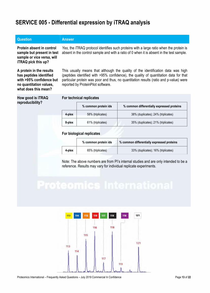
How good is iTRAQ reproducibility?
For technical replicates
% common protein ids % common differentially expressed proteins
4-plex 58% (triplicates) 38% (duplicates); 24% (triplicates)
8-plex 61% (triplicates) 35% (duplicates); 21% (triplicates)
For biological replicates
Note: The above numbers are from PI’s internal studies and are only intended to be a reference. Results may vary for individual replicate experiments.
% common protein ids % common differentially expressed proteins
4-plex 65% (triplicates) 33% (duplicates); 16% (triplicates)
Proteomics International – Frequently Asked Questions – July 2019 Commercial In Confidence Page 14 of 22
SERVICE 006 - Multiple Reaction Monitoring (MRM)
Question Answer
What is MRM? Multiple reaction monitoring (MRM, also known as Selective Reaction Monitoring – SRM) is a highly specific and sensitive mass spectrometry technique that can selectively quantify compounds within complex mixtures.
This technique uses a triple quadrupole MS that firstly targets the ion corresponding to the compound of interest with subsequent fragmentation of that target ion to produce a range of daughter ions. One (or more) of these fragment daughter ions can be selected for quantification purposes. Only compounds that meet both these criteria, i.e. specific parent ion and specific daughter ions corresponding to the mass of the molecule of interest are isolated within the mass spectrometer.
By ignoring all other ions that flow into the mass spectrometer the experiment gains sensitivity, whilst maintaining exquisite accuracy.
MRM is performed for selection of specific analytes and absolute quantitation of proteins, peptides, metabolites and lipids from plasma, serum and other biological samples.
What type of samples can be analysed?
Essentially any compound that can be ionized and then fragmented can be used for MRM analysis. The most typical examples being the measurement of proteins and peptides, metabolites, or drugs in complex mixtures such as plasma and serum. Quantification of proteins is possible through the MRM experiment on digested peptides that correspond to the protein of interest. This makes MRM analysis a common choice for biomarker analysis and for detection and quantification. As the name suggests more than one MRM can be monitored per analysis allowing detection of multiple compounds in the same experiment.
How sensitive is MRM? This is a highly sensitive technique. It is possible to detect peptide compounds not normally seen by a typical MS approach. For example peptides can be accurately quantitated at femtomole concentration. In complex samples such us plasma, peptides can be quantitated at µg/ml concentration.
Is MRM compatible with absolute or relative quantitation?
This technique can provide results for either type of quantitation. Relative quantitation is generally straightforward but for absolute quantitation appropriate standards are required. For absolute quantitation a specific peptide is quantitated against a spiked internal standard (e.g. a synthetic stable isotope labeled peptide standard) to yield a measure of protein concentration.
Is method development required?
Yes. Each project is specific and requires considerable amounts of operator time to develop and run the MRM analysis. For complex mixtures some sample clean up may be required with significant chromatographic separation. For plasma and serum extra preparation work is required and additional fees will be charged.
Analysis time and costs? Time: 2–4 weeks after sample receipt (includes method development).
Costs: Final project costs will be supplied on request (a price guide is given below)
Method Development – $1,600/day
MRM data collection and analysis – from $80/sample injection
Note: Time and costs are project specific.
Proteomics International – Frequently Asked Questions – July 2019 Commercial In Confidence Page 15 of 22
Application Examples
SERVICE 006 - Multiple Reaction Monitoring (MRM)
Question Answer
Protein Quantitation MRM Reference for article on MRM of peptides to determine relative protein quantitation: Expression of Cardiac α-Actin Spares Extraocular Muscles in Skeletal Muscle α-Actin Diseases
This article describes the development and implementation of an MRM based method to determine relative quantities of two proteins (cardiac and skeletal actin) which differ in only 4 residues out of 375 in their sequences, i.e., they are 99% homologous. The proteins were processed together and specific MRMs developed that would detect the presence of each form sensitively and specifically. Only relative quantitation was required for this application.
Peptide/Lipopeptide MRM
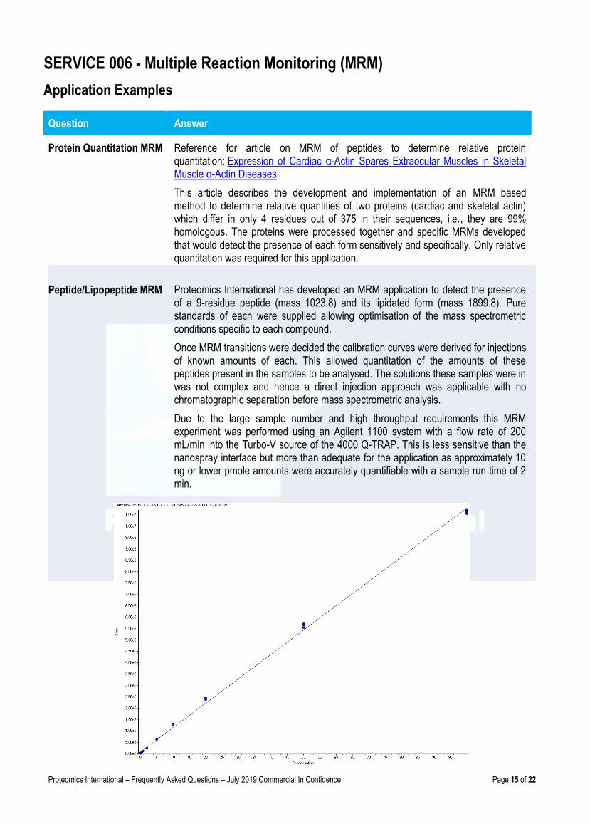
Proteomics International has developed an MRM application to detect the presence of a 9-residue peptide (mass 1023.8) and its lipidated form (mass 1899.8). Pure standards of each were supplied allowing optimisation of the mass spectrometric conditions specific to each compound.
Once MRM transitions were decided the calibration curves were derived for injections of known amounts of each. This allowed quantitation of the amounts of these peptides present in the samples to be analysed. The solutions these samples were in was not complex and hence a direct injection approach was applicable with no chromatographic separation before mass spectrometric analysis.
Due to the large sample number and high throughput requirements this MRM experiment was performed using an Agilent 1100 system with a flow rate of 200 mL/min into the Turbo-V source of the 4000 Q-TRAP. This is less sensitive than the nanospray interface but more than adequate for the application as approximately 10 ng or lower pmole amounts were accurately quantifiable with a sample run time of 2 min.
Proteomics International – Frequently Asked Questions – July 2019 Commercial In Confidence Page 16 of 22
SERVICE 008 - Intact mass analysis of proteins and peptides by mass spectrometry
This service provides the relative molecular mass (RMM) for the protein or peptide of interest. Samples are analysed intact (no enzyme digestion). Depending on the size of the molecule and the mass accuracy required MALDI-TOF MS and/or LC-MS is used. For a RMM <6,000 amu MALDI-TOF is used, and >6,000 amu LC-MS is used.
Question Answer
What is the largest size molecule that can be analysed?
160,000 amu
How should the sample be prepared?
(1) The protein must be pure, i.e. no other proteins should be present.
(2) The protein must be sent lyophilised (freeze dried) from a low salt buffer. Please state the volume of the liquid before drying.
NOTE: Gel bands and PVDF membranes cannot be analysed.
(3) The protein concentration before drying must be at least 0.1mg/ml.
(4) The solution must be a low salt buffer, e.g. 50mM ammonium hydrogen carbonate; for TRIS or phosphate buffers the maximum salt strength is 20mM. NaCl and surfactants (e.g. SDS) cannot be present.
NOTE: PI does not perform sample desalting; there are many methods described in the literature for de-salting.
How much sample is required?
For high purity protein sample a minimum of 10 ug is required. However up to 50 ug should be provided (where available) to ensure the best chance for a result. For pure peptides a minimum of 10ng is required.
Proteomics International – Frequently Asked Questions – July 2019 Commercial In Confidence Page 17 of 22
SERVICE 009 - Amino Acid Analysis (AAA)
Question Answer
(A) Sample preparation
How much sample is required?
The minimum amounts are: High sensitivity AAA – 10 μg (5μg in duplicate) Cys – 20μg (10μg in duplicate) Trp – 1mg (0.5mg in duplicate)
The sample is on PVDF membrane, is it OK?
No, the protein must be lyophilised.
The sample is in solution, is it OK?
No, the sample must be freeze dried prior to shipping to ensure maximum stability. Please state: (a) The volume of the liquid before drying. (b) Solvent/buffer used before drying. (c) Known or estimated concentration of sample.
(B) Other technical queries
What is the accuracy of Amino Acid Analysis (AAA)?
Generally most AAA laboratories accept between 10% and 15% variation from expected result. Normally the results presented have less than 10% variation, but some amino acids are more difficult. For the high sensitivity AAA (no cys or trp) the recoveries for the control sample (BSA) are typically 90% to 95% for the total of the amino acids. NOTE: This does not affect the AA composition which is the relative ratio of amino acids (i.e. mole%). For amino acid composition, the run is accepted if the result for BSA for individual amino acids is within +/- 10% of the expected mole% value. Most amino acids are within <5%.
Are some amino acids difficult to measure?
Yes, Serine is labile under acid conditions and typically for BSA there is a loss of 6-7% in the assay. Met can cause problems due to oxidation under the hydrolysis. It is also the least abundant and +/- 15% is allowed for this amino acid. Met can also be analysed by the Cys assay and some prefer this method of analysis. For Cys the recoveries are ~85% for BSA and +/- 15% from this value is acceptable. Trp can give variable results and the data is not accepted unless the BSA result is less than +/- 15% of the expected result.
My peptide contains non-standard amino acids, can they be measured?
No, the following cannot be analysed: (1) Mpr (mercaptopropionyl) (2) Harg (3) Cit-citrulline
(4) Pal (pyridyl alanine) (5) Cpa (6) Nal (naphthyl alanine)
Proteomics International – Frequently Asked Questions – July 2019 Commercial In Confidence Page 18 of 22
SERVICE 010 - N-terminal sequencing
Question Answer
(A) Sample preparation
How should the sample be prepared?
The protein should be electrophoretically transferred to a high quality (protein sequencing grade) PVDF membrane. These membranes are available from Bio-Rad, Applied Biosystems, GE, and others. The membrane should be stained with Coomassie Blue, and the target band must then be visible by eye. Blots must be free of any particulate material. NOTE: Please excise the band from the membrane and send it along with a photocopy of the membrane indicating the band. PI will not excise the band.
Which is a good membrane to use?
SequiBlot PVDF Membrane for Protein sequencing (0.2μm) [Bio-Rad Cat# 162-0182].
The sample is in a gel, can it be sequenced?
No, it must be transferred to sequencing grade PVDF membrane.
Can samples be analysed in liquid form?
Yes, provided the sample is pure and in a low salt buffer e.g. 50mM ammonium hydrogen carbonate; for TRIS or phosphate buffer the maximum salt strength is 20mM. NaCl, surfactants (e.g. SDS) and glycine cannot be present. The sample must be freeze dried or lyophilised in a micro-centrifuge tube prior to shipping. NOTE: The type of buffer and the volume of the liquid before drying must be stated on the Request Form. Complex samples containing more than one protein cannot be analysed by this process.
What amount of protein is required?
For a pure protein the sensitivity limit is approximately 1 picomole, however, 10pmol is recommended to ensure a satisfactory result. As a guide, a Coomassie stained target band must be visible by eye.
(B) Other technical queries
Is cysteine a problem? Yes, Cys can only be detected on the sequencer following special alkylation with 4-vinylpyridine. This is only possible with liquid samples and must be requested before analysis commences. NOTE: If Cys is not derivitised then it cannot be detected under normal conditions and its presence is inferred by a gap in the sequence, reported as X.
What is N-terminal blocking?
In nature, the N-terminal residue of a protein can be subject to post-translational modification. This is a biological process that occurs in the cell, and is not normally one due to sample preparation. Common N-terminal blocking groups include pyroglutamate, N-acetyl groups such as acetyl-serine or acetyl-threonine and N-formylated amino acids. NOTE: More than 50% of all eukaryote proteins are blocked.
Proteomics International – Frequently Asked Questions – July 2019 Commercial In Confidence Page 19 of 22
SERVICE 010 - N-terminal sequencing
EXAMPLE PUBLICATIONS
Question Answer
What if the protein is blocked at the N- terminus?
No sequence information will be obtained. This cannot be predicted prior to analysis, and consequently the no result fee will apply. NOTE: In such cases the best alternative to determine the protein identity is using mass spectrometry [Service 001]. Whilst this will not determine the N-terminal sequence of a blocked protein it can be used to unequivocally identify the protein. New samples would be required in the form of gel bands.
Can proteins be unblocked?
Unblocking of proteins is rarely possible and is not a service offered by PI. Unfortunately, no further analysis of blocked samples is possible.
Question Answer
Where can I find details on methods and examples of results?
http://www.proteomics.com.au/latest-news/publications/
What method is used for ProID [Service 001]?
All details required for publishing are listed in the report and published here: Bringans S.D., Kendrick T., Lui J., and Lipscombe R.J. (2008) A comparative study of the accuracy of several de novo sequencing software packages for datasets derived by matrix-assisted laser desorption/ionisation and electrospray. Rapid Commun. Mass Spectrom. 22(21): 3450-4. Note: Proteomics International does not release its SOPs (Standard Operating Procedures).
What method is used for iTRAQ [Service 005]?
All details required for publishing are listed in the report and published here: Casey T., Solomon P.S., Bringans S.D., Tan K.C., Oliver R.P., and Lipscombe R.J. (2010) Quantitative proteomic analysis of G-protein signalling in Stagonospora nodorum using Isobaric Tags for Relative and Absolute Quantification (iTRAQ). Proteomics 10, 1, 38-47. Download Copy. Note: Proteomics International does not release its SOPs (Standard Operating Procedures).
Proteomics International – Frequently Asked Questions – July 2019 Commercial In Confidence Page 20 of 22
EXAMPLE PUBLICATIONS
Question Answer
What is the workflow for biomarker discovery?
Please see model publication: http://www.absciex.com/Documents/Downloads/Literature/Biomarker-Pipeline-Diabetes-4250211-01.pdf Bringans B, T Stoll, K Winfield, T Casey, T Davis, J Albanese and R Lipscombe (2012) Protein Biomarker Research Pipeline for Developing Protein Biomarkers for Diabetic Kidney Disease. AB Sciex Technote Publication number: 4250211-01. Download Copy.
What type of results do I get from iTRAQ [Service 005]?
Please see: Casey T., Solomon P.S., Bringans S.D., Tan K.C., Oliver R.P., and Lipscombe R.J. (2010) Quantitative proteomic analysis of G-protein signalling in Stagonospora nodorum using Isobaric Tags for Relative and Absolute Quantification (iTRAQ). Proteomics 10, 1, 38-47. Download Copy.
What is MRM mass spectrometry [Service 006] and how can it help me?
Please see: Ravenscroft G., Colley S., Walker K., Clement S., Bringans S.D., Lipscombe R.J., Fabian V., Laing N., and Nowak K.J. (2008) Expression of cardiac α-actin spares extraocular muscles in skeletal muscle α-actin diseases – Quantification of striated α-actins by MRM-mass spectrometry. Neuromuscular Disorders, 18:953-958. Download Copy.
What is proteogenomics and how can it help me?
Please see: Bringans S.D., Hane J., Casey T.M., Tan K.C., Lipscombe R.J., Solomon P.S., and Oliver R.P. (2009) Deep proteogenomics; high throughput gene validation by multidimensional liquid chromatography and mass spectrometry of proteins from the fungal wheat pathogen Stagonospora nodorum. BMC Bioinformatics, 10:301-11. Download Copy.
Proteomics International – Frequently Asked Questions – July 2019 Commercial In Confidence Page 21 of 22
Other Services and Questions
Filling in the Request Form & Shipping Instructions
Question Answer
What is ISO/IEC 17025 Laboratory Standard Accreditation?
Data analysis is undertaken to ISO/IEC 17025 laboratory standard, which ensures laboratories generate technically valid results, and is the most widely used laboratory standard for USA federal testing laboratories: http://www.fda.gov/RegulatoryInformation/Guidances/ucm125434.htm ISO 17025 shares the same objectives as Good Laboratory Practice (GLP) to assure data quality. ALL proteomics and mass spectrometry services are accredited. Carbohydrate analysis is a newly offered service and this is not yet accredited. Amino acid analysis and Circular dichroism are not accredited services. N-terminal sequencing is also not covered by PI's accreditation, however, two GMP peptide suppliers have approved the procedures and the SOP's are in compliance with these procedures.
Does PI perform protein purification?
No, PI does not offer this technique as a service, however, there are many methods described in the literature.
Does PI offer peptide mass fingerprinting?
Peptide mass fingerprinting by MALDI-TOF mass spectrometry is a more basic technique for protein identification. It has been superseded by protein identification by MS [Service 001] (PMF+MS/MS).
What currency is used for pricing?
All prices are in US Dollars.
Please complete all boxes on the request form
Question Answer
What type of samples can be sent?
Any type of pure protein or protein extract (proteome) can be shipped to Proteomics International providing the permit is included. Plasma and serum samples can also be sent to PI, but special conditions apply - please contact us for details. NOTE: Tissue samples or intact cells cannot be sent.
How do I send a sample? Please consult your agent or for all shipping details see: How to send samples to Australia from ALL countries
Is a permit required? Yes, please download the permit (Australian Govt. Permit To Import Quarantine Material) and attach it to the outside of the package.

Singapore
Axil Scientific Pte Ltd
www.axilscientific.com
Telephone + 65 6775 7318
Facsimile + 65 6775 7211
Malaysia / Indonesia / Thailand
Apical Scientific Sdn. Bhd.
www.apicalscientific.com
Telephone + 603 8943 3252
Facsimile + 603 8943 3243
India
Proteomics International – India
www.proteomicsindia.com Tel +91 8527 699 192 Email: [email protected]
Hong Kong | China
Asia Bio Channel Limited www.asiabiochannel.com
Telephone +852 9845 4791
Japan
Leave a Nest Co. Ltd
www.leaveanest.com [email protected]
Telephone +81 3 6277 8041
Facsimile +81 3 6277 8042
Latin America
LinkPharma LLC www.linkpharma.co [email protected] Telephone +1 813 319 3972
Australia and other countries
Proteomics International Pty Ltd
ABN 78 096 013 455
www.proteomics.com.au [email protected]
Telephone + 61 8 9389 1992
Facsimile + 61 8 9389 1981
Address: PO Box 3008, Broadway, Nedlands
Perth, WA 6009 Australia


