Embed Size (px)
Citation preview

IOP PUBLISHING PHYSICS IN MEDICINE AND BIOLOGY
Phys. Med. Biol. 52 (2007) N149–N162 doi:10.1088/0031-9155/52/8/N01
NOTE
Front-illuminated versus back-illuminatedphoton-counting CCD-based gamma camera:important consequences for spatial resolution andenergy resolution
Jan W T Heemskerk1,2, Albert H Westra3, Peter M Linotte1,2,Kees M Ligtvoet3, Wojciech Zbijewski1,2 and Freek J Beekman1,2
1 Department of Nuclear Medicine, Image Sciences Institute, University Medical Center Utrecht,Room STR 5.203, Universiteitsweg 100, 3584 CG Utrecht, The Netherlands2 Department of Pharmacology & Anatomy, Rudolf Magnus Institute of Neuroscience,University Medical Center Utrecht, Room STR 5.203, Universiteitsweg 100, 3584 CG Utrecht,The Netherlands3 Department of Medical Technology, University Medical Center Utrecht, Room F01.114,Heidelberglaan 100, 3584 CX Utrecht, The Netherlands
E-mail: [email protected]
Received 22 September 2006, in final form 27 December 2006Published 26 March 2007Online at stacks.iop.org/PMB/52/N149
AbstractCharge-coupled devices (CCDs) coupled to scintillation crystals can be usedfor high-resolution imaging with x-rays and gamma rays. When the CCDimages can be read out fast enough, the energy and interaction position ofindividual gamma quanta can be estimated by a real-time image analysis of thescintillation light flashes (‘photon-counting mode’). The electron-multiplyingCCD (EMCCD) is well suited for fast read out, since even at high frame ratesit has extremely low read-out noise. Back-illuminated (BI) EMCCDs havemuch higher quantum efficiency than front-illuminated (FI) EMCCDs. Herewe compare the spatial and energy resolution of gamma cameras based onFI and BI EMCCDs. The CCDs are coupled to a 1000 µm thick columnarCsI(Tl) crystal for the purpose of Tc-99m and I-125 imaging. Intrinsic spatialresolutions of 44 µm for I-125 and 49 µm for Tc-99m were obtained whenusing a BI EMCCD, which is an improvement by a factor of about 1.2–2 overthe FI EMCCD. Furthermore, in the energy spectrum of the BI EMCCD, theI-125 signal could be clearly separated from the background noise, which wasnot the case for the FI EMCCD. The energy resolution of a BI EMCCD forTc-99m was estimated to be approximately 36 keV, full width at half maximum,at 141 keV. The excellent results for the BI EMCCD encouraged us toinvestigate the cooling requirements for our setup. We have found that forthe BI EMCCD, the spatial and energy resolution, as well as image noise,remained stable over a range of temperatures from −50 ◦C to −15 ◦C. This is asignificant advantage over the FI EMCCD, which suffered from loss of spatial
0031-9155/07/080149+14$30.00 © 2007 IOP Publishing Ltd Printed in the UK N149

N150 J W T Heemskerk et al
and especially energy resolution at temperatures as low as −40 ◦C. We concludethat the use of BI EMCCDs may significantly improve the imaging capabilitiesand the cost efficiency of CCD-based high-resolution gamma cameras.
1. Introduction
Compact high-resolution gamma and x-ray detectors are extremely useful for radiationmonitoring, autoradiography, x-ray imaging and high-resolution planar imaging ofradiotracers. A high intrinsic detector resolution combined with energy discriminationcapabilities is essential for the improvement of future single photon emission computedtomography (SPECT) devices, as has been shown in simulation studies by Rogulski et al(1993), Beekman and Vastenhouw (2004) and Meng et al (2006). A large number oflaboratories are currently developing high-resolution detectors for applications ranging fromastronomy and particle physics to biomedical imaging (e.g., Matteson et al (1997), Barber(1999), Menard et al (1998), He et al (1999), Vavrik et al (2002), Fiorini et al (2003), Leeset al (2003), Miyata et al (2004), De Vree et al (2005), Kataoka et al (2005), Ponchut et al(2005), Nagarkar et al (2006), Meng (2006)).
Two classes of position-sensitive gamma-ray detectors are currently of great interest forSPECT. The first use the ‘traditional’ method in which scintillating material converts gammarays into visible light, which is subsequently detected by position-sensitive light detectors, suchas (arrays of) photo-multiplier tubes (PMTs), silicon PMTs, avalanche photo diodes (APDs) orCCDs. Another important class of detectors relies on direct conversion of the gamma radiationinto electrical signals using solid-state semi-conductor materials: e.g., silicon, CdTe or CdZnTe(CZT) detectors. For medical imaging, scintillation crystals coupled to PMTs are currentlythe most commonly used gamma detectors. However, the selection of either scintillator-basedor solid state detectors must be based on a number of considerations depending on the eventualapplication of the gamma camera and costs.
One of the novel ways to obtain a very high spatial resolution (below 100 µm) with agamma camera is to use a scintillation layer composed of a bundle of parallel micro-columnarcrystals, which is read out by a CCD suitable for operating at high frame rates (figure 1).The use of columnar scintillation layers prevents resolution loss arising from light spreading.When sufficiently high frame rates (e.g., tens per second) are used, it is possible to obtainimages in which scintillation events are spatially separated so that the position and energy canbe determined for individual events (e.g., De Vree et al (2005), Beekman and De Vree (2005),Nagarkar et al (2006)). The main types of CCD-related noise that complicate the detectionof scintillation events are dark current noise and read-out noise. Dark current noise can beadequately suppressed by cooling the CCD. In most CCDs, read-out noise becomes a seriousproblem at high frame rates. To prevent this we use electron-multiplying CCDs (EMCCDs),which have sub-electron read-out noise even when operated at frame rates of tens of imagesper second. Combined with previously in-house developed read-out electronics this allowedus to construct a compact photon-counting CCD-based gamma camera that in some casesresulted in spatial resolutions of about 60 µm (De Vree et al 2005).
Further improvements in the performance of CCD-based gamma cameras are expectedwhen BI EMCCDs are used instead of FI EMCCDs. BI EMCCDs have higher quantumefficiency than FI EMCCDs because the incident light photons directly reach the depletionarea where detection takes place. In FI EMCCDs the incident photons have to pass throughthe contacts on the front of the device prior to detection, which leads to a loss of light.

Front-illuminated versus back-illuminated photon-counting CCD-based gamma camera N151
(a)
(b)
Figure 1. EMCCD chip with fibre-optic window (a) and a compact EMCCD-based gamma camera(b). A fibre-optic taper can be used to increase the effective detector area per CCD, but in ourexperiment a 3 mm thick straight taper is used as an optic window. A Peltier element cools theCCD chip, thereby reducing the dark current noise.
The quantum efficiency of the BI EMCCD at 550 nm is around 93%. Compared to thequantum efficiency of the FI EMCCD this is a gain of almost 200%. As a consequence, theamount of scintillation photons that is detected per event is around three times as high anddetermination of the position of the scintillation events can be performed more accurately.Because the amount of detected scintillation photons is an indication of the x-ray or gamma-ray energy, improved sensitivity also leads to a better energy resolution, in particular forlow-energy signals.
Finally, the use of BI EMCCDs could reduce the need for expensive cooling arrangementsfor our gamma cameras. This cooling is necessary for FI EMCCDs to decrease the dark noiseand increase the gain of the CCD. For BI EMCCDs, the increased sensitivity may compensatefor higher dark noise and lower gain at elevated operating temperatures. A reduction in coolingrequirements of CCD-based gamma cameras would result in lower cost and complexity offuture CCD-based small animal SPECT devices.
The goal of the present paper is to investigate what improvement in spatial resolutionand energy resolution can be obtained with a CCD-based gamma camera if a BI EMCCDis used instead of a FI EMCCD. To this end, we present energy spectra as well as imageprofiles of resolution measurements. In order to investigate the extent to which expensivecooling arrangements are necessary for the operation of our gamma camera, we studiedthe effects of temperature on the energy resolution, spatial resolution and noise level ofthe BI EMCCD.

N152 J W T Heemskerk et al
2. Materials and methods
2.1. EMCCD, scintillators and optic coupling
An electron-multiplying CCD (EMCCD) is a CCD with an added feature: internal gain.Because of this internal gain an EMCCD is less sensitive to read-out noise, even at highread-out speeds (e.g., several Mpixels per second). The internal gain is provided by electronmultiplication (avalanche multiplication or impact ionization) in the gain register. This gainregister is an extension of the read-out register, involving the addition of a high voltage clockedelectrode (40 to 50 V) plus a dc electrode to the standard structure of a CCD read-out register.Details of the EMCCD technology are available for example in Jerram et al (2001), Hynecek(2001) and Robbins and Hadwen (2003).
The EMCCDs used in this work are the CCD65 and CCD97 from E2V Technologies. TheCCD65 is a front-illuminated device with 288 lines and 576 pixels per line, and has an activearea of 11.52 mm × 8.64 mm (20 µm × 30 µm pixel size). The CCD97 is a back-illuminateddevice, with 512 lines of 512 pixels, and has an active area of 8.192 mm × 8.192 mm(16 µm × 16 µm pixel size). In order to have equal frame rates (50 frames per second)for both EMCCDs to facilitate comparison, the signal of the CCD97 is binned in sets of twolines. For our application, the EMCCDs were equipped with 3 mm thick straight fibre-opticwindows that were coupled with optical cement in very close proximity to the active area ofthe EMCCD (figure 1(a)).
Both the CCD65 and the CCD97 suffer from temperature-dependent dark noise.According to the manufacturer the dark noise of the CCD65 is 290 e−/pixel/s at roomtemperature (20 ◦C) and is reduced by 12% for every ◦C in temperature decrease. The darknoise of the CCD97 has a level of 400 e−/pixel/s at room temperature, and has a temperaturegradient of 14%/◦C. The dark noise of both EMCCDs reaches levels as low as 0.1 e−/pixel/sat a temperature of −40 ◦C. To ensure a very low dark current, the EMCCDs are cooled to−50 ◦C, using a Peltier element. The hot side of the Peltier element is cooled by an RS44LTcooler from FTS systems that has a heat removal power of 108 W at −40 ◦C.
For the conversion of the incident gamma rays to visible photons, we used a 1000 µm thickmicro-columnar CsI(Tl) scintillation crystal. Thallium-doped CsI has high-Z components, ahigh density of 4.51 g cm−3 and an excellent scintillation light yield; this makes it perfectlysuited for gamma-ray detection. 1000 µm thick CsI(Tl) has a capture efficiency of almost100% for I-125 and ∼30% for Tc-99m gamma photons. The scintillation module (CsI(Tl) FOSfrom Hamamatsu, type J6671) consisted of narrow CsI(Tl) needles (a few µm wide) grown ona 3 mm thick fibre-optic face plate with fibres of a diameter of 6 µm. The columnar structureyields a high spatial resolution. This scintillation module was coupled to the fibre-opticwindow of the EMCCD using optical grease.
The stopping power of the 1000 µm thick CsI(Tl) crystal for Tc-99m gamma photonsis limited to ∼30%. Use of a thicker crystal, e.g. 3000 µm, would increase the detectionefficiency for Tc-99m photons to ∼66%. Properly performing and suitably shaped thickercrystals are under development. Such crystals are interesting candidates for application insmall animal SPECT devices.
2.2. Photon-counting algorithm
The electronics board that drives and reads out the EMCCDs is connected to a digitalsignal processor (DSP), type TMS320C6416 from Texas instruments. The photon-countingalgorithm that is executed on the DSP detects and localizes scintillation events in real time byanalysing 50 entire CCD images per second.

Front-illuminated versus back-illuminated photon-counting CCD-based gamma camera N153
The algorithm proceeds as follows: first, each CCD image (Iccd) is background corrected,by subtracting the average (dark) background image A,
Ibg.corr. = Iccd − A. (1)
The background-corrected image (Ibg.corr.) is then smoothed with a 2D Gaussian kernel tosuppress high-frequency noise in the image
Ismoothed = Ibg.corr. ∗ ∗G, (2)
where ∗∗ represents a two-dimensional Gaussian convolution.In the filtered image (Ismoothed), scintillation events appear as bright regions. The position
and the intensity of the local maximum of each of these bright regions are sent to a PC (list-mode acquisition). One can set upper and lower limits for the intensity in order to distinguishscintillation events from background noise and disturbances like the direct detection of gammaphotons in the CCD or photons that have scattered within the object under investigation or thecollimator. Details of the background corrections and the algorithm are presented in De Vreeet al (2005). For a discussion on the influence of the width of the 2D Gaussian convolution onthe spatial and energy resolution we refer to section 2.3. Each of the measurements describedbelow consists of a total number of 100.000 analysed frames.
From the list-mode data, energy spectra are constructed represented by histograms ofthe intensities of the detected events. The intensity of a detected event is defined here asthe amplitude of the local maximum in the smoothed, background-corrected image, Ismoothed.A spectrum with I-125 (27–35 keV, 37 MBq) and Tc-99m (141 keV, 37 MBq) sources, as wellas a background noise spectrum was recorded for both EMCCDs.
In order to suppress effects of electronic noise and limit contributions of scattered photonsand cosmic radiation, the only events that are accepted for the determination of the spatialresolution are those with a light yield that falls within a 10% energy window (±14 keV)symmetrical around the photo-peak energy for Tc-99m and within a window ranging fromapproximately 20 keV to 50 keV for I-125.
2.3. Influence of the width of the Gaussian convolution
As is mentioned above, the photon-counting algorithm uses a 2D Gaussian convolutionto suppress high-frequency noise in the CCD image. Without such pre-processing, high-frequency noise may either be mistaken for a genuine scintillation, or, if superimposed on ascintillation flash, may lead to an incorrect localization of this flash.
In order to provide the best combination of spatial and energy resolution, the width (σ )of the Gaussian convolution that is used should fit the typical spread of a scintillation flash. Ifthe width of the convolution is too small, high-frequency noise is not adequately suppressed.In this case a single scintillation flash may even produce more than one peak in the smoothedimage. If the convolution kernel is too large the convolution will include too much noise aroundthe actual scintillation signal, which could result in a possible shift of the peak position. Aswe determine the location of a scintillation event by looking at the position of the peak valueof the convoluted signal, both effects could lead to an incorrect localization of the scintillation.Therefore, to optimize the 2D Gaussian convolution, a value of σ yielding optimal spatial andenergy resolution was first selected on the basis of experimental data. These experimentalresults are provided in section 3.1.
2.4. Measurements
To determine spatial and energy resolution of the camera, line pattern images were acquiredusing a setup with two tungsten blocks separated by a narrow slit (30 µm wide). A radioactive

N154 J W T Heemskerk et al
Table 1. Summary of the influence of the width of the Gaussian convolution (σ ) on the spatial andenergy resolution for a CCD97 at a temperature of −50 ◦C. The value of σ = 4 was selected forsubsequent experiments.
Spatial resolution FWHM (µm) Energy resolution FWHM (%)Sigma(in pixels) I-125 Tc-99m I-125 Tc-99m
2 70.2 49.0 N/A 353 46.8 48.6 65 344 44.1 49.4 57 315 46.0 51.2 57 346 51.2 53.5 55 358 69.5 56.4 55 36
source is placed behind the slit and a line pattern is projected through the slit onto thescintillator. The use of a slit to determine the line spread function is a standard method formeasuring intrinsic resolution and is widely used to characterize clinical gamma cameras.The spatial resolution is defined as the standard deviation of the full width at half maximum(FWHM) of the line spread function from the original width of the exposed slit. The resolutionvalues are corrected for the gamma beam width of 30 µm, on the assumption that both thebeam and the response from an infinitely narrow beam are Gaussian shaped. The energyresolution is determined from the FWHM of the peaks of the energy spectra. Measurementswere performed for a Tc-99m source and an I-125 source.
To investigate at what temperature dark noise and decreasing gain would inhibit detectionof scintillations, the noise on the EMCCD, as well as spatial and energy resolution, wereinvestigated for a range of temperatures between −50 and +5 ◦C. The noise on the EMCCDwas measured as the number of false counts (averaged per pixel for the total number of frames)for the I-125 energy window on a large unilluminated part of the EMCCD, consisting of ±40%of the EMCCD. For all noise calculations, the same part of the EMCCD was investigated.
3. Results
3.1. Optimization of the Gaussian convolution
Table 1 shows the values of the spatial and energy resolution that were measured when applyinga Gaussian convolution with different values of σ for the CCD97 at a temperature of −50 ◦C.The resolution values are shown for the I-125 and Tc-99m signals separately. Figure 2 showsthe dependence of the energy spectrum on the width of the convolution. Similar results andtrends as those presented here for the CCD97 have also been obtained for the CCD65.
As can be seen from table 1, the spatial resolution is dependent on both the width of theGaussian convolution and the source in question. For higher and lower values of sigma (σ =2; σ > 6) the Tc-99m signal gives a better spatial resolution than I-125, while for intermediatevalues of σ (3 � σ � 5), I-125 gives the best result. The dependence of the spatial resolutionon the convolution is much less pronounced for Tc-99m than for I-125.
For both I-125 and Tc-99m a convolution over three or four pixels gives the best result. Wecan see from figure 2 that these values for σ give an energy spectrum that allows us to adequatelydiscriminate between the I-125 and Tc-99m signals and the background. Consequently, forboth EMCCDs a value for σ of four pixels was selected, as it gives a near optimal spatialresolution as well as a good energy spectrum.

Front-illuminated versus back-illuminated photon-counting CCD-based gamma camera N155
Figure 2. Summary of the dependence of the energy spectra on the width of the 2D Gaussianconvolution (σ ).
Figure 3. Image profiles of the signal of I-125 (left) and Tc-99m (right) for the CCD65 (dashed)and CCD97 (solid line).
3.2. Resolution measurements
Figure 3 shows Tc-99m and I-125 image profiles taken perpendicularly to the line pattern forthe FI and the BI EMCCD at a temperature of −50 ◦C. One can clearly see the increase inresolution (decrease in profile width) when using the BI EMCCD (CCD97), as a result of theincrease in quantum efficiency. Furthermore, the FI EMCCD (CCD65) suffers from falselydetected events due to noise in the I-125 energy window, giving rise to a nearly uniformbackground signal, as can be seen on the left in figure 3.
The resolutions determined from full width at half maximum (FWHM) and the full widthat one tenth of the maximum (FWTM) of the profiles for both Tc-99m and I-125 are listed

N156 J W T Heemskerk et al
Table 2. Summary of the resolution measurement results, as measured from the Tc-99m andI-125 image profiles (figure 3) and the corresponding energy spectra (figure 4), for the FI and BIEMCCDs.
Tc-99m I-125
CCD65 CCD97 CCD65 CCD97
Spatial resolutionFWHM (µm) 59.4 49.4 85.3 44.1FWTM (µm) 120.1 102.9 195.2 94.6
Energy resolutionFWHM (keV) 41 36 N/A 22FWHM (%) 29 26 N/A 73
in table 2. Use of the BI EMCCD improves the spatial resolution in terms of FWHM by afactor ∼1.2 for Tc-99m and by a factor 1.5 to 2 for I-125, and improves the FWTM by factorsranging from 1.2 up to 2.
The improvement in spatial resolution is most notable for I-125. This results from thefact that the improvement of the sensitivity will have a larger influence on the detection of theweaker I-125 scintillations than on the detection of the brighter Tc-99m scintillations. Thisconsiderable improvement of the detectability of the I-125 scintillations for the BI comparedto the FI device can also be appreciated from a comparison of the energy spectra of the bothCCDs, for which we refer to section 3.3. That the I-125 signal has a slightly better spatialresolution than Tc-99m seems counterintuitive. However, two factors lead to a degradation ofthe spatial resolution of Tc-99m, which do not affect I-125:
• First, the spread in the depth-of-interaction (DOI) of the Tc-99m gamma photons resultsin a variation of the light spread for Tc-99m scintillations. This means that the width ofthe convolution of the photon-counting algorithm (σ ) cannot be optimized for the Tc-99mscintillation as well as it can be optimized for I-125 scintillations.
• Second, the Tc-99m gamma photons have enough energy to experience dual interactionswith the CsI(Tl) crystal (as has for example been observed by Miller et al (2006)). In sucha dual interaction, some of the energy of the Tc-99m gamma photon is transformed in aK-shell x-ray (that has an energy of approximately 30 keV) in a primary interaction, andthis K x-ray undergoes a secondary interaction leading to a second, nearly simultaneouslyemitted scintillation flash, often close to the primary scintillation. In many cases thiswill shift the peak obtained after convolution which leads to blurring effects since thesecondary x-rays have randomly distributed directions.
3.3. Energy spectra
Figure 4 shows the spectra acquired with Tc-99m and I-125 simultaneously for both EMCCDs.Also shown for each device is a background spectrum that was acquired by taking an imagewithout a radioactive source present. The scale is estimated by assuming that the peaks ofI-125 and Tc-99m are situated at 30 keV and 141 keV, respectively. On the basis of thisassumption the energy resolution was estimated to be 29% FWHM (41 keV) for Tc-99m forthe CCD65. For the CCD97, the energy resolution was estimated to be 26% (36 keV) forTc-99m and 73% (22 keV) for I-125. The energy resolution for both EMCCDs is listed intable 2.
Front-illuminated versus back-illuminated photon-counting CCD-based gamma camera N157
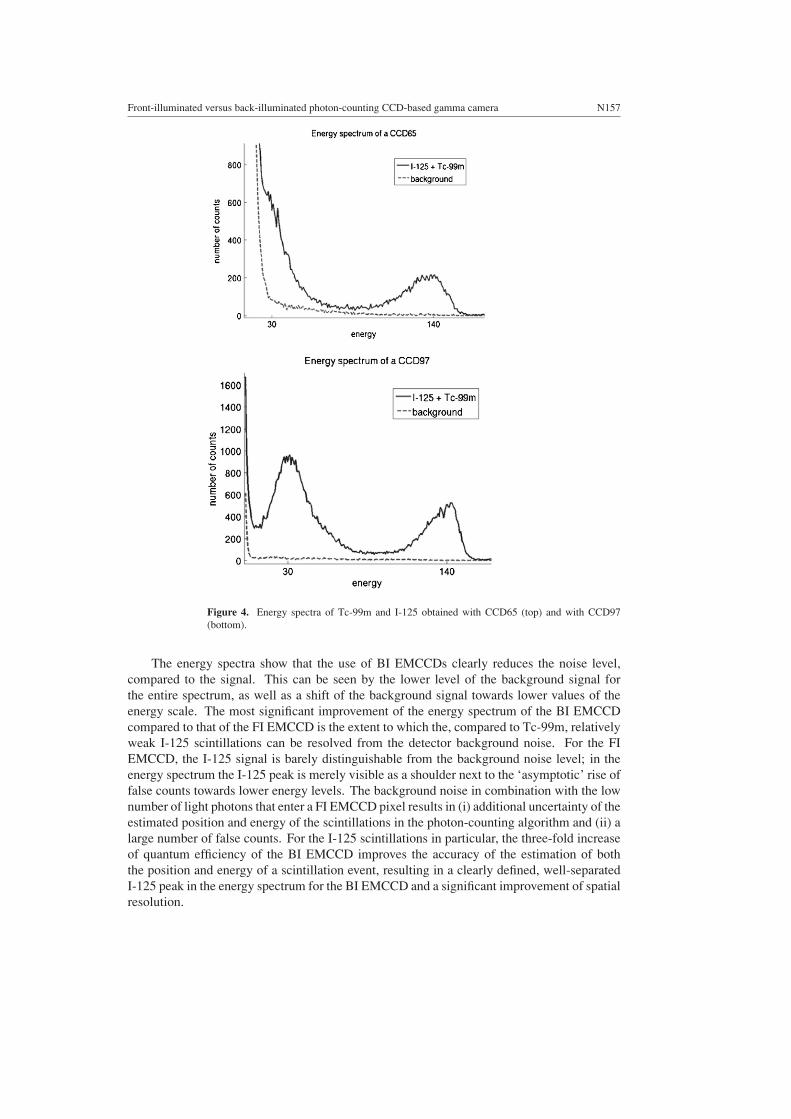
Figure 4. Energy spectra of Tc-99m and I-125 obtained with CCD65 (top) and with CCD97(bottom).
The energy spectra show that the use of BI EMCCDs clearly reduces the noise level,compared to the signal. This can be seen by the lower level of the background signal forthe entire spectrum, as well as a shift of the background signal towards lower values of theenergy scale. The most significant improvement of the energy spectrum of the BI EMCCDcompared to that of the FI EMCCD is the extent to which the, compared to Tc-99m, relativelyweak I-125 scintillations can be resolved from the detector background noise. For the FIEMCCD, the I-125 signal is barely distinguishable from the background noise level; in theenergy spectrum the I-125 peak is merely visible as a shoulder next to the ‘asymptotic’ rise offalse counts towards lower energy levels. The background noise in combination with the lownumber of light photons that enter a FI EMCCD pixel results in (i) additional uncertainty of theestimated position and energy of the scintillations in the photon-counting algorithm and (ii) alarge number of false counts. For the I-125 scintillations in particular, the three-fold increaseof quantum efficiency of the BI EMCCD improves the accuracy of the estimation of boththe position and energy of a scintillation event, resulting in a clearly defined, well-separatedI-125 peak in the energy spectrum for the BI EMCCD and a significant improvement of spatialresolution.

N158 J W T Heemskerk et al
Figure 5. Comparison of the energy spectra and the noise of a CCD65 for temperatures of−50 ◦C and −40 ◦C.
Figure 6. Comparison of the energy spectra and the noise of a CCD97 for temperatures of−50 ◦C and −15 ◦C.
3.4. Temperature dependence of the back-illuminated EMCCD
Figure 5 shows the temperature dependence of the energy resolution for the FI device (CCD65).Figure 6 shows the temperature dependence of the BI EMCCD (CCD97). Note that the graphsshown were measured at temperatures of −50 ◦C and −15 ◦C for the BI device, and at −50 ◦Cand −40 ◦C for the FI device. For the FI EMCCD, an increase of the operating temperature to−40 ◦C already causes a significant deterioration of the I-125 signal and a significant drop inthe spatial resolution (>10%) for Tc-99m. Therefore, detailed results are only listed for theBI device.

Front-illuminated versus back-illuminated photon-counting CCD-based gamma camera N159
Table 3. Summary of the noise and resolution measurements of a BI EMCCD for a range ofdifferent temperatures. For both spatial resolution and the noise, only the data from the I-125energy window are shown. The energy resolution is shown for both the I-125 and the Tc-99msignal. For the highest temperature (5.7 ◦C), there is an uncertainty in the energy resolution of theTc-99m signal due to the uncertainty in the position of the I-125 signal that prohibits an accuratescaling of the energy axis. The noise level is given as the number of false counts averaged perpixel for the total number of frames.
Temperature Spatial resolution Energy resolution Energy Resolution Noise level(◦C) (µm) I-125 (%) Tc-99m (%) (false counts)
5.7 57.5 – 67 3.73−1.3 48.2 68 75 0.04−15 45.3 59 34 0.04−50 44.3 57 31 0.04
Table 3 shows the noise on the BI EMCCD in combination with the spatial and energyresolution as a function of temperature. For the BI device, even though one can see a reductionin energy resolution with increasing temperature, the spatial resolution remains more or lessconstant, almost up to ∼ 0 ◦C, as is shown in table 3. We can see from figure 6 that at −15 ◦Cthe separation of the I-125 signal from the background noise has decreased, as the distancebetween the I-125 peak and the ‘asymptotic’ rise of background signal towards low-energyvalues is smaller. However, the number of false counts within the I-125 energy window (i.e.,the background noise level below the I-125 energy peak) has not significantly increased, itis still negligible. The different peak heights for both measurements are due to differentalignments of the sources. The sustained performance of the BI EMCCD at −15 ◦C canalso be seen in table 3. Despite a decrease in gain and increase of dark current, the use ofBI EMCCDs enables us to raise the operating temperature of the gamma camera by 35 ◦C,without significant loss of spatial or energy resolution. For applications using only Tc-99m as aradiation source, it would even be possible to operate the CCD gamma camera at temperaturesabove 0 ◦C.
One final remark to be made here is that variations in the gain setting betweenmeasurements lead to small differences in the scaling of the energy axis. This can be seen, forinstance, in the slight discrepancies between the −50 ◦C results of tables 2 and 3. The overalltrends of the noise, and energy and spatial resolution when comparing different temperatures,however, are undeniable.
4. Discussion
In the present work we have shown that the use of BI EMCCDs greatly improves both thespatial and the energy resolution of CCD-based gamma cameras. Furthermore, the increasedsensitivity of the BI EMCCD can eliminate the necessity to cool the device to very lowtemperatures (e.g., −50 ◦C). Our work has shown cases in which temperatures as high as−15 ◦C are already sufficient for performing gamma-ray imaging with BI EMCCDs withouta significant loss of spatial or energy resolution. Gamma-ray imaging with a resolution below60 µm and with energy discrimination for Tc-99m can even be possible at temperatures closeto 0 ◦C.
Further improvements in energy and spatial resolution are possible with the applicationof advanced centroiding algorithms, e.g. Anger logic or fitting algorithms, for a sub-pixeldetermination of the location of the individual scintillation events. Miller et al (2006) have

N160 J W T Heemskerk et al
developed a maximum likelihood estimation maximization (MLEM) algorithm for 3D positionand energy estimation that can partly correct for DOI effects. This 3D-MLEM algorithmdetermines the position and energy of individual scintillation events by fitting the scintillationsignal to expected signals of scintillations having a certain depth of interaction. These expectedsignals, which the measured signal is fitted to, are obtained from a calibration step. Althoughsuch an algorithm can be computationally expensive, a future real-time application of a similaralgorithm could be instrumental for further improvements in spatial and energy resolution.
The increased sensitivity of BI EMCCDs that is demonstrated in this paper offers apossibility of reducing the number of CCDs necessary for the detection area required for aSPECT imaging instrument. For example, coupling a demagnifying fibre-optic taper to theCCD will effectively increase the detector area, be it with a loss of intrinsic spatial and energyresolution. Although tapers have a transmission that decreases with increasing demagnificationfactors, the photons of a scintillation event are concentrated on a smaller number of pixels.Therefore, the loss of SNR may be smaller than one would expect considering solely the lossin total light flux. The improved sensitivity of the BI EMCCDs is elemental in surmountingthe resulting loss of SNR, and might furthermore reduce the loss of intrinsic spatial resolutionensuing from the effective increase in the pixel size, particularly in combination with improvedphoton-counting algorithms. The decrease of the number of CCDs necessary for a SPECTsystem will reduce the overall cost and of CCD-based SPECT imaging equipment.
Sufficient energy resolution is essential to discriminate scattered photons from primaryphotons. At present, the energy resolution of our CCD-based gamma camera is approachingthat of clinical NaI-based gamma cameras. Furthermore, the small sizes of the objects beingimaged in small animal SPECT result in a relatively small amount of scattered photons forisotopes like Tc-99m. Moreover, we have shown (Van der Have and Beekman 2004) that theamount of scatter in pinhole apertures is quite low, and even without energy discriminationdoes not give rise to strong contamination of projection data. The energy spectra presented inthis work clearly indicate that the prototype camera may be able to perform sufficient scatterrejection for applications such as small animal SPECT. The increased energy resolution of BIEMCCDs could also prove essential for human application of CCD-based gamma-radiationdetection, which requires extremely accurate scatter rejection. However, further improvementof the energy resolution of CCD-based gamma cameras then is necessary. This, in combinationwith the currently available sizes and high costs of BI EMCCDs, makes their application in afull-scale human SPECT system an ambitious project for a somewhat more distant future.
5. Conclusions
In the present work we have compared resolution and spectral characteristics of front-illuminated (FI) and back-illuminated (BI) EMCCD-based gamma cameras. The use ofBI EMCCDs leads to improvements in spatial resolution and energy resolution. Furtherimprovements might be possible with the use of more advanced gamma-photon detectionalgorithms. Moreover, the use of BI EMCCDs significantly reduces the cooling requirementsof the system. As has been shown in this paper, gamma cameras based on BI EMCCDs arevery promising, in particular for biomedical applications such as SPECT.
Acknowledgment
This work was sponsored in part by The Netherlands Organization for Scientific Research(NWO), grant no. 917.36.335.

Front-illuminated versus back-illuminated photon-counting CCD-based gamma camera N161
References
Anger H O 1958 Scintillation camera Rev. Sci. Instrum. 29 27–33Barber H B 1999 Applications of semiconductor detectors to nuclear medicine Nucl. Instrum. Methods Phys. Res.
A 436 102–10Beekman F J and Vastenhouw B 2004 Design and simulation of a high-resolution stationary SPECT system for small
animals Phys. Med. Biol. 49 4579–92Beekman F J, Van Der Have F, Vastenhouw B, Van Der Linden A J A, Van Rijk P P, Burbach J P H and Smidt M P
2005 U-SPECT-I: a novel system for sub-mm resolution tomography of radiolabeled molecules in mice J. Nucl.Med. 46 1194–200
Beekman F J and De Vree G A 2005 Photon-counting versus an integrating CCD-based gamma camera: importantconsequences for spatial resolution Phys. Med. Biol. 50 N109–19
Beekman F J and Van Der Have F 2007 The Pinhole: gateway to ultra-high-resolution three-dimensional radionuclideimaging Eur. J. Nucl. Med. Mol. Im. 34 151–61
Brem R F, Schoonjans J M, Kieper D A, Majewski S, Goodman S and Civelek C 2002 High-resolutionscintimammography: a pilot study J. Nucl. Med. 43 909–15
Charon Y, Laniece P and Tricoire H 1998 Radio-imaging for quantitative autoradiography in biology Nucl. Med. Biol.25 699–704
De Vree G A, Westra A H, Moody I, Van der Have F, Ligtvoet C M and Beekman F J 2005 Photon-counting gammacamera based on an electron-multiplying CCD IEEE Trans. Nucl. Sci. 52 580–8
Fiorini C, Longoni A, Perotti F, Labanti C, Rossi E, Lechner P, Soltau H and Struder L 2003 A monolithic arrayof silicon drift detectors coupled to a single scintillator for gamma-ray imaging with sub-millimeter positionresolution Nucl. Instrum. Methods Phys. Res. A 512 265–71
Gruner S M, Tate M W and Eikenberry E F 2002 Charge-coupled device area x-ray detectors Rev. Sci. Instrum.73 2815–42
He Z, Li W, Knoll G F, Wehe D K, Berry J and Stahle C M 1999 3-D position sensitive CdZnTe gamma-rayspectrometers Nucl. Instrum Methods Phys. Res. A 422 173–8
Hynecek J 2001 Impactron—a new solid state image intensifier IEEE Trans. Electron. Devices 48 2238–41Jerram P, Pool P, Bell R, Burt D, Bowring S, Spencer S, Hazelwood M, Moody I, Catlett N and Heyes P 2001 The
LLLCCD: low light imaging without the need for an intensifier Proc. SPIE 4306 178–86Kataoka J, Saito T, Kuramoto Y, Ikagawa T, Yatsu Y, Kotoku J, Arimoto M, Kawai N, Ishikawa Y and Kawabata N
2005 Recent progress of avalanche photodiodes in high-resolution X-rays and γ -rays detection Nucl. Instrum.Methods Phys. Res. A 541 398–404
Lees J E, Fraser G W, Keay A, Bassford D, Ott R and Ryder W 2003 The high resolution gamma imager (HRGI):a CCD based camera for medical imaging Nucl. Instrum. Methods Phys. Res. A 513 23–26
Matteson J L et al 1997 CdZnTe arrays for astrophysics applications Proc. SPIE 3115 160–175Menard L, Charon Y, Solal M, Laniece P, Mastrippolito R, Pinot L, Ploux L, Ricard M and Valentin L 1998 POCI: a
compact high resolution gamma camera for intra-operative surgical use IEEE Trans. Nucl. Sci. 45 1293–7Meng L J, Clinthorne N H, Skinner S, Hay R V and Gross M 2006 Design and feasibility study of a single photon
emission microscope system for small animal I-125 imaging IEEE Trans. Nucl. Sci. 53 1168–78Meng L J 2006 An intensified EMCCD camera for low energy gamma ray imaging applications IEEE Trans. Nucl.
Sci. 53 2376–84Miller B W, Bradford Barber H, Barrett H H, Shestakova I, Singh B and Nagarkar V V 2006 Single-photon spatial
and energy resolution enhancement of a columnar CsI(Tl)/EMCCD gamma-camera using maximum-likelihoodestimation Proc. SPIE 6142 61421T
Miyata E and Tamura K 2003 Novel photon-counting detector for 0.1–100 keV x-Ray imaging possessing high spatialresolution Japan. J. Appl. Phys. Part 2, 10A 42 L1201-4
Miyata E, Miki M, Tawa N, Kamiyama D and Miyaguchi K 2004 Development of new x-ray imaging device sensitiveto 0.1–100 keV Nucl. Instrum. Methods Phys. Res. A 525 122–5
Nagarkar V V, Gordon J S, Vasile S, Gothoskar P and Hopkins F 1996 High resolution x-ray sensor for non destructiveevaluation IEEE Trans. Nucl. Sci. 43 1559–63
Nagarkar V V, Tipnis S V, Gupta T K, Miller S R, Gaysinskiy V B, Klugerman Y, Squillante M R, Entine G andMoses W W 1999 High speed X-ray imaging camera using a structured CsI(Tl) scintillator IEEE Trans. Nucl.Sci. 46 232–6
Nagarkar V V, Shestakova I, Gaysinskiy V, Tipnis S V, Singh B, Barber W, Hasegawa B and Entine G 2006 ACCD-based Detector for SPECT IEEE Trans. Nucl. Sci. 53 54–8
Ponchut C, Zontone F and Graafsma H 2005 Experimental comparison of pixel detector arrays and CCD-Basedsystems for x-ray area detection on synchrotron beamlines IEEE Trans. Nucl. Sci. 52 1760–5

N162 J W T Heemskerk et al
Robbins M S and Hadwen B J 2003 The noise performance of electron multiplying charge-coupled devices IEEETrans. Electron. Devices 50 1227–32
Rogulski M M, Barber H B, Barrett H H, Shoemaker R L and Woolfenden J M 1993 Ultra-high resolution brainSPECT imaging—simulation results IEEE Trans. Nucl. Sci. 40 1123–9
Rowe R K, Aarsvold J N, Barrett H H, Chen J C, Klein W P, Moore B A, Pang I W, Patton D D and White T A 1993A stationary hemispherical SPECT imager for three-dimensional brain imaging J. Nucl. Med. 34 474–80
Van Der Have F and Beekman F J 2004 Characterization of photon penetration and scatter in micro-pinholes Phys.Med. Biol. 49 1369–86
Vavrik D, Jakubek J, Visschers J, Pospisil S, Ponchut C and Zemankova J 2002 First tests of a Medipix-1 pixeldetector for X-ray dynamic defectoscopy Nucl. Instrum. Methods Phys. Res. A 487 216–23


