Embed Size (px)
Citation preview

Functional Relevance of the BMD-AssociatedPolymorphism rs312009: Novel Involvement ofRUNX2 in LRP5 Transcriptional Regulation
Lıdia Agueda ,1 Rafael Velazquez-Cruz ,1* Roser Urreizti ,1 Guy Yoskovitz ,2 Patricia Sarrion ,1
Susana Jurado ,2 Roberto Guerri ,2 Natalia Garcia-Giralt ,2 Xavier Nogues ,2 Leonardo Mellibovsky ,2
Adolfo Dıez-Perez ,2 Pierre J Marie ,3 Susana Balcells ,1 and Daniel Grinberg1
1Department of Genetics, Faculty of Biology, University of Barcelona, IBUB, CIBERER, Barcelona, Spain2Internal Medicine, URFOA, IMIM, Hospital del Mar, Autonomous University of Barcelona, Barcelona, Spain3INSERM U606 and University Paris Diderot, Paris, France
ABSTRACT
LRP5 is an osteoporosis susceptibility gene. Association analyses reveal that individual single-nucleotide polymorphisms (SNPs)
determine variation in bone mineral density (BMD) among individuals as well as fracture risk. In a previous study, we identified a
lumbar spine BMD-associated SNP, rs312009, located in the LRP5 5’ region. A RUNX2 binding site was identified in this region by gel-shift
experiments. Here we test the functionality of this SNP and examine whether RUNX2 is indeed a regulator of LRP5 expression. Gene
reporter assays were used to test rs312009 functionality. Bioinformatic predictive tools and gel-shift and gene reporter assays were used
to identify and characterize additional RUNX2 binding elements in the 3.3-kb region upstream of LRP5. Allelic differences in the
transcriptional activity of rs312009 were observed in two osteoblastic cell lines, the T allele being a better transcriber than the C allele.
RUNX2 cotransfection in HeLa cells revealed that the LRP5 5’ region responded to RUNX2 in a dose-dependent manner and that the
previously identified RUNX2 binding site participated in this response. Also, RUNX2 inhibition by RNAi led to nearly 60% reduction of
endogenous LRP5mRNA in U-2 OS cells. Four other RUNX2 binding sites were identified in the 5’ region of LRP5. Luciferase experiments
revealed the involvement of each of them in the RUNX2 response. The allelic differences observed point to the involvement of rs312009
as a functional SNP in the observed association. To our knowledge, this is the first time that the direct action of RUNX2 on LRP5 has been
described. This adds evidence to previously described links between two important bone-regulating systems: the RUNX2 transcription-
factor cascade and the Wnt signaling pathway. � 2011 American Society for Bone and Mineral Research.
KEY WORDS: LRP5; RUNX2; SNP; LUCIFERASE; OSTEOPOROSIS
Introduction
The LRP5 gene encodes the low-density lipoprotein receptor–
related protein 5 (LRP5), a transmembrane protein that is
involved in Wnt signaling. The Wnt pathway has been shown to
be related to bone mass and metabolism.(1,2) Its role as an
essential regulator of bone mass was discovered by linkage
studies in two rare human diseases, osteoporosis-pseudoglioma
syndrome (OPPG) and high bone mass (HBM).(3) In osteoblasts,
LRP5 can transduce canonical signals to promote the renewal of
stem cells, stimulation of preosteoblast replication, induction of
osteoblastogenesis, and inhibition of osteoblast and osteocyte
apoptosis by increasing the levels of b-catenin and altering gene
expression through LEF/TCF transcription factors.(4)
RUNX2 (also known as CBFA1 or AML3) is a transcription factor
that is essential for the differentiation of osteoblasts, the cells
responsible for bone formation and skeletal development.(5) As
demonstrated by several genetic studies in mice and humans,
RUNX2 has well-defined roles in mediating osteoblast differ-
entiation and maturation, in controlling proliferation, and in
supporting normal osteogenesis. In addition, RUNX2 deficiency
and mutations affecting its function cause a severe bone
phenotype called cleidocranial dysplasia (CCD). At the molecular
level, RUNX2 activates or represses gene expression following a
specific interaction with the osteoblast-specific element (OSE)
that is present in the promoter or enhancer regions of its targets
by organizing protein complexes that can activate or repress
mammalian gene expression depending on the cellular and
ORIGINAL ARTICLE JJBMR
Received in original form May 26, 2010; revised form September 28, 2010; accepted November 5, 2010. Published online November 18, 2010.
Address correspondence to: Daniel Grinberg, PhD, Department of Genetics, Faculty of Biology, UB, Avinguda Diagonal 645, E-08028 Barcelona, Spain.
E-mail: [email protected]
*Present Address: Instituto Nacional de Medicina Genomica, Mexico City, Mexico.
Journal of Bone and Mineral Research, Vol. 26, No. 5, May 2011, pp 1133–1144
DOI: 10.1002/jbmr.293
� 2011 American Society for Bone and Mineral Research
1133

promoter/enhancer context.(6–8) RUNX2 factors interact with
other transcription factors and recruit numerous chromatin-
modifying proteins that regulate gene expression.(9) Among the
cofactors that interact with Runx proteins are coactivators such
as p300 and CREB-binding protein (CEBP) and co-repressors such
as mSin3A, transducin-like enhancer of split proteins (TLEs), and
several histone desacetylases (Hdacs).
Several RUNX2 responsive elements have been identified in
important bone-related genes, such as osteocalcin,(10–16) osteo-
protegerin,(17) osterix,(18) and bone sialoprotein,(19–21) among
others. More interestingly, functional RUNX2 binding sites have
been described in the promoters of genes of other Wnt signaling
pathway elements, such as SOST(22) and AXIN2 genes.(23)
Sclerostin is a secreted protein that can act as an inhibitor of
canonical Wnt signaling by interacting with Lrp5/6 corepressors,
whereas Axin2 is a negative regulator of this pathway by another
mechanism based on the promotion of b-catenin degradation
(reviewed by Liu and colleagues(24)).
Other links between RUNX2 and canonical Wnt signaling
pathways have been reported previously. Khaler and collea-
gues(12) described Lef1, a final effector of Wnt signaling, as a
RUNX2 transcriptional regulator. Gaur and colleagues(25) and
Reinhold and Naski(26) also reported cooperation between
LEF/TCF and RUNX2 factors in RUNX2’s own promoter and
the FGF18 promoter, respectively. Recently, McCarthy and
Centrella(27) presented novel evidence for bidirectional cross-
talk between the Wnt pathway and RUNX2 in osteoblasts.
However, regulatory involvement of RUNX2 in LRP5 transcription
has not been reported to date.
Several allelic variants in the LRP5 gene have been associated
with osteoporotic phenotypes such as bone mineral density
(BMD) and fracture in different association-analysis approaches,
such as single missense single-nucleotide polymorphism (SNP)
analysis, gene-wide analysis, and even genome-wide analysis
(GWA; reviewed by Li and colleagues(28)). Therefore LRP5 is a
confirmed osteoporosis susceptibility gene. In this sense, we
have previously reported the association of an LRP5 promoter
SNP, rs312009, with lumbar spine BMD.(29) A RUNX2 binding
element was identified at the polymorphic site by gel-shift
experiments, but there were no differences in the binding
capacities of the two possible alleles of rs312009. In this study,
we analyzed the putative functional involvement of this
polymorphism in the transcriptional activity of the LRP5 5’
region. We also characterized other putative RUNX2 binding
elements located in this region and studied their involvement in
LRP5 transcriptional regulation.
Materials and Methods
Cell culture
The human osteosarcoma cell lines Saos-2 and U-2 OS and the
human adenocarcinoma cell line HeLa were obtained from the
American Type Culture Collection (ATCC no. HTB-85, ATCC no.
HTB-96, and ATCC no. CCL-2, respectively; ATCC, Manassas, VA,
USA). All cell lines were cultured in Dulbecco’s modified Eagle’s
medium (DMEM) supplemented with L-glutamine (292mg/L),
10% heat-inactivated fetal calf serum (FCS), and 1% antibiotics
(penicillin and streptomycin; Invitrogen, Carlsbad, CA, USA).
Immortalized human neonatal calvaria cells (IHNC) were a
generous gift of Dr Eric Hay and are described in Ref. 30.
In silico prediction and alignment tools
The presence of putative RUNX2 binding sites was assessed
using two predictive tools: MatInspector from Genomatix
(Munich, Germany),(31) with the core similarity threshold set at
0.75, and TFSearch (Tokyo, Japan),(32) with the threshold for
acceptance set at 85. The DNA sequence conservation of
putative RUNX2 binding sites across available eutherian
mammals was inspected using the comparative genomic tool
BlastZ-net available in the Ensembl database (Wellcome Trust
Genome Campus, Hinxton, UK).(33)
Western blot
Nuclear cell extracts from Saos-2, U-2 OS, and HeLa cells were
prepared as described previously.(29) Twenty and thirty micro-
grams of protein were resolved by SDS-PAGE (12.5% poly-
acrylamide), transferred onto nitrocellulose membranes, and
analyzed by standard immunostaining using a horseradish
peroxidase–conjugated secondary antibody and chemilumines-
cence detection. The following primary antibodies were used:
RUNX2 M-70 at 1:800 dilution (SC-10758x; Santa Cruz
Biotechnologies, Santa Cruz, CA, USA) and nucleoporine NP62
at 1:4000 dilution (BD Biosciences, San Jose, CA, USA) for the
loading control. Horseradish peroxidase–conjugated secondary
antibodies were used (GARPO and SAMPO, respectively; Sigma,
St Louis, MO, USA). Immunoreactive bands were detected by
incubating the membrane for 2minutes in the following
solution: 10mL of 100mM Tris-HCl (pH 9.0), 50mL of 45mM
p-coumaric acid, 50mL of luminol, and 10mL of 30% H2O2.
Real-time quantitative PCR
IHNC, Saos-2 and U-2 OS cells were cultured in 60-mm dishes to
confluence. RNA extractions were performed using Trizol
reagent (Invitrogen) according to the manufacturer’s instruc-
tions. RNA quantity and quality were assessed by spectro-
photometry (Nanodrop ND-1000 Spectrophotometer; Nanodrop
Technologies Inc., Wilmington, DE, USA). Three micrograms of
total RNA were reverse transcribed using M-MLV RT (Invitrogen)
with oligo-dT priming according to the manufacturer’s instruc-
tions. Five microliters of a 1:20 dilution of the resulting cDNA was
used for subsequent amplification in a LightCycler 480 (Roche,
Basel, Switzerland) with SYBR Green fluorescent dye (ABsolute
Blue QPCR SYBR Green Mix; Thermo Scientific, Waltham, MA,
USA) under standard conditions. GAPDH mRNA was used for
normalization, and the results were expressed relative to RUNX2
expression in the IHNC cell line. For polymerase chain reaction
(PCR) amplification, the following primers were used: GAPDH
forward: 5’-CGA GAT CCC TCC AAA ATC AA-3’, and reverse: 5’-TGT
GGT CAT GAG TCC TTC CA-3’; RUNX2 forward: 5’-TCT GGC CTT
CCA CTC TCA GT-3’, and reverse: 5’-GAC TGG CGG GGT GTA AGT
AA-3’; LRP5 forward: 5’-GAA CAT CAA GCG AGC CAA G-3’, and
reverse: 5’-TGG CTC AGA GAG GTC AAA ACA-3’.
1134 Journal of Bone and Mineral Research AGUEDA ET AL.
RUNX2 RNA interference
Two siRNAs against RUNX2 (IDs: s2455 and s2456; Ambion,
Austin, TX, USA) were used. Silencer FAM-labeled Negative
Control siRNA (Ambion) was used as a reference. U-2 OS and
Saos-2 cells were seeded in 6-well plates at 1� 104 cells per well.
After 24 hours, the cells were transfected with 75 nM of siRNA
and 10mL of X-tremeGENE (Roche) in DMEM with 5% FBS
without antibiotic. Twenty-four hours after transfection, RNA was
extracted. Retrotranscription and real-time quantitative PCR
conditions were as described earlier.
Electrophoretic mobility shift assays (EMSAs)
Saos-2 human osteosarcoma cells (ATCC no. HBT-85TM) were
grown in DMEM (Gibco, Billings, MT, USA) supplemented with
10% FCS (Gibco). Nuclear extracts were prepared according to
Schreiber and colleagues(34) using a modified buffer C (10%
glycerol and 1.5mM of MgCl2). Protein concentrations were
determined by the Bradford method, and nuclear extracts were
stored at�808C until use. Single-stranded DNA oligonucleotides
(Sigma-Aldrich, St Louis, MO, USA) were synthesized automa-
tically (both forward and reverse strands). The sequences are
listed in Table 1. Double-stranded probes were obtained by
annealing complementary oligonucleotides and end labeling
with [g-32P]ATP (GE Healthcare, Piscataway, NJ, USA, or
PerkinElmer Life and Analytical Sciences, Waltham, MA, USA)
using the OptiKinase (USB, Santa Clara, CA, USA) standard
protocol. The unincorporated nucleotides were removed using a
quick-spin G-25 Sephadex column (Roche). Binding reactions
typically contained 10mg of nuclear extract, 0.5mg of double-
stranded poly(dI-dC) (Amersham Pharmacia Biotech, Waukesha,
WI, USA), 0.5mg of double-stranded poly(dA-dT) (Roche), 6mg of
acetylated BSA (Promega, San Luis Obispo, CA, USA), and
100,000 cpm of radiolabeled probe. The reaction mixtures were
incubated for 30minutes at room temperature in a buffer
containing 20mM HEPES (pH 7.9), 80mM KCl, 1mM EDTA, 1mM
DTT, and 10% glycerol in a total volume of 20mL. Protein-DNA
complexes were resolved from the free probes in nondenaturing
7% polyacrylamide (29:1) gels containing 2.5% glycerol.
Electrophoresis was performed at 48C in 1� TBE buffer at
20mA for approximately 3 hours. Gels were vacuum dried and
exposed to storage phosphor screens (Kodak, Rochester, NY,
USA) at room temperature for 3 to 12 hours. In competition
assays, the binding reactions were performed in the presence of
an excess of unlabeled competitor oligonucleotide, as indicated
in each case. For supershift experiments, 1mg of the RUNX2
polyclonal antibody (Calbiochem, Merck KGaA, Darmstadt,
Germany) or 0.5mL of SP1 polyclonal antibody (H00006667-
A01; Abnova, Taipei City, Taiwan) was preincubated with nuclear
proteins in the binding buffer on ice for 15minutes before
adding the probe.
Reporter constructs and gene-reporter assays
To generate several constructs with the LRP5 5’ region linked to a
reporter gene, an initial PCR amplicon spanning Chr11 positions
68,078,243 to 68,080,193 bp (according to the February 2009,
GRCh37/hg19 UCSC genome assembly) corresponding to
positions �1879 to þ11 from A(þ1)TG in the LRP5 promoter
region was obtained from Saos-2 DNA using the following
primers F8: 5’-GAG TCC CAG CAG AGA ACA GC-3’; and R7: 5’-GCT
GCC TCC ATG TTG TCC-3’. This fragment was cloned in pUC18/
SmaI (Amersham Pharmacia Biotech) by blunt-end ligation and
subcloned by AdeI and NcoI digestion in the pGL3 basic vector/
SmaI (Promega). This clone was named MP and contained
positions �49/�1826 of LRP5. Subsequent digestion of MP
with BlpI and NcoI produced a 729-bp fragment that was
recloned in the pGL3 basic vector/SmaI to obtain the BP
construct (�49/�778). A second PCR fragment was obtained
using the following primers: forward: 5’-GTG ATT CTC CCG CCT
CAG C-3’, and reverse: 5’-TCA TGC GTC CCC ACT TGC T-3’ (Chr11:
68,076,318 to 68,078,510, 2192 bp) again from Saos-2 DNA. This
fragment was digested with AleI and AvrII and subcloned to an
NheI- and AleI-digested MP to give the LP-C construct (�49/
�3274). The LP-T construct was obtained by site-directed
mutagenesis using the QuickChange II XL Site-Directed
Mutagenesis Kit (Stratagene Cloning Systems, La Jolla, CA,
USA). Site-directed mutagenesis also was used to incorporate 2-
bp substitutions in the RUNX2 binding site core sequences to
obtain the MUT1-C, MUT1-T, MUT2, MUT3, MUT4, and MUT5
constructs. All constructs were verified by automatic sequencing.
For the luciferase activity assays, 5� 104 Saos-2, U-2 OS, or HeLa
cells were plated in 24-well plates and cultured in DMEMwith 10%
FCS. On the following day, 1.75mg of reporter construct or
0.875mg of reporter construct plus 0.875mg of effector and 50ng
of normalizing b-galactosidase expression vector (Upstate
Biotechnology, Billerica, MA, USA) were cotransfected using
3mL of Transfast transfection reagent (Promega). The effector was
pCMV-Runx2, a mouse Runx2 isoform II (MASNS) cDNA cloned in
the pCMV5 expression vector (pCMV-Runx2; kindly provided by Dr
Karsenty, Columbia University, New York, NY, USA). This form has
97.6% homology at the amino acid level with the orthologous
isoform in humans. Empty pCMV5 vector (pCMV-EV) also was
cotransfected in order to provide the same total amount of DNA in
cotransfection experiments.
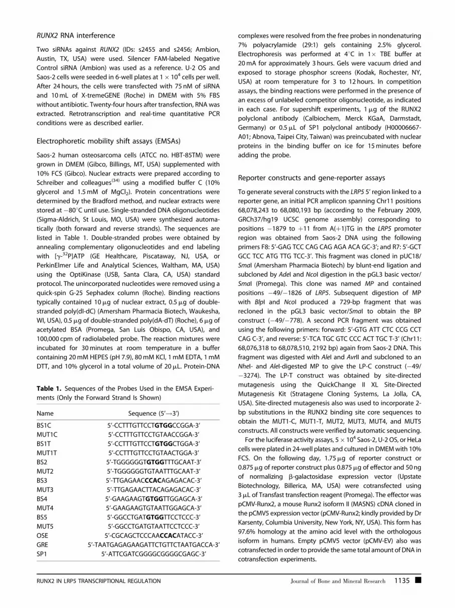
Table 1. Sequences of the Probes Used in the EMSA Experi-
ments (Only the Forward Strand Is Shown)
Name Sequence (5’!3’)
BS1C 5’-CCTTTGTTCCTGTGGCCGGA-3’
MUT1C 5’-CCTTTGTTCCTGTAACCGGA-3’
BS1T 5’-CCTTTGTTCCTGTGGCTGGA-3’
MUT1T 5’-CCTTTGTTCCTGTAACTGGA-3’
BS2 5’-TGGGGGGTGTGGTTTGCAAT-3’
MUT2 5’-TGGGGGGTGTAATTTGCAAT-3’
BS3 5’-TTGAGAACCCACAGAGACAC-3’
MUT3 5’-TTGAGAACTTACAGAGACAC-3’
BS4 5’-GAAGAAGTGTGGTTGGAGCA-3’
MUT4 5’-GAAGAAGTGTAATTGGAGCA-3’
BS5 5’-GGCCTGATGTGGTTCCTCCC-3’
MUT5 5’-GGCCTGATGTAATTCCTCCC-3’
OSE 5’-CGCAGCTCCCAACCACATACC-3’
GRE 5’-TAATGAGAGAAGATTCTGTTCTAATGACCA-3’
SP1 5’-ATTCGATCGGGGCGGGGCGAGC-3’
RUNX2 IN LRP5 TRANSCRIPTIONAL REGULATION Journal of Bone and Mineral Research 1135

Twenty-four/forty-eight hours after transfection, luciferase
and b-galactosidase activities were measured in a plate
luminometer (either SAFAS-Xenius XL, SAFAS, Monaco, or a
GloMax-Multi Detection System, Promega) using the corre-
sponding standard commercial kits (Luciferase Assay System,
Promega, and Chemiluminescent Beta-Gal Reporter Gene Assay,
Roche). The luciferase activity was normalized to the b-
galactosidase activity to correct for transfection efficiency. The
luciferase activity of the empty pGL3 vector (a measure of
background signal) was subtracted from normalized experi-
mental values (except in Fig. 2C). The results were expressed as
relative luciferase units (RLUs) in terms of the long promoter
construct with the more frequent allele of rs312009 (LP-C).
Transfections were performed in quadruplicate in at least three
independent experiments.
Statistical analyses
The results of the quantitative PCR and luciferase assays are
expressed as means� SEM. Statistical significance was deter-
mined by unpaired Student’s t tests (for comparisons between
different constructs) and by paired Student’s t tests (for
comparison of the same construct in different cotransfection
conditions). Significance is denoted as follows: �(or §)for p< .05;��(or §§)for p< .01; ���(or §§§)for p< .001; and ����(or §§§§)for
p< .0001.
Results
Binding of RUNX2 to the putative RUNX2-responsive
element at �2.9 kb of LRP5
The presence of a RUNX2 binding site at �2.9 kb from the
transcription start site of LRP5 has been reported previously.(29) In
contrast to that indicated by predictive tools (Table 2), the
presence of this binding site (BS1) was not conditioned by the
presence of the T allele of the rs312009 polymorphism, as shown
in gel-shift experiments in Fig. 1A, B. Equivalent patterns of
shifted bands and competition effects were observed for the
BS1T and BS1C probes. In both cases, the same level of
competition was observed with the specific probes (BS1T or
BS1C) without any cross-competition differences, whereas
nonspecific cold probes (GRE or SP1) displayed nearly no
competition at all. Specific competition with the osteoblast-
specific element (OSE) from the osteocalcin gene promoter
revealed the participation of RUNX2 in the retained protein
complex. In addition, when the core sequence of the predicted
RUNX2 binding site was mutated (MUT1-C and MUT1-T) and
used as a cold oligonucleotide competitor (Fig. 1C), MUT1C was
not able to compete with the binding at BS1C at any
concentration, and MUT1T showed some degree of competition
for the BS1T binding when added at high molar excess (�500).
Levels of RUNX2 mRNA and RUNX2 protein in human
osteoblast-like cells and in nonosteoblastic HeLa cells
In order to choose the appropriate cell lines for transfection
studies, two different osteoblastic cell lines, Saos-2 and U-2 OS,
were assessed for RUNX2 expression at the RNA and protein
levels. Initially, RUNX2 transcript levels were assessed in the two
osteoblastic cell lines by real-time PCR (Fig. 2A). Using IHNC as a
reference, the levels of RUNX2 mRNA in U-2 OS cells were
approximately 60% of those in Saos-2 cells. This difference was
even greater at the protein level (Fig. 2B). The nonosteoblastic
cell line HeLa showed no detectable expression of RUNX2
protein, as expected, given the osteoblast-specific expression
pattern of RUNX2.
Promoter activity of different fragments of the LRP5 5’
region in three cellular contexts
The performance of the LRP5 promoter region was assessed in
the two osteoblastic lines as well as in HeLa cells. Three different
reporter constructs bearing increasing amounts of the 5’ region
of LRP5 [BP (up to �778), MP (up to �1826), and LP-C (up to
�3274, bearing the C allele of rs312009)] were transfected, and
Table 2. The Five Predicted RUNX2 Binding Sites Found in the 3-kb LRP5 5’ Region
RUNX2
binding sites
Chromosomal Position
(Feb. 2009, GRCh37/hg19)
Matrix similarity
according to
Genomatix predictionaScore according to
TFSearch prediction Sequence
BS 1 Chr11: 68,077,228–68,077,242
(�2.9 kb)
MS:0.846 (only in presence of
T allele of rs312009)
<85 (not predicted) TTCCTGTGGCYGGAGb
BS 2 Chr11: 68,077,403–68,077,417
(�2.4 kb)
MS:0.954 100 GGGTGTGGTTTGCRAc
BS 3 Chr11: 68,078,103–68,078,117
(�1.8 kb)
MS:0.859 <85 (not predicted) TGAGAACCCACAGAG
BS 4 Chr11: 68,078,138–68,078,152
(�1.7 kb)
MS:0.949 100 AAGKGTGGTTGGAGCd
BS 5 Chr11: 68,078,794–68,078,808
(�1.3 kb)
MS:0.900 100 TGATGTGGTTCCTCC
aFor all RUNX2 sites, the optimized matrix similarity was 0.84 and the core similarity was 1.bY stands for C/T rs312009, validated frequent SNP (CEU HapMap frequence C 0.776 and T 0.224).cR stands for A/G rs4988327, validated rare SNP. Only the A allele (CEU HapMap frequence 0.941) was considered further.dK stands for T/G rs4988329, described in dbSNP but without frequency validation (only T allele considered).
1136 Journal of Bone and Mineral Research AGUEDA ET AL.

luciferase activity was measured (Fig. 2C). pGL3-EV (empty
vector) was used for normalization. All constructs showed
significantly higher luciferase gene activity than pGL3-EV in all
cell lines. The LP-C promoter activity was highest in Saos-2 cells,
showing more than threefold higher transcriptional activity than
pGL3-EV (p< .0001). In U-2 OS and HeLa cells, this activity was
double that of the empty vector (p< .0001). In all cellular
contexts, MP doubled the transcriptional activity of the LP-C
construct (p< .0001 in Saos-2 cells; p¼ .0130 in U-2 OS cells; and
p¼ .0002 in HeLa cells). BP also led to increased luciferase activity
relative to LP-C in all cell lines (p< .0001 in Saos-2 and HeLa cells,
and p¼ .0002 in U-2 OS cells) and to MP in the two osteosarcoma
cell lines (p¼ .0003 in Saos-2 cells; and p¼ 0.0493 in U-2 OS cells)
but not in HeLa cells. These data point to the presence of one or
several repressor elements between positions �778 and �1826
and between positions �1826 and �3270. In general, the LRP5
promoter activity was higher in Saos-2 cells than in other cell
lines for all three constructs.
Functional analysis of the rs312009 polymorphism
Given the genomic localization of SNP rs312009 in the LRP5
promoter region, we hypothesized that it could have functional
implications and that these might be mediated by the binding of
RUNX2. To test these hypotheses, several reporter-gene assays
were performed.
Four reporter constructs containing 3270 bp of the LRP5
5’ region were obtained: (1) LP-C, with the C allele in rs312009,
(2) LP-T, with the T allele in rs312009, (3) MUT1-C, with
mutated RUNX2 BS1 core sequence and the C allele in
rs312009, and (4) MUT1-T, with mutated RUNX2 BS1 core
sequence and the T allele in rs312009. A schematic representa-
tion of the constructs is shown in Fig. 3 together with their
relative luciferase activities after transfection in the above-
mentioned cells.
In both osteoblastic cell lines, the promoter bearing the
T allele (LP-T) showed significantly higher transcriptional
activity than LP-C. The differences between means [95%
confidence interval (CI)] were 40.7 (� 26.5), p¼ .0029, in
Saos-2 cells, and 57.8 (� 57.6), p¼ .049, in U-2 OS cells. The
difference between LP-C and LP-T did not reach statistical
significance in HeLa cells.
Mutation of the RUNX2 core sequence was tested for its effect
on the transcriptional activity of the LP-C and LP-T constructs.
The transcriptional capacity of MUT1-C was significantly higher
than LP-C in the three cell lines [differences between means
(95% CI) in Saos-2 cells, 38.27 (� 34.7), p¼ .031; in U-2 OS cells,
74.1 (� 41.1), p¼ .0011; and in HeLa cells, 333.3 (� 209.0),
Fig. 1. The DNA sequence containing the predicted RUNX2 binding site 1 (BS1) binds nuclear proteins present in osteoblast nuclear extracts, including
RUNX2. DNA binding was analyzed by gel-shift experiments. (A) Labeled double-stranded oligonucleotide containing the T allele of rs312009 (BS1T�) was
incubatedwith 10mg of Saos-2 nuclear extract. Competition experiments were performedwith cold T-allele (BS1T) or C-allele (BS1C) oligonucleotides, with
the nonspecific competitors GRE and Sp1, and with OSE, an oligonucleotide containing the RUNX2 consensus sequence of the osteocalcin promoter. (B) An
equivalent experiment was performed with the double-stranded oligonucleotide containing the C allele (BS1C�). (C) Oligonucleotides, BS1C�, and BS1T�,
competed by the corresponding cold probes, either wild type or mutated (MUT1C, MUT1T).
RUNX2 IN LRP5 TRANSCRIPTIONAL REGULATION Journal of Bone and Mineral Research 1137

p¼ .0022]. On the contrary, MUT1-T activity was significantly
lower than LP-T activity only in the U-2 OS cell line [�63.9 (�
63.2), p¼ .0476].
Effect of RUNX2 cotransfection on transcriptional activity
of the LRP5 5’ region
To assess the response of the LRP5 5’ region to exogenous
RUNX2, HeLa cells (known to lack endogenous RUNX2
expression) were cotransfected with increasing amounts of
pCMV5-RUNX2 expression vector together with the LP-C
construct, which contains the major allele of the polymorphism.
Luciferase activity was tested at 24 and 48 hours after
transfection. Figure 4A shows a dose-dependent stimulatory
effect caused by increasing amounts of RUNX2 vector. This effect
was higher at 24 hours than at 48 hours. Cotransfection of the
maximal pCMV-Runx2 concentration tested (0.875mg) caused a
more than fivefold increase at 24 hours (p¼ .0013). This
experimental condition was used in subsequent cotransfection
experiments.
The stimulatory effect of RUNX2 expression observed on
the LP-C construct was then tested on the LP-T construct, as
well as on MUT1-C and MUT1-T (Fig. 4B). LP-T, but not the
mutant constructs, was stimulated by RUNX2 (p¼ .0298),
although to a lesser extent. The differences between means
were 244.2 (95% CI 416.9–71.6) for LP-C and 102.2 (95% CI 192.9–
11.4) for LP-T. As already seen in the simple transfection
experiments (Fig. 3), the transcriptional activity of MUT1-C and
MUT1-T was higher than that of LP-C and LP-T in the absence of
RUNX2.
Taken together, these results indicate that the LPR5 5’ region is
able to respond to RUNX2 and that BS1 participates in this
response.
Effect of RUNX2 inhibition on the expression of the
endogenous LRP5 gene
To analyze the effect of RUNX2 on endogenous LRP5 gene
expression, RUNX2 was inhibited by RNAi, both in U-2 OS and
Saos-2 cells. These cell lines were transfected with siRNA s2456
against RUNX2 (Fig. 5), achieving 85% to 90% inhibition
(p< .005). In U-2 OS cells, the reduction in RUNX2 mRNA levels
led to nearly 60% reduction in LRP5 mRNA levels (p¼ .001). In
contrast, in Saos-2 cells, LRP5 mRNA levels were not modified.
Similar results were observed with a second siRNA against
RUNX2, (siRNA s2455; not shown).
Fig. 2. Characterization of RUNX2 mRNA and protein levels and assessment of LRP5 promoter constructs in different cell lines. (A) Real-Time PCR
quantification of RUNX2 isoform II mRNA levels in immortalized human neonatal calvaria cells (IHNC), Saos-2 cells, and U-2 OS cells. GAPDHwas used as the
internal normalization gene; results were expressed relative to RUNX2 in IHNC. (B) Western blot analysis of RUNX2 protein levels in the different cell lines
employed. Twenty and thirty micrograms of each nuclear extract were subjected to SDS-PAGE. Antibody RUNX2 (M-70, SCBT) was used as the primary
antibody, and Nucleoporine NP62 was used for the loading control. (C) The long promoter construct (LP-C) and two deletion constructs (MP and BP) were
assayed for transcriptional activity in the indicated cell lines. Luciferase values were normalized to b-galactosidase activity. The results are represented
relative to the pGL3 empty vector (pGL3-EV) for each cellular type. The bars represent the average luciferase activity of at least three independent
experiments with four replicates each, and the error bars represent the SEM. The significance, according to an unpaired Student’s t test, is represented by
asterisks for comparisons with the LP-C construct within each cell line and by a section mark (§) for the comparison of BP with MP within a given cell line.
1138 Journal of Bone and Mineral Research AGUEDA ET AL.

Search for additional RUNX2 binding sites in
the LRP5 5’ region
After identifying the first RUNX2 binding site (BS1), we looked for
other putative RUNX2 binding sites in the 3.3-kb LRP5 upstream
region. The Genomatix and TFSearch prediction tools identified
four additional putative RUNX2 binding sites, as summarized in
Table 2. BS2, BS4, and BS5 were perfect matches to the RUNX2
consensus binding site (5’-TGPyGGTPy-3’, where GPyGG is the
core) and also were consistent with the most frequent sequence
5’-TGTGGTT-3’. On the other hand, BS1 and BS3 differed by one
base, next to the core. Evolutionary conservation was assessed
for the five predicted sites (data not shown), and all showed a
certain degree of conservation in closely related primates.
Interestingly, none was conserved in mouse or rat.
In vitro assessment of RUNX2 binding to the LRP5
upstream region
In vitro binding of the RUNX2 transcription factor at the other
four predicted binding sites (BS2, BS3, BS4, and BS5) was
assessed by electromobility gel-shift assays. Figure 6A shows the
presence of specific binding at all the sites tested. While wild-
type competitors had a clear competitive effect, this was not the
case for the mutated probes. GRE and SP1, used as nonspecific
competitors, were not able to erase the shifted bands. Specific
competition with OSE and supershift experiments (Fig. 6B)
confirmed the involvement of RUNX2 in the protein complexes
retained in the gel shift.
Involvement of the identified RUNX2 binding sites in the
transcriptional activity of the LRP5 5’ region
The functional involvement of the five RUNX2 binding sites was
tested by gene-reporter assays (Fig. 7). The wild-type construct
(LP-C) was compared with five other constructs, each containing
the five RUNX2 sites, one of which was mutated (MUT1-C, MUT2,
MUT3, MUT4, and MUT5). Transfections were carried out in the
two osteoblastic cell lines and in HeLa cells. The mutation of any
of the sites resulted in significant changes in transcription activity
in at least one of the cell lines. Mutation of BS5 (MUT5) had the
greatest effect in the three cells types: around a twofold increase
in Saos-2 and U-2 OS cells and a four-fold increase in HeLa cells.
The differences between means were 108.4� 32.6 in Saos-2 cells
(p< .0001), 88.5� 54.0 in U-2 OS cells (p¼ .0026), and
380.3� 131.5 in HeLa cells (p< .0001). A similar effect, although
somewhat lower, was observed for site 1 (MUT1-C), as shown
previously (Fig. 3). MUT2 and MUT4 led to a small but significant
decrease in luciferase activity only in U-2 OS cells (differences
between means: �37.0� 34.7, p¼ .038, and �40.1� 33.8,
p¼ .0223, respectively), whereas MUT3 led to a small but
significant increase in Saos-2 cells (difference between means:
30.2� 21.2, p¼ .0058).
The RUNX2 expression vector was cotransfected with each of
the mutants, and the transcriptional activity was plotted as the
fold change relative to that produced by cotransfection with an
empty vector (Fig. 8). A general reduction in the RUNX2
stimulatory effect was observed for all mutants compared with
that of the LP-C wild type, suggesting a functional role for all
sites. In particular, MUT1-C and MUT3 were not significantly
stimulated by RUNX2 cotransfection, whereas MUT2 and MUT5
were slightly stimulated (p¼ .028 and p¼ .018, respectively).
Interestingly, a significant reduction in the activity of MUT4 was
observed after RUNX2 cotransfection (p¼ .018).
Discussion
Osteoporosis is a complex disease in which bone quality is
impaired and bones are prone to fracture. As in other
multifactorial diseases, a complex interplay between genetic
and environmental factors determines the phenotype. Genetic
association analyses have been used widely to study its genetic
component, and LRP5 has been demonstrated to be one of the
most relevant osteoporosis genes at the genome-wide level. A
Fig. 3. Functional effect of rs312009 on the transcriptional activity of the LRP5 5’ region. The LP construct in the two possible allelic forms of rs312009 (LP-C
and LP-T) was assayed for luciferase activity in three cell lines (Saos-2, U-2 OS, and HeLa cells). Additional constructs were obtained by selectivemutation of
the RUNX2 binding site core sequence (MUT1-C and MUT1-T). The RUNX2 binding site core sequence is depicted in bold, the rs312009 position is in bold
and italic, and the mutated base pairs are underlined. The results are expressed relative to LP-C, after b-galactosidase normalization and subtraction of
pGL3-EV values. The bars represent the average luciferase activity of at least three independent experiments with four replicates each; the error bars
represent the SEM. The significance, according to an unpaired Student’s t test, is represented by asterisks for comparisons with LP-C, except for MUT-1T
values, which are compared with those of LP-T in the corresponding cell line.
RUNX2 IN LRP5 TRANSCRIPTIONAL REGULATION Journal of Bone and Mineral Research 1139

previous study by our group reported a positive association
between the SNP rs312009 located in the 5’ region of LRP5 and
lumbar spine BMD, and we identified a RUNX2 binding site at this
SNP position.(29) RUNX2 is a master regulator in bone biology and
targets many important bone genes. Here we assessed the
possible functional role of the LRP5 rs312009 polymorphism in
the above-mentioned association. We hypothesized that RUNX2
binding at the SNP site (BS1) may be allele-dependent, leading to
differential allele-specific transcriptional capacity. Using gene-
reporter assays, we showed allele-specific differences in
transcriptional activity between the C and T alleles of this
SNP. In addition, with cotransfection, site-directed mutagenesis,
and RNA interference, we illustrated the implications of RUNX2 in
regulation of the LRP5 promoter.
We demonstrated for the first time that LRP5 is modulated by
RUNX2. RUNX2 and the Wnt signaling pathway, together with
osterix, are key regulators of osteoblast differentiation and
function.(7,35) Some interconnections between these pathways
have already been described. For example, the family of
transcription factors LEF/TCF that are downstream effectors of
the Wnt b-catenin signaling pathway have been shown to
interact with RUNX2 on some promoters(12,26) and also to act on
Fig. 4. Effect of RUNX2 cotransfection on the transcriptional activity of the LRP5 5’ region. (A) Transfection was performed in HeLa cells, and luciferase
activity was measured at 24 hours (black bars) and 48 hours (dashed bars). Increasing amounts of the RUNX2 expression vector were cotransfected with the
LP-C construct, as indicated in the table below the figure. The asterisks indicate the statistical significance (according to a paired Student’s t test) of the
difference between each transfection and LP-C with only pCMV-EV, except for the one specified by a connector. (B) 0.875mg of the RUNX2 expression
vector (dashed bars) or 0.875mg of the empty vector (black bars) was cotransfected with the following long promoter constructs: LP-C, LP-T, MUT1-C, and
MUT1-T, and luciferase activity was measured after 24 hours. The asterisks indicate the statistical significance of the difference between cotransfection
with the RUNX2 expression vector and that of the empty vector for each long promoter construct using the paired Student’s t test. The section symbols (§)
indicate the significance of the difference between MUT1-C and LP-C or MUT1-T and LP-T cotransfected with the empty vector according to an unpaired
Student’s t test. The bars represent the average luciferase activity of at least three independent experiments with four replicates each; the error bars
represent SEM.
1140 Journal of Bone and Mineral Research AGUEDA ET AL.

the RUNX2 promoter itself.(25) On the other hand, the inhibitor of
the Wnt signaling pathway sclerostin is a target of RUNX2. It
harbors a RUNX2 binding site, acting as a transcriptional activator
in its promoter.(22) Furthermore, AXIN3 is under RUNX2
regulation.(23) Here we propose another level of regulation in
which RUNX2 acts on the LRP5 promoter. Interactions between
these two master regulatory networks may be crucial for
osteoblast maturation and mature function.
Reporter-gene experiments showed allelic differences, the T
allele being a better transcriber, in both Saos-2 and U-2 OS cells.
This result is consistent with the fact that the T allele contains a
RUNX2 binding site that is more similar to the consensus than
that of the C allele. These differences could not be detected by
the EMSA experiments presumably owing to the limited
sensitivity of the technique.
Importantly, cotransfection of pCMV-Runx2 in HeLa cells
stimulated both the LP-C and LP-T promoters, revealing for the
first time the effect of this transcription factor on LRP5
expression. The effect was stronger at 24 hours than at 48 hours,
which is consistent with a direct effect of RUNX2 on the LRP5
promoter. The selective mutation of BS1 (in both allele contexts)
abolished this response, highlighting the relevance of this
binding site for RUNX2 stimulation. However, the mutation of
BS1 resulted in increased expression in HeLa cells in a RUNX2-
independent manner. These changesmay be explained either by
the destruction of a repressor binding site or by the artificial
creation of an activator binding site. Indeed, an in silico search of
transcription binding sites in the mutated sequence revealed the
possibility of a STATx recognition element, most probably
STAT6,(36) in both MUT1-C and MUT1-T.
Furthermore, the inhibition of the endogenous RUNX2 mRNA
by siRNAs in U-2 OS cells led to a decrease of the endogenous
Fig. 5. Effect of RUNX2 mRNA inhibition on endogenous LRP5 gene
expression. Transfection of the siRNA s2456 against RUNX2 was per-
formed in U-2 OS and Saos-2 cells, and LRP5mRNA levels were quantified
by qPCR 24 hours after transfection. GAPDHwas used as a reference gene.
Each cell line transfected with the Silencer FAM-labeled negative control
siRNA was taken as reference value (control). The bars represent the
average mRNA levels relative to the control cells of two independent
experiments with at least two replications each. The error bars represent
SD. The asterisks indicate the statistical significance (according to an
unpaired Student’s t test) of the difference between the control and
RUNX2-inhibited cells.
Fig. 6. (A) DNA oligonucleotides containing BS2, BS3, BS4, or BS5 specifically bind nuclear proteins that are present in osteoblastic extracts. DNA binding
was analyzed by gel-shift assays. Labeled double-stranded oligonucleotide probes were incubated with 10mg of Saos-2 nuclear extract. Competition
experiments were performed with the respective cold probes (wt) or mutated cold probes (MUT2, MUT3, MUT4, and MUT5, respectively) at increasing
molar excesses. Probes containing GRE or the Sp1-binding site were used as nonspecific competitors and the OSE probe as a RUNX2-specific competitor.
(B) An anti-RUNX2 antibody and not an anti-SP1 antibody is able to generate supershifts (arrowheads) for all four binding sites.
RUNX2 IN LRP5 TRANSCRIPTIONAL REGULATION Journal of Bone and Mineral Research 1141

LRP5mRNA levels, showing a rate-limiting role of RUNX2 on LRP5
regulation. However, in Saos-2 cells, this effect was not observed,
probably owing to the high levels of RUNX2 present in this cell
line.
To further characterize the effect of RUNX2 on the LRP5
promoter, we looked for additional RUNX2 binding sites within
the 3.3-kb region included within the LP construct. Predictive
tools allowed the identification of four other RUNX2 binding sites
(BS2, BS3, BS4, and BS5), which were confirmed by EMSA and
supershift experiments. These are located more than 1 kb
upstream of the transcription start site and are present in
primates but not in other mammals, in agreement with data
reported by Twells and colleagues,(37) which showed the lack of
LRP5 promoter conservation between human and mouse. This
lack of evolutionary conservation may indicate that human and
mouse LRP5 promoters are subjected to different regulatory
controls with different regulatory boxes or with the same boxes
but organized differently.
A series of reporter gene experiments in which each BS was
selectively mutated revealed the participation of each of them in
the RUNX2-response. All BS mutations affected transcriptional
activity, but again, the effects differed according to the site and
the cell type used. Moreover, all BSs, when mutated individually,
altered the RUNX2-stimulatory effect observed after LP-C RUNX2
cotransfection. RUNX2 acts as a repressor or activator depending
on the presence of different cofactors or genomic contexts.(38)
Further study of the genomic proximity of each RUNX2 binding
site and the presence of possible cofactor interactions may allow
determination of the precise role played by each BS in the
different cell types. Furthermore, our study of the 3.3-kb region
upstream of the LRP5 gene by means of serial deletions suggests
the existence of several repressor elements because the
luciferase activity was inversely proportional to the amount of
5’ sequence included in the construct. These repressors need to
be characterized further. To our knowledge, only one previous
study has addressed he dissection of the human LRP5
promoter.(39) In general, our results in the U-2 OS cell line are
consistent with those of Li and colleagues.
One of the limitations of this work is that transcription gene-
reporter assays are known to depend on the cellular systems
employed. Both Saos-2 and U-2 OS cells are osteosarcoma cell
lines generally used as osteoblast models. They represent
different differentiation states, and because they are trans-
formed, there are important genetic differences between them
and with normal osteoblasts. We can only speculate with the
participation of other proteins/factors that could be present in
different quantities in these cell lines. On the other hand, HeLa
cell line is not an osteoblastic cell, and it was chosen as a model
of cells lacking RUNX2 expression. The complete pattern of
transcription factors expressed by this cell type is obviously
different form that in osteoblastic cells. Other cells that were not
studied here might be relevant. The work by Yadav and
colleagues(40) suggests that the effect of LRP5 on bone depends
on its expression in the enterochromaffin cells in the duodenum.
Fig. 7. Effect of the specific mutation of each of the five RUNX2 binding sites on the transcriptional capacity of LP-C. The LP-C construct was assayed for
luciferase activity and compared with that of the MUT1-C, MUT2, MUT3, MUT4, and MUT5 constructs in three different cellular contexts (Saos-2, U-2 OS,
and HeLa cells). A schematic representation of the constructs is shown on the left. The mutated nucleotides are underlined. Luciferase values were
normalized as in Fig. 3. The bars represent the average luciferase activity of at least three independent experiments with four replications each; the error
bars represent SEM. The significance according to an unpaired Student’s t test is indicated by asterisks for differences with LP-C in the same host cell.
Fig. 8. Effect of each individual RUNX2 binding-site mutation on the
response to RUNX2 cotransfection. Hela cells were cotransfected with
0.875mg of the different long promoter constructs (LP-C, MUT1-C, MUT2,
MUT3, MUT4, and MUT5) and the same amount of pCMV-EV or pCMV-
Runx2 and were assayed for luciferase activity at 24 hours. The dashed
bars represent the fold induction of luciferase activity after RUNX2
cotransfection, and the asterisks indicate the statistical significance
according to a paired Student’s t test (for each construct with or without
RUNX2 cotransfection). The bars represent the average luciferase activity
of at least three independent experiments with four replications each,
and the error bars represent SEM.
1142 Journal of Bone and Mineral Research AGUEDA ET AL.

Thus experiments on the regulation of the LRP5 promoter in this
cell type should be very interesting. Other members of the RUNX
family of transcription factors are expressed in gastroepithelial
cells,(41) and it would be useful to discern whether they interact
with the BSs described here. It also would be important to assess
RUNX2 activity in these cells.
In conclusion, this analysis provides functional evidence of the
involvement of the BMD-associated SNP rs312009 in LRP5
promoter activity. Our functional analysis also suggests a role for
the RUNX2 transcription factor in LRP5 transcriptional regulation
in osteoblast-like cells.
Disclosures
All the authors state that they have no conflicts of interest.
Acknowledgments
S Balcells and D Grinberg contributed equally to this work.
We are grateful to M Cozar and M Bustamante for technical
help and R Rycroft for revising the English. LA was the recipient of
a fellowship from the Catalan Government and an ECTS Travel
Grant. RVC was supported in part by a grant from the Foundation
Carolina of Spain and from the Consejo Nacional de Ciencia y
Tecnologıa (CONACYT: SALUD-2008-C01-87331). This study was
supported by grants from the Carlos III Health Institute (ISCIII), the
Spanish Ministry of Science and Innovation (PI030708), the
Spanish Ministry of Education and Science (SAF2007-64654),
and the Catalan Government (2005SGR 00848 and
2009SGR971). The authors are also grateful for support from
the Centro de Investigacion Biomedica en Red de Enfermedades
Raras (CIBERER) and the Red Tematica de Investigacion Coop-
erativa en Envejecimiento y Fragilidad (RETICEF), which are
initiatives of the ISCIII.
References
1. Gong Y, Slee RB, Fukai N, et al. LDL receptor–related protein 5 (LRP5)
affects bone accrual and eye development. Cell. 2001;107:513–523.
2. Little RD, Carulli JP, Del Mastro RG, et al. A mutation in the LDL
receptor–related protein 5 gene results in the autosomal dominant
high-bone-mass trait. Am J Hum Genet. 2002;70:11–19.
3. Ralston SH, de Crombrugghe B. Genetic regulation of bone mass and
susceptibility to osteoporosis. Genes Dev. 2006;20:2492–2506.
4. Krishnan V, Bryant HU, Macdougald OA. Regulation of bone mass by
Wnt signaling. J Clin Invest. 2006;116:1202–1209.
5. Karsenty G. Transcriptional control of skeletogenesis. Annu Rev
Genomics Hum Genet. 2008;9:183–196.
6. Ducy P. Cbfa1: a molecular switch in osteoblast biology. Dev Dyn.
2000;219:461–471.
7. Lian JB, Stein GS, Javed A, et al. Networks and hubs for the transcrip-
tional control of osteoblastogenesis. Rev Endocr Metab Disord.
2006;7:1–16.
8. Schroeder TM, Kahler RA, Li X, Westendorf JJ. Histone deacetylase 3
interacts with runx2 to repress the osteocalcin promoter and regulate
osteoblast differentiation. J Biol Chem. 2004;279:41998–42007.
9. Schroeder TM, Jensen ED, Westendorf JJ. Runx2: a master organizer
of gene transcription in developing and maturing osteoblasts. Birth
Defects Res C Embryo Today. 2005;75:213–225.
10. Ducy P, Karsenty G. Two distinct osteoblast-specific cis-acting ele-
ments control expression of a mouse osteocalcin gene. Mol Cell Biol.
1995;15:1858–1869.
11. Javed A, Gutierrez S, Montecino M, et al. Multiple Cbfa/AML sites in
the rat osteocalcin promoter are required for basal and vitamin D-
responsive transcription and contribute to chromatin organization.
Mol Cell Biol. 1999;19:7491–7500.
12. Kahler RA, Westendorf JJ. Lymphoid enhancer factor-1 and beta-
catenin inhibit Runx2-dependent transcriptional activation of the
osteocalcin promoter. J Biol Chem. 2003;278:11937–11944.
13. Lee JS, Thomas DM, Gutierrez G, Carty SA, Yanagawa S, Hinds PW.
HES1 cooperates with pRb to activate RUNX2-dependent transcrip-
tion. J Bone Miner Res. 2006;21:921–933.
14. Makita K, Ishitani K, Ohta H, Horiguchi F, Nozawa S. Long-term effects
on bone mineral density and bone metabolism of 6 months’ treat-
ment with gonadotropin-releasing hormone analogues in Japanese
women: comparison of buserelin acetate with leuprolide acetate.
J Bone Miner Metab. 2005;23:389–394.
15. Pregizer S, Baniwal SK, Yan X, Borok Z, Frenkel B. Progressive
recruitment of Runx2 to genomic targets despite decreasing expres-
sion during osteoblast differentiation. J Cell Biochem. 2008;105:965–
970.
16. Xiao G, Jiang D, Ge C, et al. Cooperative interactions between
activating transcription factor 4 and Runx2/Cbfa1 stimulate osteo-
blast-specific osteocalcin gene expression. J Biol Chem. 2005;280:
30689–30696.
17. Thirunavukkarasu K, Miles RR, Halladay DL, et al. Stimulation of
osteoprotegerin (OPG) gene expression by transforming
growth factor-beta (TGF-beta). Mapping of the OPG promoter
region that mediates TGF-beta effects. J Biol Chem. 2001;276:
36241–36250.
18. Nishio Y, Dong Y, Paris M, O’Keefe RJ, Schwarz EM, Drissi H. Runx2-
mediated regulation of the zinc finger Osterix/Sp7 gene. Gene. 2006;
372:62–70.
19. Ducy P, Zhang R, Geoffroy V, Ridall AL, Karsenty G. Osf2/Cbfa1: a
transcriptional activator of osteoblast differentiation. Cell. 1997;89:
747–754.
20. Javed A, Barnes GL, Jasanya BO, et al. runt homology domain
transcription factors (Runx, Cbfa, and AML) mediate repression of
the bone sialoprotein promoter: evidence for promoter context-
dependent activity of Cbfa proteins. Mol Cell Biol. 2001;21:2891–
2905.
21. Roca H, Phimphilai M, Gopalakrishnan R, Xiao G, Franceschi RT.
Cooperative interactions between RUNX2 and homeodomain pro-
tein-binding sites are critical for the osteoblast-specific expression of
the bone sialoprotein gene. J Biol Chem. 2005;280:30845–30855.
22. Sevetson B, Taylor S, Pan Y. Cbfa1/RUNX2 directs specific expression
of the sclerosteosis gene (SOST). J Biol Chem. 2004;279:13849–13858.
23. Li Q, Lin S, Wang X, et al. Axin determines cell fate by controlling the
p53 activation threshold after DNA damage. Nat Cell Biol. 2009;
11:1128–1134.
24. Liu B, Yu HM, Hsu W. Craniosynostosis caused by Axin2 deficiency is
mediated through distinct functions of beta-catenin in proliferation
and differentiation. Dev Biol. 2007;301:298–308.
25. Gaur T, Lengner CJ, Hovhannisyan H, et al. Canonical WNT signaling
promotes osteogenesis by directly stimulating Runx2 gene expres-
sion. J Biol Chem. 2005;280:33132–33140.
26. Reinhold MI, Naski MC. Direct interactions of Runx2 and canonical
Wnt signaling induce FGF18. J Biol Chem. 2007;282:3653–3663.
27. McCarthy TL, Centrella M. Novel links among Wnt and TGF-beta
signaling and Runx2. Mol Endocrinol. 2010;24:587–597.
28. Li WF, Hou SX, Yu B, Li MM, Ferec C, Chen JM. Genetics of osteo-
porosis: accelerating pace in gene identification and validation. Hum
Genet. 2010;127:249–285.
RUNX2 IN LRP5 TRANSCRIPTIONAL REGULATION Journal of Bone and Mineral Research 1143

29. Agueda L, Bustamante M, Jurado S, et al. A haplotype-based analysis
of the LRP5 gene in relation to osteoporosis phenotypes in Spanish
postmenopausal women. J Bone Miner Res. 2008;23:1954–1963.
30. Hay E, Lemonnier J, Modrowski D, Lomri A, Lasmoles F, Marie PJ.
N- and E-cadherin mediate early human calvaria osteoblast differ-
entiation promoted by bone morphogenetic protein-2. J Cell Physiol.
2000;183:117–128.
31. Cartharius K, Frech K, Grote K, et al. MatInspector and beyond:
promoter analysis based on transcription factor binding sites. Bioin-
formatics. 2005;21:2933–2942.
32. Heinemeyer T, Wingender E, Reuter I, et al. Databases on transcrip-
tional regulation: TRANSFAC, TRRD and COMPEL. Nucleic Acids Res.
1998;26:362–367.
33. Schwartz S, Kent WJ, Smit A, et al. Human-mouse alignments with
BLASTZ. Genome Res. 2003;13:103–107.
34. Schreiber E, Matthias P, Muller MM, Schaffner W. Rapid detection of
octamer binding proteins with ’mini-extracts’, prepared from a small
number of cells. Nucleic Acids Res. 1989;17:6419.
35. Marie PJ. Transcription factors controlling osteoblastogenesis. Arch
Biochem Biophys. 2008;473:98–9105.
36. Ehret GB, Reichenbach P, Schindler U, et al. DNA binding specificity of
different STAT proteins. Comparison of in vitro specificity with natural
target sites. J Biol Chem. 2001;276:6675–6688.
37. Twells RC, Metzker ML, Brown SD, et al. The sequence and gene
characterization of a 400-kb candidate region for IDDM4 on chromo-
some 11q13. Genomics. 2001;72:231–242.
38. Otto F, Lubbert M, Stock M. Upstream and downstream targets of
RUNX proteins. J Cell Biochem. 2003;89:9–18.
39. Li J, Yang Y, Jiang B, Zhang X, Zou Y, Gong Y. Sp1 and KLF15
regulate basal transcription of the human LRP5 gene. BMC Genet.
2010;11:12.
40. Yadav VK, Ryu JH, Suda N, et al. Lrp5 controls bone formation by
inhibiting serotonin synthesis in the duodenum. Cell. 2008;135:825–
837.
41. Li QL, Ito K, Sakakura C, et al. Causal relationship between the loss of
RUNX3 expression and gastric cancer. Cell. 2002;109:113–124.
1144 Journal of Bone and Mineral Research AGUEDA ET AL.


