Embed Size (px)
Citation preview

RESEARCH ARTICLE
FUS/TLS forms cytoplasmic aggregates,inhibits cell growth and interactswith TDP-43 ina yeast model of amyotrophic lateral sclerosis
Dmitry Kryndushkin1,2, Reed B. Wickner1, Frank Shewmaker2✉
1 Laboratory of Biochemistry and Genetics, National Institute of Diabetes Digestive and Kidney Diseases, National Institutes ofHealth, Bethesda, MD 20892, USA
2 Department of Pharmacology, Uniformed Services University of the Health Sciences, Bethesda, MD 20814, USA✉ Correspondence: [email protected] February 28, 2011 Accepted March 6, 2011
ABSTRACT
Amyotrophic lateral sclerosis (ALS) is a fatal diseasecharacterized by the premature loss of motor neurons.While the underlying cellular mechanisms of neurondegeneration are unknown, the cytoplasmic aggregationof several proteins is associated with sporadic andfamilial forms of the disease. Both wild-type and mutantforms of the RNA-binding proteins FUS and TDP-43accumulate in cytoplasmic inclusions in the neurons ofALS patients. It is not known if these so-called proteino-pathies are due to a loss of function or a gain of toxicityresulting from the formation of cytoplasmic aggregates.Here we present a model of FUS toxicity using the yeastSaccharomyces cerevisiae in which toxicity is asso-ciated with greater expression and accumulation of FUSin cytoplasmic aggregates. We find that FUS and TDP-43have a high propensity for co-aggregation, unlike theaggregation patterns of several other aggregation-proneproteins. Moreover, the biophysical properties of FUSaggregates in yeast are distinctly different from manyamyloidogenic proteins, suggesting they are not com-posed of amyloid.
KEYWORDS amyotrophic lateral sclerosis (ALS), fusedin sarcoma (FUS), TLS, proteinopathy, yeast
INTRODUCTION
Amyotrophic lateral sclerosis (ALS) is a debilitating and fatalneurodegenerative disease that is linked to the selectivedegeneration of motor neurons. The disease typically
manifests in midlife and symptoms include progressivemuscle weakness with eventual paralysis; death usuallyoccurs from respiratory failure approximately three years aftersymptom onset. Most cases of ALS are apparently sporadic,but about 10% are familial, and causative mutations havebeen identified in the SOD1, TARDBP and FUS/TLS genes.In each case the protein product of the gene is observed toform aberrant cytoplasmic inclusions in the diseased neurons(Rosen et al., 1993; Sreedharan et al., 2008; Kwiatkowskiet al., 2009; Vance et al., 2009). Despite much focus on thesedisease-linked genes, a mechanistic explanation that linksmotor neuron degeneration and cytoplasmic protein aggrega-tion remains elusive.
The RNA-binding proteins FUS (fused in sarcoma) andTDP-43 (product of TARDBP gene) have each beenconnected to RNA processing and control (reviewed(Lagier-Tourenne et al., 2010)). These proteins are similar inhaving RNA-recognition motifs and glycine-rich domains, andthey both normally primarily localize to the nucleus, but alsohave been shown to shuttle between the nucleus andcytoplasm (Zinszner et al., 1997; Ayala et al., 2008). More-over, there is evidence that they are involved in the formationof stress granules (Colombrita et al., 2009; Bosco et al., 2010;Gal et al., 2010; Ito et al., 2011). It is not known if theircytoplasmic accumulation results in a loss-of-function toxicityor if the cytoplasmic inclusions themselves introduce a newtoxicity, or possibly both. In the case of TDP-43, variousstudies have not definitively shown whether increased levels,decreased function or aggregated TDP-43 is the driver ofpathogenesis. In the case of FUS, there is little data due to apaucity of model systems.
The yeast Saccharomyces cerevisiae is an established
© Higher Education Press and Springer-Verlag Berlin Heidelberg 2011 223
Protein Cell 2011, 2(3): 223–236DOI 10.1007/s13238-011-1525-0
Protein & Cell

model for studying the cellular mechanisms of toxicity that areassociated with protein misfolding and aggregation inneurodegenerative diseases (Bharadwaj et al., 2010; Braunet al., 2010; Khurana and Lindquist, 2010). Yeast containnative proteins that can aggregate and produce toxicity(McGlinchey et al., 2011), and have mechanisms that areconserved among eukaryotes to efficiently cope with proteinmisfolding and aggregation. A general research strategy,using yeast cells, involves the expression of various disease-associated and aggregation-prone proteins whose cellularaccumulation is specifically linked to disease. A huntingtinfragment with expanded polyglutamine tract (Meriin et al.,2002; Duennwald and Lindquist, 2008), α-synuclein (Outeiroand Lindquist, 2003; Sharma et al., 2006) and TDP-43(Johnson et al., 2008)—all produce strong toxicity concurrentwith increased expression in yeast cells. Furthermore,unbiased genetic screens employing these proteins havesuccessfully identified proteins that can either enhance orreduce the induced toxicity. Importantly, the modifiers thatresulted from such screens were not generally overlapping;indicating that the toxicity produced by these proteins may beattributed to their specific biological functions, and not relatedto a general aggregation phenomenon. Therefore, theapproach of yeast expression for disease-associated proteinsmay lead to the discovery of specific cellular pathwayscounteracting toxicity.
Here, we examine the expression of human FUS andTDP-43 in yeast, which has no homologues of these proteins.We show that yeast can serve as a FUS/TLS proteinopathymodel suitable to investigate the mechanisms of FUSaggregation and toxicity on the cellular level. Informationgained from a yeast model should be applicable tomammalian neuronal culture, because cellular pathwaysthat govern protein misfolding are highly conserved amongeukaryotes (Cooper et al., 2006; Yeger-Lotem et al., 2009).Additionally, we investigate the localization and character ofboth FUS and TDP-43 aggregates and find that theyrecapitulate many of the observations made in diseasedtissues.
RESULTS
FUS expression in yeast cells results in rapidaggregation and toxicity
In order to get insights into the mechanisms of FUS toxicityand aggregation, we first utilized a galactose induciblepromoter to achieve high-level expression of full-length FUSin yeast cells. To track cellular localization of FUS duringexpression, a FUS-GFP fusion was made and expressed inparallel with the untagged version. Cells were grown to logphase under non-inducing conditions (in raffinose medium)and then were plated on galactose medium to induceexpression. Both FUS and FUS-GFP similarly caused severe
cell growth inhibition during expression (Fig. 1A). Suchinhibition was previously reported for TDP-43 (Johnson etal., 2008), which likewise inhibited growth using our expres-sion system (Fig. 1A). For additional comparison, also
Figure 1. Relative toxicity of FUS, TDP-43 and polyglu-tamine in yeast. (A) Galactose-inducible (GAL1 promoter)expression vectors were used to compare the relative effects ofexpression of various FUS, TDP-43 and polyglutamine
constructs (HttQ25 and HttQ103) in Saccharomyces cerevi-
siae strain BY4741. Cells were grown in SDRaf to mid-logphase; then 5 × serial dilutions were spotted on 2% glucose or
2% galactose/1% raffinose SD agar plates and grown for threedays. Co-expression of FUS and TDP-43 in the same cellsresulted in severe toxicity that was equal to, but not obviously
greater than, expression of FUS alone (data not shown). (B)Hemagglutinin A (HA) epitope-tagged FUS and TDP-43 wereexpressed in strain BY4741 under the control of a copper-inducible promoter; 10 × serial dilutions were spotted on SD
agar plates with or without copper. (C) A western blot usinganti-HA antibody was performed on equivalent cell lysates fromcopper-induced and un-induced cells. Placing an HA tag at
either the amino-terminal or C-terminal end of TDP-43 had noobvious differential affect on expression.
224 © Higher Education Press and Springer-Verlag Berlin Heidelberg 2011
Dmitry Kryndushkin et al.Protein & Cell
expressed was HttQ103-GFP (N-terminal polyglutamine-expanded fragment of the human Huntingtin protein), whichwas shown previously to cause glutamine length-dependenttoxicity in yeast via suppression of endocytosis (Meriin et al.,2003). For control, the non-pathological human Huntingtinfragment (HttQ25) was also included.
All constructs were expressed under identical conditionsusing the galactose inducible GAL1 promoter to allowsynchronous induction of protein expression in all cells. Allthree proteins clearly caused toxicity, with FUS and FUS-GFPexhibiting the strongest effect. Milder toxicities could beachieved using a copper-inducible promoter (CUP1) that, inour experience, produces less expression than the galactose-inducible promoter (Fig. 1B). The different toxicities exhibitedby FUS and TDP-43 do not appear to be a result of differentialprotein expression (Fig. 1C).
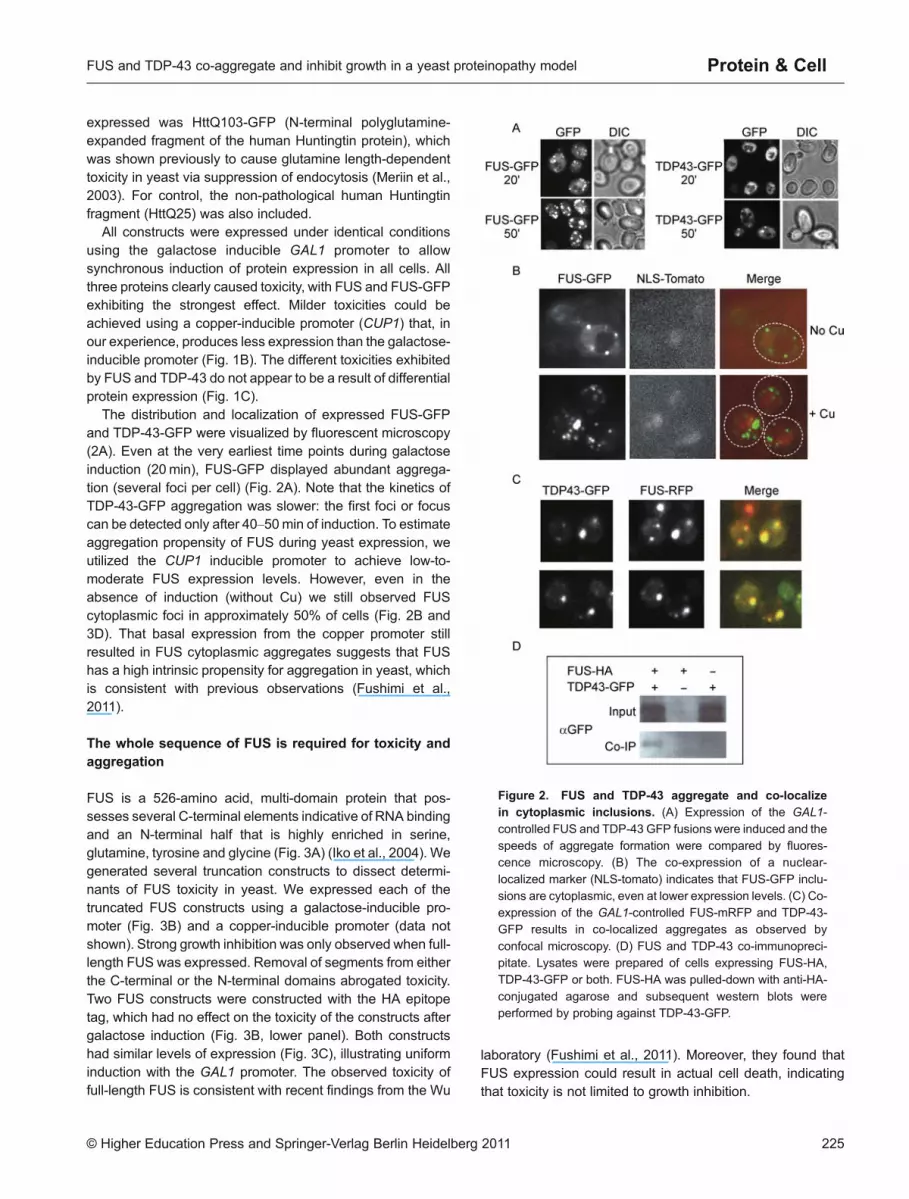
The distribution and localization of expressed FUS-GFPand TDP-43-GFP were visualized by fluorescent microscopy(2A). Even at the very earliest time points during galactoseinduction (20min), FUS-GFP displayed abundant aggrega-tion (several foci per cell) (Fig. 2A). Note that the kinetics ofTDP-43-GFP aggregation was slower: the first foci or focuscan be detected only after 40–50min of induction. To estimateaggregation propensity of FUS during yeast expression, weutilized the CUP1 inducible promoter to achieve low-to-moderate FUS expression levels. However, even in theabsence of induction (without Cu) we still observed FUScytoplasmic foci in approximately 50% of cells (Fig. 2B and3D). That basal expression from the copper promoter stillresulted in FUS cytoplasmic aggregates suggests that FUShas a high intrinsic propensity for aggregation in yeast, whichis consistent with previous observations (Fushimi et al.,2011).
The whole sequence of FUS is required for toxicity andaggregation
FUS is a 526-amino acid, multi-domain protein that pos-sesses several C-terminal elements indicative of RNA bindingand an N-terminal half that is highly enriched in serine,glutamine, tyrosine and glycine (Fig. 3A) (Iko et al., 2004). Wegenerated several truncation constructs to dissect determi-nants of FUS toxicity in yeast. We expressed each of thetruncated FUS constructs using a galactose-inducible pro-moter (Fig. 3B) and a copper-inducible promoter (data notshown). Strong growth inhibition was only observed when full-length FUS was expressed. Removal of segments from eitherthe C-terminal or the N-terminal domains abrogated toxicity.Two FUS constructs were constructed with the HA epitopetag, which had no effect on the toxicity of the constructs aftergalactose induction (Fig. 3B, lower panel). Both constructshad similar levels of expression (Fig. 3C), illustrating uniforminduction with the GAL1 promoter. The observed toxicity offull-length FUS is consistent with recent findings from the Wu
laboratory (Fushimi et al., 2011). Moreover, they found thatFUS expression could result in actual cell death, indicatingthat toxicity is not limited to growth inhibition.
Figure 2. FUS and TDP-43 aggregate and co-localizein cytoplasmic inclusions. (A) Expression of the GAL1-
controlled FUS and TDP-43 GFP fusions were induced and thespeeds of aggregate formation were compared by fluores-cence microscopy. (B) The co-expression of a nuclear-
localized marker (NLS-tomato) indicates that FUS-GFP inclu-sions are cytoplasmic, even at lower expression levels. (C) Co-expression of the GAL1-controlled FUS-mRFP and TDP-43-GFP results in co-localized aggregates as observed by
confocal microscopy. (D) FUS and TDP-43 co-immunopreci-pitate. Lysates were prepared of cells expressing FUS-HA,TDP-43-GFP or both. FUS-HA was pulled-down with anti-HA-
conjugated agarose and subsequent western blots wereperformed by probing against TDP-43-GFP.
© Higher Education Press and Springer-Verlag Berlin Heidelberg 2011 225
FUS and TDP-43 co-aggregate and inhibit growth in a yeast proteinopathy model Protein & Cell

Figure 3. The full-length FUS protein is most aggregation-prone and toxic in yeast. (A) Schematic picture of the full-lengthFUS protein as characterized by Iko and coworkers (Iko et al., 2004). SQGY denotes the region that is highly enriched for serine,glutamine, glycine and tyrosine; RGG, rich in arginine and/or glycine; RRM, RNA recognition motif; ZnF, zinc finger domain. (B)
Strain BY4741 harboring a series of GAL1-controlled FUS truncations was grown on both SD and SDGal plates as described inFig. 1A, using 10 × serial dilutions. (C) A western blot with anti-HA antibody was performed on equivalent cell lysates after 6 hours ofgalactose induction, indicating that full-length FUS toxicity was not due to greater protein expression; *, degradation product. (D)
Fluorescence microscopy was used to observe GFP fused to full-length and truncated FUS under the control of a copper-induciblepromoter (upper panel). The aggregation of FUS(1–422)-GFP did not correspond with an inhibition of cell growth (lower panel).
226 © Higher Education Press and Springer-Verlag Berlin Heidelberg 2011
Dmitry Kryndushkin et al.Protein & Cell

A reduction in toxicity was coupled with diminishedaggregation potential of truncated FUS constructs. Mostevident was the absence of aggregation of FUS deletionmutants expressed under control of the CUP1 promoter inthe absence of induction (e.g., without Cu), while under thesame conditions full-length FUS still formed fluorescent foci(Fig. 3D). The fluorescence intensities of all FUS-GFPconstructs were similar, confirming similarity in their expres-sion levels. After copper induction, both FUS(1–422)-GFP andFUS(292–526)-GFP were able to form aggregates; however, theformation of foci and toxicity was still significantly reducedcompared to full-length FUS (Fig. 3D).
FUS and TDP-43 co-localize and interact in yeast
In neurons, both FUS and TDP-43 predominantly localize tocell nuclei under physiological conditions, but both proteinswere found to co-localize in cytoplasmic inclusions in thespinal motor neurons of ALS patients (Deng et al., 2010). Co-expression of FUS-RFP and TDP-43-GFP in yeast cellsresulted in cytoplasmic aggregates that unambiguouslycontained both proteins, as evident from confocal fluorescentmicroscopy (Fig. 2C). Remarkably, perfect colocalization wasobserved in every single cell that contained both FUS-RFPand TDP-43-GFP visible foci, indicating a strong interactionbetween the two proteins. It has been proposed that FUS andTDP-43 functionally interact as part of a larger biochemicalcomplex in mammalian cells (Kim et al., 2010), and they havebeen shown to co-immunoprecipitate when expressed incultured cells (Kim et al., 2010; Ling et al., 2010). We likewisefound that TDP-43 specifically co-immunoprecipitated withFUS in yeast cell extracts, when both proteins wereexpressed (Fig. 2D).
FUS aggregates do not localize to the same compartmentas many amyloidogenic proteins, but do co-localize withpolyglutamine
It was previously reported that mammalian and yeast cellsshare common protein quality control mechanisms, withsimilar patterns of sequestration and localization of misfoldedproteins (Kaganovich et al., 2008). Many amyloidogenicproteins were reported to be localized to a single “insolubleprotein deposit,” or IPOD, a putative compartmentalization ofterminally-aggregated proteins that have exceeded othermechanisms of protein quality control. In yeast cells withexpressed FUS, we did not observe localization to thenucleus, but instead extensive cytoplasmic inclusions wereformed even at low expression levels. The localization ofthese aggregates was compared with other aggregation-prone proteins that were previously shown to localize to theIPOD. FUS aggregates did not co-localize with severaldescribed IPOD markers (Kaganovich et al., 2008), such asHsp104-GFP, Ubc9ts-GFP and aggregates of overproduced
amyloidogenic proteins Ure2-GFP or RNQ-GFP (Fig. 4A). Inaddition, no co-localization was observed with α-synuclein-GFP (another amyloid-related protein forming toxic aggre-gates in yeast cells (Outeiro and Lindquist, 2003)) (Fig. 4A).However, FUS-GFP did partially co-localize with overpro-duced huntingtin fragment containing an extended polygluta-mine track, HttQ103-GFP (Fig. 4B). This is remarkablebecause FUS was previously found to be a major componentof neuronal intranuclear polyglutamine inclusions of Hunting-ton’s disease (Woulfe et al., 2010). It is striking that whileFUS-GFP did not co-localize with many other misfoldedproteins, it did show co-localization with TDP-43 (Fig. 2C) andhuntingtin fragment, indicating strong specificity of thisinteraction.
FUS inclusions are detergent sensitive and differ fromamyloid aggregates
Saccharomyces cerevisiae has several native proteins thatcan adopt an infectious, or prion, state that is based on a self-propagating amyloid conformation (Wickner et al., 2010). TheHsp104p chaperone is required for the propagation of theseamyloid-based prions and is believed to act by fragmentingthe infectious amyloid fibers, thus creating new prion “seeds”and facilitating proliferation of the prion. Deletion of HSP104,or inhibition of Hsp104p, results in the formation of very largeaggregates and finally leads to rapid prion loss during cellulardivisions (Chernoff et al., 1995; Kryndushkin et al., 2003).Likewise, Hsp104p is necessary for the manifestation ofHttQ103 (Krobitsch and Lindquist, 2000; Meriin et al., 2002),but not TDP-43 (Johnson et al., 2008) aggregation andtoxicity. We checked if Hsp104p would similarly influenceFUS toxicity and found that deletion of the HSP104 geneneither changed the FUS aggregation pattern (data notshown), nor eliminated FUS toxicity (Fig. 5).
Amyloid-based yeast prions and proteins with polygluta-mine (polyQ) expansions share extreme resistance totreatments with strong anionic detergents, such as sodiumdodecyl sulphate (SDS) or sarcosyl (Serio et al., 2000;Speransky et al., 2001; Kryndushkin et al., 2003; Salnikovaet al., 2005). This reflects a tight packing of β-sheets withinthe amyloid backbone, making it a characteristic feature ofamyloid structure. We tested whether the FUS aggregatesthat form in yeast have similar properties. Yeast cellsexpressing FUS-GFP protein for 4 hours were harvested,disrupted and analyzed by simple sedimentation assay(centrifugation 16,000 g for 10min). In parallel, Triton X-100and RNase A were applied to cell extracts for 10min followedby the same spin. About 70% of FUS-GFP protein was foundin the pellet fractions independent of treatments (Fig. 6A),indicating the presence of FUS in very high molecular weightcomplexes that are not dependent on membrane associa-tions.
Aggregation behavior of FUS was compared with TDP-43,
© Higher Education Press and Springer-Verlag Berlin Heidelberg 2011 227
FUS and TDP-43 co-aggregate and inhibit growth in a yeast proteinopathy model Protein & Cell

HttQ103 and HttQ25 using the same sedimentation assay(Fig. 6B). Cell extracts were treated with 0.5% Triton X-100and spun down. Pellet fractions were treated with 1 × SDS-PAGE Sample buffer (see MATERIALS AND METHODS),incubated at room temperature or boiled and then immedi-ately subjected to SDS-PAGE. HttQ103 aggregates showedstriking resistance to SDS treatment at room temperatures;protein complexes were stuck in the wells of the 10%polyacrylamide gel and could be detected inside the gelonly after boiling. Similar behavior was reported for nativeyeast prion aggregates (Speransky et al., 2001; Kryndushkinet al., 2003). Instead, both FUS and TDP43 could mostlyenter the gel immediately after brief SDS treatment of thepellet fractions at room temperatures (compare lanes “P” and“P*” in Fig. 6B), indicating a significant difference in thebiophysical properties of the aggregates. However, bothproteins did show a mild resistance to SDS in the absence ofheat and prolonged incubation.
The filter retardation assay is an established method forcharacterizing misfolded and aggregated proteins in yeast(Johnson et al., 2008; Alberti et al., 2009). We treated cell
extracts with 1% SDS at room temperature for 15min priorto vacuum blotting them to a cellulose acetate membrane(0.2 μm pore size). While HttQ103 aggregates remainedSDS-insoluble and were unable to pass through themembrane, FUS aggregates were much less resistant(Fig. 6C). Similar SDS sensitivity was previously reportedfor TDP-43 (Johnson et al., 2008).
The yeast prion-like amino terminus of FUS does notpropagate as a yeast prion
The amino-terminal domain of FUS has been noted for itsstriking resemblance to yeast prion domains (Cushman et al.,2010; Udan and Baloh, 2011). Yeast prion domains aredistinct in their richness in uncharged polar residues, paucityof charged residues and their apparent native disorder(Toombs et al., 2010). The first approximately 165 aminoacids of FUS are not highly charged, but instead are highlyenriched in serine, tyrosine, glycine and glutamine (Fig. 3A),compositionally reminiscent of many yeast prion domains.The fact that FUS forms aggregates in diseased motor neural
Figure 4. FUS aggregates do not co-localize with several IPOD markers. (A) The GAL1-controlled fluorescent protein fusionsof FUS and several characterized IPODmarkers (Hsp104, Ubc9ts, Ure2, Rnq1) (Kaganovich et al., 2008), as well as amyloidogenicα-synuclein (aSYN), were co-expressed and monitored by confocal fluorescence microscopy. (B) The GAL1-controlled FUS-RFP
and a polyglutamine tract fused to GFP (HttQ103-GFP) partially co-localize. A similar result was observed for the GAL1-controlledTDP-43-GFP and HttQ103-RFP.
228 © Higher Education Press and Springer-Verlag Berlin Heidelberg 2011
Dmitry Kryndushkin et al.Protein & Cell

networks that progressively degenerate appears at leastsuperficially similar to the prion aggregates that propagatevertically through yeast populations.
A yeast Sup35p reporter assay was employed to see ifamino-terminal FUS fragments could propagate in two distinctstable states in vivo, as do yeast prions, in which there is asoluble state and a self-propagating amyloid state. Sup35p isa well studied prion protein and is responsible for the [PSI+]prion. It contains a portable N-terminal prion domain and afunctional C-terminal domain (referred to as Sup35MC),involved in translation termination. When Sup35p is in theprion state, translation is imperfectly terminated at stopcodons, resulting in some translational read-through. Apremature stop codon in the adenine biosynthesis geneADE2 is used to report Sup35 function: [PSI+] cells are white/pink and can grow on media lacking adenine, whereas prion-negative cells are unable to grow on media lacking adenine orgrow red on low-adenine media due to the accumulation of anintermediate in the adenine biosynthetic pathway. Fusing
Figure 5. FUS and TDP-43 toxicity is not dependent onHsp104 function. The toxicities of several aggregation-proneproteins were compared by over-expression using a galactose-inducible promoter in strain 74D-694 containing either wild-
type or null HSP104. Cells were grown as described in Fig. 1A.
Figure 6. FUS forms highmolecular weight SDS-sensitiveaggregates. (A) FUS sedimentation does not require associa-
tion with membranes. Protein lysates were prepared from yeastcells expressing FUS-GFP. Sedimentation of the lysates wasperformed before or after Triton X-100 and RNase A treatments;
S, supernatant and P, pellet fraction. (B) Lysates from yeastcells expressing FUS-HA, TDP-43-HA, HA-TDP-43, HttQ103-GFP or HttQ25-GFP were prepared, sedimented as above andsubjected to SDS-PAGE. T, total lysate; S, supernatant; P, pellet
(no heated incubation); P*, pellet (heated for 5 min at 96°C). (C)Filter retardation assay. Lysates of cells expressing FUS andpolyglutamine GFP fusions were treated with 1% SDS, vacuum
blotted to cellulose acetate (0.2 μm pore size) and immunos-tained with anti-GFP antibody.
© Higher Education Press and Springer-Verlag Berlin Heidelberg 2011 229
FUS and TDP-43 co-aggregate and inhibit growth in a yeast proteinopathy model Protein & Cell

230 © Higher Education Press and Springer-Verlag Berlin Heidelberg 2011
Dmitry Kryndushkin et al.Protein & Cell

protein fragments to Sup35MC, lacking its native amino-terminal prion domain, is an established method for screeningfor new prion-capable proteins (Chernoff et al., 2000;Kushnirov et al., 2000; Santoso et al., 2000; Alberti et al.,2009).
Plasmids coding FUS(1–167)-Sup35MC and FUS(1–134)-Sup35MC were plasmid swapped (using 5-FOA) in asup35Δ strain harboring SUP35MC on a URA3 plasmid(Fig. 7A and data not shown). Both FUS fusion constructscould support normal Sup35p function (Fig. 7A), indicatingthat the constructs were not catastrophic to Sup35p folding,solubility or activity. If the FUS fusions are capable of enteringa prion state, this should manifest in the appearance of Ade+isolates on media lacking adenine. Indeed, upon selection onadenine-minus medium, the fusion constructs yielded farmore Ade+ isolates than the SUP35MC control that lacks aprion domain (Fig. 7B). However, when we attempted toinduce the formation of prions by transiently over-expressingamino-terminal FUS or full-length FUS in the presence of ourFUS(1–167)-Sup35MC and FUS(1–134)-Sup35MC reporters, wedid not see a corresponding increase in Ade+ frequency (datanot shown).
Yeast prions are distinguished by their reversible curability.If FUS(1–167)-Sup35MC and FUS(1–134)-Sup35MC are formingprions, this should be marked by an ability to reverse statesand return to the prion-negative form (while retaining thecapacity to become a prion again). The conversion to theprion-negative state is greatly facilitated by the inhibition ofHsp104p function, either by gene deletion or a chemicalinhibitor (millimolar guanidine), which is necessary for thepropagation of most amyloid-based yeast prions. In ourhands, a small minority of the Ade+ isolates appeared to losethe Ade+ phenotype after growth on millimolar guanidine(Fig. 7C). However, we could not confirm a true reversion bymaking the HSP104 deletion in some clones, indicating thatthe reversion of the phenotype might be because of instabilityand differential growth rates on medium with guanidine.Furthermore, if the fusion proteins are truly able to stablypropagate as prions, they should be able to infect other cellswith the prion in a cytoplasmic mixing experiment, assumingthe recipient is coding a similar FUS-Sup35MC fusion. Wewere unable to transfer the Ade+ state from any of our Ade+isolates to recipient reporters (data not shown). Finally, theability of a specific prion amyloid to recruit GFP fusionproteins (consisting of GFP fused to the prion protein of
interest) is an established method for reporting the physicalstate of prion proteins within a yeast cell. The aggregationlevels of FUS(1–183)-GFP (CUP1 promoter) expressed in theAde+ cells versus the control Ade- cells were compared. Wecould not detect any visible aggregation of FUS(1–183)-GFP ineither case (data not shown and Fig. 7D).
Finally, the aggregation propensity of the FUS N-terminalfragment (amino acids 1–183 fused to GFP) was explored byfluorescent microscopy and filter retardation assay. A robustinduction of the FUS(1–183)-GFP expression under the GAL1promoter resulted only in subtle aggregate formation—oneaggregate per cell in 50% of the cells together with intensivecytoplasmic distribution (Fig. 7D). In contrast, under the sameexpression conditions, full-length FUS exhibited severeaggregation (Fig. 7D). Also, the filter retardation assayshowed SDS sensitivity of the FUS(1–183)-GFP aggregates(Fig. 6C), whereas SDS resistance is characteristicallyobserved for prion amyloid aggregates. The fundamentaldifferences observed between aggregation of FUS N-terminalfragments and yeast prion-forming proteins argue against thisdomain alone conferring a prion capacity of FUS in yeast.
DISCUSSION
Protein misfolding and aggregation are a pathological hall-mark of several neurodegenerative diseases, includingAlzheimer’s disease, Huntington’s disease and amyotrophiclateral sclerosis. Recently the FUS/TLS protein was identifiedas a new component of the cellular inclusions in non-SOD1type ALS, atypical fronto-temporal lobar degeneration(FTLD)-U and neuronal intermediate filament inclusiondisease (dementia) (NIFID) (Neumann et al., 2009; Denget al., 2010; Woulfe et al., 2010), indicating that thesedisorders may share a common pathogenic pathway thatultimately leads to neuronal cell death. To find potentiallyconserved mechanisms that result in (or from) abnormal FUSaggregation, we utilized a yeast model system that hasadvantages over mammalian cell lines due to its tractability.By expressing FUS in yeast cells, we were able torecapitulate some of the characteristic features observed indiseased tissue, such as formation of FUS cytoplasmicinclusions and specific co-aggregation with TDP-43 (Denget al., 2010). Importantly, our model shows that a higher levelof FUS expression leads to more severe aggregation andcellular toxicity, suggesting that abnormal cytoplasmic
Figure 7. The SQGY-rich amino terminus of FUS could not form a prion. (A) FUS1–167-Sup35MC fusion is functional. Acartoon illustrates how strains RW4827 and RW4830 were transformed. Plasmid swaps were performed in which the LEU2
plasmids with Sup35MC, FUS1–167-Sup35MC or empty control were each substituted for p1216 by 5-FOA selection. Only plasmids
coding functional Sup35 permit growth on 5-FOA under this selection. (B) Expression of FUS1–167-Sup35MC in the strains RW4827and RW4830 yieldedmore Ade+ colonies (prion candidates) than expression of Sup35MC. (C) Some candidates had unstable Ade+phenotypes, which were exacerbated by the inhibition of Hsp104 function by growth on millimolar guanidine. (D) High-level GAL1-
controlled expression of the FUS1–183-GFP results in less aggregation compared to full-length FUS-GFP as judged by fluorescencemicroscopy.
© Higher Education Press and Springer-Verlag Berlin Heidelberg 2011 231
FUS and TDP-43 co-aggregate and inhibit growth in a yeast proteinopathy model Protein & Cell

accumulation of FUS in neurons may have a similarconsequence and correlate with disease progression.
A particularly interesting conclusion that comes from ourwork is a striking similarity in FUS and TDP-43 behavior inyeast cells. Both proteins are forming toxic SDS-solubleaggregates that differ biophysically from polyglutamine andamyloid-based yeast prion aggregates (Fig. 6). Furthermore,they show perfect co-aggregation during simultaneousexpression in yeast cells, and their interaction was corrobo-rated by co-immunoprecipitation (Fig. 2C and 2D). Thisinteraction is specific since FUS (and TDP-43) aggregates didnot co-localize with overproduced amyloidogenic RNQ-GFP,Ure2-GFP or aSYN-GFP proteins, each of which also formscytoplasmic inclusions in yeast cells (Fig. 4A). Takentogether, our results strongly indicate that the mechanism(s)of cellular toxicity for FUS and TDP-43 might be similar.
Both FUS and TDP-43 are involved in formation of stressgranules in mammalian cells (Colombrita et al., 2009; Galet al., 2010; Ito et al., 2011) that may be directly connectedwith their normal function. Yeast cells do not have closehomologues of FUS and TDP-43, but they do have a similarset of proteins that form stress granules under glucosedeprivation or elevated temperature (Buchan et al., 2008).Among them are Pab1p, Pub1p, Ngr1p and Pbp1p (yeastorthologues of mammalian PABP, TIA-1, TIA-R and Ataxin-2),all of which are important components of mammalian stressgranules (Nonhoff et al., 2007; Buchan and Parker, 2009).Interestingly, Pbp1p was found recently to modify TDP-43toxicity in yeast and Drosophila (Elden et al., 2010). It wouldbe of interest to more thoroughly investigate the possibleinteractions between FUS, TDP-43 and stress granulecomponents; perhaps such an approach could contribute toa better understanding of FUS and TDP-43 cellular functions.Many stress granule-related proteins, including FUS andTDP-43, appear to share both a glutamine-rich domain aswell as an RNA-binding domain(s). Glutamine-rich domainsnot only have a strong tendency to self-aggregate, but arelikely involved in the assembly process of large stress granulecomplexes.
A close association of FUS and TDP-43 with expandedpolyglutamines deserves further investigation. FUS wasidentified to be a major component of nuclear polyglutamineaggregates in a Neuro2a cell line (Doi et al., 2008), andlikewise TDP-43 is sequestered in polyglutamine aggregates,resulting in the depletion of TDP-43 from the nucleus and theformation of detergent-resistant inclusions (Fuentealba et al.,2010). Interestingly, Ataxin-2 intermediate-length polygluta-mine expansions were found more frequently in ALS patients(Elden et al., 2010). Ataxin-2 was found to be associated withTDP-43 under some conditions and to be able to modify TDP-43 toxicity. We observed frequent co-aggregation of FUS orTDP-43 with polyglutamine aggregates (Fig. 4B), furthersupporting a potential link between these RNA-bindingproteins and polyglutamine disorders.
We tested the ability of the N-terminal fragment of FUS toserve as a prion in yeast cells. We focused on this fragmentbecause it has high compositional similarity to yeast priondomains (Toombs et al., 2010), which are highly enriched inhydrophilic amino acids like glutamine and asparagine. Suchprion domains can adopt an infectious amyloid conformationthat propagates by recruiting identical protein sequences intothe amyloid aggregate via a template mechanism. BecauseFUS forms aggregates in neurons, we hypothesized that itmay similarly form aggregates in yeast cells by a mechanismsimilar to the yeast prions. However, we were unable to findevidence that the prion-like domain of FUS has such behaviorin yeast cells. In fact, we think that the ability to substitute anative prion domain and drive prion formation may requirestrong amyloid-forming propensity that appears to be absentin the case of FUS. Because of the fundamental differencesobserved between FUS aggregation and prion-formingproteins, it appears unlikely that FUS has a pathologicalmechanism of propagation that is directly reminiscent of prionamyloids. However, it is possible that by utilizing a differentapproach and/or using different FUS fragments one will beable to isolate an infectious FUS prion. We cannot excludethe possibility that the full-length protein or its sub-domainscould form a prion.
During the preparation of this manuscript, Fushimi et al.presented a similar model of FUS proteinopathy (Fushimiet al., 2011). While many of their conclusions are similar toours, they concluded that the resistance of FUS inclusions to0.5% sarcosyl, but not to 2% sarcosyl, suggests that FUSmay be amyloidogenic in yeast. However, based on ourexperience (Kryndushkin et al., 2003; Kushnirov et al., 2006),0.5% sarcosyl may not be sufficient to dissolve all protein-protein complexes in yeast cell lysates. Instead, we used 1%SDS and found FUS aggregates to be of much greatersensitivity than polyglutamine aggregates. Amyloids, includ-ing yeast prion amyloids, generally show obvious SDSresistance and will frequently get stuck in the loading wellsof polyacrylamide gels (an observation we did not find forFUS). Though Fushimi et al. provide suggestive evidence thatFUS aggregation is amyloid-like, the nature of FUS aggre-gates appears to be distinctly different from what has beenobserved with many amyloidogenic proteins in yeast. TDP-43aggregates in yeast were previously reported to be non-amyloid in nature (Johnson et al., 2008), which is consistentwith our characterization of FUS aggregates.
We observed that FUS and TDP-43 behavior in yeastrecapitulates many of the same observations made inneurons and cell culture. This suggests that a simpleeukaryotic model could assist in the elucidation of themechanisms underlying aggregation and cytotoxicity of FUSand other disease-related/aggregation-prone proteins. Con-sidering that there are currently few options for studying FUSproteinopathy in higher organisms, the yeast model and itsamenability to high-throughput screens may prove to be a
232 © Higher Education Press and Springer-Verlag Berlin Heidelberg 2011
Dmitry Kryndushkin et al.Protein & Cell

helpful tool in understanding cellular degeneration observedwith ALS.
MATERIALS AND METHODS
Yeast strains, growth conditions and plasmids
Strains BY4741 (MATa his3 leu2 met15 ura3), W303 (MATa can1-
100 his3-11,15 leu2-3,112 trp1-1 ura3-1 ade2-1), 74D-694 (MATa
ade1-14 his3 leu2 trp1 ura3 [PIN+]) (Chernoff et al., 1995) and 74D-694 ∆hsp104::LEU2 (a kind gift of Dr. Susan Liebman) were usedfor FUS and TDP-43 expression. Strains RW4827 (MATa ade2-1
SUQ5 trp1 kar1-1 his3 leu2 ura3 sup35:kanMX) and RW4830 (MATa
ade2-1 SUQ5 leu2 ura3 lys2 kar1 sup35:kanMX trp1) were used inthe prion assay (discussed below). Strain BY4741 bearing HSP104-
GFP in the HSP104 genomic locus was purchased from Invitrogen.Yeast cells were grown at 30°C in standard synthetic defined media(SD) containing 2% glucose, or 2% raffinose (SDRaf), or 2%
galactose and 1% raffinose (SDGal) as a carbon source. Onlyrequired amino acids were added during cell growth. For proteininduction cells were grown overnight in SDRaf medium, adjusted toOD600 = 1 and then incubated in liquid SDGal medium for 4 h. For
toxicity assays, yeast cells were grown overnight in liquid SDRaf untilthey reached log phase. Cultures were then equalized by OD600,serially diluted, spotted onto solid SDGal medium and grown at 30°C
for 3 days.Plasmids used in this study are listed in Table 1. The open reading
frames corresponding to wild type FUS or TDP-43 were inserted into
yeast expression plasmids, pH392cup (centromeric, URA3 marker,CUP1 promoter), pH396gal (high copy, URA3 marker, GAL1
promoter), pH610 (high copy, TRP1, GAL1 promoter) or pH317gal
(high copy, LEU2 marker, GAL1 promoter) either intact or in framewith HA, EGFP or mRFP tags at the carboxyl-termini of the proteins.Full-length TDP-43, FUS and truncation variants were prepared usingwild-type cDNA plasmids from Open Biosystems; all constructs were
confirmed by DNA sequencing.
Sedimentation and filter retardation assays
To prepare yeast cell extracts strain BY4741 carrying expressionplasmid(s) was grown in liquid SDRaf medium overnight and switchedto SDGal (OD600 = 1) for induction of protein expression. Cells were
incubated for 4 h, harvested, washed in buffer A (25mmol/L Tris-HCl,pH 7.4, 150mmol/L NaCl, 10% glycerol, 5 mmol/L EDTA, 1mmol/Ldithiothreitol and complete protease inhibitor mixture (Roche AppliedScience, Indianapolis, IN) and lysed by vortexing with glass beads in
the same buffer. To limit protein degradation, in some cases cellswere alternatively disrupted by vortexing with glass beads in UreaLysis Buffer (10mmol/L Tris, pH 7.5, 8 mol/L urea). Cell debris was
removed by centrifugation at 200 g for 10min. Protein was measuredwith BCA reagent (Pierce, Rockford, IL).
For sedimentation analysis, cell lysate aliquots were treated with
1% Triton X-100 and/or 0.1mg/mL RNase A for 10min on ice, spundown at 16,000 g for 10min, and afterward the pellet and supernatantwere collected and compared by standard SDS-PAGE. Immediately
prior to loading the samples on an SDS-PAGE gel, the samples weremixed 3:1 with a protein-loading buffer containing 4% sodium dodecylsulfate. All fractions were heated for 5 min at 96°C, except wherenoted. Immunoblotting was performed using PVDF membranes with
anti-HA antibody or anti-GFP antibody (Roche Applied Science) andAP-conjugated secondary anti-rat IgG (Promega). Signal detectionwas performed using CDP-Star detection reagent (Perkin Elmer) and
Kodak Biomax chemoluminescence film.For comparing relative detergent resistance of separated fractions,
prior to electrophoresis, each fraction (except one pellet fraction) was
adjusted to 1% sodium dodecyl sulfate in gel loading buffer andheated for 5 min at 96°C.
The filter retardation assay using cellulose acetate membranes
(0.2 μm pore size) was performed as described previously(Muchowski et al., 2000; Johnson et al., 2008). Lysates of cellsexpressing FUS and polyglutamine GFP fusions were treated with1% SDS, vacuum blotted to cellulose acetate, washed and blotted
with anti-GFP.
Co-immunoprecipitation
The interaction between FUS-HA and TDP-43-GFP was analyzed byimmunoprecipitation using the protocol and reagents found in theProFound™ HA-Tag IP/Co-IP kit (Pierce). FUS-HA was immunopre-
cipitated from lysates that were prepared from yeast cells that weremechanically disrupted with glass beads in Non-denaturing LysisBuffer (TBS pH 7.4, 0.1% tween, 10% glycerol, 0.1mmol/L AEBSF,0.5mmol/L EDTA, protease inhibitor cocktail (Roche)). Following
immunoprecipitation, western blots were performed using an anti-GFP primary antibody and an AP-conjugated secondary antibody.Signal detection was performed as described above.
Microscopy
Spinning disc confocal imaging of live yeast cells expressing the
appropriate GFP- and RFP-tagged fusion proteins was performed ona Nikon Eclipse TE2000Umicroscope equipped with a 100 × PlanApo objective. This system is outfittedwith a Spectral Applied
Research LMM5 laser merge module to control the output of fourdiode lasers (excitation at 405, 491, 561 and 655 nm), a YokogawaCSU10 spinning disk unit and a Hamamatsu C9100-13 EM-CCDcamera. Confocal imageswere acquired using IPLab imaging soft-
ware and processed with ImageJ and Adobe Photoshop CS2. Duringthe colocalization experiments, cells were grown overnight in SDRafmedium and then 2% galactose was added for the induction of the
protein of interest. Cells were typically observed after 2–4 hours ofinduction. 5 μL of 2mg/mL Concanavalin A (Sigma) solution wasapplied to microscope slides to immobilize cells. Representative cells
were chosen for figures.
Yeast prion assay
Strains RW4827 and RW4830 are sup35 null, but are viable becauseSup35 function is supplied by plasmid p1216, which codes for the C-terminal functional domain of Sup35. Plasmid swaps were performedin which plasmids pFPS201 and pFPS208 were substituted for p1216
by 5-FOA selection. To select for adenine positive prion candidates,cells were plated to synthetic complete medium lacking adenine (SC-ade). The stability of the adenine positive phenotype was tested by
growing cells for 40 generations on 3 mmol/L guanidine and thenreturning cells to SC-ade. Cytoplasmic transfer experiments (cyto-ductions) between strains RW4827 and RW4830 were performed as
previously described (Wickner et al., 2006).
© Higher Education Press and Springer-Verlag Berlin Heidelberg 2011 233
FUS and TDP-43 co-aggregate and inhibit growth in a yeast proteinopathy model Protein & Cell

ACKNOWLEDGEMENTS
We thank Herman Edskes (NIH) for providing plasmids and for histhoughtful suggestions, Tetsade Piermartiri for her assistance withwestern blotting, Susan Liebman (University of Illinois Chicago) for
providing yeast strains, Kevin O’Connell (NIH) for expert assistancewith confocal microscopy, Judith Frydman (Stanford University) andMichael Sherman (Boston University) for plasmids. This researchwas supported by Uniformed Services University of the Health
Sciences and the Intramural Research Program of the NationalInstitutes of Health, National Institute of Diabetes and Digestive andKidney Diseases.
ABBREVIATIONS
ALS, amyotrophic lateral sclerosis; FTLD, fronto-temporal lobardegeneration; FTLD-U, FTLD with ubiquitin-positive inclusions;NIFID, neuronal intermediate filament inclusion disease (dementia);
5-FOA, 5-fluoroorotic acid
REFERENCES
Alberti, S., Halfmann, R., King, O., Kapila, A., and Lindquist, S.
(2009). A systematic survey identifies prions and illuminatessequence features of prionogenic proteins. Cell 137, 146–158.
Table 1 Plasmids used in the study
Plasmid name Promoter Marker Copy # Source
pH317gal FUS-GFP GAL1 LEU2 high copy this study
pH396gal FUS-mRFP GAL1 URA3 high copy this study
pH317gal TDP43-GFP GAL1 LEU2 high copy this study
pH392c FUS1–526-GFP CUP1 URA3 centromeric this study
pH392c FUS1–422-GFP CUP1 URA3 centromeric this study
pH392c FUS292–526-GFP CUP1 URA3 centromeric this study
pH392c FUS292–422-GFP CUP1 URA3 centromeric this study
p784 Ure2-GFP URE2 LEU2 high copy this study
pH396gal RNQ1-GFP GAL1 URA3 high copy this study
HttQ103-GFP GAL1 URA3 high copy Meriin et al., 2002
HttQ25-GFP GAL1 URA3 high copy Meriin et al., 2002
pEsc NLS-tdTomato GAL1 LEU2 high copy Kaganovich et al., 2008
pEsc CherryFP-Ubc9ts GAL1 URA3 high copy Kaganovich et al., 2008
pH396gal aSYN-GFP GAL1 URA3 high copy this study
pH396gal FUS1–183-GFP GAL1 URA3 high copy this study
pFPS201(FUS(1–167)-Sup35MC) ADH1 LEU2 centromeric this study
pFPS208(FUS(1–134)-Sup35MC) ADH1 LEU2 centromeric this study
pFPS219(FUS1–526) GAL1 TRP1 high copy this study
pFPS217(FUS1–134) GAL1 TRP1 high copy this study
pFPS257(FUS1–253) GAL1 TRP1 high copy this study
pFPS258(FUS135–526) GAL1 TRP1 high copy this study
pFPS258(FUS135–526) GAL1 TRP1 high copy this study
pFPS245(FUS254–526) GAL1 TRP1 high copy this study
pFPS251(FUS436–526) GAL1 TRP1 high copy this study
pFPS264(FUS1–526 + HA) GAL1 TRP1 high copy this study
pFPS259(FUS135–526 + HA) GAL1 TRP1 high copy this study
pFPS256(FUS254–526 + HA) GAL1 TRP1 high copy this study
pFPS229(FUS(1–183)+ GFP) GAL1 TRP1 high copy this study
pFPS266(FUS1–526) CUP1 URA3 centromeric this study
pFPS270(FUS1–526 + HA) CUP1 URA3 centromeric this study
pFPS268(FUS254–526 + HA) CUP1 URA3 centromeric this study
pFPS234(TDP-43 + HA) CUP1 URA3 centromeric this study
pFPS235(HA + TDP-43) CUP1 URA3 centromeric this study
p1216 (SUP35C) ADH1 URA3 centromeric this study
p1166 (SUP35MC) ADH1 LEU2 centromeric this study
234 © Higher Education Press and Springer-Verlag Berlin Heidelberg 2011
Dmitry Kryndushkin et al.Protein & Cell

Ayala, Y.M., Zago, P., D’Ambrogio, A., Xu, Y.F., Petrucelli, L., Buratti,
E., and Baralle, F.E. (2008). Structural determinants of the cellularlocalization and shuttling of TDP-43. J Cell Sci 121, 3778–3785.
Bharadwaj, P., Martins, R., and Macreadie, I. (2010). Yeast as a
model for studying Alzheimer's disease. FEMS Yeast Res 10,961–969.
Bosco, D.A., Lemay, N., Ko, H.K., Zhou, H., Burke, C., Kwiatkowski,
T.J. Jr, Sapp, P., McKenna-Yasek, D., Brown, R.H. Jr, andHayward, L.J. (2010). Mutant FUS proteins that cause amyotrophiclateral sclerosis incorporate into stress granules. Hum Mol Genet
19, 4160–4175.
Braun, R.J., Büttner, S., Ring, J., Kroemer, G., and Madeo, F. (2010).Nervous yeast: modeling neurotoxic cell death. Trends Biochem
Sci 35, 135–144.
Buchan, J.R., Muhlrad, D., and Parker, R. (2008). P bodies promote
stress granule assembly in Saccharomyces cerevisiae. J Cell Biol183, 441–455.
Buchan, J.R., and Parker, R. (2009). Eukaryotic stress granules: the
ins and outs of translation. Mol Cell 36, 932–941.
Chernoff, Y.O., Galkin, A.P., Lewitin, E., Chernova, T.A., Newnam, G.P., and Belenkiy, S.M. (2000). Evolutionary conservation of prion-
forming abilities of the yeast Sup35 protein. Mol Microbiol 35,865–876.
Chernoff, Y.O., Lindquist, S.L., Ono, B., Inge-Vechtomov, S.G., andLiebman, S.W. (1995). Role of the chaperone protein Hsp104 inpropagation of the yeast prion-like factor [psi +]. Science 268,880–884.
Colombrita, C., Zennaro, E., Fallini, C., Weber, M., Sommacal, A.,Buratti, E., Silani, V., and Ratti, A. (2009). TDP-43 is recruited to
stress granules in conditions of oxidative insult. J Neurochem 111,1051–1061.
Cooper, A.A., Gitler, A.D., Cashikar, A., Haynes, C.M., Hill, K.J.,
Bhullar, B., Liu, K., Xu, K., Strathearn, K.E., Liu, F., et al. (2006).Alpha-synuclein blocks ER-Golgi traffic and Rab1 rescues neuronloss in Parkinson’s models. Science 313, 324–328.
Cushman, M., Johnson, B.S., King, O.D., Gitler, A.D., and Shorter, J.(2010). Prion-like disorders: blurring the divide between transmis-sibility and infectivity. J Cell Sci 123, 1191–1201.
Deng, H.X., Zhai, H., Bigio, E.H., Yan, J., Fecto, F., Ajroud, K.,Mishra, M., Ajroud-Driss, S., Heller, S., Sufit, R., et al. (2010).FUS-immunoreactive inclusions are a common feature in sporadic
and non-SOD1 familial amyotrophic lateral sclerosis. Ann Neurol67, 739–748.
Doi, H., Okamura, K., Bauer, P.O., Furukawa, Y., Shimizu, H.,Kurosawa, M., Machida, Y., Miyazaki, H., Mitsui, K., Kuroiwa, Y.,et al. (2008). RNA-binding protein TLS is a major nuclearaggregate-interacting protein in huntingtin exon 1 with expanded
polyglutamine-expressing cells. J Biol Chem 283, 6489–6500.
Duennwald, M.L., and Lindquist, S. (2008). Impaired ERAD and ER
stress are early and specific events in polyglutamine toxicity.Genes Dev 22, 3308–3319.
Elden, A.C., Kim, H.J., Hart, M.P., Chen-Plotkin, A.S., Johnson, B.S.,
Fang, X., Armakola, M., Geser, F., Greene, R., Lu, M.M., et al.(2010). Ataxin-2 intermediate-length polyglutamine expansionsare associated with increased risk for ALS. Nature 466,
1069–1075.
Fuentealba, R.A., Udan, M., Bell, S., Wegorzewska, I., Shao, J.,Diamond, M.I., Weihl, C.C., and Baloh, R.H. (2010). Interaction
with polyglutamine aggregates reveals a Q/N-rich domain in TDP-43. J Biol Chem 285, 26304–26314.
Fushimi, K., Long, C., Jayaram, N., Chen, X., Li, L., and Wu, J.Y.(2011). Expression of human FUS/TLS in yeast leads to proteinaggregation and cytotoxicity, recapitulating key features of FUSproteinopathy. Protein Cell 2, 141–149
Gal, J., Zhang, J., Kwinter, D.M., Zhai, J., Jia, H., Jia, J., and Zhu, H.(2010). Nuclear localization sequence of FUS and induction of
stress granules by ALS mutants. Neurobiol Aging. Jul 29. [Epubahead of print]. doi:10.1016/ j.neurobiolaging.2010.06.010.
Iko, Y., Kodama, T.S., Kasai, N., Oyama, T., Morita, E.H., Muto, T.,
Okumura, M., Fujii, R., Takumi, T., Tate, S., et al. (2004). Domainarchitectures and characterization of an RNA-binding protein, TLS.J Biol Chem 279, 44834–44840.
Ito, D., Seki, M., Tsunoda, Y., Uchiyama, H., and Suzuki, N. (2011).Nuclear transport impairment of amyotrophic lateral sclerosis-linked mutations in FUS/TLS. Ann Neurol 69, 152–162.
Johnson, B.S., McCaffery, J.M., Lindquist, S., and Gitler, A.D. (2008).A yeast TDP-43 proteinopathy model: Exploring the moleculardeterminants of TDP-43 aggregation and cellular toxicity. Proc Natl
Acad Sci U S A 105, 6439–6444.
Kaganovich, D., Kopito, R., and Frydman, J. (2008). Misfolded
proteins partition between two distinct quality control compart-ments. Nature 454, 1088–1095.
Khurana, V., and Lindquist, S. (2010). Modelling neurodegeneration
in Saccharomyces cerevisiae: why cook with baker’s yeast? NatRev Neurosci 11, 436–449.
Kim, S.H., Shanware, N.P., Bowler, M.J., and Tibbetts, R.S. (2010).
Amyotrophic lateral sclerosis-associated proteins TDP-43 andFUS/TLS function in a common biochemical complex to co-regulate HDAC6 mRNA. J Biol Chem 285, 34097–34105.
Krobitsch, S., and Lindquist, S. (2000). Aggregation of huntingtin inyeast varies with the length of the polyglutamine expansion and theexpression of chaperone proteins. Proc Natl Acad Sci U S A 97,
1589–1594.
Kryndushkin, D.S., Alexandrov, I.M., Ter-Avanesyan, M.D., and
Kushnirov, V.V. (2003). Yeast [PSI +] prion aggregates are formedby small Sup35 polymers fragmented by Hsp104. J Biol Chem 278,49636–49643.
Kushnirov, V.V., Alexandrov, I.M., Mitkevich, O.V., Shkundina, I.S.,and Ter-Avanesyan, M.D. (2006). Purification and analysis of prionand amyloid aggregates. Methods 39, 50–55.
Kushnirov, V.V., Kochneva-Pervukhova, N.V., Chechenova, M.B.,Frolova, N.S., and Ter-Avanesyan, M.D. (2000). Prion propertiesof the Sup35 protein of yeast Pichia methanolica. EMBO J 19,
324–331.
Kwiatkowski, T.J. Jr, Bosco, D.A., Leclerc, A.L., Tamrazian, E.,Vanderburg, C.R., Russ, C., Davis, A., Gilchrist, J., Kasarskis, E.
J., Munsat, T., et al. (2009). Mutations in the FUS/TLS gene onchromosome 16 cause familial amyotrophic lateral sclerosis.Science 323, 1205–1208.
Lagier-Tourenne, C., Polymenidou, M., and Cleveland, D.W. (2010).TDP-43 and FUS/TLS: emerging roles in RNA processing andneurodegeneration. Hum Mol Genet 19, R46–R64.
Ling, S.C., Albuquerque, C.P., Han, J.S., Lagier-Tourenne, C.,Tokunaga, S., Zhou, H., and Cleveland, D.W. (2010). ALS-
associated mutations in TDP-43 increase its stability and promoteTDP-43 complexes with FUS/TLS. Proc Natl Acad Sci U S A 107,
© Higher Education Press and Springer-Verlag Berlin Heidelberg 2011 235
FUS and TDP-43 co-aggregate and inhibit growth in a yeast proteinopathy model Protein & Cell

13318–13323.
McGlinchey, R., Kryndushkin, D., and Wickner, R.B. (2011). Suicidal
[PSI +] is a lethal yeast prion. Proc Natl Acad Sci U S A. (In press)
Meriin, A.B., Zhang, X., He, X., Newnam, G.P., Chernoff, Y.O., andSherman, M.Y. (2002). Huntington toxicity in yeast model depends
on polyglutamine aggregation mediated by a prion-like proteinRnq1. J Cell Biol 157, 997–1004.
Meriin, A.B., Zhang, X., Miliaras, N.B., Kazantsev, A., Chernoff, Y.O.,
McCaffery, J.M., Wendland, B., and Sherman, M.Y. (2003).Aggregation of expanded polyglutamine domain in yeast leads todefects in endocytosis. Mol Cell Biol 23, 7554–7565.
Muchowski, P.J., Schaffar, G., Sittler, A., Wanker, E.E., Hayer-Hartl,M.K., and Hartl, F.U. (2000). Hsp70 and hsp40 chaperones caninhibit self-assembly of polyglutamine proteins into amyloid-like
fibrils. Proc Natl Acad Sci U S A 97, 7841–7846.
Neumann, M., Roeber, S., Kretzschmar, H.A., Rademakers, R.,
Baker, M., and Mackenzie, I.R. (2009). Abundant FUS-immunor-eactive pathology in neuronal intermediate filament inclusiondisease. Acta Neuropathol 118, 605–616.
Nonhoff, U., Ralser, M., Welzel, F., Piccini, I., Balzereit, D., Yaspo, M.L., Lehrach, H., and Krobitsch, S. (2007). Ataxin-2 interacts withthe DEAD/H-box RNA helicase DDX6 and interferes with P-bodies
and stress granules. Mol Biol Cell 18, 1385–1396.
Outeiro, T.F., and Lindquist, S. (2003). Yeast cells provide insight intoalpha-synuclein biology and pathobiology. Science 302,
1772–1775.
Rosen, D.R., Siddique, T., Patterson, D., Figlewicz, D.A., Sapp, P.,Hentati, A., Donaldson, D., Goto, J., O’Regan, J.P., Deng, H.X.,
et al. (1993). Mutations in Cu/Zn superoxide dismutase gene areassociated with familial amyotrophic lateral sclerosis. Nature 362,59–62.
Salnikova, A.B., Kryndushkin, D.S., Smirnov, V.N., Kushnirov, V.V.,and Ter-Avanesyan, M.D. (2005). Nonsense suppression in yeastcells overproducing Sup35 (eRF3) is caused by its non-heritable
amyloids. J Biol Chem 280, 8808–8812.
Santoso, A., Chien, P., Osherovich, L.Z., andWeissman, J.S. (2000).
Molecular basis of a yeast prion species barrier. Cell 100, 277–288.
Serio, T.R., Cashikar, A.G., Kowal, A.S., Sawicki, G.J., Moslehi, J.J.,Serpell, L., Arnsdorf, M.F., and Lindquist, S.L. (2000). Nucleated
conformational conversion and the replication of conformationalinformation by a prion determinant. Science 289, 1317–1321.
Sharma, N., Brandis, K.A., Herrera, S.K., Johnson, B.E., Vaidya, T.,Shrestha, R., and Debburman, S.K. (2006). alpha-Synucleinbudding yeast model: toxicity enhanced by impaired proteasomeand oxidative stress. J Mol Neurosci 28, 161–178.
Speransky, V.V., Taylor, K.L., Edskes, H.K., Wickner, R.B., andSteven, A.C. (2001). Prion filament networks in [URE3] cells of
Saccharomyces cerevisiae. J Cell Biol 153, 1327–1336.
Sreedharan, J., Blair, I.P., Tripathi, V.B., Hu, X., Vance, C., Rogelj, B.,Ackerley, S., Durnall, J.C., Williams, K.L., Buratti, E., et al. (2008).
TDP-43 mutations in familial and sporadic amyotrophic lateralsclerosis. Science 319, 1668–1672.
Toombs, J.A., McCarty, B.R., and Ross, E.D. (2010). Compositional
determinants of prion formation in yeast. Mol Cell Biol 30, 319–332.
Udan, M., and Baloh, R.H. (2011). Implications of the prion-related Q/
N domains in TDP-43 and FUS. Prion 5, 1–5.
Vance, C., Rogelj, B., Hortobágyi, T., De Vos, K.J., Nishimura, A.L.,Sreedharan, J., Hu, X., Smith, B., Ruddy, D., Wright, P., et al.
(2009). Mutations in FUS, an RNA processing protein, causefamilial amyotrophic lateral sclerosis type 6. Science 323,1208–1211.
Wickner, R.B., Edskes, H.K., and Shewmaker, F. (2006). How to finda prion: [URE3], [PSI +] and [beta]. [beta] Methods 39, 3–8.
Wickner, R.B., Shewmaker, F., Edskes, H., Kryndushkin, D.,Nemecek, J., McGlinchey, R., Bateman, D., and Winchester, C.L. (2010). Prion amyloid structure explains templating: howproteins can be genes. FEMS Yeast Res 10, 980–991.
Woulfe, J., Gray, D.A., and Mackenzie, I.R. (2010). FUS-immunor-eactive intranuclear inclusions in neurodegenerative disease.
Brain Pathol 20, 589–597.
Yeger-Lotem, E., Riva, L., Su, L.J., Gitler, A.D., Cashikar, A.G., King,O.D., Auluck, P.K., Geddie, M.L., Valastyan, J.S., Karger, D.R.,
et al. (2009). Bridging high-throughput genetic and transcriptionaldata reveals cellular responses to alpha-synuclein toxicity. NatGenet 41, 316–323.
Zinszner, H., Sok, J., Immanuel, D., Yin, Y., and Ron, D. (1997). TLS(FUS) binds RNA in vivo and engages in nucleo-cytoplasmicshuttling. J Cell Sci 110, 1741–1750.
236 © Higher Education Press and Springer-Verlag Berlin Heidelberg 2011
Dmitry Kryndushkin et al.Protein & Cell


