Embed Size (px)
Citation preview
* CorrespSciences, CPomona, CE-mail: ne
This is anprovided th
PHYTOTHERAPY RESEARCHPhytother. Res. (2014)Published online in Wiley Online Library(wileyonlinelibrary.com) DOI: 10.1002/ptr.5253
© 2014 Th
Garlic (Allium sativum) StimulatesLipopolysaccharide-induced Tumor NecrosisFactor-alpha Production from J774A.1 MurineMacrophages
Jessica Sung,1 Youssef Harfouche,1 Melissa De La Cruz,1 Martha P. Zamora,1 Yan Liu,2James A. Rego2 and Nancy E. Buckley1*1Department of Biological Sciences, California State Polytechnic University, 3801 W. Temple Ave., Pomona, CA 91768, USA2Department of Chemistry and Biochemistry, California State Polytechnic University, 3801 W. Temple Ave., Pomona, CA 91768, USA
Garlic (Allium sativum) is known to have many beneficial attributes such as antimicrobial, antiatherosclerotic,antitumorigenetic, and immunomodulatory properties. In the present study, we investigated the effects of anaqueous garlic extract on macrophage cytokine production by challenging the macrophage J774A.1 cell line withthe garlic extract in the absence or presence of lipopolysaccharide (LPS) under different conditions. The effect ofallicin, the major component of crushed garlic, was also investigated. Using enzyme-linked immunosorbent assayand reverse transcriptase-quantitative polymerase chain reaction, it was found that garlic and synthetic allicingreatly stimulated tumor necrosis factor-alpha (TNF-α) production in macrophages treated with LPS. TheTNF-α secretion levels peaked earlier and were sustained for a longer time in cells treated with garlic and LPScompared with cells treated with LPS alone. Garlic acted in a time-dependent manner. We suggest that garlic,at least partially via its allicin component, acts downstream from LPS to stimulate macrophage TNF-α secretion.© 2014 The Authors. Phytotherapy Research published by John Wiley & Sons, Ltd.
Keywords: garlic; allicin; macrophages; tumor necrosis factor-alpha (TNF-α); lipopolysaccharide (LPS).
INTRODUCTION
Garlic has been used as a home remedy for a myriad ofdiseases for thousands of years. To date, garlic and itsconstituents are known to have many beneficialattributes such as antimicrobial (Casella et al., 2013;Goncagul and Ayaz, 2010), antiatherosclerotic (Asdaqet al., 2009; Chan et al., 2013; Gonen et al., 2005;Malekpour-Dehkordi et al., 2013), antitumorigenetic(Iciek et al., 2009; Kaschula et al., 2011), and immuno-modulatory (Iciek et al., 2009) properties. Of particularinterest for our studies is the modulation of cytokineproduction by garlic as cytokines are important immunesystem mediators. Different types of garlic extractshave been shown to directly modulate cytokine pro-duction in lymphocytes (Dong et al., 2011; Liu et al.,2009; Zamani et al., 2009) and macrophages (Changet al., 2005; Dong et al., 2011; Hodge et al., 2002; Keisset al., 2003; Romano et al., 1997). Garlic is largely recog-nized as having antiinflammatory properties; however,not all reports provide evidence indicating that garlicincreases antiinflammatory [i.e. interleukin-10 (IL-10)]cytokines while decreasing pro-inflammatory (i.e. tumornecrosis-alpha (TNF-α), IL-1β, and IL-6) cytokines. Thus,we set out to investigate the effect of a water garlic extractand allicin, the major component of crushed garlic
ondence to: Nancy E. Buckley, Department of Biologicalalifornia State Polytechnic University, 3801 W. Temple Ave.,A 91768, [email protected]
open access article under the terms of the Creative Commons Attributioe original work is properly cited.
e Authors. Phytotherapy Research published by John Wiley & Sons, L
(Rybak et al., 2004), on murine macrophage cytokineproduction induced by lipopolysaccharide (LPS). TheLPS is found on the cell wall of Gram-negative bacteria(Rietschel and Brade, 1992) and has been widely usedto cause an immune response in vivo and in vitro fromvarious immune cells including the activation of macro-phages (Fiorentino et al., 1991; Parameswaran andPatial, 2010; Ulmer et al., 2000). Of the cytokines inves-tigated (IL-1β, IL-6, IL-10 and IL-12, and TNF-α), wefound that a water garlic extract consistently stimulatedLPS-induced TNF-α secretion from macrophages. Tran-scription of TNF-α was also significantly enhanced bygarlic, and allicin, in LPS-treated cells. In addition, macro-phages treated with the aqueous garlic extract alonesecreted small, albeit significant amounts of TNF-α.
MATERIALS AND METHODS
Preparation of water garlic extract. Garlic waspurchased at the local grocery store. The cloves of garlicfrom two garlic bulbs were peeled, and any discoloredparts or blemishes were cut off. Garlic was weighed(56.77 g) and added to a waring blender containing113.54mL of pyrogen-free water (HOSPIRA, Inc., LakeForest, IL). The garlic to water ratio was modeled afterthe aqueous garlic extraction method reported byGamboa-León et al. (2007). The garlic was then blendedfor 1min, and the homogenate was passed throughgrade 50 cheesecloth (Lymex, Chicopee, MA). In atissue culture hood, the flow-through was then filter-
n License, which permits use, distribution and reproduction in any medium,td.
Received 06 May 2014Revised 15 September 2014Accepted 06 October 2014
J. SUNG ET AL.
sterilized using a 150-mL Nalgene 0.2-μm pore-sizedfilter unit (Nalge Nunc International, Rochester, NY)connected to a vacuum pump. The flow-through fromthe Nalgene filter was aliquoted into microcentrifugetubes and stored at �80 °C until used. To determineendotoxin contamination, the garlic extract was sentfor endotoxin testing using the kinetic chromogenic lim-ulus amebocyte lysate assay (Charles River Laborato-ries, Charleston, SC).
Determination of allicin content in garlic. Allicin in thegarlic extracts was quantified using a reversed-phaseHPLC method that employed the Agilent-1100 HPLCsystem (Agilent, CA) with a Zorbax Eclipse XDB-C18column. The garlic extracts were eluted through thecolumn by the isocratic solvent of water/methanol (50/50)at a flow rate of 0.75mL/min, and the absorbance of theeluate was monitored at 254nm.
Allicin synthesis. Allicin was synthetized by theperoxyacid oxidation of diallyl disulphide (Alfa Aesar,Ward Hill, MA) according to the procedure of Smallet al. (1947). Pre-purification of diallyl disulphide byfractional vacuum distillation to remove diallyl sulphideand diallyl trisulphide was critical for obtaining pureallicin after oxidation. Because of the thermal instabilityof allicin, the crude product was purified by silica gelchromatography rather than distillation.
J774A.1 macrophage culture and treatment. The murinemacrophage cell line J774A.1 was purchased fromATCC (Manassas, VA) and maintained in complete me-dia: Dulbecco’s modified Eagle’s medium (ATCC), 10%fetal bovine serum (Invitrogen, Carlsbad, CA), and penicil-lin (100units/mL)/streptomycin (100mg/mL) (Invitrogen).For experiments, J774A.1 macrophages were plated at aconcentration of 1.25×10⁵ cells/mL, 200μL/well in com-plete media in 96 well tissue culture plates. Cells betweenpassages 6–15 were used for all experiments.Before treatment, the conditioned media was removed
from the cells 24 h after plating them. Fresh media wasthen added to the cells, and they were treated with thegarlic or synthetic allicin. The garlic extract was addedto the cells at varying dilutions in pyrogen-free water(1:40, 1:100, 1:200, 1:500, 1:1000, and 1:2000). Alterna-tively, allicin (1–10μg/mL) also diluted in pyrogen-freewater was added to the cells. Garlic or allicin was addedin the presence or absence of 0.1μg/mL LPS (LPSfrom Escherichia coli 055:B5, Sigma, St. Louis, MO).The cells were then incubated at 37 °C and 5% CO2for 24 h. After this incubation period, cell supernatantswere collected and stored at �80 °C until assaying forcytokine secretion. Analysis of cytokine levels revealedan effective dilution of garlic (1:500) that modulatedcytokine production.To determine the minimum LPS concentration that
would cause the maximum TNF-α stimulation in thepresence of garlic, J774A.1 macrophages were platedin 96 well plates as described earlier and treated withthe garlic extract (G1:500) and different concentrationsof LPS (0.001 to 10μg/mL). To further characterize theinteraction between LPS and the effective garlic extract
© 2014 The Authors. Phytotherapy Research published by John Wiley & Sons, L
dilution (G1:500), J774A.1 macrophages were plated in 96well plates as described earlier. The cells were then treatedwith G1:500 for different periods before, after, or at thesame time as LPS (0.1μg/mL). The cells were incubatedat 37°C for 24h (or for the time specified in the figure leg-end).At the end of this incubation period, the supernatantswere collected from the wells and stored at �80°C untilassaying for cytokine secretion. In some experiments,G1:500 was removed prior to the addition of the LPS. Inthese experiments, the conditioned media in the wellswas replaced with fresh media prior to adding the LPS.The cells were incubated at 37 °C for 24h after the addi-tion of LPS. At the end of the 24h, the supernatants werecollected from the wells and stored at �80°C untilassaying for cytokine secretion. Alternatively, in otherexperiments, LPS was added to the cells for differentperiods and removed prior to the addition of G1:500. Inthese experiments, the conditioned media in the wellswas replaced with fresh media prior to adding G1:500.These cells were then incubated at 37 °C for 24h afterthe addition of G1:500. At the end of the 24h, thesupernatants were collected from the wells and stored at�80°C until assaying for cytokine secretion.
Cell proliferation XTT {2,3-bis (2-methoxy-4-nitro-5-sulfophenyl)-5-[(phenylamino) carbonyl]-2H-tetrazoliumhydroxide} assay. A cell proliferation assay was carriedout to determine the effect of garlic or allicin treatmenton macrophage cell viability. Thus, macrophages wereplated (2.5×10⁵cells/mL, 100μL/well) in a 96 well plateand incubated at 37°C. After incubation for 24h, the com-plete media was removed, and new complete media wasadded (100μL/well). The cells were then treated with dif-ferent dilutions of the garlic extract or synthetic allicin inthe absence or presence of LPS (0.1μg/mL). The effect ofthe garlic extract on cell viability was assayed using theCellProliferation Kit II (XTT) from Roche (Indianapolis, IN)as described by the manufacturer.
Cytokine assays. Cell supernatants were assayed for thepresence of cytokines (TNF-α, IL-1β, IL-6, IL-10, andIL-12) using cytokine specific ELISAs (BD OptEIA,BD Biosciences, San Diego, CA) as indicated by themanufacturer.
mRNA quantification. To determine the effect of garlicand allicin on TNF-α and toll-like receptor 4 (TLR4)mRNA levels, J774A.1 macrophages were plated at a con-centration of 1.25×10⁵ cells/mL, 5mL/well in completeme-dia in six well tissue culture plates. The conditioned mediawas removed from the cells 24h after plating them, andfresh media was added. The cells were then treated for2hwith the garlic or synthetic allicin in the absence or pres-ence or LPS (0.1μg/mL). Preparation ofRNA, cDNA, andqPCR was carried out with reagents from Bio-Rad(Hercules, CA). Total RNA extraction and cDNA synthe-sis were carried out as indicated by the manufacturer usingthe Aurum Total RNA Mini kit and the iScript reversetranscriptase, respectively. Real-time PCR was performedin the CFX96 Touch Real-Time PCR Instrument usingSso Advanced Universal SYBR Green and primers(PrimePCR SYBR Assay) for TNF-α, Actin-β, or TLR4.
td. Phytother. Res. (2014)
GARLIC STIMULATES LPS-INDUCED TNF-α FROM MACROPHAGES
The reactionwas run at 95°C for 2min for 1cycle, 95 °C for5 s for 40cycles, 60 °C for 30 s for 40cycles, and 65–95°C(0.5 °C increments) for 5 s/step for 1cycle. The data,expressed as 2�ΔΔCq, were obtained by normalizing againstthe actin gene and then comparing the treated cells againstthe untreated cells.
Statistical analysis. Statistical analysis was performedusing GraphPad Prism Version 6.0. An unpaired one-way analysis of variance (ANOVA) was performed witha p< 0.05 considered significant. Post hoc testing wasperformed on data considered statistically significantlydifferent in the ANOVA, including a Tukey’s multiplecomparison test to compare all groups against eachother. p<0.05 was considered statistically significant.
RESULTS
The aqueous garlic extract (G) was tested at differentdilutions to investigate whether it had any effects oncytokine levels. Of the cytokines tested (TNF-α, IL-1β,IL-6, IL-10, and IL-12), G at dilutions of 1:200 to1:1000 consistently increased TNF-α secretion from themacrophages (Table 1) compared with pyrogen-freewater (PFW). Furthermore, G was very effective at stim-ulating LPS (0.1μg/mL)-induced TNF-α (Table 1) frommacrophages. Thus, we focused on the production ofTNF-α. To rule out that the stimulation of the garlicextract on TNF-α secretion was due to endotoxin contam-ination, the garlic extract was sent out for testing. Thelevels were found to be 0.0025 endotoxin units/mg in5mg/mL of garlic extract. These endotoxin levels werenegligible, and thus, we concluded that component(s)within the garlic extract was responsible for stimulatingTNF-α production from the macrophages. We next
Table 1. Garlic stimulates TNF-α secretion from J774A.1macrophages in the absence and presence of lipopolysaccharide(LPS)
Treatment TNF-α (pg/mL) avg ± SD
Pyrogen-free water 21.72 ± 8.86G1:200 357.67 ± 47.35a
G1:500 86.81 ± 16.23b
G1:1000 31.63 ± 8.98LPS 0.1 μg/mL 1471.75 ± 453.90G1:200+LPS 8401.25 ± 112.40c
G1:500+LPS 8315.00 ± 271.77c
G1:1000+LPS 3502.81 ± 427.94d
Cells were plated at 1.25 × 10⁵ cells/mL, 200 μL/well in 96 wellplates, and incubated at 37 °C. The media was replaced with freshmedia 24 h after plating. The garlic extract (G) was then added tothe cells at the indicated dilutions in the absence or presence of0.1 μg/mL LPS, and the cells were incubated for 24 h. After this in-cubation period, cell supernatants were collected and assayed forTNF-α using ELISA.ap<0.0001.bp=0.0434 compared with the control pyrogen-free water.cp<0.0001.dp=0.0002 compared with LPS 0.1 μg/mL. This is a representa-tive experiment from at least five independent experiments.
© 2014 The Authors. Phytotherapy Research published by John Wiley & Sons, L
looked at cell proliferation using the XTT assay todetermine whether garlic had a mitogenic effect. Wefound that garlic did not stimulate cell proliferation, indi-cating that the increase in TNF-α was not because of anincrease in cell number. However, garlic at a dilution of1:40, in the absence of LPS, decreased metabolic activityby 10% as compared with untreated cells (data notshown). This suggests that at this dilution, garlic had aslightly toxic effect on the cells. This finding is similar tothat by Shin et al. (2013) who also reported that at highraw garlic extract concentrations, RAW cell viability wasreduced (Shin et al., 2013).
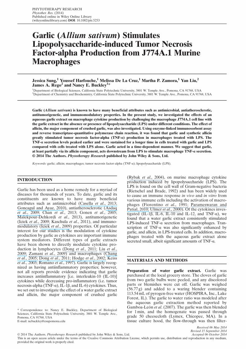
For subsequent studies, we used the highest dilution ofgarlic that caused the maximum stimulation of LPS-induced TNF-α secretion, which was seen using the1:500 dilution (G1:500).We next varied the concentrationof LPS to determine the LPS concentration to use in sub-sequent experiments. As seen in Fig. 1A, garlic stimulatedLPS-induced TNF-α macrophage secretion at all LPSconcentrations studied. Furthermore, although TNF-αsecretion increased in a concentration-dependent manner
Figure 1. Effect of garlic on LPS-induced TNF-α secretion fromJ774A.1 macrophages. Cells were plated at 1.25 × 10⁵ cells/mL,200 μL/well in 96 well plates, and incubated at 37 °C. The mediawas replaced with fresh media 24 h after plating. (A) LPS was thenadded to the cells at the indicated concentrations in the absence(open circles) or presence of G1:500 (filled-in circles). After a24-h incubation, cell supernatants were collected and assayedfor TNF-α using ELISA. ap<0.02, bp<0.008, and cp<0.0001compared with 1 ng/mL LPS and dp<0.0001 compared withcorresponding LPS-only treatment. This is a representativeexperiment from at least three independent experiments. (B) PFW(open circles) or G1:500 (filled circles) was added to the cells atthe indicated times (180, 160, 60, or 15min prior to LPS or 15,60, 120, or 180min post LPS addition). The LPS (0.1 μg/mL)was added (arrow), and the cells were incubated for an additional24 h at which time, cell supernatants were collected and assayedfor TNF-α via ELISA. G1:500+LPS (closed circles) treatmentsare significantly different (p<0.001) when compared with PFW+LPS (open circles) control for each time point. This is a represen-tative experiment of at least three independent experiments.*p<0.04 compared with the LPS �180 h time point.
td. Phytother. Res. (2014)
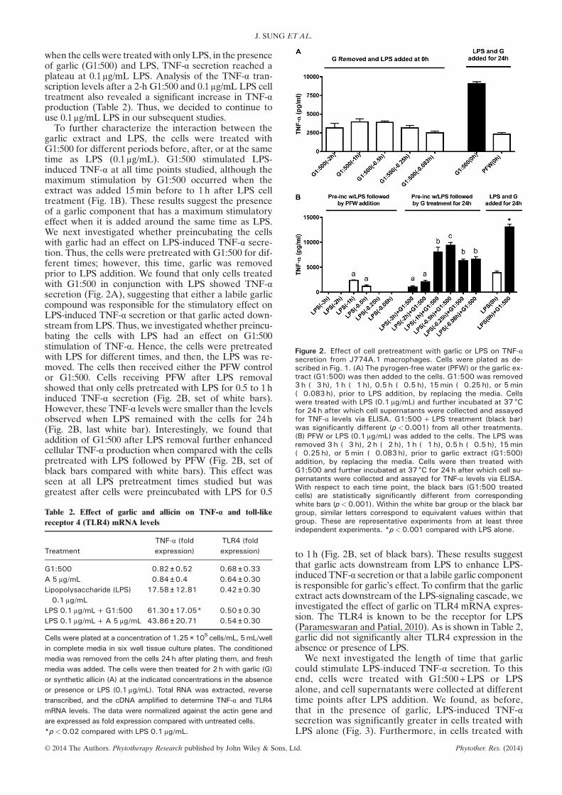
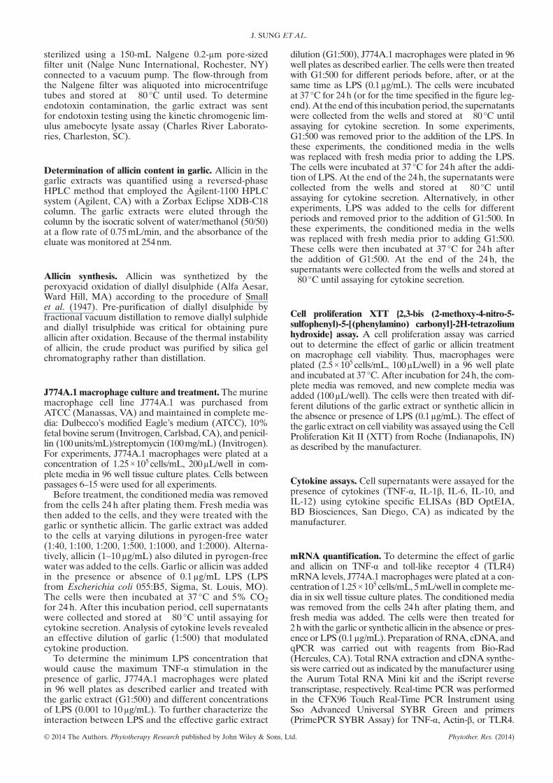
Figure 2. Effect of cell pretreatment with garlic or LPS on TNF-αsecretion from J774A.1 macrophages. Cells were plated as de-scribed in Fig. 1. (A) The pyrogen-free water (PFW) or the garlic ex-tract (G1:500) was then added to the cells. G1:500 was removed3 h (�3 h), 1 h (�1 h), 0.5 h (�0.5 h), 15min (�0.25 h), or 5min(�0.083 h), prior to LPS addition, by replacing the media. Cellswere treated with LPS (0.1 μg/mL) and further incubated at 37 °Cfor 24 h after which cell supernatants were collected and assayedfor TNF-α levels via ELISA. G1:500+LPS treatment (black bar)was significantly different (p<0.001) from all other treatments.(B) PFW or LPS (0.1 μg/mL) was added to the cells. The LPS wasremoved 3 h (�3 h), 2 h (�2 h), 1 h (�1 h), 0.5 h (�0.5 h), 15min(�0.25 h), or 5min (�0.083 h), prior to garlic extract (G1:500)addition, by replacing the media. Cells were then treated withG1:500 and further incubated at 37 °C for 24 h after which cell su-pernatants were collected and assayed for TNF-α levels via ELISA.With respect to each time point, the black bars (G1:500 treatedcells) are statistically significantly different from corresponding
J. SUNG ET AL.
when the cells were treatedwith only LPS, in the presenceof garlic (G1:500) and LPS, TNF-α secretion reached aplateau at 0.1μg/mL LPS. Analysis of the TNF-α tran-scription levels after a 2-h G1:500 and 0.1μg/mL LPS celltreatment also revealed a significant increase in TNF-αproduction (Table 2). Thus, we decided to continue touse 0.1μg/mL LPS in our subsequent studies.To further characterize the interaction between the
garlic extract and LPS, the cells were treated withG1:500 for different periods before, after, or at the sametime as LPS (0.1μg/mL). G1:500 stimulated LPS-induced TNF-α at all time points studied, although themaximum stimulation by G1:500 occurred when theextract was added 15min before to 1h after LPS celltreatment (Fig. 1B). These results suggest the presenceof a garlic component that has a maximum stimulatoryeffect when it is added around the same time as LPS.We next investigated whether preincubating the cellswith garlic had an effect on LPS-induced TNF-α secre-tion. Thus, the cells were pretreated with G1:500 for dif-ferent times; however, this time, garlic was removedprior to LPS addition. We found that only cells treatedwith G1:500 in conjunction with LPS showed TNF-αsecretion (Fig. 2A), suggesting that either a labile garliccompound was responsible for the stimulatory effect onLPS-induced TNF-α secretion or that garlic acted down-stream from LPS. Thus, we investigated whether preincu-bating the cells with LPS had an effect on G1:500stimulation of TNF-α. Hence, the cells were pretreatedwith LPS for different times, and then, the LPS was re-moved. The cells then received either the PFW controlor G1:500. Cells receiving PFW after LPS removalshowed that only cells pretreated with LPS for 0.5 to 1hinduced TNF-α secretion (Fig. 2B, set of white bars).However, these TNF-α levels were smaller than the levelsobserved when LPS remained with the cells for 24h(Fig. 2B, last white bar). Interestingly, we found thataddition of G1:500 after LPS removal further enhancedcellular TNF-α production when compared with the cellspretreated with LPS followed by PFW (Fig. 2B, set ofblack bars compared with white bars). This effect wasseen at all LPS pretreatment times studied but wasgreatest after cells were preincubated with LPS for 0.5
Table 2. Effect of garlic and allicin on TNF-α and toll-likereceptor 4 (TLR4) mRNA levels
TreatmentTNF-α (foldexpression)
TLR4 (foldexpression)
G1:500 0.82 ± 0.52 0.68 ± 0.33A 5 μg/mL 0.84 ± 0.4 0.64 ± 0.30Lipopolysaccharide (LPS)0.1 μg/mL
17.58 ± 12.81 0.42 ± 0.30
LPS 0.1 μg/mL+G1:500 61.30 ± 17.05* 0.50 ± 0.30LPS 0.1 μg/mL+A 5 μg/mL 43.86 ± 20.71 0.54 ± 0.30
Cells were plated at a concentration of 1.25×10⁵ cells/mL, 5mL/wellin complete media in six well tissue culture plates. The conditionedmedia was removed from the cells 24h after plating them, and freshmedia was added. The cells were then treated for 2h with garlic (G)or synthetic allicin (A) at the indicated concentrations in the absenceor presence or LPS (0.1μg/mL). Total RNA was extracted, reversetranscribed, and the cDNA amplified to determine TNF-α and TLR4mRNA levels. The data were normalized against the actin gene andare expressed as fold expression compared with untreated cells.*p<0.02 compared with LPS 0.1 μg/mL.
white bars (p<0.001). Within the white bar group or the black bargroup, similar letters correspond to equivalent values within thatgroup. These are representative experiments from at least threeindependent experiments. *p<0.001 compared with LPS alone.
© 2014 The Authors. Phytotherapy Research published by John Wiley & Sons, L
to 1h (Fig. 2B, set of black bars). These results suggestthat garlic acts downstream from LPS to enhance LPS-induced TNF-α secretion or that a labile garlic componentis responsible for garlic’s effect. To confirm that the garlicextract acts downstream of the LPS-signaling cascade, weinvestigated the effect of garlic on TLR4 mRNA expres-sion. The TLR4 is known to be the receptor for LPS(Parameswaran and Patial, 2010). As is shown in Table 2,garlic did not significantly alter TLR4 expression in theabsence or presence of LPS.
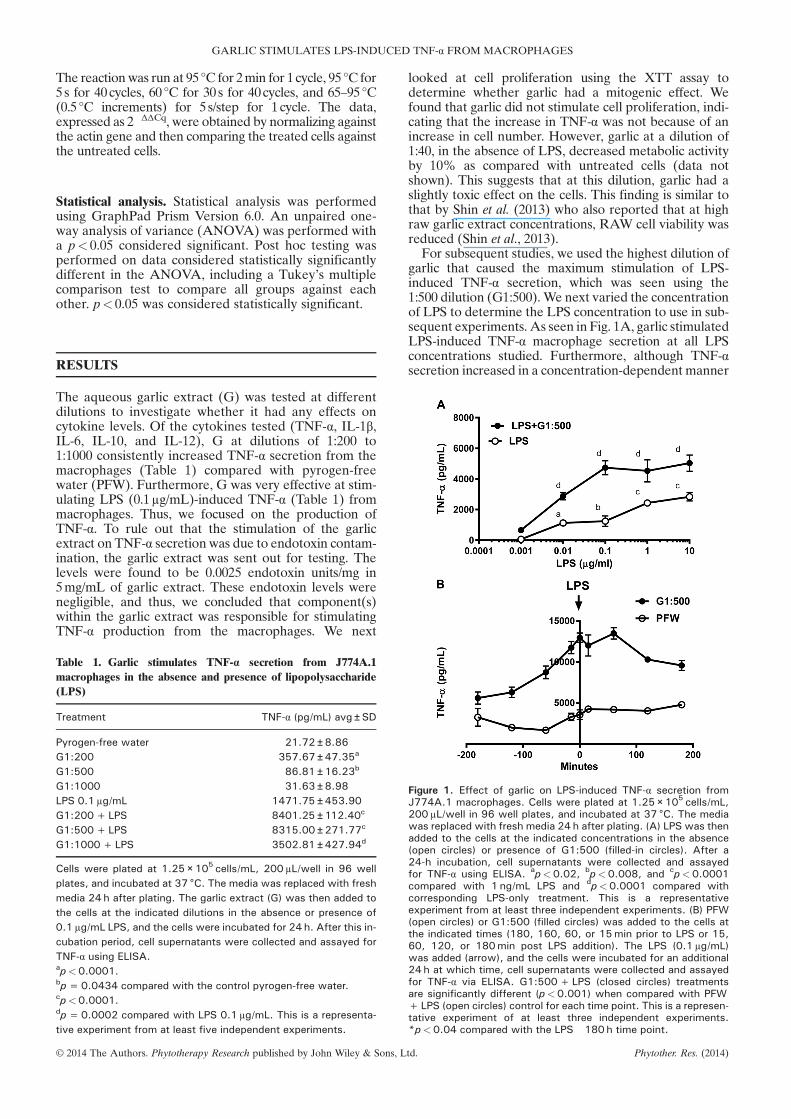
We next investigated the length of time that garliccould stimulate LPS-induced TNF-α secretion. To thisend, cells were treated with G1:500+LPS or LPSalone, and cell supernatants were collected at differenttime points after LPS addition. We found, as before,that in the presence of garlic, LPS-induced TNF-αsecretion was significantly greater in cells treated withLPS alone (Fig. 3). Furthermore, in cells treated with
td. Phytother. Res. (2014)
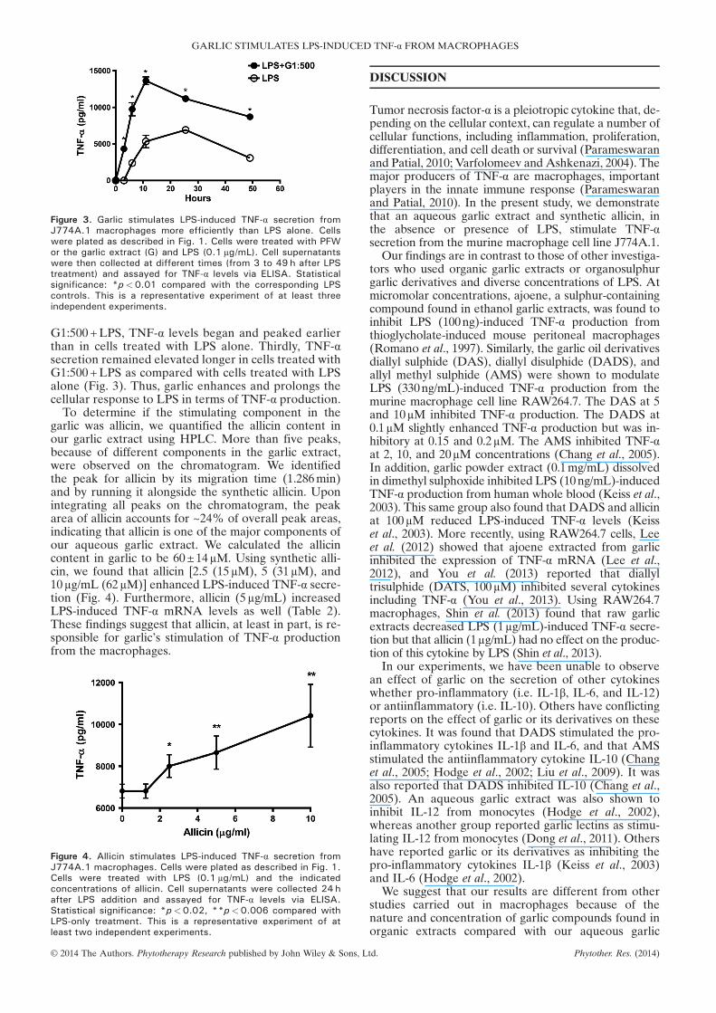
Figure 3. Garlic stimulates LPS-induced TNF-α secretion fromJ774A.1 macrophages more efficiently than LPS alone. Cellswere plated as described in Fig. 1. Cells were treated with PFWor the garlic extract (G) and LPS (0.1 μg/mL). Cell supernatantswere then collected at different times (from 3 to 49 h after LPStreatment) and assayed for TNF-α levels via ELISA. Statisticalsignificance: *p<0.01 compared with the corresponding LPScontrols. This is a representative experiment of at least threeindependent experiments.
GARLIC STIMULATES LPS-INDUCED TNF-α FROM MACROPHAGES
G1:500+LPS, TNF-α levels began and peaked earlierthan in cells treated with LPS alone. Thirdly, TNF-αsecretion remained elevated longer in cells treated withG1:500+LPS as compared with cells treated with LPSalone (Fig. 3). Thus, garlic enhances and prolongs thecellular response to LPS in terms of TNF-α production.To determine if the stimulating component in the
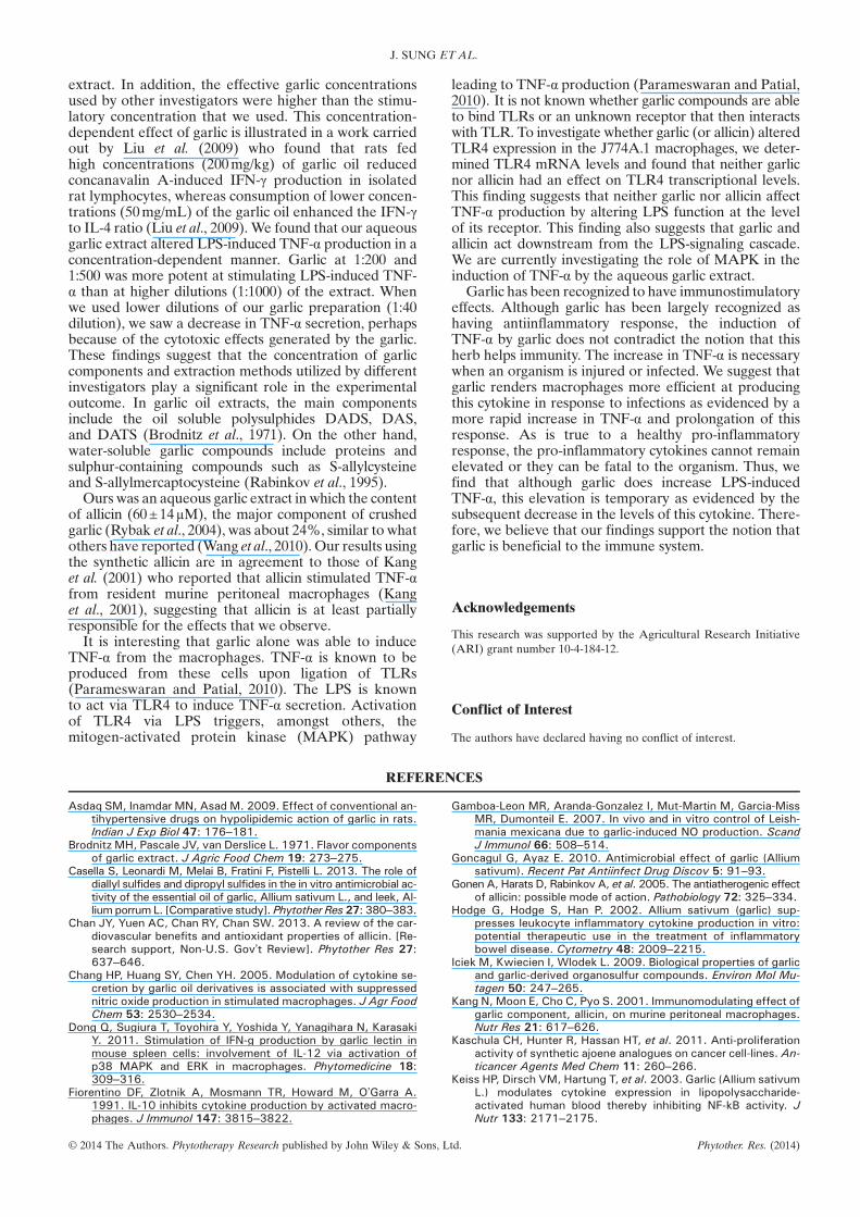
garlic was allicin, we quantified the allicin content inour garlic extract using HPLC. More than five peaks,because of different components in the garlic extract,were observed on the chromatogram. We identifiedthe peak for allicin by its migration time (1.286min)and by running it alongside the synthetic allicin. Uponintegrating all peaks on the chromatogram, the peakarea of allicin accounts for ~24% of overall peak areas,indicating that allicin is one of the major components ofour aqueous garlic extract. We calculated the allicincontent in garlic to be 60±14μM. Using synthetic alli-cin, we found that allicin [2.5 (15μM), 5 (31μM), and10μg/mL (62μM)] enhanced LPS-induced TNF-α secre-tion (Fig. 4). Furthermore, allicin (5μg/mL) increasedLPS-induced TNF-α mRNA levels as well (Table 2).These findings suggest that allicin, at least in part, is re-sponsible for garlic’s stimulation of TNF-α productionfrom the macrophages.
Figure 4. Allicin stimulates LPS-induced TNF-α secretion fromJ774A.1 macrophages. Cells were plated as described in Fig. 1.Cells were treated with LPS (0.1 μg/mL) and the indicatedconcentrations of allicin. Cell supernatants were collected 24 hafter LPS addition and assayed for TNF-α levels via ELISA.Statistical significance: *p<0.02, **p<0.006 compared withLPS-only treatment. This is a representative experiment of atleast two independent experiments.
© 2014 The Authors. Phytotherapy Research published by John Wiley & Sons, L
DISCUSSION
Tumor necrosis factor-α is a pleiotropic cytokine that, de-pending on the cellular context, can regulate a number ofcellular functions, including inflammation, proliferation,differentiation, and cell death or survival (Parameswaranand Patial, 2010; Varfolomeev and Ashkenazi, 2004). Themajor producers of TNF-α are macrophages, importantplayers in the innate immune response (Parameswaranand Patial, 2010). In the present study, we demonstratethat an aqueous garlic extract and synthetic allicin, inthe absence or presence of LPS, stimulate TNF-αsecretion from the murine macrophage cell line J774A.1.
Our findings are in contrast to those of other investiga-tors who used organic garlic extracts or organosulphurgarlic derivatives and diverse concentrations of LPS. Atmicromolar concentrations, ajoene, a sulphur-containingcompound found in ethanol garlic extracts, was found toinhibit LPS (100ng)-induced TNF-α production fromthioglycholate-induced mouse peritoneal macrophages(Romano et al., 1997). Similarly, the garlic oil derivativesdiallyl sulphide (DAS), diallyl disulphide (DADS), andallyl methyl sulphide (AMS) were shown to modulateLPS (330ng/mL)-induced TNF-α production from themurine macrophage cell line RAW264.7. The DAS at 5and 10μM inhibited TNF-α production. The DADS at0.1μM slightly enhanced TNF-α production but was in-hibitory at 0.15 and 0.2μM. The AMS inhibited TNF-αat 2, 10, and 20μM concentrations (Chang et al., 2005).In addition, garlic powder extract (0.1mg/mL) dissolvedin dimethyl sulphoxide inhibited LPS (10ng/mL)-inducedTNF-α production from human whole blood (Keiss et al.,2003). This same group also found that DADS and allicinat 100μM reduced LPS-induced TNF-α levels (Keisset al., 2003). More recently, using RAW264.7 cells, Leeet al. (2012) showed that ajoene extracted from garlicinhibited the expression of TNF-α mRNA (Lee et al.,2012), and You et al. (2013) reported that diallyltrisulphide (DATS, 100μM) inhibited several cytokinesincluding TNF-α (You et al., 2013). Using RAW264.7macrophages, Shin et al. (2013) found that raw garlicextracts decreased LPS (1μg/mL)-induced TNF-α secre-tion but that allicin (1μg/mL) had no effect on the produc-tion of this cytokine by LPS (Shin et al., 2013).
In our experiments, we have been unable to observean effect of garlic on the secretion of other cytokineswhether pro-inflammatory (i.e. IL-1β, IL-6, and IL-12)or antiinflammatory (i.e. IL-10). Others have conflictingreports on the effect of garlic or its derivatives on thesecytokines. It was found that DADS stimulated the pro-inflammatory cytokines IL-1β and IL-6, and that AMSstimulated the antiinflammatory cytokine IL-10 (Changet al., 2005; Hodge et al., 2002; Liu et al., 2009). It wasalso reported that DADS inhibited IL-10 (Chang et al.,2005). An aqueous garlic extract was also shown toinhibit IL-12 from monocytes (Hodge et al., 2002),whereas another group reported garlic lectins as stimu-lating IL-12 from monocytes (Dong et al., 2011). Othershave reported garlic or its derivatives as inhibiting thepro-inflammatory cytokines IL-1β (Keiss et al., 2003)and IL-6 (Hodge et al., 2002).
We suggest that our results are different from otherstudies carried out in macrophages because of thenature and concentration of garlic compounds found inorganic extracts compared with our aqueous garlic
td. Phytother. Res. (2014)
J. SUNG ET AL.
extract. In addition, the effective garlic concentrationsused by other investigators were higher than the stimu-latory concentration that we used. This concentration-dependent effect of garlic is illustrated in a work carriedout by Liu et al. (2009) who found that rats fedhigh concentrations (200mg/kg) of garlic oil reducedconcanavalin A-induced IFN-γ production in isolatedrat lymphocytes, whereas consumption of lower concen-trations (50mg/mL) of the garlic oil enhanced the IFN-γto IL-4 ratio (Liu et al., 2009). We found that our aqueousgarlic extract altered LPS-induced TNF-α production in aconcentration-dependent manner. Garlic at 1:200 and1:500 was more potent at stimulating LPS-induced TNF-α than at higher dilutions (1:1000) of the extract. Whenwe used lower dilutions of our garlic preparation (1:40dilution), we saw a decrease in TNF-α secretion, perhapsbecause of the cytotoxic effects generated by the garlic.These findings suggest that the concentration of garliccomponents and extraction methods utilized by differentinvestigators play a significant role in the experimentaloutcome. In garlic oil extracts, the main componentsinclude the oil soluble polysulphides DADS, DAS,and DATS (Brodnitz et al., 1971). On the other hand,water-soluble garlic compounds include proteins andsulphur-containing compounds such as S-allylcysteineand S-allylmercaptocysteine (Rabinkov et al., 1995).Ours was an aqueous garlic extract in which the content
of allicin (60±14μM), the major component of crushedgarlic (Rybak et al., 2004), was about 24%, similar to whatothers have reported (Wang et al., 2010). Our results usingthe synthetic allicin are in agreement to those of Kanget al. (2001) who reported that allicin stimulated TNF-αfrom resident murine peritoneal macrophages (Kanget al., 2001), suggesting that allicin is at least partiallyresponsible for the effects that we observe.It is interesting that garlic alone was able to induce
TNF-α from the macrophages. TNF-α is known to beproduced from these cells upon ligation of TLRs(Parameswaran and Patial, 2010). The LPS is knownto act via TLR4 to induce TNF-α secretion. Activationof TLR4 via LPS triggers, amongst others, themitogen-activated protein kinase (MAPK) pathway
© 2014 The Authors. Phytotherapy Research published by John Wiley & Sons, L
leading to TNF-α production (Parameswaran and Patial,2010). It is not known whether garlic compounds are ableto bind TLRs or an unknown receptor that then interactswith TLR. To investigate whether garlic (or allicin) alteredTLR4 expression in the J774A.1 macrophages, we deter-mined TLR4 mRNA levels and found that neither garlicnor allicin had an effect on TLR4 transcriptional levels.This finding suggests that neither garlic nor allicin affectTNF-α production by altering LPS function at the levelof its receptor. This finding also suggests that garlic andallicin act downstream from the LPS-signaling cascade.We are currently investigating the role of MAPK in theinduction of TNF-α by the aqueous garlic extract.
Garlic has been recognized to have immunostimulatoryeffects. Although garlic has been largely recognized ashaving antiinflammatory response, the induction ofTNF-α by garlic does not contradict the notion that thisherb helps immunity. The increase in TNF-α is necessarywhen an organism is injured or infected. We suggest thatgarlic renders macrophages more efficient at producingthis cytokine in response to infections as evidenced by amore rapid increase in TNF-α and prolongation of thisresponse. As is true to a healthy pro-inflammatoryresponse, the pro-inflammatory cytokines cannot remainelevated or they can be fatal to the organism. Thus, wefind that although garlic does increase LPS-inducedTNF-α, this elevation is temporary as evidenced by thesubsequent decrease in the levels of this cytokine. There-fore, we believe that our findings support the notion thatgarlic is beneficial to the immune system.
Acknowledgements
This research was supported by the Agricultural Research Initiative(ARI) grant number 10-4-184-12.
Conflict of Interest
The authors have declared having no conflict of interest.
REFERENCES
Asdaq SM, Inamdar MN, Asad M. 2009. Effect of conventional an-tihypertensive drugs on hypolipidemic action of garlic in rats.Indian J Exp Biol 47: 176–181.
Brodnitz MH, Pascale JV, van Derslice L. 1971. Flavor componentsof garlic extract. J Agric Food Chem 19: 273–275.
Casella S, Leonardi M, Melai B, Fratini F, Pistelli L. 2013. The role ofdiallyl sulfides and dipropyl sulfides in the in vitro antimicrobial ac-tivity of the essential oil of garlic, Allium sativum L., and leek, Al-lium porrum L. [Comparative study]. Phytother Res 27: 380–383.
Chan JY, Yuen AC, Chan RY, Chan SW. 2013. A review of the car-diovascular benefits and antioxidant properties of allicin. [Re-search support, Non-U.S. Gov’t Review]. Phytother Res 27:637–646.
Chang HP, Huang SY, Chen YH. 2005. Modulation of cytokine se-cretion by garlic oil derivatives is associated with suppressednitric oxide production in stimulated macrophages. J Agr FoodChem 53: 2530–2534.
Dong Q, Sugiura T, Toyohira Y, Yoshida Y, Yanagihara N, KarasakiY. 2011. Stimulation of IFN-g production by garlic lectin inmouse spleen cells: involvement of IL-12 via activation ofp38 MAPK and ERK in macrophages. Phytomedicine 18:309–316.
Fiorentino DF, Zlotnik A, Mosmann TR, Howard M, O’Garra A.1991. IL-10 inhibits cytokine production by activated macro-phages. J Immunol 147: 3815–3822.
Gamboa-Leon MR, Aranda-Gonzalez I, Mut-Martin M, Garcia-MissMR, Dumonteil E. 2007. In vivo and in vitro control of Leish-mania mexicana due to garlic-induced NO production. ScandJ Immunol 66: 508–514.
Goncagul G, Ayaz E. 2010. Antimicrobial effect of garlic (Alliumsativum). Recent Pat Antiinfect Drug Discov 5: 91–93.
Gonen A, Harats D, Rabinkov A, et al. 2005. The antiatherogenic effectof allicin: possible mode of action. Pathobiology 72: 325–334.
Hodge G, Hodge S, Han P. 2002. Allium sativum (garlic) sup-presses leukocyte inflammatory cytokine production in vitro:potential therapeutic use in the treatment of inflammatorybowel disease. Cytometry 48: 2009–2215.
Iciek M, Kwiecien I, Wlodek L. 2009. Biological properties of garlicand garlic-derived organosulfur compounds. Environ Mol Mu-tagen 50: 247–265.
Kang N, Moon E, Cho C, Pyo S. 2001. Immunomodulating effect ofgarlic component, allicin, on murine peritoneal macrophages.Nutr Res 21: 617–626.
Kaschula CH, Hunter R, Hassan HT, et al. 2011. Anti-proliferationactivity of synthetic ajoene analogues on cancer cell-lines. An-ticancer Agents Med Chem 11: 260–266.
Keiss HP, Dirsch VM, Hartung T, et al. 2003. Garlic (Allium sativumL.) modulates cytokine expression in lipopolysaccharide-activated human blood thereby inhibiting NF-kB activity. JNutr 133: 2171–2175.
td. Phytother. Res. (2014)
GARLIC STIMULATES LPS-INDUCED TNF-α FROM MACROPHAGES
Lee DY, Li H, LimHJ, Lee HJ, Jeon R, Ryu JH. 2012. Anti-inflammatoryactivity of sulfur-containing compounds from garlic. JMed Food15: 992–999.
Liu CT, Su HM, Lii CK, Sheen LY. 2009. Effects of supplementationwith garlic oil on activity of Th1 and Th2 lymphocytes fromrats. Planta Med 75: 205–210.
Malekpour-Dehkordi Z, Javadi E, Doosti M, et al. 2013. S-Allylcysteine,a garlic compound, increases ABCA1 expression in human THP-1 macrophages. [Research support, Non-U.S. Gov’t]. PhytotherRes 27: 357–361.
Parameswaran N, Patial, S. 2010. Tumor necrosis factor-alpha signal-ing in macrophages. Crit Rev Eukaryot Gene Expr 20: 87–103.
Rabinkov A, Wilchek M, Mirelman D. 1995. Alliinase (alliin lyase)from garlic (Alliium sativum) is glycosylated at ASN146 andforms a complex with a garlic mannose-specific lectin.Glycoconj J 12: 690–698.
Rietschel ET, Brade H. 1992. Bacterial endotoxins. Sci Am 267:53–61.
Romano EL, Montano RF, Brito B, et al. 1997. Effects of ajoene onlymphocyte and macrophage membrane-dependent functions.Immunopharmacol Immunotoxicol 19: 15–36.
Rybak ME, Calvey EM, Harnly JM. 2004. Quantitative determina-tion of allicin in garlic: supercritical fluid extraction and stan-dard addition of alliin. J Agric Food Chem 52: 682–687.
© 2014 The Authors. Phytotherapy Research published by John Wiley & Sons, L
Shin JH, Ryu JH, Kang MJ, Hwang CR, Han J, Kang D. 2013.Short-term heating reduces the anti-inflammatory effects offresh raw garlic extracts on the LPS-induced production ofNO and pro-inflammatory cytokines by downregulating allicinactivity in RAW 264.7 macrophages. [Research support,Non-U.S. Gov’t]. Food Chem Toxicol 58: 545–551.
Small LD, Bailey JH, Cavallito CJ. 1947. Alkyl thiolsulfinates. J AmChem Soc 69: 1710–1713.
Ulmer AJ, Flad H, Rietschel T, Mattern T. 2000. Induction of prolif-eration and cytokine production in human T lymphocytes bylipopolysaccharide (LPS). Toxicology 152: 37–45.
Varfolomeev E, Ashkenazi A. 2004. Tumor necrosis factor: anapoptosis JuNKie? Cell 116: 491–497.
Wang H, Li X, Liu S, Jin S. 2010. Quantitative determination ofallicin in Allium sativum L. bulbs by UPLC. Chromatographia71: 159–161.
You S, Nakanishi E, Kuwata H, et al. 2013. Inhibitory effects andmolecular mechanisms of garlic organosulfur compounds onthe production of inflammatory mediators. [Research support,Non-U.S. Gov’t]. Mol Nutr Food Res 57: 2049–2060.
Zamani A, Vahidinia A, Sabouri Ghannad M. 2009. The effect ofgarlic consumption on Th1/Th2 cytokines in phytohemaggluti-nin (PHA) activated rat spleen lymphocytes. Phytother Res 23:579–581.
td. Phytother. Res. (2014)


