Embed Size (px)
Citation preview
Gating of Hippocampal-Evoked Activity in PrefrontalCortical Neurons by Inputs from the Mediodorsal Thalamusand Ventral Tegmental Area
Stan B. Floresco and Anthony A. GraceDepartment of Neuroscience, University of Pittsburgh, Pittsburgh, Pennsylvania 15206
Projections from the hippocampus, the mediodorsal thalamus (MD), and the ventral tegmental area (VTA) form interconnected neuralcircuits that converge in the prefrontal cortex (PFC) to participate in the regulation of executive functions. The present study assessed theroles that the MD and VTA play in regulating the hippocampal–PFC pathway using extracellular single-unit recordings in urethane-anesthetized rats. MD stimulation inhibited PFC neuron firing (�100 msec duration) evoked by fimbria/fornix (FF) stimulation in amajority of neurons tested. However, this effect was reduced if activation of thalamocortical inputs occurred almost simultaneously (10msec) with stimulation of the FF. In a separate population of neurons, burst stimulation of the MD produced a short-term (�100 msec)inhibition or facilitation of FF-evoked firing in 66 and 33% of PFC neurons, respectively. Moreover, tetanic stimulation of the MD causeda longer-lasting (�5 min) potentiation of FF-evoked firing. Burst stimulation of the VTA inhibited FF-evoked firing in a frequency-dependent manner: firing evoked by higher-frequency trains of pulses to the FF was less inhibited than firing evoked by single-pulsestimulation. The inhibitory actions of VTA stimulation were augmented by D1 receptor antagonism and attenuated by D2 and D4
antagonists. Moreover, stimulation of the MD 10 msec before stimulation of the FF attenuated the VTA-mediated inhibition of evokedfiring. Thus, both the MD and VTA exert a complex gating action over PFC neural activity, either facilitating or inhibiting firing in thehippocampal–PFC pathway depending on the frequency and relative timing of the arrival of afferent input.
Key words: prefrontal cortex; hippocampus; ventral tegmental area; mediodorsal thalamus; dopamine; in vivo electrophysiology;gating; rat
IntroductionStudies in both animals and humans have implicated the prefron-tal cortex (PFC) and related subcortical afferent connections inmediating executive functions such as prospective coding, set-shifting, and working memory (Kolb, 1984; Hauser, 1999; Rob-bins, 1996; Goldman-Rakic, 1998; Miller, 2000). A contemporarytheory of PFC function posits that the frontal lobes play an essen-tial role in integrating different types of information, subservedby distinct cortical and subcortical networks, to organize behav-ior for the attainment of future goals (Miller, 2000; Fuster, 2001).Hippocampal projections to the PFC form one transcortical net-work that mediates cognitive processes such as working memory.In the rat, the ventral CA1/subicular region of the hippocampussends glutamatergic projections to both pyramidal neurons andGABAergic interneurons of the prelimbic region of the PFC(Conde et al., 1995; Carr and Sesack, 1996; Gabbott et al., 2002).Stimulation of hippocampal afferents evokes excitatory and in-hibitory responses in PFC neurons (Gigg et al., 1994; Jay et al.,1995, Mulder et al., 1997; Lewis and O’Donnell, 2000). The im-portance of hippocampal–PFC circuits in working memory isunderscored by the finding that disconnection between these re-
gions selectively disrupts retrieval of information during delayedresponding (Floresco et al., 1997; Aujla and Beninger, 2001).
Two other subcortical nuclei that play a role in cognitive pro-cesses mediated by the hippocampal–PFC circuits include theventral tegmental area (VTA) and the mediodorsal nucleus of thethalamus (MD). The VTA sends a dopaminergic projection to thePFC (Van Eden et al., 1987), and dopamine (DA) terminals arefound often in close proximity to hippocampal terminals on PFCneurons (Carr and Sesack, 1996). Intracellular in vivo recordingshave shown that stimulation of the VTA can produce a prolongeddepolarization in PFC neurons accompanied by a reduction inspike firing (Lewis and O’Donnell, 2000). Moreover, VTA acti-vation can exert two differential effects on hippocampal-evokedactivity of PFC neurons: (1) an inhibition of firing evoked bylow-frequency stimulation of the ventral subiculum and (2) anenhancement of long-term potentiation of the hippocampal–PFC pathway induced by high-frequency stimulation (Jay et al.,1995; Gurdren et al., 1999). These data suggest that DA inputplays an important neuromodulatory role over activity in thehippocampal–PFC pathway, a contention supported by the find-ings that selective disruption of DA D1 receptor modulation ofhippocampal inputs to the PFC impairs working memory medi-ated by hippocampal–PFC circuits (Seamans et al., 1998; Aujlaand Beninger, 2001). The functional role of D2-like (D2, D4) re-ceptors in the PFC remains to be established; however, activationof these receptors tends to inhibit PFC neural activity, either bydirect actions on PFC pyramidal neurons or by facilitating
Received Nov. 25, 2002; revised Feb. 4, 2003; accepted Feb. 18, 2003.This work was supported by United States Public Health Service Grants MH 01055, MF 57440, and MH 45156.
S.B.F. is a recipient of a Human Frontiers Science Organization Post-Doctoral Fellowship. We thank Christy Smolakand Nicole MacMurdo for their assistance with histology and Pfizer for their generous donation of CP293,019.
Correspondence should be addressed to Dr. Stan B. Floresco, Department of Neuroscience, University of Pitts-burgh, 446 Crawford Hall, Pittsburgh, PA, 15260. E-mail: [email protected] © 2003 Society for Neuroscience 0270-6474/03/233930-14$15.00/0
3930 • The Journal of Neuroscience, May 1, 2003 • 23(9):3930 –3943

GABAergic transmission (Sesack and Bunney, 1989; Rubinsteinet al., 2001; Wedzony et al., 2001).
The MD shares a reciprocal glutamatergic projection with thePFC with thalamocortical inputs synapsing on layer III and Vneurons, and possibly GABAergic interneurons as well (Krettekand Price, 1977; Groenewegen, 1988; Ray and Price, 1992; Pirot etal., 1994; Kuroda et al., 1998). Behavioral studies have implicatedthe MD in a broad range of PFC-related processes, includingprospective coding (Joyce and Robbins, 1991; Daum and Acker-mann, 1994) and strategy selection (Hunt and Aggleton, 1998).Of particular relevance is the fact that disconnection between theMD and PFC disrupts working memory processes dependent onboth hippocampal–PFC circuits and mesocortical DA (Florescoet al., 1999).
The above-mentioned findings suggest that converging corti-copetal inputs originating from the hippocampus, the MD, andthe VTA may interact in a cooperative manner to regulate exec-utive functions governed by the PFC. Despite this, it is surprisingthat there is a paucity of research investigating the mechanisms bywhich these inputs interact to influence PFC neural activity. Assuch, the present study was undertaken to assess the modulatoryactions that inputs from the MD and the VTA exert overhippocampal-evoked firing of PFC neurons, using in vivo extra-cellular single-unit recordings.
Materials and MethodsSubjects and surgeryMale Sprague Dawley rats (300 – 400 gm; Hilltop, Scottsdale, PA) wereanesthetized with urethane (1.5 gm/kg, i.p.) and mounted in a stereotaxicframe, with the incisor bar set at �3.3 mm. Body temperature was main-tained at 37°C with a temperature-controlled heating pad. In all surgicalpreparations, the scalp was incised and holes were drilled in the skulloverlying the prelimbic region of the medial PFC, the fornix/fimbria(FF), the MD, and the VTA. Concentric bipolar electrical stimulatingelectrodes (SNE-100; Kopf, Tujunga, CA) were implanted into the threeafferent regions of the PFC. The stereotaxic coordinates were as follows(flat skull): FF electrode � anteroposterior (AP) �1.3 mm (bregma),mediolateral (ML) �1.6 mm, dorsoventral (DV) �4.0 mm (cortex); MDelectrode � AP �2.9 mm, ML �0.7 mm, DV �5.3 mm; VTA elec-trode � AP �5.4 mm, ML �0.7 mm, DV �7.8 mm. Animal care andsurgical procedures were performed in accordance with the guidelinesoutlined in the NIH Guide for the Care and Use of Laboratory Animals andwere approved by the Institutional Animal Care and Use Committee ofthe University of Pittsburgh.
Extracellular recordings and cell-searching proceduresExtracellular recording microelectrodes were constructed from 2.0 mmouter diameter borosilicate glass capillary tubing (WPI) using a verticalmicropipette puller (Narishige, Tokyo, Japan). The tips of the electrodeswere broken back against a glass rod to �1 �m tip diameter and filledwith 2 M NaCl containing 2% Pontamine sky blue dye. The in vitroimpedance of the microelectrodes ranged from 5 to 10 M� as measuredat 135 Hz using a Winston Electronics BL-1000 impedance meter. After aburr hole was drilled overlying the PFC, the dura was resected, and theelectrode was lowered into the PFC (coordinates: �3.5–2.7 mm anteriorfrom bregma, 0.6 – 0.8 mm lateral from the midline, 2.2–5.0 mm ventralfrom brain surface) with a hydraulic microdrive (Kopf Model 640). Theelectrode signal was amplified, filtered (400 – 4000 Hz), and discrimi-nated from noise using a combination amplification and window dis-crimination unit for extracellular recording (Fintronics, Orange, CT)and displayed on an oscilloscope (Tektronics, Wilsonville, OR). Thedata were acquired, stored, and analyzed using custom-designedcomputer software (Neuroscope) running on an Intel-based personalcomputer with a data acquisition board interface (Microstar Labora-tories, Bellevue, WA).
After the glass microelectrodes had been lowered to the dorsal borderof the PFC, a cell-searching procedure began. In this procedure, the
microelectrode was lowered incrementally through the PFC while alter-nating stimuli were delivered to the FF and the MD (1500 �A) at 1 secintervals (i.e., each afferent was stimulated at 0.5 Hz). Although previousin vitro studies in hippocampus neurons have shown that prolongedlow-frequency stimulation can induce long-term depression (Mulkeyand Malenka, 1992), other studies in vivo have shown that extendedbouts of low-frequency stimulation (1 Hz, 15 min) of the hippocampusdoes not produce any reliable change in synaptic efficacy in the PFC(Burette et al., 1997). Cathodal constant current pulses (0.2 msec dura-tion) were delivered to the FF and MD through an Iso-Flex optical iso-lator (A.M.P.I., Jerusalem, Israel) via a Master-8 programmable pulsegenerator (A.M.P.I.) using the parameters noted below. Once a cell wasdetected, the position of the microelectrode was adjusted to maximizethe spike amplitude relative to background noise. Neurons that re-sponded only to FF stimulation or MD stimulation or received converg-ing input from both regions were identified by their robust excitatoryresponse after stimulation of the respective afferent region. Only neuronsthat responded with an orthodromic, monosynaptic response and dis-played a signal-to-noise ratio of at least 3:1 were used in the data analysis.Evoked firing was characterized as monosynaptic/orthodromic if the re-sponse displayed spike jitter of at least 2 msec and a shift in spike latencywith increasing current amplitude, followed by paired-pulse stimulationat 50 Hz (otherwise characterized as polysynaptic), but failed to follow400 Hz paired-pulse stimulation (otherwise characterized at antidromic)(Pirot et al., 1994; Mulder et al., 1997).
With respect to MD-evoked responses, previous studies have demon-strated that electrical stimulation of the MD can evoke two types ofmonosynaptic responses in PFC neurons. Single-pulse stimulation canevoke a short latency (�10 msec) action potential that likely representsorthodromic activation of thalamocortical axons. However, higher-frequency (10 Hz) stimulation yields longer latency (�13 msec) spikesthat are thought to be caused by antidromic activation of axons of PFCprojection neurons that terminate in the MD but also have collateralsthat synapse onto other PFC neurons (Pirot et al., 1994). Experimentsassessing the conduction velocities of MD neurons projecting to the PFChave revealed that antidromically evoked firing of MD neurons occurs2–11 msec after PFC stimulation (Pirot et al., 1994). In the present study,we observed that orthodromic monosynaptic action potentials evoked bysingle-pulse stimulation of the MD displayed a latency range of 6 –22msec (mean � 13.4 msec). However, to maximize the possibility thatthese responses were driven by activation of thalamocortical projectionsand not axon collaterals of PFC projection neurons, we included onlyPFC neurons that displayed MD-evoked orthodromic firing with laten-cies of �12 msec. Whenever a PFC neuron was encountered that dis-played an antidromic spike after MD stimulation (but no response to FFstimulation), the location of that neuron was noted, but no other datawere taken.
Stimulation protocolsAfter establishing that firing evoked by stimulation of the FF or MD wasmonosyaptic and orthodromic, stimulation currents were adjusted tosubmaximal stimulation intensity (range 100 –1800 �A) so that stimula-tion of the FF or MD would evoke an action potential �60% of the time(range 40 –75%, depending on the experiment) in response to single-pulse stimulation delivered at 0.2 Hz. In each of these experiments, datawere compiled using a minimum of 25 sweeps. We used various stimu-lation protocols designed to investigate different types of interactionsamong hippocampal, MD, and VTA inputs to the PFC.
Interactions between converging inputs from the hippocampal and MD inthe PFC: sequential-pulse protocols. In PFC neurons that displayed amonosynaptic orthodromic spike after stimulation of both the FF andMD, we conducted a series of sequential single-pulse stimulation exper-iments to assess how stimulation of one input could influence firingevoked by stimulation of the second input. In these experiments, eachneuron received a conditioning pulse of one input followed by a test pulseof the other input, at intervals ranging between 10 and 500 msec, in acounterbalanced order, using the same stimulation intensity for eachinterstimulus interval (ISI). Paired-pulse facilitation/depression was also
Floresco and Grace • Hippocampal–Thalamic–VTA Interactions in PFC J. Neurosci., May 1, 2003 • 23(9):3930 –3943 • 3931

examined for each individual input, using 25–100 msec intervals betweenthe conditioning pulse and the test pulse.
Modulation of hippocampal-evoked firing by burst stimulation of theMD. For some PFC neurons that only fired in response to FF and not MDstimulation, a series of experiments assessed the effect of burst stimula-tion of the MD on firing evoked by FF stimulation. In these studies, thestimulation intensity of the FF was adjusted so that the firing probabilitywas �50% and kept constant throughout the course of the experiment.The firing probability was maintained at �50% so that we would be ableto observe either inhibition or facilitation of FF-evoked firing by MDburst stimulation. After establishing baseline firing probabilities, we ap-plied a four-pulse, 20 Hz train to the MD (800 –1000 �A) 10, 25, or 50msec before the FF pulse, with 25 sweeps collected for each interstimulusinterval. This firing pattern mimics that displayed by MD neurons duringlearning (Oyoshi et al., 1996). Any neuron that displayed reliable firing inresponse to MD burst stimulation was not included in the data analysis.After each series of 25 sweeps, FF-evoked firing probability was moni-tored at the same stimulation intensity for 3–5 min using single pulses tothe FF delivered at 0.2 Hz. This continued until the FF-evoked firingprobability returned to �50%.
We also assessed effects of tetanic stimulation of the MD on longerlasting (i.e., minutes) changes of FF-evoked firing in PFC neurons. In thisexperiment, baseline probability of evoked firing in response to singlepulses delivered to the FF were recorded over �10 min, using repeatedsweeps administered every 2.5 min. Once stable levels of evoked-firingactivity were observed (�15% variation in spike probability over 5–10min, three to four sweeps), 25 trains (four pulses, 20 Hz ISI) were deliv-ered to the MD at a frequency of 0.2 Hz. Thirty seconds after this tetanus(assigned as time 0), the FF was again stimulated with single pulses at 0.2Hz using the same stimulation current as before tetanus, and changes inFF-evoked firing probability were assessed for another 10 min with re-peated sweeps recorded every 2.5 min. Data were normalized to theaverage firing probability observed over 5 min before tetanus and ana-lyzed in terms of percentage change in firing probability. For both of theabove-mentioned protocols, any neuron displaying reliable firing in re-sponse to MD burst stimulation was not included in the data analysis.
VTA modulation of FF-evoked firing of PFC neurons. For these experi-ments, FF stimulation intensities were adjusted to evoke an action po-tential �60 –75% of the time after single-pulse stimulation of the FFdelivered at 0.2 Hz. Once baseline firing probabilities were established,the VTA was stimulated in a burst pattern (two four-pulse 20 Hz trains ofpulses; interburst interval � 200 msec) 10 or 50 msec before stimulationof the FF. Usually we stimulated the VTA with an intensity of 600 �A, butin some instances, the intensity was increased to up to 800 �A to inducea noticeable inhibition of FF-evoked firing. To assess whether activationof the VTA exerted a frequency-dependent modulation of evoked firing,we stimulated the FF first with single pulses and then with five-pulse, 20Hz trains, each delivered at 0.2 Hz. The 20 Hz stimulation frequencymimics the way hippocampal projection neurons fire during workingmemory tasks (Hampson et al., 2000). We used a minimum of 25 sweepsper interburst interval to compile the data for all experiments assessingthe effects of VTA stimulation.
Pharmacological manipulationsFor some experiments involving burst stimulation of the VTA, separategroups of rats were implanted with intravenous jugular catheters, con-sisting of PE 10 tubing attached to a 30 ga needle and a 1 ml syringe. Theseexperiments used a between-subjects design, and every animal receivedonly one drug injection. After isolation of each PFC neuron, we con-firmed that burst stimulation of the VTA inhibited hippocampal-evokedfiring induced by single-pulse stimulation of the FF before drug admin-istration. Baseline levels of FF-evoked firing were established, after whichdrugs were administered. We would then wait 10 –20 min after druginjection before stimulating the VTA again. All drugs were purchasedfrom Sigma (St. Louis, MO), with the exception of CP 293,019, whichwas donated by Pfizer (Groton, CT). The selective D1 receptor antagonistSCH23390 (0.2 mg/kg), the selective D2 antagonist eticlopride (0.25 mg/kg), and the selective NMDA receptor antagonist 3-(2-carboxypiperazin-4-yl)-propyl-1-phosphonic acid (CPP) (2.0 mg/kg) were dissolved in
physiological saline. The D2/D4 antagonist haloperidol (0.5 mg/kg) wasdissolved in dilute lactic acid. The selective D4 antagonist CP 293,019 (10mg/kg) was immersed in a drop of NaOH and 40% solution of cyclodex-trin and sonicated until dissolved. The concentrations of these solutionswere set so that injection volumes would range between 0.15 and 0.30 ml,and no more than one drug injection was given per animal. The doses ofthese drugs were chosen from previous studies (Gioanni et al., 1998;Mansbach et al., 1998; Floresco et al., 2001).
HistologyAt the end of each experiment, the recording site in the PFC was markedvia iontophoretic ejection of Pontamine sky blue dye from the tip of therecording electrode (30 �A constant current for 20 –30 min). Iron de-posits were made in the fimbria, the MD, and the VTA stimulation sitesby passing DC current (100 �A for 10 sec) through the stimulating elec-trode. After dye ejection, brains were removed and fixed in formalincontaining 0.1% potassium ferricyanide for at least 24 hr. The brainswere then immersed in phosphate-buffered sucrose solution (25%) untilsaturated. The tissue was sectioned into 40 �M coronal slices, mounted,and stained with cresyl violet to enable histological determination ofrecording and stimulating electrode sites. Representative locations ofdifferent classes of PFC neurons and stimulating electrodes are presentedin Figure 1.
Data analysisThe data were analyzed in terms of evoked firing probability in responseto stimulation of the FF or MD, or percentage change of baseline-evokedfiring probability. Evoked firing probabilities were calculated by dividingthe number of action potentials observed by the number of stimuli ad-ministered (typically 25–50 pulses) � 100. For the sequential-pulse ex-periments that measured the interactions between hippocampal and MDinputs, the baseline firing probabilities evoked by each input were calcu-lated by taking the mean firing probability evoked when that input wasstimulated as the conditioning pulse. Changes in these probabilities wereused as an index of the influence that MD or VTA inputs exerted overhippocampal-evoked firing of PFC neurons. The type of statistical anal-ysis depended on the particular experiment, but typically entailed two- orthree-way repeated measures ANOVA, with the exception of the phar-macological studies, which also included drug as a between-subjects fac-tor. Multiple comparisons were made using two-tailed Dunnett’s test forrepeated measures.
ResultsGeneral properties of PFC4Hipp neurons: convergence ofinputs and projections to the MD and VTAThe data from a total of 114 PFC neurons that displayed a mono-synaptic orthodromic spike after stimulation of the FF are pre-sented here (hereafter referred to as PFC4Hipp neurons). Thesecells displayed a mean basal spontaneous firing rate of 3.1 0.5Hz (range 0.0 –17.2 Hz; median firing rate 2.9 Hz) and had amean evoked firing latency of 11 0.8 msec (range 7–17 msec),consistent with previous findings (Mulder et al., 1997). Most ofthese 114 neurons that fired in response to FF stimulation werealso tested for an antidromic response to MD or VTA stimula-tion. Ten of these cells (10 of 109; 9.2%) were confirmedPFC3MD projection neurons (mean antidromic spike la-tency � 11.2 0.9 msec; range 7–18 msec), whereas 24.6% ofthese neurons (17 of 69) responded with an antidromic spikeafter VTA stimulation (mean antidromic spike latency � 9.6 1.0 msec; range 4 –19 msec). In addition, 4 of 60 neurons (6.7%)responded with an antidromic spike after stimulation of both theVTA and MD, suggesting that a subpopulation of PFC neuronsthat receive hippocampal input send axon collaterals to both re-gions (Fig. 1E). It is notable that many of the PFC4Hipp neu-rons were located in the same vertical electrode track where otherPFC neurons that were antidromically activated by MD stimula-tion were observed. This suggests that most of the PFC4Hipp
3932 • J. Neurosci., May 1, 2003 • 23(9):3930 –3943 Floresco and Grace • Hippocampal–Thalamic–VTA Interactions in PFC

neurons that were recorded in the present study were located inthe projection layers of the PFC. Last, and most pertinent to thepresent study, 13 of 105 PFC4Hipp neurons (12.4%) also dis-played converging orthodromic input from the MD, exhibiting amonosynaptic action potential after MD stimulation that waswithin the latency range of our inclusion criterion (mean spikelatency � 7.6 0.6 msec). Representative locations of theseneurons are shown in Figure 1. PFC4Hipp neurons that pro-jected to the MD were clustered in the more rostral regions of thePFC and found in both prelimbic and infralimbic areas, whereasPFC4Hipp neurons that projected to the VTA clustered morerostrally and were located in the dorsal prelimbic region.
Interactions between converging hippocampal and MDinputs to the PFCIn six PFC neurons that displayed a monosynaptic spike in re-sponse to stimulation of both the FF and the MD (six rats), weadministered a sequential-pulse stimulation protocol to assesshow stimulation of one input could influence firing driven by the
other input. In these cells, the basal firing probabilities evoked byeither FF or MD single-pulse stimulation did not differ (FF �55.7 4/7%; MD � 53.6 7.3%; F(1,5) � 0.04; NS). Applicationof a conditioning pulse to the FF 10 –500 msec before a test pulseto the MD resulted in a profound and significant ( p � 0.05)depression of MD evoked firing probability (Fig. 2A, blacksquares) compared with single-pulse stimulation of the MDalone (Fig. 2A, gray square) (F(6,30) � 3.5, p � 0.01; and Dun-nett’s, p � 0.05). The suppression of MD-evoked firing probabil-ity was maximal when the conditioning pulse to the FF was ad-ministered 10 msec before MD stimulation (�94 3%), whereaslonger intervals (25–500 msec) produced suppression of firing tobetween 45 and 75% of the baseline MD-evoked firing probabil-ity. In these same neurons, a similar profile was observed when aconditioning pulse was applied to the MD before a test pulse tothe FF. In this instance, FF-evoked firing probability was signifi-cantly reduced by �60% when a conditioning pulse was admin-istered 25–100 msec before FF stimulation (Fig. 2B, black circles)when compared with single pulse of the FF alone (Fig. 2B, gray
Figure 1. Histology. A, Schematic of coronal sections of the rat brain showing representative placements of recording electrodes where different PFC neurons were observed. PFC4Hipp neurons, blacksquare; neurons that responded with an orthodromic action potential after MD stimulation, gray circles; PFC4Hipp cells that were confirmed to project to the MD, open circles; PFC4Hipp cells that wereconfirmed to project to the VTA; PFC4Hipp cells that were antidromically activated by both the VTA and the MD, stars; PFC3MD projection neurons that did not respond to FF stimulation, triangles. Numberscorrespond to millimeters from bregma. B–D, Photographs of a representative placement of a stimulating electrode in the FF ( B ), the MD (C ), and the VTA ( D ). Arrows highlight the location of stimulatingelectrodeplacements.cc,Corpuscallosum;hipp,hippocampus.E, Individualdata.TenoverlaidtracesrecordedfromaPFCneuronthatdisplayedanantidromic(AD)spikeafterMDstimulationandanorthodromic(ortho) spike after FF stimulation (E1). This same neuron could also be activated antidromically by stimulation of the VTA (E2 ) (5 traces).
Floresco and Grace • Hippocampal–Thalamic–VTA Interactions in PFC J. Neurosci., May 1, 2003 • 23(9):3930 –3943 • 3933

circle). However, application of a condi-tioning pulse to the MD either 10 or 250 –500 msec before FF stimulation did notsignificantly effect the FF evoked firingprobability (Fig. 2B, white circles) (F(6,30)
� 5.6; p � 0.01). The duration of the inhi-bition of MD-evoked spiking by the FF wassubstantially longer (�500 msec) than thereduction in FF-evoked firing mediated bythe MD (�100 msec). This difference mayhave been caused by differences in the du-ration of feed-forward inhibition evokedby each afferent. To investigate this differ-ence further, we analyzed the duration ofinhibition of spontaneous activity evokedby single-pulse stimulation of each affer-ent. In accordance with our findings usingsequential-pulse protocols, single-pulsestimulation of the MD evoked an initialmonosynaptic spike, which was followedby a period of inhibition of spontaneousfiring that lasted 160 21 msec. In con-trast, inhibition of spontaneous firingevoked by FF stimulation had a substan-tially longer duration (403 27 msec).
Subsequent analyses of these data wereconducted to assess whether the neuron fired an action potentialto the conditioning pulse to either MD or FF stimulation had anyeffect on the firing probability observed in response to the testpulse to the FF or the MD. Analysis of these data revealed that FFstimulation caused the same level of depression of MD-evokedspike firing independent of whether the FF conditioning pulseevoked a spike (F(1,5) � 0.2; NS). The reverse was also true: MDstimulation caused a similar depression in the response to FFstimulation independent of whether the MD condition pulseevoked an action potential (F(1,5) � 2.7; NS). Thus, activity in thehippocampal–PFC pathway 10 –500 msec before impulse activityin the MD–PFC pathway can profoundly reduce the probabilityof firing induced by thalamocortical inputs, an effect that is notrelated to FF-evoked firing. The MD–PFC pathway exerts a similargating action over hippocampal-evoked firing, although this inhib-itory action occurs at a more temporally discrete range (25–100msec). Importantly, the inhibitory influence that this thalamocorti-cal pathway exerts over hippocampal-evoked firing of PFC neuronscan be offset if impulse activity from the hippocampus arrives almostsimultaneously with inputs from the MD (i.e., within �10 msec).
We also assessed paired-pulse facilitation/depression in thehippocampal–PFC or MD–PFC pathway individually, using 25,50, and 100 msec interstimulus intervals between conditioningand test pulses (Table 1). For PFC4Hipp neurons (n � 10; sixrats), delivering paired pulses to the FF at intervals of 50 or 100msec, but not at 25 msec, resulted in significant paired-pulsefacilitation ( p � 0.01). In contrast, for neurons PFC4MD (n �9; five rats), there was a significant ( p � 0.05) paired-pulse de-pression of the test pulse at a 100 msec interval, but not at the 25or 50 msec intervals (Fig. 2B) (F(2,34) � 6.9; p � 0.01).
Burst activation of the MD produces differential short-termgating of hippocampal-evoked firing in subpopulations ofPFC neurons that are not activated by MD stimulationIn light of the dense thalamocortical projection to the PFC, wewere surprised to observe that a relatively small proportion(�15%) of PFC4Hipp neurons fired a monosynaptic action
potential after stimulation of the MD. However, this may be anunderestimation of the total number of PFC4Hipp neurons thatmay be modulated by activity in the MD–PFC pathway. For ex-ample, it is possible that a greater proportion of PFC4Hippneurons also received direct or indirect excitatory input from theMD, but the strength of these inputs was only sufficient to evokea subthreshold EPSP unobservable using extracellular record-ings. Alternatively, a proportion of MD axons may make connec-tions with GABAergic interneurons in the PFC, which in turnsynapse on PFC4Hipp neurons. With this in mind, we assessedhow activation of the MD would affect hippocampal-evoked fir-ing in PFC neurons that only displayed an extracellular spike afterFF stimulation but not after MD stimulation.
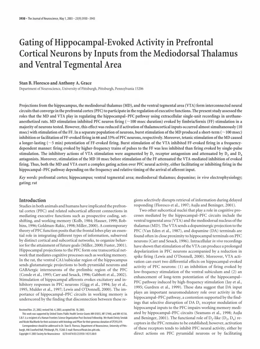
Eighteen PFC4Hipp neurons that showed no reliable excita-tory or inhibitory response to single-pulse MD stimulation weretested in this manner. In these cells, single-pulse stimulation ofthe MD 10 –50 msec before a single pulse to the FF had no dis-cernable effect over FF-evoked firing (Fig. 3C1). We then appliedrepeated four-pulse, 20 Hz train to the MD 10 –50 msec beforesingle-pulse stimulation of the FF. In all PFC neurons that weretested in this manner (n � 18; eight rats), burst stimulation of theMD had some effect over FF-evoked firing, revealing two distinctpopulations of PFC neurons. In 12 of these cells (67%), burststimulation of the MD caused a pronounced reduction in thefiring probability evoked by FF stimulation at all intervals tested,when compared with the firing probability observed after single-pulse stimulation of the FF alone (F(2,22) � 3.7, p � 0.05; andDunnett’s, p � 0.01) (Fig. 3A,C2). Four of these cells were testedfurther using intervals of 100 and 250 msec between the MD trainand the single pulse to the FF. This protocol revealed that theinhibitory effects of MD stimulation were still apparent at inter-vals of 250 msec after an MD burst (data not shown). In contrastto the above-mention effects, in another six PFC4Hipp neurons(33%), burst stimulation of the MD had the opposite effect: anincreased FF-evoked firing probability (F(1,5) � 6.92; p � 0.05)(Fig. 3B). The location of these two types of PFC neurons isshown in Figure 3D. We observed no consistent pattern of local-
Figure 2. Examples of sequential-pulse interactions of the PFC and MD inputs to the PFC. A, In neurons that fired in response tostimulation of both the FF and MD, a conditioning pulse applied to the FF inhibited firing evoked by a test pulse to the MD (blacksquares; mean � SEM) compared with a single MD pulse alone with the same intensity (gray square, hatched line). B, In thesesame neurons, a conditioning pulse applied to the MD significantly inhibited firing evoked by an FF test pulse compared with firingevoked by single pulses to the FF (gray circles). Here, the inhibition was significant ( p � 0.05) only at intervals of 25–100 msec(black circles) but not at intervals of 10 or 250 –500 msec (white circles) when compared with the firing probability observed usingsingle-pulse stimulation (gray circle). Bottom panels diagram the stimulation protocols used for A and B, respectively.
3934 • J. Neurosci., May 1, 2003 • 23(9):3930 –3943 Floresco and Grace • Hippocampal–Thalamic–VTA Interactions in PFC
ization of these two types of neurons within the PFC; both typeswere found in the dorsal and ventral regions of the deep layers ofthe prelimbic and infralimbic cortex. Thus, these data imply thatnatural-type bursting activity in the thalamocortical pathway canexert differential gating actions on PFC neural activity driven byhippocampal inputs. Most PFC neurons are inhibited by burstingactivity in the thalamocortical pathway, whereas another groupof neurons displays a facilitation of hippocampal-evoked firingafter MD burst stimulation.
Repeated burst stimulation of the MD produces a longer lastingpotentiation of hippocampal-evoked firing in PFC neuronsOver the course of the above-mentioned series of experiments,we noticed an increase in the FF-evoked firing probability afterrepetitive burst stimulation of the MD (i.e., 25 bursts delivered at
0.2 Hz frequency), regardless of whether MD activation inhibitedor facilitated FF-evoked firing, an effect that lasted a number ofminutes. Therefore, we conducted another experiment to for-mally assess this heterosynaptic potentiation of FF-evoked firingby burst stimulation of the MD. Repeated burst stimulation of theMD resulted in a robust potentiation of FF-evoked firing proba-bility in all neurons tested (n � 8; four rats) (Fig. 4A,B1,B2). Thispotentiation of hippocampal evoked firing probability reached apeak at 1 min after tetanus (�46.7 14%) (Fig. 3B1,B2) andremained significantly elevated for another 6 – 8 min before re-turning to baseline levels of evoked firing probability (F(7,91) �4.7, p � 0.01; and Dunnett’s, p � 0.05, 0.01). To confirm that thepotentiation of hippocampal-evoked firing was not merelycaused by repetitive single-pulse stimulation of the FF, a separategroup of neurons (n � 6; three rats) were tested under conditions
Table 1. Homosynaptic paired-pulse facilitation/depression in the hippocampal–PFC and MD–PFC pathways
ISI
Mean (SEM) FF-evoked firing probabilities Mean (SEM) MD-evoked firing probabilities
Conditioning pulse Test pulse Conditioning pulse Test pulse
25 msec 46.4 (3.4) 53 (10.7) 48 (6.1) 44.4 (8.8)50 msec 40.6 (5.8) 60 (6.3)* 50 (6.1) 42.2 (9.3)100 msec 44.6 (6.1) 67 (10.5)* 55.1 (5.3) 36.8 (8.6)*
*Significant differences between conditioning and test pulse firing probability at p � 0.05.
Figure 3. Burst stimulation of the MD modulates hippocampal-evoked firing in PFC neurons. A, In 12 of 18 PFC4Hipp neurons that did not fire in response to MD stimulation, burst stimulationof the MD inhibited FF-evoked firing (black bars; mean � SEM), relative to the firing probability observed after single-pulse stimulation of the FF alone at the same stimulation intensity (gray bars;mean � SEM). B, In another 6 of 18 PFC4Hipp neurons, burst stimulation of the MD facilitated FF-evoked firing. C, Representative data from individual neurons. C1, Ten overlaid traces showingthe effect of single-pulse stimulation of the MD (100 �A) 25 msec before stimulation of the FF (680 �A). C2, In the same neuron, 10 traces showing the effect of burst stimulation of the MD (last 2pulses shown) before FF stimulation. Over 10 sweeps, single-pulse stimulation of the MD had no effect over FF-evoked firing, but burst stimulation substantially reduced the probability of firing overthe same number of trails. D, Location of PFC4Hipp neurons the evoked firing of which was inhibited (open squares and minus sign) and facilitated (black squares) by burst stimulation of the MD.E, Diagrams of the stimulation protocol used in this experiment. Here, the MD was stimulated with a four-pulse, 20 Hz train (1) 10 –50 msec before single-pulse stimulation of the FF (2). MDstimulation did not evoke firing in any of these neurons. Significant difference versus single-pulse stimulation of the FF: **p � 0.01.
Floresco and Grace • Hippocampal–Thalamic–VTA Interactions in PFC J. Neurosci., May 1, 2003 • 23(9):3930 –3943 • 3935

in which the MD was not stimulated. These neurons displayed nosignificant change in the probability of firing evoked by repetitivesingle-pulse FF stimulations over a 15 min period (F(7,91) � 0.32,NS) (Fig. 4A, gray circles). Thus, repetitive bursting activity inthis thalamocortical pathway can produce a robust, short-termpotentiation of hippocampal-evoked firing of PFC neurons,thereby priming PFC neurons to be more responsive to informa-tion conveyed by the hippocampus.
Interactions between hippocampal and VTA inputs to the PFC
Single-pulse stimulation of the FFIn a separate group of PFC4Hipp neurons, we assessed the effectof burst stimulation of the VTA on FF-evoked firing of PFC neu-rons. All of the neurons tested (n � 54; 51 rats) fired in responseto FF stimulation but showed no monosynaptic response afterMD stimulation. These data are presented in Figure 5. In accor-dance with previous findings (Jay et al., 1995; Gurden et al.,1999), burst stimulation of the VTA produced a drastic inhibitionof FF-evoked firing (F(1,22) � 80.0, p � 0.001; and Dunnett’s, p �0.01) (Fig. 5). This inhibitory effect of VTA stimulation was ap-parent when the interval between the last pulse in the VTA burstand the single stimulation of the FF was either 10 or 50 msec(F(1,22) � 8.7; p � 0.01). Others have shown that single-pulsestimulation of the VTA inhibits hippocampal-evoked activityonly at intervals of �25 msec (Jay et al., 1995; Gurden et al.,
1999). Thus, burst stimulation of the VTA, which is known to bemore effective at releasing mesolimbic DA than single pulses(Garris and Wightman, 1994), inhibits firing of PFC neuronsevoked by single-pulse stimulation of the hippocampus, and thiseffect lasts at least 50 msec after the end of the VTA burst.
Train stimulation (20 Hz) of the FFA number of recent studies have shown that the actions of DA onevoked activity in PFC neurons are dependent on the frequencyat which excitatory inputs are stimulated (Jay et al., 1995; Otani etal., 1998; Gurden et al., 1999; Seamans et al., 2001). In light ofthese data, we assessed the effect of burst stimulation of the VTAon hippocampal-evoked firing of PFC neurons when the FF wasstimulated with a higher-frequency train of pulses. FF stimula-tion consisted of a five-pulse, 20 Hz train. Stimulation of the FF inthis manner resulted in an equivalent probability of firing (75–90%) in PFC neurons throughout each pulse in the train (Figs.6A, gray circles, 7 A1). However, activation of the VTA before 20Hz train stimulation of the FF altered this firing profile in aninteresting manner. Burst stimulation of the VTA produced aprofound reduction in the firing probability observed after thefirst pulse in the five-pulse train delivered to the FF, when com-pared with the firing probability observed at the same time pointwhen no VTA stimulation was given (75.8 4 vs 7.4 3%) (Fig.6A, gray square). However, the firing probability observed in thelatter parts of the train was significantly less attenuated comparedwith the firing probability evoked by the first pulse (Figs. 6A,black squares, 7A2) (mean � 52.3 7%; F(4,36) � 7.1; p � 0.01).This effect emerged by the second pulse in the train, and the firing
Figure 4. Repeated burst stimulation of the MD potentiates FF-evoked firing in PFC neurons.A, In neurons that did not fire in response to MD stimulation, tetanic burst stimulation of the MD(25 4-pulse 20 Hz trains; interburst interval of 5 sec; arrow) potentiated FF-evoked firing for�10 min (black squares). In contrast, repeated single-pulse stimulation of the FF alone had noeffect (gray circles). Symbols represent mean percentage change (�SEM) in baseline FF-evoked firing. B, Peristimulus time histograms showing a typical response from a single PFCneuron at baseline (B1) 2 min before and 1 min after MD tetanus (B2) (25 sweeps each). In thiscell, MD tetanus substantially potentiated FF-evoked firing probability. Arrows represent timepoints at which single pulses were administered to the FF (450 �A stimulation intensity forboth). * p � 0.05 and ** p � 0.01, respectively, versus baseline.
Figure 5. Burst stimulation of the VTA inhibits FF-evoked firing in PFC neurons. Mean firingprobability (�SEM) evoked by single-pulse stimulation of the FF alone (gray bars) or after burststimulation of the VTA (black bars) either 10 or 50 msec before FF stimulation. Bottom paneldiagrams stimulation protocol used in this experiment. Here, the VTA was stimulated in a burstpattern (1) (2 4-pulse 20 Hz trains; interburst interval of 200 msec) 10 or 50 msec beforesingle-pulse stimulation of the FF (2).
3936 • J. Neurosci., May 1, 2003 • 23(9):3930 –3943 Floresco and Grace • Hippocampal–Thalamic–VTA Interactions in PFC
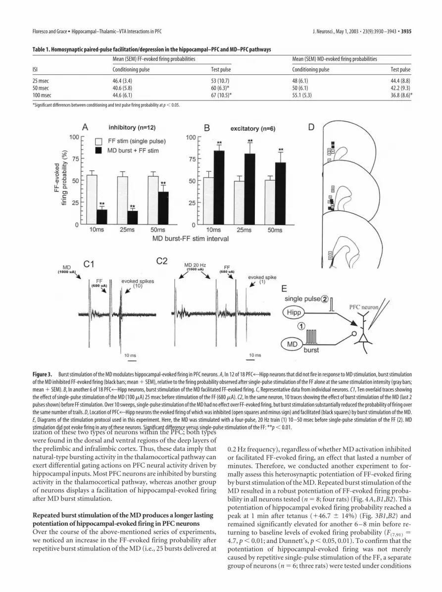
probability evoked by pulses 2–5 did not differ throughout thecourse of the train. It is important to note that burst stimulationof the VTA still caused a significant reduction ( p � 0.01) in thefiring probability evoked by later pulses in the train comparedwith the firing probability at the same time points of the trainwhen no VTA stimulation was administered, but the magnitudeof this inhibition was substantially reduced compared with thatobserved during the first pulse (first pulse firing probability �7.4 3%; pulses 2–5 � 52.3 7%; p � 0.01). From these data itis apparent that the inhibitory actions that the VTA exerts overhippocampal-evoked firing in PFC neurons are frequency depen-dent. Burst stimulation of the VTA can drastically inhibit firingevoked by single-pulse stimulation of the hippocampus. How-ever, if impulse traffic in the hippocampal–PFC pathway occursin the form of a higher-frequency train of action potentials, theVTA-mediated inhibition is reduced substantially in the latterportions of the train.
Selective DA receptor antagonists alter the effects of VTAstimulation on FF-evoked firingTo ascertain the role that DA receptors play in the effects of VTAstimulation, we administered selective DA receptor antagonistsbefore burst stimulation of the VTA. Analysis of these data re-vealed significant drug � pulse number � VTA stimulation in-teraction (F(20,192) � 1.9; p � 0.05). Simple main effects analysisrevealed that when no VTA stimulation was administered, therewere no significant differences between drug treatment groupswith respect to the firing probabilities evoked by train stimula-tion of the FF (F(5,48) � 0.1; NS). This finding indicates that anydifferences between the control condition and the drug treatmentgroup could not be attributed to group differences in the firingprobabilities evoked by train stimulation alone. Furthermore, thefact that blockade of D1, D2, or D4 receptors did not alter firingprobabilities evoked by 20 Hz train stimulation implies that anyincrease in mesocortical DA release that may be caused by stim-
Figure 6. The effects of D1 , D2 , D4 , and NMDA receptor blockaded on VTA-induced inhibition of FF-evoked firing in PFC neurons. For all panels, symbols represent mean (�SEM) firing probabilityevoked by each pulse in a five-pulse train delivered to the FF. Gray circles represent FF-evoked firing probability for each pulse in the five-pulse train, when no VTA stimulation was given. Squaresrepresent FF-evoked firing probability after a VTA burst. Black squares denote significant within-group difference ( p � 0.05), and white squares denote no significant within-group difference infiring probability observed for latter pulses in the train compared with the firing probability observed during the first pulse (gray square). For comparative purposes, the gray line in B–F representsfiring probability of the control condition (VTA � FF). *p � 0.05, **p � 0.01, significant difference versus probabilities within groups, observed at the same time point in the train with and withoutVTA stimulation; †p � 0.05, significant difference in firing probabilities between groups versus those observed at the same time point in the control condition (gray line). A, Burst stimulation of theVTA dramatically reduced firing evoked by the first pulse in the train but was less effective for pulses 2–5. B, Administration of the D1 antagonist SCH23390 augmented the VTA-induced inhibition.C, The D2 antagonist eticlopride abolished the inhibition of firing observed in the latter parts of the train, whereas haloperidol ( D) almost completely attenuated the inhibition. The D4 antagonist CP293,019 ( E) also attenuated the inhibitory effect of VTA stimulation. The NMDA receptor antagonist CPP ( F) was without effect. G, Diagrams of the stimulation protocol used in this experiment. Here,the VTA was stimulated in a burst pattern (1) before five-pulse, 20 Hz train stimulation of the FF (2).
Floresco and Grace • Hippocampal–Thalamic–VTA Interactions in PFC J. Neurosci., May 1, 2003 • 23(9):3930 –3943 • 3937

ulation of hippocampal afferents (Gurdenet al., 2000) does not influence spike firingevoked during train stimulation.
D1 receptor blockadeAdministration of the D1 antagonist SCH23390 (0.2 mg/kg, i.v.; n � 9) markedlyenhanced the inhibitory actions of VTAstimulation on firing evoked by train stim-ulation of the FF (Figs. 6B, 7B). Burst stim-ulation of the VTA caused a pronouncedinhibition of firing evoked by the secondand third pulse of the five-pulse FF train.In fact, the firing probabilities at these timepoints did not differ from the firing prob-ability evoked by first pulse (Fig. 6B, whitesquares). Moreover, the firing probabilityat the middle portion of the train was sig-nificantly inhibited compared with theprobability observed at the same timepoints for control neurons. The firingprobability evoked by the last two pulses inthe train was significantly reduced ( p �0.01) when compared with the firing prob-ability at the same time points of the trainwhen no VTA stimulation was adminis-tered but was significantly higher ( p �0.01) than the firing probability evoked bythe first pulse of the train. Thus, D1 recep-tor blockade revealed an underlying po-tent VTA-mediated inhibition during thelatter parts of the train.
D2 receptor blockadeIn contrast to the effects of D1 receptor blockade, intravenousadministration of the D2 receptor antagonist eticlopride (0.25mg/kg; n � 10) abolished the inhibitory actions of VTA stimula-tion on firing evoked by the latter parts (pulses 3–5) of trainstimulation of the FF but had no significant effect on the inhibi-tion observed during the early parts of the train (Fig. 6C). Thisattenuation of the VTA-induced inhibition was even more pro-nounced after pretreatment with the D2/D4 antagonist haloperi-dol (0.5 mg/kg; n � 8). In these cases, VTA stimulation had noeffect over the firing probabilities evoked by pulses 2–5, whencompared with the probabilities observed when no VTA stimu-lation was given (Fig. 6D). Moreover, in the presence of haloper-idol, VTA stimulation was less effective at inhibiting firing in-duced by the first pulse in the train, when compared with thecontrol condition ( p � 0.05).
D4 receptor blockadeHaloperidol has a higher affinity for DA D4 receptors than eticlo-pride (Durcan et al., 1995; Seeman and Van Tol, 1995; Seeman etal., 1997) and was more effective than eticlopride in attenuatingthe inhibitory actions of VTA stimulation on FF-evoked activity.In addition, application of D4 antagonists can increase the excit-ability of PFC neurons (Rubinstein et al., 2001). In keeping withthese observations, administration of the selective DA D4 recep-tor antagonist CP 293,019 (10 mg/kg; n � 9) caused an effect likethat of haloperidol but unlike that of eticlopride. D4 receptorblockade attenuated significantly ( p � 0.05) the VTA-inducedinhibition of the firing probability evoked by the first pulse of thefive-pulse FF train, when compared with neurons in the controlcondition (Fig. 6E). The effect of CP 293,019 on the firing prob-
ability evoked in the latter parts of the train were mixed; D4
receptor blockade appeared to reduce the VTA-induced inhibi-tion of firing evoked by pulses 2– 4, but the firing probability atthese time points was still significantly lower when comparedwith the probabilities observed when no VTA stimulation wasgiven. VTA stimulation did not affect the firing probabilityevoked by the fifth pulse in the train when compared with thesame time point when no VTA stimulation was administered.
NMDA receptor blockadePrevious studies in vitro have shown that the facilitatory effects ofD1 receptor activity on synaptic transmission in the PFC may beco-mediated by the NMDA receptor (Seamans et al., 2001).Therefore, we conducted an experiment to assess whether thefrequency-dependent effects observed in the present study weremediated by NMDA receptors. Surprisingly, in eight cells tested,administration of the NMDA receptor antagonist CPP (2 mg/kg)caused no change in the firing probability evoked by train stim-ulation of the hippocampus after VTA stimulation (Fig. 6F). Asobserved in the control condition, burst stimulation of the VTAdrastically reduced the firing probability evoked by the first pulsein the train, whereas firing probability evoked by latter pulses inthe train was significantly higher when compared with the firstpulse. The discrepancy between the present in vivo study and theeffects reported by Seamans et al. (2001) in vitro may be attrib-uted to a number of procedural differences, such as blockade ofNa� channels in the study by Seamans and colleagues, the dura-tion of the train used (5 vs 15 pulses), the dependent variable(spike firing vs subthreshold EPSPs), and method of DA receptoractivation (VTA stimulation vs bath application of a selective D1
receptor agonist).The protocol used in the pharmacological experiments used
between-subjects comparisons between animals that received in-
Figure 7. Representative data from individual neurons recorded from experiments using 20 Hz train stimulation of the FF. Eachpanel represents 10 overlaid traces, and gray arrows denote time points where the FF was stimulated. A, Example of a neuron inthe control condition (stimulation intensities: FF � 1000 �A, VTA � 750 �A). Train stimulation of the FF by itself reliably evokedfiring over all five pulses of the train (A1). Burst stimulation of the VTA 10 msec before train stimulation of the FF drasticallyreduced firing evoked by the first pulse in the train, whereas firing evoked by the latter pulses was relatively unaffected (A2). Forclarity, only the last pulse of the VTA burst (VTA stim) is shown. B, Example of a neuron pretreated with the D1 receptor antagonistSCH23390 (stimulation intensities: FF � 680 �A, VTA � 600 �A). As was observed in control neurons, train stimulation of the FFby itself reliably evoked firing over all five pulses of the train (B1). However, in the presence of SCH23390, burst stimulation of theVTA completely abolished firing evoked by pulses 1–2 and substantially reduced firing evoked by latter pulses of the train (B2).
3938 • J. Neurosci., May 1, 2003 • 23(9):3930 –3943 Floresco and Grace • Hippocampal–Thalamic–VTA Interactions in PFC

jections of DA antagonists and those that received no drug treat-ment. However, as noted above, the effects of VTA stimulationon firing evoked by single-pulse stimulation of the FF was as-sessed for each individual cell, before any drug administration.Thus, we were able to conduct a within-subjects analysis of theeffects of DA receptor blockade on the VTA-induced inhibitionof firing evoked by single-pulse stimulation of the FF. Analysis ofthese data revealed that before drug administration, burst stim-ulation of the VTA drastically inhibited FF-evoked firing of allneurons in all groups [�80% reduction in firing probability (Fig.8, gray bars) when compared with single-pulse stimulation alone(Fig. 8, white bar)]. Similar to the effects observed with trainstimulation of the FF, the VTA-induced inhibition of firingevoked by single-pulse stimulation of the FF was not affected bySCH 23390 and was significantly (but not completely) attenuatedby eticlopride, haloperidol, and CP 293,019 (F(4,32) � 3.0, p �0.05; and Dunnett’s, p � 0.05, p � 0.01) (Fig. 8, black bars). Thus,the finding that blockers for D2 and D4 receptors reduced theeffects of burst stimulation of the VTA suggests that the inhibi-tory actions of VTA stimulation on evoked firing of PFC4Hippneurons is mediated at least in part by the mesocortical DAprojection.
To summarize, activation of the VTA exerts a frequency-dependent inhibition over firing driven by hippocampal inputsto the PFC. VTA stimulation potently inhibits hippocampal-evoked firing evoked by single-pulse stimulation. However, whenthis pathway is activated at a higher frequency, the inhibition ofhippocampal-evoked firing is attenuated, and inputs from thetemporal lobe are much more likely to evoke spike firing. Theinhibitory actions of the VTA were reduced after blockade of DAD2 and D4 receptors. Moreover, the DA-mediated inhibition in-duced by stimulation of the VTA appears to have both an earlyand a late component. Inhibition that occurs �100 msec after theend of a VTA burst is reduced by D4 receptor blockade (CP293,019) (Fig. 6E), whereas a later onset inhibition (100 –250msec after a VTA burst) appears to be mediated more promi-nently by D2 receptors (eticlopride) (Fig. 6C). Accordingly, treat-
ment with an antagonist that has affinity for both D2 and D4
receptors (haloperidol) (Fig. 6D) almost completely abolishedthe inhibitory actions of VTA stimulation. The finding that an-tagonists for both D2 and D4 receptors did alleviate much of theinhibitory actions of VTA stimulation implies that these effectsare mediated primarily by DA and not the mesocortical GABAprojection (Carr and Sesack, 2000). Last, blockade of D1 recep-tors disrupted the frequency-dependent modulation of impulseactivity in this pathway, augmenting the VTA-mediated inhibi-tion of FF-evoked firing.
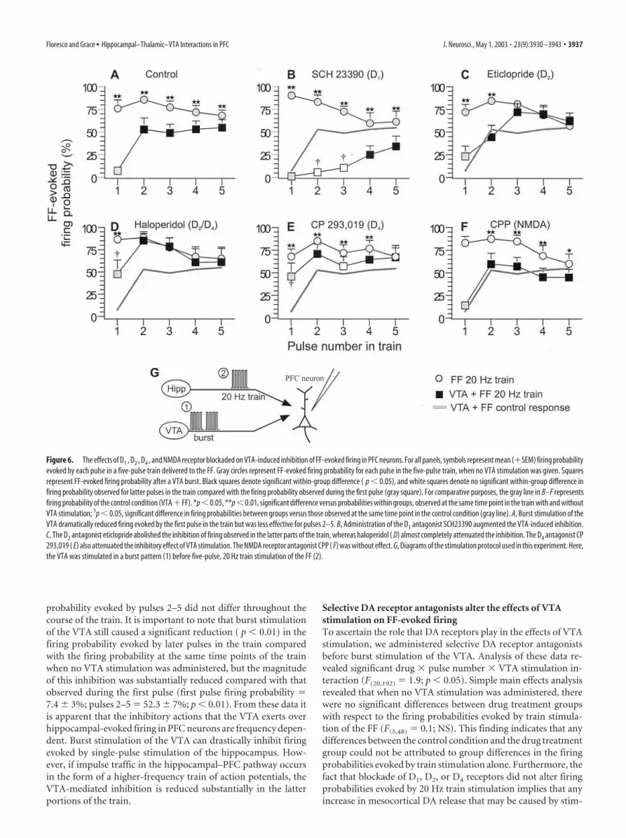
Interactions among hippocampal, MD, and VTA inputs tothe PFCThe above-mentioned findings indicate that bursting activity ofthe VTA can exert a frequency-dependent gating action over PFCneuron firing driven by inputs from the hippocampus. In light ofthese data, we were interested in assessing what role the VTA mayplay in modulating the interactions between MD and hippocam-pal inputs to the PFC. In nine PFC4Hipp neurons (nine rats)that also displayed a monosynaptic action potential in responseto MD stimulation, we assessed how burst stimulation of the VTAmodulated MD gating exerted over hippocampal-evoked firing,using our sequential-pulse stimulation protocol. For these exper-iments, we only used interpulse intervals of 10 –50 msec, becausewe did not observe any significant effect of MD conditioningpulses applied �100 msec before FF test pulses, and the magni-tude of inhibition of firing by MD conditioning pulses was equiv-alent at intervals of 25–100 msec.
Analysis of these data revealed a significant MD pulse � VTAstimulation interaction (F(2,16) � 5.8; p � 0.05) (Fig. 9A). In thesecells, the baseline firing probability evoked by MD stimulationand FF stimulation did not differ (FF mean � 59.5 4%; MDmean � 63.9 6%; F(1,8) � 0.7; NS). As observed previously (Fig.2), delivery of a conditioning pulse to the MD 10 or 50 msecbefore stimulation of the FF significantly inhibited FF-evokedfiring, when compared with firing evoked by single-pulse stimu-lation of the FF alone. The inhibition of FF-evoked firing by aconditioning pulse to the MD was significantly greater ( p � 0.05)when the interval between pulses was 50 versus 10 msec (Fig. 9A).Burst stimulation of the VTA 10 msec before single-pulse stimu-lation of the FF also inhibited FF-evoked spike firing ( p � 0.05).However, application of a conditioning pulse to the MD 10 msecafter VTA stimulation, and 10 msec before FF stimulation (i.e., aVTA burst–MD pulse–FF pulse sequence), attenuated the VTA-mediated inhibition of FF-evoked firing. The hippocampal-evoked firing probability was significantly higher ( p � 0.05)when compared with firing probability observed after VTAburst–FF stimulation alone. Moreover, at the 10 msec intervalbetween MD and FF stimulation, there was no difference in thefiring probabilities observed after a VTA–MD–FF sequence whencompared with an MD–FF paired-pulse sequence. This effect wasnot observed when a longer, 50 msec interval between the MDconditioning pulse and FF test pulse was used. In this instance, aconditioning pulse to the MD 10 msec after VTA stimulation and50 msec before FF stimulation caused no significant change in theFF-evoked firing probability, when compared with the reducedfiring probability displayed after VTA stimulation alone. Thus, asobserved previously, activation of either VTA or MD inputs tothe PFC can inhibit evoked firing in PFC4Hipp neurons. How-ever, during periods of VTA bursting activity, activation of PFCneurons by the MD that occurs nearly simultaneously with acti-vation by hippocampal inputs converging on the same neuron
Figure 8. D2 and D4 but not D1 receptor blockade attenuates VTA-mediated inhibition offiring evoked by single-pulse stimulation of the FF. Bars represent the percentage change infiring probability in response to single pulses to the FF after burst stimulation of the VTA beforedrug administration (gray bar) and 10 –20 min after drug administration (black bars). Thesevalues are expressed as a percentage change relative to the evoked-firing probability observedwhen the FF was stimulated alone (open bar; for comparative purposes). Thus, a score of 0%indicates that no spike firing is elicited by FF stimulation after VTA stimulation, whereas FFstimulation alone is always 100%. Only the D2 antagonist eticlopride, the D2 /D4 antagonisthaloperidol, and the D4 antagonist CP 293,019 were effective in attenuating the VTA-inducedinhibition, whereas the D1 antagonist SCH23390 and the NMDA antagonist CPP were withouteffect. Significance at *p � 0.05, **p � 0.01 versus pre-drug condition.
Floresco and Grace • Hippocampal–Thalamic–VTA Interactions in PFC J. Neurosci., May 1, 2003 • 23(9):3930 –3943 • 3939
can reduce the inhibitory actions that the VTA exerts over spikefiring driven by the hippocampus.
DiscussionUsing different stimulation protocols, we report four primaryobservations: (1) in the majority of PFC4Hipp neurons tested,inputs from the MD exert a powerful inhibitory gating actionover hippocampal-evoked firing; (2) repetitive burst stimulationof the MD potentiates hippocampal-evoked activity that lastsseveral minutes; (3) stimulation of the VTA inhibitshippocampal-evoked firing with a preferential attenuation oflow-frequency inputs, an effect that was altered after selectiveblockade of DA receptors. D1 receptors facilitate higher-frequency transmission in the hippocampal-PFC pathway,whereas D2 and D4 receptors inhibit firing, and (4) the relativetiming of inputs from the MD or VTA determines their impact onPFC neuron responses to hippocampal inputs. Activation of theMD �10 msec before activation of hippocampal inputs was lesslikely to inhibit hippocampal-evoked firing than at longer inter-vals. This last finding suggests that inputs from the MD may playa role in heterosynaptic coincidence detection (Usrey, 2002), in-
hibiting inputs from the temporal lobe unless they arrive syn-chronously with diencephalic inputs.
Modulation of the hippocampal–PFC pathway by the MDThalamocortical inputs from the MD typically exerted a pro-nounced inhibition of hippocampal-evoked firing in PFC neu-rons. Although there is only preliminary ultrastructural evidencethat inputs from the MD synapse on PFC GABAergic interneu-rons (Kuroda et al., 1998), such an arrangement is commonlyreported in other thalamocortical projection systems (Freund etal., 1985; Staiger et al., 1996), and activation of thalamocorticalaxons yields a threefold larger amplitude EPSP on fast-spikinginterneurons as compared with pyramidal neurons (Beierleinand Connors, 2002). Furthermore, stimulation of the MD pro-duces an EPSP–IPSP sequence in PFC neurons (Gigg et al., 1994;Lewis and O’Donnell, 2000), and chemical stimulation of the MDactivates c-fos immunoreactivity in PFC GABA-containing neu-rons (Bubser et al., 1998). In the present study, the inhibition ofhippocampal-evoked firing after activation of glutamatergic MDafferents is likely mediated by feedforward inhibitory circuits.The fact that this effect was observed in the majority ofPFC4Hipp neurons suggests that a large proportion of informa-tion that arrives from the hippocampus is under the modulatorycontrol of the MD.
For PFC4Hipp neurons that did not fire in response to MDstimulation, activation of thalamocortical afferents with a train ofstimuli (but not single pulses) attenuated hippocampal-evokedfiring in a majority of neurons. This finding suggests that al-though relatively few PFC4Hipp neurons appeared to receivedirect monosynaptic input from the MD, a larger proportion ofthese cells can still be modulated by MD inputs in a frequency-dependent manner. Reyes et al. (1998) reported that activity ofneocortical GABAergic interneurons was facilitated when excita-tory inputs were stimulated with a 10 Hz train. Thus, an increasein activity of MD inputs to the PFC in the 10 – 40 Hz range canfilter a large ensemble of PFC4Hipp neurons, attenuating firingdriven by temporal lobe inputs. This finding parallels phenom-ena observed in other thalamocortical systems, whereby thalamicnuclei can gate the flow of sensory inputs to the neocortex, pro-ducing widespread suppression of activity, thereby focusing cor-tical sensory representations (Castro-Alamancos, 2002; Castro-Alamancos and Oldford, 2002).
In contrast to the above-mentioned effects, 33% of thePFC4Hipp neurons displayed a facilitation of hippocampal-evoked firing after train stimulation of the MD, although MDstimulation alone did not evoke firing. Moreover, repeated trainsof thalamic stimulation facilitated hippocampal-evoked firingfor minutes after tetanus. These data, combined with those de-scribed above, suggests the presence of two distinct populationsof PFC4Hipp neurons: one receives a predominantly inhibitory(presumably polysynaptic) input from the MD, and a secondreceives a weak excitatory input that by itself is insufficient toevoke spike firing. With respect to this latter population, intra-cellular recordings have shown that train stimulation of thalamicnuclei can produce an “augmenting response” in sensorimotorcortex, causing a sustained depolarization lasting hundreds ofmilliseconds (Castro-Alamancos and Connors, 1996; Steriade etal., 1998). This phenomenon is thought to originate in deep cor-tical layers and activates reverberatory circuits via intralaminarand horizontal collaterals. A similar mechanism may explain theeffects observed here: high-frequency activation of subthresholdinputs to the MD would have depolarized a subpopulation pro-portion of PFC neurons (distinct from those that are inhibited by
Figure 9. Interactions among hippocampal, MD, and VTA inputs converging on the samePFC neurons. A, FF-evoked firing probabilities in PFC4Hipp neurons that also responded withan orthodromic monosynaptic action potential after stimulation of the MD. Application of aconditioning pulse to the MD 10 or 50 msec before a test pulse to the MD (gray bars) significantlyreduced the evoked firing probability relative to that observed when the FF was stimulatedalone (gray hatched bar). Burst stimulation of the VTA also attenuated FF-evoked firing (blackbars). However, application of a conditioning pulse to the MD 10 msec before an FF test pulsesignificantly (*p � 0.05) attenuated the VTA-mediated inhibition. Stars denote significantdifference versus firing probability evoked by FF single-pulse stimulation. B, Location of allneurons observed in this study (open stars) that responded with an orthodromic, monosynapticaction potential after stimulation of the FF and the MD. Numbers beside each plate correspondto millimeters from bregma. C, Diagram of the stimulation protocol used in this experiment.Here the VTA was stimulated in a burst pattern (1) 10 msec before administering MD–FFsequential-pulse stimulation (2, 3). Both the MD and FF were stimulated with single pulses thatwere separated by 10 or 50 msec.
3940 • J. Neurosci., May 1, 2003 • 23(9):3930 –3943 Floresco and Grace • Hippocampal–Thalamic–VTA Interactions in PFC

MD activation) and made them more responsive to hippocampalinput.
VTA modulation of hippocampal-evoked firing ofPFC neuronsBurst stimulation of the VTA induced a frequency-dependentinhibition over evoked firing in PFC4Hipp neurons; these ef-fects were altered by systemic administration of DA antagonists.This observation complements previous findings in which acti-vation of DA receptors promotes a frequency-dependent inhibi-tion over synaptically evoked activity of PFC neurons (Jay et al.,1995; Gurden et al., 1999; Seamans et al., 2001). The faster-onset(�100 msec) D4-mediated inhibition and a longer-lasting D2-mediated attenuation of spike firing (�300 msec) after a VTAburst may be related to the location of these receptors in the PFC.In addition to being localized on some pyramidal neurons, D2-like receptors in the PFC also reside on GABAergic interneurons(Mrzijak et al., 1996; Wedzony et al., 2001) and on excitatorypresynaptic terminals (Sesack et al., 1995). Thus, multiple cellu-lar mechanisms exist by which D2 and D4 receptors could de-crease the responsivity of PFC neurons to hippocampal inputs,including facilitation of GABA release (Retaux et al., 1991;Grobin and Deutch, 1998; Zhou and Hablitz, 1999), modulationof the resting membrane potential (Gulledge and Jaffe, 1998,Yang et al., 1999), and presynaptic inhibition of hippocampalinputs. Indeed, in the nucleus accumbens, application of D2 ago-nists altered hippocampal terminal excitability, implying that DAinhibits hippocampal inputs presynaptically (Yang and Mogen-son, 1986). We observed a dramatic alteration in the paired-pulseratio of FF-evoked firing after VTA stimulation (Fig. 6A), sug-gesting that mesocortical DA can also inhibit hippocampal inputspresynaptically.
Blockade of D1 receptors augmented the VTA-mediated inhi-bition of hippocampal-evoked firing, abolishing the frequency-dependent inhibition that was observed under control condi-tions. This finding implies that D1 receptor activity can offset theinhibitory actions of D2-like receptors on PFC neuron firing,thereby augmenting the probability that higher-frequency inputswill evoke spiking. These effects parallel those observed in vitroduring which excitatory inputs to PFC neurons were stimulatedusing 20 Hz trains (Seamans et al., 2001). Bath application of a D1
receptor agonist reduced the EPSP evoked in early parts of thetrain but enhanced the EPSP in latter parts of the train. In thatstudy, it was found that the facilitatory effects of D1 receptorswere co-mediated by NMDA receptors. We did not observe a rolefor NMDA receptor in the D1 modulation of evoked firing, sug-gesting that D1–NMDA interactions may play a more importantrole in modulating subthreshold EPSPs (Seamans et al., 2001;Wang and O’Donnell, 2001). Alternatively, D1 receptors couldincrease the excitability of PFC neurons by facilitating persistentNa� (Yang and Seamans, 1996; Gorelova and Yang, 2000; Lavinand Grace, 2001) or Ca 2� conductances (Hernandez-Lopez et al.,1997; Wang and O’Donnell, 2001), which would enhance neuronexcitability, facilitate temporal summation of inputs, and in-crease the probability that subsequent EPSPs evoked by latterparts of a train will evoke spiking.
A similar mechanism may underlie the interactions amongMD, hippocampal, and VTA inputs to PFC neurons. VTA acti-vation normally inhibits firing evoked by single pulses to the FF.However, impulses from the MD arriving nearly simultaneouslywith those from the hippocampus may depolarize the neuron,activating intrinsic currents that are susceptible to D1 receptormodulation (Lavin and Grace, 2001), which in turn would make
the cell more responsive to hippocampal inputs. Collectively,these data suggest that during periods of increased VTA neuronbursting, impulse activity from the hippocampus that arrives ei-ther at a higher frequency or in close temporal proximity to im-pulses from the MD can be augmented by D1 receptor activityand are less likely to be inhibited by D2–like receptors. As such,inputs from the VTA may be viewed as a “high-pass filter,” per-mitting higher-frequency inputs that exert a stronger depolariz-ing action and greater control over spike firing of PFC neurons.
Functional implicationsMnemonic processes mediated by the hippocampal–PFC cir-cuits, such as working memory, are dependent on functionalinteractions between MD and mesocortical DA input (Gabriel,1993; Freeman et al., 1996; Floresco et al., 1997, 1999). Similarly,inputs from both the MD and the mesocortical DA system havebeen implicated in other executive functions mediated by thePFC, such as behavioral flexibility (Hunt and Aggleton, 1998;Ragozzino et al., 1999; Ragozzino, 2002). The present data pro-vide important insights regarding the cellular mechanisms thatunderlie cognitive processes subserved by neural networks incor-porating the frontal and temporal lobes and thalamic and mid-brain nuclei. For example, during periods in which reward isanticipated, increased bursting of VTA DA neurons (Schultz,1998) should limit the flow of information from the hippocam-pus to the PFC, permitting only the most salient information,arriving in the form of higher-frequency trains of impulses, toevoke firing. Blockade of D1 receptors in the PFC would be ex-pected to attenuate mnemonic-related activity in PFC4Hippneurons and impair working memory (Seamans et al., 1998). Inaddition, MD neurons also show increased delay-period activityduring a delayed response task (Oyoshi et al., 1996). Our datasuggest that a prolonged increase in MD activity may facilitateworking memory processes, augmenting the excitability of PFCneurons and making them more responsive to information arriv-ing from the hippocampus after a delay. On the other hand, theability of both the MD and VTA DA inputs to facilitate or inhibitthe activity of PFC4Hipp neurons may be viewed as a cellularcomponent that permits strategy switching, facilitating the learn-ing of a new behavioral strategy while at the same time inhibitinga previously learned response. Collectively, these data suggestthat inputs from the MD and VTA that modulate the activity ofPFC4Hipp neurons are essential components of cortical–sub-cortical networks that regulate information processing by thefrontal lobes. Disruption in these components would be expectedto result in disorganized information processing, behavioral in-flexibility, and deficits in working memory. Interestingly, each ofthese symptoms are observed in schizophrenia, the pathophysi-ology of which is characterized by dysfunction in both the MDand hippocampal–PFC circuits, as well as a decrease in PFC D1
receptor activity (Weinberger et al., 1994; Okubo et al., 1997; Akilet al., 1999; Friedman et al., 1999; Shenton et al., 2001). Thepresent data suggest that decreased D1 receptor activity com-bined with a hyperexcitable mesolimbic DA system would reducethe transfer of information between the temporal and frontallobes. Our finding that antipsychotic agents, acting on D2-likereceptors, can alleviate the VTA-mediated inhibition ofPFC4Hipp neurons suggests that their efficacy in treating psy-chosis may be attributable in part to their ability to normalizeneural activity in these circuits that has been perturbed by alter-ations in D1 receptor activity.
Floresco and Grace • Hippocampal–Thalamic–VTA Interactions in PFC J. Neurosci., May 1, 2003 • 23(9):3930 –3943 • 3941

ReferencesAkil M, Pierri JN, Whitehead RE, Edgar CL, Mohila C, Sampson AR, Lewis
DA (1999) Lamina-specific alterations in the dopamine innervation ofthe prefrontal cortex in schizophrenic subjects. Am J Psychiatry156:1580 –1589.
Aujla H, Beninger RJ (2001) Hippocampal-prefrontocortical circuits: PKAinhibition in the prefrontal cortex impairs delayed nonmatching in theradial maze in rats. Behav Neurosci 115:1204 –1211.
Beierlein M, Connors BW (2002) Short-term dynamics of thalamocorticaland intracortical synapses onto layer 6 neurons in neocortex. J Neuro-physiol 4:1924 –1932.
Bubser M, De Brabander JM, Timmerman W, Feenstra MGP, Erdtsieck-Ernste EB, Rinnens A, Van Uum JFM, Westerink BHC (1998) Disinhi-bition of the mediodorsal thalamus induces Fos-like immunoreactivity inboth pyramidal and GABA-containing neurons in the medial prefrontal cor-tex of rats, but does not affect prefrontal levels of GABA. Synapses30:156–165.
Burette F, Jay TM, Laroche S (1997) Reversal of LTP in the hippocampalafferent fibre system to the prefrontal cortex in vivo with low-frequencypatterns of stimulation that do not produce LTD. J Neurophysiol78:1155–1160.
Carr DB, Sesack SR (1996) Hippocampal afferents to the rat prefrontal cor-tex: synaptic targets and relation to dopamine terminals. J Comp Neurol369:1–15.
Carr DB, Sesack SR (2000) Projections from the rat prefrontal cortex to theventral tegmental area: target specificity in the synaptic associations withmesoaccumbens and mesocortical neurons. J Neurosci 19:3723–3730.
Castro-Alamancos MA (2002) Role of thalamocortical sensory suppressionduring arousal: focusing sensory inputs in neocortex. J Neurosci 22:9651–9655.
Castro-Alamancos MA, Connors BW (1996) Cellular mechanisms of theaugmenting response: short term plasticity in a thalamocortical pathway.J Neurosci 16:7742–7756.
Castro-Alamancos MA, Oldford E (2002) Cortical sensory suppression dur-ing arousal is due to the activity-dependent depression of thalamocorticalsynapses. J Physiol (Lond) 541:319 –331.
Conde F, Maire-Lepoivre E, Audinat E, Crepel F (1995) Afferent connec-tions of the medial frontal cortex of the rat. II. Cortical and subcorticalafferents. J Comp Neurol 325:567–593.
Daum I, Ackermann H (1994) Frontal-type memory impairment with tha-lamic damage. Int J Neurosci 77:187–198.
Durcan MJ, Rigdon GC, Norman MH, Morgan PF (1995) Is clozapine se-lective for the dopamine D4 receptor? Life Sci 57:275–283.
Floresco SB, Seamans JK, Phillips AG (1997) Selective roles for hippocam-pal prefrontal cortical, and ventral striatal circuits in radial-arm mazetasks with or without a delay. J Neurosci 17:1880 –1890.
Floresco SB, Braaksma DN, Phillips AG (1999) Thalamic– cortical–striatalcircuitries mediate working memory during delayed responding on aradial-arm maze. J Neurosci 19:11061–11071.
Floresco SB, Blaha CD, Yang CR, Phillips AG (2001) Modulation of hip-pocampal and amygdalar-evoked activity of nucleus accumbens neuronsby dopamine: cellular mechanisms of input selection. J Neurosci 21:2851–2860.
Freeman JH, Cuppernell C, Flannery K, Gabriel M (1996) Context-specificmulti-site cingulate cortical, limbic thalamic, and hippocampal neuronalactivity during concurrent discriminative approach and avoidance train-ing in rabbits. J Neurosci 16:1538 –1549.
Freund TF, Martin KA, Somogyi P, Whitteridge D (1985) Innervation of catvisual areas 17 and 18 by physiologically identified X and Y-type afferentsII: identification of postsynaptic targets by GABA immunochemistry andGolgi impregnation. J Comp Neurol 242:275–291.
Friedman JI, Temporini H, Davis KL (1999) Pharmacologic strategies foraugmenting cognitive performance in schizophrenia. Biol Psychiatry45:1–16.
Fuster JM (2001) The prefrontal cortex—an update: time is of the essence.Neuron 30:319 –333.
Gabbott P, Headlam A, Bubsy S (2002) Morphological evidence that CA1hippocampal afferents monosynaptically innervate PV-containing neu-rons and NADPH-diaphorase reactive cells in the medial prefrontal cor-tex (areas 25/32) of the rat. Brain Res 946:314 –322.
Gabriel M (1993) Discriminative avoidance learning: a model system. In:Neurobiology of the cingulate cortex and limbic thalamus: a comprehen-
sive handbook (Vogt BA, Gabriel M eds), pp 478 –523. Boston, MA:Birkhauser.
Garris PA, Wightman RM (1994) Different kinetics govern dopaminergictransmission in the amygdala, prefrontal cortex, and striatum: an in vivovoltammetric study. J Neurosci 14:442– 450.
Gigg J, Tan AM, Finch DM (1994) Glutamatergic hippocampal formationprojections to prefrontal cortex in the rat are regulated by GABAergicinhibition and show convergence with glutamatergic projections fromlimbic thalamus. Hippocampus 4:189 –198.
Gioanni Y, Thierry AM, Glowinski J, Tassin JP (1998) Alpha1-adrenergic,D1, and D2 receptors interactions in the prefrontal cortex: implicationsfor the modality of action of different types of neuroleptics. Synapse30:362–370.
Goldman-Rakic PS (1998) The prefrontal landscape: implications of func-tional architecture for understanding human mentation and the centralexecutive. In: The prefrontal cortex: executive and cognitive functions(Roberts AC, Robbins TW, Weizkrantz L, eds), pp 67– 86. Oxford: OxfordUP.
Gorelova N, Yang CR (2000) Dopamine D1/D5 receptor activation modu-lates a persistent sodium current in rat prefrontal cortical neurons invitro. J Neurophysiol 84:75– 87.
Grobin AC, Deutch AY (1998) Dopaminergic regulation of extracellulargamma-aminobutyric acid levels in the prefrontal cortex of the rat.J Pharmacol Exp Ther 285:350 –357.
Groenewegen HJ (1988) Organization of the afferent connections of themediodorsal thalamic nucleus in the rat, related to the mediodorsal-prefrontal topography. Neuroscience 24:379 – 431.
Gulledge AT, Jaffe DB (1998) Dopamine decreases the excitability of layer Vpyramidal cells in rat prefrontal cortex. J Neurosci 18:9139 –9151.
Gurden H, Tassin J-P, Jay TM (1999) Integrity of the mesocortical dopami-nergic system is necessary for complete expression of in vivo hippocampal-prefrontal cortex long-term potentiation. Neuroscience 94:1019–1027.
Gurden H, Takita M, Jay TM (2000) Essential role of D1 but not D2 receptorsin the NMDA receptor-dependent long-term potentiation at hippocampal-prefrontal cortex synapses in vivo. J Neurosci 20:RC106(1–5).
Hampson RE, Hedberg T, Deadwyler SA (2000) Differential informationprocessing by hippocampal and subicular neurons. Ann NY Acad Sci911:151–165.
Hauser MD (1999) Perseveration, inhibition and the prefrontal cortex: anew look. Curr Opin Neurobiol 9:214 –222.
Hernandez-Lopez S, Bargas J, Surmeier DJ, Reyes A, Galarraga E (1997) D1receptor activation enhances evoked discharge in neostriatal mediumspiny neurons by modulating an L-type Ca 2� conductance. J Neurosci17:3334 –3342.
Hunt PR, Aggleton JP (1998) Neurotoxic lesions of the dorsomedial thala-mus impair the acquisition but not the performance of delayed matchingto place by rats: a deficit in shifting response rules. J Neurosci 18:10045–10052.
Jay TM, Glowinski J, Thierry AM (1995) Inhibition of hippocampo-prefrontal cortex excitatory responses by the mesocortical DA system.NeuroReport 6:1845–1848.
Joyce EM, Robbins TW (1991) Frontal lobe function in Korsakoff and non-Korsakoff alcoholics: planning and spatial working memory. Neuropsy-chologia 29:709 –723.
Kolb B (1984) Functions of the frontal cortex of the rat: a comparative re-view. Brain Res 320:65–98.
Krettek JE, Price J (1977) The cortical projections of the mediodorsal nu-cleus and adjacent thalamic nuclei in the rat. J Comp Neurol 171:157–192.
Kuroda M, Yokofujita J, Murakami K (1998) An ultrastructural study of theneural circuit between the prefrontal cortex and the mediodorsal nucleusof the thalamus. Prog Neurobiol 54:417– 458.
Lavin A, Grace AA (2001) Stimulation of D1-type dopamine receptors en-hances excitability in prefrontal cortical pyramidal neurons in a state-dependent manner. Neuroscience 104:335–346.
Lewis BL, O’Donnell P (2000) Ventral tegmental area afferents to the pre-frontal cortex maintain membrane potential “up” states in pyramidalneurons via D1 dopamine receptors. Cereb Cortex 10:1168 –1175.
Mansbach RS, Brooks EW, Sanner MA, Zorn SH (1998) Selective dopamineD4 receptor antagonists reverse apomorphine-induced blockade of pre-pulse inhibition. Psychopharmacology 135:194 –200.
Miller EK (2000) The prefrontal cortex and cognitive control. Nat Rev Neu-rosci 1:59 – 65.
3942 • J. Neurosci., May 1, 2003 • 23(9):3930 –3943 Floresco and Grace • Hippocampal–Thalamic–VTA Interactions in PFC

Mrzijak L, Bergson C, Pappy M, Huff R, Levenson R, Goldman-Rakic PS(1996) Localization of dopamine D4 receptors in GABAergic neurons ofthe primate brain. Nature 381:245–248.
Mulder AB, Arts MPM, Lopes da Silva FH (1997) Short- and long-termplasticity of the hippocampus to nucleus accumbens and prefrontal cor-tex pathways in the rat, in vivo. Eur J Neurosci 9:1603–1611.
Mulkey RM, Malenka RC (1992) Mechanisms underlying induction of ho-mosynaptic long-term depression in area CA1 of the hippocampus. Neu-ron 9:967–975.
Okubo Y, Suhara T, Suzuki K, Kobayashi K, Inoue O, Terasaki O, Someya Y,Sassa T, Sudo Y, Matsushima E, Iyo M, Tateno Y, Toru M (1997) De-creased prefrontal dopamine D1 receptors in schizophrenia revealed byPET. Nature 385:634 – 636.
Otani S, Blond O, Desce JM, Crepel F (1998) Dopamine facilitates long-term depression of glutamatergic transmission in rat prefrontal cortex.Neuroscience 85:669 – 676.
Oyoshi T, Nishijo H, Asakura T, Takamura Y, Ono T (1996) Emotional andbehavioral correlates of mediodorsal thalamic neurons during associativelearning in rats. J Neurosci 16:5812–5829.
Pirot S, Jay TM, Glowinski J, Thierry AM (1994) Anatomical and electro-physiological evidence for an excitatory amino acid pathway from thethalamic mediodorsal nucleus to the prefrontal cortex in the rat. EurJ Neurosci 6:1225–1234.
Ragozzino ME (2002) The effects of dopamine D1 receptor blockade in theprelimbic-infralimbic areas on behavioral flexibility. Learn Mem 9:18–28.
Ragozzino ME, Detrick S, Kesner RP (1999) Involvement of the prelimbic-infralimbic areas of the rodent prefrontal cortex in behavioral flexibilityfor place and response learning. J Neurosci 19:4585– 4594.
Ray JP, Price JL (1992) The organization of the thalamocortical connectionsof the mediodorsal thalamic nucleus in the rat, related to the ventralforebrain-prefrontal cortex topography. J Comp Neurol 323:167–197.
Retaux S, Besson MJ, Penit-Soria J (1991) Opposing effects of dopamine D2receptor stimulation on the spontaneous and the electrically evoked releaseof [ 3H]GABA on rat prefrontal cortex slices. Neuroscience 42:61–71.
Reyes A, Lujan R, Rozov A, Burnashev N, Somogyi P, Sakmann B (1998)Target-cell-specific facilitation and depression in neocortical circuits. NatNeurosci 4:279 –285.
Robbins TW (1996) Dissociating executive functions of the prefrontal cor-tex. Philos Trans R Soc Lond B Biol Sci 351:1463–1470.
Rubinstein M, Cepeda C, Hurst RS, Flores-Hernandez J, Ariano MA, FalzoneTL, Kozell LB, Meshul CK, Bunzow JR, Low MJ, Levine MS, Grandy DK(2001) Dopamine D4 receptor-deficient mice display cortical hyperex-citability. J Neurosci 21:3756 –3763.
Schultz W (1998) Predictive reward signal of dopamine neurons. J Neuro-physiol 80:1–27.
Seeman P, Van Tol HH (1995) Deriving the therapeutic concentrations forclozapine and haloperidol: the apparent dissociation constant of a neuro-leptic at the dopamine D2 or D4 receptor varies with the affinity of thecompeting radioligand. Eur J Pharmacol 291:59 – 66.
Seeman P, Corbett R, Van Tol HH (1997) Atypical neuroleptics have low
affinity for dopamine D2 receptors or are selective for D4 receptors. Neu-ropsychopharmacology 16:93–110.
Seamans JK, Floresco SB, Phillips AG (1998) D1 receptor modulation ofhippocampal-prefrontal cortical circuits integrating spatial memory withexecutive functions in the rat. J Neurosci 18:1613–1621.
Seamans JK, Durstewitz D, Christie B, Stevens CF, Sejnowski TJ (2001) Do-pamine D1/D5 receptor modulation of excitatory synaptic inputs to layerV prefrontal cortex neurons. Proc Natl Acad Sci USA 98:301–306.
Sesack SR, Bunney BS (1989) Pharmacological characterization of the re-ceptor mediating electrophysiological responses to dopamine in the ratmedial prefrontal cortex: a microiontophoretic study. J Pharmacol ExpTher 248:1323–1333.
Sesack SR, King SW, Bressler CN, Watson SJ, Lewis DA (1995) Electronmicroscopic visualization of dopamine D2 receptors in the forebrain:cellular, regional, and species comparisons. Soc Neurosci Abstr 21:365.
Shenton ME, Dickey CC, Frumin M, McCarley RW (2001) A review of MRIfindings in schizophrenia. Schizophrenia Res 49:1–52.
Staiger JF, Zilles K, Freund TF (1996) Distribution of GABAergic elementspostsynaptic to ventroposteromedial thalamic projections in layer IV ofrat barrel cortex. Eur J Neurosci 11:2273–2285.
Steriade M, Timofeev I, Grenier F, Durmuller N (1998) Role of thalamicand cortical neurons in augmenting responses and self-sustained activity:dual intracellular recordings in vivo. J Neurosci 18:6425– 6443.
Usrey WM (2002) The role of spike timing for thalamocortical processing.Curr Opin Neurobiol 12:411– 417.
Van Eden CG, Hoorneman EMD, Buijs RM, Mathussen MAH, Geffard M,Uylings HBM (1987) Immunohistochemical localization of dopaminein the prefrontal cortex of the rat at the light, electron microscopal level.Neuroscience 22:849 – 862.
Wang J, O’Donnell P (2001) D1 dopamine receptors potentiate NMDA-mediated excitability increase in layer V prefrontal cortical pyramidalneurons. Cereb Cortex 11:452– 462.
Wedzony K, Chocyk A, Mackowiak M, Fijal K, Czyrak A (2001) Corticallocalization of dopamine D4 receptors in the rat brain-immunocyto-chemical study. J Physiol Pharmacol 51:205–221.
Weinberger DR, Aloia MS, Goldberg TE, Berman KF (1994) The frontallobes and schizophrenia. J Neuropsychiatry Clin Neurosci 6:419 – 427.
Yang CR, Mogenson GJ (1986) Dopamine enhances terminal excitability ofhippocampal-accumbens neurons via D2 receptor: role of dopamine inpresynaptic inhibition. J Neurosci 6:2470 –2478.
Yang CR, Seamans JK (1996) Dopamine D1 receptor actions in layers V–VIrat prefrontal cortex neurons in vitro: modulation of dendritic-somaticsignal integration. J Neurosci 16:1922–1935.
Yang CR, Seamans JK, Gorelova N (1999) Developing a neuronal model forthe pathophysiology of schizophrenia based on the nature of electrophys-iological actions of dopamine in the prefrontal cortex. Neuropsychophar-macology 21:161–194.
Zhou F-M, Hablitz JJ (1999) Dopamine modulation of membrane and syn-aptic properties of interneurons in rat cerebral cortex. J Neurophysiol81:967–976.
Floresco and Grace • Hippocampal–Thalamic–VTA Interactions in PFC J. Neurosci., May 1, 2003 • 23(9):3930 –3943 • 3943


