Embed Size (px)
Citation preview
Gender-Specific Association of the Plasminogen
Activator Inhibitor-1 4G/5G Polymorphism
With Central Arterial Blood Pressure
Hanna Björck, Per Eriksson, Urban Alehagen, Rachel Debasso, Liza Ljungberg,
Karin Persson, Ulf Dahlström and Toste Länne
Linköping University Post Print
N.B.: When citing this work, cite the original article.
Original Publication:
Hanna Björck, Per Eriksson, Urban Alehagen, Rachel Debasso, Liza Ljungberg, Karin
Persson, Ulf Dahlström and Toste Länne, Gender-Specific Association of the Plasminogen
Activator Inhibitor-1 4G/5G Polymorphism With Central Arterial Blood Pressure, 2011,
American Journal of Hypertension, (24), 7, 802-808.
http://dx.doi.org/10.1038/ajh.2011.63
Copyright: Nature Publishing Group
http://npg.nature.com/
Postprint available at: Linköping University Electronic Press
http://urn.kb.se/resolve?urn=urn:nbn:se:liu:diva-69831
1
Word count abstract: 236
Word count text: 2965
References: 40
Tables: 4
Gender-specific association of the plasminogen activator inhibitor-1 4G/5G
polymorphism with central arterial blood pressure
Running headline: PAI-1 and central blood pressure
Hanna M. Björcka, Per Eriksson
b, Urban Alehagen
a, Rachel De Basso
a, Liza U. Ljungberg
a,c,
Karin Perssonc, Ulf Dahlström
a, Toste Länne
a
aDivision of Cardiovascular Medicine, Department of Medical and Health Sciences, Faculty
of Health Sciences, Linköping University, Sweden.
bAtherosclerosis Research Unit, Center for Molecular Medicine, Department of Medicine,
Karolinska Institute, Sweden.
cDivision of Drug Research, Department of Medical and Health Sciences, Faculty of Health
Sciences, Linköping University, Sweden.
Disclosure: None to declare.
Corresponding author:
Hanna M. Björck, Faculty of Health Sciences, Department of Medicine and Health Sciences/
pharmacology, SE-58185 Linköping, Sweden.
E-mail: [email protected], Phone: +46(0)10 1038057, Fax: +46(0)13 149106
Keywords: aorta, arterial stiffness, genetics, hypertension, pressure pulse wave
2
Abstract
Background. The functional plasminogen activator inhibitor-1 (PAI-1) 4G/5G polymorphism
has previously been associated with hypertension. In recent years, central blood pressure,
rather than brachial has been argued a better measure of cardiovascular damage and clinical
outcome. The aim of this study was to investigate the possible influence of the 4G/5G
polymorphism on central arterial blood pressure in a cohort of elderly individuals.
Methods. We studied 410 individuals, 216 men and 194 women, aged 70-88. Central
pressures and pulse waveforms were calculated from the radial artery pressure waveform by
the use of the SphygmoCor system and a generalized transfer function. Brachial pressure was
recorded using oscillometric technique (Dinamap). PAI-1 antigen was determined in plasma.
Results. The results showed that central pressures were higher in women carrying the PAI-1
4G/4G genotype compared to female carriers of the 5G/5G genotype, (P=0.025, P=0.002 and
P=0.002 for central systolic-, diastolic- and mean arterial pressure, respectively). The
association remained after adjustment for potentially confounding factors related to
hypertension. No association of the PAI-1 genotype with blood pressure was found in men.
Multiple regression analysis revealed an association between PAI-1 genotype and plasma
PAI-1 levels (P=0.048).
Conclusions. Our findings show a gender-specific association of the PAI-1 4G/5G
polymorphism with central arterial blood pressure. The genotype effect was independent of
other risk factors related to hypertension, suggesting that impaired fibrinolytic potential may
play an important role in the development of central hypertension in women.
3
Introduction
Hypertension is a major risk factor for the development of cardiovascular disease.
Plasminogen activator inhibitor-1 (PAI-1) is the principal inhibitor of the fibrinolytic system,
interfering with the activation of plasminogen and subsequent matrix degradation. Circulating
PAI-1 influence smooth muscle cell proliferation as well as cell migration1,2
, and has
previously been associated with increased risk of thrombotic events3,4
. Furthermore, plasma
PAI-1 has been positively correlated with brachial blood pressure5,6
and plasma PAI-1 activity
is significantly increased in hypertensive patients compared to normotensive subjects7. The
functional 4G/5G polymorphism in the PAI-1 promoter is associated with variations in
plasma PAI-1 levels, 4G/4G individuals having higher levels of circulating PAI-1 than 5G
carriers8,9
. Consequently, the PAI-1 4G/4G genotype has been associated with with higher
relative risk of hypertension10
, as well as with an increased risk of myocardial infarction
(reviewed by Boekholdt et al11
).
In late years, central blood pressure has been argued a better measure of left ventricular load
than brachial pressure, and suggested as an independent predictor of cardiovascular damage
and clinical outcome12-14
. No study so far has however investigated the effect of the PAI-1
4G/5G polymorphism on central hemodynamics. The aim of this study was to examine the
possible influence of the PAI-1 4G/5G polymorphism on central arterial blood pressure in a
cohort of elderly individuals. Gender-dependent effects of the polymorphism were of specific
interest as plasma PAI-1 levels previously have been shown to differ between genders15,16
.
4
Methods
Study population
A total of 410 individuals (216 men and 194 women), aged 70-88 were studied. All were
members of a previous longitudinal study involving elderly inhabitants in Kinda municipality,
South East of Sweden17
. The present study population has been described in detail
elsewhere18
. In brief, in the original study, all inhabitants in Kinda municipality aged 65-82
(n=1130) were invited, 876 of whom agreed to participate. During a follow-up study on 675
individuals in years 2003-2005, all participants were asked to take part in the present study. A
total of 452 individuals accepted, of which pulse wave analysis (PWA) and PAI-1 genotyping
were successful in 410 subjects. All examinations were performed by the same two
investigators on one single occasion. Subjects were requested to refrain from tobacco, coffee
and tea 4 hours prior to assessment. Each participant provided written informed consent and
the study protocol was approved by the Regional Ethical Review board in Linköping,
Sweden. The study was conducted in accordance with the principles stated in the Declaration
of Helsinki.
Biochemical measurements and variable definitions
Blood samples were drawn following overnight fasting and collected in pre-chilled plastic
vacutainer tubes containing EDTA (Terumo EDTA K-3). Plasma was prepared by
centrifugation at 3000 g for 10 minutes at 4˚C. All samples were stored at -70˚C pending
analyses. Body mass index (BMI) was calculated from height and weight measurements.
Fasting glucose, low-density lipoprotein (LDL), high-density lipoprotein (HDL), creatinine
and C-reactive protein (CRP) were determined in plasma. Glomerular filtration rate (GFR)
was calculated according to the Cockcroft-Gault formula19
. Information on conventional
cardiovascular risk factors, history of cardiovascular events and medications were obtained by
5
a standardized interview during the examination phase. Diabetes was defined as use of anti-
diabetic medication and/or a fasting glucose concentration equal to or above 7.0 mmol L-1
.
Hypertension was defined as a blood pressure equal or above 140/90 mmHg or a previous
diagnosis of hypertension. Ischemic heart disease was defined as a history of angina pectoris
or a verified myocardial infarction.
Determination of PAI-1 antigen
Levels of PAI-1 antigen were analyzed in plasma using TriniLIZE PAI-1 antigen (T6003)
assay (Trinity Biotech, NY, USA), according to the manufacturer’s instructions. The same
positive control was included in all analyses to control for inter-assay variation.
Blood pressure measurements
Brachial blood pressure was recorded following 15 minutes of rest with an oscillometric
device (Dinamap model PRO 200 Monitor; Critikon, Tampa, FL, USA). The subjects were in
the supine position with the left arm in a relaxed position and the cuff in the same position as
estimated position of the left atrium of the heart.
Measurement of intima-media thickness and lumen diameter in the abdominal aorta
Intima-media thickness (IMT) of the posterior wall was measured in diastole. Diastolic lumen
diameter and pulsatile diameter change were measured between the posterior and the anterior
wall and used for calculation of systolic lumen diameter. The aorta was examined at the
midpoint between the renal arteries and the aortic bifurcation. Measurements were carried out
using an ultrasound scanner (Esaote AU5, Esaote Biomedica, Florence, Italy) equipped with a
7.5 MHz linear transducer or a 7.3 MHz curved transducer. The ultrasound system was
connected to a PC equipped with the Wall Track System software (WTS2, Pie Medical,
6
Maastricht, The Netherlands). Details of the ultrasound technique have been described
elsewhere20,21
. All measurements were performed with subjects in the supine position, directly
following brachial blood pressure registrations. Mean values from three consecutive
recordings were used for statistical analysis.
Calculations
Distensibility coefficient (DC) (10-3
kPa-1
) and compliance coefficient (CC) (mm2 kPa
-1) were
calculated as measures of arterial stiffness22
. DC ((2 x Dd x ΔD + ΔD2)/(Dd
2 x ΔP)) is the
relative change in arterial diameter for a given increase in pressure. CC (π(2 x Dd x ΔD +
ΔD2)/(4 x ΔP)) is the absolute increase in cross-sectional area for a given increase in arterial
pressure, assuming that the vessel length is constant during the pulse wave22
. Dd is the end-
diastolic diameter (mm), ΔD is the diameter change between systole and diastole (mm) and
ΔP is the pressure change between systole and diastole (kPa). There is a nonlinear pressure-
diameter relationship of the aortic wall, meaning that distensibility characteristics of the
abdominal aorta are somewhat dependent on blood pressure. However, the index stiffness β
seems to be less sensitive to pressure changes23
. Stiffness β was calculated as follows;
Stiffness β = ln(Psys / Pdia)/(ΔD / Dd)24
. Psys and Pdia are the maximal systolic and end-diastolic
blood pressure levels (mmHg). Details of all measures of arterial stiffness have been
described previously18
.
Applanation tonometry
Radial artery pulse waves were obtained noninvasively by the use of the SphygmoCor system
(version 7.0, Model MM3, AtCor Medical, Sydney, Australia), equipped with a Millar
pressure tonometer. For PWA, the central pressure waveform was derived using a generalized
transfer function, calculated from an 11 seconds recording of the radial artery pressure
7
waveform. Brachial systolic- and diastolic blood pressure were measured prior to pulse wave
recordings and used for calibration of pressure waves. All pulse wave registrations were
repeated at least three times, and average data from three registrations were used for statistical
analysis. Only recordings with high quality curves were used for PWA. Augmentation index
(AIx) and pressure augmentation (AugP) were calculated from the central pressure waveform.
AIx is the pressure augmentation expressed as percentage of the central pulse pressure (PP):
AIx = (AugP/PP) × 100.
Determination of PAI-1 4G/5G genotype
Genomic DNA was prepared from peripheral blood using the QIAamp® DNA Mini Kit
(Qiagen, Hilden, Germany), and stored at -20˚C pending analysis. PAI-1 4G/5G genotype was
determined using PCR and endonuclease digestion, as described elsewhere25
. A total of 20 ng
was amplified in 20 µl reaction volumes.
Statistical analysis
All data are presented as mean values ± standard deviation (SD) unless otherwise stated.
Hardy-Weinberg equilibrium was evaluated for the PAI-1 4G/5G genotype using Pearsons
chi-squared test; allele-frequencies were determined by gene counting. Differences in
continuous data were analyzed using one-way ANOVA followed by Bonferroni post hoc test,
or independent-samples t-test. Categorical data was evaluated by the use of Pearsons chi-
squared test. Adjustments for potentially confounding factors were made using general linear
models. Linear regression analysis was performed to evaluate the relationship between plasma
PAI-1 level and blood pressure. Multiple regression analysis was used to assess determinants
of plasma PAI-1 levels. To address whether significant associations were gender-specific,
two-way ANOVA including PAI-1 genotype, gender and a gender-genotype interaction were

8
performed. Logarithmic transformation was performed on skewed data before analysis were
made. A two-tailed P-value <0.05 was considered statistically significant. Statistical analyses
were performed using SPSS 15.0 for Windows software (SPSS Inc., Chicago, IL, USA).
9
Results
Characteristics of the study population
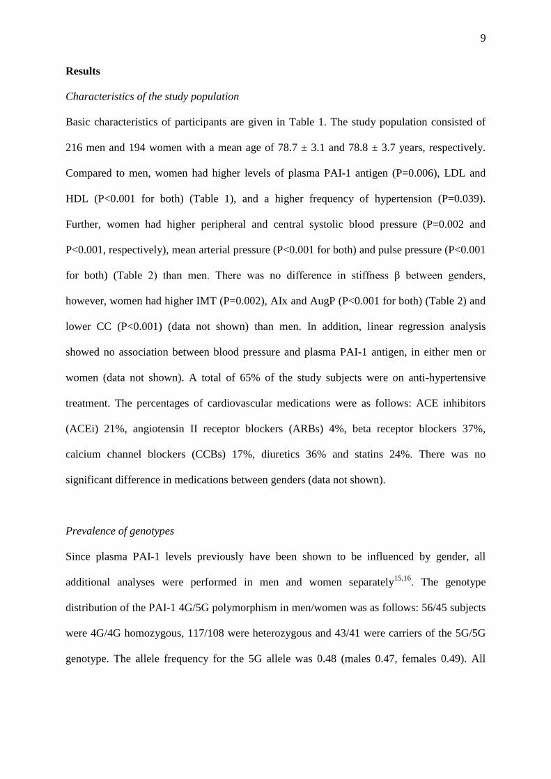
Basic characteristics of participants are given in Table 1. The study population consisted of
216 men and 194 women with a mean age of 78.7 ± 3.1 and 78.8 ± 3.7 years, respectively.
Compared to men, women had higher levels of plasma PAI-1 antigen (P=0.006), LDL and
HDL (P<0.001 for both) (Table 1), and a higher frequency of hypertension (P=0.039).
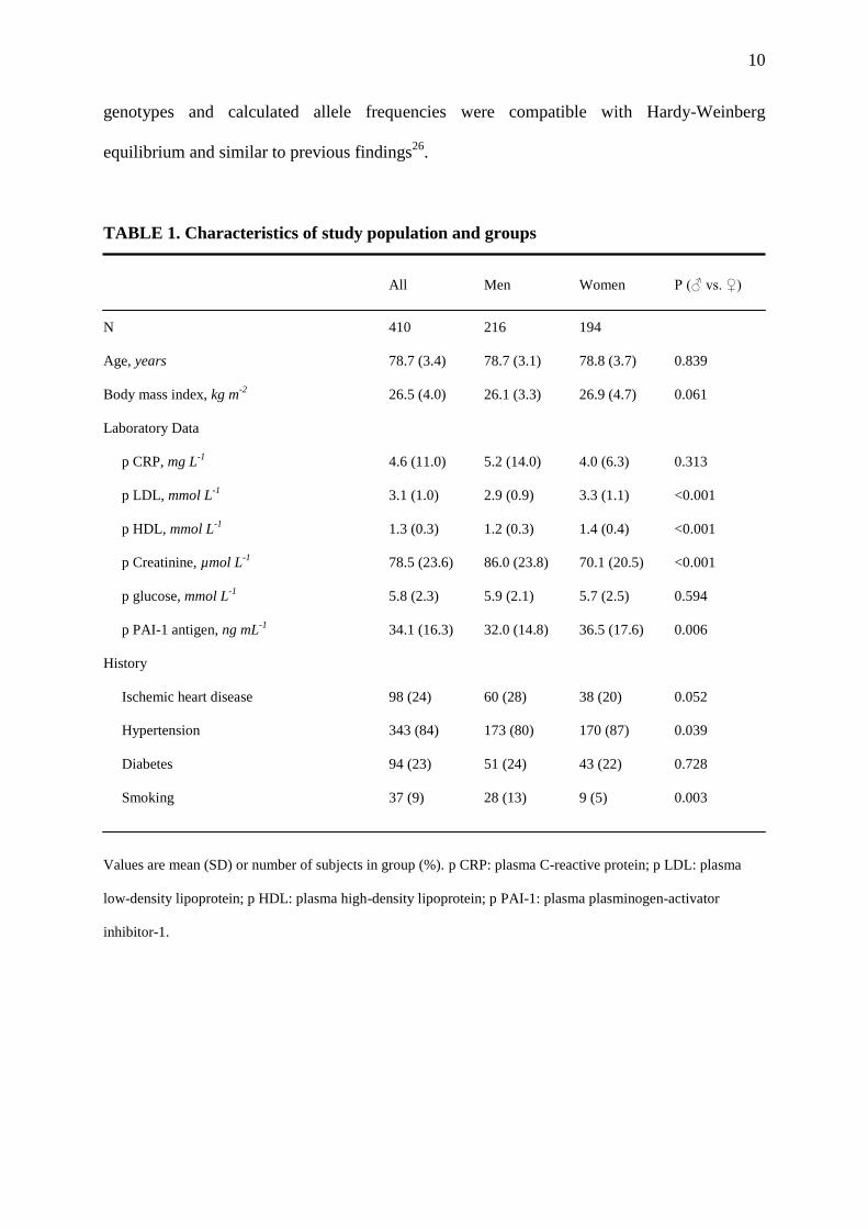
Further, women had higher peripheral and central systolic blood pressure (P=0.002 and
P<0.001, respectively), mean arterial pressure (P<0.001 for both) and pulse pressure (P<0.001
for both) (Table 2) than men. There was no difference in stiffness β between genders,
however, women had higher IMT (P=0.002), AIx and AugP (P<0.001 for both) (Table 2) and
lower CC (P<0.001) (data not shown) than men. In addition, linear regression analysis
showed no association between blood pressure and plasma PAI-1 antigen, in either men or
women (data not shown). A total of 65% of the study subjects were on anti-hypertensive
treatment. The percentages of cardiovascular medications were as follows: ACE inhibitors
(ACEi) 21%, angiotensin II receptor blockers (ARBs) 4%, beta receptor blockers 37%,
calcium channel blockers (CCBs) 17%, diuretics 36% and statins 24%. There was no
significant difference in medications between genders (data not shown).
Prevalence of genotypes
Since plasma PAI-1 levels previously have been shown to be influenced by gender, all
additional analyses were performed in men and women separately15,16
. The genotype
distribution of the PAI-1 4G/5G polymorphism in men/women was as follows: 56/45 subjects
were 4G/4G homozygous, 117/108 were heterozygous and 43/41 were carriers of the 5G/5G
genotype. The allele frequency for the 5G allele was 0.48 (males 0.47, females 0.49). All
10
genotypes and calculated allele frequencies were compatible with Hardy-Weinberg
equilibrium and similar to previous findings26
.
TABLE 1. Characteristics of study population and groups
All Men Women P (♂ vs. ♀)
N 410 216 194
Age, years 78.7 (3.4) 78.7 (3.1) 78.8 (3.7) 0.839
Body mass index, kg m-2
26.5 (4.0) 26.1 (3.3) 26.9 (4.7) 0.061
Laboratory Data
p CRP, mg L-1
4.6 (11.0) 5.2 (14.0) 4.0 (6.3) 0.313
p LDL, mmol L-1
3.1 (1.0) 2.9 (0.9) 3.3 (1.1) <0.001
p HDL, mmol L-1
1.3 (0.3) 1.2 (0.3) 1.4 (0.4) <0.001
p Creatinine, µmol L-1
78.5 (23.6) 86.0 (23.8) 70.1 (20.5) <0.001
p glucose, mmol L-1
5.8 (2.3) 5.9 (2.1) 5.7 (2.5) 0.594
p PAI-1 antigen, ng mL-1
34.1 (16.3) 32.0 (14.8) 36.5 (17.6) 0.006
History
Ischemic heart disease 98 (24) 60 (28) 38 (20) 0.052
Hypertension 343 (84) 173 (80) 170 (87) 0.039
Diabetes 94 (23) 51 (24) 43 (22) 0.728
Smoking 37 (9) 28 (13) 9 (5) 0.003
Values are mean (SD) or number of subjects in group (%). p CRP: plasma C-reactive protein; p LDL: plasma
low-density lipoprotein; p HDL: plasma high-density lipoprotein; p PAI-1: plasma plasminogen-activator
inhibitor-1.
11
TABLE 2. Pressure and stiffness characteristics
Men Women P
Peripheral BP, mmHg
Systolic 143 (20) 152 (24) <0.001
Diastolic 75 (10) 76 (12) 0.388
Mean arterial pressure 97 (12) 103 (15) <0.001
Pulse pressure 68 (17) 77 (19) <0.001
Central BP, mmHg
Systolic 132 (20) 142 (23) <0.001
Diastolic 75 (10) 77 (12) 0.216
Mean arterial pressure 97 (12) 103 (15) <0.001
Pulse pressure 56 (17) 65 (18) <0.001
Augmentation index, % 30.4 (9.5) 35.0 (8.8) <0.001
Pressure augmentation, mmHg 18.1 (9.4) 23.4 (10.4) <0.001
Wall properties abdominal aorta
Intima-media thickness, mm 0.55 (0.14) 0.60 (0.22) 0.002
Stiffness β 28.4 (20.5) 27.0 (23.3) 0.493
Values are mean (SD). BP: blood pressure.
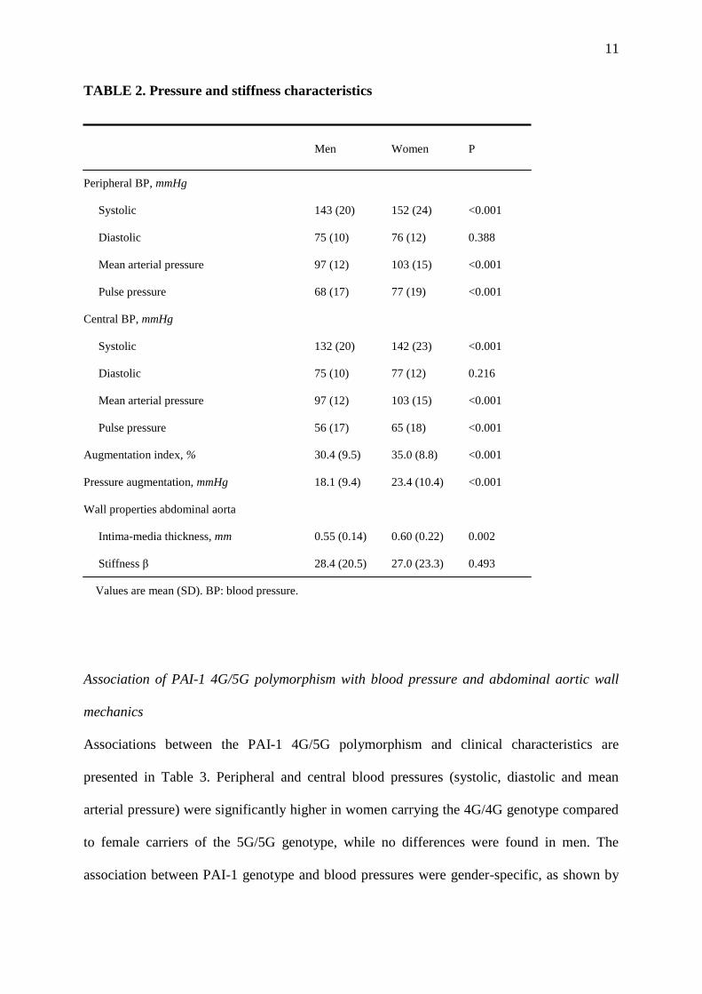
Association of PAI-1 4G/5G polymorphism with blood pressure and abdominal aortic wall
mechanics
Associations between the PAI-1 4G/5G polymorphism and clinical characteristics are
presented in Table 3. Peripheral and central blood pressures (systolic, diastolic and mean
arterial pressure) were significantly higher in women carrying the 4G/4G genotype compared
to female carriers of the 5G/5G genotype, while no differences were found in men. The
association between PAI-1 genotype and blood pressures were gender-specific, as shown by
12
TABLE 3. Characteristics according to PAI-1 4G/5G genotype
Men Women
4G/4G 4G/5G 5G/5G P PAdja 4G/4G 4G/5G 5G/5G P PAdj
a
N 56 117 43 45 108 41
Age, years 78.7 (2.9) 78.8 (3.3) 78.4 (2.9) 0.832 78.7 (2.9) 78.8 (3.9) 78.6 (3.9) 0.929
Body mass index, kg m-2
26.2 (3.6) 25.9 (3.2) 26.3 (2.8) 0.774 28.0 (5.0) 26.6 (4.6) 26.2 (4.5) 0.170
Plasma PAI-1 antigen, ng mL-1
32.2 (14.2) 33.1 (15.8) 28.9 (12.7) 0.285 0.893b 40.0 (16.2) 35.5 (17.3) 35.0 (19.5) 0.300 0.123
b
Peripheral BP, mmHg
Systolic 138 (21) 145 (20) 143 (19) 0.088 0.184 160 (22) 152 (24) 145 (25) 0.011d 0.034
Diastolic 74 (9) 75 (10) 75 (10) 0.635 0.727 80 (11) 75 (12) 72 (11) 0.003e 0.007
Mean arterial pressure 95 (12) 98 (12) 98 (12) 0.291 0.508 109 (14) 103 (15) 98 (16) 0.003f 0.010
Pulse pressure 64 (18) 70 (16) 69 (16) 0.094 0.181 80 (17) 77 (20) 73 (18) 0.233 0.293
Central BP, mmHg
Systolic 126 (21) 134 (20) 132 (19) 0.062 0.152 148 (21) 142 (23) 134 (23) 0.031g 0.048
Diastolic 74 (9) 76 (10) 75 (10) 0.663 0.751 81 (11) 76 (12) 73 (11) 0.003h, i
0.006
Mean arterial pressure 95 (12) 98 (12) 98 (12) 0.293 0.510 109 (14) 103 (15) 98 (16) 0.003j 0.010
Pulse pressure 52 (18) 58 (16) 57 (16) 0.066 0.147 66 (17) 65 (19) 62 (17) 0.449 0.348
Augmentation index, % 27.9 (8.9) 31.5 (9.6) 30.8 (9.6) 0.068 33.0 (10.1) 35.3 (8.4) 36.1 (8.3) 0.227
Pressure augmentation, mmHg 15.4 (8.9) 19.2 (9.7) 18.5 (8.5) 0.043 22.7 (10.6) 24.0 (10.9) 22.6 (8.9) 0.671
Wall properties abdominal aorta
Intima-media thickness, mm 0.53 (0.12) 0.55 (0.14) 0.55 (0.16) 0.446 0.59 (0.20) 0.60 (0.19) 0.62 (0.29) 0.816
Stiffness β 31.9 (25.6) 25.7 (18.2) 31.1 (18.1) 0.231 0.294c 35.2 (35.1) 25.1 (19.1) 22.8 (13.7) 0.090 0.129
c
CC, mm2 kPa
-1 2.03 (1.68) 1.97 (8.54) 1.81 (1.26) 0.603 1.21 (0.93) 1.49 (0.96) 1.61 (1.11) 0.121
DC, 10-3
kPa-1
9.43 (8.54) 9.04 (6.10) 7.28 (5.41) 0.298 7.39 (5.93) 9.12 (6.24) 9.29 (5.22) 0.074
Values are mean (SD) or number of subjects in group. PAI-1: Plasminogen activator inhibitor-1; BP: blood pressure; CC: compliance coefficient; DC: distensibility coefficient. a Adjusted for
age, body mass index, low-density lipoprotein, high-density lipoprotein, intima-media thickness, glomerular filtration rate, diabetes and smoking; b 4G/4G subject vs. 5G-carriers; c Adjusted for
age and mean arterial pressure.
Bonferroni Post hoc test d P-value (0.008) of subjects with 4G/4G genotype vs. subjects with 5G/5G genotype h P-value (0.042) of subjects with 4G/4G genotype vs. subjects with 4G/5G genotype e P-value (0.003) of subjects with 4G/4G genotype vs. subjects with 5G/5G genotype i P-value (0.002) of subjects with 4G/4G genotype vs. subjects with 5G/5G genotype f P-value (0.002) of subjects with 4G/4G genotype vs. subjects with 5G/5G genotype j P-value (0.002) of subjects with 4G/4G genotype vs. subjects with 5G/5G genotype
g P-value (0.025) of subjects with 4G/4G genotype vs. subjects with 5G/5G genotype
13
significant interactions between PAI-1 genotype and gender (e.g. P=0.006 for central systolic
pressure). Adjustment for potentially confounding factors related to hypertension (age, BMI,
LDL, HDL, IMT, GFR, diabetes and smoking) had no or little effect on the associations.
There was no difference in plasma PAI-1 antigen between genotypes, although women
carrying the 4G/4G genotype showed a tendency towards higher antigen levels compared to
5G carriers (4G/4G: 40.0±16.2 ng/ml; 5G carriers: 35.4±17.9 ng/ml; P=0.123). However,
multiple regression analysis controlling for a large number of potentially confounding factors
(sex, age, BMI, diabetes, smoking, LDL-cholesterol and CRP) showed a weak but significant
association between PAI-1 genotype and plasma PAI-1 antigen (P=0.048). In addition, BMI
(P<0.000), sex (P=0.004) and current smoking (P=0.002) were shown to be strong
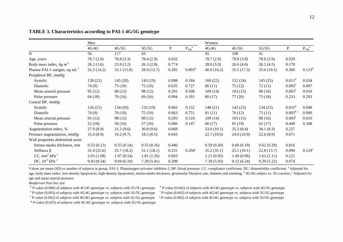
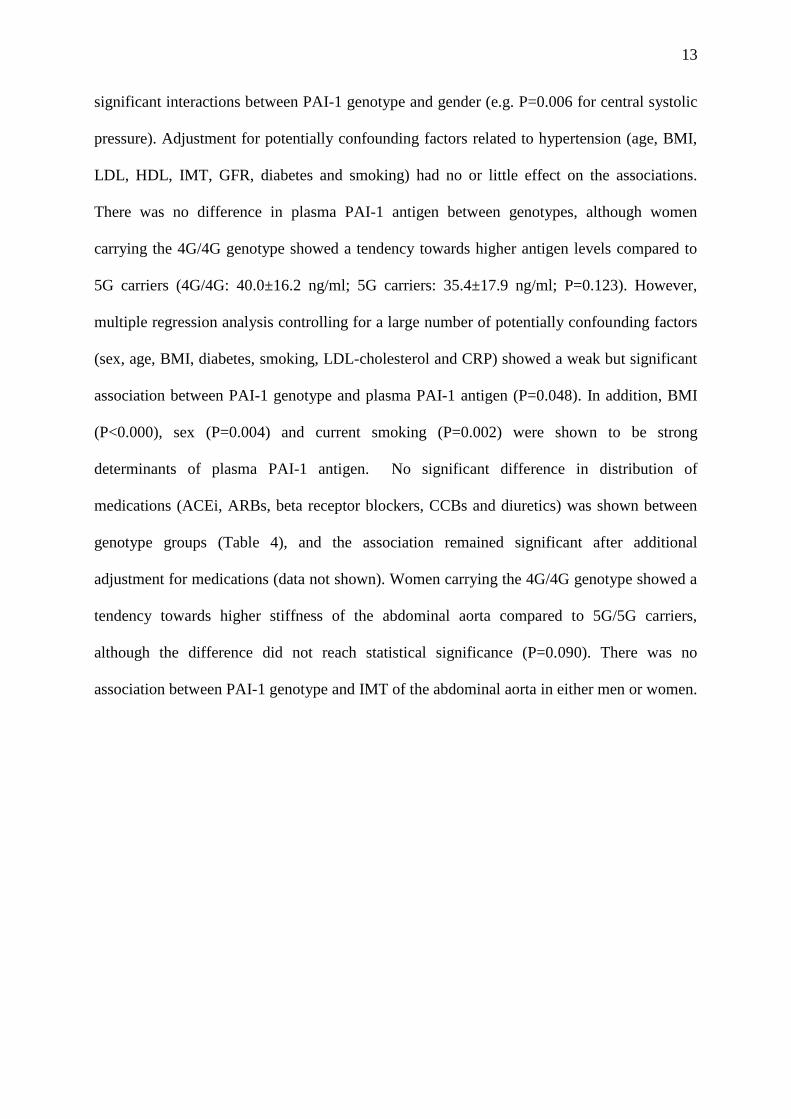
determinants of plasma PAI-1 antigen. No significant difference in distribution of
medications (ACEi, ARBs, beta receptor blockers, CCBs and diuretics) was shown between
genotype groups (Table 4), and the association remained significant after additional
adjustment for medications (data not shown). Women carrying the 4G/4G genotype showed a
tendency towards higher stiffness of the abdominal aorta compared to 5G/5G carriers,
although the difference did not reach statistical significance (P=0.090). There was no
association between PAI-1 genotype and IMT of the abdominal aorta in either men or women.
14
TABLE 4. Distribution of medications according to PAI-1 4G/5G genotype
Men Women
4G/4G 4G/5G 5G/5G P 4G/4G 4G/5G 5G/5G P
N 56 117 43 45 108 41
Medication, n (%)
ACE inhibitor 13 (23) 18 (15) 12 (28) 0.164 8 (18) 25 (23) 10 (24) 0.712
ARB 2 (4) 5 (4) 2 (5) 0.962 4 (9) 4 (4) 1 (2) 0.287
Beta receptor blocker 24 (43) 41 (35) 17 (40) 0.595 18 (40) 36 (33) 16 (39) 0.668
Ca2+
-channel blocker 8 (14) 26 (22) 7 (16) 0.405 7 (16) 14 (13) 7 (17) 0.792
Diuretic 15 (27) 40 (34) 15 (35) 0.578 21 (47) 41 (38) 15 (37) 0.545
Values are number of subjects in group (%). ARB: Angiotensin receptor blocker.
15
Discussion
The PAI-1 gene has previously been suggested as a candidate gene predisposing peripheral
hypertension. In the present study, the possible influence of the single 4G/5G guanosine
polymorphism on central arterial blood pressure was investigated. We show that the PAI-1
4G/4G genotype is associated with higher central systolic-, diastolic- and mean arterial blood
pressure in women, whereas no association was found in men. The associations remained
after adjustment for potentially confounding factors, i.e. age, BMI, LDL, HDL, IMT, GFR,
diabetes and smoking.
The present study highlights two major findings. Firstly, the PAI-1 4G/5G polymorphism is
associated not only with peripheral blood pressure, but more importantly also with central
blood pressure. Secondly, the 4G/4G genotype-phenotype association was only found in
women, suggesting a gender-specific biology of PAI-1.
The brachial artery has for many years been the standard site for measurement of blood
pressure. However, it is the central blood pressure to which the major organs affected by
hypertension are exposed. Therefore, central pressure has lately been argued as a better
measure of left ventricular afterload than peripheral pressure, and has been shown to be an
independent predictor of end organ damage12-14
. In addition, central and peripheral pressures
are differently affected by antihypertensive drugs, and large-scale trials have emphasized
central hemodynamics as a meaningful target of treatment27
. Increased central pressure is
associated with elevated aortic stiffness, also a predictor of cardiovascular events. In the
present study, a tendency towards higher stiffness of the abdominal aorta in 4G/4G women,
compared to women carrying the 5G/5G genotype was shown. The difference did not
however reach statistical significance.
16
Central pressure can be reliable assessed by a range of different noninvasive techniques. In
the present study, a generalized transfer function was used to calculate aortic pressure from
non-invasive calibrations of the radial artery pressure waveform. Although the use of this
technique may introduce errors, such as underestimations of the central pressure28,29
, it will
most likely not affect comparative studies between different groups of subjects.
The present study suggests a gender-specific biology of PAI-1 as the 4G/4G genotype-
phenotype association only was found in women. Gender has previously been associated with
plasma PAI-1 levels, men having higher levels of circulating PAI-1 than women30,31
.
Surprisingly, in the present study the association with central pressure was only seen in
women, which might seem counterintuitive. However, previous reports have shown that
gender differences in PAI-1 levels disappear to a large extent with age, increasing with age in
women but stay roughly the same in men5. In our study, with a population mean age of ~80
years, women had significantly higher plasma PAI-1 antigen levels compared to men. An
increase in plasma PAI-1 could possibly result in elevated blood pressure, however, linear
regression analysis showed no association between blood pressure and plasma PAI-1 antigen.
The synthesis of PAI-1 is transcriptionally regulated by the 4G/5G promoter polymorphism.
The 4G allele binds an activator, whereas the 5G allele binds an activator and a repressor.
This results in increased transcription and higher plasma PAI-1 levels in individuals carrying
the 4G allele8,9
. The allele-specific increase in plasma PAI-1 has been shown in several
populations and in both men and women9,32,33
. This indicates that the gender-related
genotype-phenotype association found in the present investigation may be a consequence of
later events rather than regulation on a transcriptional level. However, arguing against this is
17
the finding that the 4G/5G polymorphism previously been implicated in an allele-specific
response to plasma triglycerides34
, instead suggesting a gene-environmental interaction.
Dyslipidemia, as a component of the metabolic syndrome, is associated with increased arterial
stiffness, and this association is particularly pronounced in women35
. Further, human adipose
tissue has previously been suggested an important source of plasma PAI-1, especially under
obese conditions36
, and BMI has been strongly related to plasma PAI-1 levels in both men and
women15,16
. In the present study, women had higher LDL (P<0.001) and slightly higher BMI
(P=0.061) than men. Unfortunately, triglyceride measurements were not available, leaving the
genotype-phenotype relationship to be further elucidated.
Based on previous findings, it might be reasonable to believe that the genotype-phenotype
association, found in the present study is mediated by variations in plasma PAI-1. Although
no difference in plasma PAI-1 antigen was seen between genotypes, multiple regression
analysis controlling for a large number of potential confounders revealed a weak but
significant association between PAI-1 genotype and plasma PAI-1 antigen (P=0.048). In
addition, sex, BMI and current smoking were shown to be strong determinant of plasma PAI-
1 antigen. Hence, there seem to be an association between PAI-1 4G/5G genotype and plasma
PAI-1 antigen levels, although in an elderly population like ours other factors may interfere.
A number of medical therapies, commonly used in an elderly population have been shown to
influence plasma PAI-1 levels. Blood pressure lowering drugs such as ACEi reduce plasma
PAI-1 antigen levels, in postmenopausal women37
, in patients with hypertension38
, and in
subjects with chronic activation of the renin-angiotensin-aldosterone system39
. Additional
adjustment for ACEi, ARBs, beta receptor blockers, CCBs or diuretics had no significant
effect on the association between PAI-1 genotype and central pressures. Furthermore,
18
hormonal replacement therapy (HRT) is effective in lowering PAI-1 levels in postmenopausal
women40
. There are no data available on HRT use in the present study. However, as all
women included in this study live in a rural community, and are relatively old, the usage of
HRT is most likely negligible.
Limitations of study
Due to shortage of plasma we could not measure triglyceride levels. Such data would
potentially further disentangle the allele- and gender-dependent association found in the
present investigation. In addition, a larger sample of subjects would possibly have
strengthened our findings.
In summary, the study presents a gender-specific association of the PAI-1 4G/5G
polymorphism with peripheral, and more importantly, central arterial blood pressure. The
genotype-phenotype association remained after correction for potentially confounding factors.
These findings may provide valuable insight into the understanding of the development of
hypertension, however, further studies are required to disentangle the molecular mechanism
underlying the association.
Acknowledgements
The authors wish to thank all individuals participating in the study. The study was supported
by grants from the Swedish Research Council (12660 and 12661), the Swedish Heart-Lung
foundation (20060425), the European Commission (FAD; Health-F2-2008-200647), County
Council of Östergötland (LIO-27071, LIO-28721 and LIO-82541) and the Medical Research
Council of Southeast Sweden (FORSS-12680 and FORSS-5762).

19
Disclosure
No conflict of interest.
20
References
1. Stefansson S, Lawrence DA. The serpin PAI-1 inhibits cell migration by blocking
integrin alpha V beta 3 binding to vitronectin. Nature 1996; 383:441-443.
2. Kouri FM, Queisser MA, Konigshoff M, Chrobak I, Preissner KT, Seeger W,
Eickelberg O. Plasminogen activator inhibitor type 1 inhibits smooth muscle cell
proliferation in pulmonary arterial hypertension. Int J Biochem Cell Biol 2008;
40:1872-1882.
3. Hamsten A, de Faire U, Walldius G, Dahlen G, Szamosi A, Landou C, Blomback M,
Wiman B. Plasminogen activator inhibitor in plasma: risk factor for recurrent
myocardial infarction. Lancet 1987; 2:3-9.
4. Hamsten A, Wiman B, de Faire U, Blomback M. Increased plasma levels of a rapid
inhibitor of tissue plasminogen activator in young survivors of myocardial infarction.
N Engl J Med 1985; 313:1557-1563.
5. Sundell IB, Nilsson TK, Ranby M, Hallmans G, Hellsten G. Fibrinolytic variables are
related to age, sex, blood pressure, and body build measurements: a cross-sectional
study in Norsjo, Sweden. J Clin Epidemiol 1989; 42:719-723.
6. Poli KA, Tofler GH, Larson MG, Evans JC, Sutherland PA, Lipinska I, Mittleman
MA, Muller JE, D'Agostino RB, Wilson PW, Levy D. Association of blood pressure
with fibrinolytic potential in the Framingham offspring population. Circulation 2000;
101:264-269.
7. Jeng JR. Association of PAI-1 gene promoter 4g/5g polymorphism with plasma PAI-1
activity in Chinese patients with and without hypertension. Am J Hypertens 2003;
16:290-296.
8. Dawson SJ, Wiman B, Hamsten A, Green F, Humphries S, Henney AM. The two
allele sequences of a common polymorphism in the promoter of the plasminogen
21
activator inhibitor-1 (PAI-1) gene respond differently to interleukin-1 in HepG2 cells.
J Biol Chem 1993; 268:10739-10745.
9. Eriksson P, Kallin B, van 't Hooft FM, Bavenholm P, Hamsten A. Allele-specific
increase in basal transcription of the plasminogen-activator inhibitor 1 gene is
associated with myocardial infarction. Proc Natl Acad Sci U S A 1995; 92:1851-1855.
10. Martinez-Calatrava MJ, Gonzalez-Sanchez JL, Zabena C, Martinez-Larrad MT,
Luque-Otero M, Serrano-Rios M. Is the plasminogen activator inhibitor-1 gene a
candidate gene predisposing to hypertension? Results from a population-based study
in Spain. J Hypertens 2007; 25:773-777.
11. Boekholdt SM, Bijsterveld NR, Moons AH, Levi M, Buller HR, Peters RJ. Genetic
variation in coagulation and fibrinolytic proteins and their relation with acute
myocardial infarction: a systematic review. Circulation 2001; 104:3063-3068.
12. Roman MJ, Devereux RB, Kizer JR, Lee ET, Galloway JM, Ali T, Umans JG,
Howard BV. Central pressure more strongly relates to vascular disease and outcome
than does brachial pressure: the Strong Heart Study. Hypertension 2007; 50:197-203.
13. Safar ME, Blacher J, Pannier B, Guerin AP, Marchais SJ, Guyonvarc'h PM, London
GM. Central pulse pressure and mortality in end-stage renal disease. Hypertension
2002; 39:735-738.
14. Jankowski P, Kawecka-Jaszcz K, Bryniarski L, Czarnecka D, Brzozowska-Kiszka M,
Posnik-Urbanska A, Kopec G, Dragan J, Klecha A, Dudek D. Fractional diastolic and
systolic pressure in the ascending aorta are related to the extent of coronary artery
disease. Am J Hypertens 2004; 17:641-646.
15. Asselbergs FW, Williams SM, Hebert PR, Coffey CS, Hillege HL, Navis G, Vaughan
DE, van Gilst WH, Moore JH. Gender-specific correlations of plasminogen activator
22
inhibitor-1 and tissue plasminogen activator levels with cardiovascular disease-related
traits. J Thromb Haemost 2007; 5:313-320.
16. Schoenhard JA, Asselbergs FW, Poku KA, Stocki SA, Gordon S, Vaughan DE,
Brown NJ, Moore JH, Williams SM. Male-female differences in the genetic regulation
of t-PA and PAI-1 levels in a Ghanaian population. Hum Genet 2008; 124:479-488.
17. Alehagen U, Ericsson A, Dahlstrom U. Are there any significant differences between
females and males in the management of heart failure? Gender aspects of an elderly
population with symptoms associated with heart failure. J Card Fail 2009; 15:501-
507.
18. Bjorck HM, Lanne T, Alehagen U, Persson K, Rundkvist L, Hamsten A, Dahlstrom U,
Eriksson P. Association of genetic variation on chromosome 9p21.3 and arterial
stiffness. J Intern Med 2009; 265:373-381.
19. Cockcroft DW, Gault MH. Prediction of creatinine clearance from serum creatinine.
Nephron 1976; 16:31-41.
20. Kool MJ, van Merode T, Reneman RS, Hoeks AP, Struyker Boudier HA, Van Bortel
LM. Evaluation of reproducibility of a vessel wall movement detector system for
assessment of large artery properties. Cardiovasc Res 1994; 28:610-614.
21. Hoeks AP, Willekes C, Boutouyrie P, Brands PJ, Willigers JM, Reneman RS.
Automated detection of local artery wall thickness based on M-line signal processing.
Ultrasound Med Biol 1997; 23:1017-1023.
22. van der Heijden-Spek JJ, Staessen JA, Fagard RH, Hoeks AP, Boudier HA, van Bortel
LM. Effect of age on brachial artery wall properties differs from the aorta and is
gender dependent: a population study. Hypertension 2000; 35:637-642.
23
23. Lanne T, Stale H, Bengtsson H, Gustafsson D, Bergqvist D, Sonesson B, Lecerof H,
Dahl P. Noninvasive measurement of diameter changes in the distal abdominal aorta
in man. Ultrasound Med Biol 1992; 18:451-457.
24. Kawasaki T, Sasayama S, Yagi S, Asakawa T, Hirai T. Non-invasive assessment of
the age related changes in stiffness of major branches of the human arteries.
Cardiovasc Res 1987; 21:678-687.
25. Margaglione M, Grandone E, Vecchione G, Cappucci G, Giuliani N, Colaizzo D,
Celentano E, Panico S, Di Minno G. Plasminogen activator inhibitor-1 (PAI-1)
antigen plasma levels in subjects attending a metabolic ward: relation to
polymorphisms of PAI-1 and angiontensin converting enzyme (ACE) genes.
Arterioscler Thromb Vasc Biol 1997; 17:2082-2087.
26. Festa A, D'Agostino R, Jr., Rich SS, Jenny NS, Tracy RP, Haffner SM. Promoter
(4G/5G) plasminogen activator inhibitor-1 genotype and plasminogen activator
inhibitor-1 levels in blacks, Hispanics, and non-Hispanic whites: the Insulin
Resistance Atherosclerosis Study. Circulation 2003; 107:2422-2427.
27. Williams B, Lacy PS, Thom SM, Cruickshank K, Stanton A, Collier D, Hughes AD,
Thurston H, O'Rourke M. Differential impact of blood pressure-lowering drugs on
central aortic pressure and clinical outcomes: principal results of the Conduit Artery
Function Evaluation (CAFE) study. Circulation 2006; 113:1213-1225.
28. Hope SA, Tay DB, Meredith IT, Cameron JD. Use of arterial transfer functions for the
derivation of aortic waveform characteristics. J Hypertens 2003; 21:1299-1305.
29. Davies JI, Band MM, Pringle S, Ogston S, Struthers AD. Peripheral blood pressure
measurement is as good as applanation tonometry at predicting ascending aortic blood
pressure. J Hypertens 2003; 21:571-576.
24
30. Krishnamurti C, Tang DB, Barr CF, Alving BM. Plasminogen activator and
plasminogen activator inhibitor activities in a reference population. Am J Clin Pathol
1988; 89:747-752.
31. van Harmelen V, Wahrenberg H, Eriksson P, Arner P. Role of gender and genetic
variance in plasminogen activator inhibitor-1 secretion from human adipose tissue.
Thromb Haemost 2000; 83:304-308.
32. Asselbergs FW, Williams SM, Hebert PR, Coffey CS, Hillege HL, Navis G, Vaughan
DE, van Gilst WH, Moore JH. The gender-specific role of polymorphisms from the
fibrinolytic, renin-angiotensin, and bradykinin systems in determining plasma t-PA
and PAI-1 levels. Thromb Haemost 2006; 96:471-477.
33. Sartori MT, Vettor R, De Pergola G, De Mitrio V, Saggiorato G, Della Mea P, Patrassi
GM, Lombardi AM, Fabris R, Girolami A. Role of the 4G/5G polymorphism of PaI-1
gene promoter on PaI-1 levels in obese patients: influence of fat distribution and
insulin-resistance. Thromb Haemost 2001; 86:1161-1169.
34. Panahloo A, Mohamed-Ali V, Lane A, Green F, Humphries SE, Yudkin JS.
Determinants of plasminogen activator inhibitor 1 activity in treated NIDDM and its
relation to a polymorphism in the plasminogen activator inhibitor 1 gene. Diabetes
1995; 44:37-42.
35. Lin HF, Liu CK, Liao YC, Lin RT, Chen CS, Juo SH. The risk of the metabolic
syndrome on carotid thickness and stiffness: sex and age specific effects.
Atherosclerosis 210:155-159.
36. Eriksson P, Reynisdottir S, Lonnqvist F, Stemme V, Hamsten A, Arner P. Adipose
tissue secretion of plasminogen activator inhibitor-1 in non-obese and obese
individuals. Diabetologia 1998; 41:65-71.
25
37. Fogari R, Zoppi A, Preti P, Fogari E, Malamani G, Mugellini A. Differential effects of
ACE-inhibition and angiotensin II antagonism on fibrinolysis and insulin sensitivity in
hypertensive postmenopausal women. Am J Hypertens 2001; 14:921-926.
38. Sakata K, Shirotani M, Yoshida H, Urano T, Takada Y, Takada A. Differential effects
of enalapril and nitrendipine on the fibrinolytic system in essential hypertension. Am
Heart J 1999; 137:1094-1099.
39. Brown NJ, Agirbasli MA, Williams GH, Litchfield WR, Vaughan DE. Effect of
activation and inhibition of the renin-angiotensin system on plasma PAI-1.
Hypertension 1998; 32:965-971.
40. Koh KK, Mincemoyer R, Bui MN, Csako G, Pucino F, Guetta V, Waclawiw M,
Cannon RO, 3rd. Effects of hormone-replacement therapy on fibrinolysis in
postmenopausal women. N Engl J Med 1997; 336:683-690.