Embed Size (px)
Citation preview

Gene Expression Profiling in the Brains of Human CocaineAbusers
MICHAEL J. BANNON1,2,3, GREGORY KAPATOS1,3, and DAWN N. ALBERTSON1
1 Department of Psychiatry & Behavioural Neurosciences, Wayne State University School of Medicine
2 Department of Pharmacology, Wayne State University School of Medicine
3 Center for Molecular Medicine & Genetics, Wayne State University School of Medicine
AbstractChronic cocaine abuse induces long-term neurochemical, structural and behavioural changes thoughtto result from altered gene expression within the nucleus accumbens and other brain regions playinga critical role in addiction. Recent methodological advances now allow the profiling of geneexpression in human postmortem brain. In this article, we review studies in which we have usedAffymetrix oligonucleotide microarrays to identify transcripts that are differentially expressed in thenucleus accumbens of cocaine abusers in comparison to well-matched control subjects. Of theapproximately 39 000 gene transcripts interrogated, the expression of only a fraction of 1% issignificantly modified in cocaine abusers. Found within this list are equivalent incidences ofincreased and decreased transcript abundance, including known gene transcripts clustered intoseveral functional categories. A striking exception is a group of myelin-related genes, consisting ofmultiple transcripts representing myelin basic protein (MBP), proteolipid protein (PLP) and myelin-associated oligodendrocyte basic protein (MOBP), which as a group are substantially decreased incocaine abusers compared to controls. These data, suggesting a possible dysregulation of myelin incocaine abusers, are discussed in the context of myelin-related changes in other human braindisorders. Finally, the effects of cocaine abuse on the profile of gene expression in some other brainregions critical for addiction (the prefrontal cortex and ventral midbrain) are briefly reviewed.
IntroductionDrug addiction, which poses a serious threat to public health in terms of lost productivity andlives (Office of Applied Statistics 2000; National Institute on Drug Abuse 2003), is amultifaceted disorder involving tolerance, dependence, craving and relapse (Nestler, 2002). Abetter understanding of the molecular mechanisms underlying drug addiction can be expectedto facilitate the development of more successful drug treatment strategies. Although themolecular basis of drug abuse is not fully understood, more is known about the neural systemssubserving this disorder. In particular, animal studies have identified the nucleus accumbensas a brain region that plays a critical role in addiction (Dackis and O’Brien, 2001; Everitt andWolf, 2002). Furthermore, in animal models, chronic exposure to cocaine induces structuraland functional changes in the nucleus accumbens that are presumably mediated by altered geneexpression (Norrholm et al., 2003; Toda et al., 2002). At the same time, it is difficult to modelin laboratory animals the uniquely human aspects of cocaine abuse, namely the spontaneousself-administration of cocaine, most often in a binging pattern of abuse, over a period of yearsor decades.
Correspondence to: Michael J. Bannon, Department of Psychiatry and Behavioural Sciences, Wayne State University School of Medicine,2309 Scott Hall, 540 E. Canfield Avenue, Detroit, MI 48201, USA. E-mail: [email protected].
NIH Public AccessAuthor ManuscriptAddict Biol. Author manuscript; available in PMC 2008 January 28.
Published in final edited form as:Addict Biol. 2005 March ; 10(1): 119–126.
NIH
-PA Author Manuscript
NIH
-PA Author Manuscript
NIH
-PA Author Manuscript

With the sequencing of the human genome and the advent of microarray technologies, it seemsincumbent upon neuroscientists to characterize gene expression patterns within the humanbrain that underlie complex, presumably polygenic disorders such as drug abuse. Although itis possible in some instances (e.g. neurosurgery patients) to obtain brain tissue biopsies, mosthuman brain tissue becomes available only at autopsy. Fortunately, numerous studies over thelast 20 years have demonstrated that mRNA is remarkably stable post mortem, and that changesin the expression of individual transcripts can be assessed readily using autopsy material(Bannon et al., 1992a, 1992b, 2002; Wilson et al., 1996; Segal et al., 1997; Albertson et al.,2004). Analysis of post-mortem brain provides a unique opportunity to examine changes ingene expression in the human drug abuser. We have used microarray technology to investigatechanges in gene expression in the nucleus accumbens of chronic cocaine abusers relative tomatched control subjects. Of the relatively small number of differentially expressed genesdetected, the most robust finding was a decreased expression of several myelin-related genes(Albertson et al., 2004). We review the findings of these studies as well as the other publishedreports of gene expression profiling in the brains of human cocaine abusers.
Methodologies employedTissue acquisition and subject characterization
Brain specimens were collected as part of the routine autopsy process, as described previously(Albertson et al., 2004). Cause and manner of death were determined after medicolegalexamination by the Medical Examiner. Study I consisted of five subjects whose deaths wereruled chronic cocaine abuse (based on toxicology, history of drug use and cardiovascularfindings (Karch, 2002)) and five drug-free control subjects matched pairwise with cocaineabusers for age, gender and race. Study II consisted of five subjects who exhibited a positivetoxicology for cocaine and/or its metabolites (but died of gunshot wound-related trauma) andfive controls matched for demographics and cause of death. Post hoc analysis revealed nosignificant differences between studies of cocaine abusers and control subjects on anydemographic, with the exception that Study II subjects were younger than subjects in Study I(Albertson et al., 2004).
Sample preparation and microarray hybridizationCoronal sections (2 – 3 cm thickness) were taken throughout the rostrocaudal extent of thebasal ganglia at the time of autopsy. As illustrated in Fig. 1, the nucleus accumbens, definedas the ventral extension of the caudate immediately below the anterior limb of the internalcapsule, was dissected free while excluding adjacent external capsule as described (Albertsonet al., 2004). Isolation of RNA, elimination of contaminating genomic DNA, and assessmentof RNA abundance and quality have been described in detail elsewhere (Albertson et al.,2004).
Affymetrix oligonucleotide arrays (Santa Clara, CA, USA) were used in all studies. Before usein full-scale experiments, the quality of all RNA samples was verified by test arrayhybridization (test 2 or test 3). The 3′/5′ ratios of glyceraldehyde-3-phosphate dehydrogenase(GAPDH, a housekeeping gene) were generated from these test chips as a further measure ofsample quality and efficiency of the reverse transcription-polymerase chain reaction (RT-PCR)and in vitro transcription. According to Affymetrix quality control parameters, this ratio shouldnot be more than 3.0. For subsequent full-scale analysis, Study I samples were hybridizedseparately to five oligonucleotide arrays representing distinct probe sets: human u95Av2,u95B, u95C, u133A and u133B, for a total of 50 microarrays. Study II samples were analysedon both the u133A and u133B arrays. The sample labelling, hybridization and scanningfollowed the Affymetrix GeneChip® Expression Analysis Technical Manual(www.affymetrix.com).
BANNON et al. Page 2
Addict Biol. Author manuscript; available in PMC 2008 January 28.
NIH
-PA Author Manuscript
NIH
-PA Author Manuscript
NIH
-PA Author Manuscript

Microarray data analysisMicroarray data were analysed with the Affymetrix Microarray Suite 5.0 software package.Images were scaled for signal intensity to account for any differences in hybridizationefficiencies. Data were analysed in pairs, comparing each cocaine sample with its matchedcontrol. Significant differences between subject pairs were calculated using the Wilcoxonsigned rank test (p ≤ 0.05); marginal calls were considered non-significant. Although initiallyanalysed separately, data from Study I and Study II were pooled for increased statistical power.Only transcripts that were increased or decreased in the majority ( 6 of 10) of subject pairs,representing 0.2% of the total transcripts, were considered differentially expressed. Transcriptsmeeting this criterion were examined post hoc for the statistical significance of differencesbetween groups using Mann – Whitney U-tests (p ≤ 0.05). Functional groups were createdusing probe information provided by Affymetrix.
In order to look for similar patterns of gene expression across subjects, hierarchical clusteringwas performed with Spotfire Decision Site software (Somerville, MA, USA). Microarraysignal data were converted to Z scores for comparison across studies. The clustering was carriedout using unweighted averages, giving equal importance to each gene, and the Euclidiandistance default with subsequent logistic regression analysis with SPSS version 10.0.5(Chicago, IL, USA).
Methods for validation of microarray findingsRNA from all 20 subjects was used for verification of the microarray data. Equivalent amountsof RNA from each subject were pooled to create standard curves assayed in parallel withreplicate samples consisting of 5 ng RNA from individual subjects. RT was performed(Sensiscript RT Kit, Qiagen, Valencia, CA, USA) using random hexamers, followed by PCRusing the Qiagen SYBR Green PCR Kit and specific primers for myelin basic protein (MBP),proteolipid protein (PLP), myelin-associated oligodendrocyte basic protein (MOBP), cocaine-and amphetamine-regulated transcript (CART) or β-actin. LightCycler3 Data Analysissoftware (Roche, Palo Alto, CA, USA) used standard curve data to create a regression modelfrom which transcript concentrations were calculated. For sample normalization, MBP, PLP,MOBP or CART values were divided by the subject’s β-actin values. Details of this assay arepublished (Albertson et al., 2004). In addition, representative subject pairs were assessed forthe level of MBP-immunoreactivity as described (Albertson et al., 2004).
Results of nucleus accumbens gene profiling studiesQuality control experiments
The quality of the post-mortem samples used in these experiments was assessed initially bybrain pH (a reliable indicator of RNA quality and stability; Kingsbury et al., 1995) followedby RNA analysis using spectrophotometric and electropherographic techniques (Albertson etal., 2004). Sample quality was further demonstrated by hybridization to Affymetrixoligonucleotide test arrays. The 3′/5′ GAPDH ratios of all samples ( <2.0) documented thequality of input RNA as well as efficiency of the RT-PCR and in vitro transcription reactionspreceding hybridization to microarrays. Thus all samples passed multiple quality measuresbefore inclusion in the subsequent analyses. No significant differences were found amongstudies or groups in terms of brain pH, 3′/5′ GAPDH ratios, tissue weight, RNA yield or 260/280ratios (Albertson et al., 2004).
In order to determine test – retest reliability for the overall microarray procedure an experimentwas performed by splitting human nucleus accumbens RNA (not otherwise used in subsequentexperiments) into two aliquots that were then treated as unique samples. These samples werelabelled several months apart with separate RT, PCR and in vitro transcription reactions and
BANNON et al. Page 3
Addict Biol. Author manuscript; available in PMC 2008 January 28.
NIH
-PA Author Manuscript
NIH
-PA Author Manuscript
NIH
-PA Author Manuscript

hybridized onto two individual u95Av2 arrays. The reproducibility of the entire procedure isshown in Fig. 2 and demonstrates a significant correlation in the abundance of presenttranscripts (n = 5558) between the two samples as determined by Pearson’s product moment(R2 = 0.98, p = 0.002). None the less, because of the enormous number of genes interrogated,even a small residual variance means that scores of genes may be detected as differentiallyexpressed when they are not, thus necessitating multiple microarray experiments and validationof targets using QRT-PCR.
Comparison of gene expression in cocaine abusers and matched controlsTranscripts detected in the nucleus accumbens of all subject pairs and differentially expressedin the majority of cocaine abusers relative to their matched controls represent onlyapproximately 0.2% of the ~ 45 000 oligonucleotide probes on the u133 series Affymetrixmicroarrays (a total of 110 genes; see Albertson et al., 2004 for details). One-half of these wereunannotated; the remainder could be clustered into broad functional categories (Fig. 3). Thelargest groups of annotated genes were related to signal transduction, translation/transcription/RNA processing, neurotransmission/synaptic function/membrane recycling, glia andstructural/cell adhesion. Post-hoc examination (by Mann – Whitney U test) confirmed thestatistical significance of the differential expression for the majority of transcripts (Albertsonet al., 2004). Most functional categories encompassed both increases and decreases in cocaine-related expression patterns, such that there was little overall change seen by functional group.This could be due to a number of factors, not least of which are the broad categories employedand current limitations in gene annotation and functional clustering tools.
It was interesting to note that cocaine- and amphetamine-regulated transcript (CART) mRNAlevels were increased in the nucleus accumbens of human cocaine abusers, a finding confirmedby hybridization to a second distinct (u95) array as well as by RT-PCR (Albertson et al.,2004). CART represents a gene whose expression is induced by cocaine in rodent forebrain(Douglass et al., 1995), supporting the notion that at least some of the gene expression changesseen in the cocaine-exposed human brain are consonant with previous animal model data.
Although cocaine abuse was associated with equivalent incidences of increased and decreasedgene expression overall (seen as positive and negative signal log ratios, respectively; Fig. 4),a striking exception was the group of myelin-related genes, consisting of multiple transcriptsrepresenting myelin basic protein (MBP), proteolipid protein (PLP) and myelin associatedoligodendrocyte basic protein (MOBP). As a group, these transcripts showed a substantialdecrease in cocaine abusers compared to controls (Fig. 4). Changes in myelin-related transcriptabundance found using the u133 microarrays were confirmed in separate analyses using u95series microarrays and RNA from Study I subjects (Albertson et al., 2004).
We performed hierarchical clustering (Fig. 5) to illustrate the relatedness and relativeexpression levels of transcripts differentially expressed between cocaine abusers and controlsubjects. Microarray signal data were converted into Z scores for this analysis, with eachcolumn representing transcript expression from individual subjects. When looking at overallpatterns of expression (Fig. 5, left side), some clusters of genes with both increased expression(red) and decreased expression (green) are evident in the cocaine subjects. The tight branchingof MBP and MOBP (Fig. 5, right side) is indicative of a decline in cocaine subjects and suggestsa similarity in their transcriptional regulation. Two genes not associated previously with MBPand MOBP, namely dishevelled associated activator of morphogenesis 2 (DAAM2) andpeanut-like 2 (PNUTL2) were also found in this cluster. We performed a logistic regression,creating a mathematical model for the prediction of group affiliation (cocaine vs. control). Thisanalysis revealed that the relative abundance of MBP and MOBP was sufficient to classifywith 90% accuracy our subjects as either cocaine abusers or controls (χ2(2) = 14.14, p = 0.001).When data from all four genes in this cluster (MBP, MOBP, DAAM2 and PNUTL2) were
BANNON et al. Page 4
Addict Biol. Author manuscript; available in PMC 2008 January 28.
NIH
-PA Author Manuscript
NIH
-PA Author Manuscript
NIH
-PA Author Manuscript

used, the classification of cocaine abusers and controls was 100% accurate (χ2(4) = 27.73, p =0.0001) regardless of cause of death. No such predictive value was seen with a randomly chosenset of transcripts (Albertson et al., 2004). The ability of this limited cluster of genes to identifysamples derived from cocaine abusers supports the microarray data and suggests that changesin myelin-related gene expression are a fundamental characteristic of cocaine abusers.
Confirmation of changes in myelin-related transcripts was obtained using quantitative RT-PCR. Cocaine abusers who exhibited decreases in MBP, MOBP and PLP relative to theircontrols on the microarrays were also found to have decreased abundances of these mRNAsby QRT-PCR (Albertson et al., 2004). Pearson’s correlation revealed a significant correlationbetween the subject pairs’ QRT-PCR and microarray data (Fig. 6; MBP for Study II subjectsshown; R2 = 0.98). In addition, tissue sections from representative subject pairs processed forimmunohistochemistry revealed a significant decrease in the number of MBP-immunoreactiveoligodendrocytes within the nucleus accumbens (Albertson et al., 2004).
DiscussionGene expression profiles in the nucleus accumbens of human cocaine abusers:dysregulation of myelin-related genes
Human cocaine abusers typically self-administer cocaine in a binging pattern over a period ofyears or decades. To the extent that animal models fail to duplicate these and other aspects ofhuman cocaine abuse, they may fall short in revealing relevant changes in gene expression.This has led us to investigate changes in gene expression in human postmortem brain usingmicroarray. This unbiased approach allowed us to explore fully differences in the nucleusaccumbens of human cocaine abusers unconstrained by a priori hypotheses. Previous studieshave demonstrated the stability of human brain RNA post mortem (Perrett et al., 1988;Kobayashi et al., 1990; Bannon et al., 1992a,b; Kingsbury et al., 1995) and its suitability foruse with microarray (Mirnics et al., 2000; Hakak et al., 2001; Hemby et al., 2002). In theexperiments described here, subject pairs were matched carefully and sample quality wasconfirmed by multiple measures (brain pH, RNA characterization and test array hybridization).With these samples, we found that a very small percentage of all the transcripts expressed inthe nucleus accumbens was affected by chronic cocaine abuse. A major finding of these studiesis the decreased expression of myelin-related genes in the nucleus accumbens of human cocaineabusers, accompanied by an apparent loss of MBP-positive oligodendrocytes. These myelin-related findings were the most robust and consistent findings from our study, cross-validatedwith different microarray types, multiple experimental techniques and two independent cohortsof cocaine-abusing individuals with different causes of death.
Our observations find support in several studies suggesting that drug administration in animalsor humans may change myelin expression (Volkow et al., 1988a; Filley et al., 1990; Wigginsand Ruiz, 1990; Korbo, 1999; Kittler et al., 2000; Mayfield et al., 2002; Saito et al., 2002). Inneuroimaging studies, white matter hyperintensities and chemical changes indicative of whitematter pathology are often seen in cocaine abusers (Chang et al., 1997, 1999; Bartzokis et al.,1999a,b). More recently, two studies have demonstrated a loss of white matter volume(Bartzokis et al., 2002) and microstructure integrity in chronic human cocaine abusers (Lim etal., 2002). Paralleling our findings, Lehrmann et al. (2003) have reported decreased PLP mRNAin the prefrontal cortex of human cocaine abusers (discussed below).
One possible explanation for these documented effects on white matter might be thevasoconstrictive effects of cocaine (Kaufman et al., 1998). The normal adult human braincontains oligodendrocyte progenitors with the capacity for extensive continued myelinationthrough the fourth decade of life (Chang et al., 2000; Bartzokis et al., 2001). Cerebralvasoconstriction has been linked to hypoperfusion, which in turn has been shown to decrease
BANNON et al. Page 5
Addict Biol. Author manuscript; available in PMC 2008 January 28.
NIH
-PA Author Manuscript
NIH
-PA Author Manuscript
NIH
-PA Author Manuscript

MBP over time (Kurumatani et al., 1998). It is possible that the vascular effects of cocainecould interfere with the continued myelination in adult brain, accounting for our findings ofdecreased myelin transcripts and decreased MBP-immunoreactive oligodendrocytes(Albertson et al., 2004). Alternatively, the effects of cocaine on myelin-related gene expressionmay be directly related to the increases in extracellular dopamine (DA) that cocaine elicits inthe nucleus accumbens (Pettit and Justice, 1989; Hemby et al., 1997). Oligodendrocytesexpress D2 and D3 DA receptors (Bogarzone et al., 1998; Howard et al., 1998), and it has beenreported that DA receptor stimulation decreases the conversion from immature to matureoligodendrocytes (Bogarzone et al., 1998). It is plausible that in the DA-rich nucleusaccumbens, cocaine diminishes generation of mature myelin-producing oligodendrocytesthrough the overstimulation of oligodendrocyte DA receptors. The extent to which these effectsare localized solely to the nucleus accumbens is unknown, as we also observed an apparentdecrease in MBP-immunoreactive oligodendrocytes in the white matter immediatelysurrounding the nucleus accumbens of cocaine abusers (data not shown). Further studies arerequired to distinguish between these possibilities. Although the literature connecting myelinand cocaine is relatively modest, perhaps reflective of the unexpectedness of the association,a link between altered myelination and the cognitive and motoric deficits associated withcocaine abuse (Bauer, 1996; Strickland et al., 1998; Robinson et al., 1999; Fillmore and Rush,2002) has face validity. It has been reported that the majority of long-term cocaine users havefocal perfusion defects, a subtle form of cerebrovascular dysfunction, which have beenassociated with moderate to severe cognitive impairment (Volkow et al., 1988b; Holman etal., 1991; Strickland et al., 1993). Both the cognitive and focal vascular defects reportedlypersist in periods of abstinence.
‘Another possibility that cannot be discounted at present is that the decreased expression ofmyelin-associated genes is not secondary to cocaine’s neurochemical effects, but representsinstead a predisposing phenotype associated with cocaine abuse. In this regard, it may be worthnoting that chronic cocaine abusers such as the subjects in our study (half of whom died directlyas a result of drug abuse), represent a very small proportion of individuals in the generalpopulation who have self-administered cocaine at some time. The dysregulation of myelin ina number of other central nervous system (CNS) disorders is discussed below.
Gene expression profiles in other brain regions from human cocaine abusersAnother recent gene expression profiling study has focused on changes occurring within theventral midbrain, the site of origin for DA neurones that project to the nucleus accumbens andother forebrain areas (Tang et al., 2003). In this study, the midbrain ventral tegmental area andlateral substantia nigra from 10 cocaine abusers and 11 matched controls were analysed usingcustom neural macroarray blots consisting of 81 cDNAs corresponding to DA, glutamate andGABA receptors, regulators of G protein signalling and a few other transcripts. Thepredominant effect observed in this small subset of genes was an up-regulation of severalglutamate receptor transcripts in the ventral tegmental area, a finding broadly supported byimmunoblot data. It is also interesting to note that the neurotransmitter CART is down-regulated in the ventral tegmental area of cocaine abusers (Tang et al., 2003) although up-regulated in the nucleus accumbens (Albertson et al., 2004).
In addition to the nucleus accumbens, the prefrontal cortex is thought to play an important rolein mediating responses to cocaine in animals and man (Lehrmann et al., 2003). The profile ofgene expression in the dorsolateral prefrontal cortex has been examined using samples fromseven cocaine abusers and matched controls with a pairwise design and an 1100 cloneneuroarray (Lehrmann et al., 2003). Sixty-five differentially expressed transcripts wereidentified, representing changes in broad functional categories including energy metabolism,cytoskeleton, cell signalling, transcription and oligodendrocyte function. The authors noted
BANNON et al. Page 6
Addict Biol. Author manuscript; available in PMC 2008 January 28.
NIH
-PA Author Manuscript
NIH
-PA Author Manuscript
NIH
-PA Author Manuscript

that each subject pair fell into one of two distinct patterns of gene expression, with thesubgroups often showing opposite directions of change for any given gene. The presence ofanhydroecgonine (a cocaine metabolite generated by smoking crack cocaine) in most of thecocaine abusers from one of the two subgroups led to the suggestion that the distinct patternsof gene expression might reflect an acute response to crack cocaine exposure in one group vs.a post-drug refractory state in the other cohort. This intriguing hypothesis warrants furtherinvestigation.
In this latter study, a significant decrease in PLP expression was seen in the prefrontal cortexof a subset of cocaine abusers (Lehrmann et al., 2003), paralleling the striking changes we haveseen in PLP and other myelin-related genes in the nucleus accumbens (Albertson et al.,2004). Surprisingly, decreased expression of PLP and numerous other myelin-related geneshave also been observed in postmortem brains from alcoholics (Lewohl et al., 2000; Mayfieldet al., 2002), schizophrenics (Hakak et al., 2001; Pongrac et al., 2002; Tkachev et al., 2003)and bipolar disorder patients (Tkachev et al., 2003). The common factors among these disparatediseases that could underlie similar changes in myelin-related gene expression are, at present,obscure. The fact that several other brain diseases do not exhibit changes in myelin-relatedgene expression (Tkachev et al., 2003) suggests some specificity to this finding. Clearly, thisintriguing commonality of decreased myelin-related gene expression warrants furtherinvestigation.
The difficulties in comparing data across CNS gene profiling studies are manifold. One obviousdifficulty with any study of the CNS is that each brain region encompasses a differentcomplement of neural types with distinct interneuronal connections and responsivity to stimuli.Another potential confound of human addiction studies is the cohorts studied, their history ofdrug exposure and comorbidity for other brain disorders. Of course, the nature of the geneexpression platform employed (macroarray vs. microarray, custom designed vs. commercial)is critical, as expression changes cannot be detected for genes that are not assessed. Lessobvious to the scientist not engaged in these types of studies is the importance of experimentaldesign (e.g. pairwise vs. group analysis) and statistical analyses on the outcome of these studies.Hopefully, many of these limitations will be overcome successfully through sample and datasharing arrangements and further standardization of experimental approaches. Even with theselimitations, gene expression profiling of human postmortem brain holds great promise forproviding new and unanticipated insights into the pathogenesis of addictive disorders,ultimately suggesting novel therapeutic strategies.
Acknowledgements
The authors would like to extend their gratitude to Drs John Kamholz, Robert Skoff, Anthony Campagnoni, RobinFisher and Sharon K. Michelhaugh for helpful discussions and Donald M. Kuhn, Carl J. Schmidt and Barb Pruetz fortheir participation in the studies reviewed above. We would also like to thank Dr Susan Land, Daniel Lott, TaraTwomey, Bin Yao and Dan Liu at Wayne State University’s Applied Genomics Technology Center.
ReferencesAlbertson DN, Pruetz B, Schmidt CJ, Kuhn DM, Kapatos G, Bannon MJ. Gene expression profile of the
nucleus accumbens of human cocaine abusers: evidence for dysregulation of myelin. J Neurochem2004;88:1211–1219. [PubMed: 15009677]
Bannon MJ, Poosch MS, Haverstick DM, Mandall A, Xue IC-H, Shibata K, Dragovic LJ.Preprotachykinin gene expression in the human basal ganglia: characterization of mRNAs and pre-mRNAs produced by alternate RNA splicing. Brain Res Mol Brain Res 1992a;12:225–231. [PubMed:1312203]
Bannon MJ, Poosch MS, Xia Y, Goebel DJ, Cassin B, Kapatos G. Dopamine transporter mRNA contentin human substantia nigra decreases precipitously with age. Proc Natl Acad Sci USA 1992b;89:7095–7099. [PubMed: 1353885]
BANNON et al. Page 7
Addict Biol. Author manuscript; available in PMC 2008 January 28.
NIH
-PA Author Manuscript
NIH
-PA Author Manuscript
NIH
-PA Author Manuscript

Bannon MJ, Pruetz B, Manning-Bog AB, Whitty CJ, Michelhaugh SK, Sacchetti P, Granneman JG, MashDC, Schmidt CJ. Decreased expression of the transcription factor NURR1 in dopamine neurons ofcocaine abusers. Proc Natl Acad Sci USA 2002;99:6382–6385. [PubMed: 11959923]
Bartzokis G, Beckson M, Hance DB, Lu PH, Foster JA, Mintz J, Ling W, Bridge P. Magnetic resonanceimaging evidence of ‘silent’ cerebrovascular toxicity in cocaine dependence. Biol Psychiatry 1999a;45:1203–1211. [PubMed: 10331113]
Bartzokis G, Goldstein IB, Hance DB, Beckson M, Shapiro D, Lu PH, Edwards N, Mintz J, Bridge P.The incidence of T2-weighted MR imaging signal abnormalities in the brain of cocaine-dependentpatients is age-related and region-specific. Am J Neuroradiol 1999b;20:1628–1635. [PubMed:10543632]
Bartzokis G, Beckson M, Lu PH, Nuechterlein KH, Edwards N, Mintz J. Age-related changes in frontaland temporal lobe volumes in men: a magnetic resonance imaging study. Arch Gen Psychiatry2001;58:461–465. [PubMed: 11343525]
Bartzokis G, Beckson M, Lu PH, Edwards N, Bridge P, Mintz J. Brain maturation may be arrested inchronic cocaine addicts. Biol Psychiatry 2002;51:605–611. [PubMed: 11955460]
Bauer LO. Resting hand tremor in abstinent cocaine-dependent, alcohol-dependent, and polydrug-dependent patients. Alcohol Clin Exp Res 1996;20:1196–1201. [PubMed: 8904970]
Bogarzone ER, Howard SG, Schonmann V, Campagnoni AT. Identification of the dopamine D3 receptorin oligodendrocyte precursors: potential role in regulating differentiation and myelin formation. JNeurosci 1998;18:5344–5353. [PubMed: 9651217]
Chang L, Mehringer CM, Ernst T, Melchor R, Myers H, Forney D, Satz P. Neurochemical alterations inasymptomatic abstinent cocaine users: a proton magnetic resonance spectroscopy study. BiolPsychiatry 1997;42:1105–1114. [PubMed: 9426880]
Chang L, Ernst T, Strickland T, Mehringer CM. Gender effects on persistent cerebral metabolite changesin the frontal lobes of abstinent cocaine users. Am J Psychiatry 1999;156:716–722. [PubMed:10327904]
Chang A, Nishiyama A, Peterson J, Prineas J, Trapp BD. NG2-positive oligodendrocyte progenitor cellsin adult human brain and multiple sclerosis lesions. J Neurosci 2000;20:6404–6412. [PubMed:10964946]
Dackis CA, O’Brien CP. Cocaine dependence: a disease of the brain’s reward centers. J Subst AbuseTreat 2001;21:111–117. [PubMed: 11728784]
Douglass J, McKinzie AA, Couceyro P. PCR differential display identifies a rat brain mRNA that istranscriptionally regulated by cocaine and amphetamine. J Neurosci 1995;15:2471–2481. [PubMed:7891182]
Everitt BJ, Wolf ME. Dopamine release in the dorsal striatum during cocaine-seeking behaviour underthe control of a drug-associated cue. J Neurosci 2002;22:3312–3320. [PubMed: 11978805]
Filley CM, Heaton RK, Rosenberg NL. White matter dementia in chronic toluene abuse. Neurology1990;40:532–534. [PubMed: 2314597]
Fillmore MT, Rush CR. Impaired inhibitory control of behaviour in chronic cocaine users. Drug AlcoholDep 2002;66:265–273.
Hakak Y, Walker JR, Li C, Wong WH, Davis KL, Buxbaum JD, Haroutunian V, Fienberg AA. Genome-wide expression analysis reveals dysregulation of myelination-related genes in chronicschizophrenia. Proc Natl Acad Sci USA 2001;98:4746–4751. [PubMed: 11296301]
Hemby SE, Co C, Koves TR, Smith JE, Dworkin SI. Differences in extracellular dopamine concentrationsin the nucleus accumbens during response-dependent and response-independent cocaineadministration in the rat. Psychopharmacology 1997;133:7–16. [PubMed: 9335075]
Hemby SE, Ginsberg SD, Brunk B, Arnold SE, Trojanowski JQ, Eberwine JH. Gene expression profilefor schizophrenia: discrete neuron transcription patterns in the entorhinal cortex. Arch Gen Psychiatry2002;59:631–640. [PubMed: 12090816]
Holman BL, Carvalho PA, Mendelson JH, Teoh SK, Nardin R, Hallgring E, Hebben N, Johnson KA.Brain perfusion is abnormal in cocaine-dependent polydrug users: a study using technetium-99m-HMPAO and ASPECT. J Nucl Med 1991;32:1206–1210. [PubMed: 2045934]
BANNON et al. Page 8
Addict Biol. Author manuscript; available in PMC 2008 January 28.
NIH
-PA Author Manuscript
NIH
-PA Author Manuscript
NIH
-PA Author Manuscript

Howard S, Landry C, Fisher R, Bezouglaia O, Handley V, Campagnoni A. Postnatal localization andmorphogenesis of cells expressing the dopaminergic D2 receptor gene in rat brain: expression in non-neuronal cells. J Comp Neurol 1998;391:87–98. [PubMed: 9527544]
Karch, SB. Karch’s pathology of drug abuse. 3. Washington, DC: CRC Press; 2002. p. 1-186.Kaufman MJ, Levin JM, Ross MH, Lange N, Rose SL, Kukes TJ, Mendelson JH, Lukas SE, Cohen BM,
Renshaw PF. Cocaine-induced cerebral vasoconstriction detected in humans with magneticresonance angiography. JAMA 1998;279:376–380. [PubMed: 9459471]
Kingsbury AE, Foster OJF, Nisbet AP, Cairns N, Bray L, Eve DJ, Lees AJ, Marsden CD. Tissue pH asan indicator of mRNA preservation in human post-mortem brain. Brain Res Mol Brain Res1995;28:311–318. [PubMed: 7723629]
Kittler JT, Grigorenko EV, Clayton C, Zhuang S-Y, Bundey SC, Trowler MM, Wallace D, Hampson R,Deadwyler S. Large-scale analysis of gene expression changes during acute and chronic exposure to[Delta]9-THC in rats. Physiol Genomics 2000;3:175–185. [PubMed: 11015613]
Kobayashi H, Sakimura K, Kuwano R, Sato S, Ikuta F, Takahashi Y, Miyatake T, Tsuji S. Stability ofmessenger RNA in postmortem human brains and construction of human brain cDNA libraries. JMol Neurosci 1990;2:29–34. [PubMed: 1701658]
Korbo L. Glial cell loss in the hippocampus of alcoholics. Alcohol Clin Exp Res 1999;23:164–168.[PubMed: 10029219]
Kurumatani T, Kudo T, Ikura Y, Takeda M. White matter changes in the gerbil brain under chroniccerebral hypoperfusion. Stroke 1998;29:1058–1062. [PubMed: 9596257]
Lehrmann E, Oyler J, Vawter MP, Hyde TM, Kolachana B, Kleinman JE, Huestis MA, Becker KG, FreedWJ. Transcriptional profiling in the human prefrontal cortex: evidence for two activational statesassociated with cocaine abuse. Pharmacogenomics J 2003;3:27–40. [PubMed: 12629581]
Lewohl JM, Wang L, Miles MF, Zhang L, Dodd PR, Harris RA. Gene expression in human alcoholism:microarray analysis of frontal cortex. Alcohol Clin Exp Res 2000;24:1873–1882. [PubMed:11141048]
Lim KO, Choi SJ, Pomara N, Wolkin A, Rotrosen JP. Reduced frontal white matter integrity in cocainedependence: a controlled diffusion tensor imaging study. Biol Psychiatry 2002;51:890–895.[PubMed: 12022962]
Mayfield RD, Lewohl JM, Dodd PR, Herlihy A, Liu J, Harris RA. Patterns of gene expression are alteredin the frontal and motor cortices of human alcoholics. J Neurochem 2002;81:802–813. [PubMed:12065639]
Mirnics K, Middleton FA, Marquez A, Lewis DA, Levitt P. Molecular characterization of schizophreniaviewed by microarray analysis of gene expression in prefrontal cortex. Neuron 2000;28:53–67.[PubMed: 11086983]
National Institute on Drug Abuse (NIDA). NIDA info facts: costs to society. Rockville, MD: NationalInstitutes of Health, US Department of Health and Human Services; 2003.
Nestler EJ. From neurobiology to treatment: progress against addiction. Nature Neurosci 2002;5:1076–1079. [PubMed: 12403990]
Norrholm SD, Bibb JA, Nestler EJ, Ouimet CC, Taylor JR, Greengard P. Cocaine-induced proliferationof dendritic spines in nucleus accumbens is dependent on the activity of cyclin-dependent kinase-5.Neuroscience 2003;116:19–22. [PubMed: 12535933]
Office of Applied Statistics. Drug abuse warning network annual medical examiner data 1998. Rockville,MD: SAMHSA, US Department of Health and Human Services; 2000.
Perrett CW, Marchbanks RM, Whatley SA. Characterisation of messenger RNA extracted post-mortemfrom the brains of schizophrenic, depressed and control subjects. J Neurol Neurosurg Psychiatry1988;51:325–331. [PubMed: 2452240]
Pettit HO, Justice JB Jr. Dopamine in the nucleus accumbens during cocaine self-administration as studiedby in vivo microdialysis. Pharmacol Biochem Behav 1989;34:899–904. [PubMed: 2623043]
Pongrac J, Middleton FA, Lewis DA, Levitt P, Mirnics K. Gene expression profiling with DNAmicroarrays: advancing our understanding of psychiatric disorders. Neurochem Res 2002;27:1049–1063. [PubMed: 12462404]
Robinson JE, Heaton RK, O’Malley SS. Neuropsychological functioning in cocaine abusers with andwithout alcohol dependence. J Int Neuropsychol Soc 1999;5:10–19. [PubMed: 9989019]
BANNON et al. Page 9
Addict Biol. Author manuscript; available in PMC 2008 January 28.
NIH
-PA Author Manuscript
NIH
-PA Author Manuscript
NIH
-PA Author Manuscript
Saito M, Smiley J, Toth R, Vadasz C. Microarray analysis of gene expression in rat hippocampus afterchronic ethanol treatment. Neurochem Res 2002;27:1221–1229. [PubMed: 12462420]
Segal DM, Moraes CT, Mash DC. Up-regulation of D3 dopamine receptor mRNA in the nucleusaccumbens of human cocaine fatalities. Brain Res Mol Brain Res 1997;45:335–339. [PubMed:9149110]
Strickland TL, Mena I, Villanueva-Meyer J, Miller BL, Cummings J, Mehringer CM, Satz P, Myers H.Cerebral perfusion and neuropsychological consequences of chronic cocaine use. J NeuropsychiatryClin Neurosci 1993;5:419–427. [PubMed: 8286941]
Strickland TL, Miller BL, Kowell A, Stein R. Neurobiology of cocaine-induced organic brain impairment:contributions from functional neuroimaging. Neuropsychol Rev 1998;8:1–9. [PubMed: 9585919]
Tang WX, Fasulo WH, Mash DC, Hemby SE. Molecular profiling of midbrain dopamine regions incocaine overdose victims. J Neurochem 2003;85:911–924. [PubMed: 12716423]
Tkachev D, Mimmack ML, Ryan MM, Wayland M, Freeman T, Jones PB, Starkey M, Webster MJ,Yolken RH, Bahn S. Oligodendrocyte dysfunction in schizophrenia and bipolar disorder. Lancet2003;362:798–805. [PubMed: 13678875]
Toda S, McGinty JF, Kalivas PW. Repeated cocaine administration alters the expression of genes incorticolimbic circuitry after a 3-week withdrawal: a DNA macroarray study. J Neurochem2002;82:1290–1299. [PubMed: 12358776]
Volkow ND, Valentine A, Kulkarni M. Radiological and neurological changes in the drug abuse patient:a study with MRI. J Neuroradiol 1988a;15:288–293. [PubMed: 3266757]
Volkow ND, Mullani N, Gould KL, Adler S, Krajewski K. Cerebral blood flow in chronic cocaine users:a study with positron emission tomography. Br J Psychiatry 1988b;152:641–648. [PubMed:3262397]
Wiggins RC, Ruiz B. Development under the influence of cocaine. II. Comparison of the effects ofmaternal cocaine and associated undernutrition on brain myelin development in the offspring. MetabBrain Dis 1990;5:101–109. [PubMed: 2385213]
Wilson JM, Levey AI, Bergeron C, Kalasinsky K, Ang L, Peretti F, Adams VI, Smialek J, Anderson WR,Shannak K, Deck J, Niznik HB, Kish SJ. Striatal dopamine, dopamine transporter, and vesicularmonoamine transporter in chronic cocaine users. Ann Neurol 1996;40:428–439. [PubMed: 8797532]
BANNON et al. Page 10
Addict Biol. Author manuscript; available in PMC 2008 January 28.
NIH
-PA Author Manuscript
NIH
-PA Author Manuscript
NIH
-PA Author Manuscript

Figure 1.Coronal section of human forebrain illustrating the dissection of the nucleus accumbens. Arrowon the midline indicates the area dissected.
BANNON et al. Page 11
Addict Biol. Author manuscript; available in PMC 2008 January 28.
NIH
-PA Author Manuscript
NIH
-PA Author Manuscript
NIH
-PA Author Manuscript

Figure 2.Validation of microarray procedure using human tissue. Sample derived from human nucleusaccumbens was hybridized to separate Affymetrix U95A chips as described in the text.
BANNON et al. Page 12
Addict Biol. Author manuscript; available in PMC 2008 January 28.
NIH
-PA Author Manuscript
NIH
-PA Author Manuscript
NIH
-PA Author Manuscript
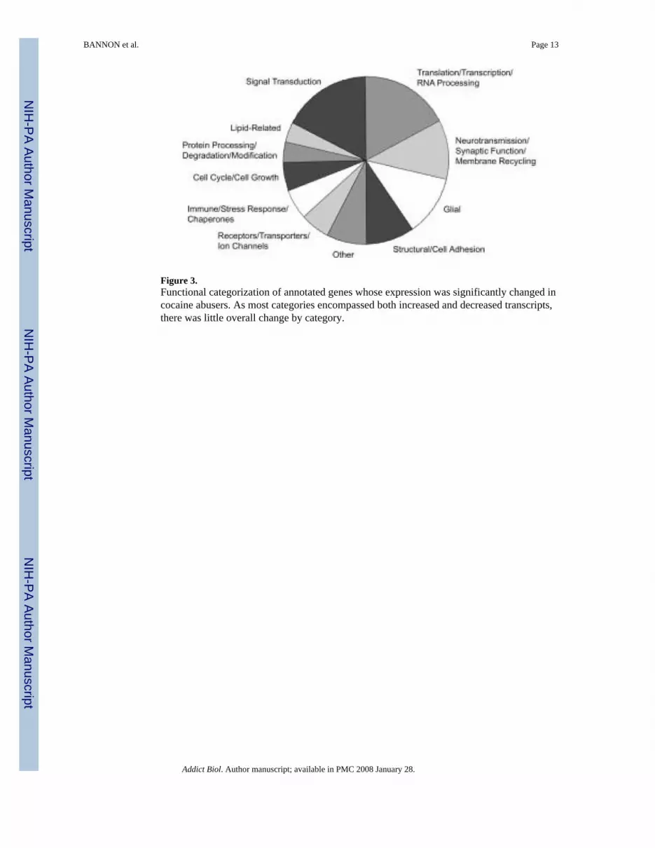
Figure 3.Functional categorization of annotated genes whose expression was significantly changed incocaine abusers. As most categories encompassed both increased and decreased transcripts,there was little overall change by category.
BANNON et al. Page 13
Addict Biol. Author manuscript; available in PMC 2008 January 28.
NIH
-PA Author Manuscript
NIH
-PA Author Manuscript
NIH
-PA Author Manuscript

Figure 4.Decreased expression of myelin-related transcripts in cocaine abusers. The signal log ratio(base 2) distribution of all transcripts detected in the majority of subject pairs was plotted afterbinning averages into groups of 0.05 (n = 18055, mean = 0.01). The myelin-specific group(n = 5, mean = −0.81) was found to be significantly decreased from the mean of all transcriptspresent (t(4) = −7.95, p = 0.001).
BANNON et al. Page 14
Addict Biol. Author manuscript; available in PMC 2008 January 28.
NIH
-PA Author Manuscript
NIH
-PA Author Manuscript
NIH
-PA Author Manuscript

Figure 5.Hierarchical cluster showing the relatedness and relative expression levels of transcriptsdifferentially expressed between cocaine and control groups. Each column represents transcriptexpression from individual subjects. Z-score data were converted to color for ease ofinterpretation: black represents the mean across subjects, with red for values above the mean,while green indicates expression below the mean. Branch lengths represent relatedness ofsignal expression across subjects. Right side of the figure shows the parallel changes inexpression of four myelin-related transcripts (two MBP, two MOBP), and two closely clusteredtranscripts, as a function of cocaine abuse. MBP: myelin basic protein; MOBP: myelin-associated oligodendrocyte basic protein; DAAM2: dishevelled associated activator ofmorphogenesis 2; PNUTL2: peanut-like 2.
BANNON et al. Page 15
Addict Biol. Author manuscript; available in PMC 2008 January 28.
NIH
-PA Author Manuscript
NIH
-PA Author Manuscript
NIH
-PA Author Manuscript

Figure 6.Scatter plot demonstrating the correlation between MBP microarray and quantitative RT-PCRdata. Difference scores for each Study II subject pair were converted to Z-scores and correlatedby Pearson’s product moment.
BANNON et al. Page 16
Addict Biol. Author manuscript; available in PMC 2008 January 28.
NIH
-PA Author Manuscript
NIH
-PA Author Manuscript
NIH
-PA Author Manuscript


