Embed Size (px)
Citation preview

Letters to the Editor 921
in anticipation that the side effects will prove to be min-imal, we propose to evaluate the safety and efficacy ofNTBC for alkaptonuria patients. With the cooperationof Dr. C. Ronald Scott of the University of Washington,we now are attempting to secure NTBC for use in thetreatment of alkaptonuria.
Whereas gene therapy generally involves specific tissuelocalization, pharmacotherapy routinely employs a widerange of targets. For many metabolic disorders, this pro-vides a distinct advantage. For example, in the treatmentof cystinosis, cysteamine has beneficial effects upon avariety of organs and tissues (Gahl et al. 1995), includingthe kidney, muscle, cornea, and thyroid (Kimonis et al.1995). NTBC could have multisystemic salutary effectsas well, meaning that we really are ready to try to curealkaptonuria.
YAIR ANIKSTER,1,2 WILLIAM L. NYHAN,3 AND
WILLIAM A. GAHL2
1Medical Genetics Branch, National Human GenomeResearch Institute, and 2Section on HumanBiochemical Genetics, Heritable Disorders Branch,National Institute of Child Health and HumanDevelopment, National Institutes of Health, Bethesda,MD; and 3Department of Pediatrics, University ofCalifornia–San Diego, San Diego
Electronic-Database Information
Accession number and URL for data in this article are asfollows:
Online Mendelian Inheritance in Man (OMIM), http://www.ncbi.nlm.nih.gov/Omim (alkaptonuria [MIM 203500])
References
Gahl WA, Schneider JA, Aula P (1995) Lysosomal transportdisorders: cystinosis and sialic acid storage diseases. In:Scriver CR, Beaudet AL, Sly W, Valle D (eds) The metabolicand molecular bases of inherited disease, 7th ed. McGraw-Hill, New York, pp 3763–3797
Gibbs TC, Payan J, Brett EM, Lindstedt S, Holme E, ClaytonPT (1993) Peripheral neuropathy as the presenting featureof tyrosinaemia type I and effectively treated with an inhib-itor of 4-hydroxyphenylpyruvate dioxygenase. J NeurolNeurosurg Psychiatry 56:1129–1132
Grompe M, Lindstedt S, Al-Dhalimy M, Kennaway NG, Pa-paconstantinou J, Torres-Ramos CA, Ou C-N, et al (1995)Pharmacological correction of neonatal lethal hepatic dys-function in a murine model of hereditary tyrosinaemia typeI. Nat Genet 10:453–459
Kimonis VE, Troendle J, Yang ML, Rose SR, Markello TC,Gahl WA (1995) Effects of early cysteamine therapy on thy-roid function and growth in nephropathic cystinosis. J ClinEndocrinol Metab 80:3257–3261
La Du BN (1995) Alkaptonuria. In: Scriver CR, Beaudet AL,Sly W, Valle D (eds) The metabolic and molecular bases of
inherited disease, 7th ed. McGraw-Hill, New York, pp1371–1386
——— (1998) Are we ready to try to cure alkaptonuria? AmJ Hum Genet 62:765–767
Lindstedt S, Holme E, Lock EA, Hjalmarson O, Strandvik B(1992) Treatment of hereditary tyrosinaemia type I by in-hibition of 4-hydroxyphenylpyruvate dioxygenase. Lancet340:813–817
Mitchell GA, Lambert M, Tanguay RM (1997) Hypertyrosi-nemia. In: Scriver CR, Beaudet AL, Sly W, Valle D (eds) Themetabolic and molecular bases of inherited disease, 7th ed.McGraw-Hill, New York (CD-ROM)
Pronicka E, Rowinska E, Bentkowski Z, Zawadski J, HolmeE, Lindstedt S (1996) Treatment of two children with he-reditary tyrosinaemia type I and long-standing renal diseasewith a 4-hydroxyphenylpyruvate dioxygenase inhibitor(NTBC). J Inherit Metab Dis 19:234–238
Schulz A, Ort O, Beyer P, Kleinig H (1993) SC-0051, a 2-benzoyl-cyclohexane-1,3-dione bleaching herbicide, is a po-tent inhibitor of the enzyme p-hydroxyphenylpyruvate diox-ygenase. FEBS Lett 318:162–166
Address for correspondence and reprints: Dr. William A. Gahl, 10 CenterDrive, MSC 1830, Building 10, Room 9S-241, National Institute of Child Healthand Human Development, National Institutes of Health, Bethesda, MD 20892-1830. E-mail: [email protected]
� 1998 by The American Society of Human Genetics. All rights reserved.0002-9297/98/6303-0042$02.00
Am. J. Hum. Genet. 63:921–926, 1998
Gene Localization for Aculeiform Cataract, onChromosome 2q33-35
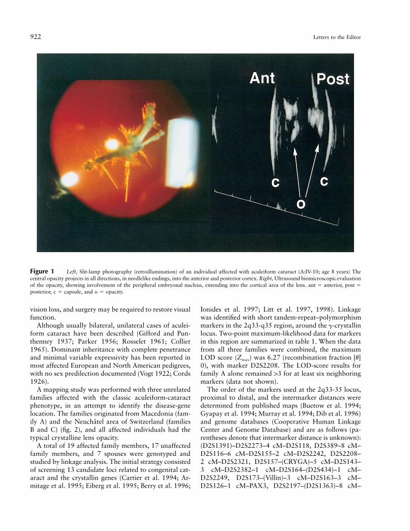
To the Editor:Aculeiform cataract (MIM 115700) is a form of con-genital crystalline cataract that originally was describedby Vogt in 1922 and was referred to as “Spiesskatarakt”(Vogt 1922). Since its original description, this entityalso has been referred to as “frosted cataract,” “needle-shaped cataract,” or “fasciculiform cataract” (Parker1956). This phenotype is characterized by fiberglasslikeor needlelike crystals projecting in different directions,through or close to the axial region of the lens (fig. 1).Some crystals may be 11 mm in length, and their bio-chemical composition is not known. This type of cata-ract is considered to be different from the corraliformcataract, which does not show the needlelike projections.This opacity does not appear to respect the sutures orthe direction of the lens fibers (Francois 1963) and ap-pears to originate from the fetal and postnatal nuclei,suggesting a congenital origin with some postnatal pro-gression, if any. The opacity causes a variable degree of
922 Letters to the Editor
Figure 1 Left, Slit-lamp photography (retroillumination) of an individual affected with aculeiform cataract (A:IV-10; age 8 years) Thecentral opacity projects in all directions, in needlelike endings, into the anterior and posterior cortex. Right, Ultrasound biomicroscopic evaluationof the opacity, showing involvement of the peripheral embryonal nucleus, extending into the cortical area of the lens. ant � anterior, post �posterior, c � capsule, and o � opacity.
vision loss, and surgery may be required to restore visualfunction.
Although usually bilateral, unilateral cases of aculei-form cataract have been described (Gifford and Pun-thenney 1937; Parker 1956; Rosselet 1961; Collier1965). Dominant inheritance with complete penetranceand minimal variable expressivity has been reported inmost affected European and North American pedigrees,with no sex predilection documented (Vogt 1922; Cords1926).
A mapping study was performed with three unrelatedfamilies affected with the classic aculeiform-cataractphenotype, in an attempt to identify the disease-genelocation. The families originated from Macedonia (fam-ily A) and the Neuchatel area of Switzerland (familiesB and C) (fig. 2), and all affected individuals had thetypical crystalline lens opacity.
A total of 19 affected family members, 17 unaffectedfamily members, and 7 spouses were genotyped andstudied by linkage analysis. The initial strategy consistedof screening 13 candidate loci related to congenital cat-aract and the crystallin genes (Cartier et al. 1994; Ar-mitage et al. 1995; Eiberg et al. 1995; Berry et al. 1996;
Ionides et al. 1997; Litt et al. 1997, 1998). Linkagewas identified with short tandem-repeat–polymorphismmarkers in the 2q33-q35 region, around the g-crystallinlocus. Two-point maximum-likelihood data for markersin this region are summarized in table 1. When the datafrom all three families were combined, the maximumLOD score (Zmax) was 6.27 (recombination fraction [v]0), with marker D2S2208. The LOD-score results forfamily A alone remained 13 for at least six neighboringmarkers (data not shown).
The order of the markers used at the 2q33-35 locus,proximal to distal, and the intermarker distances weredetermined from published maps (Buetow et al. 1994;Gyapay et al. 1994; Murray et al. 1994; Dib et al. 1996)and genome databases (Cooperative Human LinkageCenter and Genome Database) and are as follows (pa-rentheses denote that intermarker distance is unknown):(D2S1391)–D2S2273–4 cM–D2S118, D2S389–8 cM–D2S116–6 cM–D2S155–2 cM–D2S2242, D2S2208–2 cM–D2S2321, D2S157–(CRYGA)–5 cM–D2S143–3 cM–D2S2382–1 cM–D2S164–(D2S434)–1 cM–D2S2249, D2S173–(Villin)–3 cM–D2S163–3 cM–D2S126–1 cM–PAX3, D2S2197–(D2S1363)–8 cM–

Figure 2 Pedigrees of families studied, with haplotypes for selected markers relevant to recombinant breakpoints on chromosome 2q33-35. Blackened squares and circles denote affected individuals, and diamonds denote nonparticipating relatives. A dash within a marker orderdenotes an untyped marker (deemed not critical to the identification of recombination events), which is not considered to be within the disease-gene interval. A haplotype cosegregating with the affected status is indicated by a blackened bar; the critical crossovers defining the proximaland distal boundaries of the aculeiform candidate region are shown in family B, individuals III:3 and IV:4, placing the disease locus betweenthe markers D2S2273 and D2S143. Unblackened and patterned bars denote the non–disease-associated haplotypes.

924 Letters to the Editor
Table 1
Two-Point Linkage Data for Aculeiform-Cataract Phenotype and Markers of2q33-35 Region
MARKER
LOD SCORE AT v �MAXIMUM
v Zmax.00 .05 .10 .20 .30 .40
D2S1391 �1.96 2.18 2.17 1.79 1.22 1.08 .05 2.18D2S118 5.25 4.77 4.28 3.22 2.09 .97 .00 5.25D2S389 6.12 5.53 4.91 3.60 2.28 1.59 .00 6.12D2S116 5.39 4.89 4.39 3.34 2.25 1.10 .00 5.39D2S155 6.08 5.51 4.93 3.69 2.39 1.14 .00 6.08D2S2208 6.27 5.64 5.04 3.79 2.48 1.15 .00 6.27D2S2321 3.07 2.77 2.47 1.86 1.24 .61 .00 3.07D2S157 3.36 3.08 2.77 2.15 1.47 .75 .00 3.36CRYGA 1.36 1.26 1.14 .83 .53 .22 .00 1.36D2S143 5.83 5.34 4.82 4.29 3.71 3.13 .00 5.83D2S2382 3.33 3.13 2.87 2.22 1.49 .71 .00 3.33D2S164 4.18 3.90 3.51 2.75 1.85 .88 .00 4.18D2S126 �1.33 2.81 2.79 2.36 1.89 .89 .07 2.88PAX3 2.81 2.49 2.18 1.61 1.01 .45 .00 2.81D2S1363 3.39 3.15 2.84 2.16 1.41 .64 .00 3.39D2S159 �10.04 �.60 �.13 .17 .12 .05 .20 .17
NOTE.—Linkage analysis was performed with the LINKAGE program package(version 5.1), and MLINK was used for pairwise analysis. A full-penetrance, equalallele frequency and a disease-gene frequency of .0001 were assumed for the diseaselocus.
Table 2
Haplotype Analysis of Aculeiform Cataract
MARKER
INTERMARKER
DISTANCE(cM)
AFFECTED
HAPLOTYPE
IN FAMILYa
A B C
D2S116 3 4 7
D2S155 6 13 6
D2S2242 2 8 1 1D2S2208 0 6 8 8D2S2321 2 4 2 2D2S157 0 5 6 6CRYGA 2 1 1D2S143 5 5 3 3
D2S2382 3 65 5
D2S164 1 9 10 11D2S434 5 4 3
a The region of allele sharing is circumscribedby the box, and the alleles that define the disease-gene interval when the recombination eventsshown in figure 2 are taken into account areunderlined.
D2S159. The marker CRYGA was an intragenic poly-morphism of the g-crystallin–A gene.
Critical recombination events observed in affected in-dividuals defined an initial disease-gene interval of 27cM between markers D2S2273 and D2S143 (fig. 2). Fur-thermore, observation of recombination events in theunaffected allele of individual C:IV-1 allowed orderingof markers D2S2321 and D2S157 (cen-D2S2321-D2S157-tel), which were nonrecombinant on the Ge-nethon map (Dib et al. 1996).
Haplotype analysis showed a common affected hap-lotype for seven markers (D2S2242, D2S2208,D2S2321, D2S157, CRYGA, D2S143, and D2S2382)over a 10-cM interval in families B and C (see alleleswithin the box in table 2). Although no common an-cestor could be identified through genealogical studies,both families are from the relatively small Neuchatelarea of Switzerland (population ∼170,000). The sharedhaplotype, together with the recombination events ob-served between markers D2S2242 and D2S143, definea disease-gene interval of 7 cM (see the underlined allelesin table 2).
Several candidate genes are of interest in this interval,the most relevant being the g-crystallin–gene cluster,CRYG (2q33-35). Although the precise position ofCRYGA is unclear, haplotype analysis and observationof recombination events in families A and B suggest thatCRYGA is distal to D2S155 and centromeric to D2S143.Another crystallin gene, CRYBA2, has been mapped tothe 2q34-36 region (Hulsebos et al. 1995). However,
physical mapping using radiation-hybrid cell lines placedCRYGA separate from and centromeric to CRYBA2(Hulsebos et al. 1995). The gene order in the human2q33-36 segment appears to be syntenic with that ofgenes on mouse chromosome 1, and, in the mouse,Cryba2 is nonrecombinant with Villin (Vil) (10.6 cMtelomeric to Cryg) (Hulsebos et al. 1995). Genotyping

Letters to the Editor 925
the three families using a dinucleotide repeat close toVillin confirms its location as being telomeric toCRYGA, since it is mapped below the recombinationbreakpoint in individual A:IV-14 (fig. 2). If synteny be-tween the mouse genome and the human genome is as-sumed for this region, CRYBA2 would be located out-side the disease-gene interval of interest. A develop-mental gene, PAX-3, was documented at the telomericend of the interval. However, observation of recombi-nation events centromeric to this gene, in the familiesstudied, excluded the potential role of PAX-3 in thiscataract phenotype (fig. 2).
The human g-crystallin genes constitute a multigenefamily whose members are expressed only in the eye lens.The g-crystallin–gene cluster contains six highly con-served genes (A–F), all mapped to chromosome 2q33-q35 (den Dunnen et al. 1985; Meakin et al. 1985) andspecific to mammals (Cveki and Piatgorsky 1996) . Therelative position of the g-crystallins B–E have been es-tablished on a 40-kb DNA segment, but the exact lo-cations for g-crystallins A and F in the gene cluster areyet to be determined (Meakin et al. 1985). The g-crys-tallin cluster is of great interest in the study of congenitalcataract, since it is expressed early in development andis presumed to play a role in both fiber differentiationand maintenance of lens-fiber transparency (Papacon-stantinou 1967). Furthermore, this locus has been as-sociated with hereditary cataract in mouse and human(Oda et al. 1980; Lubsen et al. 1987; Cartier et al. 1992;Santhiya et al. 1995).
Although g-crystallins E and F are considered to bepseudogenes, by virtue of an in-frame stop codon(Meakin et al. 1985), a low level of g-crystallin–E tran-script has been detected (Brakenhoff et al. 1994). Lubsenet al. (1987) reported a tight linkage between the g-crystallin–gene cluster on chromosome 2 and a pheno-type referred to as “Coppock-like cataract,” confined tothe embryonic nucleus (clearly distinct from the aculei-form cataract) (Lubsen et al. 1987). Recent work hasdemonstrated that sequence changes upstream from theg-crystallin–E pseudogene result in a 10-fold increase inthe activity of the g-crystallin–E promoter. These datasuggest a potential role for the g-crystallin–E peptide inthe Coppock-like cataract of human (Brakenhoff et al.1994).
Of interest in the Elo and the Cat2 mutant-mousemodels, the g-crystallin–E gene is the target of mutationsand also is responsible for cataract phenotypes (Oda etal. 1980; Cartier et al. 1992; Santhiya et al. 1995). Inboth these mutants, the opacity involves the embryonicnucleus.
Recently, Rogaev et al. (1996) studied a large family,from the isolated Nokhurli population of Turkmenia,that is affected with polymorphic congenital cataract(PCC). This phenotype also mapped to the 2q33 locus,
and it was characterized by a progressive, mostly pe-ripheral, and highly variable opacity (Ginter et al. 1983,1991). Whether PCC, Coppock-like cataract, and acu-leiform cataract are allelic variants remains to be elu-cidated, but they clearly are three distinct clinicalentities.
In summary, the localization of a gene for aculeiformcataract has been identified on chromosome 2q33-35,within a 7-cM interval. This condition appears to begenetically homogeneous. Refinement of the disease-gene interval and analysis of the g-crystallin–gene clusterare currently underway, in an attempt to identify thedisease-causing mutation(s). The molecular characteri-zation of this phenotype may shed light on the complexcascade of events modulating lens differentiation.
Acknowledgments
The authors are grateful to the families for their enthusiasticparticipation and to Ms. Megan Priston for her excellent tech-nical work. This work has been supported by Swiss NationalScience Foundation grant 32-43619.95 (to F.L.M. and D.F.S.)and by the Glaucoma Research Society of Ontario, the WestonFoundation, and the Imperial Oil Research Fund (all providedsupport to E.H.).
ELISE HEON,1,2 SEN LIU,2 GAIL BILLINGSLEY,2 OTTAVIO
BERNASCONI,3 CATHY TSIFILDIS,2 DANIEL F. SCHORDERET,4
AND FRANCIS L. MUNIER3,4
1Department of Ophthalmology, University of Toronto, and2Eye Research Institute of Canada, Toronto; and 3HopitalOphtalmique Jules Gonin and 4Unit of Molecular Genetics,Division of Medical Genetics, Lausanne, Switzerland
Electronic-Database Information
URLs for data in this article are as follows:
Cooperative Human Linkage Center (CHLC), http://www.chlc.org
Genethon, http://www.genethon.frGenome Database, http://gdbwww.gdb.orgOnline Mendelian Inheritance in Man (OMIM), http://
www.ncbi.nlm.nih.gov/omim
References
Armitage M, Kivlin J, Ferrell R (1995) A progressive earlyonset cataract gene maps to chromosome 17q24. Nat Genet9:37–40
Berry V, Ionides A, Moore A, Plant C, Bhattacharya S, ShielsA (1996) A locus for autosomal dominant anterior polarcataract on chromosome 17p. Hum Mol Genet 5:415–419
Brakenhoff R, Henskens H, van Rossum M, Lubsen N,Schoenmakers G (1994) Activation of the gE-crystallin pseu-dogene in the human hereditary Coppock-like cataract.Hum Mol Genet 3:279–283
Buetow K, Weber J, Ludwigsen S, Scherpbier-Heddema T,Duyk G, Sheffield V, Wang Z, et al (1994) Integrated human

926 Letters to the Editor
genome-wide maps constructed using the CEPH referencepanel. Nat Genet 6:391–393
Cartier M, Breitman M, Tsui L-C (1992) A frame-shift mu-tation in the gammaE-crystallin gene of the Elo mouse. NatGenet 2:42–45
Cartier M, Tsui L, Ball S, Lubsen N (1994) Crystallins genesand cataract. In: Wright AF, Jay B (eds) Molecular geneticsof inherited eye disorders. Harwood Academic, Edinburgh,pp 413–443
Collier M (1965) Cataracte aculeiforme unilaterale droite. BullSoc Ophtalmol 65:881–884
Cords R (1926) Uber Speisskatarakt. Klinische Monatsblatterfur Augenheilkunde 76:125–126
Cvekl A, Piatgorsky J (1996) Lens development and crystallingene expression: many roles for Pax-6. BioEssays 18:621–630
den Dunnen J, Moorman R, Bremers F, Schoenmakers J (1985)Two human g-crystallin genes are linked and riddled withAlu-repeats. Gene 38:197–204
Dib C, Faure S, Fizames C, Samson D, Drouot N, Vignal A,Millasseau P, et al (1996) A comprehensive genetic map ofthe human genome based on 5,264 microsatellites. Nature380:152–154
Eiberg H, Lund M, Warburg M, Rosenberg T (1995) Assign-ment of congenital cataract Volkmann type (CCV) to chro-mosome 1p36. Hum Genet 96:33–38
Francois J (1963) Varieties of congenital cataracts. In: Con-genital cataracts. Royal Van Gorcum, Assen, Netherlands,pp 164–165
Gifford S-R, Punthenney I (1937) Coralliform cataract and anew form of congenital cataract with crystals in the lens.Arch Ophthalmol 17:884–892
Ginter E, Petrin A, Spitsyn V, Rogaev E (1991) An attempt atmapping human congenital cataract gene using linkage. Ge-netika 27:1840–1849
Ginter E, Turaeva S, Revasov A, Panteleeva O, Artikov A,Michailova L (1983) Medical-genetic studies of the popu-lation of Turkmenia. IV. Hereditary pathology in the Nok-hurli Turkmens. Genetika 19:1344–1351
Gyapay G, Morissette J, Vignal A, Dib C, Fizames C, Millas-seau P, Marc S, et al (1994) The 1993–94 Genethon humangenetic linkage map. Nat Genet 7:246–339
Hulsebos TJM, Cerosaletti KM, Fournier REK, Sinke RJ, Roc-chi M, Marzella R, Jenkins NA, et al (1995) Identificationof the human beta-A2 crystallin gene (CRYBA2): localiza-tion of the gene on human chromosome 2 and the homol-ogous gene on mouse chromosome 1. Genomics 28:543–548
Ionides A, Berry V, MacKay D, Moore A, Bhattacharya S,
Shiels A (1997) A locus for autosomal dominant posteriorpolar cataract on chromosome 1p. Hum Mol Genet 6:47–51
Litt M, Carrero-Valenzuela R, LaMorticella DM, Schultz DW,Mitchell TN, Kramer P, Maumenee IH (1997) Autosomaldominant cerulean cataract is associated with a chain ter-mination mutation in the human b-crystallin gene CRYBB2.Hum Mol Genet 6:665–668
Litt M, Kramer P, LaMorticella DM, Murphey W, Lovrien EW,Weleber RG (1998) Autosomal dominant congenital cata-ract associated with a missense mutation in the human alphacrystallin gene CRYAA. Hum Mol Genet 7:471–474
Lubsen N, Renwick J, Tsui L-C, Breitman M, SchoenmackersJ (1987) A locus of a human hereditary cataract is closelylinked to the gamma-crystallin gene family. Proc Natl AcadSci USA 84:489–492
Meakin S, Breitman M, Tsui L (1985) Structural and evolu-tionary relationships among five members of the human g-crystallins gene family. Mol Cell Biol 5:1408–1414
Murray J, Buetow K, Weber J, Ludwigsen S, Scherpbier-Hed-dema T, Manion F, Quillen J, et al (1994) A comprehensivehuman linkage map with centimorgan density: CooperativeHuman Linkage Center. Science 265:2049–2054
Oda S-I, Watanabe T, Kondo K (1980) A new mutation, eyelens obsolescence, Elo on chromosome 1 in the mouse. JpnJ Genet 55:71–75
Papaconstantinou J (1967) Molecular aspects of lens differ-entiation. Science 156:338–446
Parker C (1956) Spear cataract. Arch Ophthalmol 55:23–24Rogaev EI, Rogaeva EA, Korovaitseva GI, Farrer LA, Petrin
AN, Keryanov SA, Turaeva S, et al (1996) Linkage of poly-morphic congenital cataract to the g-crystallin gene locuson human chromosome 2q33-35. Hum Mol Genet 5:699–703
Rosselet E (1961) Cataracte aculeiforme. Ophthalmologica141:425–427
Santhiya ST, Abd-alla SM, Loster J, Graw J (1995) Reducedlevels of gamma-crystallin transcripts during embryonic de-velopment of murine Cat2nop mutant lenses. Graefes ArchClin Exp Ophthalmol 233:795–800
Vogt A (1922) Weitere Ergebnisse der Spaltlampenmikrosko-pie des vorderen Bulbusabschnittes III. Angeborene und fruherworbene Linsenveranderungen. Graefes Arch Ophthalmol108:182–191
Address for correspondence and reprints: Dr. Elise Heon, Eye Research In-stitute of Canada, 399 Bathurst Street, Room 6-412, Toronto, M5T 2S8 Ontario,Canada. E-mail: [email protected]
� 1998 by The American Society of Human Genetics. All rights reserved.0002-9297/98/6303-0043$02.00


