Embed Size (px)
Citation preview
ORIGINAL ARTICLE
Gene Signature Distinguishes Patients with Chronic UlcerativeColitis Harboring Remote Neoplastic LesionsJoel Pekow, MD,* Urszula Dougherty, MS,* Yong Huang, PhD,* Edward Gometz, MD,* Jeff Nathanson, MD,*Greg Cohen, MD,* Shawn Levy, PhD,† Masha Kocherginsky, PhD,‡ Nanda Venu, MD,*Maria Westerhoff, MD,§ John Hart, MD,§ Amy E. Noffsinger, MD,§ Stephen B. Hanauer, MD,*Roger D. Hurst, MD,k Alessandro Fichera, MD,k Loren J. Joseph,§ Qiang Liu, MD,§ and Marc Bissonnette, MD*
Background: Individuals with ulcerative colitis (UC) are at increased risk for colorectal cancer. The standard method of surveillance for neoplasia inUC by colonoscopy is invasive and can miss flat lesions. We sought to identify a gene expression signature in nondysplastic mucosa without activeinflammation that could serve as a marker for remote neoplastic lesions.
Methods: Gene expression was analyzed by complementary DNA microarray in 5 normal controls, 4 UC patients without dysplasia, and 11 UCpatients harboring remote neoplasia. Common gene ontology pathways of significantly differentially expressed genes were identified. Expression ofgenes which were progressively and significantly upregulated from controls to UC without neoplasia, to UC with remote neoplasia were evaluated byreal-time polymerase chain reaction. Several gene products were also examined by immunohistochemistry.
Results: Four hundred and sixty-eight genes were significantly upregulated, and 541 genes were significantly downregulated in UC patients withneoplasia compared with UC patients without neoplasia. Nine genes (ACSL1, BIRC3, CLC, CREM, ELTD1, FGG, S100A9, THBD, and TPD52L1) wereprogressively and significantly upregulated from controls to nondysplastic UC to UC with neoplasia. Immunostaining of proteins revealed increasedexpression of S100A9 and REG1a in UC-associated cancer and in nondysplastic tissue from UC patients harboring remote neoplasia compared with UCpatients without neoplasia and controls.
Conclusions: Gene expression changes occurring as a field effect in the distal colon of patients with chronic UC identify patients harboring remoteneoplastic lesions. These markers may lead to a more accurate and less invasive method of detection of neoplasia in patients with inflammatory boweldisease.
(Inflamm Bowel Dis 2013;19:461–470)
Key Words: inflammatory bowel disease, ulcerative colitis, dysplasia, colorectal cancer, gene expression
P atients with ulcerative colitis (UC) are at increased risk forcolorectal cancer (CRC), although mechanisms of carcinogen-
esis remain poorly understood.1–3 The risk of inflammatory boweldisease (IBD)–associated colon cancer is related to the extent,duration, and severity of inflammation.4–6 With chronic inflamma-
tion, the epithelium undergoes a transformation from injured andregenerating yet nondysplastic tissue to progressively neoplasticepithelium, ranging from low-grade dysplasia (LGD) to high-gradedysplasia (HGD) and eventually to adenocarcinoma.3,7 Molecularand genetic changes accompany this histological progression.3,8–11
Previous reports have also identified molecular abnormalitiesin normal-appearing nondysplastic mucosa from patients with UCwho harbor a remote dysplastic lesion.11–15 Aneuploidy, chromo-somal alterations, p53 mutations, and p53 loss of heterozygosityappear in large areas of normal-appearing mucosa adjacent to areaswith cancer.12–14 Furthermore, chromosomal instability can be seenin normal-appearing mucosa before the development of dysplasia.15
Earlier microarray studies have demonstrated changes ingene expression from mucosa in patients with UC compared withnormal controls and patients with Crohn’s disease.16,17 Althoughone study identified gene expression changes in UC tissue remotefrom neoplastic lesions compared with UC without dysplasia, thestudy included tissue with inflammatory activity, which is knownto alter gene expression.18 No study has addressed gene expressionchanges in quiescent UC at risk for colon cancer. We hypothesized
Supplemental digital content is available for this article. Direct URL citationsappear in the printed text and are provided in the HTML and PDF versions of thisarticle on the journal’s Web site (www.ibdjournal.org).
Received for publication June 21, 2012; Accepted July 3, 2012.
From the *Section of Gastroenterology, Hepatology, and Nutrition, University ofChicago, Chicago, Illinois; †Department of Biomedical Informatics, Vanderbilt Uni-versity, Nashville, Tennessee; ‡Departments of Health Studies, §Pathology, andkSurgery, University of Chicago, Chicago, Illinois.
The authors have no conflicts of interest to disclose.
Supported by the International Organization for the Study of InflammatoryBowel Disease (M.B. and G.C.), the Crohn’s and Colitis Foundation of America(J.P.), and National Institutes of Health Grant K08DK090152 (J.P).
Reprints: Joel Pekow, MD, 900 East 57th Street, MB#9, Chicago, IL 60637(e-mail: [email protected]).
Copyright © 2013 Crohn’s & Colitis Foundation of America, Inc.
DOI 10.1097/MIB.0b013e3182802bac
Published online 6 February 2013.
Inflamm Bowel Dis � Volume 19, Number 3, March 2013 www.ibdjournal.org | 461
Copyright © 2013 Crohn’s & Colitis Foundation of America, Inc. Unauthorized reproduction of this article is prohibited.

that gene expression profiles in random normal-appearing UCmucosa might provide a “genetic signature” to distinguish colonsharboring dysplasia from nondysplastic colons. Identification ofgene expression changes could provide valuable insights intomechanisms driving cancer development in IBD and potentiallyidentify biomarkers for patients with IBD harboring neoplasticlesions.
MATERIALS AND METHODS
PatientsThe study was approved by the institutional review board at
the University of Chicago. Informed consent was obtained frompatients with UC undergoing surveillance colonoscopy, colono-scopy for follow-up of a previous diagnosis of dysplasia, orsurgical colectomy with a diagnosis of dysplasia. Microarray datawere obtained from colonic mucosal biopsies from normalcontrols, UC patients without dysplasia, and nondysplasticmucosa from patients with UC harboring a remote neoplasticlesion. Patients with UC were included if they had a previousclinical diagnosis of UC confirmed by an expert gastrointestinalpathologist, a disease duration of .7 years, and an extent ofdisease .20 cm proximal to the anal verge. Background infor-mation was collected on the age of the patient, duration and extentof disease, history of primary sclerosing cholangitis, and a familyhistory of colon cancer in a first-degree relative.
Histology ReviewOriginal surgical pathology was reviewed for neoplasia by
2 expert gastrointestinal pathologists (J.H./A.N.). All rectosig-moid samples submitted to pathology were reviewed from thesame date as the tissue biopsies obtained for the study. Onhistological review, all patients had IBD with quiescent disease(no active inflammation) or with minimal inflammatory activity inthe rectosigmoid colon. No patient had a neoplastic lesion at thesite where study biopsies were obtained.
Sample CollectionDuring endoscopic procedures, 4 mucosal biopsies were
obtained from 10 to 20 cm proximal to the anal verge and placedin RNAlater. After 24 to 48 hours at 48C, the biopsies were thenstored at 2808C until RNA extraction was performed. Specimensobtained at the time of surgery were acquired through biopsy of tissuefrom the rectosigmoid colon immediately after the removal of thecolon during colectomy. Surgical specimens were placed in RNAlaterwithout delay and transferred to 48C storage for 24 to 48 hours, andthen stored at 2808C until RNA extraction was performed.
RNA ExtractionRNA was extracted using the RNeasy Kit (Quiagen,
Hilden, Germany) that included a 1-column DNase step accordingto the manufacturer’s specifications. RNA purity and concentra-tion were determined using the Agilent nanodrop ND-1000 spec-trophotometer and RNA integrity assessed by Agilent 2100 N
(Agilent Technologies, Santa Clara, CA). All samples used inthe analysis had a RNA integrity number .7.
Microarray AnalysisMicroarray assays were carried out on Affymetrix Human
U133p2 platform at the Vanderbilt Functional Genomics Facilityfollowing Affymetrix protocols (Affymetrix, Santa Clara, CA). Theautomated hybridization, washing, staining, and scanning of thearrays were completed on the Affymetrix Gene Titan Instrument.The quality check and data normalization were performed usingR/Bioconductor package “affy” with “rma” approach.19 The micro-array .cel files and normalized data were deposited in NCBI GEOrepository with accession number GSE37283.
Gene filtering and sample classification were performed usingdChip software (http://biosun1.harvard.edu/complab/dchip/) withPearson’s correlation matrix and centroid-linkage method. Probe setswere filtered by the criteria of SD .0.7 and ,2, and probe setintensity .5 in at least 50% of samples, resulting in 2450 probe setsdemonstrating sufficient variation across individual samples. Unsu-pervised hierarchical clustering of samples was performed usingthese probe sets to determine whether the clinical conditions couldbe distinguished by global gene expression pattern.
To identify differentially expressed genes, 2-group comparisonwas performed using Significance Analysis of Microarrays softwareusing fold change .2.0 and false discovery rate ,5%. Differentiallyexpressed genes between UC without neoplasia (UC) and UCpatients with neoplasia (UCN) were further parsed into clusters withdistinct expression pattern across the 3 clinical conditions in thesequence of normal, UC, and UCN using Self-Organizing-Map im-plemented in GeneCluster2 software (www.broadinstitute.org/cancer/software/genecluster2/gc2.html).20 Functional enrichment analysis ofthe identified differentially expressed genes or SOM clusters wasperformed using Onto-Express software.21 The criteria of significantGene Ontology Biological Processes was set at gene number$4 andfalse discovery rate #5%.
Quantitative Real-Time PolymeraseChain Reaction
Complementary DNAs were prepared using 100 ng of totalRNA, gene-specific primers, and SuperScript III Platinum Two-Step qRT-polymerase chain reaction (PCR) (Applied Biosystems,Foster City, CA). SYBR Green quantitative real-time reversetranscription-PCR (qPCR) was performed with the ABI PRISM7700 Sequence Detection System (Applied Biosystems). Primersets for THBD, ACSL1, BIRC3, ELTD1, TPD52L1, FGG, CREM,S100A9, CLC, and REG1a were synthesized by Invitrogen(Carlsbad, CA) (see Table, Supplemental Digital Content 1,http://links.lww.com/IBD/A70). The PCR thermal profile was 508Cfor 2 minutes, 958C for 2 minutes, and 40 cycles at 958C for 3seconds followed by 608C for 30 seconds in a Roche LightCycler480 II (Indianapolis, IN).
For qPCRs, statistical analyses were performed with SAS(Cary, NC). Gene expression levels were compared using thecomparative CT method (DCT) and normalized to b-actin.22
Pekow et al Inflamm Bowel Dis � Volume 19, Number 3, March 2013
462 | www.ibdjournal.org
Copyright © 2013 Crohn’s & Colitis Foundation of America, Inc. Unauthorized reproduction of this article is prohibited.

Significance between individual groups and across the 3 groups(normal, UC, and UC with remote neoplasia) was calculated usinga mixed-effect analysis of variance.23
ImmunohistochemistrySigmoid colon mucosa was compared between normal controls,
UC patients without neoplasia, nondysplastic tissue from UC patientswith a remote neoplatic lesion, and UC-associated dysplastic tissue.Tissue from UC patients with a remote neoplastic lesion were froma subgroup of the same patients used in the microarray and PCRanalysis. Five-micron sections from formalin-fixed paraffin-embeddedtissue were cut and mounted on Vectabond-coated Superfrost Plusslides. A tissue microarray was also prepared with punch biopsiesfrom 27 formalin-fixed paraffin-embedded UC-associated cancers.After deparaffinization, the slides were hydrated in a graded series ofethanol washes. Antigen retrieval was accomplished by heating in 0.01mol/L citrate buffer (pH 6; CTNNB1) in a steamer for 20 minutes. Toblock endogenous peroxidases, the slides were incubated in methanol/H2O2 (3%) protected from light. Protein block (Dako, Denmark) wasused to inhibit nonspecific staining. The sections were incubated withthe primary antibody overnight at 4 degrees [1:50 dilution forS100A9 (H00006280-M01; Abnova, Taiwan); 1:800 for fibrinogen(A0080; Dako), 1:50 for clusterin (SC-6420; Santa Cruz Biotechnol-ogy, Dallas, TX), 1:100 for annexin A3 (AB33068; Abcam, Cam-bridge, MA), and 1:200 for REG1a (RD181078100; LaboratoryMedicine, Czech Republic)]. The slides were incubated witha 1:200 dilution of secondary antibody (Dako) for 35 minutes andstained with 3,30-diaminobenzidine solution. The slides were counter-stained with hematoxylin and bluing reagent. For negative controls,primary antibodies were omitted. Control sections showed no specificstaining. For S100A9, semiquantitative intensity scores were indepen-dently assigned by 3 investigators blinded to tissue diagnosis usingthe immunoreactive score described by Remmele and Stegner.24
When there was disagreement among reviewers, the slides wererereviewed and a consensus was reached. For the tissue array withS100A9, positive staining was reported as stromal, glandular, or both.REG1a positivity was reported as the percent positive staining cryptsby 3 blinded investigators and averaged. Comparisons were madeusing 1-way analysis of variance model, and residuals were checkedfor normality. P values for the pairwise comparisons were Bonferroniadjusted. For the REG1a tissue array, positive staining of tumorglandular cells was characterized as none, focal weak, diffuse weak,focal strong, and diffuse strong. Because the data were not normallydistributed, the overall difference between 3 groups was assessedusing the Kruskal–Wallis test, and pairwise group comparisons weredone using the Wilcoxon’s rank sum test. Analyses were performedusing Stata v12 (College Station, TX).
RESULTS
PatientsMicroarray results were obtained in 24 patients. Of these
patients, 5 were normal controls, 4 had quiescent UC, and 15 had
UC with neoplasia. After removal of 2 patients with primarysclerosing cholangitis and 2 patients because of poor RNA qualityby normalization, 11 patients with remote neoplastic lesions wereincluded in the analysis. Of the patients with neoplasia, 4 had LGD,3 had HGD, and 4 had adenocarcinomas. Two neoplastic lesionswere located in the ascending colon, 3 in the transverse colon, 1 inthe descending colon, 3 in the sigmoid colon, and 2 in the rectum.
The mean age of the patients analyzed was 46 years in thecontrol group, 48.3 years in patients with quiescent UC, and 46.9years in patients with UC-associated neoplasia. The mediandisease duration was 13.5 years in UC patients with neoplasiaand 20 years in UC patients without neoplasia. All patientsincluded in the analysis were men. Ten of the 11 patients withUC-associated neoplasia had pancolitis. Of the 4 UC patientswithout dysplasia, 2 had extensive colitis and 2 had left-sidedcolitis (Table 1).
MicroarrayIn UC patients without neoplasia compared with normal
controls, 324 genes were significantly upregulated and 17genes were significantly downregulated. In UCN comparedwith normal controls, 1667 genes were significantly upregu-lated and 508 genes were significantly downregulated. In UCNcompared with UC, 488 genes were significantly upregulatedand 541 genes were significantly downregulated (Fig. 1;Table 2). The 25 genes with greatest upregulation and down-regulation between groups are presented in Tables, Supplemen-tal Digital Content 2, 3, and 4, http://links.lww.com/IBD/A71,http://links.lww.com/IBD/A72, and http://links.lww.com/IBD/A73.
Nine genes (ACSL1, BIRC3, CLC, CREM, ELTD1, FGG,S100A9, THBD, and TPD52L1) were progressively and signifi-cantly upregulated from healthy controls to quiescent UC without
TABLE 1. Baseline Patient Characteristics
Normal UCUC withNeoplasia
N 5 4 11
Sex (M/F) 5/0 4/0 11/0
Race (AA/C/A/U) 2/3/0/0 0/3/0/1 1/6/1/3
Age (mean) 46 48.3 46.9
Disease duration (median) X 20 13.5Left-sided colitis X 2 1
Pancolitis X 2 10
Dysplasia histology(LGD/HGD/adenocarcinoma)
X X 4/3/4
Medications
Mesalamine 4 8
Immunomodulators 2 3Antitumor necrosis factor 0 1
AA, African American; C, white; A, Asian; U, unknown.
Inflamm Bowel Dis � Volume 19, Number 3, March 2013 Patients with Chronic Ulcerative Colitis
www.ibdjournal.org | 463
Copyright © 2013 Crohn’s & Colitis Foundation of America, Inc. Unauthorized reproduction of this article is prohibited.

neoplasia to patients with UC harboring remote neoplasia(Table 3). No genes were progressively downregulated acrossthese patient groups. For the purpose of this pilot study, wefocused on progressively upregulated genes for further validationby real-time PCR and immunostaining.
Real-Time PCRChanges in 9 genes that progressively and significantly
increased in expression from normal controls to UC patientswithout neoplasia to UC patients with neoplasia in microarrayanalysis were tested by real-time PCR. Real-time PCR wasperformed on 5 normal controls, 4 patients with UC withoutdysplasia, and 7 patients with UC with neoplasia remote from thebiopsy site from the same patient samples used in the microarrayanalysis. The highest expression levels in all 9 genes analyzed
were in UC patients with neoplasia. Furthermore, all genesdisplayed significant expression differences between UC withneoplasia and normal controls. In comparing UC without neo-plasia to normal controls, 5 genes were significantly upregulated(CREM, S100A9, THBD, FGG, and TPD52L1) and 1 gene wassignificantly downregulated (ELTD1). Compared with UC with-out neoplasia, 7 of the 9 genes displayed significantly increasedexpression in UC patients with neoplasia (ACSL1, BIRC3, CREM,ELTD1, FGG, THBD, and TPD52L1) (Table 3).
We also performed real-time PCR analyses for REG1a asthis gene had the greatest differential expression between UCwithout dysplasia and UC tissue harboring remote neoplasia inthe microarray. The real-time PCR findings confirmed the micro-array results with highest gene expression seen in UC patientsharboring remote neoplasia. As shown in Figure 3, REG1a wasupregulated 2.3 (1.1–4.9)-fold in patients with UC (P ¼ 0.03)and 19.2 (10.0–36.8)-fold in patients harboring remote neoplasia(P , 0.001) compared with normal controls. Compared with UCpatients without dysplasia, patients with UC harboring remoteneoplasia had 8.2 (4.1–16.5)-fold upregulation of REG1a (P ,0.001) (Fig. 2).
Biological Processes Enriched withDifferentially Expressed Genes
To cluster differentially expressed genes by functionalclassification, we created 5 groups based on expression patternsfrom normal controls to UC without neoplasia to UC withneoplasia (C0–C4; Fig. 3). We focused on groups C3 and C4because genes clustered into functional ontology classificationsin these groups with less random distribution than C0, C1, andC2. One hundred and sixty-nine genes had expression patternsconsistent with C3. These genes belonged to pathways of immuneresponse, innate immune response, chemotaxis, inflammatory
TABLE 2. Prioritization of Differentially ExpressedGenes Underlying Disease Progression
Two Group
Comparison
False
Discovery
Rate%
Fold
Change Upregulated Downregulated
UC versusnormal
5.2 2.0 324 17
UCN versusnormal
2.5 2.0 1667 508
UCN versusUC
5.0 2.0 468 541
UC, UC without neoplasia; UCN, UC harboring remote neoplasia.Gene filtering criteria and number of significantly upregulated or downregulated gene lociindentified by Significance Analysis of Microarrays software.
TABLE 3. Real-time PCR Results for Genes Which Were Progressively and Significantly Upregulated BetweenNormal Controls to UC to UCN
Symbol Gene Name
UC versus Normal UCN versus Normal UCN versus UC
Fold Change P Fold Change P Fold Change P
ACSL1 Acyl-CoA synthetase long-chain family member 1 1.4 (0.9–2.0) 0.1 2.1 (1.5–2.9) ,0.0001 1.5 (1.1–2.2) 0.02
BIRC3 Baculoviral IAP repeat-containing 3 1.3 (1.0–1.8) 0.08 6.0 (4.6–7.8) ,0.0001 4.6 (3.4–6.1) ,0.0001
CLC Charcot–Leyden crystal protein 1.5 (1.0–2.3) 0.3 1.7 (1.2–2.5) 0.008 1.1 (0.8–1.7) 0.6
CREM cAMP-responsive element modulator 2.5 (1.6–2.8) ,0.0001 4.5 (3.1–6.5) ,0.0001 1.8 (1.2–2.7) 0.004ELTD1 EGF, latrophilin and 7 transmembrane domain
containing 10.2 (0.2–0.3) ,0.0001 2.5 (1.8–3.4) ,0.0001 10.2 (7.2–14.4) ,0.0001
FGG Fibrinogen gamma chain 1.9 (1.2–3.0) 0.007 7.2 (4.8–10.9) ,0.0001 3.8 (2.4–5.8) ,0.0001
S100A9 S100 calcium binding protein A9 10.3 (6.3–16.7) ,0.0001 13.2 (8.7–20.2) ,0.0001 1.3 (0.8–2.0) 0.3
THBD Thrombomodulin 2.4 (1.5–3.7) 0.0004 7.2 (4.8–10.7) ,0.0001 3.0 (2.0–4.7) ,0.0001
TPD52L1 Tumor protein D52-like 1 5.5 (3.6–8.2) 0.004 10.9 (7.6–15.7) ,0.0001 2.0 (1.4–3.0) 0.0006
UC, UC without neoplasia; UCN, UC harboring remote neoplasia.Results are expressed as fold change (95% CIs) and P value between the groups indicated.
Pekow et al Inflamm Bowel Dis � Volume 19, Number 3, March 2013
464 | www.ibdjournal.org
Copyright © 2013 Crohn’s & Colitis Foundation of America, Inc. Unauthorized reproduction of this article is prohibited.

response, G-protein coupled receptor signaling, elevation of cyto-solic calcium ion concentration, and signal transduction (Fig. 4).There were 299 genes in group C4 with progressive upregulationfrom normal controls to UC without dysplasia to UC harboringremote neoplasia. These genes belong to pathways of cellularcalcium ion homeostasis, complement activation, cell–cell signal-ing, B-cell activation, innate immune response, signal transduc-tion, response to lipopolysaccharide, inflammatory response,chemotaxis, and immune response (Fig. 4).
ImmunohistochemistryBy immunohistochemistry, we examined several proteins
whose transcripts were significantly upregulated in UC with remoteneoplasia suggesting that these proteins could be potentially usefulclinical biomarkers. Protein expression examined by immunohis-tochemistry included the following: annexin A3, clusterin, fibrin-ogen gamma (FGG), REG1a, and S100A9. Because of the diffusestromal staining of annexin A3, clusterin, and FGG, the antibodieswere not suitable to demonstrate clinically relevant differencesbetween groups (data not shown).
S100A9 immunostaining was performed on nondysplasticmucosa from the sigmoid colons of normal controls (n ¼ 6), UCpatients without dysplasia (n ¼ 6), patients with UC harboringa remote neoplastic lesion (n ¼ 9), UC-associated dysplasia (n ¼5), and on a tissue array of UC-associated colon cancer tissues (n¼ 27). S100A9 antibodies stained epithelial stroma in all samples.Compared with controls {average score 2.7 (95% confidenceinterval [CI], 20.04 to 5.4)}, S100A9 increased to 4.8 (95%CI, 2.1–7.5) in UC without dysplasia (P ¼ 0.80) to 8.9 (95%CI, 6.7–11.1) in nondysplastic tissue from patients with UC har-boring remote neoplasia (P ¼ 0.002) and to 10.2 (95% CI, 7.2–13.2) in UC-associated cancer (P , 0.001). Compared with qui-escent nondysplastic UC, increases in S100A9 expression weresignificant in UC-associated dysplasia (P ¼ 0.03) and nearlyreached significance in nondysplastic tissue in patients with UCharboring remote neoplasia (P ¼ 0.06). All tumors displayedstromal staining for S100A9, and 17/27 tumors also showed stain-ing in epithelial cells (Fig. 5).
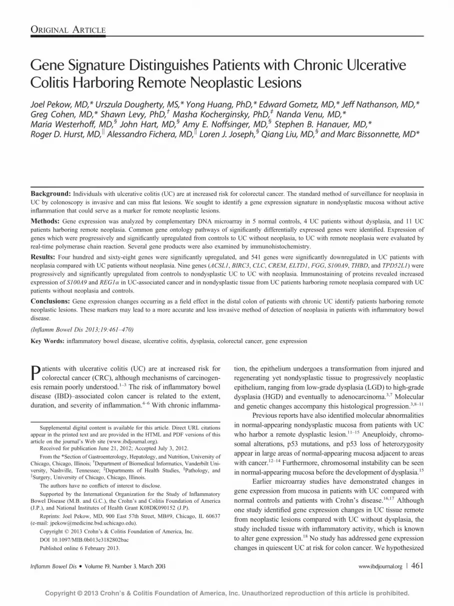
REG1a immunostaining was performed on nondysplasticmucosa from the sigmoid colons of normal controls (n ¼ 3), UCpatients without dysplasia (n ¼ 5), patients with UC harboringa remote neoplastic lesion (n ¼ 6), and on a tissue array ofUC-associated colon cancer tissues (n ¼ 27). Staining differenceswere statistically significant between controls, UC, and UC withremote dysplasia patients (P ¼ 0.05, Kruskal–Wallis test). Theproportion of positive crypts was increased in patients with UCharboring remote neoplasia compared with normal controls (P ¼0.04) but not in UC patients with dysplasia compared to normal
FIGURE 1. Heat map with genome-wide analysis of gene expression.Using 2-way unsupervised hierarchical clustering, patients clusteredinto phenotypic groups with the exception of 1 UC patient withoutdysplasia (S29) who had a gene expression profile more characteristicof those harboring remote neoplasia.
Inflamm Bowel Dis � Volume 19, Number 3, March 2013 Patients with Chronic Ulcerative Colitis
www.ibdjournal.org | 465
Copyright © 2013 Crohn’s & Colitis Foundation of America, Inc. Unauthorized reproduction of this article is prohibited.

controls (P ¼ 0.12). In control patients, no staining was identifiedby immunohisochemistry. Patients with UC harboring remoteneoplasia had higher increase in crypt cell staining compared withUC patients without dysplasia (median percent positive staining0.97% versus 22.9%; P ¼ 0.09). In a tissue microarray of 27 UC-associated cancers, 25/27 cancers had positive epithelial staining.Of these, 9 had focal weak glandular positivity, 2 had diffuseweak positivity, 8 had focal strong positivity, and 6 had diffusestrong positivity (Fig. 6).
DISCUSSIONIn this cross-sectional analysis, we demonstrated that patients
with UC harboring a neoplastic lesion display differential geneexpression in remote quiescent nondysplastic mucosa comparedwith UC patients without dysplasia and normal controls. Geneontology analysis indicated that differentially expressed genes areinvolved in pathways of innate immune response and Toll receptorsignaling supporting the hypothesis that neoplastic lesions in long-standing UC likely result from activation of tumor-promoting
pathways secondary to long-standing inflammation. In agreementwith previous studies demonstrating molecular changes occurringin nondysplastic mucosa of patients with IBD remote froma dysplastic lesion, these findings provide further evidence thatthe entire colonic mucosa is at risk for neoplastic transformation inlongstanding UC.
For the purpose of this study, we validated genes by real-timePCR and focused on genes that were progressively upregulatedfrom normal to neoplastic mucosa, because these genes maycontribute causally to neoplastic transformation from normalmucosa to regenerating nondysplastic tissue to mucosa at high riskfor neoplasia. We confirmed by real-time PCR that all theprogressively upregulated genes displayed higher expression inUC patients with neoplasia compared with UC patients withoutneoplasia and normal controls with varying levels of significance.Of these 9 genes, BIRC3, THBD, and S100A9 have been reportedto be dysregulated with colonic carcinogenesis in the previousstudies.25–29 Only 2 of the genes, BIRC3 and S100A9, have pre-viously been associated with IBD without neoplastic changes.17,30
Although FGG and S100A9 are known to be upregulated in colitis-associated cancer in an animal model, none of the genes withupregulation confirmed by real-time PCR have been describedbefore in IBD-associated neoplasia.29,31
While there is limited data implicating these progressivelyupregulated genes in IBD carcinogenesis, the function of themajority of these genes has been described in the previous studies.ACSL1 and ELTD1 are regulators of signal transduction. ACSL1is an enzyme that converts long-chain fatty acids into fatty acyl-CoA esters.32 ELTD1 is a member of the epidermal growth fac-tor–transmembrane 7 subfamily of receptors and is known to beupregulated in endothelial cells of the microvasculature. Membersof this class are involved in the innate and acquired immuneresponse, although there is no data in IBD or CRC withELTD1.33,34 CREM is a transcription factor that binds to thecyclic adenosine monophospate response element and downregu-lates cyclic adenosine monophospate–induced transcription.35,36
BIRC3 inhibits apoptosis by binding to tumor necrosis factorreceptor–associated factors. Upregulation of BIRC3 has previ-ously been described in IBD and sporadic colon cancer.25,30
Fibrinogen gamma is the gene that encodes for the gamma chainof fibrinogen. Fibrinogen seems to act as an oncogene as assessedin experimental loss of function mutant mice.31 Previous studies
FIGURE 2. REG1a expression by real-time PCR. Fold change in REG1aexpression in UC patients without dysplasia and patients with UCharboring remote neoplasia is compared with normal controls. Errorbars are model-based standard error estimates.
FIGURE 3. Genes clustered into 5 groups (C0–C4) based on the expression patterns between normal, UC, and UC harboring remote neoplasia. Thenumber of genes in each group is listed at the top of each box. The blue line represents the expression pattern with SD of the mean in red. Theblack boxes represent the average rank of gene expression levels by clinical condition (normal, left; UC, middle; and UCN, right).
Pekow et al Inflamm Bowel Dis � Volume 19, Number 3, March 2013
466 | www.ibdjournal.org
Copyright © 2013 Crohn’s & Colitis Foundation of America, Inc. Unauthorized reproduction of this article is prohibited.

FIGURE 4. Gene Ontology Pathways Common to Groups C3 (A) and C4 (B). Gene ontology categories were identified by Ontoexpress software;only biological processes with $4 genes and a P value ,0.05 are displayed. The numbers to the right of the column represent the number ofunique genes in each pathway.
FIGURE 5. S100A9 immunohistochemistry. Representative images from the sigmoid colon in (A) normal colon (·20), (B) quiescent UC (·20), and(C ·10, D ·20) nondysplastic mucosa in a UC patient with a remote dysplastic lesion. Note increased stromal staining in (C) and (D) compared with(A) and (B). (E–H, ·10) IBD cancer tissue array: representative images from 2 sections with positive stromal staining (E and F) and 2 sections withglandular staining (G and H). Insets are at ·40 magnification.
Inflamm Bowel Dis � Volume 19, Number 3, March 2013 Patients with Chronic Ulcerative Colitis
www.ibdjournal.org | 467
Copyright © 2013 Crohn’s & Colitis Foundation of America, Inc. Unauthorized reproduction of this article is prohibited.
have demonstrated that fibrinogen is synthesized by colon tumors,and elevated serum levels are associated with colon cancer and maybe of prognostic significance.37,38 TPD52L1 inhibits MAP3K4-induced apoptosis. It is known to be upregulated in breast cancer,although it has not been previously described in association withcolon cancer.39,40 Thrombomodulin, THBD, is a membrane recep-tor that binds thrombin resulting in the activation of protein C.Although there are no data on THBD in colon cancer, it is upregu-lated in other cancers, including melanoma.41 CLC is the gene thatencodes for Charcot–Leyden crystal protein. This protein issecreted by eosinophils and overexpressed in acute myeloid leuke-mia. Although it increases in allergic inflammation, it has not pre-viously been described in association with IBD.42
By immunohistochemistry, we examined several proteinswhich we found were associated with progressively upregulatedgene expression in mucosa from normal controls to UC withoutneoplasia to UC with neoplasia. In addition to S100A9, we alsoinvestigated proteins for fibrinogen gamma (FGG), clusterin, and
annexin A3. Because of the diffuse stromal staining of theseadditional proteins, the antibodies were not suitable to demon-strate clinically relevant differences between groups. In futureexperiments, we plan to confirm these findings by in situhybridization. We did demonstrate that S100A9 is increased inthe colonic stroma of patients with UC harboring remoteneoplasia and in the stroma and tumor glands of IBD-associatedcancer.
We also examined REG1a transcript and protein expres-sion. REG1a is a growth factor involved in the regeneration of theepithelium. Previous studies have shown that REG1a has mito-genic and/or antiapoptotic properties in both gastric cancer andUC-associated cancer.43,44 Two previous analyses have demon-strated increased epithelial staining for REG1a in UC-associateddysplasia and in colon cancer.44,45 In our microarray studies,REG1a expression was 35-fold upregulated in mucosa frompatients with UC harboring remote neoplasia compared withmucosa from patients with quiescent UC without dysplasia. We
FIGURE 6. REG1a immunohistochemistry. (A) Normal colon, (B and C) UC without dysplasia, and (D–F) UC harboring a remote neoplastic lesion. Inpatients with UC harboring remote neoplasia, there is increased crypt staining, primarily in base of crypts. IBD cancer tissue array: (G) a cancer withno positive staining, (H) cancer with diffuse weak positive staining, (I) a cancer with focal strong positive staining, and (J–L) cancers with diffusestrong glandular positivity. Images A–G at ·10 magnification. Images G–L at ·5 magnification.
Pekow et al Inflamm Bowel Dis � Volume 19, Number 3, March 2013
468 | www.ibdjournal.org
Copyright © 2013 Crohn’s & Colitis Foundation of America, Inc. Unauthorized reproduction of this article is prohibited.

confirmed strong protein expression of REG1a in UC-associatedneoplastic tissues. Although REG1a expression was increased byPCR in nondysplastic UC tissue compared with normal controls,it was not significantly increased by immunohistochemistry asdemonstrated in the previous studies.44 This observation likelyreflects the fact that we did not include mucosa with significantinflammation, because inflammation is known to increase REG1aexpression.44 It bears emphasis that this is the first study to showincreased expression of REG1a in nondysplastic tissue frompatients with UC harboring remote neoplasia.
A previous study by Watanabe et al18 has examined geneexpression changes occurring in nondysplastic mucosa in patientswith UC harboring remote dysplasia. That study identified 40genes that discriminated between patients with UC harboringremote neoplasia and UC patients without neoplasia. Althoughthere were similarities in the results of several key genes and geneontology pathways between the 2 studies, we were able to identifymany genes which were differentially expressed in nondysplasticmucosa in patients harboring remote dysplasia which have notbeen previously described. The discrepancy in our findings canbe explained at least in part by several key differences betweenthe 2 studies. In contrast to the study by Watanabe et al, weselected patients without active inflammation because inflamma-tory activity alone may account for many changes in gene expres-sion. Furthermore, we were able to demonstrate differences ingene expression not only for patients harboring UC-CRC but alsofor patients with LGD and HGD. Lastly, we validated several keygenes by real-time PCR and immunohistochemistry; qPCR ismore sensitive for quantitative measurement, and protein mea-surement is important because these molecules mediate manybiological processes in carcinogenesis. Although there were nosignificant differences in disease-specific patient characteristicsbetween the 2 studies, there was variation in gender and ethnicitybetween the 2 study populations; the impact of these differencesin gene expression is not known.
This study has several limitations. As this study wasa cross-sectional analysis, we cannot conclude that the identifiedRNA expression changes and protein markers are predictive offuture risk of developing a dysplastic lesion. Furthermore, wewere not able to assess the impact of tumor location, grade ofdysplasia, or impact of medical therapy on gene expressionbecause of the small sample size. Although we were able tocontrol for endoscopic and histologic inflammation between studygroups, several genes which were upregulated are involved ininflammatory pathways. This finding may indicate that ongoinginflammatory activity at a molecular level is more common inpatients with a dysplastic lesion. As such, the ability of thesegenes and proteins to act as clinical markers of dysplasia will needto be validated in a larger cohort of patients which includescontrols with active inflammation. The mode of sample collectionmay also have impacted gene expression differences. Samplesfrom the majority of UC patients with neoplasia were collected atthe time of surgery, whereas most of the UC patients withoutdysplasia and normal controls were collected by colonoscopic
biopsy. Although these methods both yield high-quality RNA, thedepth and size of the biopsy specimens may have varied betweenthe groups, potentially altering gene expression levels. Because ofthis potential confounding, we validated several proteins ofinterest through immunohistochemistry. However, a future largerprospective study that controls for method of tissue collection willbe needed to further validate these findings for use as predictivebiomarkers.
In conclusion, this is the first study to identify geneexpression changes occurring as a field effect in UC patientswithout active inflammation who harbor a remote neoplasticlesion. Patients with UC who harbor a neoplastic lesion displaydifferential gene expression in normal-appearing mucosa com-pared with UC patients without dysplasia. This includes genesthat regulate signal transduction, immune function, inflammation,proliferation, and apoptosis. Further characterization of thesegenes might elucidate pathways of carcinogenesis in IBD. Thesefindings may also lead to the development of more accurate, lessinvasive markers of dysplasia in those at increased risk.
REFERENCES1. Ekbom A, Helmick C, Zack M, et al. Ulcerative colitis and colorectal
cancer. A population-based study. N Engl J Med. 1990;323:1228–1233.2. Eaden JA, Abrams KR, Mayberry JF. The risk of colorectal cancer in
ulcerative colitis: a meta-analysis. Gut. 2001;48:526–535.3. Itzkowitz SH. Molecular biology of dysplasia and cancer in inflammatory
bowel disease. Gastroenterol Clin North Am. 2006;35:553–571.4. Rutter M, Saunders B, Wilkinson K, et al. Severity of inflammation is
a risk factor for colorectal neoplasia in ulcerative colitis. Gastroenterol-ogy. 2004;126:451–459.
5. Gupta RB, Harpaz N, Itzkowitz S, et al. Histologic inflammation is a riskfactor for progression to colorectal neoplasia in ulcerative colitis: a cohortstudy. Gastroenterology. 2007;133:1099–1105; quiz 1340–1091.
6. Rubin DT. The changing face of colorectal cancer in inflammatory boweldisease: progress at last! Gastroenterology. 2006;130:1350–1352.
7. van der Woude CJ, Kleibeuker JH, Jansen PL, et al. Chronic inflamma-tion, apoptosis and (pre-)malignant lesions in the gastro-intestinal tract.Apoptosis. 2004;9:123–130.
8. Xie J, Itzkowitz SH. Cancer in inflammatory bowel disease. WorldJ Gastroenterol. 2008;14:378–389.
9. Svec J, Musilkova J, Bryndova J, et al. Enhanced expression of propro-liferative and antiapoptotic genes in ulcerative colitis-associated neoplasia.Inflamm Bowel Dis. 2010;16:1127–1137.
10. Willenbucher RF, Aust DE, Chang CG, et al. Genomic instability is anearly event during the progression pathway of ulcerative-colitis-relatedneoplasia. Am J Pathol. 1999;154:1825–1830.
11. Burmer GC, Rabinovitch PS, Haggitt RC, et al. Neoplastic progressionin ulcerative colitis: histology, DNA content, and loss of a p53 allele.Gastroenterology. 1992;103:1602–1610.
12. Brentnall TA, Crispin DA, Rabinovitch PS, et al. Mutations in thep53 gene: an early marker of neoplastic progression in ulcerative colitis.Gastroenterology. 1994;107:369–378.
13. Rubin CE, Haggitt RC, Burmer GC, et al. DNA aneuploidy in colonicbiopsies predicts future development of dysplasia in ulcerative colitis.Gastroenterology. 1992;103:1611–1620.
14. Chen R, Rabinovitch PS, Crispin DA, et al. DNA fingerprinting abnormal-ities can distinguish ulcerative colitis patients with dysplasia and cancerfrom those who are dysplasia/cancer-free. Am J Pathol. 2003;162:665–672.
15. Rabinovitch PS, Dziadon S, Brentnall TA, et al. Pancolonic chromosomalinstability precedes dysplasia and cancer in ulcerative colitis. Cancer Res.1999;59:5148–5153.
16. Okahara S, Arimura Y, Yabana T, et al. Inflammatory gene signature inulcerative colitis with cDNA macroarray analysis. Aliment PharmacolTher. 2005;21:1091–1097.
Inflamm Bowel Dis � Volume 19, Number 3, March 2013 Patients with Chronic Ulcerative Colitis
www.ibdjournal.org | 469
Copyright © 2013 Crohn’s & Colitis Foundation of America, Inc. Unauthorized reproduction of this article is prohibited.

17. Lawrance IC, Fiocchi C, Chakravarti S. Ulcerative colitis and Crohn’sdisease: distinctive gene expression profiles and novel susceptibility can-didate genes. Hum Mol Genet. 2001;10:445–456.
18. Watanabe T, Kobunai T, Toda E, et al. Gene expression signature andthe prediction of ulcerative colitis-associated colorectal cancer by DNAmicroarray. Clin Cancer Res. 2007;13:415–420.
19. Irizarry RA, Hobbs B, Collin F, et al. Exploration, normalization, andsummaries of high density oligonucleotide array probe level data.Biostatistics. 2003;4:249–264.
20. Tamayo P, Slonim D, Mesirov J, et al. Interpreting patterns of geneexpression with self-organizing maps: methods and application to hema-topoietic differentiation. Proc Natl Acad Sci U S A. 1999;96:2907–2912.
21. Draghici S, Khatri P, Bhavsar P, et al. Onto-Tools, the toolkit of themodern biologist: Onto-Express, Onto-Compare, Onto-Design andOnto-Translate. Nucleic Acids Res. 2003;31:3775–3781.
22. Livak KJ, Schmittgen TD. Analysis of relative gene expression datausing real-time quantitative PCR and the 2(-Delta Delta C(T)) Method.Methods. 2001;25:402–408.
23. Yuan JS, Reed A, Chen F, et al. Statistical analysis of real-time PCR data.BMC Bioinformatics. 2006;7:85.
24. Remmele W, Stegner HE. [Recommendation for uniform definition of animmunoreactive score (IRS) for immunohistochemical estrogen receptordetection (ER-ICA) in breast cancer tissue]. Pathologe. 1987;8:138–140.
25. Endo T, Abe S, Seidlar HB, et al. Expression of IAP family proteins incolon cancers from patients with different age groups. Cancer ImmunolImmunother. 2004;53:770–776.
26. Pucci S, Bonanno E, Sesti F, et al. Clusterin in stool: a new biomarker forcolon cancer screening? Am J Gastroenterol. 2009;104:2807–2815.
27. Marshall KW, Mohr S, Khettabi FE, et al. A blood-based biomarker panelfor stratifying current risk for colorectal cancer. Int J Cancer. 2010;126:1177–1186.
28. Hanly AM, Redmond M, Winter DC, et al. Thrombomodulin expressionin colorectal carcinoma is protective and correlates with survival.Br J Cancer. 2006;94:1320–1325.
29. Turovskaya O, Foell D, Sinha P, et al. RAGE, carboxylated glycansand S100A8/A9 play essential roles in colitis-associated carcinogenesis.Carcinogenesis. 2008;29:2035–2043.
30. Seidelin JB, Vainer B, Andresen L, et al. Upregulation of cIAP2 inregenerating colonocytes in ulcerative colitis. Virchows Arch. 2007;451:1031–1038.
31. Steinbrecher KA, Horowitz NA, Blevins EA, et al. Colitis-associated canceris dependent on the interplay between the hemostatic and inflammatory
systems and supported by integrin alpha(M)beta(2) engagement of fibrino-gen. Cancer Res. 2010;70:2634–2643.
32. Mashek DG, Bornfeldt KE, Coleman RA, et al. Revised nomenclature forthe mammalian long-chain acyl-CoA synthetase gene family. J Lipid Res.2004;45:1958–1961.
33. Wallgard E, Larsson E, He L, et al. Identification of a core set of 58 genetranscripts with broad and specific expression in the microvasculature.Arterioscler Thromb Vasc Biol. 2008;28:1469–1476.
34. Yona S, Lin HH, Stacey M. Immunity and adhesion-GPCRs. Adv ExpMed Biol. 2010;706:121–127.
35. Foulkes NS, Laoide BM, Schlotter F, et al. Transcriptional antagonistcAMP-responsive element modulator (CREM) down-regulates c-foscAMP-induced expression. Proc Natl Acad Sci U S A. 1991;88:5448–5452.
36. Foulkes NS, Borrelli E, Sassone-Corsi P. CREM gene: use of alternativeDNA-binding domains generates multiple antagonists of cAMP-inducedtranscription. Cell. 1991;64:739–749.
37. Shi HJ, Stubbs R, Hood K. Characterization of de novo synthesizedproteins released from human colorectal tumour explants. Electrophoresis.2009;30:2442–2453.
38. Grande M, Milito G, Attina GM, et al. Evaluation of clinical, laboratoryand morphologic prognostic factors in colon cancer. World J Surg Oncol.2008;6:98.
39. Byrne JA, Mattei MG, Basset P. Definition of the tumor protein D52(TPD52) gene family through cloning of D52 homologues in human(hD53) and mouse (mD52). Genomics. 1996;35:523–532.
40. Abba MC, Sun H, Hawkins KA, et al. Breast cancer molecular signaturesas determined by SAGE: correlation with lymph node status. Mol CancerRes. 2007;5:881–890.
41. de Wit NJ, Rijntjes J, Diepstra JH, et al. Analysis of differential geneexpression in human melanocytic tumour lesions by custom made oligo-nucleotide arrays. Br J Cancer. 2005;92:2249–2261.
42. Benson M, Langston MA, Adner M, et al. A network-based analysis ofthe late-phase reaction of the skin. J Allergy Clin Immunol. 2006;118:220–225.
43. Sekikawa A, Fukui H, Fujii S, et al. REG Ialpha protein may function asa trophic and/or anti-apoptotic factor in the development of gastric cancer.Gastroenterology. 2005;128:642–653.
44. Sekikawa A, Fukui H, Fujii S, et al. Possible role of REG Ialpha protein inulcerative colitis and colitic cancer. Gut. 2005;54:1437–1444.
45. Tanaka H, Fukui H, Fujii S, et al. Immunohistochemical analysis of REGIalpha expression in ulcerative colitis-associated neoplastic lesions. Diges-tion. 2011;83:204–209.
Pekow et al Inflamm Bowel Dis � Volume 19, Number 3, March 2013
470 | www.ibdjournal.org
Copyright © 2013 Crohn’s & Colitis Foundation of America, Inc. Unauthorized reproduction of this article is prohibited.


