Embed Size (px)
Citation preview
BioMed CentralBMC Developmental Biology
ss
Open AcceResearch articleGeneration of FGF reporter transgenic zebrafish and their utility in chemical screensGabriela A Molina1, Simon C Watkins2 and Michael Tsang*1,3Address: 1Department of Molecular Genetics and Biochemistry, University of Pittsburgh, School of Medicine. BST3-5062, 3051 Fifth Avenue, Pittsburgh, PA 15213, USA, 2Department of Cell Biology and Physiology, Center for Biological Imaging, University of Pittsburgh, School of Medicine S362 Biomedical Science Towers, 3500 Terrace Street. Pittsburgh, PA 15261 USA and 3LMG/NICHD/NIH, Building 6B, 9000 Rockville Pike. Bethesda, MD 20892 USA
Email: Gabriela A Molina - [email protected]; Simon C Watkins - [email protected]; Michael Tsang* - [email protected]
* Corresponding author
AbstractBackground: Fibroblast Growth Factors (FGFs) represent a large family of secreted proteins thatare required for proper development and physiological processes. Mutations in mouse andzebrafish FGFs result in abnormal embryogenesis and lethality. A key to understanding the preciserole for these factors is to determine their spatial and temporal activity during embryogenesis.
Results: Expression of Dual Specificity Phosphatase 6 (dusp6, also known as Mkp3) is controlled byFGF signalling throughout development. The Dusp6 promoter was isolated from zebrafish and usedto drive expression of destabilized green fluorescent protein (d2EGFP) in transgenic embryos(Tg(Dusp6:d2EGFP)). Expression of d2EGFP is initiated as early as 4 hours post-fertilization (hpf)within the future dorsal region of the embryo, where fgf3 and fgf8 are initially expressed. At laterstages, d2EGFP is detected within structures that correlate with the expression of Fgf ligands andtheir receptors. This includes the mid-hindbrain boundary (MHB), pharyngeal endoderm, oticvesicle, hindbrain, and Kupffer's vesicle. The expression of d2EGFP is under the control of FGFsignalling as treatment with FGF Receptor (FGFR) inhibitors results in the suppression of d2EGFPexpression. In a pilot screen of commercially available small molecules we have evaluated theeffectiveness of the transgenic lines to identify specific FGF inhibitors within the class ofindolinones. These compounds were counter screened with the transgenic line Tg(Fli1:EGFP)y1, thatserves as an indirect read-out for Vascular Endothelial Growth Factor (VEGF) signalling in order todetermine the specificity between related receptor tyrosine kinases (RTKs). From these assays itis possible to determine the specificity of these indolinones towards specific RTK signallingpathways. This has enabled the identification of compounds that can block specifically the VEGFRor the FGFR signalling pathway.
Conclusion: The generation of transgenic reporter zebrafish lines has allowed direct visualizationof FGF signalling within the developing embryo. These FGF reporter transgenic lines provide a toolto screen for specific compounds that can distinguish between two conserved members of the RTKfamily.
Published: 6 June 2007
BMC Developmental Biology 2007, 7:62 doi:10.1186/1471-213X-7-62
Received: 26 January 2007Accepted: 6 June 2007
This article is available from: http://www.biomedcentral.com/1471-213X/7/62
© 2007 Molina et al; licensee BioMed Central Ltd. This is an Open Access article distributed under the terms of the Creative Commons Attribution License (http://creativecommons.org/licenses/by/2.0), which permits unrestricted use, distribution, and reproduction in any medium, provided the original work is properly cited.
Page 1 of 14(page number not for citation purposes)
BMC Developmental Biology 2007, 7:62 http://www.biomedcentral.com/1471-213X/7/62
BackgroundThe complex process of embryogenesis is directed by theregulation of signalling pathways that are achieved in partby the activity of a variety of secreted ligands. Thus under-standing the temporal and spatial activity of signallingpeptides is key to determining the role for these factors incontrolling cellular fates. For example, Fibroblast GrowthFactors (FGFs), a family of secreted glycoproteins, per-form crucial functions that include the establishment ofembryo polarity, the formation of organizing centres, andthe induction of limb outgrowth [1-3]. These ligands areexpressed in discrete domains during development andtheir actions are restricted to cells that express integralmembrane proteins that can bind FGFs [2,4]. The FGFreceptors (FGFRs) are members of the receptor tyrosinekinase (RTK) class of transmembrane proteins and acti-vate several signalling cascades, including the phospholi-pase C gamma (PLC-γ), phosphatidylinositol-3 kinase(PI3K) which activates Akt/protein kinase B, and Raswhich activates extracellular signal-regulated proteinkinase (ERK, also known as MAPK) pathways [5,6]. FGFactivity results in the control of gene expression throughthe modification of transcription factors by activated ERKsand AKT. As a consequence of altered gene expression, cel-lular proliferation, survival and fate determination can begoverned by FGF activity. How FGFs control gene expres-sion and the nature of the genes that they regulate duringdevelopment is still not completely established.
One step towards defining FGF target genes is to deter-mine the temporal and spatial activity of FGFs duringdevelopment. This will provide an activity map of whereand when these factors act to control developmental proc-esses. Since activation of FGF signalling results in thephosphorylation of Erk, one approach to illustrate FGFactivity during development has been to detect the spatialand temporal presence of phosphorylated ERKs in theembryo. This has resulted in mapping the location of FGFactivity during mouse, chick, Xenopus laevis and zebrafishembryogenesis [7-12]. While these studies provide adetailed analysis of FGF activity during development, it isnot possible to visualize FGF activity in the live embryoand observe the dynamic changes in FGF signalling as theembryo develops.
We have previously identified several FGF regulated genesin zebrafish, including the Dual Specificity Phosphatase 6,dusp6 (also known as Map Kinase Phosphatase 3, mkp3), andSef, two genes that exhibit almost identical expression tofgf8 and fgf3 during development [11,13-15]. Dusp6 func-tions to dephosphorylate activated p44 and p42 ERKs andover-expression of Dusp6 results in the suppression ofFGF activity in the embryo [11,16-18]. Expression of dusp6was suppressed in embryos treated with SU5402, an FGFRinhibitor, or by the ectopic expression of dominant nega-
tive FGFR, indicating that dusp6 transcription is regulatedby FGFs [11,16-18]. Genetic studies in mouse have iden-tified the requirement for FGFRs in maintaining Dusp6expression, as loss of either FGFR1 or FGFR2 resulted inthe depletion of Dusp6 transcripts [19]. It is clear fromthese studies that Dusp6 expression is regulated by FGF lig-ands and receptors, however it has been controversial asto which signalling pathway downstream of the receptoris required for Dusp6 gene transcription.
Experiments described in the chick, mouse and zebrafishembryos have provided clues that Dusp6 gene regulationis context dependent [11,16-18,20,21]. In several studies,the PI3K inhibitor, LY294002, was used in the chick limbbud to show that blocking the PI3K/AKT pathway resultsin the suppression of Dusp6 expression within the distallimb bud [17,20]. Likewise, implantation beads soaked inLY294002 could suppress Dusp6 expression within themid-hindbrain boundary (MHB) in mouse and chickembryos [18,21]. In contrast, genetic studies in the mousehas revealed that the PI3K pathway is not required asknock-out of PDK1, an upstream activator of PI3K, stillallowed expression of Dusp6 albeit in a disorganized fash-ion [20].
Analysis of the RAS/MAPK pathway under similar experi-mental conditions indicates that this pathway is alsoimportant for Dusp6 gene expression. For instance, theimplantation of beads soaked with PD184352, a specificinhibitor of Mek can also block Dusp6 expression in thechick limb bud and somites, implicating the RAS/MAPKpathway in regulating Dusp6 expression [16,20,22]. Fur-ther, over-expression of Dusp6 itself, which would resultin the removal of activated ERKs and hence the shutdownthe RAS/MAPK pathway, resulted in the down regulationof Dusp6 transcription [11,20]. Although these studieshighlight the complex nature of Dusp6 gene regulationdownstream of the FGF receptor it is however clear thatDusp6 is a direct target of FGF signalling. This concept issupported by the fact that the presence of Erk phosphor-ylation correlates with Dusp6 expression throughout chickembryogenesis [12]. A detailed map of ERK activation wasdescribed and Dusp6 expression was found to colocalizewith activated ERKs in all tissues throughout chick devel-opment [12]. Thus FGF activity can be directly measuredby either the presence of phosphorylated ERKs or indi-rectly by the presence of Dusp6 transcripts.
The generation of transgenic lines that express fluorescentreporters in response to FGFs would allow live visualiza-tion of FGF activity during development. Since zebrafishembryos develop ex utero, the direct visualization ofd2EGFP in the embryos can provide an indirect biosensorfor FGF activity in vivo. In this report we have generatedtransgenic zebrafish lines that expresses a destabilized
Page 2 of 14(page number not for citation purposes)
BMC Developmental Biology 2007, 7:62 http://www.biomedcentral.com/1471-213X/7/62
form of Green Fluorescent Protein (d2EGFP) under thecontrol of active FGF signalling. To achieve this we iso-lated the promoter region of the dusp6 gene and fused it tod2EGFP reporter gene. d2EGFP fluorescence can be visual-ised in transgenic embryos within multiple tissues whereactivated ERKs (phosphorylated) have been detected, andwhere expression of FGF ligands, receptors and targetgenes such as sef, sprouty4, pea3 and erm have beendescribed [9,10,13,15,23-31].
The zebrafish has become a viable model organism forchemical screens [32-34]. With the generation of fluores-cent transgenic reporter zebrafish lines it is possible to dis-cover specific molecules that can alter differentiationevents during organogenesis [35]. We have validated theFGF reporter lines as tools to identify novel FGF modula-tors and in counter screens it is possible to determinecompound specificity towards two closely related RTKpathways. The generation of in vivo reporters for FGF activ-ity will provide a valuable tool to screen for genes orchemicals that modulate FGF signalling.
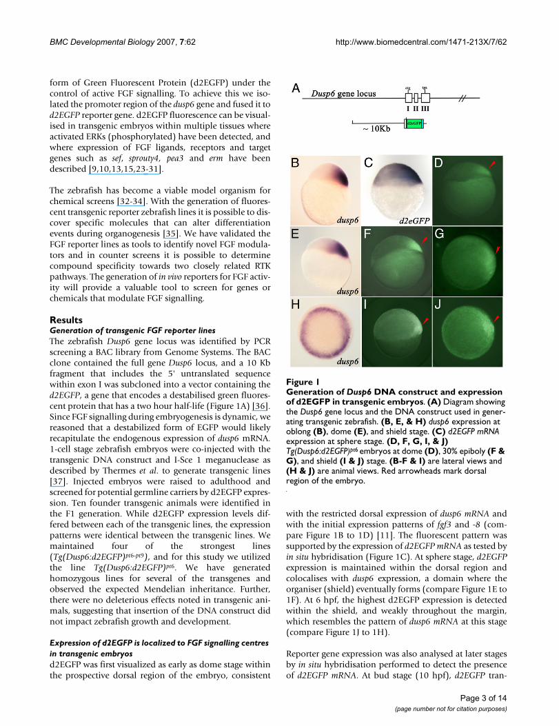
ResultsGeneration of transgenic FGF reporter linesThe zebrafish Dusp6 gene locus was identified by PCRscreening a BAC library from Genome Systems. The BACclone contained the full gene Dusp6 locus, and a 10 Kbfragment that includes the 5' untranslated sequencewithin exon I was subcloned into a vector containing thed2EGFP, a gene that encodes a destabilised green fluores-cent protein that has a two hour half-life (Figure 1A) [36].Since FGF signalling during embryogenesis is dynamic, wereasoned that a destabilized form of EGFP would likelyrecapitulate the endogenous expression of dusp6 mRNA.1-cell stage zebrafish embryos were co-injected with thetransgenic DNA construct and I-Sce 1 meganuclease asdescribed by Thermes et al. to generate transgenic lines[37]. Injected embryos were raised to adulthood andscreened for potential germline carriers by d2EGFP expres-sion. Ten founder transgenic animals were identified inthe F1 generation. While d2EGFP expression levels dif-fered between each of the transgenic lines, the expressionpatterns were identical between the transgenic lines. Wemaintained four of the strongest lines(Tg(Dusp6:d2EGFP)pt6-pt9), and for this study we utilizedthe line Tg(Dusp6:d2EGFP)pt6. We have generatedhomozygous lines for several of the transgenes andobserved the expected Mendelian inheritance. Further,there were no deleterious effects noted in transgenic ani-mals, suggesting that insertion of the DNA construct didnot impact zebrafish growth and development.
Expression of d2EGFP is localized to FGF signalling centres in transgenic embryosd2EGFP was first visualized as early as dome stage withinthe prospective dorsal region of the embryo, consistent
with the restricted dorsal expression of dusp6 mRNA andwith the initial expression patterns of fgf3 and -8 (com-pare Figure 1B to 1D) [11]. The fluorescent pattern wassupported by the expression of d2EGFP mRNA as tested byin situ hybridisation (Figure 1C). At sphere stage, d2EGFPexpression is maintained within the dorsal region andcolocalises with dusp6 expression, a domain where theorganiser (shield) eventually forms (compare Figure 1E to1F). At 6 hpf, the highest d2EGFP expression is detectedwithin the shield, and weakly throughout the margin,which resembles the pattern of dusp6 mRNA at this stage(compare Figure 1J to 1H).
Reporter gene expression was also analysed at later stagesby in situ hybridisation performed to detect the presenceof d2EGFP mRNA. At bud stage (10 hpf), d2EGFP tran-
Generation of Dusp6 DNA construct and expression of d2EGFP in transgenic embryosFigure 1Generation of Dusp6 DNA construct and expression of d2EGFP in transgenic embryos. (A) Diagram showing the Dusp6 gene locus and the DNA construct used in gener-ating transgenic zebrafish. (B, E, & H) dusp6 expression at oblong (B), dome (E), and shield stage. (C) d2EGFP mRNA expression at sphere stage. (D, F, G, I, & J) Tg(Dusp6:d2EGFP)pt6 embryos at dome (D), 30% epiboly (F & G), and shield (I & J) stage. (B-F & I) are lateral views and (H & J) are animal views. Red arrowheads mark dorsal region of the embryo.
Page 3 of 14(page number not for citation purposes)
BMC Developmental Biology 2007, 7:62 http://www.biomedcentral.com/1471-213X/7/62
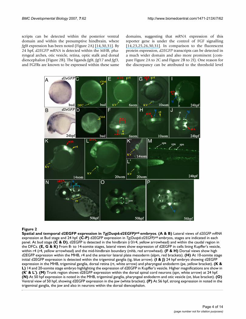
scripts can be detected within the posterior ventraldomain and within the presumptive hindbrain, wherefgf8 expression has been noted (Figure 2A) [14,30,31]. By24 hpf, d2EGFP mRNA is detected within the MHB, pha-ryngeal arches, otic vesicle, retina, optic stalk and dorsaldiencephalon (Figure 2B). The ligands fgf8, fgf17 and fgf3,and FGFRs are known to be expressed within these same
domains, suggesting that mRNA expression of thisreporter gene is under the control of FGF signalling[14,23,25,26,30,31]. In comparison to the fluorescentprotein expression, d2EGFP transcripts can be detected ina much wider domain and also more prominent (com-pare Figure 2A to 2C and Figure 2B to 2I). One reason forthe discrepancy can be attributed to the threshold level
Spatial and temporal d2EGFP expression in Tg(Dusp6:d2EGFP)pt6 embryosFigure 2Spatial and temporal d2EGFP expression in Tg(Dusp6:d2EGFP)pt6 embryos. (A & B) Lateral views of d2EGFP mRNA expression at Bud stage and 24 hpf. (C-P) d2EGFP expression in Tg(Dusp6:d2EGFP)pt6 embryos, stages are indicated in each panel. At bud stage (C & D), d2EGFP is detected in the hindbrain (r3/r4, yellow arrowhead) and within the caudal region in the DFCs. (E, G & K) From 8- to 14-somite stages, lateral views show expression of d2EGFP in cells lining Kupffer's vesicle, within r4 (r4, yellow arrowhead) and the mid-hindbrain boundary (mhb, red arrowhead). (F & H) Dorsal views show high d2EGFP expression within the MHB, r4 and the anterior lateral plate mesoderm (alpm, red brackets). (H) At 10-somite stage initial d2EGFP expression is detected within the trigeminal ganglia (tg, blue arrow). (I & J) 24 hpf embryo showing d2EGFP expression in the MHB, trigeminal ganglia, dorsal retina (rt, white arrow) and pharyngeal endoderm (pe, yellow bracket). (K & L) 14 and 20-somite stage embryo highlighting the expression of d2EGFP in Kupffer's vesicle. Higher magnifications are show in (K' & L'). (M) Trunk region shows d2EGFP expression within the dorsal spinal cord neurons (spn, white arrow) at 24 hpf. (N) At 50 hpf expression is noted in the MHB, trigeminal ganglia, pharyngeal endoderm and otic vesicle (ot, blue bracket). (O) Ventral view of 50 hpf, showing d2EGFP expression in the jaw (white bracket). (P) At 56 hpf, strong expression in noted in the trigeminal ganglia, the jaw and also in neurons within the dorsal diencephalon.
Page 4 of 14(page number not for citation purposes)
BMC Developmental Biology 2007, 7:62 http://www.biomedcentral.com/1471-213X/7/62
required for visualisation of d2EGFP protein as comparedto the detection of transcripts by in situ hybridisation.
At bud stage, fluorescent protein can be visualized withinthe medial neural plate, presumptive hindbrain and at aposition that corresponds to the site where the DorsalForerunner Cells (DFCs) in the posterior domain of trans-genic embryos have been described (Figure 2C &2D)[38,39]. The DFCs represent a specialized group of non-involuting cells that migrate just ahead of the shield dur-ing gastrulation and eventually forms a fluid filled struc-ture known as Kupffer's vesicle, a transient organparticular to teleosts [38,39]. Kupffer's vesicle is thoughtto serve equivalent functions in establishing left-rightpolarity as the mouse node [40-43].
By the 6-somite stage, strong d2EGFP fluorescence can bedetected in the MHB, the hindbrain with strongest expres-sion in r4, the anterior lateral plate mesoderm and theKupffer's vesicle (Figure 2E &2F). Approximately twohours later at the 10-somite stage expression of d2EGFP islocated within the same domains as described for the 6-somite stage with the addition of the trigeminal ganglia(Figure 2G &2H). The expression within Kupffer's vesicleis quite striking as the structure is completely outlined byd2EGFP positive cells from the 6-somite onwards (Figure2C, 2E, 2G, 2K and 2K'). In the zebrafish, a role for FGFsignalling has been suggested in the formation ofKupffer's vesicle, as fgf8 is expressed in the DFCs at gastru-lation, and this structure is absent in about 30% oface(fgf8) mutants [44]. The Tg(Dusp6:d2EGFP)pt6 line con-firms that the DFCs and Kupffer's vesicle receive FGF sig-nals from the time when the DFCs begin to coalesce andright through to the 20-somite stage when Kupffer's vesi-cle begins to collapse (Figure 2K, 2L, 2K', &2L'). Timelapse imaging of the transgenic embryos shows fluores-cent DFCs migration towards the posterior region of theembryo and the formation of Kupffer's Vesicle at the 6-somite stage [see Additional files 1, 2, 3 and 4]. d2EGFPexpression is still noted throughout these stages and evenafter the collapse of Kupffer's vescles when these epithelialcells migrate towards the tail bud and contribute to meso-dermal tissues such as notochord, posterior somites andthe tail bud (see Additional files 2, 3 and 4) [38,39]. Bythe 26-somite stage, the Kupffer's vesicle cells have losttheir expression of d2EGFP, suggesting that these cells nolonger receive FGF signals (data not shown).
At 24 hpf, d2EGFP can be visualized within the dorsal ret-ina, trigeminal ganglia, otic vesicles, within the dorsaldiencephalon, and dorsal spinal cord neurons (Figure 2I,2J &2M). These domains of d2EGFP expression are con-sistent with expression of FGF ligands, receptors andknown target genes such as erm, pea3, sef and sprouty4,suggesting that the transgenic line reports on FGF activity
in vivo [13,15,24,27-29]. d2EGFP expression persiststhroughout the next day of development in a majority ofthe same domains as at 24 hpf and continues up to 56 hpf(Figure 2N, 2O &2P). The one exception is that dorsal ret-ina expression is lost, while lens expression becomesstronger from 36 hpf onwards (Figure 2N). Expression atlater stages can also be detected within the developing jawand further refined within the pharyngeal arches and thepectoral fins (Figure 2N &2O, pectoral fins not shown). Adorsal view of a 56 hpf transgenic embryo reveals d2EGFPexpression within distinct cells in the dorsal dien-cephalon, suggesting that these particular cells areresponding to FGF signals (Figure 2P). Of particular inter-est is that there seems to be an asymmetric distribution offluorescent cells with respect to the left-right axis, imply-ing that within this region of the diencephalon, FGF sig-nalling is asymmetrical (Figure 2P). An alternative view isthat the asymmetric expression of d2EGFP is a result ofanatomical or stochastic (random) differences betweenthe left and right side of the diencephalon. Studies haveshown that the left habenular nuclei is larger and the par-apineal gland is situated to the left of the midline, suggest-ing that d2EGFP expression reflects structural differencebetween the left and right dorsal diencephalon [45,46].
The domains of d2EGFP expression in the transgenicembryos were consistent with many of the regions whereendogenous dusp6 transcripts have been detected. How-ever, dusp6 mRNA expression was not fully recapitulatedin the transgenic embryos, in particular FGF activity anddusp6 expression have been well documented within thesomites and tail bud and yet no expression of d2EGFPmRNA or protein were detected in these regions (Figure 2)[11]. One explanation is that only 10 Kb of upstreamDusp6 promoter sequence was used to generate the DNAconstruct for transgenesis, hence it is likely that somiticand tail bud enhancers were not present within thissequence.
In summary we have generated several transgenic linesthat express d2EGFP under the control of the Dusp6 pro-moter, a gene that is directly regulated by FGF signallingduring development. Since d2EGFP has a half life of justtwo hours, it is likely that domains of high d2EGFPexpression represents cells that respond to active FGF sig-nalling as opposed to cells inheriting fluorescent proteinfrom their ancestors.
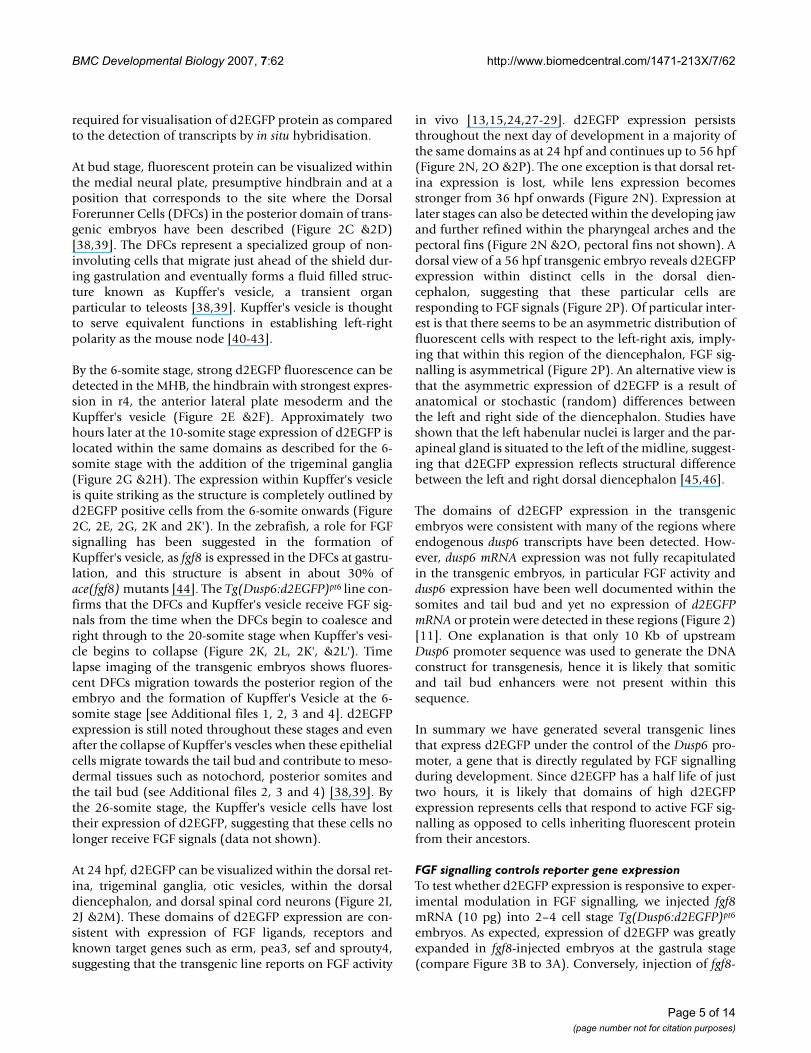
FGF signalling controls reporter gene expressionTo test whether d2EGFP expression is responsive to exper-imental modulation in FGF signalling, we injected fgf8mRNA (10 pg) into 2–4 cell stage Tg(Dusp6:d2EGFP)pt6
embryos. As expected, expression of d2EGFP was greatlyexpanded in fgf8-injected embryos at the gastrula stage(compare Figure 3B to 3A). Conversely, injection of fgf8-
Page 5 of 14(page number not for citation purposes)
BMC Developmental Biology 2007, 7:62 http://www.biomedcentral.com/1471-213X/7/62
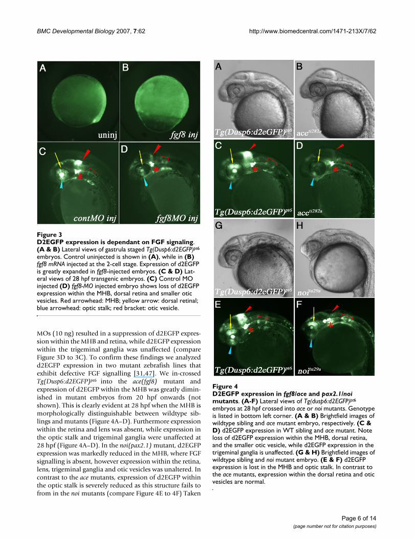
MOs (10 ng) resulted in a suppression of d2EGFP expres-sion within the MHB and retina, while d2EGFP expressionwithin the trigeminal ganglia was unaffected (compareFigure 3D to 3C). To confirm these findings we analyzedd2EGFP expression in two mutant zebrafish lines thatexhibit defective FGF signalling [31,47]. We in-crossedTg(Dusp6:d2EGFP)pt6 into the ace(fgf8) mutant andexpression of d2EGFP within the MHB was greatly dimin-ished in mutant embryos from 20 hpf onwards (notshown). This is clearly evident at 28 hpf when the MHB ismorphologically distinguishable between wildtype sib-lings and mutants (Figure 4A–D). Furthermore expressionwithin the retina and lens was absent, while expression inthe optic stalk and trigeminal ganglia were unaffected at28 hpf (Figure 4A–D). In the noi(pax2.1) mutant, d2EGFPexpression was markedly reduced in the MHB, where FGFsignalling is absent, however expression within the retina,lens, trigeminal ganglia and otic vesicles was unaltered. Incontrast to the ace mutants, expression of d2EGFP withinthe optic stalk is severely reduced as this structure fails tofrom in the noi mutants (compare Figure 4E to 4F) Taken
D2EGFP expression in fgf8/ace and pax21/noi mutantsFigure 4D2EGFP expression in fgf8/ace and pax2.1/noi mutants. (A-F) Lateral views of Tg(dusp6:d2EGFP)pt6
embryos at 28 hpf crossed into ace or noi mutants. Genotype is listed in bottom left corner. (A & B) Brightfield images of wildtype sibling and ace mutant embryo, respectively. (C & D) d2EGFP expression in WT sibling and ace mutant. Note loss of d2EGFP expression within the MHB, dorsal retina, and the smaller otic vesicle, while d2EGFP expression in the trigeminal ganglia is unaffected. (G & H) Brightfield images of wildtype sibling and noi mutant embryo. (E & F) d2EGFP expression is lost in the MHB and optic stalk. In contrast to the ace mutants, expression within the dorsal retina and otic vesicles are normal.
D2EGFP expression is dependant on FGF signalingFigure 3D2EGFP expression is dependant on FGF signaling. (A & B) Lateral views of gastrula staged Tg(Dusp6:d2EGFP)pt6
embryos. Control uninjected is shown in (A), while in (B) fgf8 mRNA injected at the 2-cell stage. Expression of d2EGFP is greatly expanded in fgf8-injected embryos. (C & D) Lat-eral views of 28 hpf transgenic embryos. (C) Control MO injected (D) fgf8-MO injected embryo shows loss of d2EGFP expression within the MHB, dorsal retina and smaller otic vesicles. Red arrowhead: MHB; yellow arrow: dorsal retinal; blue arrowhead: optic stalk; red bracket: otic vesicle.
Page 6 of 14(page number not for citation purposes)
BMC Developmental Biology 2007, 7:62 http://www.biomedcentral.com/1471-213X/7/62
together, these results show that we have generated atransgenic reporter line that expresses d2EGFP under thecontrol of FGF signalling during development.
Treatment of Tg(Dusp6:d2EGFP)pt6 embryos with known FGF pathway inhibitorsGiven that Tg(Dusp6:d2EGFP)pt6 embryos respond tochanges in FGF signalling, these embryos can provide avaluable tool to screen for molecules that modulate FGFsignalling in vivo. To validate that these reporter lines can
measure changes in FGF activity in a chemical screen,Tg(Dusp6:d2EGFP)pt6 embryos were treated with knowninhibitors of the FGF pathway. For our studies we selectedtransgenic embryos at 24 hpf and treated with com-pounds for 8 hours. We reasoned that since the half-life ofd2EGFP is 2 hours, then an inhibitor of FGF signallingshould substantially reduce d2EGFP fluorescence after 6–8 hours of treatment. Furthermore, since embryos at thisdevelopmental stage (24 hpf) have already formed manyof the structures that express d2EGFP such as the MHB,trigeminal ganglia and otic vesicles, then the assay woulddirectly measure loss of d2EGFP fluorescence as a conse-quence of FGF inhibition and not per se a loss of these tis-sues.
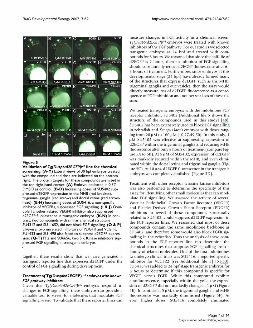
We treated transgenic embryos with the indolinone FGFreceptor inhibitor, SU5402 [Additional file 5 shows thestructure of the compounds used in this study] [48].SU5402 has been extensively used to block FGF signallingin zebrafish and Xenopus laevis embryos with doses rang-ing from 20 μM to 160 μM [10,27,49,50]. In this study, 1μM SU5402 was effective at suppressing expression ofd2EGFP within the trigeminal ganglia and reducing MHBfluorescence after only 8 hours of treatment (compare Fig-ure 5A to 5B). At 5 μM of SU5402, expression of d2EGFPwas markedly reduced within the MHB, and even elimi-nated within the dorsal retina and trigeminal ganglia (Fig-ure 5C). At 10 μM, d2EGFP fluorescence in the transgenicembryos was completely abolished (Figure 5D).
Treatment with other receptor tyrosine kinase inhibitorswas also performed to determine the specificity of thisassay for identifying other small molecules that can mod-ulate FGF signalling. We assessed the activity of severalVascular Endothelial Growth Factor Receptor (VEGFR)and Platelet Derived Growth Factor Receptor (PDGFR)inhibitors to reveal if these compounds, structurallyrelated to SU5402, could suppress d2EGFP expression inthe FGF reporter lines. We reasoned that most of thesecompounds contain the same indolinone backbone asSU5402, and therefore some would also block FGFR sig-nalling in the zebrafish. Thus the analysis of these com-pounds in the FGF reporter line can determine thechemical structures that suppress FGF signalling from afamily of related molecules. One of the first indolinonesto undergo clinical trials was SU5416, a reported specificinhibitor for VEGFR2 [see Additional file 5] [51-53].SU5416 was added to 24 hpf stage transgenic embryos for6 hours to determine if this compound is specific forVEGFR versus FGFR. While this compound exhibitsautofluorescence, especially within the yolk, the expres-sion of d2EGFP did not markedly change at 1 μM (Figure5E). In contrast at 5 μM, the trigeminal ganglia and MHBfluorescence was markedly diminished (Figure 5F). Ateven higher doses, SU5416 completely eliminated
Validation of Tg(Dusp6:d2EGFP)pt6 line for chemical screeningFigure 5Validation of Tg(Dusp6:d2EGFP)pt6 line for chemical screening. (A-Y) Lateral views of 30 hpf embryos treated with the compound and dose are indicated on the bottom right. The protein targets for these compounds are listed in the top right hand corner. (A) Embryo incubated in 0.5% DMSO as control. (B-D) Increasing doses of SU5402 sup-pressed d2EGFP expression in the MHB (red bracket), trigeminal ganglia (red arrow) and dorsal retina (red arrow-head). (E-H) Increasing doses of SU5416, a non-specific inhibitor of VEGFRs, suppressed FGF signalling. (I & J) Oxin-dole I another related VEGFR inhibitor also suppressed d2EGFP fluorescence in transgenic embryos. (K-N) In con-trast, two compounds with similar chemical structure SU4312 and SU11652, did not block FGF signalling. (O & P) Likewise, two unrelated inhibitors of PDGFR and VEGFR, SU1433 and SU1498 also failed to suppress d2EGFP expres-sion. (Q-T) PP2 and SU6656, two Src Kinase inhibitors sup-pressed FGF signalling in transgenic embryos.
Page 7 of 14(page number not for citation purposes)
BMC Developmental Biology 2007, 7:62 http://www.biomedcentral.com/1471-213X/7/62
d2EGFP expression within the transgenic embryos, eventhough the treated embryos exhibited strong fluorescentyolks, the expression of d2EGFP within the MHB wasclearly eliminated (Figure 5G &5H). These results suggestthat SU5416 is not completely specific to VEGFR2 at theseconcentrations and it can block FGF signalling. Similarly,Oxindole I was also effective at blocking d2EGFP expres-sion, however a much higher dose (40 μM) was required[see Additional file 5], and (Figure 5I &5J). SU4312 repre-sents another indolinone inhibitor of VEGFR, which hasbeen shown to block autophosphorylation of VEGFRs[54]. In contrast to SU5416, treatment up to 40 μM ofSU4312 had little effect in the expression of d2EGFPwithin the MHB and trigeminal ganglia in theTg(Dusp6:d2EGFP)pt6 embryos [see Additional file 5] and(Figure 5K &5L). A newer generation of indolinone com-pound was also tested to determine its specificity towardsVEGFR/PDGFR versus FGFR. SU11652, has been shownto have at least 100-fold greater inhibitory effect targetinga VEGFR and PDGFR versus FGFR [see Additional file 5][55,56]. In our assays, SU11652 did not alter d2EGFPexpression suggesting that this compound does not blockFGF signalling (Figure 5M &5N). We also treated trans-genic embryos with two structurally unrelated VEGF sig-nalling inhibitors, SU1498 and SU1433 (also known asAG1433) to determine the specificity of these molecules[see Additional file 5] [57-59]. Both compounds havebeen shown to be effective at blocking VEGFR, and to alesser extent basic FGF in HUVEC tubulogenesis assays[58]. Treatment with high doses of SU1433 or SU1498did not alter d2EGFP fluorescence in the transgenicembryos, suggesting that these chemically divergent com-pounds do not block FGF signalling in the zebrafish at thedoses indicated (Figure 5O &5P).
We next determined the activity of Src Kinase inhibitorsand their role in FGF signalling in the early embryo as pre-vious reports have shown that several Src family membersfunction to relay FGF signalling [60,61]. PP2, an inhibitorof several Src Kinases including Fyn and Lck as well asPDGFRs and Bcr-Abl, only mildly altered d2EGFP expres-sion at 20 μM (Figure 5Q), a dose that is several foldhigher than what has been shown to inhibit Fyn and Lckin cell culture [62,63]. SU6656, a more specific inhibitorof Src kinases, exhibited robust inhibitory activity in theseassays. At concentrations of 1 μM, expression of d2EGFPwas completely abolished within the MHB, trigeminalganglia and the retina. The concentration of SU6656 waswithin the range that is known to be specific for blockingmouse Src kinase activity and not other related tyrosinekinases (Figure 5R–T) [63]. In fact 1 μM SU6656 is likelyto inhibit only the Src members, Yes, Fyn, Src and Lyn,with Yes kinase exhibiting higher sensitivity, suggestingthat Yes is required for FGF signalling in the zebrafishembryo [63].
The results from these experiments support the use oftransgenic FGF reporter lines to screen for small moleculesthat affect FGF signalling. Given the rapid nature of thesescreens as d2EGFP expression can be suppressed in just 6hours, it is likely that a chemical screen would identifycompounds that directly modulate FGF signalling.
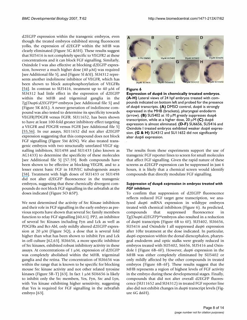
Suppression of dusp6 expression in embryos treated with FGF inhibitorsTo confirm that suppression of d2EGFP fluorescencereflects reduced FGF target gene transcription, we ana-lyzed dusp6 mRNA expression in wildtype embryostreated with chemical inhibitors (Figure 6). As predicted,compounds that suppressed fluorescence inTg(Dusp6:d2EGFP)pt6embryos also resulted in a reductionof dusp6 transcripts (Figure 6B to 6F). SU5402, SU6656,SU5416 and Oxindole I all suppressed dusp6 expressionafter 10hr treatment at the dose indicated. In particular,dusp6 expression within the dorsal diencephalon, pharyn-geal endoderm and optic stalks were greatly reduced inembryos treated with SU5402, S6656, SU5416 and Oxin-dole I (Figure 6B–6F). However, dusp6 expression in theMHB was either completely eliminated by SU5402 oronly mildly affected by the other compounds in treatedembryos (Figure 6B–6F). These results suggest that theMHB represents a region of highest levels of FGF activityin the embryo during these developmental stages. Finally,compounds that did not alter overall d2EGFP fluores-cence (SU11652 and SU4312) in treated FGF reporter linealso did not exhibit changes in dusp6 transcript levels (Fig-ure 6G &6H).
Expression of dusp6 in chemically treated embryosFigure 6Expression of dusp6 in chemically treated embryos. (A-H) Lateral views of 24 hpf embryos treated with com-pounds indicated on bottom left and probed for the presence of dusp6 transcripts. (A) DMSO control, dusp6 is strongly expressed in the MHB (brackets), pharyngeal endoderm (arrow). (B) SU5402 at 10 μM greatly suppresses dusp6 transcription, while at a higher dose, 20 μM (C) dusp6 expression is almost eliminated. (D-F) SU6656, SU5416 and Oxindole I treated embryos exhibited weaker dusp6 expres-sion. (G & H) SU4312 and SU11652 did not significantly alter dusp6 expression.
Page 8 of 14(page number not for citation purposes)
BMC Developmental Biology 2007, 7:62 http://www.biomedcentral.com/1471-213X/7/62
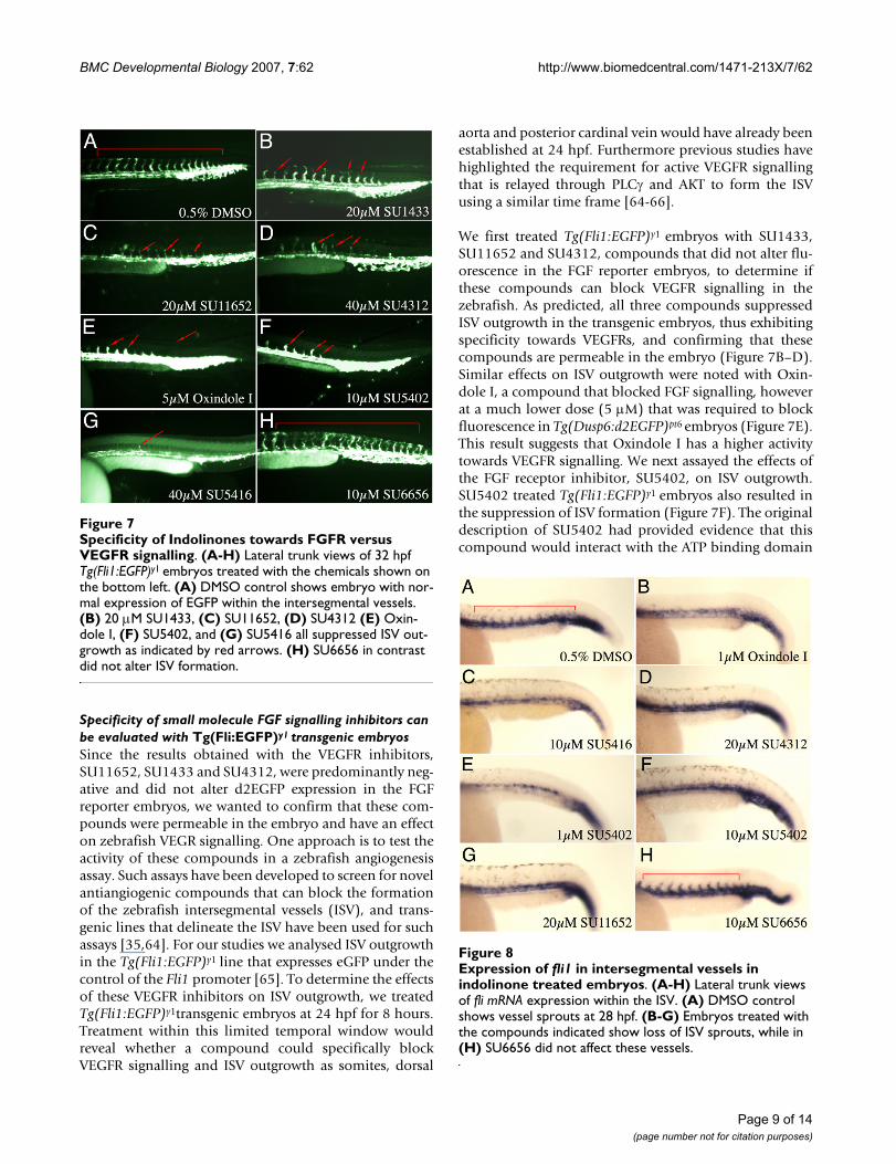
Specificity of small molecule FGF signalling inhibitors can be evaluated with Tg(Fli:EGFP)y1 transgenic embryosSince the results obtained with the VEGFR inhibitors,SU11652, SU1433 and SU4312, were predominantly neg-ative and did not alter d2EGFP expression in the FGFreporter embryos, we wanted to confirm that these com-pounds were permeable in the embryo and have an effecton zebrafish VEGR signalling. One approach is to test theactivity of these compounds in a zebrafish angiogenesisassay. Such assays have been developed to screen for novelantiangiogenic compounds that can block the formationof the zebrafish intersegmental vessels (ISV), and trans-genic lines that delineate the ISV have been used for suchassays [35,64]. For our studies we analysed ISV outgrowthin the Tg(Fli1:EGFP)y1 line that expresses eGFP under thecontrol of the Fli1 promoter [65]. To determine the effectsof these VEGFR inhibitors on ISV outgrowth, we treatedTg(Fli1:EGFP)y1transgenic embryos at 24 hpf for 8 hours.Treatment within this limited temporal window wouldreveal whether a compound could specifically blockVEGFR signalling and ISV outgrowth as somites, dorsal
aorta and posterior cardinal vein would have already beenestablished at 24 hpf. Furthermore previous studies havehighlighted the requirement for active VEGFR signallingthat is relayed through PLCγ and AKT to form the ISVusing a similar time frame [64-66].
We first treated Tg(Fli1:EGFP)y1 embryos with SU1433,SU11652 and SU4312, compounds that did not alter flu-orescence in the FGF reporter embryos, to determine ifthese compounds can block VEGFR signalling in thezebrafish. As predicted, all three compounds suppressedISV outgrowth in the transgenic embryos, thus exhibitingspecificity towards VEGFRs, and confirming that thesecompounds are permeable in the embryo (Figure 7B–D).Similar effects on ISV outgrowth were noted with Oxin-dole I, a compound that blocked FGF signalling, howeverat a much lower dose (5 μM) that was required to blockfluorescence in Tg(Dusp6:d2EGFP)pt6 embryos (Figure 7E).This result suggests that Oxindole I has a higher activitytowards VEGFR signalling. We next assayed the effects ofthe FGF receptor inhibitor, SU5402, on ISV outgrowth.SU5402 treated Tg(Fli1:EGFP)y1 embryos also resulted inthe suppression of ISV formation (Figure 7F). The originaldescription of SU5402 had provided evidence that thiscompound would interact with the ATP binding domain
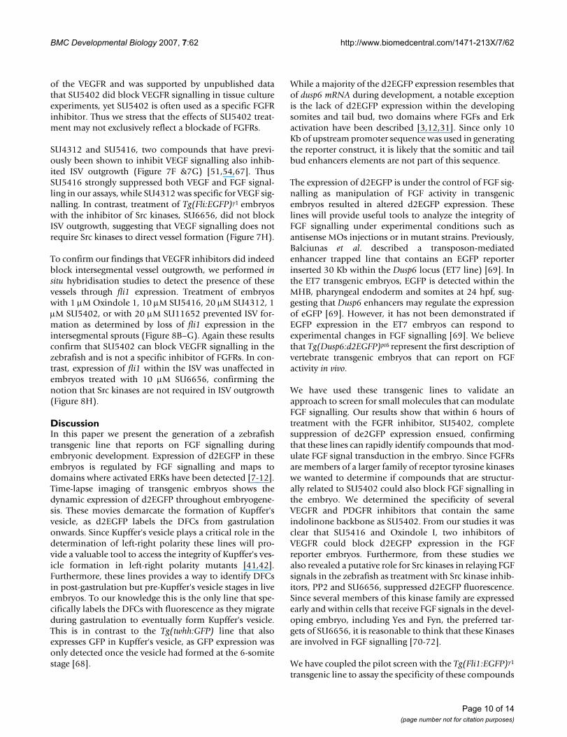
Expression of fli1 in intersegmental vessels in indolinone treated embryosFigure 8Expression of fli1 in intersegmental vessels in indolinone treated embryos. (A-H) Lateral trunk views of fli mRNA expression within the ISV. (A) DMSO control shows vessel sprouts at 28 hpf. (B-G) Embryos treated with the compounds indicated show loss of ISV sprouts, while in (H) SU6656 did not affect these vessels.
Specificity of Indolinones towards FGFR versus VEGFR sig-nallingFigure 7Specificity of Indolinones towards FGFR versus VEGFR signalling. (A-H) Lateral trunk views of 32 hpf Tg(Fli1:EGFP)y1 embryos treated with the chemicals shown on the bottom left. (A) DMSO control shows embryo with nor-mal expression of EGFP within the intersegmental vessels. (B) 20 μM SU1433, (C) SU11652, (D) SU4312 (E) Oxin-dole I, (F) SU5402, and (G) SU5416 all suppressed ISV out-growth as indicated by red arrows. (H) SU6656 in contrast did not alter ISV formation.
Page 9 of 14(page number not for citation purposes)
BMC Developmental Biology 2007, 7:62 http://www.biomedcentral.com/1471-213X/7/62
of the VEGFR and was supported by unpublished datathat SU5402 did block VEGFR signalling in tissue cultureexperiments, yet SU5402 is often used as a specific FGFRinhibitor. Thus we stress that the effects of SU5402 treat-ment may not exclusively reflect a blockade of FGFRs.
SU4312 and SU5416, two compounds that have previ-ously been shown to inhibit VEGF signalling also inhib-ited ISV outgrowth (Figure 7F &7G) [51,54,67]. ThusSU5416 strongly suppressed both VEGF and FGF signal-ling in our assays, while SU4312 was specific for VEGF sig-nalling. In contrast, treatment of Tg(Fli:EGFP)y1 embryoswith the inhibitor of Src kinases, SU6656, did not blockISV outgrowth, suggesting that VEGF signalling does notrequire Src kinases to direct vessel formation (Figure 7H).
To confirm our findings that VEGFR inhibitors did indeedblock intersegmental vessel outgrowth, we performed insitu hybridisation studies to detect the presence of thesevessels through fli1 expression. Treatment of embryoswith 1 μM Oxindole 1, 10 μM SU5416, 20 μM SU4312, 1μM SU5402, or with 20 μM SU11652 prevented ISV for-mation as determined by loss of fli1 expression in theintersegmental sprouts (Figure 8B–G). Again these resultsconfirm that SU5402 can block VEGFR signalling in thezebrafish and is not a specific inhibitor of FGFRs. In con-trast, expression of fli1 within the ISV was unaffected inembryos treated with 10 μM SU6656, confirming thenotion that Src kinases are not required in ISV outgrowth(Figure 8H).
DiscussionIn this paper we present the generation of a zebrafishtransgenic line that reports on FGF signalling duringembryonic development. Expression of d2EGFP in theseembryos is regulated by FGF signalling and maps todomains where activated ERKs have been detected [7-12].Time-lapse imaging of transgenic embryos shows thedynamic expression of d2EGFP throughout embryogene-sis. These movies demarcate the formation of Kupffer'svesicle, as d2EGFP labels the DFCs from gastrulationonwards. Since Kupffer's vesicle plays a critical role in thedetermination of left-right polarity these lines will pro-vide a valuable tool to access the integrity of Kupffer's ves-icle formation in left-right polarity mutants [41,42].Furthermore, these lines provides a way to identify DFCsin post-gastrulation but pre-Kupffer's vesicle stages in liveembryos. To our knowledge this is the only line that spe-cifically labels the DFCs with fluorescence as they migrateduring gastrulation to eventually form Kupffer's vesicle.This is in contrast to the Tg(twhh:GFP) line that alsoexpresses GFP in Kupffer's vesicle, as GFP expression wasonly detected once the vesicle had formed at the 6-somitestage [68].
While a majority of the d2EGFP expression resembles thatof dusp6 mRNA during development, a notable exceptionis the lack of d2EGFP expression within the developingsomites and tail bud, two domains where FGFs and Erkactivation have been described [3,12,31]. Since only 10Kb of upstream promoter sequence was used in generatingthe reporter construct, it is likely that the somitic and tailbud enhancers elements are not part of this sequence.
The expression of d2EGFP is under the control of FGF sig-nalling as manipulation of FGF activity in transgenicembryos resulted in altered d2EGFP expression. Theselines will provide useful tools to analyze the integrity ofFGF signalling under experimental conditions such asantisense MOs injections or in mutant strains. Previously,Balciunas et al. described a transposon-mediatedenhancer trapped line that contains an EGFP reporterinserted 30 Kb within the Dusp6 locus (ET7 line) [69]. Inthe ET7 transgenic embryos, EGFP is detected within theMHB, pharyngeal endoderm and somites at 24 hpf, sug-gesting that Dusp6 enhancers may regulate the expressionof eGFP [69]. However, it has not been demonstrated ifEGFP expression in the ET7 embryos can respond toexperimental changes in FGF signalling [69]. We believethat Tg(Dusp6:d2EGFP)pt6 represent the first description ofvertebrate transgenic embryos that can report on FGFactivity in vivo.
We have used these transgenic lines to validate anapproach to screen for small molecules that can modulateFGF signalling. Our results show that within 6 hours oftreatment with the FGFR inhibitor, SU5402, completesuppression of de2GFP expression ensued, confirmingthat these lines can rapidly identify compounds that mod-ulate FGF signal transduction in the embryo. Since FGFRsare members of a larger family of receptor tyrosine kinaseswe wanted to determine if compounds that are structur-ally related to SU5402 could also block FGF signalling inthe embryo. We determined the specificity of severalVEGFR and PDGFR inhibitors that contain the sameindolinone backbone as SU5402. From our studies it wasclear that SU5416 and Oxindole I, two inhibitors ofVEGFR could block d2EGFP expression in the FGFreporter embryos. Furthermore, from these studies wealso revealed a putative role for Src kinases in relaying FGFsignals in the zebrafish as treatment with Src kinase inhib-itors, PP2 and SU6656, suppressed d2EGFP fluorescence.Since several members of this kinase family are expressedearly and within cells that receive FGF signals in the devel-oping embryo, including Yes and Fyn, the preferred tar-gets of SU6656, it is reasonable to think that these Kinasesare involved in FGF signalling [70-72].
We have coupled the pilot screen with the Tg(Fli1:EGFP)y1
transgenic line to assay the specificity of these compounds
Page 10 of 14(page number not for citation purposes)
BMC Developmental Biology 2007, 7:62 http://www.biomedcentral.com/1471-213X/7/62
between FGFR and VEGFR signalling. We confirm thatSU5416, SU4312, SU11652, Oxindole I and surprisinglySU5402 exhibited inhibitory effects on ISV outgrowth andprobably a result of VEGFR inhibition. In the originalstudy, a crystal structure of SU5402 complexed with theFGFR1 kinase domain highlighted the exact peptide resi-dues that interfaced with the compound [48]. Since theseresidues are also highly conserved with the VEGFRs kinasedomain, it was postulated that SU5402 could interactwith the VEGFRs [48]. Furthermore the authors refer tounpublished observations that SU5402 did inhibitVEGFR signalling in living cells [48]. Our studies revealthat SU5402 can block ISV outgrowth, which we interpretas a result of VEGFR inhibition in the zebrafish embryo.Alternatively, our data could imply that FGFRs arerequired for ISV outgrowths given that FGFs are known toplay a role in angiogenesis [53]. However from the pub-lished literature, FGFR1-4 are not expressed in the ISV tosupport the role for this pathway in vasculogenesis[25,26]. SU5402 has been used extensively as a FGFRinhibitor in many studies. In light of these findings, otherexperiments should be considered to test whether SU5402might also block VEGFR signalling to elicit the observedphenotypes.
ConclusionThis study describes the generation of transgeniczebrafish, Tg(Dusp6:d2EGFP)pt6, that reports on FGF activ-ity during development. The expression of d2EGFP mir-rors the expression of several FGF ligands in the earlyembryo and will provide a tool to analyse FGF signallingunder various experimental conditions. We have per-formed a pilot screen to validate these lines in chemicalscreens to identify novel compounds that can modulateFGF activity. This rapid screening protocol can be coupledwith the Tg(Fli1:EGFP)y1 line to eliminate compoundsthat can potentially cross react with the VEGFR pathway.Finally, acridine orange staining will further eliminatetoxic compounds, thus by following these procedures it ispossible to identify chemicals that specifically modulateFGF signalling in vivo.
MethodsGeneration of Tg(Dusp6:d2EGFP) zebrafishA Dusp6 BAC clone was identified by PCR from BAC DNApools as directed by manufacturers protocols (GenomeSystems). A 10 Kb Kpn1 fragment was identified that con-tain parts of Exon 1 (441 bp) and approximately 9.5 Kb ofupstream promoter sequence (see Figure 1A). This Kpn1fragment was subcloned into the Kpn1 site ofpSce1d2EGFP vector (pSce1 vector with d2EGFP cDNAcloned into the multiple cloning site). 20 pg ofpDusp6:d2EGFP plasmid DNA was injected into the 1-cellembryo with I-Sce 1 (New England Biolabs, Ipswich, MA)restriction enzyme as described in [37]. These Founder F0
injected embryos were raised to adulthood and incrossedto identify transgenic founders. We identified 10 founderlines that expressed d2EGFP within regions of known FGFactivity in the developing embryo. Four lines exhibitedstrong expression throughout development and weremaintained. Tg(Dusp6:d2EGFP)pt6 was used predomi-nantly in this study.
Zebrafish Microinjection of RNA and antisense Morpholinos10 pg fgf8 mRNA, 20 ng control-MO(5'-CCTCTTACCT-CAGTTACAATTTATA-3'), and 10 ng fgf8-MO (5'-GAGTCTCATGTTTATAGCCTCAGTA-3') was injected intothe 2-cell stage transgenic embryos as described by Tsanget. al and Araki et. al, respectively [13,73]. Morpholinoswere obtained from Gene-tools inc. (Philomath, OR).Embryos were incubated for the desired stage before visu-alisation under a Leica stereomicroscope and photo-graphed by a Retiga Exi camera (Qimaging, Burnaby, BCCanada). Images were analyzed in Photoshop CS (Adobe,San Jose, CA).
In situ hybridisationZebrafish embryos were fixed in 4% paraformaldehydeand whole mount in situ hybridisation was preformedwith d2EGFP, dusp6 and fli1 RNA probes [11]. In Situmethodology as described in Kudoh et. al. [14].
Chemical treatment of transgenic embryosFive Tg(Dusp6:d2EGFP)pt6 embryos at 24 hpf were arrayedinto individual wells in a 96-well plate. 100 μl of E3, 0.5%DMSO solution was added along with compound at thedose indicated. SU5402 was kindly provided by Pfizer.SU1498, SU11652, AG1433 (SU1433), SU6656, Oxin-dole I, were all obtained from Calbiochem (EMD bio-sciences, Inc. San Diego, CA) and SU5416, SU4312 fromSigma-Aldrich (St. Louis, MO). Embryos were analyzed at6–8 hour post treatment after manual dechorinonationand treatment with tricaine to immobilise for photogra-phy. Treated embryos were photographed under the samesettings for exposure, gain and magnification for each pic-ture using a MZFLIII (leica) microscrope and fluorescentillumination for GFP using endow cube (Chroma Tech-nology Corp., Rockingham, VT). Qimaging software andthe Retiga Exi camera (Qimaging, Burnaby, BC Canada)was used to capture the images. Each experiment wasrepeated three times to show reproducibility of the assayand at least 4 of the 5 treated embryos exhibited the samephenotype. For treatment of Tg(Fli:EGFP)y1 embryos, fivetransgenic embryos were placed into 96-well plates at 24hpf and incubated with compound until 32 hpf. Treatedembryos were manually dechorionated and photo-graphed as described for the FGF reporter assay. Imageswere analyzed in Adobe Photoshop CS (San Jose, CA) andfalse coloured under the same parameters.
Page 11 of 14(page number not for citation purposes)
BMC Developmental Biology 2007, 7:62 http://www.biomedcentral.com/1471-213X/7/62
Zebrafish imagingTg(Dusp6:d2EGFP)pt6 embryos were placed into a MatTekglass bottom culture dish (MatTek Corp.) at gastrula stage(6 hpf) and held in place with 1% low melting point aga-rose. Embryos were photographed under low magnifica-tion differential contract (DIC) microscopy andfluorescent illumination for GFP using endow cube(Chroma Technology Corp., Rockingham, VT) at 5 minintervals until 24 hpf. Images were analyzed and proc-essed into movies with Metamorph imaging software(Molecular Devices, Dowlingtown PA).
Authors' contributionsThe experiments described in this paper were planned,conducted and analyzed by GAM, SCW and MT as a jointeffort. MT isolated the Dusp6 promoter, generated thetransgenic lines, described the expression of d2EGFP, andperformed experiments to determine that these lines areresponsive to FGFs. MT and GAM conceived the chemicalscreen test and GAM performed the experiments detailedin the pilot chemical screen and the in situ hybridisation.SCW was responsible for the time-lapse imaging of theselines. MT drafted the manuscript and all authors read andapproved the final version.
Additional material
AcknowledgementsWe would like to thanks Drs Tom Smithgall, Michael Rebagliati, Neil Hukriede, Beth Roman and Igor Dawid for insightful discussion, and critical reading of the manuscript. SU5402 was kindly provided by Pfizer. The I-Sce-I meganuclease vector and the Tg(Fli1:EGFP)y1 line was provided by Dr J. Wittbrodt and Dr Brant Weinstein, respectively. This work is supported by grants from the American Heart Association (0565400U) and Pennsyl-vania Department of Health to MT, and from the National Institutes of Health (1U54 RR02241-01) to SCW.
References1. Rhinn M, Brand M: The midbrain--hindbrain boundary organ-
izer. Curr Opin Neurobiol 2001, 11(1):34-42.2. Martin GR: The roles of FGFs in the early development of ver-
tebrate limbs. Genes Dev 1998, 12(11):1571-1586.3. Furthauer M, Thisse C, Thisse B: A role for FGF-8 in the dorsov-
entral patterning of the zebrafish gastrula. Development 1997,124(21):4253-4264.
4. Thisse B, Thisse C: Functions and regulations of fibroblastgrowth factor signaling during embryonic development. DevBiol 2005, 287(2):390-402.
5. Powers CJ, McLeskey SW, Wellstein A: Fibroblast growth factors,their receptors and signaling. Endocr Relat Cancer 2000,7(3):165-197.
6. Tsang M, Dawid IB: Promotion and attenuation of FGF signalingthrough the Ras-MAPK pathway. Sci STKE 2004,2004(228):pe17.
7. Corson LB, Yamanaka Y, Lai KM, Rossant J: Spatial and temporalpatterns of ERK signaling during mouse embryogenesis.Development 2003, 130(19):4527-4537.
8. Schohl A, Fagotto F: Beta-catenin, MAPK and Smad signalingduring early Xenopus development. Development 2002,129(1):37-52.
9. Sawada A, Shinya M, Jiang YJ, Kawakami A, Kuroiwa A, Takeda H: Fgf/MAPK signalling is a crucial positional cue in somite boundaryformation. Development 2001, 128(23):4873-4880.
10. Shinya M, Koshida S, Sawada A, Kuroiwa A, Takeda H: Fgf signallingthrough MAPK cascade is required for development of thesubpallial telencephalon in zebrafish embryos. Development2001, 128(21):4153-4164.
11. Tsang M, Maegawa S, Kiang A, Habas R, Weinberg E, Dawid IB: A rolefor MKP3 in axial patterning of the zebrafish embryo. Develop-ment 2004, 131(12):2769-2779.
12. Lunn JS, Fishwick KJ, Halley PA, Storey KG: A spatial and temporalmap of FGF/Erk1/2 activity and response repertoires in theearly chick embryo. Dev Biol 2007, 302:536-552.
13. Tsang M, Friesel R, Kudoh T, Dawid IB: Identification of Sef, anovel modulator of FGF signalling. Nat Cell Biol 2002,4(2):165-169.
14. Kudoh T, Tsang M, Hukriede NA, Chen X, Dedekian M, Clarke CJ,Kiang A, Schultz S, Epstein JA, Toyama R, Dawid IB: A gene expres-sion screen in zebrafish embryogenesis. Genome Res 2001,11(12):1979-1987.
Additional file 1Movie showing d2EGFP expression in Dorsal Forerunner Cells (DFCs). Movie of developing Tg(Dusp6:d2EGFP)pt6 embryo from late gastrula stage until bud stage. The movie shows a group of fluorescent cells, the DFCs as they coalesce towards the caudal region of the embryo.Click here for file[http://www.biomedcentral.com/content/supplementary/1471-213X-7-62-S1.mov]
Additional file 2Movie showing d2EGFP expression in the developing embryo. Time-laspe imaging of developing Tg(Dusp6:d2EGFP)pt6 embryo from gastrula stage until 24-somite stage. Movie highlights the dynamic expression of d2EGFP within the developing embryo. The movie of the fluorescent embryo was overlayed onto the DIC-imaged movie to show clearly the developing embryo. Note expression of d2EGFP within the developing hindbrain, MHB and Kupffer's vesicle.Click here for file[http://www.biomedcentral.com/content/supplementary/1471-213X-7-62-S2.mov]
Additional file 3Movie showing d2EGFP expression in Kupffer's vesicle. Time-laspe imag-ing of Tg(Dusp6:d2EGFP)pt6 embryo from gastrula stage until 24-somite stage. GFP movie showing the formation of Kupffer's vesicle from the 6-somite stage right until after this structure collapses at the 20-somite stage. Note Kupffer's vesicle cells collapse inwards and begin to migrate towards the tail bud.Click here for file[http://www.biomedcentral.com/content/supplementary/1471-213X-7-62-S3.mov]
Additional file 4Movie showing d2EGFP expression in Kupffer's vesicle with DIC overlay. This is the same movie as Supplemental Movie 3, but overlayed with the DIC-imaged movie to show structures of the developing embryo.Click here for file[http://www.biomedcentral.com/content/supplementary/1471-213X-7-62-S4.mov]
Additional file 5Diagram of the small molecules used in this study. The majority of the chemicals used in the pilot screen are related in structure and contain the indolinone backbone as described for SU5402.Click here for file[http://www.biomedcentral.com/content/supplementary/1471-213X-7-62-S5.png]
Page 12 of 14(page number not for citation purposes)
BMC Developmental Biology 2007, 7:62 http://www.biomedcentral.com/1471-213X/7/62
15. Furthauer M, Lin W, Ang SL, Thisse B, Thisse C: Sef is a feedback-induced antagonist of Ras/MAPK-mediated FGF signalling.Nat Cell Biol 2002, 4(2):170-174.
16. Eblaghie MC, Lunn JS, Dickinson RJ, Munsterberg AE, Sanz-Ezquerro JJ,Farrell ER, Mathers J, Keyse SM, Storey K, Tickle C: Negative feed-back regulation of FGF signaling levels by Pyst1/MKP3 inchick embryos. Curr Biol 2003, 13(12):1009-1018.
17. Kawakami Y, Rodriguez-Leon J, Koth CM, Buscher D, Itoh T, Raya A,Ng JK, Esteban CR, Takahashi S, Henrique D, Schwarz MF, Asahara H,Izpisua Belmonte JC: MKP3 mediates the cellular response toFGF8 signalling in the vertebrate limb. Nat Cell Biol 2003,5(6):513-519.
18. Echevarria D, Martinez S, Marques S, Lucas-Teixeira V, Belo JA: Mkp3is a negative feedback modulator of Fgf8 signaling in themammalian isthmic organizer. Dev Biol 2005, 277(1):114-128.
19. Li C, Scott DA, Hatch E, Tian X, Mansour SL: Dusp6 (Mkp3) is anegative feedback regulator of FGF-stimulated ERK signalingduring mouse development. Development 2007, 134(1):167-176.
20. Smith TG, Karlsson M, Lunn JS, Eblaghie MC, Keenan ID, Farrell ER,Tickle C, Storey KG, Keyse SM: Negative feedback predominatesover cross-regulation to control ERK MAPK activity inresponse to FGF signalling in embryos. FEBS Lett 2006,580(17):4242-4245.
21. Vieira C, Martinez S: Experimental study of MAP kinase phos-phatase-3 (Mkp3) expression in the chick neural tube in rela-tion to Fgf8 activity. Brain Res Brain Res Rev 2005, 49(2):158-166.
22. Smith TG, Sweetman D, Patterson M, Keyse SM, Munsterberg A:Feedback interactions between MKP3 and ERK MAP kinasecontrol scleraxis expression and the specification of rib pro-genitors in the developing chick somite. Development 2005,132(6):1305-1314.
23. David NB, Saint-Etienne L, Tsang M, Schilling TF, Rosa FM: Require-ment for endoderm and FGF3 in ventral head skeleton for-mation. Development 2002, 129(19):4457-4468.
24. Furthauer M, Reifers F, Brand M, Thisse B, Thisse C: sprouty4 actsin vivo as a feedback-induced antagonist of FGF signaling inzebrafish. Development 2001, 128(12):2175-2186.
25. Scholpp S, Groth C, Lohs C, Lardelli M, Brand M: Zebrafish fgfr1 isa member of the fgf8 synexpression group and is required forfgf8 signalling at the midbrain-hindbrain boundary. Dev GenesEvol 2004, 214(6):285-295.
26. Tonou-Fujimori N, Takahashi M, Onodera H, Kikuta H, Koshida S,Takeda H, Yamasu K: Expression of the FGF receptor 2 gene(fgfr2) during embryogenesis in the zebrafish Danio rerio.Gene Expr Patterns 2002, 2(3-4):183-188.
27. Raible F, Brand M: Tight transcriptional control of the ETSdomain factors Erm and Pea3 by Fgf signaling during earlyzebrafish development. Mech Dev 2001, 107(1-2):105-117.
28. Roehl H, Nusslein-Volhard C: Zebrafish pea3 and erm are gen-eral targets of FGF8 signaling. Curr Biol 2001, 11(7):503-507.
29. Munchberg SR, Ober EA, Steinbeisser H: Expression of the Etstranscription factors erm and pea3 in early zebrafish develop-ment. Mech Dev 1999, 88(2):233-236.
30. Reifers F, Adams J, Mason IJ, Schulte-Merker S, Brand M: Overlappingand distinct functions provided by fgf17, a new zebrafishmember of the Fgf8/17/18 subgroup of Fgfs. Mech Dev 2000,99(1-2):39-49.
31. Reifers F, Bohli H, Walsh EC, Crossley PH, Stainier DY, Brand M: Fgf8is mutated in zebrafish acerebellar (ace) mutants and isrequired for maintenance of midbrain-hindbrain boundarydevelopment and somitogenesis. Development 1998,125(13):2381-2395.
32. MacRae CA, Peterson RT: Zebrafish-based small molecule dis-covery. Chem Biol 2003, 10(10):901-908.
33. Peterson RT, Link BA, Dowling JE, Schreiber SL: Small moleculedevelopmental screens reveal the logic and timing of verte-brate development. Proc Natl Acad Sci U S A 2000,97(24):12965-12969.
34. Zon LI, Peterson RT: In vivo drug discovery in the zebrafish. NatRev Drug Discov 2005, 4(1):35-44.
35. Cross LM, Cook MA, Lin S, Chen JN, Rubinstein AL: Rapid analysisof angiogenesis drugs in a live fluorescent zebrafish assay.Arterioscler Thromb Vasc Biol 2003, 23(5):911-912.
36. Li X, Zhao X, Fang Y, Jiang X, Duong T, Fan C, Huang CC, Kain SR:Generation of destabilized green fluorescent protein as atranscription reporter. J Biol Chem 1998, 273(52):34970-34975.
37. Thermes V, Grabher C, Ristoratore F, Bourrat F, Choulika A, Witt-brodt J, Joly JS: I-SceI meganuclease mediates highly efficienttransgenesis in fish. Mech Dev 2002, 118(1-2):91-98.
38. Cooper MS, D'Amico LA: A cluster of noninvoluting endocyticcells at the margin of the zebrafish blastoderm marks the siteof embryonic shield formation. Dev Biol 1996, 180(1):184-198.
39. Melby AE, Warga RM, Kimmel CB: Specification of cell fates at thedorsal margin of the zebrafish gastrula. Development 1996,122(7):2225-2237.
40. Hashimoto H, Rebagliati M, Ahmad N, Muraoka O, Kurokawa T, HibiM, Suzuki T: The Cerberus/Dan-family protein Charon is a neg-ative regulator of Nodal signaling during left-right patterningin zebrafish. Development 2004, 131(8):1741-1753.
41. Essner JJ, Amack JD, Nyholm MK, Harris EB, Yost HJ: Kupffer's ves-icle is a ciliated organ of asymmetry in the zebrafish embryothat initiates left-right development of the brain, heart andgut. Development 2005, 132(6):1247-1260.
42. Kramer-Zucker AG, Olale F, Haycraft CJ, Yoder BK, Schier AF, Drum-mond IA: Cilia-driven fluid flow in the zebrafish pronephros,brain and Kupffer's vesicle is required for normal organogen-esis. Development 2005, 132(8):1907-1921.
43. Long S, Ahmad N, Rebagliati M: The zebrafish nodal-related genesouthpaw is required for visceral and diencephalic left-rightasymmetry. Development 2003, 130(11):2303-2316.
44. Albertson RC, Yelick PC: Roles for fgf8 signaling in left-right pat-terning of the visceral organs and craniofacial skeleton. DevBiol 2005, 283(2):310-321.
45. Halpern ME, Liang JO, Gamse JT: Leaning to the left: laterality inthe zebrafish forebrain. Trends Neurosci 2003, 26(6):308-313.
46. Concha ML, Burdine RD, Russell C, Schier AF, Wilson SW: A nodalsignaling pathway regulates the laterality of neuroanatomicalasymmetries in the zebrafish forebrain. Neuron 2000,28(2):399-409.
47. Lun K, Brand M: A series of no isthmus (noi) alleles of thezebrafish pax2.1 gene reveals multiple signaling events indevelopment of the midbrain-hindbrain boundary. Develop-ment 1998, 125(16):3049-3062.
48. Mohammadi M, McMahon G, Sun L, Tang C, Hirth P, Yeh BK, HubbardSR, Schlessinger J: Structures of the tyrosine kinase domain offibroblast growth factor receptor in complex with inhibitors.Science 1997, 276(5314):955-960.
49. Delaune E, Lemaire P, Kodjabachian L: Neural induction in Xeno-pus requires early FGF signalling in addition to BMP inhibi-tion. Development 2005, 132(2):299-310.
50. Maroon H, Walshe J, Mahmood R, Kiefer P, Dickson C, Mason I: Fgf3and Fgf8 are required together for formation of the otic pla-code and vesicle. Development 2002, 129(9):2099-2108.
51. Fong TA, Shawver LK, Sun L, Tang C, App H, Powell TJ, Kim YH,Schreck R, Wang X, Risau W, Ullrich A, Hirth KP, McMahon G:SU5416 is a potent and selective inhibitor of the vascularendothelial growth factor receptor (Flk-1/KDR) that inhibitstyrosine kinase catalysis, tumor vascularization, and growthof multiple tumor types. Cancer Res 1999, 59(1):99-106.
52. Giles FJ, Cooper MA, Silverman L, Karp JE, Lancet JE, Zangari M, ShamiPJ, Khan KD, Hannah AL, Cherrington JM, Thomas DA, Garcia-Man-ero G, Albitar M, Kantarjian HM, Stopeck AT: Phase II study ofSU5416--a small-molecule, vascular endothelial growth fac-tor tyrosine-kinase receptor inhibitor--in patients withrefractory myeloproliferative diseases. Cancer 2003,97(8):1920-1928.
53. Cross MJ, Claesson-Welsh L: FGF and VEGF function in angio-genesis: signalling pathways, biological responses and thera-peutic inhibition. Trends Pharmacol Sci 2001, 22(4):201-207.
54. Kendall RL, Rutledge RZ, Mao X, Tebben AJ, Hungate RW, ThomasKA: Vascular endothelial growth factor receptor KDR tyro-sine kinase activity is increased by autophosphorylation oftwo activation loop tyrosine residues. J Biol Chem 1999,274(10):6453-6460.
55. Liao AT, Chien MB, Shenoy N, Mendel DB, McMahon G, CherringtonJM, London CA: Inhibition of constitutively active forms ofmutant kit by multitargeted indolinone tyrosine kinase inhib-itors. Blood 2002, 100(2):585-593.
56. Heryanto B, Lipson KE, Rogers PA: Effect of angiogenesis inhibi-tors on oestrogen-mediated endometrial endothelial cellproliferation in the ovariectomized mouse. Reproduction 2003,125(3):337-346.
57. Strawn LM, McMahon G, App H, Schreck R, Kuchler WR, Longhi MP,Hui TH, Tang C, Levitzki A, Gazit A, Chen I, Keri G, Orfi L, Risau W,Flamme I, Ullrich A, Hirth KP, Shawver LK: Flk-1 as a target fortumor growth inhibition. Cancer Res 1996, 56(15):3540-3545.
58. Boguslawski G, McGlynn PW, Harvey KA, Kovala AT: SU1498, aninhibitor of vascular endothelial growth factor receptor 2,causes accumulation of phosphorylated ERK kinases andinhibits their activity in vivo and in vitro. J Biol Chem 2004,279(7):5716-5724.
59. Arbiser JL, Larsson H, Claesson-Welsh L, Bai X, LaMontagne K, WeissSW, Soker S, Flynn E, Brown LF: Overexpression of VEGF 121 inimmortalized endothelial cells causes conversion to slowly
Page 13 of 14(page number not for citation purposes)
BMC Developmental Biology 2007, 7:62 http://www.biomedcentral.com/1471-213X/7/62
Publish with BioMed Central and every scientist can read your work free of charge
"BioMed Central will be the most significant development for disseminating the results of biomedical research in our lifetime."
Sir Paul Nurse, Cancer Research UK
Your research papers will be:
available free of charge to the entire biomedical community
peer reviewed and published immediately upon acceptance
cited in PubMed and archived on PubMed Central
yours — you keep the copyright
Submit your manuscript here:http://www.biomedcentral.com/info/publishing_adv.asp
BioMedcentral
growing angiosarcoma and high level expression of the VEGFreceptors VEGFR-1 and VEGFR-2 in vivo. Am J Pathol 2000,156(4):1469-1476.
60. Boilly B, Vercoutter-Edouart AS, Hondermarck H, Nurcombe V, LeBourhis X: FGF signals for cell proliferation and migrationthrough different pathways. Cytokine Growth Factor Rev 2000,11(4):295-302.
61. Kusakabe M, Masuyama N, Hanafusa H, Nishida E: Xenopus FRS2 isinvolved in early embryogenesis in cooperation with the Srcfamily kinase Laloo. EMBO Rep 2001, 2(8):727-735.
62. Tatton L, Morley GM, Chopra R, Khwaja A: The Src-selectivekinase inhibitor PP1 also inhibits Kit and Bcr-Abl tyrosinekinases. J Biol Chem 2003, 278(7):4847-4853.
63. Blake RA, Broome MA, Liu X, Wu J, Gishizky M, Sun L, CourtneidgeSA: SU6656, a selective src family kinase inhibitor, used toprobe growth factor signaling. Mol Cell Biol 2000,20(23):9018-9027.
64. Chan J, Bayliss PE, Wood JM, Roberts TM: Dissection of angiogenicsignaling in zebrafish using a chemical genetic approach. Can-cer Cell 2002, 1(3):257-267.
65. Lawson ND, Weinstein BM: In vivo imaging of embryonic vascu-lar development using transgenic zebrafish. Dev Biol 2002,248(2):307-318.
66. Lawson ND, Mugford JW, Diamond BA, Weinstein BM: phospholi-pase C gamma-1 is required downstream of vascularendothelial growth factor during arterial development. GenesDev 2003, 17(11):1346-1351.
67. Serbedzija GN, Flynn E, Willett CE: Zebrafish angiogenesis: a newmodel for drug screening. Angiogenesis 1999, 3(4):353-359.
68. Du SJ, Dienhart M: Zebrafish tiggy-winkle hedgehog promoterdirects notochord and floor plate green fluorescence proteinexpression in transgenic zebrafish embryos. Dev Dyn 2001,222(4):655-666.
69. Balciunas D, Davidson AE, Sivasubbu S, Hermanson SB, Welle Z, EkkerSC: Enhancer trapping in zebrafish using the Sleeping Beautytransposon. BMC Genomics 2004, 5(1):62.
70. Sharma D, Holets L, Zhang X, Kinsey WH: Role of Fyn kinase in sig-naling associated with epiboly during zebrafish development.Dev Biol 2005, 285(2):462-476.
71. Jopling C, den Hertog J: Fyn/Yes and non-canonical Wnt signal-ling converge on RhoA in vertebrate gastrulation cell move-ments. EMBO Rep 2005, 6(5):426-431.
72. Tsai WB, Zhang X, Sharma D, Wu W, Kinsey WH: Role of Yeskinase during early zebrafish development. Dev Biol 2005,277(1):129-141.
73. Araki I, Brand M: Morpholino-induced knockdown of fgf8 effi-ciently phenocopies the acerebellar (ace) phenotype. Genesis2001, 30(3):157-159.
Page 14 of 14(page number not for citation purposes)