Embed Size (px)
Citation preview

REVIEWS
The selective advantage of sensitive hearing to monitorthe environment and communicate has produced vari-ous morphologies of hearing organs in vertebratespecies1,2. Although the physiological mechanisms thatunderlie the sensitivity and frequency selectivity ofhearing might differ between mammalian and non-mammalian species2,3, there is a recurrent architecturaltheme in hearing organs: they all use hair cells to detectmechanical stimuli and convert them into afferent nervesignals that are directed towards the brain4.
The mammalian inner ear contains approximately15,000–30,000 neurosensory hair cells. These cells arelocated inside the temporal bone of the cranium, whichmakes their post-mortem isolation a daunting task inmany mammalian species, including humans. For thisreason, much of what is known about the structure andfunction of hair cells has been derived from studies ofnon-mammalian species, including frogs, turtles andchickens. However, many of the proteins that areinvolved in the morphogenesis of hair cells have beenidentified through the positional cloning of genes thatare responsible for different forms of hereditary deafnessin humans, or through the analysis of mouse models ofhereditary hearing loss5. Surprisingly, many intracellularmotor and intercellular adhesion molecules have beenimplicated in hair cell morphogenesis. Only recently has
it become possible to incorporate the insights from all ofthese studies into integrated, testable models of hair cellmorphogenesis. Here, we review these models and thegenes involved in the ‘micro-morphogenesis’ of struc-tural elements that are required for the mechanosensi-tivity of hair cells. These morphogenetic events includechanges in actin packing and the contour of the haircell’s mechanosensory organelle (the STEREOCILIUM),adhesion of adjacent stereocilia to form a hair bundleand elongation of stereocilia to form a staircase-shapedbundle. Irreversible hair cell degeneration is a commonfeature of many genetic and environmental forms ofhearing loss, and our evolving knowledge of hair cellmorphogenesis could eventually lead to strategies toprevent, reduce or reverse hearing loss caused by haircell damage.
Development of the inner ear in mammalsThe mammalian inner ear consists of the cochlea, asnail-shaped organ that mediates sound transduc-tion, and the vestibular labyrinth, which detects grav-itational force, and angular and linear accelerations(FIG. 1a,b). The extraordinary ability of the mammaliancochlea to detect and distinguish sounds over a widerange of frequencies depends on the precise organiza-tion of its highly specialized neurosensory epithelium,
GENETIC INSIGHTS INTO THEMORPHOGENESIS OF INNER EARHAIR CELLSGregory I. Frolenkov*, Inna A. Belyantseva‡, Thomas B. Friedman‡ and Andrew J. Griffith*§
The mammalian inner ear is a sensory organ that has specialized hair cells that detect sound, aswell as orientation and movement of the head. The ‘hair’ bundle on the apical surface of thesecells is a mechanosensitive organelle that consists of precisely organized actin-filled projectionsknown as stereocilia. Alterations in hair-bundle morphogenesis can result in hearing loss, balancedefects or both. Positional cloning of genes that underlie hereditary hearing loss, coupled with thecharacterization of corresponding mouse models, is revealing how hair cells have adapted themolecular mechanisms of intracellular motility and intercellular adhesion for the morphogenesis oftheir apical surfaces.
STEREOCILIUM
(Pl. stereocilia). A large, rigid,actin-filled microvillus on theapical surface of hair cells in theinner ear.
NATURE REVIEWS | GENETICS VOLUME 5 | JULY 2004 | 489
*Section on Gene Structureand Function; ‡Section on Human Genetics, Laboratory ofMolecular Genetics; §Hearing Section,Neuro-Otology Branch,National Institute onDeafness and OtherCommunication Disorders,National Institutes of Health,Rockville, Maryland 20850,USA.Correspondence to A.J.G.e-mail:[email protected]:10.1038/nrg1377
F O C U S O N O R G A N O G E N E S I S

490 | JULY 2004 | VOLUME 5 www.nature.com/reviews/genetics
R E V I E W S
From microvillus to stereociliumIn spite of the misleading name that indicates a relation-ship with true cilia, hair cell stereocilia are specializedderivatives of actin-based MICROVILLI18. The core ofboth microvilli and stereocilia consists of parallel ACTIN
FILAMENTS that are held together by different sets of actin-bundling proteins, such as espin and fimbrin/plastin inhair cell stereocilia, or villin, fimbrin/plastin and ‘smallespin’ in intestinal epithelial microvilli19. The filamentsare unidirectionally aligned with their barbed end —the site of high-affinity actin polymerization — ori-ented away from the surface of the cell20. Growth ofboth microvilli and stereocilia therefore occurs by theaddition of new actin monomers to their tips21–23.
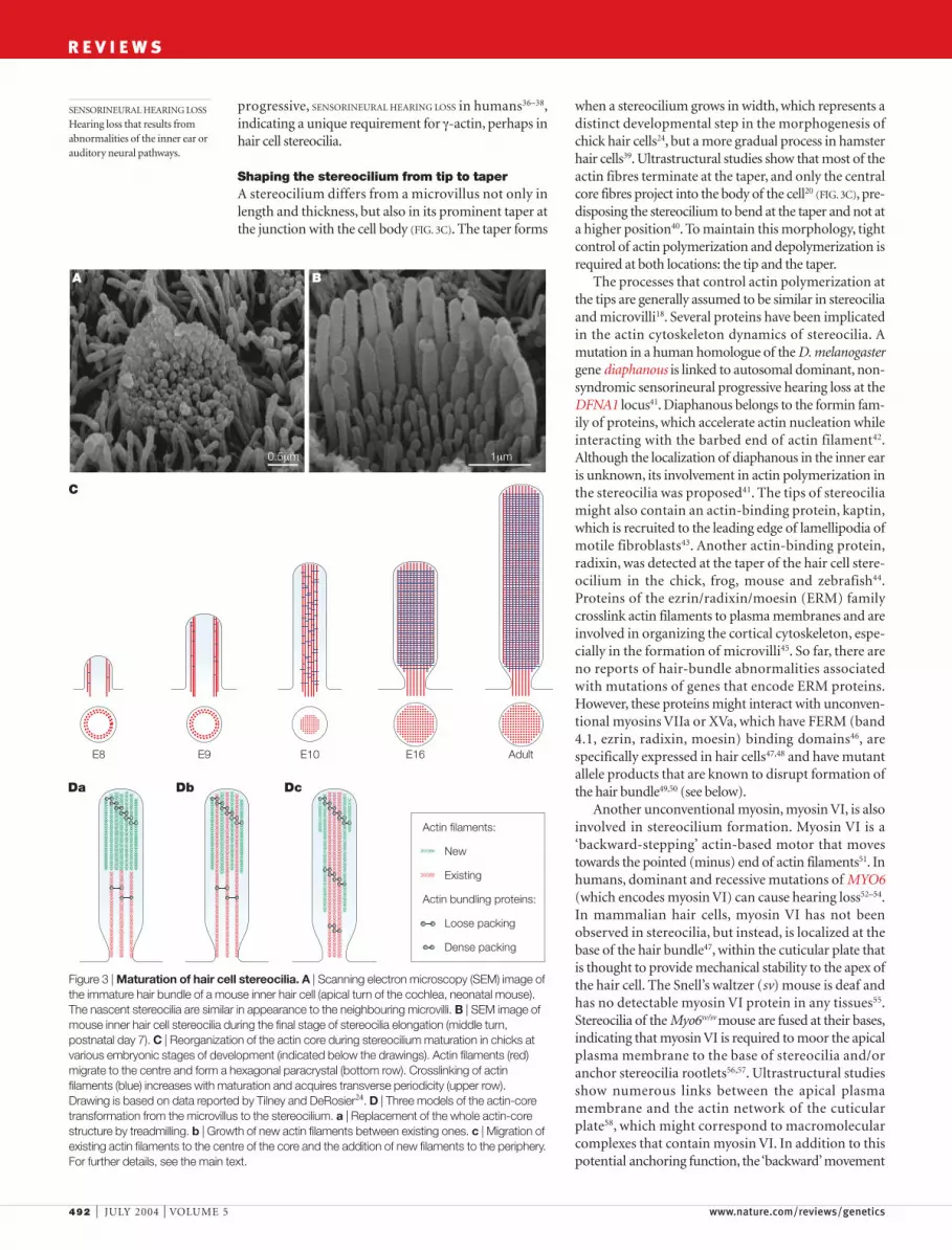
When stereocilia emerge from the apical surface of ahair cell, they have a similar appearance to microvilli24
(FIG. 3A). The loosely packed actin filaments of the pre-cursor microvillus are then replaced by the dense,hexagonally ordered paracrystalline array of actin fila-ments in a mature stereocilium25 (FIG. 3B,C). There areat least three models for this transformation (FIG. 3D).The first model posits that some signal might initiatethe growth of a new, denser actin core that begins at thetip of the stereocilium (FIG. 3Da). The new core wouldprogressively replace the old actin fibres by a treadmillprocess, in which actin monomers are depolymerizedfrom the pointed end while new monomers are addedto the barbed end, resulting in overall downwardmovement of the entire array26. Alternatively, newactin fibres might be added between existing fibres(FIG. 3Db). In a third model, the hair cell might modify the
known as the organ of Corti (FIG. 1c). Given this com-plexity, it is therefore not surprising that many genesare required for the development of the mammalianinner ear.
The inner ear develops from the otic placode, athickened region of cranial ECTODERM that invaginatesand closes to form an otic vesicle, where early develop-ment and differentiation of epithelial and neuronalstructures occur6,7. The genetic basis of these early mor-phogenetic events has been reviewed elsewhere6,7, butmorphogenesis of the mechanosensory cells of theinner ear occurs at the later stages of inner ear develop-ment. Investigations of these later stages have beengreatly facilitated in small rodents including the mouse,in which the inner ear continues to develop through thefirst three postnatal weeks8. In the mouse, formation ofsensory hair cells begins when cells of the inner ear sen-sory epithelium synchronously exit the cell cycle atembryonic day 12.5–13.5 (REF. 9). Some of them subse-quently differentiate into hair cells, the highly specializedmechanoreceptors with hair-like projections on theirapical surfaces (FIG. 2). These projections, known as stere-ocilia (FIG. 2b,c), have mechanosensitive ion channels10,11
of unknown molecular identity and are precisely orga-nized to detect sub-nanometer deflections12,13 (FIG. 2d).Unlike hair cells in birds and fish, mammalian cochlearhair cells do not regenerate and their loss or damageresults in irreversible deafness or hearing impairment.Hair cells of mammalian vestibular organs show somepotential for regeneration14–17, although the mechanismof renewal is unknown.
ECTODERM
Embryonic tissue that is theprecursor of the epidermis andthe nervous system.
MICROVILLUS
(Pl. microvilli). A thin,cylindrical, membrane-coveredprojection on the surface of ananimal cell that contains a corebundle of actin filaments.
ACTIN FILAMENT
A helical protein filament that isformed by the polymerization ofglobular actin molecules.
VESTIBULAR AREFLEXIA
An abnormal absent response toartificial caloric (hot or cold)stimulation of the neurosensoryorgans of balance in the innerear.
a b c
External ear
Ossicles
Middle ear
Inner ear
Vestibular labyrinth
Cochlea
Cochlearpartition
Inner hair cell
Tectorial membrane
Outerhair cells
Stereocilia
Basilar membrane
Figure 1 | Structure and function of the mammalian ear. a | Detection of environmental sound begins when incoming soundwaves reach the external ear. Sound vibrates the chain of ossicles in the middle ear, which transmit it into the inner ear, wheremechanical vibrations are converted into electrical nerve impulses. b | The fluid-filled hearing end-organ, known as the ‘cochlea’, is a continuous coiled duct that is separated longitudinally by a cochlear partition. Mechanical stiffness and resonant frequency of abasilar membrane that underlies the cochlear partition gradually decreases from the base of the cochlea to its apex. Soundvibrations of a particular frequency cause the basilar membrane to resonate at a particular location that is determined by its stiffness.c | A sensory part of cochlear partition, the organ of Corti, has two types of specialized mechanosensitive cell: inner and outer haircells, both of which convert mechanical stimuli into variations of intracellular potential. Only inner hair cells seem to transmitinformation to the brain, whereas outer hair cells receive abundant efferent innervation (yellow) but have few afferent fibres (darkgreen). Outer hair cells demonstrate a unique ATP-independent voltage-driven motility that might provide positive cycle-by-cyclefeedback to amplify sound-induced vibrations of the organ of Corti3. Both cochlear amplification and outer hair cell motility areabsent in mice that are deficient for prestin123, a novel voltage-sensing motor protein124 that is localized exclusively to the lateralplasma membrane of these cells125. Hair cells can be lost or damaged by ageing, noise exposure, ototoxic drugs such asaminoglycoside antibiotics, or by the inheritance of mutant alleles of genes that are essential for hearing. For further details, see the online supplementary information S1 (movie).

F O C U S O N O R G A N O G E N E S I S
NATURE REVIEWS | GENETICS VOLUME 5 | JULY 2004 | 491
possible. In the stereocilia of chicken hair cells, develop-mental changes of actin filaments seem to follow thethird model (FIG. 3C,Dc), although there are no data thatcompare the rates of growing new filaments at theperiphery and treadmilling of the existing filaments atthe centre. An obvious molecular candidate for mediat-ing the transition from loose to dense packing of actinfilaments in chick hair cell stereocilia is espin, the expres-sion of which coincides with stages when stereociliabecome thicker27. In Drosophila melanogaster, the replace-ment of Forked by Fascin controls the proper formationof the actin core in the sensory bristles28.Whether a simi-lar developmental mechanism is functioning in mam-malian hair cells is not known, but two lines of evidencesupport this possibility. First, L-plastin, a homologue ofthe actin-bundling protein fimbrin, is transiently presentduring mammalian stereocilia bundle formation, butnot in mature bundles29. Second, espin is an importantstructural element of the hair bundle of mammalian haircells, and a recessive mutation of the gene (Espn) thatencodes espin in the deaf jerker mouse results in failureto accumulate detectable amounts of this protein in thehair bundle, which leads to shortening, loss of mechani-cal stiffness and eventual disintegration of stereocilia30.In humans, recessive mutations of ESPN at the DFNB36locus cause profound prelingual hearing loss andperipheral VESTIBULAR AREFLEXIA31.
Stereocilia and microvilli compared. In various mam-malian and non-mammalian cells, microvilli aredynamic structures that continuously form and disas-semble with an average lifetime of approximately 12minutes32. By contrast, stereocilia of auditory hair cellsretain their mature shape throughout the life of theorganism. Nevertheless, the actin core of a stereociliumseems to be renewed continuously through an actintreadmill mechanism22. This renewal process is at leasttwo orders of magnitude slower than the cytoskeletalrenewal in typical actin bundle-based structures such asFILOPODIA33 or intestinal epithelial microvilli21,23. Theextraordinary stability of a stereocilium was attributedlong ago to the remarkable paracrystalline structure ofits actin core20,25. Recent structural studies indicate thatthe assembly of similar actin–fimbrin arrays in vitroproceeds as a whole crystal unit and requires strongcoupling between actin polymerization, fimbrin bind-ing, cross-bridge formation and conformational changesof actin34. Therefore, the marked differences in the stabil-ity of stereocilia compared with microvilli mightresult from differences in actin crosslinking and/orthe predominant isoform of actin. In chick hair cells,the β-isoform of actin seems to be targeted selectively tostereocilia, where it is crosslinked by fimbrin/plastin,which has a higher binding affinity for β-actin than forγ-actin35. γ-actin, which differs from β-actin in only fouramino acids at the amino terminus, is found through-out the hair cell, including microvilli35, as well asthroughout many other cell types, and could be consid-ered as a housekeeping gene product. Nevertheless,there are several dominant missense alleles of ACTG1that encode γ-actin that result in non-syndromic,
actin-bundling components by adding or removing cer-tain proteins, which would allow denser packing ofactin filaments. According to this last model, existing fil-aments migrate to the centre of the stereocilium andnew filaments are added at the periphery (FIG. 3Dc).Certainly, some combination of these models is also
FILOPODIUM
(Pl. filopodia). A thin, spike-likeprotrusion with an actinfilament core that is generatedon the leading edge of a motileanimal cell.
a
b
c
d
e
Excitation
5 µm
1 µm
1 µm 200 nm
Figure 2 | The hair bundle of mammalian auditory sensory cells. a | Scanning electronmicroscopy (SEM) image of three rows of outer hair cells that is viewed from the top of the organ ofCorti and that shows unidirectional orientation of V/W-shaped hair bundles. b | Close-up view of one of these almost-mature hair bundles that consists of three rows of individual stereocilia and a true cilium, the kinocilium (white arrow), which is always positioned at the vertex of thebundle. Images in panels a and b were obtained from the middle cochlear turn of a wild-typemouse at postnatal day 7. c | Side view of a mature outer hair cell bundle that shows the precisestaircase organization of stereocilia rows. d | Sound-induced, nanometer-scale deflections of thehair bundle13 open mechanically gated ion channels (shown in red)10,11. These channels arethought to be located at the ends of tip-links126. e | The tip-link92 (indicated by white arrows) is atiny filament that connects neighbouring stereocilia. Images in panels c and e were obtained fromthe middle cochlear turn of an adult guinea pig.
492 | JULY 2004 | VOLUME 5 www.nature.com/reviews/genetics
R E V I E W S
when a stereocilium grows in width, which represents adistinct developmental step in the morphogenesis ofchick hair cells24, but a more gradual process in hamsterhair cells39. Ultrastructural studies show that most of theactin fibres terminate at the taper, and only the centralcore fibres project into the body of the cell20 (FIG. 3C), pre-disposing the stereocilium to bend at the taper and not ata higher position40. To maintain this morphology, tightcontrol of actin polymerization and depolymerization isrequired at both locations: the tip and the taper.
The processes that control actin polymerization atthe tips are generally assumed to be similar in stereociliaand microvilli18. Several proteins have been implicatedin the actin cytoskeleton dynamics of stereocilia. Amutation in a human homologue of the D. melanogastergene diaphanous is linked to autosomal dominant, non-syndromic sensorineural progressive hearing loss at theDFNA1 locus41. Diaphanous belongs to the formin fam-ily of proteins, which accelerate actin nucleation whileinteracting with the barbed end of actin filament42.Although the localization of diaphanous in the inner earis unknown, its involvement in actin polymerization inthe stereocilia was proposed41. The tips of stereociliamight also contain an actin-binding protein, kaptin,which is recruited to the leading edge of lamellipodia ofmotile fibroblasts43. Another actin-binding protein,radixin, was detected at the taper of the hair cell stere-ocilium in the chick, frog, mouse and zebrafish44.Proteins of the ezrin/radixin/moesin (ERM) familycrosslink actin filaments to plasma membranes and areinvolved in organizing the cortical cytoskeleton, espe-cially in the formation of microvilli45. So far, there areno reports of hair-bundle abnormalities associatedwith mutations of genes that encode ERM proteins.However, these proteins might interact with unconven-tional myosins VIIa or XVa, which have FERM (band4.1, ezrin, radixin, moesin) binding domains46, arespecifically expressed in hair cells47,48 and have mutantallele products that are known to disrupt formation ofthe hair bundle49,50 (see below).
Another unconventional myosin, myosin VI, is alsoinvolved in stereocilium formation. Myosin VI is a‘backward-stepping’ actin-based motor that movestowards the pointed (minus) end of actin filaments51. Inhumans, dominant and recessive mutations of MYO6(which encodes myosin VI) can cause hearing loss52–54.In mammalian hair cells, myosin VI has not beenobserved in stereocilia, but instead, is localized at thebase of the hair bundle47, within the cuticular plate thatis thought to provide mechanical stability to the apex ofthe hair cell. The Snell’s waltzer (sv) mouse is deaf andhas no detectable myosin VI protein in any tissues55.Stereocilia of the Myo6sv/sv mouse are fused at their bases,indicating that myosin VI is required to moor the apicalplasma membrane to the base of stereocilia and/oranchor stereocilia rootlets56,57. Ultrastructural studiesshow numerous links between the apical plasmamembrane and the actin network of the cuticularplate58, which might correspond to macromolecularcomplexes that contain myosin VI. In addition to thispotential anchoring function, the ‘backward’movement
progressive, SENSORINEURAL HEARING LOSS in humans36–38,indicating a unique requirement for γ-actin, perhaps inhair cell stereocilia.
Shaping the stereocilium from tip to taperA stereocilium differs from a microvillus not only inlength and thickness, but also in its prominent taper atthe junction with the cell body (FIG. 3C). The taper forms
SENSORINEURAL HEARING LOSS
Hearing loss that results fromabnormalities of the inner ear orauditory neural pathways.
A B
0.5µm 1µm
E8 E9 E10 E16 Adult
C
Da Db Dc
Actin filaments:
New
Existing
Actin bundling proteins:
Loose packing
Dense packing
Figure 3 | Maturation of hair cell stereocilia. A | Scanning electron microscopy (SEM) image ofthe immature hair bundle of a mouse inner hair cell (apical turn of the cochlea, neonatal mouse).The nascent stereocilia are similar in appearance to the neighbouring microvilli. B | SEM image ofmouse inner hair cell stereocilia during the final stage of stereocilia elongation (middle turn,postnatal day 7). C | Reorganization of the actin core during stereocilium maturation in chicks atvarious embryonic stages of development (indicated below the drawings). Actin filaments (red)migrate to the centre and form a hexagonal paracrystal (bottom row). Crosslinking of actinfilaments (blue) increases with maturation and acquires transverse periodicity (upper row).Drawing is based on data reported by Tilney and DeRosier24. D | Three models of the actin-coretransformation from the microvillus to the stereocilium. a | Replacement of the whole actin-corestructure by treadmilling. b | Growth of new actin filaments between existing ones. c | Migration ofexisting actin filaments to the centre of the core and the addition of new filaments to the periphery.For further details, see the main text.
F O C U S O N O R G A N O G E N E S I S
NATURE REVIEWS | GENETICS VOLUME 5 | JULY 2004 | 493
cuticular plate and projects into the cell63,64. However,the orientation and overall arrangement of the bun-dles are generally intact in these conditions, indicat-ing the presence of a mechanism that stabilizes theoverall orientation of the cuticular plate and hairbundle as an integrated complex. This mechanismmight involve myosin VIIa, which, anchored by vezatinto a cadherin–catenins complex, could link the corticalcytoskeleton to the adhesion junctions between haircells and neighbouring supporting cells65. Consistentwith this hypothesis, mutations in myosin VIIa and acadherin-related protein, cadherin 23, result in loss ofthe V/W configuration and disorientation of thebundle, respectively49,66.
Orientation of the stereocilia bundles. Stereocilia bun-dles are directionally sensitive67 and their precise orienta-tion (FIG. 2a) is necessary for normal auditory perception68.Deflections towards the tallest stereocilia increase theMECHANOTRANSDUCTION current, whereas deflections in theopposite direction decrease the current67 (FIG. 2d). Inmost mammalian and non-mammalian species, a spe-cialized extracellular matrix, the tectorial membrane,overlies the auditory neuroepithelium and deflects hairbundles in response to sound (FIG. 1c). In the chick,growth and concomitant radial movement of the tector-ial membrane was proposed to orient hair bundles by atraction mechanism69. However, in mammals, the ori-entation of cochlear hair cell bundles becomes evidentas soon as microvilli emerge, well before the tectorialmembrane is formed70. Furthermore, targeted deletionof α-tectorin, one of three key glycoproteins that consti-tute the tectorial membrane, seems to cause completedetachment of the tectorial membrane from the mouseorgan of Corti71,72, but does not affect the orientation orstructure of the hair cell bundles72. The signalling pro-cesses that control hair-bundle orientation might besimilar to those that control PLANAR-CELL POLARITY in theeye of D. melanogaster. Indeed, Flamingo, a member ofthe cadherin superfamily of adhesion proteins, thetransmembrane protein Van Gogh and the secretedmolecule Wingless, have been implicated in the regula-tion of planar-cell polarity in flies73,74. The mammalianhomologues of each of these molecules is also invol-ved in the polarization of cochlear hair cell stereociliabundles75–77.
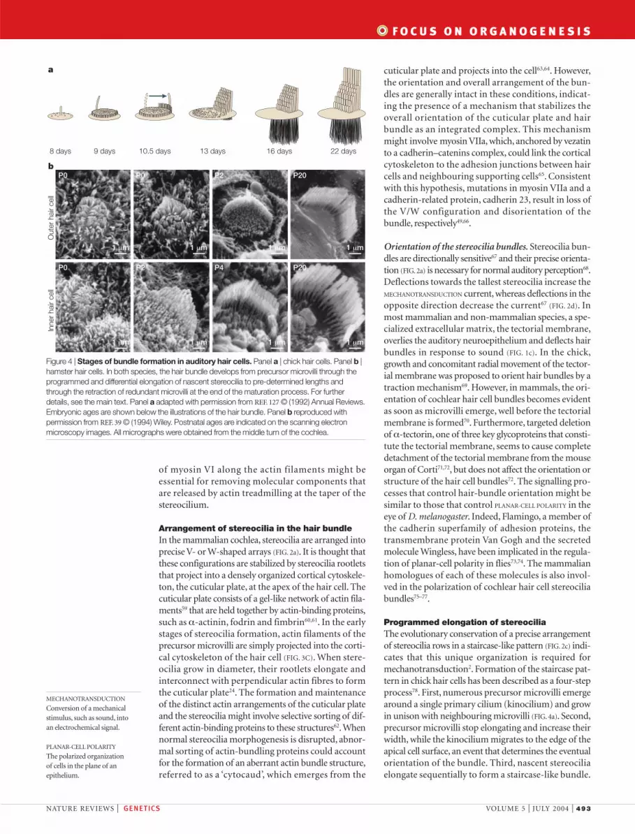
Programmed elongation of stereociliaThe evolutionary conservation of a precise arrangementof stereocilia rows in a staircase-like pattern (FIG. 2c) indi-cates that this unique organization is required formechanotransduction2. Formation of the staircase pat-tern in chick hair cells has been described as a four-stepprocess78. First, numerous precursor microvilli emergearound a single primary cilium (kinocilium) and growin unison with neighbouring microvilli (FIG. 4a). Second,precursor microvilli stop elongating and increase theirwidth, while the kinocilium migrates to the edge of theapical cell surface, an event that determines the eventualorientation of the bundle. Third, nascent stereociliaelongate sequentially to form a staircase-like bundle.
of myosin VI along the actin filaments might beessential for removing molecular components thatare released by actin treadmilling at the taper of thestereocilium.
Arrangement of stereocilia in the hair bundleIn the mammalian cochlea, stereocilia are arranged intoprecise V- or W-shaped arrays (FIG. 2a). It is thought thatthese configurations are stabilized by stereocilia rootletsthat project into a densely organized cortical cytoskele-ton, the cuticular plate, at the apex of the hair cell. Thecuticular plate consists of a gel-like network of actin fila-ments59 that are held together by actin-binding proteins,such as α-actinin, fodrin and fimbrin60,61. In the earlystages of stereocilia formation, actin filaments of theprecursor microvilli are simply projected into the corti-cal cytoskeleton of the hair cell (FIG. 3C). When stere-ocilia grow in diameter, their rootlets elongate andinterconnect with perpendicular actin fibres to formthe cuticular plate24. The formation and maintenanceof the distinct actin arrangements of the cuticular plateand the stereocilia might involve selective sorting of dif-ferent actin-binding proteins to these structures62. Whennormal stereocilia morphogenesis is disrupted, abnor-mal sorting of actin-bundling proteins could accountfor the formation of an aberrant actin bundle structure,referred to as a ‘cytocaud’, which emerges from the
MECHANOTRANSDUCTION
Conversion of a mechanicalstimulus, such as sound, into an electrochemical signal.
PLANAR-CELL POLARITY
The polarized organization of cells in the plane of anepithelium.
Out
er h
air
cell
Inne
r ha
ir ce
ll
P0 P0 P2 P20
P0 P2 P4 P20
b
a
8 days 9 days 10.5 days 13 days 16 days 22 days
1 µm
1 µm
1 µm
1 µm
1 µm
1 µm
1 µm
1 µm
Figure 4 | Stages of bundle formation in auditory hair cells. Panel a | chick hair cells. Panel b |hamster hair cells. In both species, the hair bundle develops from precursor microvilli through theprogrammed and differential elongation of nascent stereocilia to pre-determined lengths andthrough the retraction of redundant microvilli at the end of the maturation process. For furtherdetails, see the main text. Panel a adapted with permission from REF. 127 © (1992) Annual Reviews.Embryonic ages are shown below the illustrations of the hair bundle. Panel b reproduced withpermission from REF. 39 © (1994) Wiley. Postnatal ages are indicated on the scanning electronmicroscopy images. All micrographs were obtained from the middle turn of the cochlea.

494 | JULY 2004 | VOLUME 5 www.nature.com/reviews/genetics
R E V I E W S
Mutations in several genes are known to cause hear-ing loss by disrupting bundle morphogenesis (FIG. 5b;TABLE 1). Mutations of MYO15A, which encodesunconventional myosin XVa, cause non-syndromicsensorineural deafness in humans (DFNB3) as wellas deafness and vestibular disorders in the shaker-2 mouse50,80,81. Hair cell stereocilia in homozygousshaker-2 mice are present and properly positioned, butare much shorter than wild-type stereocilia50. In theshaker-2 mouse, all stereocilia within a bundle areapproximately the same length and there is no staircaseorganization of the mature hair bundle. Myosin XVa isdiscretely located at the tip of every stereocilium in
Finally, excess microvilli on the apical cell surface areresorbed (FIG. 4a). Morphogenesis of the hair bundle inmammalian auditory hair cells generally follows thesame principles39,79, although the stages are less distinct(FIG. 4b). Moreover, the kinocilium of mammalian audi-tory hair cells (FIG. 2b) degenerates shortly after forma-tion of the bundle, indicating that it is not required tomaintain bundle structure and function. However, itspersistence throughout the lifetime of other types ofhair cell with the capability to regenerate (mammalianvestibular hair cells, and hair cells in frogs and chicks)indicates that it might indeed be important for bundlemorphogenesis.
PDZ-ligand
?
? ? ?
?
?
?
??
? ?
a c
b
Vezatin
F-actin
Myosin VIIa
SANS
Motor
5 IQ FERM FERMSH3
MyTH4
Ank1 Ank2 Ank3 SAM
Harmonin b
Harmonin b
PDZ3 PST PDZ2 PDZ1CC2 CC1
PDZ3PSTPDZ2PDZ1 CC2CC1
EC27EC26 EC1
EC27EC26 EC1
68
68
Adhesion proteins:
Cadherin 23
Protocadherin 15
Motor proteins:
Myosin XVa
Myosin VIIa
Myosin VI
Scaffolding proteins:
Whirlin
Harmonin b
SANS
? ?
Cadherin 23 (+68)
?
Tip links
Top links
Side links
Ankle links
Vezatin
Figure 5 | Adhesion in the hair bundle. a | Four known types of link between adjacent stereocilia86. b | Schematized illustration of proteins that constitute adhesion complexes on the plasma membrane of stereocilia. Experimentally demonstrated interactionsbetween myosin VIIa, harmonin, cadherin 23, SANS and vezatin are shown, as well as the interaction of myosin XVa with whirlin(I.A.B. et al., unpublished observations). c | Interacting domains among the proteins that are essential for stereocilia micro-morphogenesis and function. These proteins were identified through positional cloning of genes that underlie type 1 Ushersyndrome. Ank, ankyrin repeat domain that is thought to be involved in protein–protein interactions; CC, coiled coil domain thatmediates dimerization; EC, cadherin extracellular repeat; FERM, domain that is also known as the talin homology domain, which isthought to be important for linking cytoskeletal proteins to the membrane; IQ, a motif that serves as a binding site for myosin lightchains; Motor, a domain that mediates actin binding, ATP binding and hydrolysis, and force generation; MyTH4, a domain ofunknown function in some myosin and kinesin tails; PDZ, a domain that mediates interactions with other proteins that contain a PDZligand sequence and that is thought to be important for targeting signalling molecules to sub-membranous sites; PST, a proline-,serine- and threonine-rich region; SAM, a sterile α-motif, a domain that is found in many signalling proteins and that is thought to beinvolved in protein–protein interactions; SH3, a Src homology-3 domain that is involved in protein–protein interactions.

F O C U S O N O R G A N O G E N E S I S
NATURE REVIEWS | GENETICS VOLUME 5 | JULY 2004 | 495
Role of stereocilia links in morphogenesisTypes of link and their distribution. Ultrastructuralstudies have revealed four different types of link betweenstereocilia that provide cohesiveness to the stereociliabundle and might mediate signalling events during mor-phogenesis (FIG. 5a). From the top to the base of the stere-ocilia, these links are designated: tip-links, top-links, shaftconnections or side-links, and ankle-links, which arelocated at the beginning of the stereocilia taper86. Thereare also links that connect the kinocilium to the adjacentstereocilia of the tallest row87. All of these link types canbe distinguished by their differential sensitivities totreatment with CALCIUM CHELATORS and a specific enzyme,subtilisin88. After treating the bundle with the calciumchelator BAPTA, tip-links break and multiple linkagessprout from the tips of the stereocilia over the next 12hours89. Inappropriate links subsequently disappear,leaving only regenerated tip-links at 24 hours after treat-ment. Similar link dynamics were described for develop-ing bundles39,90. The molecular identities of stereocilialinks are under investigation, but their appearance earlyin development indicates that they might be importantfor the morphogenesis of hair bundles. Ankle-links pro-gressively disappear within the first postnatal monthin mouse hair cells but persist throughout life inchick hair cells65. Tip-links are present in mature haircells (FIG. 2e) and are believed to be crucial inmechan-otransduction91,92. Side-links are also present in adulthair cells93 but do not seem to be a principal factorholding stereocilia in the developing bundle. Haircells of transgenic mice with different mutations inprotein tyrosine phosphatase receptor Q (Ptprq) donot have side links but form transient, normal-appearing bundles that subsequently degenerateshortly after birth94.
Usher syndrome and hair-bundle morphology. Inter-cellular adhesion is a key signalling process that initiatescytoskeleton rearrangement during morphogenesis95,96.The genetic investigation of type 1 Usher syndrome(USH1) in humans and the characterization of corre-sponding mouse models has revealed that hair cellsmight use analogous adhesion mechanisms to controlthe morphogenesis of the hair bundle. Usher syndrome
wild-type auditory and vestibular hair cells, where itappears just before the staircase emerges, which indi-cates that it is required for the elongation and formationof the stereocilia-bundle staircase48.
In a hair bundle, longer stereocilia have moremyosin XVa at their tips compared with the adjacentrow of shorter stereocilia48,82. Similarly, the rate of actinrenewal82 and the amount of espin23 are proportionalto the length of the stereocilium. Overexpression ofespin in CL4 porcine kidney epithelial cells initiates theformation of abnormally long microvilli23, whereasoverexpression of myosin XVa in wild-type mouse haircells or in COS-7 cells does not induce additionalgrowth of stereocilia or filopodia, respectively48. Oneinterpretation of these data is that myosin XVa partici-pates in the development of the staircase morphologyof the hair bundle by an increased delivery of essentialstructural or regulatory elements to the tips of thetaller stereocilia48,83.
Localization of myosin XVa to the extreme tips ofstereocilia raises the possibility that it is tethered there byintegral membrane proteins48. Although the proteinsthat interact with myosin XVa are not known, it has twopredicted FERM domains that could mediate interac-tions with ERM proteins. Perhaps more interestingly,myosin XVa has a PDZ ligand sequence that could inter-act with PDZ domain-containing proteins to coordinatea macromolecular complex at the tips of stereocilia.PDZ scaffold proteins serve as organizing centres ofmacromolecular functional complexes84. One such PDZprotein, whirlin, has recently been described85. Recessivemutations in WHRN and Whrn (which encode whirlin)cause deafness in humans (DFNB31) and in whirlermice, respectively. Stereocilia of whirler mice are abnor-mally short and arrayed in a near-normal configurationon the apical hair cell surface85. The overall inner earphenotype of whirler mice is strikingly similar to that ofshaker-2 mice, raising the possibility that whirlin andmyosin XVa interact physically within a macromolec-ular complex that is responsible for programmed stere-ocilia elongation83. Whirlin also has a C-terminal PDZligand sequence that could interact with one of thePDZ domains of another whirlin protein to organizetheir multimerization into a higher-order structure.
CALCIUM CHELATORS
Substances that reversibly bindcalcium, usually with highaffinity, to remove free calciumions from a solution.
Table 1 | Genes required for morphogenesis of the hair cell bundle
Biological role Gene product Gene symbol Location Human locus Mouse model Refs(human) (human) (gene symbol)
Cytoskeleton γ-actin ACTG1 17q25 DFNA20, A26 N/A 36–38formation diaphanous 1 HDIA1 5q31 DFNA1 N/A 41,42
espin ESPN 1p36 DFNB36 Jerker (je) 27,30,31
Adhesion cadherin 23 CDH23 10q21-q22 DFNB12, USH1D Waltzer (v) 66,100,101,112,113protocadherin 15 PCDH15 10q21 DFNB23, USH1F Ames waltzer (av) 107,111,128
Intracellular myosin VI MYO6 6q13 DFNA22, B37 Snell’s waltzer (sv) 52–56motors myosin VIIa MYO7A 11q12.3 DFNB2, A11, USH1BShaker-1 (sh1) 49,97,129
myosin XVa MYO15A 17p11.2 DFNB3 Shaker-2 (sh2) 50,81
Scaffolding harmonin USH1C 11p15.1 DFNB18, USH1C Deaf circler (dfcr) 98,99,108,109proteins SANS SANS 17q24-q25 USH1G Jackson circler (js) 104
whirlin WRHN 9q32-q34 DFNB31 Whirler (wi) 85
DFNAx, autosomal dominant deafness type ‘x’; DFNBx, autosomal recessive deafness type ‘x’; N/A, not available; USHx, Ushersyndrome type ‘x’.

496 | JULY 2004 | VOLUME 5 www.nature.com/reviews/genetics
R E V I E W S
ankle-links are consistent with an additional role: anadhesion complex that regulates actin–myosin-basedtrafficking through the taper of a stereocilium. Althoughthere is no direct evidence to support this model, a bar-rier at the junction of the taper and the main body ofthe stereocilium could maintain the segregation of themolecular determinants of these distinct structuraldomains.
Mechanotransduction and morphogenesis Mature mechanotransduction currents can be detectedin mammalian vestibular (mouse utricle) hair cells byembryonic day 16 or 17 (REF. 115), in cochlear hair cellsearlier than postnatal day 0 (REF. 116) and in chick auditoryhair cells by embryonic day 12 (REF. 117). At all of thesedevelopmental stages, the morphological maturation ofthe hair bundles is not fully complete. This temporal rela-tionship raises the possibility that mechanotransductioncurrents are required to complete the morphogenesis ofthe hair bundle, and that some or all of the mechan-otransduction apparatus is assembled at the tips ofstereocilia before the final elongation of stereociliaoccurs. Before these observations, Tilney and co-workersspeculated that growth of the tallest row of stereociliaincreases tension on the tip-links and opens transduc-tion channels in the next row of stereocilia to initiatetheir growth and generate a staircase morphology114.Extending this hypothesis further, a threshold levelof transduction current activity might determinewhether a stereocilium is incorporated into the bun-dle or resorbed as a microvillus, providing a potentialexplanation for the conserved number of rows ofstereocilia in mammalian auditory hair cells. Calciumthat enters the cell through mechanotransductionchannels might destabilize calcium-sensitive cross-links between actin filaments in the microvilli thatare adjacent to nascent stereocilia. By contrast, stereo-cilia would be unaffected because their actin core iscrosslinked by calcium-insensitive espin30.
Missing pieces of hair-bundle morphogenesisThe genetic investigation of hearing loss has revealedvarious genes and steps that are required for hair-bundlemorphogenesis. The remaining pathways might bedetermined by characterizing additional mousemutants, such as the recessive pirouette (pi) and domi-nant Tasmanian devil mutants that have thin stereocilia,which are abnormally short in pi118 and abnormallylong in Tasmanian devil mice119. Tailchaser (Tlc) is adominant mutation in which stereocilia fail to form thenormal V-shaped configuration on outer hair cells120.Similarly, Headbanger is a dominant mutation that dis-rupts development of the V-shaped configuration, butonly in the apex of the cochlea, whereas the inner haircell bundles in the apex fuse postnatally to form giantstereocilia, and the stereocilia of one of the vestibularend organs (the utricle) are abnormally long and thin121.The last three mutants were generated and identified ina large-scale ethylnitrosourea (ENU) mutagenesisscreen for deafness and vestibular phenotypes122,which has been a valuable adjunct to the finite supply
is an autosomal recessive disorder that is characterizedby sensorineural hearing loss and progressive loss ofvision owing to RETINITIS PIGMENTOSA. So far, 5 genesthat underlie USH1 have been cloned: myosin VIIa(MYO7A)97, harmonin (USH1C)98,99, cadherin 23(CDH23)66,100,101, protocadherin 15 (PCDH15)102,103 andSANS104. There are corresponding mutant mouse modelsand, in some cases, several mutant alleles for each gene:shaker-1 (Myo7ash1), deaf circler (Ush1cdfcr), waltzer(Cdh23v), Ames waltzer (Pcdh15av) and Jackson shaker(Sansjs). These mutant phenotypes are all characterizedby deafness, vestibular dysfunction and disorganized,splayed stereocilia in homozygous mice49,66,105–107. On thebasis of similar mutant phenotypes, as well as in vitroprotein-interaction studies, it was suggested that the pro-tein products of genes that are involved in USH1 mightform a macromolecular complex (FIG. 5c) that providescohesiveness to the stereocilia bundles104,108,109. Two ofthese gene products, harmonin and SANS, representPDZ domain-containing proteins. Harmonin hasbeen shown to interact with myosin VIIa and cad-herin 23108,109, which has a PDZ ligand sequence at itsC-terminus. SANS has also been implicated in an inter-action with harmonin104, and, therefore, SANS and har-monin might act as cytoplasmic scaffold organizers ofproteins that are involved in USH1.
Cadherins in the hair bundle. Cadherin 23 and proto-cadherin 15 are members of the cadherin superfamily ofintegral membrane proteins that are responsible forintercellular adhesion as well as signalling96,110. Homo-philic interaction of these proteins might form links thatinterconnect stereocilia within a bundle. Protocadherin15 appears in the stereocilia of developing mammalianhair cells and persists in adult hair cells along the lengthof stereocilia, indicating that it might be important forthe long-term maintenance of lateral connections (side-links) between stereocilia111. Cadherin 23 is located atthe tips of the bundle in frog and zebrafish hair cells andhas been proposed as an essential component of tip-links112,113. During development in mammals, cadherin23 and harmonin transiently appear together in stere-ocilia, and have been reported to diminish to unde-tectable amounts in adult stereocilia108. However, a recentdescription of cadherin 23 persisting in the tips of adultmammalian stereocilia112 indicates that its contributionto the maintenance of mature stereocilia links remainsto be resolved.
At least some hair-bundle adhesion complexes seemto be linked by unconventional myosin VIIa to the actincore at sites of links between stereocilia (FIG. 5b,c). At thelateral surface, myosin VIIa probably links USH1macromolecular complexes (harmonin–cadherin23–SANS) to the actin filaments during hair-bundlematuration104,108,109. At the base of the stereocilium,myosin VIIa interacts with a novel transmembrane pro-tein, vezatin, and could comprise part of an adhesioncomplex that is associated with ankle-links65.Ankle-linksmight determine the precise positioning and concomi-tant growth of individual stereocilia within a bundle114.The unique location and putative composition of
RETINITIS PIGMENTOSA
An aetiologically heterogeneousdisorder that is characterized byprogressive loss of vision andretinal photoreceptordegeneration.

F O C U S O N O R G A N O G E N E S I S
NATURE REVIEWS | GENETICS VOLUME 5 | JULY 2004 | 497
incomplete. It is prudent to continue positional cloningof genes that underlie hearing or balance disorders inhumans and mice, especially genes that are expressed atlow levels that elude detection by other screeningmethods. As some mutations might escape detectionin forward-genetic screens owing to embryonic orearly postnatal lethal phenotypes, reverse-geneticapproaches, including conditional knockouts, areneeded to fill in the molecular gaps in hair cell mor-phogenesis. Candidates might initially be identifiedthrough protein-interaction screens, proteomic analy-ses, genomic approaches or a combination of thesemethodologies. This integrated approach should leadto a comprehensive understanding of hair-bundlemorphogenesis and insights into the pathogenesis ofhearing loss and balance disorders, as well as strategiesfor their prevention and treatment.
of spontaneous mouse mutants. Finally, human fami-lies that segregate deafness continue to be a fruitfulresource to identify genes that are required for hearingand hair-bundle morphogenesis.
ConclusionsSeveral developmental themes have emerged from theidentification of genes that are required for the morpho-genesis of the hair cell bundle. Many of these genesencode intracellular motors, adhesion proteins or scaf-folding proteins that interact with them (TABLE 1). Haircells have therefore adapted and integrated mechanismsof cell-to-cell adhesion and intracellular motility to gen-erate their precisely organized hair bundles. However, itis clear that there are still crucial genes and pathwaysthat remain to be determined, indicating that thegenetic investigation of hair-bundle morphogenesis is
1. Fay, R. R. & Popper, A. N. Evolution of hearing in vertebrates:the inner ears and processing. Hear. Res. 149, 1–10 (2000).
2. Manley, G. A. Cochlear mechanisms from a phylogeneticviewpoint. Proc. Natl Acad. Sci. USA 97, 11736–11743 (2000).
3. Dallos, P. & Fakler, B. Prestin, a new type of motor protein.Nature Rev. Mol. Cell Biol. 3, 104–111 (2002).
4. Popper, A. N. & Fay, R. R. Evolution of the ear and hearing:issues and questions. Brain Behav. Evol. 50, 213–221 (1997).
5. Friedman, T. B. & Griffith, A. J. Human nonsyndromicsensorineural deafness. Annu. Rev. Genomics Hum. Genet.4, 341–402 (2003).A comprehensive and critical review of the genes thatare implicated in non-syndromic sensorineuraldeafness in humans.
6. Fekete, D. M. & Wu, D. K. Revisiting cell fate specification inthe inner ear. Curr. Opin. Neurobiol. 12, 35–42 (2002).
7. Kelley, M. W. Cell adhesion molecules during inner ear andhair cell development, including notch and its ligands. Curr.Top. Dev. Biol. 57, 321–356 (2003).
8. Ehret, G. Postnatal development in the acoustic system ofthe house mouse in the light of developing maskedthresholds. J. Acoust. Soc. Am. 62, 143–148 (1977).
9. Chen, P., Johnson, J. E., Zoghbi, H. Y. & Segil, N. The role of Math1 in inner ear development: uncoupling theestablishment of the sensory primordium from hair cell fatedetermination. Development 129, 2495–2505 (2002).
10. Corey, D. P. & Hudspeth, A. J. Ionic basis of the receptorpotential in a vertebrate hair cell. Nature 281, 675–677 (1979).
11. Ohmori, H. Mechano-electrical transduction currents inisolated vestibular hair cells of the chick. J. Physiol. 359,189–217 (1985).
12. Russell, I. J., Richardson, G. P. & Kossl, M. The responses ofcochlear hair cells to tonic displacements of the sensory hairbundle. Hear. Res. 43, 55–69 (1989).
13. Hudspeth, A. J. Hair-bundle mechanics and a model formechanoelectrical transduction by hair cells. Soc. Gen.Physiol. Ser. 47, 357–370 (1992).
14. Warchol, M. E., Lambert, P. R., Goldstein, B. J., Forge, A. &Corwin, J. T. Regenerative proliferation in inner ear sensoryepithelia from adult guinea pigs and humans. Science 259,1619–1622 (1993).
15. Forge, A., Li, L., Corwin, J. T. & Nevill, G. Ultrastructuralevidence for hair cell regeneration in the mammalian innerear. Science 259, 1616–1619 (1993).
16. Rubel, E. W., Dew, L. A. & Roberson, D. W. Mammalianvestibular hair cell regeneration. Science 267, 701–707 (1995).
17. Zheng, J. L., Keller, G. & Gao, W. Q. Immunocytochemicaland morphological evidence for intracellular self-repair as an important contributor to mammalian hair cell recovery. J. Neurosci. 19, 2161–2170 (1999).
18. DeRosier, D. J. & Tilney, L. G. F-actin bundles are derivativesof microvilli: what does this tell us about how bundles mightform? J. Cell Biol. 148, 1–6 (2000).
19. Bartles, J. R. Parallel actin bundles and their multiple actin-bundling proteins. Curr. Opin. Cell Biol. 12, 72–78 (2000).
20. Tilney, L. G., Derosier, D. J. & Mulroy, M. J. The organizationof actin filaments in the stereocilia of cochlear hair cells. J. Cell Biol. 86, 244–259 (1980).
21. Tyska, M. J. & Mooseker, M. S. MYO1A (brush bordermyosin I) dynamics in the brush border of LLC-PK1-CL4cells. Biophys. J. 82, 1869–1883 (2002).
22. Schneider, M. E., Belyantseva, I. A., Azevedo, R. B. &Kachar, B. Rapid renewal of auditory hair bundles. Nature418, 837–838 (2002).Demonstration of actin renewal in auditory hairbundles.
23. Loomis, P. A. et al. Espin crosslinks cause the elongation ofmicrovillus-type parallel actin bundles in vivo. J. Cell Biol.163, 1045–1055 (2003).
24. Tilney, L. G. & DeRosier, D. J. Actin filaments, stereocilia,and hair cells of the bird cochlea. IV. How the actin filamentsbecome organized in developing stereocilia and in thecuticular plate. Dev. Biol. 116, 119–129 (1986).Description of maturational changes in actin packingduring hair cell stereocilia development.
25. DeRosier, D. J., Tilney, L. G. & Egelman, E. Actin in the innerear: the remarkable structure of the stereocilium. Nature287, 291–26 (1980).First description of paracrystalline organization ofactin filaments in hair cell stereocilia.
26. Neuhaus, J. M., Wanger, M., Keiser, T. & Wegner, A.Treadmilling of actin. J. Muscle Res. Cell. Motil. 4, 507–527(1983).
27. Li, H. et al. Correlation of expression of the actin filament-bundling protein espin with stereociliary bundle formation inthe developing inner ear. J. Comp. Neurol. 468, 125–134(2004).
28. Tilney, L. G., Connelly, P. S., Vranich, K. A., Shaw, M. K. &Guild, G. M. Why are two different crosslinkers necessary foractin bundle formation in vivo and what does each crosslinkcontribute? J. Cell Biol. 143, 121–133 (1998).
29. Daudet, N. & Lebart, M. C. Transient expression of the t-isoform of plastins/fimbrin in the stereocilia of developingauditory hair cells. Cell Motil. Cytoskeleton 53, 326–336(2002).
30. Zheng, L. et al. The deaf jerker mouse has a mutation in thegene encoding the espin actin-bundling proteins of hair cellstereocilia and lacks espins. Cell 102, 377–385 (2000).Positional cloning of the mouse jerker mutationrevealed that espin is required for hair cell stereociliadevelopment.
31. Naz, S. et al. Mutations of ESPN cause autosomal recessivedeafness and vestibular dysfunction. J. Med. Genet. (in thepress).
32. Gorelik, J. et al. Dynamic assembly of surface structures inliving cells. Proc. Natl Acad. Sci. USA 100, 5819–5822(2003).
33. Mallavarapu, A. & Mitchison, T. Regulated actin cytoskeletonassembly at filopodium tips controls their extension andretraction. J. Cell Biol. 146, 1097–1106 (1999).
34. Volkmann, N., DeRosier, D., Matsudaira, P. & Hanein, D. Anatomic model of actin filaments crosslinked by fimbrin andits implications for bundle assembly and function. J. CellBiol. 153, 947–956 (2001).
35. Hofer, D., Ness, W. & Drenckhahn, D. Sorting of actinisoforms in chicken auditory hair cells. J. Cell. Sci. 110,765–770 (1997).
36. Zhu, M. et al. Mutations in the γ-actin gene (ACTG1) areassociated with dominant progressive deafness(DFNA20/26). Am. J. Hum. Genet. 73, 1082–1091 (2003).
37. Morell, R. J. et al. A new locus for late-onset, progressive,hereditary hearing loss DFNA20 maps to 17q25. Genomics63, 1–6 (2000).
38. van Wijk, E. et al. A mutation in the γ-actin 1 (ACTG1) genecauses autosomal dominant hearing loss (DFNA20/26). J. Med. Genet. 40, 879–884 (2003).
39. Kaltenbach, J. A., Falzarano, P. R. & Simpson, T. H.Postnatal development of the hamster cochlea. II. Growthand differentiation of stereocilia bundles. J. Comp. Neurol.350, 187–198 (1994).
40. Tilney, L. G., Egelman, E. H., DeRosier, D. J. & Saunder, J. C.Actin filaments, stereocilia, and hair cells of the bird cochlea.II. Packing of actin filaments in the stereocilia and in thecuticular plate and what happens to the organization whenthe stereocilia are bent. J. Cell Biol. 96, 822–834 (1983).
41. Lynch, E. D. et al. Nonsyndromic deafness DFNA1 associatedwith mutation of a human homolog of the Drosophila genediaphanous. Science 278, 1315–1318 (1997).
42. Higashida, C. et al. Actin polymerization-driven molecularmovement of mDia1 in living cells. Science 303, 2007–2010(2004).
43. Bearer, E. L. & Abraham, M. T. 2E4 (kaptin): a novel actin-associated protein from human blood platelets found inlamellipodia and the tips of the stereocilia of the inner ear.Eur. J. Cell Biol. 78, 117–126 (1999).
44. Pataky, F., Pironkova, R. & Hudspeth, A. J. Radixin is aconstituent of stereocilia in hair cells. Proc. Natl Acad. Sci.USA 101, 2601–2606 (2004).
45. Tsukita, S. & Yonemura, S. Cortical actin organization:lessons from ERM (ezrin/radixin/moesin) proteins. J. Biol.Chem. 274, 34507–34510 (1999).
46. Oliver, T. N., Berg, J. S. & Cheney, R. E. Tails of unconventionalmyosins. Cell. Mol. Life Sci. 56, 243–2457 (1999).
47. Hasson, T. et al. Unconventional myosins in inner-earsensory epithelia. J. Cell Biol. 137, 1287–1307 (1997).
48. Belyantseva, I. A., Boger, E. T. & Friedman, T. B. Myosin XValocalizes to the tips of inner ear sensory cell stereocilia and isessential for staircase formation of the hair bundle. Proc.Natl Acad. Sci. USA 100, 13958–13963 (2003).Demonstration that myosin XVa is located at the tipsof stereocilia and is required for staircase formation ofthe hair bundle.
49. Self, T. et al. Shaker-1 mutations reveal roles for myosin VIIAin both development and function of cochlear hair cells.Development 125, 557–566 (1998).
50. Probst, F. J. et al. Correction of deafness in shaker-2 miceby an unconventional myosin in a BAC transgene. Science280, 1444–14447 (1998).
51. Wells, A. L. et al. Myosin VI is an actin-based motor thatmoves backwards. Nature 401, 505–508 (1999).
52. Melchionda, S. et al. MYO6, the human homologue of thegene responsible for deafness in Snell’s waltzer mice, ismutated in autosomal dominant nonsyndromic hearing loss.Am. J. Hum. Genet. 69, 635–640 (2001).
53. Ahmed, Z. M. et al. Mutations of MYO6 are associated withrecessive deafness, DFNB37. Am. J. Hum. Genet. 72,1315–1322 (2003).
54. Mohiddin, S. A. et al. Novel association of hypertrophiccardiomyopathy, sensorineural deafness, and a mutation inunconventional myosin VI (MYO6). J. Med. Genet. 41,309–314 (2004).
55. Avraham, K. B. et al. The mouse Snell’s waltzer deafnessgene encodes an unconventional myosin required forstructural integrity of inner ear hair cells. Nature Genet. 11,369–375 (1995).
56. Self, T. et al. Role of myosin VI in the differentiation ofcochlear hair cells. Dev. Biol. 214, 331–341 (1999).
498 | JULY 2004 | VOLUME 5 www.nature.com/reviews/genetics
R E V I E W S
57. Altman, D., Sweeney, H. L. & Spudich, J. A. The mechanismof myosin VI translocation and its load-induced anchoring.Cell 116, 737–749 (2004).
58. Hirokawa, N. & Tilney, L. G. Interactions between actinfilaments and between actin filaments and membranes inquick-frozen and deeply etched hair cells of the chick ear. J. Cell Biol. 95, 249–261 (1982).
59. DeRosier, D. J. & Tilney, L. G. The structure of the cuticularplate, an in vivo actin gel. J. Cell Biol. 109, 2853–2867 (1989).
60. Slepecky, N. B. & Ulfendahl, M. Actin-binding andmicrotubule-associated proteins in the organ of Corti. Hear.Res. 57, 201–215 (1992).
61. Zine, A., Hafidi, A. & Romand, R. Fimbrin expression in thedeveloping rat cochlea. Hear. Res. 87, 165–169 (1995).
62. Drenckhahn, D. et al. Three different actin filamentassemblies occur in every hair cell: each contains a specificactin crosslinking protein. J. Cell Biol. 112, 641–651 (1991).
63. Anniko, M., Sobin, A. & Wersall, J. Vestibular hair cellpathology in the Shaker-2 mouse. Arch. Otorhinolaryngol.226, 45–50 (1980).
64. Beyer, L. A. et al. Hair cells in the inner ear of the pirouette andshaker 2 mutant mice. J. Neurocytol. 29, 227–240 (2000).
65. Kussel-Andermann, P. et al. Vezatin, a novel transmembraneprotein, bridges myosin VIIA to the cadherin–cateninscomplex. EMBO J. 19, 6020–6029 (2000).
66. Di Palma, F. et al. Mutations in Cdh23, encoding a new typeof cadherin, cause stereocilia disorganization in waltzer, themouse model for Usher syndrome type 1D. Nature Genet.27, 103–107 (2001).
67. Shotwell, S. L., Jacobs, R. & Hudspeth, A. J. Directionalsensitivity of individual vertebrate hair cells to controlleddeflection of their hair bundles. Ann. N. Y. Acad. Sci. 374,1–10 (1981).
68. Yoshida, N. & Liberman, M. C. Stereociliary anomaly in theguinea pig: effects of hair bundle rotation on cochlearsensitivity. Hear. Res. 131, 29–38 (1999).
69. Cotanche, D. A. & Corwin, J. T. Stereociliary bundlesreorient during hair cell development and regeneration in thechick cochlea. Hear. Res. 52, 379–402 (1991).
70. Coleman, G. B., Kaltenbach, J. A. & Falzarano, P. R.Postnatal development of the mammalian tectorialmembrane. Am. J. Otol. 16, 620–627 (1995).
71. Verhoeven, K. et al. Mutations in the human α-tectorin genecause autosomal dominant non-syndromic hearingimpairment. Nature Genet. 19, 60–62 (1998).
72. Legan, P. K. et al. A targeted deletion in α-tectorin revealsthat the tectorial membrane is required for the gain andtiming of cochlear feedback. Neuron 28, 273–285 (2000).
73. Das, G., Reynolds-Kenneally, J. & Mlodzik, M. The atypicalcadherin Flamingo links Frizzled and Notch signaling inplanar polarity establishment in the Drosophila eye. Dev. Cell2, 655–666 (2002).
74. Mlodzik, M. Planar cell polarization: do the samemechanisms regulate Drosophila tissue polarity andvertebrate gastrulation? Trends Genet. 18, 564–571 (2002).
75. Curtin, J. A. et al. Mutation of Celsr1 disrupts planar polarityof inner ear hair cells and causes severe neural tube defectsin the mouse. Curr. Biol. 13, 1129–1133 (2003).
76. Montcouquiol, M. et al. Identification of Vangl2 and Scrb1 asplanar polarity genes in mammals. Nature 423, 173–177(2003).
77. Dabdoub, A. et al. Wnt signaling mediates reorientation ofouter hair cell stereociliary bundles in the mammaliancochlea. Development 130, 2375–2384 (2003).
78. Tilney, L. G. & Tilney, M. S. Functional organization of thecytoskeleton. Hear. Res. 22, 55–77 (1986).
79. Zine, A. & Romand, R. Development of the auditory receptorsof the rat: a SEM study. Brain Res. 721, 49–58 (1996).
80. Friedman, T. B. et al. A gene for congenital, recessivedeafness DFNB3 maps to the pericentromeric region ofchromosome 17. Nature Genet. 9, 86–91 (1995).
81. Wang, A. et al. Association of unconventional myosinMYO15 mutations with human nonsyndromic deafnessDFNB3. Science 280, 1447–1451 (1998).
82. Rzadzinska, A. K., Schneider, M. E., Davies, C., Riordan, G. P.& Kachar, B. An actin molecular treadmill and myosinsmaintain stereocilia functional architecture and self-renewal.J. Cell Biol. 164, 887–897 (2004).
83. Belyantseva, I. A., Labay, V., Boger, E. T., Griffith, A. J. &Friedman, T. B. Stereocilia: the long and the short of it.Trends Mol. Med. 9, 458–461 (2003).
84. Harris, B. Z. & Lim, W. A. Mechanism and role of PDZdomains in signaling complex assembly. J. Cell. Sci. 114,3219–3231 (2001).
85. Mburu, P. et al. Defects in whirlin, a PDZ domain moleculeinvolved in stereocilia elongation, cause deafness in thewhirler mouse and families with DFNB31. Nature Genet. 34,421–428 (2003).Positional cloning of the mouse whirler mutationrevealed that a novel PDZ domain protein, whirlin, isrequired for stereocilia elongation.
86. Goodyear, R. & Richardson, G. Distribution of the 275 kDhair cell antigen and cell surface specialisations on auditoryand vestibular hair bundles in the chicken inner ear. J. Comp. Neurol. 325, 243–256 (1992).
87. Ernstson, S. & Smith, C. A. Stereo-kinociliar bonds inmammalian vestibular organs. Acta Otolaryngol. 101,395–402 (1986).
88. Goodyear, R. & Richardson, G. The ankle-link antigen: anepitope sensitive to calcium chelation associated with thehair-cell surface and the calycal processes ofphotoreceptors. J. Neurosci. 19, 3761–3772 (1999).
89. Zhao, Y., Yamoah, E. N. & Gillespie, P. G. Regeneration ofbroken tip links and restoration of mechanical transduction inhair cells. Proc. Natl Acad. Sci. USA 93, 15469–15474 (1996).
90. Pickles, J. O., von Perger, M., Rouse, G. W. & Brix, J. Thedevelopment of links between stereocilia in hair cells of thechick basilar papilla. Hear. Res. 54, 153–163 (1991).
91. Assad, J. A., Shepherd, G. M. & Corey, D. P. Tip-link integrityand mechanical transduction in vertebrate hair cells. Neuron7, 985–994 (1991).
92. Pickles, J. O., Comis, S. D. & Osborne, M. P. Crosslinksbetween stereocilia in the guinea pig organ of Corti, andtheir possible relation to sensory transduction. Hear. Res.15, 103–112 (1984).
93. Furness, D. N. & Hackney, C. M. Crosslinks betweenstereocilia in the guinea pig cochlea. Hear. Res. 18,177–188 (1985).
94. Goodyear, R. J. et al. A receptor-like inositol lipidphosphatase is required for the maturation of developingcochlear hair bundles. J. Neurosci. 23, 9208–9219 (2003).
95. McNeill, H. Sticking together and sorting things out:adhesion as a force in development. Nature Rev. Genet. 1,100–108 (2000).
96. Jamora, C. & Fuchs, E. Intercellular adhesion, signalling andthe cytoskeleton. Nature Cell Biol. 4, E101–E108 (2002).
97. Weil, D. et al. Defective myosin VIIA gene responsible forUsher syndrome type 1B. Nature 374, 60–61 (1995).
98. Verpy, E. et al. A defect in harmonin, a PDZ domain-containing protein expressed in the inner ear sensory haircells, underlies Usher syndrome type 1C. Nature Genet. 26,51–55 (2000).
99. Bitner-Glindzicz, M. et al. A recessive contiguous genedeletion causing infantile hyperinsulinism, enteropathy anddeafness identifies the Usher type 1C gene. Nature Genet.26, 56–60 (2000).
100. Bork, J. M. et al. Usher syndrome 1D and nonsyndromicautosomal recessive deafness DFNB12 are caused by allelicmutations of the novel cadherin-like gene CDH23. Am. J.Hum. Genet. 68, 26–37 (2001).
101. Bolz, H. et al. Mutation of CDH23, encoding a new memberof the cadherin gene family, causes Usher syndrome type1D. Nature Genet. 27, 108–112 (2001).
102. Alagramam, K. N. et al. Mutations in the novel protocadherinPCDH15 cause Usher syndrome type 1F. Hum. Mol. Genet.10, 1709–1718 (2001).
103. Ahmed, Z. M. et al. Mutations of the protocadherin genePCDH15 cause Usher syndrome type 1F. Am. J. Hum.Genet. 69, 25–34 (2001).
104. Weil, D. et al. Usher syndrome type I G (USH1G) is causedby mutations in the gene encoding SANS, a protein thatassociates with the USH1C protein, harmonin. Hum. Mol.Genet. 12, 463–471 (2003).
105. Raphael, Y. et al. Severe vestibular and auditory impairmentin three alleles of Ames waltzer (av) mice. Hear. Res. 151,237–249 (2001).
106. Alagramam, K. N. et al. Neuroepithelial defects of the innerear in a new allele of the mouse mutation Ames waltzer.Hear. Res. 148, 181–191 (2000).
107. Hampton, L. L., Wright, C. G., Alagramam, K. N., Battey, J. F.& Noben-Trauth, K. A new spontaneous mutation in themouse Ames waltzer gene, Pcdh15. Hear. Res. 180, 67–75(2003).
108. Boeda, B. et al. Myosin VIIa, harmonin and cadherin 23,three Usher I gene products that cooperate to shape thesensory hair cell bundle. EMBO J. 21, 6689–6699 (2002).A combination of approaches that indicate thatinteractions of USH1 gene products are required formorphogenesis of the hair bundle.
109. Siemens, J. et al. The Usher syndrome proteins cadherin 23and harmonin form a complex by means of PDZ-domaininteractions. Proc. Natl Acad. Sci. USA 99, 14946–14951(2002).A similar paper to that of reference 108 thatdemonstrates that USH1 gene products interact toform a complex.
110. Nelson, W. J. & Nusse, R. Convergence of Wnt, β-catenin, and cadherin pathways. Science 303,1483–1487 (2004).
111. Ahmed, Z. M. et al. PCDH15 is expressed in theneurosensory epithelium of the eye and ear and mutantalleles are responsible for both USH1F and DFNB23. Hum.Mol. Genet. 12, 3215–3223 (2003).Localization of the USH1F gene productprotocadherin 15 in hair cell stereocilia providedevidence that this protein also directly participates instereocilia adhesion.
112. Siemens, J. et al. Cadherin 23 is a component of the tip linkin hair-cell stereocilia. Nature 428, 950–955 (2004).
113. Sollner, C. et al. Mutations in cadherin 23 affect tip links inzebrafish sensory hair cells. Nature 428, 955–959 (2004).
114. Tilney, L. G., Cotanche, D. A. & Tilney, M. S. Actinfilaments, stereocilia and hair cells of the bird cochlea. VI.How the number and arrangement of stereocilia aredetermined. Development 116, 213–226 (1992).
115. Geleoc, G. S. & Holt, J. R. Developmental acquisition ofsensory transduction in hair cells of the mouse inner ear.Nature Neurosci. 6, 1019–1020 (2003).
116. Kennedy, H. J., Evans, M. G., Crawford, A. C. &Fettiplace, R. Fast adaptation of mechanoelectricaltransducer channels in mammalian cochlear hair cells.Nature Neurosci. 6, 832–836 (2003).
117. Si, F., Brodie, H., Gillespie, P. G., Vazquez, A. E. &Yamoah, E. N. Developmental assembly of transductionapparatus in chick basilar papilla. J. Neurosci. 23,10815–10826 (2003).
118. Deol, M. S. The anatomy and development of themutants pirouette, shaker-1 and waltzer in the mouse.Proc. R. Soc. Lond. B. Biol. Sci. 145, 206–213 (1956).
119. Erven, A. et al. A novel stereocilia defect in sensory haircells of the deaf mouse mutant Tasmanian devil. Eur. J.Neurosci. 16, 1433–1441 (2002).
120. Kiernan, A. E. et al. Tailchaser (Tlc): a new mousemutation affecting hair bundle differentiation and hair cellsurvival. J. Neurocytol. 28, 969–985 (1999).
121. Rhodes, C. R. et al. Headbanger: an ENU induced mousemutant with stereocilia bundle defects. Abstracts of theMidwinter Meeting of the ARO 525 [online], < http://www.aro.org/archives/2003/2003_525.html> (2003).
122. Nolan, P. M. et al. A systematic, genome-wide,phenotype-driven mutagenesis programme for genefunction studies in the mouse. Nature Genet. 25,440–443 (2000).
123. Liberman, M. C. et al. Prestin is required forelectromotility of the outer hair cell and for the cochlearamplifier. Nature 419, 300–304 (2002).
124. Zheng, J. et al. Prestin is the motor protein of cochlearouter hair cells. Nature 405, 149–155 (2000).
125. Belyantseva, I. A., Adler, H. J., Curi, R., Frolenkov, G. I. &Kachar, B. Expression and localization of prestin and thesugar transporter GLUT-5 during development ofelectromotility in cochlear outer hair cells. J. Neurosci.20, RC116 (2000).
126. Denk, W., Holt, J. R., Shepherd, G. M. & Corey, D. P.Calcium imaging of single stereocilia in hair cells:localization of transduction channels at both ends of tiplinks. Neuron 15, 1311–1321 (1995).
127. Tilney, L. G., Tilney, M. S. & DeRosier, D. J. Actinfilaments, stereocilia, and hair cells: how cells count andmeasure. Annu. Rev. Cell Biol. 8, 257–274 (1992).
128. Alagramam, K. N. et al. The mouse Ames waltzer hearing-loss mutant is caused by mutation of Pcdh15, a novelprotocadherin gene. Nature Genet. 27, 99–102 (2001).
129. Gibson, F. et al. A type VII myosin encoded by the mousedeafness gene shaker-1. Nature 374, 62–64 (1995).
AcknowledgementsWe thank P. Belyantsev for the drawings and movies, R. Leapman forproviding access to electron microscopy instruments, E. Boger forhelpful discussions, and D. Drayna, R. Morell, M. Kelley and D. Wu forcritically reading the manuscript. Work in the laboratories of T.B.F. andA.J.G. was supported by intramural research funds from the NationalInstitute on Deafness and Other Communication Disorders.
Competing interests statementThe authors declare that they have no competing financial interests.
Online links
DATABASESThe following terms in this article are linked online to:Entrez: http://www.ncbi.nih.gov/Entrez/ACTG1 | CDH23 | DFNA1 | DFNB36 | diaphanous | Espn | MYO6 |MYO7A | MYO15A | PCDH15 | pi | SANS | Tlc | USH1C | WhrnOMIM: http://www.ncbi.nlm.nih.gov/Omim/type 1 Usher syndrome
FURTHER INFORMATIONAndrew Griffith’s web page:http://www.nidcd.nih.gov/research/scientists/griffith.aspHereditary hearing loss homepage (human): http://dnalab-www.uia.ac.be/dnalab/hhh/Hereditary hearing impairment in mice:http://www.jax.org/hmr/index.htmlThomas Friedman’s web page:http://www.nidcd.nih.gov/research/scientists/friedmant.asp
SUPPLEMENTARY INFORMATIONSee online article: S1 (movie)Access to this links box is available online.


