Embed Size (px)
Citation preview

REVIEW ARTICLE
Genome walking in eukaryotesClaudia Leoni1, Mariateresa Volpicella1, Francesca De Leo2, Raffaele Gallerani1 and Luigi R. Ceci2
1 Department of Biochemistry and Molecular Biology, University of Bari, Italy
2 Institute of Biomembranes and Bioenergetics, Italian National Research Council, Bari, Italy
Keywords
gene tagging; gene therapy; gene trapping;
genome walking; insertional mutagenesis;
multigene family; next generation
sequencing; T-DNA; transposable elements;
virus integration
Correspondence
L. R. Ceci, Institute of Biomembranes and
Bioenergetics, CNR, Via Amendola 165 ⁄ A,
70126 Bari, Italy
Fax: +39 080 544 3317
Tel: +39 080 544 3311
E-mail: [email protected]
(Received 1 April 2011, accepted 12 August
2011)
doi:10.1111/j.1742-4658.2011.08307.x
Genome walking is a molecular procedure for the direct identification of
nucleotide sequences from purified genomes. The only requirement is the
availability of a known nucleotide sequence from which to start. Several
genome walking methods have been developed in the last 20 years, with con-
tinuous improvements added to the first basic strategies, including the recent
coupling with next generation sequencing technologies. This review focuses
on the use of genome walking strategies in several aspects of the study of
eukaryotic genomes. In a first part, the analysis of the numerous strategies
available is reported. The technical aspects involved in genome walking are
particularly intriguing, also because they represent the synthesis of the talent,
the fantasy and the intelligence of several scientists. Applications in which
genome walking can be employed are systematically examined in the second
part of the review, showing the large potentiality of this technique, including
not only the simple identification of nucleotide sequences but also the analy-
sis of large collections of mutants obtained from the insertion of DNA of
viral origin, transposons and transfer DNA (T-DNA) constructs. The enor-
mous amount of data obtained indicates that genome walking, with its large
range of applicability, multiplicity of strategies and recent developments, will
continue to have much to offer for the rapid identification of unknown
sequences in several fields of genomic research.
Introduction
Identification of unknown nucleotide sequences flank-
ing already characterized DNA regions can be pursued
by a number of different PCR-based methods com-
monly known as genome walking (GW).
In times of high-throughput DNA sequencing tech-
nologies, when more than 1000 genomes have been
completely sequenced, the development of GW strate-
gies can appear as an out-of-date laboratory activity.
Nevertheless, papers describing applications of GW
methods and improvements of several available strate-
gies continue to be published with a steady positive
trend. Reasons for such constant interest can be found
both in the relatively low difficulty of the different
strategies, which do not require expensive equipment
or highly trained personnel, and in the increasing
possibilities of applying GW methods to eukaryotic
genomes. Furthermore, in some highly sophisticated
applications, GW strategies have recently been com-
bined with pyrosequencing technology allowing the
production of hundreds of thousands of sequences per
Abbreviations
ASLV, avian sarcoma-leukosis virus; blocked DLA, blocked digestion–ligation–amplification; EC-PCR, extension and cassette PCR; E-GW,
extension-based GW; ET-PCR, extension and tailing PCR; FLEA-PCR, flanking sequence exponential anchored PCR; GW, genome walking;
I-PCR, inverted-PCR; LAM-PCR, linear amplification mediated PCR; LM-PCR, ligation mediated PCR; MLV, murine leukaemia virus; P-GW,
primer-based GW; RAGE, rapid amplification of genomic ends; R-GW, restriction-based GW; SHP-PCR, sequential hybrid primer PCR;
TAIL-PCR, thermal asymmetric inter-laced PCR; T-DNA, transfer DNA; TVL-PCR, TOPO� vector-ligation PCR; UFW, universal fast walking.
FEBS Journal 278 (2011) 3953–3977 Journal compilation ª 2011 FEBS. No claim to original Italian government works 3953

single experiment and opening new application areas
for GW.
Review articles on GW are not numerous. After a
first paper by Hengen dated 1995 [1], where a limited
number of strategies were compared, a complete sur-
vey of the available strategies was published by Hui
et al. [2] more than a decade ago. Recently, two
reviews have been dedicated to the description of GW
strategies and their applications, but limited to micro-
organisms [3,4]. This review is intended to provide the
missing information by describing the application of
GW techniques to eukaryotic genomes. Numerous
reports can be found in which such technology has
been successfully used, avoiding in many cases the
time-consuming process of the construction and
screening of large genomic libraries.
A first section gives a general overview of the avail-
able GW methods, classified according to the basic
strategy adopted. Most of these methods can be easily
executed in any molecular biology laboratory. In addi-
tion, a list of commercial resources (kits and customer
services) is also provided. A second part of the paper
deals with the different applications of GW. Main
areas of interest have been identified and the results
obtained for specific applications are reported. Owing
to the large diffusion of GW methods, this survey can-
not by any means be complete. We have done our best
to show the huge potential of these methods and we
apologize to colleagues whose work has not been
reported.
GW methods and resources
GW methods
GW methods differ in the strategies adopted to obtain
the substrate for a final PCR step, in which a primer
specific for the known sequence (sequence specific pri-
mer) is coupled with a primer dictated by the specific
strategy of walking (walking primer).
In Table 1, the basic GW methods and related
improvements that have been developed are listed.
Methods are classified into three different groups
according to their first (and sometimes conditioning)
step: restriction-based (R-GW) methods, primer-based
(P-GW) methods and extension-based (E-GW) meth-
ods. GW methods using a combination of two of the
basic strategies are catalogued according to the first
step of the procedure. Most of the methods listed in
Table 1 have already been critically reviewed [1–4] and
are not described further in this paper. Only more
recent methods, together with those previously not
reviewed, are examined in this report. These methods
are highlighted in Table 1. Graphical representations
of the strategies at the basis of GW methods are sche-
matically reported in Fig. 1.
R-GW methods require a preliminary digestion of
the genomic DNA by suitable restriction enzymes,
whose sites must be located at a proper distance from
the boundary between known and unknown sequences
(not too far in order to allow subsequent PCR amplifi-
cation; not too close to avoid amplification of short
fragments). Restriction fragments can then be either
self-circularized or ligated to specifically designed
adaptors.
In the first case, the sub-group of the inverted-PCR
(I-PCR) methods, first described by Triglia et al. [5], is
obtained (Table 1). An improvement to this strategy,
named rolling circle I-PCR, has recently been reported
[8]. In this case circularized genomic DNA restriction
molecules are subjected to rolling circle amplification
by using random hexamers and employing the strand-
displacement property of Phi29 polymerase.
In the latter case, a wide range of methods have
been developed starting from the first strategy named
single-specific-primer PCR [9]. These methods are cata-
logued according to Tonooka and Fujishima [3] as
‘cassette PCR’, for the use of double-stranded DNA
linkers to ligate to genomic DNA restriction frag-
ments. We prefer the term ‘cassette PCR’ instead of
‘ligation mediated PCR’, which is sometimes also used
to indicate these methods ([7,21,22,28,44], for exam-
ple), since the term ‘ligation mediated’ has been used
since 1989 to indicate one of the first GW strategies,
here classified among the E-GW methods (Table 1).
Once the cassette has been ligated to genomic DNA
restriction fragments, generating what is commonly
known as the GW library, a PCR amplification of the
region encompassing the boundary between the known
and unknown sequences can be carried out using a
sequence specific primer and a cassette specific primer.
One major concern in the cassette PCR based methods
is the background of non-specific products due to the
cassette specific primer. To overcome this problem a
number of tricks have been devised, such as those
adopted in vectorette PCR [11], capture PCR [13] and
other strategies reported below.
The strategy known as transfer DNA (T-DNA) fin-
gerprinting PCR [20] adopted for GW an amplified
fragment length polymorphism method developed for
studying the number of Agrobacterium tumefaciens
T-DNA insertions in transgenic plants. Amplified frag-
ments corresponding to T-DNA ⁄plant DNA junctions,
identifiable thanks to a labelled T-DNA specific pri-
mer, are eluted from polyacrylamide gel, re-amplified
and sequenced.
Genome walking in eukaryotes C. Leoni et al.
3954 FEBS Journal 278 (2011) 3953–3977 Journal compilation ª 2011 FEBS. No claim to original Italian government works

Table 1. GW strategies. GW methods are catalogued as R-GW, P-GW and E-GW. When available, the specific name of the method as given
by the authors is reported. Otherwise the name of the first author is given. Methods in gray boxes are detailed in the GW methods section.
P and E columns refer to the analysis of prokaryotic and eukaryotic genomes, respectively.
Group Sub-group Name References P E
1 R-GW Inverted PCR Inverted PCR 5 ·2 Long range inverted PCR 6 ·3 Bridged inverted PCR 7 ·4 Rolling circle inverted PCR 8 ·5 Cassette PCR Single-specific-primer PCR 9 ·6 Fors et al. 10 ·7 Vectorette PCR 11 ·8 Cassette ligation 12 ·9 Capture PCR 13 ·
10 Splinkerette PCR 14 ·11 Boomerang DNA amplification 1 ·12 Suppression PCR GW 15 ·13 Padegimas and Reichert 16 ·14 Step-down PCR 17 ·15 Simplified oligo-cassette 18 ·16 Cottage et al. 19 ·17 T-DNA fingerprinting PCR 20 ·18 T-linker PCR 21 ·19 Versatile cassette 22 ·20 Barcoding pyrosequencing 23,24 ·21 One-base excess adaptor ligation 25 ·22 Straight walk 26 ·23 Blocked DLA 27 ·24
Others
Template-blocking PCR 28 · ·25 TVL-PCR 29 ·26 Panhandle PCR 30 ·27 Supported PCR 31 ·28 RAGE 32 ·29 Restriction site extension PCR 33 ·30 P-GW Targeted gene-walking PCR 34 ·31 Restriction-site PCR 35 ·32 Restricted PCR (novel Alu PCR) 36 ·33 TAIL-PCR 37 ·34 Uneven PCR 38 ·35 Semi-random PCR 39 ·36 Mishra et al. 40 ·37 UFW PCR 41,42 ·38 Lariat-dependent nested PCR 43 ·39 Site finding PCR 44 ·40 Touchdown PCR-based 45 ·41 Walser et al. 46 ·42 Nested PCR-based 47 ·43 Self-formed adaptor PCR 48 ·44 Two-step gene walking PCR 49 ·45 High-genome walking 50 ·46 SD-PCR 51 ·47 SHP-PCR 52 ·48 E-GW EC-PCR LM-PCR 53 ·49 LAM-PCR 54 ·50 ET-PCR Long distance genome walking PCR 55 ·51 Leoni et al. 56,57 ·52 Others Single-primer amplification 58 ·53 FLEA-PCR 59 ·
C. Leoni et al. Genome walking in eukaryotes
FEBS Journal 278 (2011) 3953–3977 Journal compilation ª 2011 FEBS. No claim to original Italian government works 3955

Digestion withrestriction enzyme
Self-ligation(I-PCR)
Cassette ligation
Linearization(2nd restriction enzyme)
Nested PCR with454 barcode primer
Pyrosequencing
Sequencing primer
BarcodeLTR primer
Genomic DNA(R-GW)
Genomic DNA(P-GW)
Hemi-specific PCR
c-c-c-c-c-c-c-c-c-
c-c-c-c-c-c-c-c-c-g-g-g-g-g-g-g-g-g-
EC-PCR ET-PCR
Primer extension
Genomic DNA
gniliaTnoitagilettessaC
(E-GW)
Fig. 1. Main GW strategies. R-GW, P-GW and E-GW strategies are schematically illustrated. Gray and white regions correspond to known
and unknown DNA sequences, respectively. Horizontal black arrows correspond to sequence specific primers; gray arrows indicate cassette
specific primers. A wavy gray arrow corresponds to a random (or degenerate) primer. Dotted lines indicate in vitro synthesized ssDNA. The
symbols and indicate restriction sites. White vertical arrows correspond to the penultimate step of the procedure, after which (with the
exception of the pyrosequencing procedure) the obtained product can be subjected to PCR (nested PCR), cloning and sequencing.
Genome walking in eukaryotes C. Leoni et al.
3956 FEBS Journal 278 (2011) 3953–3977 Journal compilation ª 2011 FEBS. No claim to original Italian government works

The association of a cassette PCR GW method with
the powerful pyrosequencing technology introduced by
Wang et al. [24] is of great interest since it allows mas-
sive parallel sequencing of thousands and thousands of
amplification fragments. A further improvement to this
method was obtained by the same group, by applica-
tion of a DNA barcoding strategy [23] (Fig. 1). By
using cassette primers with different barcodes, authors
were able to identify more than 160 000 integration
sites for lentiviral and gamma-retroviral vectors in sev-
eral tissue samples from mice. A similar approach was
reported for the analysis of maize transposons by Liu
et al. [27] in the blocked digestion–ligation–amplifica-
tion (blocked DLA) method. The method adopts a sin-
gle-strand adaptor provided with a 3¢-terminus
annealable with 3¢-overhangs of genomic restriction
fragments obtained by NspI (RCATG•Y) digestion.
After the first step, the library fragments are blocked
in 3¢-termini by addition of dideoxynucleotide and
then amplified with sequence specific and adaptor
primers. In the so-called MuClone strategy, DLA is
adapted to the identification of gene sequences flanking
the highly active maize mutator (Mu) transposon, by
using nested gene specific primers ending, downstream
of the CATG NspI sequence, with all the possible three
nucleotide tags starting with a C or T (so to be com-
patible with the complete NspI site). By combining the
MuClone strategy with pyrosequencing technology the
authors obtained about 965 000 reads [60].
Among the latest R-GW strategies, the straight walk
method [26] is characterized by the use of a 3¢-NH2
blocked double-stranded linker, to avoid the fill-in
reaction by the DNA polymerase and preventing
extension of the walking primer in the first PCR cycle.
The ssDNA produced from the sequence specific pri-
mer is then the substrate for the PCR amplification.
More recently, in the template-blocking PCR method
[28] genomic DNA restriction fragments are 3¢-blockedby addition of a dideoxynucleotide before ligation with
the DNA cassette, ensuring that subsequent PCR can
start only from the specific sequence primer. Taking
advantage of the ligation activity present on the
pCR�4-TOPO� vector (Invitrogen, San Diego, CA,
USA), Orcheski and Davis recently developed the
TOPO� vector-ligation PCR (TVL-PCR) method [29],
an improved strategy derived from the T-linker PCR
[21] which overcomes the addition of a ligase enzyme.
Under the R-GW group, four additional methods
can be catalogued in which genomic DNA restriction
fragments are not directly ligated to the double-strand
DNA cassette. In panhandle PCR [30] a single-strand
oligonucleotide is ligated to allow subsequent PCR
amplification of the unknown region.
The supported PCR method [31] combines restric-
tion digestion of the genomic DNA with linear amplifi-
cation of a sequence specific primer. A biotinylated
DNA fragment is synthesized by elongation of a gene
specific primer using Taq DNA polymerase and bio-
11-dUTP, starting with denatured DNA restriction
fragments. The streptavidin-purified molecule is then
ligated to a double-stranded cassette, allowing the
assembly of a suitable substrate for PCR.
The rapid amplification of genomic ends (RAGE)
method [32] is unique among the R-GW methods since
it does not need a ligation step. In this method, restric-
tion fragments are polyadenylated with terminal trans-
ferase before the final PCR amplification, carried out
in the presence of oligo-dT and sequence specific
primers.
In the restriction site extension PCR [33], a walking
primer ending with a 3¢-sequence corresponding to a
restriction site is annealed to an ssDNA molecule
obtained by elongation of a gene specific primer on
restricted genomic DNA fragments. A quick elonga-
tion (5 s) of the restricted ssDNA allows a suitable
template to be generated for a subsequent PCR
amplification.
P-GW methods are characterized by the use of vari-
ously designed walking primers containing either
degenerate or random sequences. These primers are
coupled to sequence specific primers in a number of
different PCR strategies. In addition to the methods
already described in previous review papers [2–4], some
other strategies have to be mentioned (Table 1).
The restricted PCR method [36] was used for the
identification of human DNA sequences flanked by
highly repetitive elements. The authors improved the
applicability of the Alu element mediated PCR by
introducing in the reaction mix a competitor copy of
the Alu primer, which, owing to the presence of
3¢-deoxyadenosine, cannot be extended by the DNA
polymerase. By finding the most appropriate ratio
between the Alu primer and its competitor, amplifica-
tion of portions of the retinoblastoma susceptibility
gene and of the ribosomal protein SI7 gene have been
obtained.
An improvement of their original universal fast
walking (UFW) method was achieved by Myrick and
Gelbart [41] by introducing an in-gel agarase digestion
for the quantitative recovery of amplicons.
The Shine-Dalgarno PCR (SD-PCR) [51] takes
advantage of the presence of the so-called Shine–Dal-
garno sequences in prokaryotic genomes to identify
sequences upstream of specific genes. The method is
based on the use of hexameric degenerate primers,
based on the Shine–Dalgarno sequences, which are
C. Leoni et al. Genome walking in eukaryotes
FEBS Journal 278 (2011) 3953–3977 Journal compilation ª 2011 FEBS. No claim to original Italian government works 3957

coupled with prokaryotic gene specific primers in the
amplification reaction.
The sequential hybrid primer PCR (SHP-PCR)
method [52] relies on the use of two or three PCR
amplifications carried out on the product of a first
amplification in which a gene specific primer and a
degenerate primer are used. Successive PCR amplifica-
tions are carried out by coupling sequence specific
nested primers and ‘hybrid’ walking primers provided
with 3¢-ends complementary to a target sequence of
the degenerate primer (or to the hybrid primers of the
preceding round) and a 5¢-end which constitutes the
target for the next hybrid primer.
Among the E-GW methods (Table 1) it is possible
to distinguish between methods in which the final PCR
step is carried out on the ligation product between a
DNA cassette and the DNA synthesized by the linear
amplification of a specific primer (extension and cas-
sette PCR, EC-PCR), and methods in which the final
PCR has as substrate the product of a 3¢-tailing reac-
tion performed on an ssDNA (extension and tailing
PCR, ET-PCR) (Fig. 1).
The ligation mediated PCR (LM-PCR) [53] is based
on the primer extension of a specific oligonucleotide
carried out on a chemically nicked DNA. A double-
strand DNA cassette is then ligated to the obtained
blunt-end DNA providing the substrate for the final
PCR. The linear amplification mediated PCR (LAM-
PCR) [54] differs in the strategy used to obtain the
double-strand DNA fragment to ligate with the DNA
cassette. In this case a 5¢-biotinylated specific primer
is extended on the genomic DNA. After capture by
streptavidin beads of the extension product, a second
strand is synthesized by random hexanucleotide prim-
ing. The double-strand DNA obtained is then
digested with a four nucleotide recognizing restriction
enzyme and ligated to a proper DNA cassette. The
ligation product is subjected to a nested PCR amplifi-
cation, whose products are further selected by gel
electrophoresis analysis, eluted, re-amplified and
sequenced.
In the ET-PCR methods the final PCR amplification
adopts a tail-specific walking primer coupled with a
sequence specific primer. The basic idea was settled in
the so-called long distance genome walking PCR [55]
in which sequences of 3–4 kb were obtained starting
from the elongation of primers for the hexamerin and
hairy genes from mosquito and Drosophila, respec-
tively. More recently a slightly different strategy has
been reported by Leoni et al. [56,57] for the simulta-
neous identification of members of the light harvesting
protein Lhcb1 multigene family in the spinach genome.
This strategy, based on the optimization of experimen-
tal conditions for the primer extension reaction, gives
the possibility of obtaining multiple information on
the different members of a multigene family by using a
single, highly conserved, sequence specific primer.
Two other E-GW methods cannot be catalogued as
either EC-PCR or ET-PCR strategies. The single-primer
amplification [58] introduces an E-GW method based
on the capture of a biotinylated ssDNA molecule
obtained by extension of a sequence specific primer, and
its successive PCR amplification with a nested primer.
The same nested primer must first misprime and extend
on the ssDNA, allowing the formation of double-strand
DNA substrate, amplifiable with a single primer. The
amplification fragments obtained are screened by south-
ern hybridization before sequencing.
The flanking sequence exponential anchored PCR
(FLEA-PCR) method [59] is based on the use of a
walking primer, provided with a known 5¢-flankingsequence and a six nucleotide degenerate 3¢-terminus,
to couple with the sequence specific primer in the final
PCR amplification of the ssDNA.
GW kits and customer services
As an alternative to the use of in-house assembled GW
methods, a number of commercial kits are available
from several companies (Table 2). The most used are
the Genome Walker Kit (Clontech, Mountain View,
CA, USA) and the Vectorette Genomic System (Sigma,
St. Louis, MO, USA). These methods rely on the fre-
quently employed suppression PCR GW and vectorette
GW methods, respectively (Table 1). The DNA Walk-
ing SpeedUp Kit (Seegene, Seoul, Korea) is based on an
E-GW strategy in which short oligomers are used in
combination with sequence specific primers, under opti-
mal conditions, in a series of nested PCRs (typically
three). The final product can be either directly
sequenced or cloned. The TOPO Walker Kit (Invitro-
gen) is based on the TVL-PCR strategy (Table 1).
Additionally, two companies offer a customer GW
service. The APA Walking Service (BIO S&T, Mon-
treal, Quebec, Canada) is based on the extension of a
biotinylated specific primer and its capture on strepta-
vidin paramagnetic beads. The immobilized ssDNA is
then ligated to a so-called universal walking primer,
forming the substrate for a successive PCR. The Gen-
ome Walking Service (Evrogen, Moscow, Russia) is
based on the suppression PCR GW (Table 1).
GW patents
The development of so many GW methods gave rise
to the application of numerous patents, claiming either
Genome walking in eukaryotes C. Leoni et al.
3958 FEBS Journal 278 (2011) 3953–3977 Journal compilation ª 2011 FEBS. No claim to original Italian government works

a methodological innovation of the process or the
application of a GW strategy for the resolution of a
specific problem. We thought it useful to collect them
in this review (Table S1). Patent retrieval was per-
formed by using the Orbit (http://www.orbit.com) plat-
form (Questel, Paris), a web resource specialized in
intellectual property. The search was executed by
browsing the FAMPAT database (Comprehensive
Worldwide Patent Family Database, Questel, Paris,
France), which covers patents from more than 90
national offices, grouped in invention-based families.
Patents were collected using the name of GW methods
(Table 1) and subdivided into processes or applications
(Table S1). Information on single patents can be
retrieved by their patent number from the Esp@cenet
portal (http://www.espacenet.com).
GW applications
GW finds application in topics where the immediate
acquisition of genomic nucleotide sequences is neces-
sary. They can be schematically catalogued in the two
main areas of insertional mutagenesis, in which the inte-
gration of DNA of viral origin, transposons and T-
DNA is studied, and de novo sequencing, in which sev-
eral aspects of genes and genome sequencing can be con-
sidered (Fig. 2). Within these areas more specific sub-
areas can be identified which have been indicated as spe-
cific molecular objectives in the third column of Fig. 2.
Furthermore, molecular objectives can be associated
with different applications (fourth column of Fig. 2). In
the following sections specific applications of GW are
reported in accordance with the scheme of Fig. 2.
Insertional mutagenesis
The genomes of both prokaryotes and eukaryotes are
often the site of insertion of viral DNA and transpos-
able elements. These natural events have been used as
tools for genetic studies in medicine and biotechnolo-
gies. In order to report about the contribution and
development of GW techniques in the field of inser-
tional mutagenesis we divided the subject according to
the molecular objective of the investigation, i.e. identi-
fication of the insertion sites of DNA of viral origin,
transposons or T-DNA.
Table 2. Commercial resources for GW. Names of GW methods are as in Table 1. DW-ACP, DNA walking-annealing control primer.
Kit name Customer service GW technique Company (website)
Genome WalkerTM Kit Suppression PCR GW Clontech (http://www.clontech.com)
VectoretteTM Genomic
Systems
Vectorette Sigma Aldrich (http://www.sigmaaldrich.com)
TOPOTM Walker Kit TVL-PCR Invitrogen (http://www.invitrogen.com)
DNA Walking SeedUp
PremixTM Kit
DW-ACP Seegene (http://www.seegene.co.kr)
Genome Walking
Service
Suppression PCR GW Evrogen (http://www.evrogen.com)
APAgene GOLD
Genome Walking Kit
APA walking
service
Bio-Primer
extension ⁄ ligation
UWP on ssDNA
Bio S&T (http://www.biost.com)
Area of interest Molecular objectives
Applications
Methylation
Genome walking
De novosequencing
NucleotideModification/mutation
TransposonsInsertional mutagenesis
Virus integration
Gene marking
Gene therapy
Retrovirus studies
Genetic studies
Transgenic plantsT-DNA
Regulatory regions
Cytoplasmic malesterility
Gene identification
Multigene families
Metagenomics
BAC/YAC termini
Fig. 2. Classification of GW applications.
The scheme summarizes the development
of the section GW applications.
C. Leoni et al. Genome walking in eukaryotes
FEBS Journal 278 (2011) 3953–3977 Journal compilation ª 2011 FEBS. No claim to original Italian government works 3959

Virus integration
A large number of investigations employing GW strat-
egies have been developed to identify and characterize
the insertion sites of retroviral cDNAs or retrovirus
derived vectors in the human genome, furnishing pre-
cious data for understanding the mechanisms of retro-
viral replication and viral pathogenesis, and also
contributing to designing safer retrovirus derived vec-
tors. In this section, the description of the use of GW
in the characterization of retroviral vectors in the
human genome precedes the discussion about the anal-
ysis of the integration of retroviral cDNA.
Gene marking protocols were the first examples in
the early 1990s of clinical gene transfer by retroviral
vectors [61]. Gene marking studies have benefited con-
siderably from GW techniques. The first analysis by
GW of a retroviral-mediated gene marking of bone
marrow cells used for autologous transplantion in
patients with neuroblastoma was reported by Rill et al.
[62]. By I-PCR it was possible to detect neuroblasts
containing the marker gene in relapsing patients, estab-
lishing that malignant cells had been reintroduced in
treated patients. The contribution of gene marking to
cell and gene therapy has recently been reviewed by
Tey and Brenner [63], who also described the advan-
tage in this field of LAM-PCR and FLEA-PCR GW
techniques (Table 1) (see the section on Critical evalua-
tions of GW methods).
‘Gene marking’ efforts preceded the use of retroviral
derived vectors in gene therapy as delivery vehicles of
therapeutic genes. Unfortunately their use in gene ther-
apy is not without serious drawbacks. Indeed, even if
retroviral vectors are depleted of most of their genes in
order to prevent dangerous infections, insertional
mutagenesis can disrupt important genes, such as those
involved in the control of cell growth and division,
leading to cancer onset. Additionally, introduced viral
promoters and enhancer can activate transcription of
proto-oncogenes [64,65]. For these reasons, analysis of
integration sites in the host genomes has to be consid-
ered as an essential step in gene therapy protocols.
Emblematic was the case of patients with a form of
severe combined immunodeficiency caused by a defect
in a gene of the X chromosome, who first received a
retrovirus derived vector engineered with the correct
human gene [66]. Unfortunately, after 3 years, two
patients developed a leukemia-like condition owing to
the insertion of the retroviral vector in proximity to
the LMO-2 (LIM domain only 2) proto-oncogene, as
demonstrated by LAM-PCR GW analysis [66].
The first extensive GW studies for the detection of
integration sites of retrovirus derived vectors in mam-
malian chromosomes, which have been highly
exploited by researchers interested in gene therapy,
were those by Schmidt et al. [54,67]. In the first report
the authors introduced a modification of the cassette
PCR method by Fors et al. (Table 1) by using a bioti-
nylated primer for the capture on streptavidin beads of
the product of the extension reaction. By this approach
they were able to identify the integration sites of retro-
virus derived vectors both in transduced HeLa cells
and in a murine transplant model. The second GW
strategy is the already described LAM PCR (Table 1).
By this approach, authors could analyze in two pri-
mate models the contribution of marked primitive cells
to hematopoiesis.
A significant improvement in the analysis of integra-
tion sites of virus derived vectors has been obtained by
applying to GW the powerful barcoding linked DNA
pyrosequencing technique. This has been clearly illus-
trated in the report by Wang et al. [23] (see above)
who were able to analyze more than 160 000 integra-
tion sites of gamma-retroviral and lentiviral vectors in
mice.
As analysis of the use of retroviral vectors pro-
ceeded, a number of studies have also been dedicated
to the integration of retroviruses in the human gen-
ome, since integration of proviral cDNA into chromo-
somes can influence subsequent latency or active
transcription of viral genes. The first analysis focused
on the integration of HIV, or HIV vectors [68], murine
leukaemia virus (MLV) and avian sarcoma-leukosis
virus (ASLV) [69,70]. All these studies employed the
Genome Walker Kit (Clontech) for the identification
of insertion sites. Results showed that gene sequences
act as preferential integration sites of HIV cDNA, with
a weak bias in favor of active genes. Interestingly, no
favored integration sites could be detected in the anal-
ysis of control naked DNA, suggesting an influence of
DNA interacting proteins (either histones or transcrip-
tion factors) in the integration process. As for the inte-
gration of MLV and ASLV, it has been observed that,
while MLV strongly favors integration near to tran-
scription start regions with a weak preference for
active genes, ASLV shows the weakest bias toward
integration in active genes and has no preference for
transcription start regions.
Also for the analysis of retrovirus integration Wang
et al. [24] combined GW with pyrosequencing technol-
ogy, being able to identify 40 569 unique HIV integra-
tion sites. Ontology analysis of HIV hosting genes
revealed preferential integration in a group of house-
keeping genes involved in metabolism, cell cycle and
RNA metabolism. A detailed description of the
pyrosequencing GW method applied to the detection
Genome walking in eukaryotes C. Leoni et al.
3960 FEBS Journal 278 (2011) 3953–3977 Journal compilation ª 2011 FEBS. No claim to original Italian government works

of HIV integration sites in the human genome has
recently been published [71].
Retroviruses have also been employed for ‘gene
trapping’ approaches (see Transposons below for a
more detailed description of gene trapping). Hansen
et al. [72] developed an automated I-PCR procedure
for a large-scale gene trapping analysis of mouse
embryonic stem cells, in which all the steps were per-
formed in 384-well plates. In such a way more than
10 000 mutated genes could be identified.
Transposons
Transposon insertional mutagenesis is a basic tool for
addressing gene function through analysis of mutant
phenotypes and identification of mutated genes in
eukaryotic genomes [73–83]. Together with the classical
‘transposon tagging’ approach, other strategies based
on insertional mutagenesis have been devised for the
identification of genes that do not produce easily
observable phenotypes when knocked-out. In the
‘activation tagging’ method [84], strong activating
sequences, such as the cauliflower mosaic virus
(CaMV) 35S enhancer, are inserted into transposon
sequences. Tagged genes can consequently be overex-
pressed resulting in gain-of-function phenotypes. In
‘gene trapping’ and ‘enhancer trapping’, transposon
constructs containing a reporter gene that can respond
to cis-acting transcriptional signals at the site of inser-
tion are employed. The host gene is identified by
observing the reporter gene expression pattern and
sequencing of tagged sites. Different trapping systems
have been reviewed by Springer [85].
In such a complex scenario, GW has contributed
significantly to the analysis of transposition events,
providing valuable data for reverse genetic analysis.
The first applications of GW for the identification of
transposon integration sites in eukaryotes can be found
in the review by Hui et al. [2]. In the following years,
additional research was performed in which GW was
employed not only for the identification of knocked-
out genes but also for the characterization of specific
transposons. To facilitate the description of the appli-
cations of GW to the study of transposable elements,
this section has been divided into the subsections
Plants, Invertebrates, Vertebrates and Yeast.
Plants
Gene inactivation by transposon insertion has been
employed for functional genomics in several plant spe-
cies. It is worth mentioning that also the T-DNA of
the Ti-plasmid of A. tumefaciens has been used for this
purpose. Although T-DNA mediated insertional muta-
genesis is discussed separately in this review, a clear
distinction between reports dealing with insertion anal-
ysis for transposons or T-DNA is not always possible
since in numerous applications T-DNA also served as
the launch-pad for transposons engineered in it [86].
Analysis of transposon tagging and characterization of
insertion sites for specific transposons in plants are
reported in Table 3.
Among the analyses reported in Table 3, of particu-
lar interest is the I-PCR based strategy for the identifi-
cation of mutated genes in the case of high copy
number transposable elements, which combines I-PCR
with differential screening of amplification products
[100]. An alternative strategy for identification of
tagged genes in the case of high copy number trans-
posable elements is the transposon display method,
based on a cassette GW strategy [101]. In this
approach, a dTph1 specific primer is coupled with a
cassette specific primer to amplify all the possible
insertion sites. Amplified fragments are then analyzed
through a high resolution polyacrylamide gel system.
Large-scale analyses of insertion sites have been per-
formed by high-throughput modification of GW strate-
gies. By means of the thermal asymmetric inter-laced
PCR (TAIL-PCR) and suppression PCR GW methods
more than 42 000 insertion sites of the Tos17 retro-
transposon in the rice genome were analyzed [107]. An
automated TAIL-PCR approach was developed [96]
for analysis of the collection of insertional mutants of
Ac ⁄Ds and Ac transposons in two cultivars of rice.
The DLA GW strategy associated with pyrosequencing
allowed the identification of about 965 000 sequences
flanking the highly active maize mutator (Mu) transpo-
son [60]. These analyses allowed the development of
specific insertional mutant databases. In the case of
Arabidopsis thaliana the ATIDB database (http://atid-
b.org) was established [110], which also contains informa-
tion for T-DNA mutants. For rice, the OryGenesDB
database (http://orygenesdb.cirad.fr/) was developed
[111].
Invertebrates
Among invertebrates, Drosophila melanogaster has been
the model organism on which the widest range of studies
on transposon-based functional genomes has been
developed. Insertional mutagenesis analysis can also be
found for other insects and other invertebrate metazoa,
such as the insects Tribolium castaneum and the worm
Caenorhabditis elegans. An overview of the application
of GW analysis for transposon characterization among
these organisms, allowing us to appreciate the
achievements reached in GW development, is reported
in Table 4 and below.
C. Leoni et al. Genome walking in eukaryotes
FEBS Journal 278 (2011) 3953–3977 Journal compilation ª 2011 FEBS. No claim to original Italian government works 3961

With regard to D. melanogaster, the main tool of
transposon insertional mutagenesis studies has been
the P-element, employed in several strategies of gene
tagging, enhancer trapping and gene trapping. I-PCR
was first used as an alternative to the so-called ‘site-
selected mutagenesis’ procedure for the selection of
transformed lines [112]. By the same approach, large
studies have been carried out allowing the analysis of
P-element integration sites [113]. They showed that the
insertion of P-elements is not a random process. Most
of the insertions occur within a few hundred bases of
the transcription start site of a gene. Databases of
Drosophila insertional mutants are available at the
Berkeley Drosophila Genome Project [130] and at the
FlyBase Consortium [131]. A specific I-PCR protocol
is available at http://www.fruitfly.org/about/methods/
inverse.pcr.html.
A wide array of studies using the enhancer trap
strategy, known as the GAL4 enhancer trap for the
employment of the yeast transcriptional activator
GAL4 [132], have also been carried out for Drosophila.
In this strategy P-elements are used as vectors for
specific genes engineered downstream of a yeast
upstream activation sequence (UAS). A comprehensive
overview on the GAL4 ⁄UAS enhancer trap system in
Drosophila can be found in the special issue of Genesis
[133]. I-PCR and more recently splinkerette PCR [115]
have been used as GW procedures for the identifica-
tion of GAL4 enhancer trap insertions. Vectorette
PCR has been proposed as a GW method for investi-
gating trapped genes directly from their mRNAs [134].
For the red flour beetle T. castaneum a large collection
of piggyBac mutants has been produced recently by
applying three different GW methods: I-PCR, restric-
tion site PCR and vectorette PCR [121].
The UFW GW method has been used to study the
diffusion and evolution of transposable elements in
Darwinulidae, a family of non-marine ostracods (Crus-
tacea), allowing two novel families of non long termi-
nal repeat retrotransposons (Syrinx and Daphne) in
Darwinula stevensoni to be characterized [124].
For insertional mutagenesis in C. elegans a first
study adopted the vectorette PCR as the GW strategy
to obtain a map of the Tc1 transposon in the genome
of a mutator strain [125]. The same approach has been
further developed by coupling electrophoresis analysis
of vectorette amplified fragment wild-type and mutant
lines (transposon display). Co-segregating bands are
excised from gel and further analyzed by nucleotide
sequencing [126].
Table 3. Transposon analysis in plant genomes by GW approaches. Transposons are listed in alphabetical order. Names of GW approaches
and kits are as in Tables 1 and 2 respectively.
Transposon Plant GW approach ⁄ kit Note References
Ac ⁄ Ds Transgenic tobacco I-PCR T-DNA launch-pad 74
A. thaliana 87
Tomato Enhancer trappinga 88
Maize a 89
Lotus japonicus T-DNA launch-pad 90
A. thaliana Activation tagging 91
TAIL-PCR Enhancer trapping 92
Gene trapping 93–95
Rice High-throughput 96
Gene trapping 97
Barley Gene trapping 98
Transgenic tobacco cells LM-PCR 99
dTph1 Petunia hybrida I-PCR See text 100
Cassette PCR 101
En A. thaliana I-PCR 102,103
TAIL-PCR 103
En ⁄ Spm-like Zingeria biebersteiniana DNA Walking SpeedUp kit 104
Antirrhinum majus Genome Walker kit 105
Mu Maize DLA GW High-throughput 60
Spm I-PCR 74
Tos17 Rice a 106
TAIL-PCR High-throughput 107
Suppression PCR
Ty-1 Apple Site finding PCR 108
Genome Walker kit 109
aAnalysis of transposon distribution.
Genome walking in eukaryotes C. Leoni et al.
3962 FEBS Journal 278 (2011) 3953–3977 Journal compilation ª 2011 FEBS. No claim to original Italian government works

The distributions of different transposon families
(ITm, hAT, piggyBac, helitron, foldback) have been
analyzed in several rotifer species by using the UFW
method [129]. In these cases telomeric regions were
found to be particularly rich in transposable elements,
whereas gene-rich regions were transposon-free.
Vertebrates
Transposon-mediated insertional mutagenesis, origi-
nally developed for plants and invertebrates, has also
been widely applied to vertebrates thanks to the identi-
fication of both reconstructed and naturally occurring
active vertebrate transposon systems. Several GW
methods have been employed for the development and
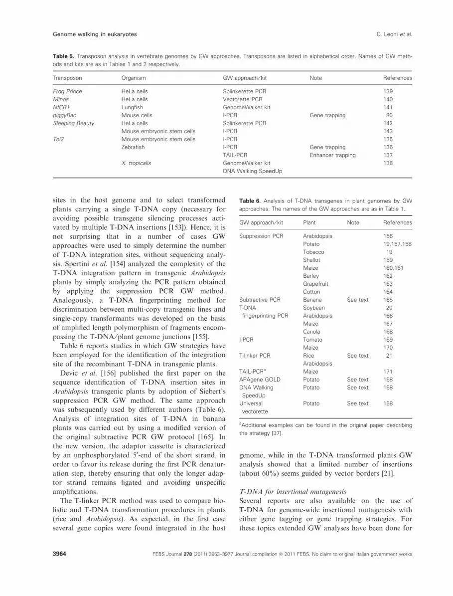
characterization of such systems (Table 5).
Recent review papers on insertional mutagenesis strat-
egies in vertebrates, where the application of GW meth-
ods is reported, are those by Izsvak et al. [144]
and Hackett et al. [145] for the analysis of the
Sleeping Beauty transposon in human cells, Yergeau and
Mead [73] on the use of transposable elements in Xeno-
pus, Clark et al. [146] dealing with the transposons in pig,
and Friedel and Soriano [83] for gene trap mutagenesis
in mouse. A specific database, containing insertion
sequences obtained by I-PCR for the piggyBac transpo-
son system in mice cells, was recently established [147].
Yeast
Transposon mutagenesis has also been applied to yeast
(Saccharomyces cerevisiae). Transposition events of the
yeast transposon Ty1 have been analyzed by I-PCR,
showing that insertions of this element occur only rarely
(about 3%) in ORF regions [148]. For yeast genome-
wide transposon mutagenesis, a more efficient shuttle
mutagenesis strategy was developed [149] in which a
library of yeast genomic DNA, mutagenized with a bac-
terial transposon, is first produced in Escherichia coli;
mutant alleles are subsequently transferred into yeast for
functional analysis. Kumar and Snyder [150] reported a
detailed protocol for shuttle mutagenesis, in which vec-
torette PCR was the chosen GW strategy for the identifi-
cation of insertion sites into the yeast genome. Reports
on the use of either vectorette PCR [151] or I-PCR [152]
for the analysis of insertional mutants obtained by
screening shuttle libraries have been published recently.
T-DNA
The T-DNA of A. tumefaciens Ti-vector is a mobile
element widely used either for ‘plant transformation’
with heterologous genes or for insertional mutagenesis
analysis of plant genomes. In any case establishing the
fate of the engineered T-DNA in the host genome
stands as one of the classical GW applications. In the
following discussion the two different topics will be
treated separately.
T-DNA for gene transfer
Before field trials of genetically modified crops it is of
primary importance both to identify T-DNA insertion
Table 4. Transposon analysis in invertebrate genomes by GW approaches. Organisms are listed according to an ascending taxonomic order,
from Insecta (D. melanogaster, Orseolia oryzae, T. castaneum, Bombyx mori), back to Crustacea (D. stevensoni), Chelicerata (Metaseiu-
lus occidentalis) and Pseudocoelomata (C. elegans, Rotifera sp.). Names of GW approaches are as in Table 1.
Organism Transposon GW approach ⁄ kit Note Reference
D. melanogaster P-element I-PCR See text 112,113
Vectorette PCR See text 114
Splinkerette PCR See text 115
piggyBac I-PCR 116,117
P-element ⁄ piggyBac Splinkerette PCR a 115
Orseolia oryzae Mariner I-PCR 118
T. castaneum Woot Restriction site PCR 119
piggyBac Restriction site PCR, I-PCR,
vectorette-PCR
120,121
Bombyx mori piggyBac I-PCR a 122
D. stevensoni Syrinx UFW 124
Daphne
Metaseiulus occidentalis Mariner I-PCR 123
C. elegans Tc1 Vectorette PCR 125
Tc3 Vectorette PCR See text 126
Mos1 I-PCR 127,128
Rotifer sp. ITm, hAT, piggyBac,
helitron, foldback
UFW 129
aEnhancer trapping analysis.
C. Leoni et al. Genome walking in eukaryotes
FEBS Journal 278 (2011) 3953–3977 Journal compilation ª 2011 FEBS. No claim to original Italian government works 3963
sites in the host genome and to select transformed
plants carrying a single T-DNA copy (necessary for
avoiding possible transgene silencing processes acti-
vated by multiple T-DNA insertions [153]). Hence, it is
not surprising that in a number of cases GW
approaches were used to simply determine the number
of T-DNA integration sites, without sequencing analy-
sis. Spertini et al. [154] analyzed the complexity of the
T-DNA integration pattern in transgenic Arabidopsis
plants by simply analyzing the PCR pattern obtained
by applying the suppression PCR GW method.
Analogously, a T-DNA fingerprinting method for
discrimination between multi-copy transgenic lines and
single-copy transformants was developed on the basis
of amplified length polymorphism of fragments encom-
passing the T-DNA ⁄plant genome junctions [155].
Table 6 reports studies in which GW strategies have
been employed for the identification of the integration
site of the recombinant T-DNA in transgenic plants.
Devic et al. [156] published the first paper on the
sequence identification of T-DNA insertion sites in
Arabidopsis transgenic plants by adoption of Siebert’s
suppression PCR GW method. The same approach
was subsequently used by different authors (Table 6).
Analysis of integration sites of T-DNA in banana
plants was carried out by using a modified version of
the original subtractive PCR GW protocol [165]. In
the new version, the adaptor cassette is characterized
by an unphosphorylated 5¢-end of the short strand, in
order to favor its release during the first PCR denatur-
ation step, thereby ensuring that only the longer adap-
tor strand remains ligated and avoiding unspecific
amplifications.
The T-linker PCR method was used to compare bio-
listic and T-DNA transformation procedures in plants
(rice and Arabidopsis). As expected, in the first case
several gene copies were found integrated in the host
genome, while in the T-DNA transformed plants GW
analysis showed that a limited number of insertions
(about 60%) seems guided by vector borders [21].
T-DNA for insertional mutagenesis
Several reports are also available on the use of
T-DNA for genome-wide insertional mutagenesis with
either gene tagging or gene trapping strategies. For
these topics extended GW analyses have been done for
Table 5. Transposon analysis in vertebrate genomes by GW approaches. Transposons are listed in alphabetical order. Names of GW meth-
ods and kits are as in Tables 1 and 2 respectively.
Transposon Organism GW approach ⁄ kit Note References
Frog Prince HeLa cells Splinkerette PCR 139
Minos HeLa cells Vectorette PCR 140
NfCR1 Lungfish GenomeWalker kit 141
piggyBac Mouse cells I-PCR Gene trapping 80
Sleeping Beauty HeLa cells Splinkerette PCR 142
Mouse embryonic stem cells I-PCR 143
Tol2 Mouse embryonic stem cells I-PCR 135
Zebrafish I-PCR Gene trapping 136
TAIL-PCR Enhancer trapping 137
X. tropicalis GenomeWalker kit 138
DNA Walking SpeedUp
Table 6. Analysis of T-DNA transgenes in plant genomes by GW
approaches. The names of the GW approaches are as in Table 1.
GW approach ⁄ kit Plant Note References
Suppression PCR Arabidopsis 156
Potato 19,157,158
Tobacco 19
Shallot 159
Maize 160,161
Barley 162
Grapefruit 163
Cotton 164
Subtractive PCR Banana See text 165
T-DNA
fingerprinting PCR
Soybean 20
Arabidopsis 166
Maize 167
Canola 168
I-PCR Tomato 169
Maize 170
T-linker PCR Rice See text 21
Arabidopsis
TAIL-PCRa Maize 171
APAgene GOLD Potato See text 158
DNA Walking
SpeedUp
Potato See text 158
Universal
vectorette
Potato See text 158
aAdditional examples can be found in the original paper describing
the strategy [37].
Genome walking in eukaryotes C. Leoni et al.
3964 FEBS Journal 278 (2011) 3953–3977 Journal compilation ª 2011 FEBS. No claim to original Italian government works

the Arabidopsis and rice genomes, which are discussed
separately below.
Large-scale analysis of T-DNA insertions in the Ara-
bidopsis genome has been achieved by different groups
who developed high-throughput versions of the original
suppression PCR GW method [172–177]. Sequencing
results are available at various databases (http://genop-
lante-info.infobiogen.fr [178], http://www.gabi-kat.de/
[177] and http://signal.salk.edu/cgi-bin/tdnaexpress [176]).
The TAIL-PCR strategy was also used for sequenc-
ing T-DNA insertions by various groups [179–182]. In
particular, Sessions et al. [182] developed a high-
throughput TAIL-PCR GW strategy for the analysis
of about 100 000 T-DNA transformants of Arabidopsis
plants. In this case, T-DNA insertion sites showed a
higher presence in promoter regions (44%) than in
transcribed regions (30%) and intergenic regions
(26%). A library of the identified sites was developed
and made available for external users (http://
www.tmri.org.) [182]. Analysis of T-DNA insertion
sites has also been carried out by applying both long
inverted PCR and long tail PCR GW methods [183],
resulting in fragments longer than 6 kb.
Large-scale T-DNA insertional mutagenesis has been
applied to rice. Analysis of thousands of insertion sites
has been achieved by both I-PCR [184] and suppres-
sion PCR GW strategies [185,186]. More recently a
large-scale analysis of T-DNA insertions was per-
formed by TAIL-PCR in about 63 000 transgenic
plants. In all cases, inserts were found to be distributed
all over the 12 chromosomes. Information on T-DNA
transformed rice lines has been collected in the SHIP
(Shangai T-DNA Insertion Population) collection and
can be found at http://ship.plantsignal.cn [187].
The Arabidopsis genome has also been (and still is) a
major field of application of several gene trapping
T-DNA analyses. The first report about the production
and analysis of a collection of Arabidopsis enhancer trap
lines dates back to 1999 [188]. In this case TAIL-PCR
and I-PCR were used to analyze flanking sequences of
inflorescence related mutants. Similar protocols are still
currently used [189–191]. A detailed protocol for pro-
moter trapping and analysis of T-DNA flanking regions
by TAIL-PCR can be found in the method paper by
Blanvillain and Gallois [192]. Suppression PCR has been
used for screening a gene trap T-DNA ⁄ uidA collection
of about 10 000 transgenic Arabidopsis lines developed
for the detection of GUS activity during seed germina-
tion [193]. Further applications of GW methods to gene
trapping in Arabidopsis can be found in the review paper
by Radhamony et al. [194].
In rice the first T-DNA gene trapping studies
adopted TAIL-PCR for the analysis of constructs con-
taining Ac ⁄Ds transposable element and the uidA
reporter gene [195,196]. Since then the numerous gene
trapping studies in rice have been mostly accompanied
by this sequencing procedure. A comprehensive
description of gene trapping achievements in rice,
including also the use of GW techniques, can be found
in the paper by An et al. [197].
Analysis of large T-DNA gene trapping collections
in rice have been carried out by I-PCR [198], high-
throughput adaptation of the suppression PCR GW
method [186] or, more recently, TAIL-PCR [199,200].
In these cases also specific databases have been devel-
oped: OTL (Oryza Tag Line) database (http://urgi.ver-
sailles.inra.fr/OryzaTagLine/) [201], TRIM (Taiwan
Rice Insertion Mutants) database (http://trim.sini-
ca.edu.tw) [202] and RMD (Rice Mutant Database)
(http://rmd.ncpgr.cn) [203].
TAIL-PCR has also been chosen as the GW method
for the analysis of gene trapping in barley [98] and
banana genomes [204].
De novo sequencing
A basic application of GW is the identification of
nucleotide sequences in the course of the characteriza-
tion of genes and genomes. This can be aimed either
at the characterization of unknown sequences or at
the identification of nucleotide modifications and
mutations.
Identification of unknown sequences
Browsing the literature in this area, it can be seen that
most efforts have been devoted to the identification of
‘regulatory regions’, while a minor number of reports
deal with the use of GW in ‘gene identification’,
‘sequencing of BAC and YAC clones’, ‘cytoplasmic
male sterility’ (large modifications occurring in plant
nuclear ⁄mitochondrial genomes, which are at the basis
of the cytoplasmic male sterility phenotypic trait) and
‘multigene families’. Table 7 summarizes studies con-
ducted by GW for the analysis of regulatory regions,
while the other applications have been reported in
Table 8. Additionally, it is worth mentioning the appli-
cation of GW to metagenomics analysis, even if this
topic is outside the scope of this review. Related infor-
mation is available in the review paper by Singh et al.
[205].
Siebert et al. [15] developed the well-known suppres-
sion PCR GW method to walk upstream of the 5¢-endcoding regions of the human TPA (tissue-type plasmin-
ogen activator) and transferrin genes for a valuable
distance (4.5 and 6 kb, respectively). This technique is
C. Leoni et al. Genome walking in eukaryotes
FEBS Journal 278 (2011) 3953–3977 Journal compilation ª 2011 FEBS. No claim to original Italian government works 3965

one of the most used GW methods, finding application
in several different cases.
Padegimas and Reichert [16] succeeded in isolating
promoter regions from three different maize peroxidase
genes, improving the splinkerette strategy with the
introduction of 3¢-blocked adaptors and removal of
unligated genomic DNA by ExoIII digestion.
The identification of regulatory regions of multigene
families can be pursued by different approaches. Leoni
et al. [56] adopted an ET-GW strategy (Table 1) in
which highly conserved regions of a multigene family
were chosen to design common primers to be used as
gene specific primers. In this way it has been possible
to simultaneously identify regulatory elements of the
spinach multigene family coding for isoforms of the
light harvesting protein Lhcb1. Additionally, novel
gene members of the same family could be detected by
this approach. In a second case, the TVL-PCR GW
method, applied to identify the regulatory regions of
strawberry SUPERMAN-like genes [29], has been used
for the identification of multiple members of a gene
family using degenerate primers based on conserved
sequences as priming sites. It must be noted also that
the Universal GW kit (Clontech) has been employed
for the analysis of a sugarcane BAC clone containing
multiple copies of the sugarcane DIRIGENT gene
[206].
Analysis of nucleotide modifications and
mutations
The application of GW to the analysis of nucleotide
modifications and mutations is essentially based on
the LM-PCR strategy due to Mueller and Wold [53].
Indeed, although originally presented as a footprint-
ing technique, LM-PCR has also been successfully
regarded as a GW technique. Pfeifer et al. [241]
illustrated its application for both genome sequencing
and methylation analysis of the human X-linked
phosphoglycerate kinase (PGK-1) gene. An automated
version of the LM-PCR usable for GW analysis of
DNA methylation, DNA damage and protein-DNA
footprints has also been developed [242]. More
recently the method was further improved (see also
[67]) and widely used for mapping DNA damage in
carcinogenesis etiology [243,244]. LM-PCR applica-
tions for the analysis of mitochondrial DNA damage
due to chemicals or ageing have also been reported
[245,246].
Critical evaluations of GW methods
The issue about which GW method better fits the spe-
cific experimental conditions is not easy to deal with
exhaustively because of the numerous methods available
Table 7. Regulatory regions identified in eukaryotes by GW approaches. Since more than 200 papers can be found in the literature dealing
with this topic, mostly reporting the use of commercially available kits, here only papers which describe the development of a specific GW
method are reported. Analyses are reported in chronological order. Names of GW methods are as in Table 1.
Gene Organism GW approach ⁄ kit Note References
Po Shark Cassette PCR 10
TPA Man Suppression PCR 15
Transferrin
At23 Arabidopsis RAGE-GW 32
PR-10 Parsley
Peroxidase Maize Modified splinkerette See text 16
Sucrose phosphate synthase Banana Single primer amplification 58
Actin Sugarcane
S15 ribosomal protein Dunaliella tertiolecta
Several cDNAs Brassica juncea Mishra et al. 40
Pennisetum glaucum
Gibberellin 20-oxidase Rice T-linker PCR 21
Squalene synthase Ganoderma lucidum Self-formed adaptor PCR 48
Ascorbate peroxidaseHsp70 Hsp10 P. glaucum High-throughput genome walking 50
Gst Salicornia brachiata
Lhcb1 family Spinach Leoni et al. 56,57
LRDEF Lily Straight walk 26
OgGSTZ2 Rice
SuRB Tobacco
PGK1 Pichia ciferrii Template blocking PCR 28
SUPERMAN-like Strawberry TVL-PCR 29
Genome walking in eukaryotes C. Leoni et al.
3966 FEBS Journal 278 (2011) 3953–3977 Journal compilation ª 2011 FEBS. No claim to original Italian government works

and the multiplicity of variables in the different assays.
Nevertheless some general comments can be made.
The first issue to consider is whether a single
sequencing (as in the case of the study of a single gene)
or multiple sequencing data (as in the case of large
insertional mutagenesis analysis) are necessary. In the
first case it can be assumed that most of the methods
give satisfactory results. This is clearly shown in the
case of the identification of gene regulatory regions
(Table 7), where at least 12 different methods have
been employed. In contrast, in the identification of
multiple sequences only a limited number of methods
have been successfully used (I-PCR, vectorette,
splinkerette, suppression, TAIL-PCR), which can
therefore be considered as first choice in planning GW
experiments.
In any case, some differences clearly exist among the
GW strategies, and at least three parameters can be
considered for their critical evaluation: specificity, sen-
sitivity and efficiency. As for specificity, it can gener-
Table 8. Genes identified in eukaryotes by GW approaches. Most of the data were obtained by using the Universal GW kit (Clontech) or
other common GW approaches described in the text. Voices are listed in alphabetical order for main taxonomic groups. Applications are as
in Fig. 2.
Species Gene Application References
Animals
Drosophila melanogaster Sup 4 BAC ⁄ YAC termini 207
Homo sapiens FLEB14-14 BAC ⁄ YAC termini 208
Scyb11 BAC ⁄ YAC termini 209
HPRT Gene sequencing 210
LRP1B Gene sequencing 211
ELF3 Gene sequencing 212
Dystrophin Gene sequencing 213
Macropus eugenii LTB, TNF and LTA BAC ⁄ YAC termini 214
Schistosoma mansoni SmHox1 SmHox4 and SmHox4 BAC ⁄ YAC termini 215
Salmo salar Hox genes BAC ⁄ YAC termini 216
Pogona vitticeps Z and W chromosome fragment Gene sequencing 217
Fungi
Latimeria menadoensis Hox Gene sequencing 218
Penicillium pinophilum Endo-b-1.4-glucanase gene 5 Gene sequencing 219
Phoma betae Aphidicolan-16b-ol synthase Gene sequencing 220
Phomopsis amygdali PbGGs, ACS, PbP450-1, PbP450-2, PbTP, PbTF Gene sequencing 220
PaDC1 and PaDC2 Gene sequencing 221
Diterpene hydrocarbon phomopsene Gene sequencing 222
Pucciniomycotina RHA1, RHA2 and RHA3 Gene sequencing 223
Plants
Allium cepa Orf725 CMS 224
Capsicum annuum Rf flanking region CMS 225
Cicer arientinum Pi-ta-2 and xa5 Gene sequencing 226
Coffea arabica; C. canephora ManS1 and GMGT1 Gene sequencing 227
Malus domestica Mal D3 genes Gene sequencing 228
MdAGP1, MdAGP2 and MdAGP3 genes Gene sequencing 229
Oryza sativa Slender glume BAC ⁄ YAC termini 230
OsPE Gene sequencing 231
Spinacia oleracea Lhcb1 Multigene family 56
Sugar beet Restorer-of-fertility BAC ⁄ YAC termini ⁄ CMS 232
Sugarcane DIRIGENT Multigene family 206
Taxus media Geranyl geranyl diphosphate Gene sequencing 233
Triticum aestivum L LMW-GS genes BAC ⁄ YAC termini 234
BAC ⁄ YAC termini 235
Protozoa
Cryptobia salmositica Adenosylmethionone synthetase Gene sequencing 236
Cathepsin L-like cysteine proteinase Gene sequencing 237
MSP-1 Gene sequencing 238
Neospora caninum NcSAG4 Gene sequencing 239
NcBSR4 Gene sequencing 240
C. Leoni et al. Genome walking in eukaryotes
FEBS Journal 278 (2011) 3953–3977 Journal compilation ª 2011 FEBS. No claim to original Italian government works 3967

ally be assumed that it mostly relies on the specificity
of the gene specific primer used in the GW approach.
I-PCR which adopts two specific primers should there-
fore be considered as the most specific method. Never-
theless, all the other methods that use a gene specific
primer coupled with an adaptor ⁄ tail specific walking
primer can be regarded as highly specific as well.
Methods adopting random ⁄degenerate primers, con-
versely, may show lower specificity. A precautionary
note must be added for cassette PCR methods that do
not take countermeasures to prevent the synthesis of
non-specific PCR products deriving from the walking
primer. The blocked DLA GW method properly
addresses this point, showing that blocking the adaptor
extension in the first cycle of the final PCR amplifica-
tion can increase specificity of PCR products from
44% to 100% [27].
Blocked DLA was also compared with splinkerette
PCR for sensitivity. The higher sensitivity of the first
method is clearly demonstrated by the relative intensity
of the electrophoretic bands of amplification products.
In the course of a screening experiment for the identifi-
cation of P-elements in Drosophila, Eggert et al. [114]
combined in several ratios flies carrying or not a
defined transposon. They showed that vectorette PCR
can be more sensitive than I-PCR in the identification
of transgenic P-elements, allowing detection of a spe-
cific insertion in a ratio of 1 : 6000–10 000.
It must noted, however, that some technical
improvements have undoubtedly improved the general
sensitivity of GW methods. This is the case of intro-
ducing biotinylation of adaptors and primers. Nielsen
et al. [247] showed the possibility that, when adopting
solid-phase purification of biotinylated fragments, GW
can reach a very high sensitivity, able to detect about
two copies of a target sequence in a DNA background
of 25 ng.
Recently three commercial kits [APAgene GOLD
Genome Walking Kit (BIO S&T), DNA Walking
SpeedUp Kit II (Seegene) and Universal Vectorette Sys-
tem (Sigma)] and the suppression PCR GW method (as
modified by Spertini et al. [154]) were compared for the
identification of T-DNA flanking regions in transgenic
potato. In this analysis, the two methods based on the
extension of gene specific primers and PCR amplifica-
tion with degenerated primers (APAgene� and DNA
Walking SpeedUp� II) showed higher success rates
than the two cassette PCR methods, which identified a
lower number of flanking regions [158].
As for efficiency, some strategies have to be men-
tioned for the reported capacity to read more than 3 kb
for single walk, as in the case of LD-GW PCR [55], sup-
pression PCR [15], long I-PCR and TAIL-PCR [183].
A last issue to consider for the choice of a GW
method is the possibility of its scale-up for
high-throughput analysis, if needed. This has been
demonstrated to be possible for suppression PCR
[107,172–177], TAIL-PCR [96,107,182,199], I-PCR
[72,88], high-throughput GW [248], LM-PCR [242],
straight walk high-throughput [26] and restriction site
extension PCR [33].
Conclusions and perspectives
GW encompasses an array of easy-to-use strategies for
the identification of genome nucleotide sequences, use-
ful for both insertional mutagenesis analysis and
de novo sequencing. In the first case it has largely con-
tributed to advances in reverse genetic analysis, and to
the development of databases of mutants of many
eukaryotic genomes. In the second case, GW is partic-
ularly advantageous for the identification of specific
sequences in cases where whole genome sequencing
projects have not been undertaken. It is noteworthy to
observe that most of the different GW strategies
or improvements have been developed in the course of
de novo sequencing approaches (see Identification of
unknown sequences, for example).
The extreme flexibility of GW strategies makes its
application possible in every standardly equipped
research laboratory. In addition, the possibility of
merging GW strategies to next generation sequencing
approaches will undoubtedly extend the future applica-
tion of this by now basic technique of molecular biol-
ogy.
Acknowledgement
This work has been partly supported by a grant from
the Italian Ministry of Instruction University and
Research: PRIN 20087ATS57 ‘Food allergens’.
References
1 Hengen PN (1995) Vectorette, splinkerette and boomer-
ang DNA amplification. Trends Biochem Sci 20, 372–
373.
2 Hui EK, Wang PC & Lo SJ (1998) Strategies for clon-
ing unknown cellular flanking DNA sequences from
foreign integrants. Cell Mol Life Sci 54, 1403–1411.
3 Tonooka Y & Fujishima M (2009) Comparison and
critical evaluation of PCR-mediated methods to walk
along the sequence of genomic DNA. Appl Microbiol
Biotechnol 85, 37–43.
4 Kotik M (2009) Novel genes retrieved from environ-
mental DNA by polymerase chain reaction: current
Genome walking in eukaryotes C. Leoni et al.
3968 FEBS Journal 278 (2011) 3953–3977 Journal compilation ª 2011 FEBS. No claim to original Italian government works

genome-walking techniques for future metagenome
applications. J Biotechnol 144, 75–82.
5 Triglia T, Peterson MG & Kemp DJ (1988) A proce-
dure for in vitro amplification of DNA segments that
lie outside the boundaries of known sequences. Nucleic
Acids Res 16, 8186.
6 Benkel BF & Fong Y (1996) Long range-inverse PCR
(LR-IPCR): extending the useful range of inverse PCR.
Genet Anal Biomolec Eng 13, 123–127.
7 Kohda T & Taira K (2000) A simple and efficient
method to determine the terminal sequences of restric-
tion fragments containing known sequences. DNA Res
7, 151–155.
8 Tsaftaris A, Pasentzis K & Argiriou A (2010) Rolling
circle amplification of genomic templates for inverse
PCR (RCA-GIP): a method for 5¢- and 3¢-genome walk-
ing without anchoring. Biotechnol Lett 32, 157–161.
9 Shyamala V & Ames GF (1989) Genome walking by
single-specific-primer polymerase chain reaction: SSP-
PCR. Gene 84, 1–8.
10 Fors L, Saavedra RA & Hood L (1990) Cloning of the
shark Po promoter using a genomic walking technique
based on the polymerase chain reaction. Nucleic Acids
Res 18, 2793–2799.
11 Riley J, Butler R, Ogilvie D, Finniear R, Jenner D,
Powell S, Anand R, Smith JC & Markham AF (1990)
A novel, rapid method for the isolation of terminal
sequences from yeast artificial chromosome (YAC)
clones. Nucleic Acids Res 18, 2887–2890.
12 Rosenthal A & Jones DS (1990) Genomic walking and
sequencing by oligo-cassette mediated polymerase chain
reaction. Nucleic Acids Res 18, 3095–3096.
13 Lagerstrom M, Parik J, Malmgren H, Stewart J,
Pettersson U & Landegren U (1991) Capture PCR: -
efficient amplification of DNA fragments adjacent to a
known sequence in human and YAC DNA. PCR
Methods Appl 1, 111–119.
14 Devon RS, Porteous DJ & Brookes AJ (1995) Splinke-
rettes – improved vectorettes for greater efficiency in
PCR walking. Nucleic Acids Res 23, 1644–1645.
15 Siebert PD, Chenchik A, Kellogg DE, Lukyanov KA
& Lukyanov SA (1995) An improved PCR method for
walking in uncloned genomic DNA. Nucleic Acids Res
23, 1087–1088.
16 Padegimas LS & Reichert NA (1998) Adaptor ligation-
based polymerase chain reaction-mediated walking.
Anal Biochem 260, 149–153.
17 Zhang Z & Gurr SJ (2000) Walking into the unknown:
a ‘step down’ PCR-based technique leading to the
direct sequence analysis of flanking genomic DNA.
Gene 253, 145–150.
18 Kilstrup M & Kristiansen KN (2000) Rapid genome
walking: a simplified oligo-cassette mediated polymer-
ase chain reaction using a single genome-specific
primer. Nucleic Acids Res 28, E55.
19 Cottage A, Yang A, Maunders H, de Lacy RC &
Ramsay NA (2001) Identification of DNA sequences
flanking T-DNA insertions by PCR-walking. Plant
Mol Biol Rep 19, 321–327.
20 Windels P, Taverniers I, Depicker A, Van Bockstaele E
& De Loose M (2001) Characterisation of the
Roundup Ready soybean insert. Eur Food Res Technol
213, 107–112.
21 Yuanxin Y, Chengcai A, Li L, Jiayu G, Guihong T &
Zhangliang C (2003) T-linker-specific ligation PCR
(T-linker PCR): an advanced PCR technique for chro-
mosome walking or for isolation of tagged DNA ends.
Nucleic Acids Res 31, e68.
22 Nthangeni MB, Ramagoma F, Tlou MG & Litthauer
D (2005) Development of a versatile cassette for direc-
tional genome walking using cassette ligation-mediated
PCR and its application in the cloning of complete
lipolytic genes from Bacillus species. J Microbiol Meth
61, 225–234.
23 Wang GP et al. (2008) DNA bar coding and pyrose-
quencing to analyze adverse events in therapeutic gene
transfer. Nucleic Acids Res 36, e49.
24 Wang GP, Ciuffi A, Leipzig J, Berry CC & Bushman FD
(2007) HIV integration site selection: analysis by mas-
sively parallel pyrosequencing reveals association with
epigenetic modifications. Genome Res 17, 1186–1194.
25 Tonooka Y, Mizukami Y & Fujishima M (2008) One-
base excess adaptor ligation method for walking uncl-
oned genomic DNA. Appl Microbiol Biotechnol 78,
173–180.
26 Tsuchiya T, Kameya N & Nakamura I (2009) Straight
walk: a modified method of ligation-mediated genome
walking for plant species with large genomes. Anal
Biochem 388, 158–160.
27 Liu S, Dietrich CR & Schnable PS (2009) DLA-based
strategies for cloning insertion mutants: cloning the gl4
locus of maize using Mu transposon tagged alleles.
Genetics 183, 1215–1225.
28 Bae JH & Sohn JH (2010) Template-blocking PCR:
an advanced PCR technique for genome walking. Anal
Biochem 398, 112–116.
29 Orcheski BB & Davis TM (2010) An enhanced method
for sequence walking and paralog mining: TOPO(R)
vector-ligation PCR. BMC Res Notes 3, 61.
30 Jones DH & Winistorfer SC (1992) Sequence specific
generation of a DNA panhandle permits PCR amplifi-
cation of unknown flanking DNA. Nucleic Acids Res
20, 595–600.
31 Rudenko GN, Rommens CM, Nijkamp HJ & Hille J
(1993) Supported PCR: an efficient procedure to
amplify sequences flanking a known DNA segment.
Plant Mol Biol 21, 723–728.
32 Cormack RS & Somssich IE (1997) Rapid amplifica-
tion of genomic ends (RAGE) as a simple method to
clone flanking genomic DNA. Gene 194, 273–276.
C. Leoni et al. Genome walking in eukaryotes
FEBS Journal 278 (2011) 3953–3977 Journal compilation ª 2011 FEBS. No claim to original Italian government works 3969

33 Ji J & Braam J (2010) Restriction site extension PCR:
a novel method for high-throughput characterization
of tagged DNA fragments and genome walking. PLoS
ONE 5, e10577.
34 Parker JD, Rabinovitch PS & Burmer GC (1991) Tar-
geted gene walking polymerase chain reaction. Nucleic
Acids Res 19, 3055–3060.
35 Sarkar G, Turner RT & Bolander ME (1993) Restric-
tion-site PCR: a direct method of unknown sequence
retrieval adjacent to a known locus by using universal
primers. PCR Methods Appl 2, 318–322.
36 Puskas LG, Fartmann B & Bottka S (1994) Restricted
PCR: amplification of an individual sequence flanked
by a highly repetitive element from total human DNA.
Nucleic Acids Res 22, 3251–3252.
37 Liu YG & Whittier RF (1995) Thermal asymmetric
interlaced PCR: automatable amplification and
sequencing of insert end fragments from P1 and YAC
clones for chromosome walking. Genomics 25, 674–681.
38 Chen X & Wu R (1997) Direct amplification of
unknown genes and fragments by uneven polymerase
chain reaction. Gene 185, 195–199.
39 Ge Y & Charon NW (1997) Identification of a large
motility operon in Borrelia burgdorferi by semi-random
PCR chromosome walking. Gene 189, 195–201.
40 Mishra RN, Singla-Pareek SL, Nair S, Sopory SK &
Reddy MK (2002) Directional genome walking using
PCR. BioTechniques 33, 830–834.
41 Myrick KV & Gelbart WM (2007) A modified univer-
sal fast walking method for single-tube transposon
mapping. Nat Protoc 2, 1556–1563.
42 Myrick KV & Gelbart WM (2002) Universal fast walk-
ing for direct and versatile determination of flanking
sequence. Gene 284, 125–131.
43 Park DJ (2005) LaNe RAGE: a new tool for genomic
DNA flanking sequence determination. Electron J
Biotechnol 8, 218–225.
44 Tan G, Gao Y, Shi M, Zhang X, He S, Chen Z & An
C (2005) SiteFinding-PCR: a simple and efficient PCR
method for chromosome walking. Nucleic Acids Res
33, e122.
45 Levano-Garcia J, Verjovski-Almeida S & da Silva AC
(2005) Mapping transposon insertion sites by touch-
down PCR and hybrid degenerate primers. BioTech-
niques 38, 225–229.
46 Walser JC, Evgen’ev MB & Feder ME (2006) A geno-
mic walking method for screening sequence length
polymorphism. Mol Ecol Notes 6, 563–567.
47 Guo H & Xiong J (2006) A specific and versatile
genome walking technique. Gene 381, 18–23.
48 Wang S, He J, Cui Z & Li S (2007) Self-formed adap-
tor PCR: a simple and efficient method for chromo-
some walking. Appl Environ Microbiol 73, 5048–5051.
49 Pilhofer M, Bauer AP, Schrallhammer M, Richter L,
Ludwig W, Schleifer KH & Petroni G (2007) Charac-
terization of bacterial operons consisting of two tubu-
lins and a kinesin-like gene by the novel two-step gene
walking method. Nucleic Acids Res 35, e135.
50 Reddy PS, Mahanty S, Kaul T, Nair S, Sopory SK &
Reddy MK (2008) A high-throughput genome-walking
method and its use for cloning unknown flanking
sequences. Anal Biochem 381, 248–253.
51 Ping L, Vogel H & Boland W (2008) Cloning of pro-
karyotic genes by a universal degenerate primer PCR.
FEMS Microbiol Lett 287, 192–198.
52 Martin-Harris MH, Bartley PA & Morley AA (2010)
Gene walking using sequential hybrid primer polymer-
ase chain reaction. Anal Biochem 399, 308–310.
53 Mueller PR & Wold B (1989) In vivo footprinting of a
muscle specific enhancer by ligation mediated PCR.
Science 246, 780–786.
54 Schmidt M et al. (2002) Polyclonal long-term repopu-
lating stem cell clones in a primate model. Blood 100,
2737–2743.
55 Min GS & Powell JR (1998) Long-distance genome
walking using the long and accurate polymerase chain
reaction. BioTechniques 24, 398–400.
56 Leoni C, Volpicella M, Placido A, Gallerani R & Ceci
LR (2010) Application of a genome walking method
for the study of the spinach Lhcb1 multigene family.
Journal of Plant Physiology 167, 138–143.
57 Leoni C, Gallerani R & Ceci LR (2008) A genome
walking strategy for the identification of eukaryotic
nucleotide sequences adjacent to known regions.
BioTechniques 44, 229, 232–235.
58 Hermann SR, Miller JA, O’Neill S, Tsao TT, Harding
RM & Dale JL (2000) Single-primer amplification
of flanking sequences. BioTechniques 29, 1176–1178,
1180.
59 Pule MA, Rousseau A, Vera J, Heslop HE, Brenner
MK & Vanin EF (2008) Flanking-sequence exponential
anchored-polymerase chain reaction amplification: a
sensitive and highly specific method for detecting retro-
viral integrant-host-junction sequences. Cytotherapy 10,
526–539.
60 Liu S et al. (2009) Mu transposon insertion sites and
meiotic recombination events co-localize with epige-
netic marks for open chromatin across the maize gen-
ome. PLoS Genet 5, e1000733.
61 Brenner M (1996) Gene marking. Hum Gene Ther 7,
1927–1936.
62 Rill DR et al. (1994) Direct demonstration that autolo-
gous bone marrow transplantation for solid tumors
can return a multiplicity of tumorigenic cells. Blood 84,
380–383.
63 Tey SK & Brenner MK (2007) The continuing contri-
bution of gene marking to cell and gene therapy. Mol
Ther 15, 666–676.
64 Bushman FD (2007) Retroviral integration and human
gene therapy. J Clin Invest 117, 2083–2086.
Genome walking in eukaryotes C. Leoni et al.
3970 FEBS Journal 278 (2011) 3953–3977 Journal compilation ª 2011 FEBS. No claim to original Italian government works

65 Modlich U & Baum C (2009) Preventing and exploit-
ing the oncogenic potential of integrating gene vectors.
J Clin Invest 119, 755–758.
66 Hacein-Bey-Abina S et al. (2003) LMO2-associated
clonal T cell proliferation in two patients after gene
therapy for SCID-X1. Science 302, 415–419.
67 Schmidt M et al. (2001) Detection and direct genomic
sequencing of multiple rare unknown flanking DNA
in highly complex samples. Hum Gene Ther 12, 743–
749.
68 Schroder AR, Shinn P, Chen H, Berry C, Ecker JR &
Bushman F (2002) HIV-1 integration in the human
genome favors active genes and local hotspots. Cell
110, 521–529.
69 Wu X, Li Y, Crise B & Burgess SM (2003) Transcrip-
tion start regions in the human genome are favored
targets for MLV integration. Science 300, 1749–1751.
70 Mitchell RS, Beitzel BF, Schroder AR, Shinn P, Chen
H, Berry CC, Ecker JR & Bushman FD (2004) Retro-
viral DNA integration: ASLV, HIV, and MLV show
distinct target site preferences. PLoS Biol 2, E234.
71 Ciuffi A & Barr SD (2010) Identification of HIV inte-
gration sites in infected host genomic DNA. Methods
53, 39–46.
72 Hansen GM et al. (2008) Large-scale gene trapping in
C57BL ⁄ 6N mouse embryonic stem cells. Genome Res
18, 1670–1679.
73 Yergeau DA & Mead PE (2007) Manipulating the
Xenopus genome with transposable elements. Genome
Biol 8(Suppl 1), S11.
74 Earp DJ, Lowe B & Baker B (1990) Amplification of
genomic sequences flanking transposable elements in
host and heterologous plants: a tool for transposon
tagging and genome characterization. Nucleic Acids
Res 18, 3271–3279.
75 Walbot V (1992) Strategies for mutagenesis and gene
cloning using transposon tagging and T-DNA inser-
tional mutagenesis. Plant Physiol 43, 49–82.
76 Izawa T et al. (1997) Transposon tagging in rice. Plant
Mol Biol 35, 219–229.
77 Feschotte C, Jiang N & Wessler SR (2002) Plant trans-
posable elements: where genetics meets genomics. Nat
Rev Genet 3, 329–341.
78 Candela H & Hake S (2008) The art and design of
genetic screens: maize. Nat Rev Genet 9, 192–203.
79 Ryder E & Russell S (2003) Transposable elements as
tools for genomics and genetics in Drosophila. Brief
Funct Genomic Proteomic 2, 57–71.
80 Wu S, Ying G, Wu Q & Capecchi MR (2007) Toward
simpler and faster genome-wide mutagenesis in mice.
Nat Genet 39, 922–930.
81 Ivics Z, Li MA, Mates L, Boeke JD, Nagy A,
Bradley A & Izsvak Z (2009) Transposon-mediated
genome manipulation in vertebrates. Nat Methods 6,
415–422.
82 Kumar A (2008) Multipurpose transposon insertion
libraries for large-scale analysis of gene function in
yeast. Methods Mol Biol 416, 117–129.
83 Friedel RH & Soriano P (2010) Gene trap mutagenesis
in the mouse. Methods Enzymol 477, 243–269.
84 Tani H et al. (2004) Activation tagging in plants: a tool
for gene discovery. Funct Integr Genomics 4, 258–266.
85 Springer PS (2000) Gene traps: tools for plant develop-
ment and genomics. Plant Cell 12, 1007–1020.
86 Muskett PR, Clissold L, Marocco A, Springer PS, Mart-
ienssen R & Dean C (2003) A resource of mapped disso-
ciation launch pads for targeted insertional mutagenesis
in the Arabidopsis genome. Plant Physiol 132, 506–516.
87 Long D, Martin M, Sundberg E, Swinburne J, Puang-
somlee P & Coupland G (1993) The maize transposable
element system Ac ⁄Ds as a mutagen in Arabidopsis:
identification of an albino mutation induced by Ds
insertion. Proc Natl Acad Sci USA 90, 10370–10374.
88 Meissner R, Chague V, Zhu Q, Emmanuel E, Elkind
Y & Levy AA (2000) Technical advance: a high
throughput system for transposon tagging and pro-
moter trapping in tomato. Plant J 22, 265–274.
89 Kolkman JM et al. (2005) Distribution of Activator
(Ac) throughout the maize genome for use in regional
mutagenesis. Genetics 169, 981–995.
90 Tirichine L, Herrera-Cervera JA & Stougaard J (2005)
Ds Gene-Tagging. In Lotus japonicus Handbook
(Marquez AJ ed.), pp 211–215. Springer, Amsterdam.
91 Wilson K, Long D, Swinburne J & Coupland G (1996)
A dissociation insertion causes a semidominant muta-
tion that increases expression of TINY, an Arabidopsis
gene related to APETALA2. Plant Cell 8, 659–671.
92 Klimmek F, Sjodin A, Noutsos C, Leister D & Jansson
S (2006) Abundantly and rarely expressed Lhc protein
genes exhibit distinct regulation patterns in plants.
Plant Physiol 140, 793–804.
93 Sundaresan V, Springer P, Volpe T, Haward S, Jones
JD, Dean C, Ma H & Martienssen R (1995) Patterns
of gene action in plant development revealed by enhan-
cer trap and gene trap transposable elements. Genes
Dev 9, 1797–1810.
94 Gu Q, Ferrandiz C, Yanofsky MF & Martienssen R
(1998) The Fruitfull MADS-box gene mediates cell dif-
ferentiation during Arabidopsis fruit development.
Development 125, 1509–1517.
95 Klimyuk VI, Nussaume L, Harrison K & Jones JD
(1995) Novel GUS expression patterns following trans-
position of an enhancer trap Ds element in Arabidop-
sis. Mol Gen Genet 249, 357–365.
96 van Enckevort LJ et al. (2005) EU-OSTID: a collection
of transposon insertional mutants for functional ge-
nomics in rice. Plant Mol Biol 59, 99–110.
97 Upadhyaya NM, Zhu QH & Bhat RS (2011) Transpo-
son insertional mutagenesis in rice. Methods Mol Biol
678, 147–177.
C. Leoni et al. Genome walking in eukaryotes
FEBS Journal 278 (2011) 3953–3977 Journal compilation ª 2011 FEBS. No claim to original Italian government works 3971

98 Lazarow K & Lutticke S (2009) An Ac ⁄Ds-mediated
gene trap system for functional genomics in barley.
BMC Genomics 10, 55.
99 Ott T, Nelsen-Salz B & Doring HP (1992) PCR-aided
genomic sequencing of 5¢ subterminal sequences of the
maize transposable element Activator (Ac) in trans-
genic tobacco plants. Plant J 2, 705–711.
100 Souer E, Quattrocchio F, de Vetten N, Mol J & Koes
R (1995) A general method to isolate genes tagged by
a high copy number transposable element. Plant J 7,
677–685.
101 Van den Broeck D, Maes T, Sauer M, Zethof J, De
Keukeleire P, D’Hauw M, Van Montagu M & Gerats
T (1998) Transposon Display identifies individual
transposable elements in high copy number lines. Plant
J 13, 121–129.
102 Aarts MG, Dirkse WG, Stiekema WJ & Pereira A
(1993) Transposon tagging of a male sterility gene in
Arabidopsis. Nature 363, 715–717.
103 Marsch-Martinez N, Greco R, Van Arkel G, Herrera-
Estrella L & Pereira A (2002) Activation tagging using
the En-I maize transposon system in Arabidopsis.
Plant Physiol 129, 1544–1556.
104 Altinkut A, Raskina O, Nevo E & Belyayev A (2006)
En ⁄ Spm-like transposons in Poaceae species: transpos-
ase sequence variability and chromosomal distribution.
Cell Mol Biol Lett 11, 214–230.
105 Roccaro M, Li Y, Sommer H & Saedler H (2007) RO-
SINA (RSI) is part of a CACTA transposable element,
TamRSI, and links flower development to transposon
activity. Mol Genet Genomics 278, 243–254.
106 Yamazaki M et al. (2001) The rice retrotransposon
Tos17 prefers low-copy-number sequences as integra-
tion targets. Mol Genet Genomics 265, 336–344.
107 Miyao A et al. (2003) Target site specificity of the
Tos17 retrotransposon shows a preference for inser-
tion within genes and against insertion in retrotrans-
poson-rich regions of the genome. Plant Cell 15,
1771–1780.
108 Zhao G, Zhang Z, Sun H, Li H & Dai H (2007) Isola-
tion of Ty1-copia-like retrotransposon sequences from
the apple genome by chromosome walking based on
modified SiteFinding-polymerase chain reaction. Acta
Biochim Biophys Sin (Shanghai) 39, 675–683.
109 Zhao G, Dai H, Chang L, Ma Y, Sun H, He P &
Zhang Z (2009) Isolation of two novel complete
Ty1-copia retrotransposons from apple and demonstra-
tion of use of derived S-SAP markers for distinguishing
bud sports of Malus domestica cv. Fuji. Tree Genetics
Genomes 6, 149–159.
110 Pan X, Liu H, Clarke J, Jones J, Bevan M & Stein L
(2003) ATIDB: Arabidopsis thaliana insertion database.
Nucleic Acids Res 31, 1245–1251.
111 Droc G et al. (2006) OryGenesDB: a database for rice
reverse genetics. Nucleic Acids Res 34, D736–D740.
112 Sentry JW & Kaiser K (1994) Application of inverse
PCR to site-selected mutagenesis of Drosophila. Nucleic
Acids Res 22, 3429–3430.
113 Liao GC, Rehm EJ & Rubin GM (2000) Insertion site
preferences of the P transposable element in Drosophila
melanogaster. Proc Natl Acad Sci USA 97, 3347–3351.
114 Eggert H, Bergemann K & Saumweber H (1998)
Molecular screening for P-element insertions in a large
genomic region of Drosophila melanogaster using poly-
merase chain reaction mediated by the vectorette.
Genetics 149, 1427–1434.
115 Potter CJ & Luo L (2010) Splinkerette PCR for map-
ping transposable elements in Drosophila. PLoS ONE
5, e10168.
116 Thibault ST et al. (2004) A complementary transposon
tool kit for Drosophila melanogaster using P and piggy-
Bac. Nat Genet 36, 283–287.
117 Bonin CP & Mann RS (2004) A piggyBac transposon
gene trap for the analysis of gene expression and func-
tion in Drosophila. Genetics 167, 1801–1811.
118 Behura SK, Nair S & Mohan M (2001) Polymorphisms
flanking the mariner integration sites in the rice gall
midge (Orseolia oryzae Wood–Mason) genome are bio-
type-specific. Genome 44, 947–954.
119 Beeman RW & Stauth DM (1997) Rapid cloning of
insect transposon insertion junctions using ‘universal’
PCR. Insect Mol Biol 6, 83–88.
120 Lorenzen MD, Berghammer AJ, Brown SJ, Denell RE,
Klingler M & Beeman RW (2003) piggyBac-mediated
germline transformation in the beetle Tribolium casta-
neum. Insect Mol Biol 12, 433–440.
121 Trauner J et al. (2009) Large-scale insertional mutagen-
esis of a coleopteran stored grain pest, the red flour
beetle Tribolium castaneum, identifies embryonic lethal
mutations and enhancer traps. BMC Biol 7, 73.
122 Uchino K et al. (2008) Construction of a piggyBac-
based enhancer trap system for the analysis of gene
function in silkworm Bombyx mori. Insect Biochem
Mol Biol 38, 1165–1173.
123 Jeyaprakash A & Hoy MA (1995) Complete sequence
of a mariner transposable element from the predatory
mite Metaseiulus occidentalis isolated by an inverse
PCR approach. Insect Mol Biol 4, 31–39.
124 Schon I & Arkhipova IR (2006) Two families of non-
LTR retrotransposons, Syrinx and Daphne, from the
Darwinulid ostracod,Darwinula stevensoni. Gene 371,
296–307.
125 Korswagen HC, Durbin RM, Smits MT & Plasterk
RH (1996) Transposon Tc1-derived sequence-tagged
sites in Caenorhabditis elegans as markers for gene
mapping. Proc Natl Acad Sci USA 93, 14680–14685.
126 Wicks SR, de Vries CJ, van Luenen HG & Plasterk
RH (2000) CHE-3, a cytosolic dynein heavy chain, is
required for sensory cilia structure and function in
Caenorhabditis elegans. Dev Biol 221, 295–307.
Genome walking in eukaryotes C. Leoni et al.
3972 FEBS Journal 278 (2011) 3953–3977 Journal compilation ª 2011 FEBS. No claim to original Italian government works

127 Bessereau JL, Wright A, Williams DC, Schuske K,
Davis MW & Jorgensen EM (2001) Mobilization of a
Drosophila transposon in the Caenorhabditis elegans
germ line. Nature 413, 70–74.
128 Granger L, Martin E & Segalat L (2004) Mos as a tool
for genome-wide insertional mutagenesis in Caenor-
habditis elegans: results of a pilot study. Nucleic Acids
Res 32, e117.
129 Arkhipova IR & Meselson M (2005) Diverse DNA
transposons in rotifers of the class Bdelloidea. Proc
Natl Acad Sci USA 102, 11781–11786.
130 Spradling AC, Stern D, Beaton A, Rhem EJ, Laverty
T, Mozden N, Misra S & Rubin GM (1999) The
Berkeley Drosophila Genome Project gene disruption
project: single P-element insertions mutating 25% of
vital Drosophila genes. Genetics 153, 135–177.
131 Tweedie S et al. (2009) FlyBase: enhancing Drosophila
gene ontology annotations. Nucleic Acids Res 37,
D555–D559.
132 Brand AH & Perrimon N (1993) Targeted gene expres-
sion as a means of altering cell fates and generating
dominant phenotypes. Development 118, 401–415.
133 Various Authors (2002) Special Issue: GAL4 ⁄UAS in
Drosophila. Genesis 34, 1–173.
134 Lukacsovich T & Yamamoto D (2001) Trap a gene
and find out its function: toward functional genomics
in Drosophila. J Neurogenet 15, 147–168.
135 Kawakami K & Noda T (2004) Transposition of the
Tol2 element, an Ac-like element from the Japanese
medaka fish Oryzias latipes, in mouse embryonic stem
cells. Genetics 166, 895–899.
136 Kawakami K, Takeda H, Kawakami N, Kobayashi M,
Matsuda N & Mishina M (2004) A transposon-medi-
ated gene trap approach identifies developmentally reg-
ulated genes in zebrafish. Dev Cell 7, 133–144.
137 Parinov S, Kondrichin I, Korzh V & Emelyanov A
(2004) Tol2 transposon-mediated enhancer trap to
identify developmentally regulated zebrafish genes
in vivo. Dev Dyn 231, 449–459.
138 Hamlet MR, Yergeau DA, Kuliyev E, Takeda M, Ta-
ira M, Kawakami K & Mead PE (2006) Tol2 transpo-
son-mediated transgenesis in Xenopus tropicalis.
Genesis 44, 438–445.
139 Miskey C, Izsvak Z, Plasterk RH & Ivics Z (2003) The
Frog Prince: a reconstructed transposon from Rana pi-
piens with high transpositional activity in vertebrate
cells. Nucleic Acids Res 31, 6873–6881.
140 Klinakis AG, Zagoraiou L, Vassilatis DK & Savakis C
(2000) Genome-wide insertional mutagenesis in human
cells by the Drosophila mobile element Minos. EMBO
Rep 1, 416–421.
141 Sirijovski N, Woolnough C, Rock J & Joss JMP
(2005) nfCR1, the first non-LTR retrotransposon
characterized in the Australian lungfish genome,
Neoceratodus forsteri, shows similarities to CR1-like
elements. J Exp Zool Pt B: Molec Develop Evol 304,
40–49.
142 Ivics Z, Hackett PB, Plasterk RH & Izsvak Z (1997)
Molecular reconstruction of Sleeping Beauty, a Tc1-
like transposon from fish, and its transposition in
human cells. Cell 91, 501–510.
143 Luo G, Ivics Z, Izsvak Z & Bradley A (1998) Chromo-
somal transposition of a Tc1 ⁄mariner-like element in
mouse embryonic stem cells. Proc Natl Acad Sci USA
95, 10769–10773.
144 Izsvak Z, Chuah MK, Vandendriessche T & Ivics Z
(2009) Efficient stable gene transfer into human cells
by the Sleeping Beauty transposon vectors. Methods
49, 287–297.
145 Hackett PB, Largaespada DA & Cooper LJ (2010) A
transposon and transposase system for human applica-
tion. Mol Ther 18, 674–683.
146 Clark KJ, Carlson DF & Fahrenkrug SC (2007) Pigs
taking wing with transposons and recombinases.
Genome Biol 8(Suppl. 1), S13.
147 Sun LV et al. (2008) PBmice: an integrated database
system of piggyBac (PB) insertional mutations and
their characterizations in mice. Nucleic Acids Res 36,
D729–D734.
148 Ji H, Moore DP, Blomberg MA, Braiterman LT, Voy-
tas DF, Natsoulis G & Boeke JD (1993) Hotspots for
unselected Ty1 transposition events on yeast chromo-
some III are near tRNA genes and LTR sequences.
Cell 73, 1007–1018.
149 Burns N, Grimwade B, Ross-Macdonald PB, Choi EY,
Finberg K, Roeder GS & Snyder M (1994) Large-scale
analysis of gene expression, protein localization, and
gene disruption in Saccharomyces cerevisiae. Genes Dev
8, 1087–1105.
150 Kumar A & Snyder M (2001) Genome-wide transpo-
son mutagenesis in yeast. In Curr Protoc Mol Biol,
Genome-Wide Transposon Mutagenesis in Yeast,
chapter 13, pp.13.3.1–13.3.15, John Wiley & Sons, New
York.
151 Estruch F, Peiro-Chova L, Gomez-Navarro N, Durban
J, Hodge C, Del Olmo M & Cole CN (2009) A genetic
screen in Saccharomyces cerevisiae identifies new genes
that interact with mex67-5, a temperature-sensitive
allele of the gene encoding the mRNA export receptor.
Mol Genet Genomics 281, 125–134.
152 Hontz RD, Niederer RO, Johnson JM & Smith JS
(2009) Genetic identification of factors that modulate
ribosomal DNA transcription in Saccharomyces
cerevisiae. Genetics 182, 105–119.
153 Flavell RB (1994) Inactivation of gene expression in
plants as a consequence of specific sequence duplica-
tion. Proc Natl Acad Sci USA 91, 3490–3496.
154 Spertini D, Beliveau C & Bellemare G (1999) Screening
of transgenic plants by amplification of unknown geno-
mic DNA flanking T-DNA. BioTechniques 27, 308–314.
C. Leoni et al. Genome walking in eukaryotes
FEBS Journal 278 (2011) 3953–3977 Journal compilation ª 2011 FEBS. No claim to original Italian government works 3973
155 Theuns I, Windels P, De Buck S, Depicker A, Van
Bockstaele E & De Loose M (2002) Identification and
characterization of T-DNA inserts by T-DNA finger-
printing. Euphytica 123, 75–84.
156 Devic M, Albert S, Delseny M & Roscoe TJ (1997)
Efficient PCR walking on plant genomic DNA. Plant
Physiol Biochem 35, 331–339.
157 Cote MJ, Meldrum AJ, Raymond P & Dollard C (2005)
Identification of genetically modified potato (Solanum
tuberosum) cultivars using event specific polymerase
chain reaction. J Agric Food Chem 53, 6691–6696.
158 Cullen D, Harwood W, Smedley M, Davies H & Tay-
lor M (2011) Comparison of DNA walking methods
for isolation of transgene-flanking regions in GM
potato. Mol Biotechnol 49, 19–31.
159 Zheng SJ, Henken B, Sofiari E, Jacobsen E, Krens
FA & Kik C (2001) Molecular characterization of
transgenic shallots (Allium cepa L.) by adaptor liga-
tion PCR (AL-PCR) and sequencing of genomic
DNA flanking T-DNA borders. Transgenic Res 10,
237–245.
160 Holck A, Va M, Didierjean L & Rudi K (2002) 5¢-Nuclease PCR for quantitative event-specific detection
of the genetically modified Mon810 MaisGard maize.
Eur Food Res Technol 214, 449–453.
161 Collonnier C et al. (2005) Characterization and event
specific detection by quantitative real-time PCR of T25
maize insert. J AOAC Int 88, 536–546.
162 Salvo-Garrido H, Travella S, Bilham LJ, Harwood
WA & Snape JW (2004) The distribution of transgene
insertion sites in barley determined by physical and
genetic mapping. Genetics 167, 1371–1379.
163 Rai M (2006) Refinement of the Citrus tristeza virus
resistance gene (Ctv) positional map in Poncirus trifoli-
ata and generation of transgenic grapefruit (Citrus
paradisi) plant lines with candidate resistance genes in
this region. Plant Mol Biol 61, 399–414.
164 Akritidis P, Pasentsis K, Tsaftaris AS, Mylona PV &
Polidoros AN (2008) Identification of unknown geneti-
cally modified material admixed in conventional cotton
seed and development of an event-specific detection
method. Electron J Biotechnol 11, 76–83.
165 Perez-Hernandez JB, Swennen R & Sagi L (2006)
Number and accuracy of T-DNA insertions in trans-
genic banana (Musa spp.) plants characterized by an
improved anchored PCR technique. Transgenic Res 15,
139–150.
166 Windels P, De Buck S, Van Bockstaele E, De Loose M
& Depicker A (2003) T-DNA integration in Arabidop-
sis chromosomes. Presence and origin of filler DNA
sequences. Plant Physiol 133, 2061–2068.
167 Windels P, Bertrand S, Depicker A, Moens W, Bock-
staele E & Loose M (2003) Qualitative and event-spe-
cific PCR real-time detection methods for StarLink
maize. Eur Food Res Technol 216, 259–263.
168 Taverniers I, Windels P, Vaitilingom M, Milcamps A,
Van Bockstaele E, Van den Eede G & De Loose M
(2005) Event-specific plasmid standards and real-time
PCR methods for transgenic Bt11, Bt176, and GA21
maize and transgenic GT73 canola. J Agric Food Chem
53, 3041–3052.
169 Knapp S, Larondelle Y, Rossberg M, Furtek D &
Theres K (1994) Transgenic tomato lines containing Ds
elements at defined genomic positions as tools for tar-
geted transposon tagging. Mol Gen Genet 243, 666–
673.
170 Ronning SB, Vaitilingom M, Berdal KG & Holst-Jen-
sen A (2003) Event specific real-time quantitative PCR
for genetically modified Bt11 maize (Zea mays). Eur
Food Res Technol 216, 347–354.
171 Yang L, Xu S, Pan A, Yin C, Zhang K, Wang Z,
Zhou Z & Zhang D (2005) Event specific qualitative
and quantitative polymerase chain reaction detection
of genetically modified MON863 maize based on the
5¢-transgene integration sequence. J Agric Food Chem
53, 9312–9318.
172 Balzergue S et al. (2001) Improved PCR-walking for
large-scale isolation of plant T-DNA borders. Biotech-
niques, 30, 496–498, 502, 504.
173 Brunaud V et al. (2002) T-DNA integration into the
Arabidopsis genome depends on sequences of pre-inser-
tion sites. EMBO Rep 3, 1152–1157.
174 Alonso JM & Ecker JR (2006) Moving forward in
reverse: genetic technologies to enable genome-wide
phenomic screens in Arabidopsis. Nat Rev Genet 7,
524–536.
175 O’Malley RC, Alonso JM, Kim CJ, Leisse TJ & Ecker
JR (2007) An adapter ligation-mediated PCR method
for high-throughput mapping of T-DNA inserts in the
Arabidopsis genome. Nat Protoc 2, 2910–2917.
176 O’Malley RC & Ecker JR (2010) Linking genotype to
phenotype using the Arabidopsis unimutant collection.
Plant J 61, 928–940.
177 Rosso MG, Li Y, Strizhov N, Reiss B, Dekker K &
Weisshaar B (2003) An Arabidopsis thaliana T-DNA
mutagenized population (GABI-Kat) for flanking
sequence tag-based reverse genetics. Plant Mol Biol 53,
247–259.
178 Samson F, Brunaud V, Balzergue S, Dubreucq B, Lepi-
niec L, Pelletier G, Caboche M & Lecharny A (2002)
FLAGdb ⁄FST: a database of mapped flanking inser-
tion sites (FSTs) of Arabidopsis thaliana T-DNA trans-
formants. Nucleic Acids Res 30, 94–97.
179 McElver J et al. (2001) Insertional mutagenesis of
genes required for seed development in Arabidopsis tha-
liana. Genetics 159, 1751–1763.
180 Budziszewski GJ et al. (2001) Arabidopsis genes essen-
tial for seedling viability: isolation of insertional
mutants and molecular cloning. Genetics 159, 1765–
1778.
Genome walking in eukaryotes C. Leoni et al.
3974 FEBS Journal 278 (2011) 3953–3977 Journal compilation ª 2011 FEBS. No claim to original Italian government works

181 Krysan PJ, Young JC, Jester PJ, Monson S, Copen-
haver G, Preuss D & Sussman MR (2002) Character-
ization of T-DNA insertion sites in Arabidopsis
thaliana and the implications for saturation mutagene-
sis. Omics 6, 163–174.
182 Sessions A et al. (2002) A high-throughput Arabidopsis
reverse genetics system. Plant Cell 14, 2985–2994.
183 Szabados L et al. (2002) Distribution of 1000
sequenced T-DNA tags in the Arabidopsis genome.
Plant J 32, 233–242.
184 An S et al. (2003) Generation and analysis of end
sequence database for T-DNA tagging lines in rice.
Plant Physiol 133, 2040–2047.
185 Sallaud C et al. (2003) Highly efficient production and
characterization of T-DNA plants for rice (Oryza sati-
va L.) functional genomics. Theor Appl Genet 106,
1396–1408.
186 Sallaud C et al. (2004) High throughput T-DNA inser-
tion mutagenesis in rice: a first step towards in silico
reverse genetics. Plant J 39, 450–464.
187 Fu FF, Ye R, Xu SP & Xue HW (2009) Studies on
rice seed quality through analysis of a large-scale
T-DNA insertion population. Cell Res 19, 380–391.
188 Campisi L et al. (1999) Generation of enhancer trap
lines in Arabidopsis and characterization of expression
patterns in the inflorescence. Plant J 17, 699–707.
189 Gardner MJ, Baker AJ, Assie JM, Poethig RS, Hasel-
off JP & Webb AA (2009) GAL4 GFP enhancer trap
lines for analysis of stomatal guard cell development
and gene expression. J Exp Bot 60, 213–226.
190 Kim SG, Lee S, Kim YS, Yun DJ, Woo JC & Park
CM (2010) Activation tagging of an Arabidopsis SHI-
related sequence gene produces abnormal anther dehis-
cence and floral development. Plant Mol Biol 74,
337–351.
191 Kuroha T, Okuda A, Arai M, Komatsu Y, Sato S,
Kato T, Tabata S & Satoh S (2009) Identification of
Arabidopsis subtilisin-like serine protease specifically
expressed in root stele by gene trapping. Physiol Plant
137, 281–288.
192 Blanvillain R & Gallois P (2008) Promoter trapping
system to study embryogenesis. Methods Mol Biol 427,
121–135.
193 Dubreucq B, Berger N, Vincent E, Boisson M, Pelletier
G, Caboche M & Lepiniec L (2000) The Arabidopsis
AtEPR1 extensin-like gene is specifically expressed in
endosperm during seed germination. Plant J 23, 643–
652.
194 Radhamony RN, Prasad AM & Srinivasan R (2005)
T-DNA insertional mutagenesis in Arabidopsis: a tool
for functional genomics. Electron J Biotechnol 8,
82–106.
195 Chin HG et al. (1999) Molecular analysis of rice plants
harboring an Ac ⁄Ds transposable element-mediated
gene trapping system. Plant J 19, 615–623.
196 Jeon JS et al. (2000) T-DNA insertional mutagenesis
for functional genomics in rice. Plant J 22, 561–570.
197 An G, Jeong DH, Jung KH & Lee S (2005) Reverse
genetic approaches for functional genomics of rice.
Plant Mol Biol 59, 111–123.
198 Ryu CH et al. (2004) Generation of T-DNA tagging
lines with a bidirectional gene trap vector and the
establishment of an insertion-site database. Plant Mol
Biol 54, 489–502.
199 Hsing YI et al. (2007) A rice gene activation ⁄ knockoutmutant resource for high throughput functional ge-
nomics. Plant Mol Biol 63, 351–364.
200 Zhang J et al. (2007) Non-random distribution of
T-DNA insertions at various levels of the genome hier-
archy as revealed by analyzing 13 804 T-DNA flanking
sequences from an enhancer-trap mutant library. Plant
J 49, 947–959.
201 Larmande P et al. (2008) Oryza Tag Line, a phenotypic
mutant database for the Genoplante rice insertion line
library. Nucleic Acids Res 36, D1022–D1027.
202 Chern CG et al. (2007) A rice phenomics study – phe-
notype scoring and seed propagation of a T-DNA
insertion-induced rice mutant population. Plant Mol
Biol 65, 427–438.
203 Zhang J, Li C, Wu C, Xiong L, Chen G, Zhang Q &
Wang S (2006) RMD: a rice mutant database for func-
tional analysis of the rice genome. Nucleic Acids Res
34, D745–D748.
204 Santos E, Remy S, Thiry E,Windelinckx S, Swennen R&
Sagi L (2009) Characterization and isolation of a T-DNA
tagged banana promoter active during in vitro culture
and low temperature stress. BMCPlant Biol 9, 77.
205 Singh J, Behal A, Singla N, Joshi A, Birbian N, Singh
S, Bali V & Batra N (2009) Metagenomics: concept,
methodology, ecological inference and recent advances.
Biotechnol J 4, 480–494.
206 Damaj MB et al. (2010) Isolating promoters of multi-
gene family members from the polyploid sugarcane
genome by PCR-based walking in BAC DNA. Genome
53, 840–847.
207 Wesley CS, Myers MP & Young MW (1994) Rapid
sequential walking from termini of cosmid, P1 and
YAC inserts. Nucleic Acids Res 22, 538–539.
208 Asakawa S et al. (1997) Human BAC library: construc-
tion and rapid screening. Gene 191, 69–79.
209 Meyer M, Erdel M, Duba HC, Werner ER & Werner-
Felmayer G (2000) Cloning, genomic sequence, and
chromosome mapping of Scyb11, the murine homo-
logue of SCYB11 (alias betaR1 ⁄H174 ⁄ SCYB9B ⁄ I-TAC ⁄ IP-9 ⁄CXCL11). Cytogenet Cell Genet 88,
278–282.
210 Nelson DL, Ledbetter SA, Corbo L, Victoria MF,
Ramirez-Solis R, Webster TD, Ledbetter DH & Cas-
key CT (1989) Alu polymerase chain reaction: a
method for rapid isolation of human-specific sequences
C. Leoni et al. Genome walking in eukaryotes
FEBS Journal 278 (2011) 3953–3977 Journal compilation ª 2011 FEBS. No claim to original Italian government works 3975

from complex DNA sources. Proc Natl Acad Sci USA
86, 6686–6690.
211 Liu CX, Musco S, Lisitsina NM, Yaklichkin SY &
Lisitsyn NA (2000) Genomic organization of a new
candidate tumor suppressor gene, LRP1B. Genomics
69, 271–274.
212 Kaplan MH, Wang XP, Xu HP & Dosik MH (2004)
Partially unspliced and fully spliced ELF3 mRNA,
including a new Alu element in human breast cancer.
Breast Cancer Res Treat 83, 171–187.
213 Zhong M, Pan SY, Cai LD, Lu BX & Zhang GZ
(2006) Strategy of localizing the deletion mutation
point in large introns of Dystrophin gene. Chinese
Journal of Clinical Rehabilitation 10, 120–121.
214 Cross JGR, Harrison GA, Coggill P, Sims S, Beck S,
Deakin JE & Marshall Graves JA (2005) Analysis of
the genomic region containing the tammar wallaby
(Macropus eugenii) orthologues of MHC class III
genes. Cytogenet Genome Res 111, 110–117.
215 Pierce RJ et al. (2005) Evidence for a dispersed Hox
gene cluster in the platyhelminth parasite Schistosoma
mansoni. Mol Biol Evol 22, 2491–2503.
216 Mungpakdee S, Seo HC, Angotzi AR, Dong X, Akalin
A & Chourrout D (2008) Differential evolution of the
13 Atlantic salmon Hox clusters. Mol Biol Evol 25,
1333–1343.
217 Quinn AE, Ezaz T, Sarre SD, Graves JM & Georges
A (2010) Extension, single-locus conversion and physi-
cal mapping of sex chromosome sequences identify the
Z microchromosome and pseudo-autosomal region in a
dragon lizard, Pogona vitticeps. Heredity 104, 410–417.
218 Koh EGL, Lam K, Christoffels A, Erdmann MV,
Brenner S & Venkatesh B (2003) Hox gene clusters in
the Indonesian coelacanth, Latimeria menadoensis. Proc
Natl Acad Sci USA 100, 1084–1088.
219 Jeya M, Joo AR, Lee KM, Sim WI, Oh DK, Kim YS,
Kim IW & Lee JK (2010) Characterization of endo-
beta-1,4-glucanase from a novel strain of Penicillium pi-
nophilum KMJ601. Appl Microbiol Biotechnol 85, 1005–
1014.
220 Toyomasu T et al. (2004) Cloning of a gene cluster
responsible for the biosynthesis of diterpene aphidico-
lin, a specific inhibitor of DNA polymerase alpha. Bio-
sci Biotechnol Biochem 68, 146–152.
221 Toyomasu T et al. (2008) Identification of diterpene
biosynthetic gene clusters and functional analysis of
labdane-related diterpene cyclases in Phomopsis amyg-
dali. Biosci Biotechnol Biochem 72, 1038–1047.
222 Toyomasu T et al. (2009) Biosynthetic gene-based sec-
ondary metabolite screening: a new diterpene, methyl
phomopsenonate, from the fungus Phomopsis amygdali.
J Org Chem 74, 1541–1548.
223 Coelho MA, Rosa A, Rodrigues N, Fonseca A & Gon-
calves P (2008) Identification of mating type genes in
the bipolar basidiomycetous yeast Rhodosporidium toru-
loides: first insight into the MAT locus structure of the
Sporidiobolales. Eukaryot Cell 7, 1053–1061.
224 Kim S, Lee ET, Cho DY, Han T, Bang H, Patil BS,
Ahn YK & Yoon MK (2009) Identification of a novel
chimeric gene, orf725, and its use in development of a
molecular marker for distinguishing among three cyto-
plasm types in onion (Allium cepa L.). Theor Appl
Genet 118, 433–441.
225 Min WK, Lim H, Lee YP, Sung SK, Kim BD & Kim
S (2008) Identification of a third haplotype of the
sequence linked to the Restorer-of-fertility (Rf) gene
and its implications for male-sterility phenotypes in
peppers (Capsicum annuum L.). Molec Cells 25, 20–29.
226 Iruela M, Piston F, Cubero JI, Millan T, Barro F &
Gil J (2009) The marker SCK13(603) associated with
resistance to ascochyta blight in chickpea is located in
a region of a putative retrotransposon. Plant Cell Rep
28, 53–60.
227 Pre M, Caillet V, Sobilo J & McCarthy J (2008)
Characterization and expression analysis of genes
directing galactomannan synthesis in coffee. Ann Bot
102, 207–220.
228 Gao ZS et al. (2005) Linkage map positions and allelic
diversity of two Mal d 3 (non-specific lipid transfer
protein) genes in the cultivated apple (Malus domes-
tica). Theor Appl Genet 110, 479–491.
229 Choi YO et al. (2010) Isolation and promoter analysis
of anther-specific genes encoding putative arabinoga-
lactan proteins in Malus x domestica. Plant Cell Rep
29, 15–24.
230 Teraishi M, Hirochika H, Okumoto Y, Horibata A,
Yamagata H & Tanisaka T (2001) Identification of
YAC clones containing the mutable slender glume
locus slg in rice (Oryza sativa L.). Genome 44, 1–6.
231 Puri A, Basha PO, Kumar M, Rajpurohit D, Randha-
wa GS, Kianian SF, Rishi A & Dhaliwal HS (2010)
The polyembryo gene (OsPE) in rice. Funct Integrative
Genom 10, 359–366.
232 Hagihara E, Matsuhira H, Ueda M, Mikami T &
Kubo T (2005) Sugar beet BAC library construction
and assembly of a contig spanning Rf1, a restorer-of-
fertility gene for Owen cytoplasmic male sterility. Mol
Genet Genomics 274, 316–323.
233 Liao Z et al. (2005) An intron-free methyl jasmonate
inducible geranylgeranyl diphosphate synthase gene
from Taxus media and its functional identification in
yeast. Molec Biol 39, 11–17.
234 Huang XQ & Cloutier S (2007) Hemi-nested touch-
down PCR combined with primer-template mismatch
PCR for rapid isolation and sequencing of low molecu-
lar weight glutenin subunit gene family from a hexa-
ploid wheat BAC library. BMC Genet 8, 18.
235 Huang XQ & Cloutier S (2008) Molecular character-
ization and genomic organization of low molecular
weight glutenin subunit genes at the Glu-3 loci in
Genome walking in eukaryotes C. Leoni et al.
3976 FEBS Journal 278 (2011) 3953–3977 Journal compilation ª 2011 FEBS. No claim to original Italian government works

hexaploid wheat (Triticum aestivum L.). Theor Appl
Genet 116, 953–966.
236 Jesudhasan PR & Woo PTK (2007) An S-adenosylme-
thionine synthetase gene from the pathogenic piscine
hemoflagellate, Cryptobia salmositica. Parasitol Res
100, 1401–1406.
237 Jesudhasan PR, Tan CW, Hontzeas N & Woo PT
(2007) A cathepsin L-like cysteine proteinase gene from
the protozoan parasite, Cryptobia salmositica. Parasitol
Res 100, 881–886.
238 Jesudhasan PR, Tan CW & Woo PTK (2007) A
metalloproteinase gene from the pathogenic piscine
hemoflagellate, Cryptobia salmositica. Parasitol Res 100,
899–904.
239 Fernandez-Garcia A, Risco-Castillo V, Zaballos A,
Alvarez-Garcia G & Ortega-Mora LM (2006) Identifi-
cation and molecular cloning of the Neospora caninum
SAG4 gene specifically expressed at bradyzoite stage.
Mol Biochem Parasitol 146, 89–97.
240 Risco-Castillo V, Fernandez-Garcia A, Zaballos A, Ag-
uado-Martinez A, Hemphill A, Rodriguez-Bertos A,
Alvarez-Garcia G & Ortega-Mora LM (2007) Molecu-
lar characterisation of BSR4, a novel bradyzoite-spe-
cific gene from Neospora caninum. Intl J Parasitol 37,
887–896.
241 Pfeifer GP, Steigerwald SD, Mueller PR, Wold B &
Riggs AD (1989) Genomic sequencing and methylation
analysis by ligation mediated PCR. Science 246, 810–
813.
242 Dai SM, Chen HH, Chang C, Riggs AD & Flanagan
SD (2000) Ligation-mediated PCR for quantitative in
vivo footprinting. Nat Biotechnol 18, 1108–1111.
243 Besaratinia A & Pfeifer GP (2009) DNA-lesion map-
ping in mammalian cells. Methods 48, 35–39.
244 Besaratinia A & Pfeifer GP (2006) Investigating human
cancer etiology by DNA lesion footprinting and muta-
genicity analysis. Carcinogenesis 27, 1526–1537.
245 LeDoux SP, DruzhynaNM,Hollensworth SB, Harrison
JF&Wilson GL (2007)Mitochondrial DNA repair:
a critical player in the response of cells of the CNS to
genotoxic insults.Neuroscience 145, 1249–1259.
246 Lezza AM, Fallacara FP, Pesce V, Leeuwenburgh C,
Cantatore P & Gadaleta MN (2008) Localization of
abasic sites and single-strand breaks in mitochondrial
DNA from brain of aged rat, treated or not with calo-
ric restriction diet. Neurochem Res 33, 2609–2614.
247 Nielsen CR, Berdal KG & Holst-Jensen A (2008)
Anchored PCR for possible detection and characterisa-
tion of foreign integrated DNA at near single molecule
level. Eur Food Res Technol 226, 949–956.
248 Reddy MK, Nair S & Sopory SK (2002) A new
approach for efficient directional genome walking using
polymerase chain reaction. Anal Biochem 306, 154–158.
Supporting information
The following supplementary material is available:
Table S1. List of patents related to GW methods.
This supplementary material can be found in the
online version of this article.
Please note: As a service to our authors and readers,
this journal provides supporting information supplied
by the authors. Such materials are peer-reviewed and
may be re-organized for online delivery, but are not
copy-edited or typeset. Technical support issues arising
from supporting information (other than missing files)
should be addressed to the authors.
C. Leoni et al. Genome walking in eukaryotes
FEBS Journal 278 (2011) 3953–3977 Journal compilation ª 2011 FEBS. No claim to original Italian government works 3977


