Embed Size (px)
Citation preview
Genome-wide annotation, expression profiling, and proteininteraction studies of the core cell-cycle genes in Phalaenopsisaphrodite
Hsiang-Yin Lin • Jhun-Chen Chen •
Miao-Ju Wei • Yi-Chen Lien • Huang-Hsien Li •
Swee-Suak Ko • Zin-Huang Liu • Su-Chiung Fang
Received: 23 April 2013 / Accepted: 3 September 2013 / Published online: 25 September 2013
� The Author(s) 2013. This article is published with open access at Springerlink.com
Abstract Orchidaceae is one of the most abundant and
diverse families in the plant kingdom and its unique
developmental patterns have drawn the attention of many
evolutionary biologists. Particular areas of interest have
included the co-evolution of pollinators and distinct floral
structures, and symbiotic relationships with mycorrhizal
flora. However, comprehensive studies to decipher the
molecular basis of growth and development in orchids
remain scarce. Cell proliferation governed by cell-cycle
regulation is fundamental to growth and development of
the plant body. We took advantage of recently released
transcriptome information to systematically isolate and
annotate the core cell-cycle regulators in the moth orchid
Phalaenopsis aphrodite. Our data verified that Phalaen-
opsis cyclin-dependent kinase A (CDKA) is an evolution-
arily conserved CDK. Expression profiling studies
suggested that core cell-cycle genes functioning during the
G1/S, S, and G2/M stages were preferentially enriched in
the meristematic tissues that have high proliferation
activity. In addition, subcellular localization and pairwise
interaction analyses of various combinations of CDKs and
cyclins, and of E2 promoter-binding factors and dimer-
ization partners confirmed interactions of the functional
units. Furthermore, our data showed that expression of the
core cell-cycle genes was coordinately regulated during
pollination-induced reproductive development. The data
obtained establish a fundamental framework for study of
the cell-cycle machinery in Phalaenopsis orchids.
Keywords Cell cycle � Phalaenopsis aphrodite �Orchid � Protocorm � Meristematic tissues
Introduction
Cell reproduction is a basic biological process that is
essential for the growth of all organisms (Hall et al. 2004).
Cell-cycle regulation plays a pivotal role in the cell pro-
liferation required for plant growth. The core cell-cycle
genes are central regulators of cell division and hence a
common converging point for internal and external signals
during growth and development. In plants, growth derives
from meristematic tissues. Cell proliferation regulated by
intrinsic developmental signals and extrinsic environmen-
tal cues is required for meristem replenishment and orga-
nization. In addition, cell division machinery governing
cell proliferation has to be carefully regulated in order to
meet differentiation and fate specification requirements
during plant development.
The eukaryotic cell cycle is controlled by an evolu-
tionarily conserved set of cell-cycle proteins (Mironov
et al. 1999). Despite the universal principle of cell-cycle
regulation, the core cell-cycle genes in plants are dupli-
cated and have diverged to accommodate complex
Electronic supplementary material The online version of thisarticle (doi:10.1007/s11103-013-0128-y) contains supplementarymaterial, which is available to authorized users.
H.-Y. Lin � J.-C. Chen � M.-J. Wei � Y.-C. Lien � H.-H. Li �S.-S. Ko � S.-C. Fang (&)
Biotechnology Center in Southern Taiwan, Academia Sinica,
No. 59, Siraya Blvd., Xinshi District, Tainan 741, Taiwan
e-mail: [email protected]
H.-Y. Lin � J.-C. Chen � M.-J. Wei � Y.-C. Lien � H.-H. Li �S.-S. Ko � S.-C. Fang
Agricultural Biotechnology Research Center, Academia Sinica,
Taipei 115, Taiwan
H.-Y. Lin � Z.-H. Liu
Institute of Tropical Plant Sciences, National Cheng Kung
University, Tainan 701, Taiwan
123
Plant Mol Biol (2014) 84:203–226
DOI 10.1007/s11103-013-0128-y
developmental requirements. Based on a homology-based
annotation strategy, more than 90 cell-cycle genes have
been classified in Arabidopsis (Vandepoele et al. 2002;
Menges et al. 2005). However, how these cell-cycle pro-
teins are coordinated to drive cell division and regulate
differentiation programs in plants remains largely unclear.
Cyclin-dependent kinases (CDKs) are central cell-cycle
regulators whose activities are subject to multiple levels of
regulation and whose expression oscillates in a periodic
manner to drive cell-cycle phase transitions (Morgan
2007). Based on the nomenclature derived from genome-
wide classification of the Arabidopsis cell-cycle genes,
there are eight classes of CDKs, designated as A to G type
and CDK-like (L-type) CDKs (Vandepoele et al. 2002;
Menges et al. 2005). CDK activity is activated by binding
to cyclins (CYCs), inhibited by CDK inhibitors, and reg-
ulated by phosphorylation. Plant cell-cycle transitions are
controlled by two types of CDKs—A-type CDKs (CDKAs)
and B-type CDKs (CDKBs). Plant CDKA contains a
canonical cyclin-binding motif PSTAIRE in the alpha-1
helix that is conserved across different kingdoms (Inze and
De Veylder 2006). CDKA has been shown to play a pivotal
role at both the G1/S and G2/M transitions and can func-
tionally rescue CDK-deficient yeast mutants (Ferreira et al.
1991; Hirayama et al. 1991). Members of the CDKB gene
family are plant specific (Burssens et al. 1998; Mironov
et al. 1999). They have been reported to play roles during
the G2-M and S-G2-M phases (Fobert et al. 1996; Magyar
et al. 1997; Umeda et al. 1999; Meszaros et al. 2000;
Porceddu et al. 2001; Sorrell et al. 2001; Breyne et al.
2002; Menges et al. 2002; Corellou et al. 2005).
Cyclins activate the kinase activity of the CDKs and
control timely entry into the cell cycle. In plants and ani-
mals, three main classes of CYCs are required for cell-
cycle progression. Generally, but not exclusively, D-type
CYCs function in the G1 stage and are required to regulate
G1/S transition, A-type CYCs function during S phase, and
B-type CYCs are important in regulation of S phase and
mitosis. Transcripts of CDKB and CYCB gene family
members are preferentially accumulated during S, G2, and
M phases and are often used as markers for dividing cells
(Inze and De Veylder 2006). Plant A- and B-type CYCs
have orthologs in animals. Plant D-type CYCs show less
similarity to D-type CYCs from animals and are therefore
referred to as plant specific (Wang et al. 2004). There are at
least 30 CYCs with potential roles in cell-cycle regulation
in Arabidopsis (Vandepoele et al. 2002). Because D-type
CYCs regulate G1-S transition and the commitment to
enter the cell cycle, they are thought to be primary targets
for external and internal signals leading to cell proliferation
(Nieuwland et al. 2009). However, many exceptions to this
simple functional assignment have been reported (Inze and
De Veylder 2006; Vanneste et al. 2011). Therefore, the
functional role of individual CYCs may be oversimplified
and their roles during cell-cycle progression need to be
tested experimentally.
Cyclin-dependent kinase activity is also modulated by
CDK-activating kinases (CAKs). Two types of CAKs have
been found in higher plants that have been designated as D-
and F-type. CDKDs are functionally related to vertebrate
CAKs, whereas CDKFs are plant specific. D- and F-type
CDKs have been shown to activate A-type CDKs via
phosphorylation (Umeda et al. 1998; Yamaguchi et al.
2000; Shimotohno et al. 2003). In Arabidopsis, cyclin
H-activated CDKDs are capable of phosphorylating CDKs
as well as the C-terminal domain of the largest subunit of
RNA polymerase II (Shimotohno et al. 2004). Conversely,
F-type CDKs do not require a CYC interacting partner and
are capable of phosphorylating CDKs (Shimotohno et al.
2004, 2006). Arabidopsis CDKF has been shown to phos-
phorylate and activate CDKD (Shimotohno et al. 2004).
Cyclin-dependent kinase inhibitors (CKIs) negatively
regulate the CDK activity by direct binding. Plants contain
CKIs that show weak similarity to the N-terminally located
CKI domain of the mammalian Cip/Kip proteins (Wang et al.
1997; Lui et al. 2000) and are, therefore, commonly referred to
as Kip-related proteins (KRPs). The Arabidopsis KRP protein
family has seven members. Work with Arabidopsis has shown
that the KRP family proteins are expressed in both mitotically
dividing and endoreduplicating cells (Ormenese et al. 2004).
The involvement of some KRP genes in regulating endore-
duplication (part of the plant differentiation program) has
been confirmed by overexpression approaches. For instance,
overexpression of either Arabidopsis KRP1 or KRP2, pref-
erentially expressed in endoreduplicating cells, reduces cell
division and affects the switch to the endoreduplication cycle
(Wang et al. 2000; De Veylder et al. 2001; Verkest et al.
2005b; Gutierrez 2005; De Veylder et al. 2007; Verkest et al.
2005a). The defects caused by overexpression of KRP1 are
overcome by simultaneous overexpression of D-type CYCs
(Jasinski et al. 2002; Schnittger et al. 2003; Zhou et al. 2003).
Additionally, KRP6 whose expression is present in both
mitotically dividing and endoreduplicating cells (Ormenese
et al. 2004) is required for timely regulation of cell-cycle
progression during gametogenesis (Liu et al. 2008).
In yeast and animal systems, the kinase activity of the
CDKs is negatively regulated by the WEE1 family kinases
(Kellogg 2003; Perry and Kornbluth 2007). WEE1 protein
kinases phosphorylate a conserved Tyr residue of the
CDKs and negatively regulate CDK activity. Such negative
regulation is necessary to coordinate transition between
DNA replication and mitosis (Russell and Nurse 1987;
Gould and Nurse 1989; Jin et al. 1996). In Arabidopsis,
WEE1 transcripts are induced under DNA damage condi-
tions. WEE1 deficient plants grow normally without
obvious cell division or endoreduplication phenotype but
204 Plant Mol Biol (2014) 84:203–226
123
display hypersensitivity to DNA-damaging agents (De
Schutter et al. 2007).
The retinoblastoma (RB)/E2 promoter-binding factors
(E2F)/dimerization partners (DP) pathway plays a pivotal
role in control of G1/S transition in eukaryotic organisms
(Weinberg 1995; Gutierrez 1998, 2005). Within this para-
digm, RB protein represses transcription of E2F-regulated
genes by physically interacting with the heterodimeric
E2F/DP transcription factor complex (Harbour and Dean
2000). The mitogenic signal induces cyclin-activated
CDKA activity, which in turn phosphorylates RB and
releases RB from E2F/DP promoter complexes. Active
transcriptional activity of E2F/DP protein then allows
expression of S-phase genes and cell cycle entry. Like
animal Rb, plant retinoblastoma-related (RBR) protein is
phosphorylated by CYCD-activated CDKA activity in a
cell cycle-dependent manner (Boniotti and Gutierrez 2001;
Nakagami et al. 2002; Koroleva et al. 2004). However, the
biological consequences of RBR phosphorylation/depho-
sphorylation in regulating cell proliferation and fate spec-
ification during plant development remains elusive. In
Arabidopsis, RBR knock-out mutants are sterile because of
excess mitotic divisions in the mature female megagame-
tophyte (Ebel et al. 2004). RBR is also required to regulate
differentiation in root stem cells (Wildwater et al. 2005).
Reduction of RBR protein negatively influences the
establishment of cell differentiation. For instance, meri-
stematic stem cells such as shoot apical meristem cells,
meristemoid mother cells, and procambial cells fail to
produce differentiated cells subsequently resulting in
defects in lateral organ formation (Borghi et al. 2010). In
addition, RBR is required for developmental phase transi-
tion (Gutzat et al. 2011) and asymmetric stem cell division
in roots (Cruz-Ramirez et al. 2012). In summary, RBR is
important in the regulation of cell division and differenti-
ation during different stages of plant development.
Plants encode multiple E2F and DP family members
(Gutierrez et al. 2002; De Veylder et al. 2002; De Veylder
et al. 2003; Dewitte and Murray 2003). E2F/DP protein
complexes can serve as transcription activators or repressors
(Muller and Helin 2000; Bracken et al. 2004; Gutierrez
2005). In Arabidopsis, overexpression of AtE2Fa and
AtE2Fb with AtDPa induced cell proliferation in differen-
tiated tissues (De Veylder et al. 2002; Rossignol et al. 2002;
Magyar et al. 2005; Sozzani et al. 2006). Recent work
revealed that AtE2Fa stimulates cell proliferation by form-
ing a stable complex with AtRBR1 protein to inhibit
endoreduplication and premature differentiation (Magyar
et al. 2012). E2Fc and DPb, on the other hand, work together
to exert a negative effect on cell proliferation (del Pozo et al.
2002). In addition to the typical E2F transcription factors,
plants have evolved atypical E2F or DP-E2F-like (DEL)
factors that lack an RBR-binding motif and possess two
DNA-binding domains that allow them to bind as a mono-
mer in a DP-independent manner to E2F target genes
(Mariconti et al. 2002; Lammens et al. 2009). These atypical
E2F proteins do not contain a transactivation domain and
play versatile roles during plant development.
The Orchidaceae family is one of the largest families of
flowering plants. Orchid species inhabit a wide range of
ecological environments and possess highly specialized
morphological, physiological, and developmental charac-
teristics (Dresser 1990). For example, ovule development
does not initiate until pollination occurs (Nadeau et al.
1996). Fertilization only occurs after ovules and female
gametophytes are fully developed, which normally takes
approximately 60–70 days. Additionally, orchid embryos
do not have the obvious organized domains for specification
of the future organs. During germination, orchid seeds pass
through a transitional stage during which a specialized small
spherical tuber-like structure termed the protocorm is pro-
duced. The protocorm has a meristematic domain at the
anterior end where new leaves and roots are derived
(Nishimura 1981). Given such unique developmental pro-
grams, cell-cycle regulators in orchids may have evolved and
be regulated differently to those of other plants to accom-
modate specialized and unique developmental decisions.
Although many studies have reported the isolation and
molecular functions of the core cell-cycle genes in Arabi-
dopsis and rice, similar effort has not been paid to non-
model plant species with specialized developmental pro-
grams. Despite the general principle of cell-cycle regula-
tion, there are important variations in how cell-cycle
programs are modified to deal with environmental cues or
developmental decisions across plant species. For instance,
endoreduplication (the modified cell-cycle program) is
incorporated into DNA stress adaptation in Arabidopsis
(Adachi et al. 2011). Rice plants, on the other hand, deal
with DNA stress by reducing endoreduplication (Endo
et al. 2011). Hence, biological consequences derived from
functional studies of the cell-cycle regulators in Arabi-
dopsis might not directly translate to the functions of
orthologous counterparts in other plants. Identification and
expression profiling analysis of the cell-cycle genes in
other species might provide novel insights into diversifi-
cation of the cell-cycle program across the plant kingdom.
In this study, we conducted a genome-wide study of the
core cell-cycle genes of the moth orchid Phalaenopsis aph-
rodite and profiled their expression patterns. We chose P.
aphrodite because it is an important parent plant for com-
mercial breeding programs in Taiwan, and its transcriptome
database is publicly available (Su et al. 2011; An et al. 2011;
Fu et al. 2011). We present a comprehensive analysis of
transcriptional regulation of the core cell-cycle genes during
orchid development. We used yeast two-hybrid (Y2H) and
bimolecular fluorescence complementation (BiFC) assays to
Plant Mol Biol (2014) 84:203–226 205
123
confirm the interaction network of the selected cell-cycle
genes. Furthermore, a protein–protein interaction map pro-
vides molecular evidence of the functional units of cell-cycle
protein complexes. Taken together, our data represent the first
comprehensive characterization of the core cell-cycle genes
in the Phalaenopsis orchid. In addition, distinct expression
patterns of cell-cycle regulators during reproductive devel-
opment have been documented.
Materials and methods
Plant materials
Phalaenopsis aphrodite Subsp. formosana (v1656, v1644
or v1642) seedlings in 2.5 in. pots were purchased from
Chain Port Orchid Nursery (Ping Tung, Taiwan). All plants
were grown in alternating 12 h light (23 �C)/12 h dark
(18 �C) cycles in a growth chamber with regular irrigation
and fertilization.
Annotation strategy
Comprehensive searches for core cell-cycle genes including
CDKs, CYCs, RB-related, E2Fs and DPs, WEE1, and CKIs
were conducted on the Orchidstra Phalaenopsis Genome
Annotation Database (Su et al. 2011). We used the tran-
scripts of core cell-cycle genes from either Arabidopsis or
rice (Supplementary Table S1) to run genome-wide Basic
Local Alignment Search Tool (BLAST) searches against
Orchidstra Phalaenopsis Genome Annotation Database
http://orchidstra.abrc.sinica.edu.tw/none/. The initial search
yielded a few high-scoring positives (E-value of 1e-005 was
set as the cutoff). The sequences of tentative EST contigs
were retrieved and blasted against the National Center for
Biotechnology Information (NCBI) non-redundant protein
database to confirm their identification, and against the
Phalaenopsis Genome Annotation Database to identify
additional family members. To categorize gene members
from the same family, the BLAST search was repeated with
the Phalaenopsis candidate genes against the protein data-
base at The Arabidopsis Information Resource and The
Michigan State University Rice Genome Annotation Project
Database and Resource. The signature motifs conserved
during evolution were taken into consideration during
annotation. To correct errors of assembled EST contigs and
obtain full-length gene models, rapid amplification of cDNA
ends (RACE)-PCR and reverse transcription were carried
out to confirm and complete the gene models. Based on this
strategy, we were able to identify and isolate most of the
core cell-cycle genes in P. aphrodite. The results from
individual gene families are discussed below. The annota-
tion results are summarized in Supplementary Table S2.
Phylogenetic tree construction
Protein sequences were aligned by ClustalW. The resulting
alignments were used to construct phylogenetic trees in
Mega 5.05 (Tamura et al. 2011). The Maximum-likelihood
method was used to generate phylogenetic trees and 1,000
replicates were used for bootstrapping. Bootstrap values of
50 % or higher were shown for each clade. The evolu-
tionary distances were computed using the Poisson cor-
rection method and the rate variation among sites was
modeled with a gamma distribution. Gene identification
numbers used to generate phylogenetic trees are listed in
Supplementary Table S3.
Yeast two-hybrid assay
cDNA of PaDP1 or PaDP2 gene was introduced into
pDEST32 vector and an in-frame fusion to the GAL4 DNA
binding domain was generated as bait. cDNA of PaE2F3
gene was introduced into pDEST22 vector and an in-frame
fusion to the GAL4 activation domain was generated as a
prey (Invitrogen, USA). cDNAs of the PaE2F1 and
PaE2F2 failed to be cloned into the Gateway system.
Alternatively, they were introduced into a pGADT7 vector
(Clontech, USA) to make an in-frame fusion to the GAL4
activation domain. cDNA of PaDP1 or PaDP2 was cloned
into pGBKT7 vector (Clontech, USA) to make an in-frame
fusion to the GAL4 DNA binding domain. The pairs of
constructs to be tested were co-transformed into AH109
yeast competent cells using a lithium acetate method
according to the manufacturer’s instructions (Invitrogen,
USA). Co-transformed yeast cells were selected on plates
with Leu (for pDEST32 and pGADT7 plasmids) and Trp
(for pDEST22 and pGBKT7 plasmids) drop-out medium
for 3–8 days at 30 �C. Transformants were tested for spe-
cific interactions by growing on SC–Leu–Trp–His plates
with 10 or 20 mM 3-amino-1,2,4 triazole (3AT).
Complementation of a S. cerevisiae cdc28-as1 mutant
PaCDKA1 cDNA was cloned into a pYES-DEST52 vector
(Invitrogen, USA). The construct was transformed into the
cdc28-as1 mutant by the lithium acetate method described
above. The transformants were allowed to grow in the
presence of 1,000 nM 4-Amino-1-tert-butyl-3-(10-naph-
thylmethyl) pyrazolo [3,4-d] pyrimidine (1NM-PP1),
Merck Millipore Chemicals, USA) and scored for viability.
Sample collection and RNA extraction
For gametophytic and embryonic tissues, orchid flowers
were hand pollinated and developing ovaries were har-
vested at the specified day. Only the interior tissues from
206 Plant Mol Biol (2014) 84:203–226
123
developing ovaries were scooped and pooled for RNA
extraction. For root samples, 2-cm tip tissue containing
root apical meristems was collected. For stalk samples,
5–10 cm long stalks were collected. For protocorm-like
body (PLB) samples, one-month-old tissues were collected.
For protocorm samples, 20- and 30-day-old tissues were
pooled and collected. The collected samples were flash
frozen in liquid nitrogen and stored in a freezer at -80 �C.
RNA was isolated using MaestroZol RNA Plus extraction
reagent (Maestrogen, USA) according to the manufac-
turer’s instructions. The isolated total RNA was treated
with RNase-free DNase (Qiagen, USA) followed by
RNeasy mini-column purification according to the manu-
facturer’s instructions (Qiagen, USA).
Real-time quantitative RT-PCR and RACE-PCR
Three micrograms (for experiments shown in Fig. 5) or
5 lg (for experiments shown in Fig. 9) total RNA was used
for cDNA synthesis. DNA-free RNA was reverse tran-
scribed in the presence of a mixture of oligo dT and ran-
dom primers (9:1 ratio) using the GoScript Reverse
Transcription System (Promega, USA) according to the
manufacturer’s instructions. Ten microliters of RT-PCR
reaction contained 2.5 ll of 1/20 diluted cDNA, 0.2 mM
(for experiments shown in Fig. 5) or 0.25 mM (for exper-
iments shown in Fig. 9) of primers, and 5 ll of 29 KAPA
SYBR FAST master mix (KAPA Biosystems, USA). The
following program was used for amplification: 95 �C for
1 min, 40 cycles of 95 �C for 5 s and 58 �C for 20 s. PCR
was performed in triplicate, and the experiments were
repeated twice with RNA isolated from independent sam-
ples. For PaCYCA3;1 and PaCYCD5;4, annealing and
extension temperature was 60 �C. Real-time PCR was
performed using a CFX96 Real-Time PCR detection sys-
tem (Bio-Rad, USA). Quantification analysis was carried
out by CFX Manager Software following manufacturer’s
instructions (BioRad, USA). Primers used for qPCR are
listed in Supplementary Table S4. 50 and 30 RACE PCR
were carried out using a SMARTerTM RACE cDNA
amplification kit according to manufacturer’s instructions
(Clontech, USA). Primers used for RACE-PCR are listed in
Supplementary Table S5.
In situ hybridization
Twenty-eight-day old protocorms, female gametophytes,
and developing embryos were collected and fixed imme-
diately in 4 % paraformaldehyde, 4 % dimethylsulfoxide,
0.25 % glutaraldehyde, 0.1 % Tween 20, and 0.1 % Triton
X-100 in diethylpyrocarbonate-treated H2O at 4 �C over-
night. Tissues were then dehydrated and infiltrated with
Paraplast (Leica, USA) using a KOS Rapid Microwave
Labstation (Milestone, USA). Tissues (10 lm thick) were
sectioned using a MICROM 315R microtome (Thermo
Scientific, USA) and mounted onto a poly-L-lysine-coated
slide (Matsunami, Japan). Sections were then de-paraffi-
nized in xylene, rehydrated in decreasing concentrations of
ethanol, and digested with 2 mg/ml proteinase K at 37 �C
for 30 min. In situ hybridization was performed as previ-
ously described (Bi et al. 2005) with slight modification.
Briefly, hybridization was performed at 59 �C in the pre-
sence of 40 ng of DIG-labeled RNA probe. Sense and
antisense probes were synthesized using a SP6/T7 digoxi-
genin RNA labeling kit according to the manufacturer’s
instructions (Roche, USA). Hybridization signals were
detected by NBT/BCIP detection kit (Roche, USA). Tissue
sections and in situ hybridization were photographed on a
Zeiss Axio Scope A1 microscope equipped with an Axio-
Cam HRc camera (Zeiss, Germany).
BiFC assay and microscopy
cDNAs encoding the CDK genes were introduced into
pE3134 [for details, please see http://www.bio.purdue.edu/
people/faculty/gelvin/nsf/protocols_vectors.htm (Tzfira
et al. 2005; Citovsky et al. 2006)]. The N-terminal (amino
acid residues 1–174) half of YFP was C-terminally in-
frame fused to the CDK protein. cDNAs encoding the CYC
genes were introduced into the pE3130 or pE3132 vectors.
The C-terminal (amino acid residues 175–239) half of YFP
was N-terminally or C-terminally in-frame fused to the
CYC proteins (N(cEYFP)-PaCYC or C(cEYFP)-PaCYC).
For PACYCB1;2 and PaCYCD2;1, only the N-terminally
tagged version, N(cEYFP)-PaCYCB1;2 and N(cEYFP)-
PaCYCD2;1, was obtained. Ballistic bombardment-medi-
ated transient transformation was carried out as previously
described (Hsu et al. 2011a) with slight modification.
Briefly, 1 lg DNA coated on gold particles (1 lm in
diameter) was bombarded into white petals of Phalaen-
opsis Soga Yukidian ‘V3’ using a Biolistic PDS-1000-He
particle delivery system (Bio-Rad, USA) at the following
settings: helium pressure of projectile, 1,100 psi;
27 mmHg partial vacuum; and target-distance, 9 cm. Flo-
rescence images were photographed on a LSM 780 Plus
ELYRA S.1 Confocal Microscope with Plan-Apochromat
409/1.4 oil objective lens (Zeiss, Germany).
Results
Annotation of core cell-cycle genes in the P. aphrodite
database
BLAST searches were conducted to identify the major cell-
cycle genes in a P. aphrodite transcriptome database.
Plant Mol Biol (2014) 84:203–226 207
123
Sixty-five potential genes encoding the core cell-cycle
proteins important for cell-cycle control were identified.
The isolated genes belonged to the CDK, CYC, Rb,
E2F/DP, Wee1, and CKI gene families.
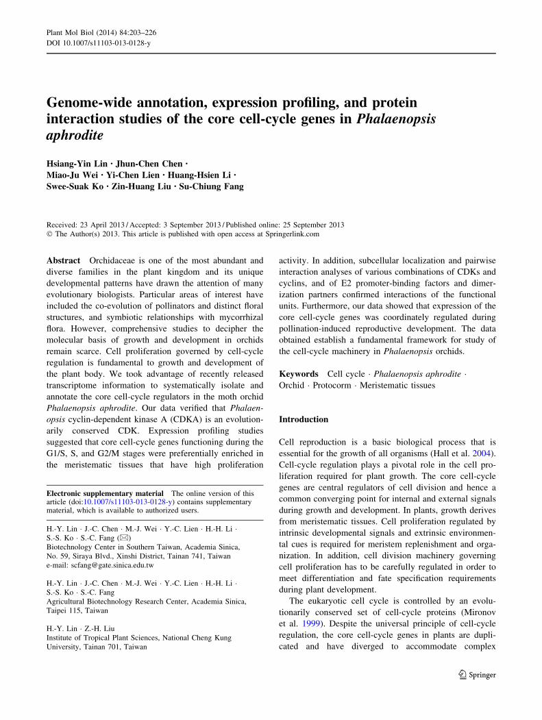
CDK family
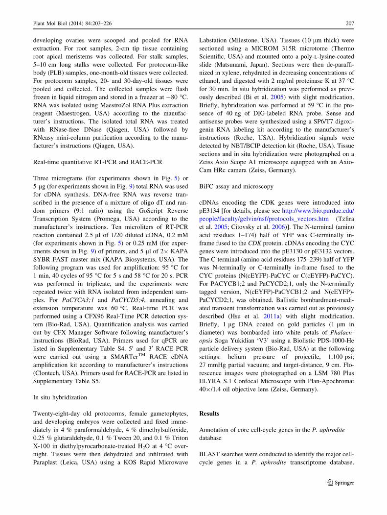
At least one member of each class of CDK was identified in
the P. aphrodite genome (Supplementary Table S1).
A- and B-type CDKs are the major CDKs that control cell-
cycle transitions in plant cells. P. aphrodite contains at
least one ortholog of the CDKA genes, which was desig-
nated as PaCDKA1. Like other plant CDKA proteins,
PaCDKA1 protein has a conserved PSTAIRE signature in
the alpha-1 helix, a hallmark CYC-binding motif (Fig. 1a).
In addition, the T-loop and the phosphorylating threonine
residue required for activation are also conserved in PaC-
DKA1 (Fig. 1a). The plant CDKB family has been classi-
fied into two subgroups (Vandepoele et al. 2002; Dewitte
and Murray 2003; Inze and De Veylder 2006). The CDKB1
subgroup has PPTALRE as the CYC binding motif. The
CDKB2 subgroup is characterized by a P[S/P]TTLRE
signature motif. Orthologs of B-type CDKs designated as
PaCDKB1 and PaCDKB2 were annotated. PaCDKB1 and
PaCDKB2 share 64 % identity and 81 % similarity at the
amino acid level. Based on a previous classification
(Dewitte and Murray 2003; Inze and De Veylder 2006;
Vandepoele et al. 2002), PaCDKB1 contains a PPTTLRE
motif and therefore belongs to the type 2 CDKB subgroup
(Fig. 1b). However, phylogenetic analysis indicated that
PaCDKB1 was clustered with type 1 CDKB members that
contain the PPTALRE motif (Fig. 1c). PaCDKB2, on the
other hand, carried a modified signature motif—PAT-
TLRE—and formed a cluster with a group of monocot
CDKB family members (Fig. 1b, c). The PATTLRE motif
was also found in CDKB family members of the moss
Physcomitrella patens (unpublished data). Of the CAKs, at
least one member each of the CDKD and CDKF family
was identified from the Phalaenopsis annotation database
(Supplementary Tables S1, S2). The other CDK classes are
C-type, E-type, G-type, and CDK-like (CKL) families. At
least one gene from each of the CDKC and CDKE families,
two CDKG and 13 CDK-like (CKL) genes were identified
from the P. aphrodite transcriptome database (Supple-
mentary Table S1). Plant C-type CDKs and E-type CDKs
are characterized by their similarity to mammalian CDK7
and CDK8, respectively (Joubes et al. 2000; Kitsios et al.
2008). They are assumed to regulate transcription in a
similar manner to their counterparts in mammals (Inagaki
and Umeda 2011). Arabidopsis CDKC has been shown to
be involved in splicing-related transcriptional regulation
(Kitsios et al. 2008). Like other plant CDKC protein
kinases (Joubes et al. 2000), the CDKC1 of P. aphrodite
also carries the PITAIRE motif (Supplementary Table S2).
The G-type CDK class is homologous to the human p58
galactosyltransferase protein whose role is important for
cytokinesis (Menges et al. 2005). Co-purification of
CDKGs with CYCL1 suggests that CYCL1 may be an
interacting partner of CDKG (Van Leene et al. 2010).
CYCL was also identified from the P. aphrodite tran-
scriptome database (Supplementary Table S1). The func-
tions of plant CKL protein kinases remain to be clarified.
Because A- and B-type CDKs are the only CDKs that
directly regulate cell-cycle progression in plants (Vande-
poele et al. 2002; Menges et al. 2005), we chose to focus
the remainder of our study on A- and B-type CDKs.
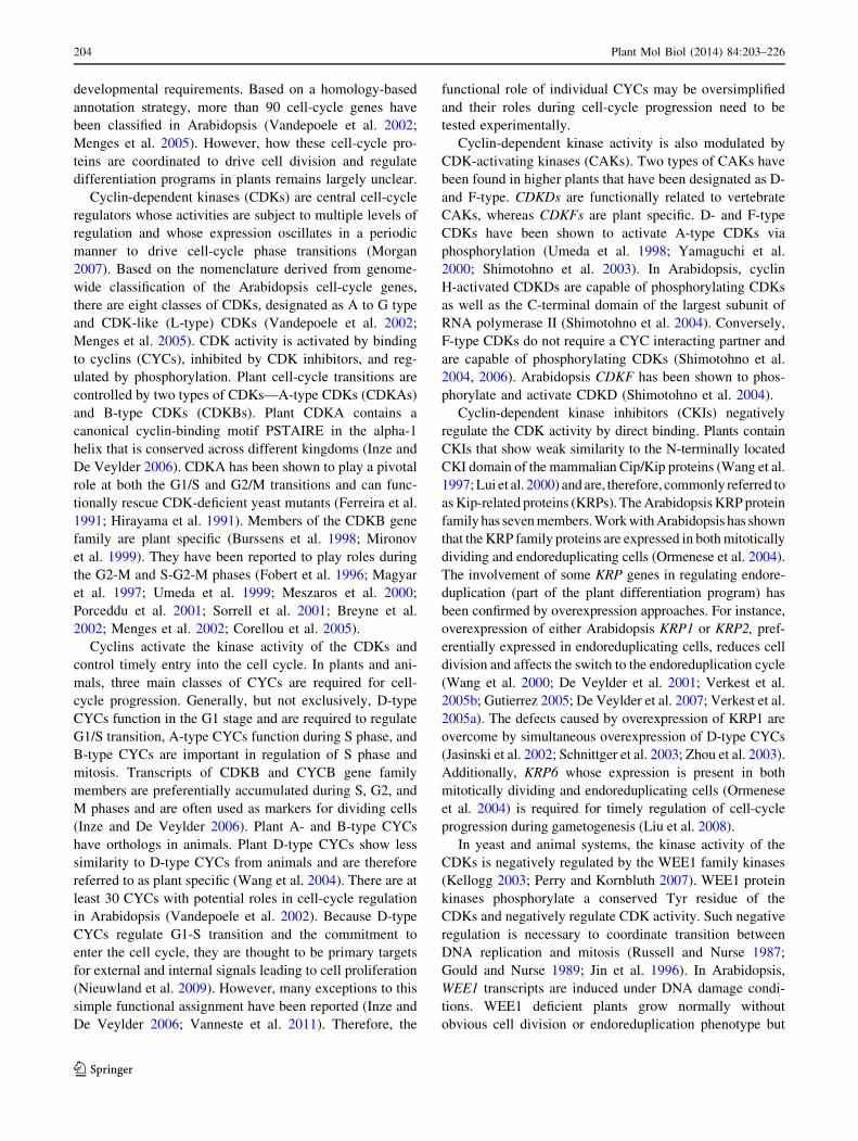
Cyclin family
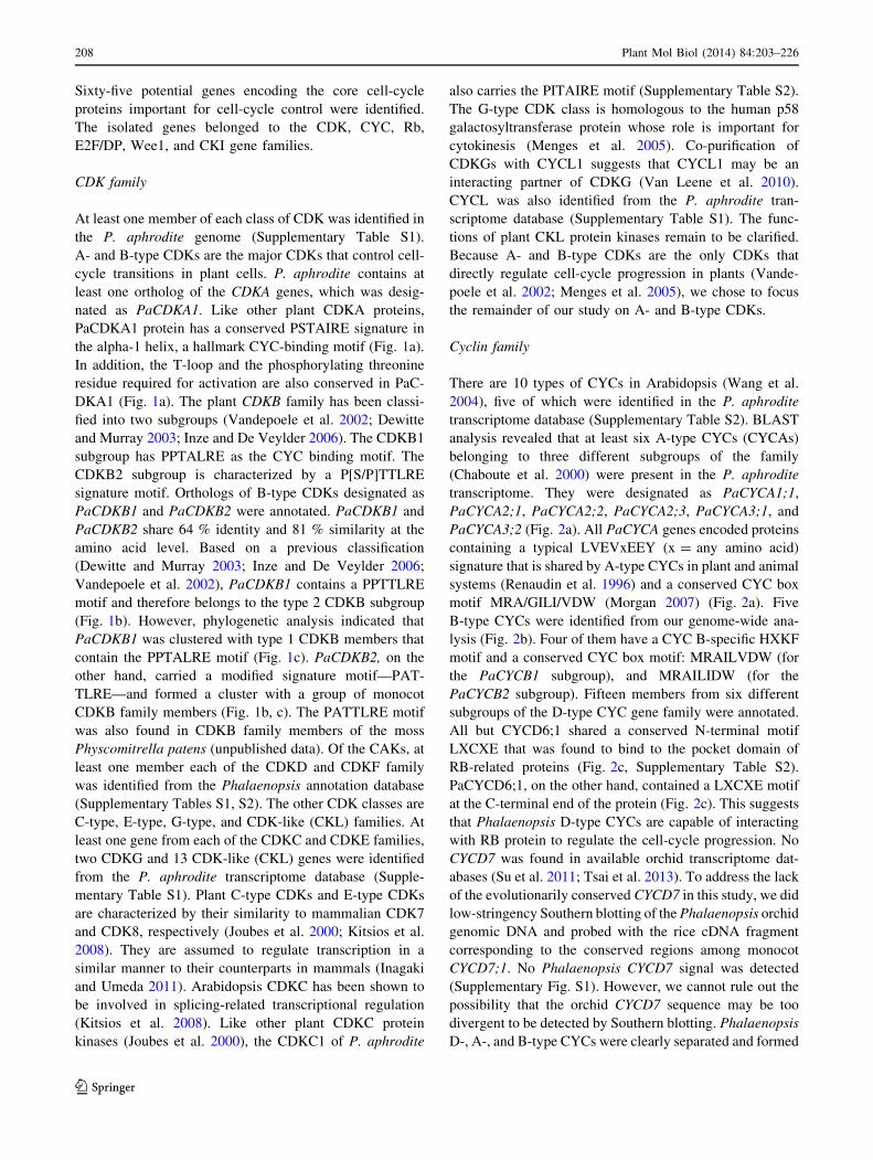
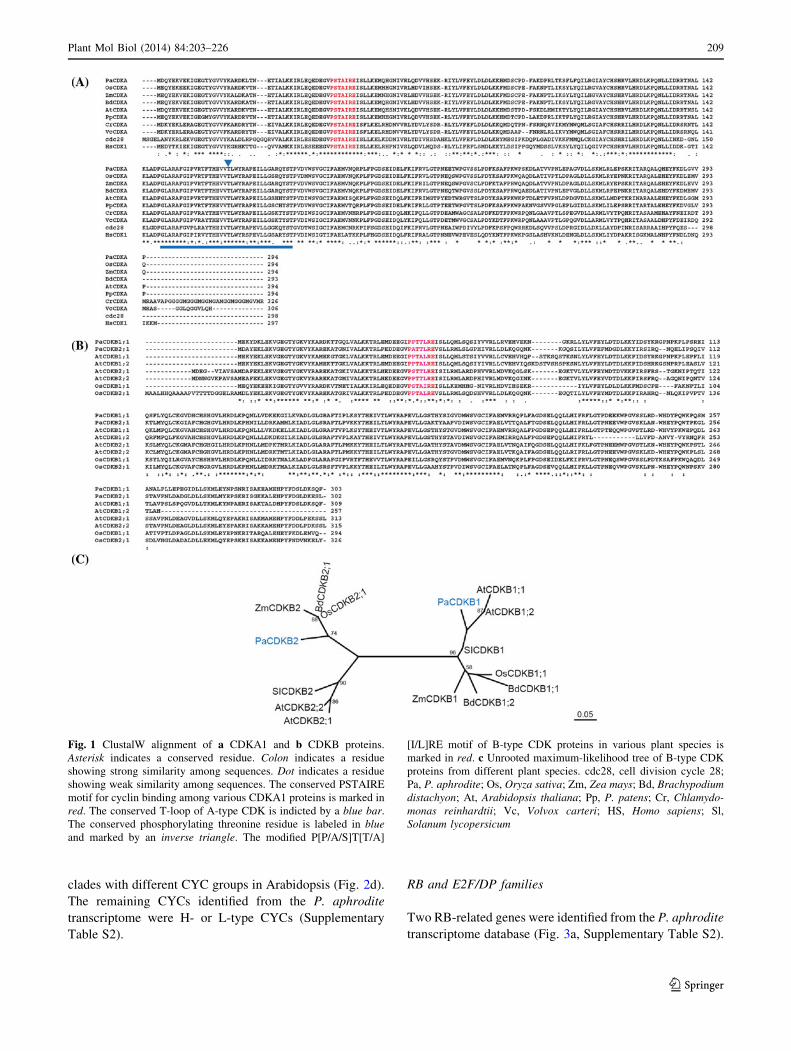
There are 10 types of CYCs in Arabidopsis (Wang et al.
2004), five of which were identified in the P. aphrodite
transcriptome database (Supplementary Table S2). BLAST
analysis revealed that at least six A-type CYCs (CYCAs)
belonging to three different subgroups of the family
(Chaboute et al. 2000) were present in the P. aphrodite
transcriptome. They were designated as PaCYCA1;1,
PaCYCA2;1, PaCYCA2;2, PaCYCA2;3, PaCYCA3;1, and
PaCYCA3;2 (Fig. 2a). All PaCYCA genes encoded proteins
containing a typical LVEVxEEY (x = any amino acid)
signature that is shared by A-type CYCs in plant and animal
systems (Renaudin et al. 1996) and a conserved CYC box
motif MRA/GILI/VDW (Morgan 2007) (Fig. 2a). Five
B-type CYCs were identified from our genome-wide ana-
lysis (Fig. 2b). Four of them have a CYC B-specific HXKF
motif and a conserved CYC box motif: MRAILVDW (for
the PaCYCB1 subgroup), and MRAILIDW (for the
PaCYCB2 subgroup). Fifteen members from six different
subgroups of the D-type CYC gene family were annotated.
All but CYCD6;1 shared a conserved N-terminal motif
LXCXE that was found to bind to the pocket domain of
RB-related proteins (Fig. 2c, Supplementary Table S2).
PaCYCD6;1, on the other hand, contained a LXCXE motif
at the C-terminal end of the protein (Fig. 2c). This suggests
that Phalaenopsis D-type CYCs are capable of interacting
with RB protein to regulate the cell-cycle progression. No
CYCD7 was found in available orchid transcriptome dat-
abases (Su et al. 2011; Tsai et al. 2013). To address the lack
of the evolutionarily conserved CYCD7 in this study, we did
low-stringency Southern blotting of the Phalaenopsis orchid
genomic DNA and probed with the rice cDNA fragment
corresponding to the conserved regions among monocot
CYCD7;1. No Phalaenopsis CYCD7 signal was detected
(Supplementary Fig. S1). However, we cannot rule out the
possibility that the orchid CYCD7 sequence may be too
divergent to be detected by Southern blotting. Phalaenopsis
D-, A-, and B-type CYCs were clearly separated and formed
208 Plant Mol Biol (2014) 84:203–226
123
clades with different CYC groups in Arabidopsis (Fig. 2d).
The remaining CYCs identified from the P. aphrodite
transcriptome were H- or L-type CYCs (Supplementary
Table S2).
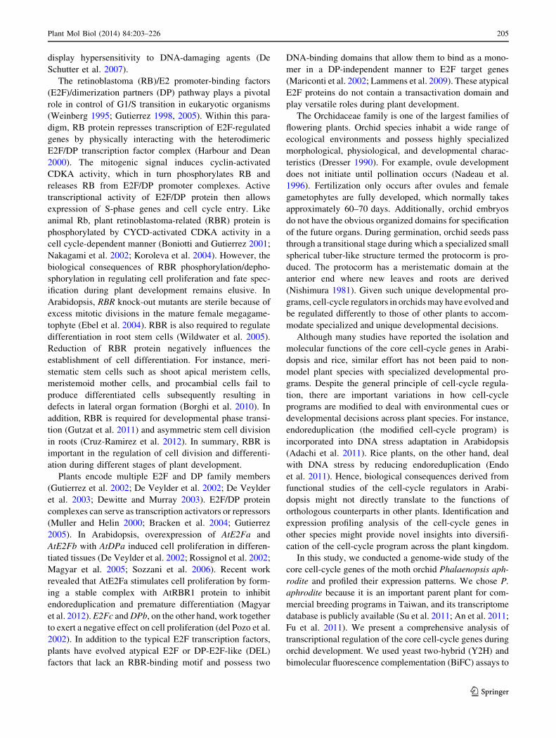
RB and E2F/DP families
Two RB-related genes were identified from the P. aphrodite
transcriptome database (Fig. 3a, Supplementary Table S2).
Fig. 1 ClustalW alignment of a CDKA1 and b CDKB proteins.
Asterisk indicates a conserved residue. Colon indicates a residue
showing strong similarity among sequences. Dot indicates a residue
showing weak similarity among sequences. The conserved PSTAIRE
motif for cyclin binding among various CDKA1 proteins is marked in
red. The conserved T-loop of A-type CDK is indicted by a blue bar.
The conserved phosphorylating threonine residue is labeled in blue
and marked by an inverse triangle. The modified P[P/A/S]T[T/A]
[I/L]RE motif of B-type CDK proteins in various plant species is
marked in red. c Unrooted maximum-likelihood tree of B-type CDK
proteins from different plant species. cdc28, cell division cycle 28;
Pa, P. aphrodite; Os, Oryza sativa; Zm, Zea mays; Bd, Brachypodium
distachyon; At, Arabidopsis thaliana; Pp, P. patens; Cr, Chlamydo-
monas reinhardtii; Vc, Volvox carteri; HS, Homo sapiens; Sl,
Solanum lycopersicum
Plant Mol Biol (2014) 84:203–226 209
123
210 Plant Mol Biol (2014) 84:203–226
123
Because the sequence of PaRBL2 cDNA in the database was
incorrect, the full-length cDNA was identified and con-
firmed by reverse transcription PCR followed by sequenc-
ing. Both PaRBL1 and PaRBL2 contained canonical A and
B domains that showed high similarity to RB proteins of
other plant species (Fig. 3a). The A and B domains are
important to form a binding pocket for E2F transcription
factors. Four E2F and three DP genes were identified from
the P. aphrodite transcriptome database (Supplementary
Table S2). The full-length cDNAs of PaE2F1, PaE2F2, and
PaE2F3 were isolated and the encoded proteins contained a
DNA binding domain, a dimerization domain, a marked
box, and a conserved C-terminal region that potentially
mediates their interactions with the PaRBL proteins
(Fig. 3b). The full-length cDNAs of PaDP1 and PaDP2
were also isolated and verified. Both PaDPs encode a pro-
tein containing the DP canonical DNA binding and dimer-
ization domains (Fig. 3c). We were not able to obtain the
full-length cDNAs of PaE2F4 and PaDP3. Therefore,
PaE2F4 and PaDP3 were not included in the following
analyses.
In addition to the typical E2F and DP gene family
members, an atypical DP-E2F-like (DEL) gene was identi-
fied in the P. aphrodite transcriptome database. Like other
plant atypical DEL proteins (Vandepoele et al. 2002;
Lammens et al. 2009), PaDEL1 contained a duplicated
DNA binding domain highly similar to that of the E2F
transcription factor (Fig. 3d). Based on previous studies, the
atypical DEL proteins do not heterodimerize with DP and
instead form homodimers to exert their function (Di Stefano
et al. 2003). In plants, DEL proteins are involved in regu-
lation of cell wall biosynthesis and endoreduplication (Ra-
mirez-Parra et al. 2004; Vlieghe et al. 2005; Lammens et al.
2008).
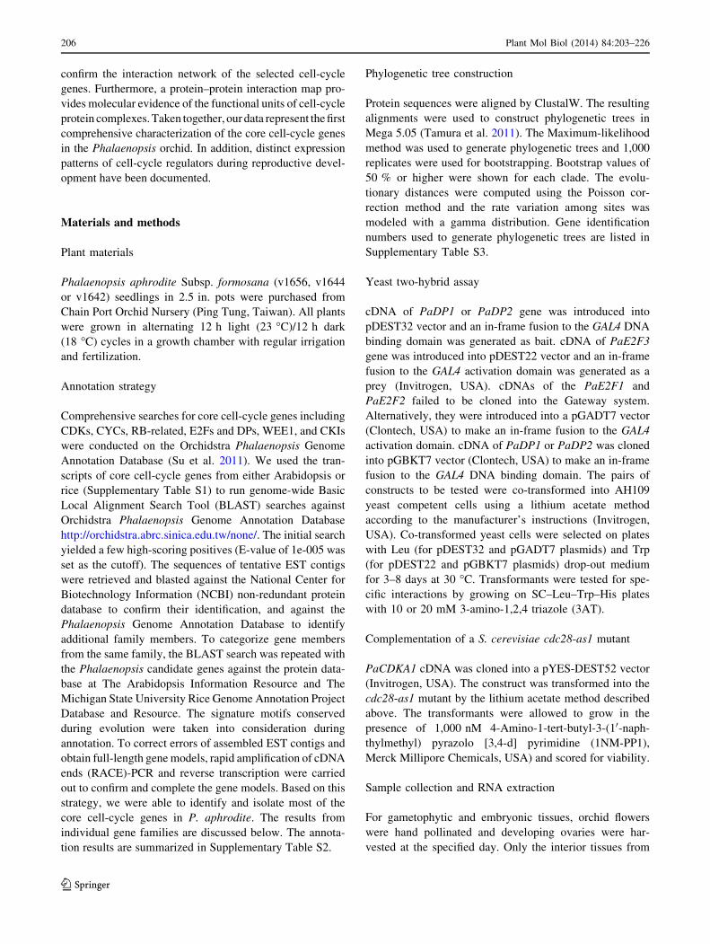
WEE1 and CKI gene families
One WEE1 ortholog was found in the Phalaenopsis
annotation database. The encoded protein displays 47 %
identity to AtWEE1 and 57 % identity to rice WEE1 pro-
teins at the amino acid sequence level. The Phalaenopsis
WEE1 protein has a conserved catalytic domain including
the ATP binding site of the protein kinase family (Fig. 4a).
It also contains a conserved E/DGD triplet motif that dis-
tinguishes WEE1-related kinases from other kinase fami-
lies (Booher et al. 1993; Sorrell et al. 2002).
Three potential CDK kinase inhibitor (CKI) genes were
annotated and identified in P. aphrodite. All three carried a
CKI domain (pfam02234) at the C-terminal region
(Fig. 4b). PaKRP1 shares 69 % amino acid sequence iden-
tity with PaKRP3. PaKRP2 shares 52 and 49 % amino acid
sequence identity with PaKRP1 and PaKRP3, respectively.
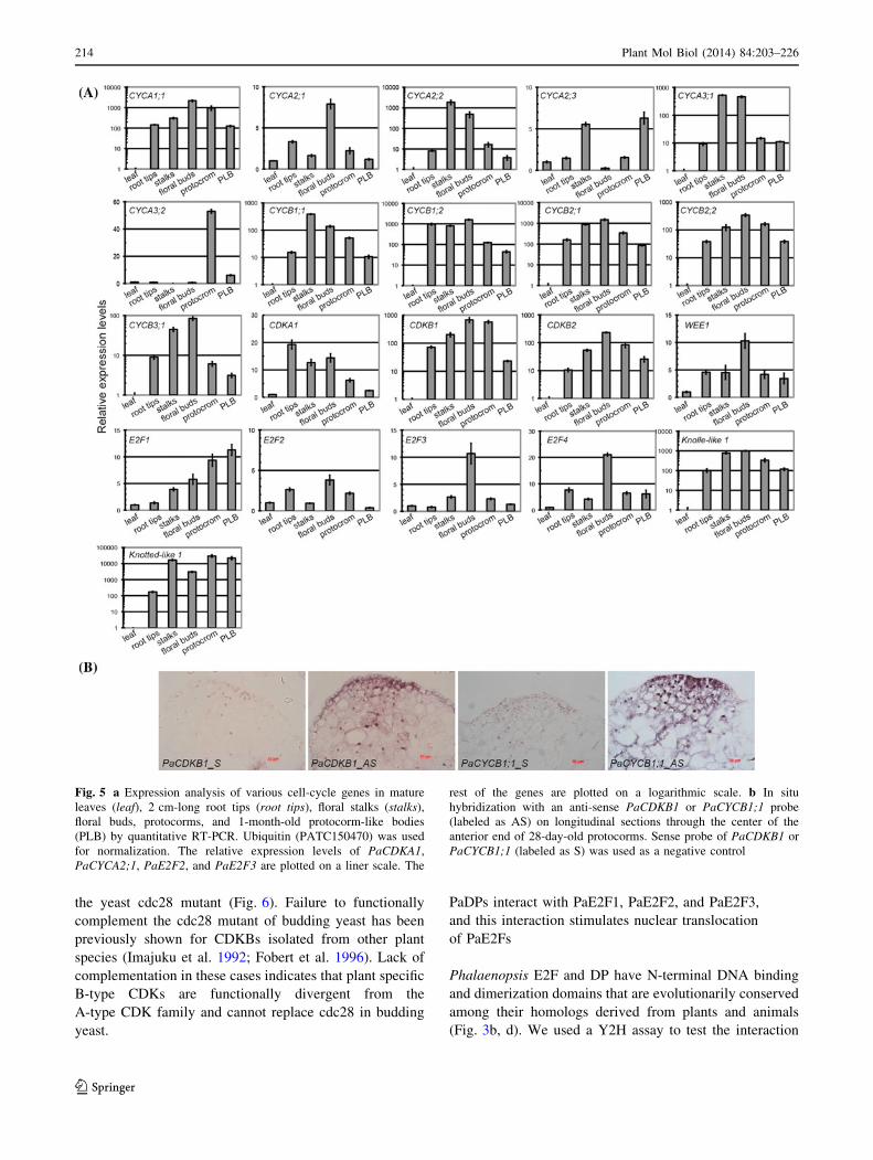
Cell-cycle gene expression profiles
To investigate the expression patterns of the cell-cycle genes
in Phalaenopsis orchid, we used quantitative RT-PCR (qRT-
PCR) analysis to compare relative transcript abundance in
various tissues. Meristematic tissues with a high rate of cell
division were marked by cytokinesis-specific syntaxin
(PATC147993), KNOLLE (Lauber et al. 1997), and a mer-
istematic marker, KNOTTED-like (PATC127065) homeo-
box transcription factor (Fig. 5a). Our expression analysis
showed that PaCYCA1;1, PaCYCB1;1, PaCYCB1;2, Pa-
CYCB2;1, PaCYCB2;2, PaCDKB1, and PaCDKB2 mRNAs
accumulated to relatively high levels in meristems of root
tips, young stalks, young floral buds, PLBs, and protocorm-
containing actively dividing cells, but were almost absent or
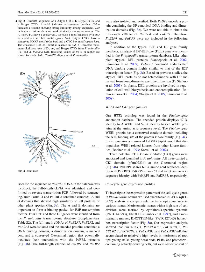
Fig. 2 continued
Fig. 2 ClustalW alignment of a A-type CYCs, b B-type CYCs, and
c D-type CYCs. Asterisk indicates a conserved residue. Colon
indicates a residue showing strong similarity among sequences. Dot
indicates a residue showing weak similarity among sequences. The
A-type CYCs have a conserved LVEVxEEY motif (marked by a blue
bar) and a CYC box motif (green bar). B-type CYCs have a
conserved HXKF motif (blue bar) and a CYC box motif (green bar).
The conserved LXCXC motif is marked in red. d Unrooted maxi-
mum-likelihood tree of D-, A-, and B-type CYCs from P. aphrodite
(Pa) and A. thaliana (At). Bootstrap values of 50 % or higher are
shown for each clade. ClustalW alignment of P. aphrodite
b
Plant Mol Biol (2014) 84:203–226 211
123
barely detectable in the fully differentiated leaves (Fig. 5a,
Supplementary Fig. S2). PaCDKB1 and PaCYCB1;1
mRNAs were concentrated in the shoot apical meristems of
the developing protocorms (Fig. 5b). Other distinct CYC
gene expression patterns were also observed. For example,
transcripts of PaCYCA2;2, PaCYCA3;1, and PaCYCB3;1
212 Plant Mol Biol (2014) 84:203–226
123
were highly enriched in floral stalks and floral buds (Fig. 5a).
The accumulation of PaCYCA2;3 mRNA was enriched in
floral stalks and PLBs. PaCYCA3;2 mRNA showed prefer-
ential accumulation in developing protocorms. In addition to
CYC genes, PaE2F1, PaE2F2, PaE2F3 and PaE2F4 also
showed distinct expression patterns in the surveyed tissues
(Fig. 5a). The mRNA of PaCDKA1 was detected in
both meristematic and differentiated tissues (Supplementary
Fig. S2). This is consistent with expression patterns of CDKA
genes in the other plant species (Mironov et al. 1999).
However, the levels of PaCDKA1 mRNA varied slightly in
different tissues (Fig. 1).
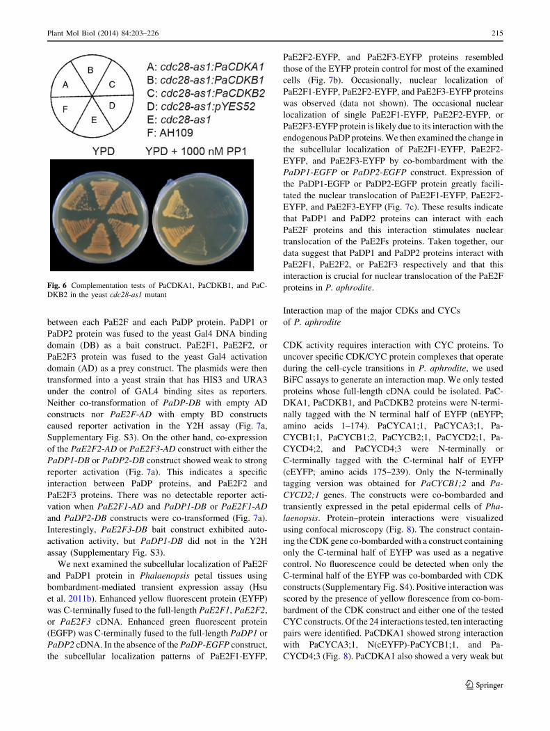
PaCDKA1 is a bona fide cyclin-dependent protein
kinase
To test whether PaCDKA1 and two PaCDKB genes encode
functional equivalents of the evolutionarily conserved
CDKs, PaCDKA1, PaCDKB1 and PaCDKB1 genes were
expressed in an inhibitor-sensitive allele of yeast CDK,
cdc28-as1 mutant (Bishop et al. 2000). The cdc28-as1 allele
has an enlarged ATP-binding pocket, allowing it to bind the
cell permeable ATP analog 1NM-PP1, and treatment of cells
with 1NM-PP1 results in rapid and specific down-regulation
of Cdc28 kinase activity. In the presence of 1NM-PP1, the
yeast strain carrying the cdc28-as1 allele failed to grow.
Introduction of PaCDKA1 rescued the proliferation defect of
cdc28 mutant when 1NM-PPA was present (Fig. 6). This
experiment demonstrated that PaCDKA1 is a functional
homolog of the cdc28 CDK family. However, neither PaC-
DKB1 nor PaCDKB2 rescued inhibitor-sensitive alleles of
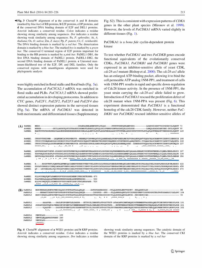
Fig. 4 ClustalW alignment of a WEE1 proteins and b KRP proteins.
Asterisk indicates a conserved residue. Colon indicates a residue
showing strong similarity among sequences. Dot indicates a residue
showing weak similarity among sequences. The catalytic domain of
the WEE1 proteins is marked by a blue bar. The conserved CKI
domain of the KRP proteins is marked by a red bar
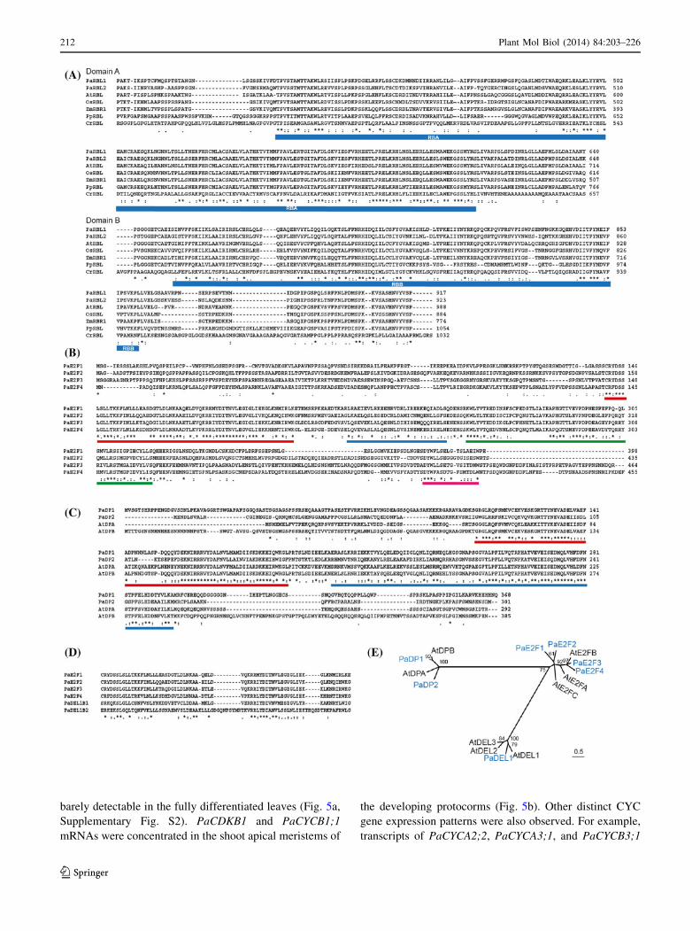
Fig. 3 ClustalW alignment of a the conserved A and B domains
(marked by blue bar) of RB proteins, b E2F proteins, c DP proteins, and
d the conserved DNA binding domain of E2F and DEL1 proteins.
Asterisk indicates a conserved residue. Colon indicates a residue
showing strong similarity among sequences. Dot indicates a residue
showing weak similarity among sequences. Pa, P. aphrodite; At, A.
thaliana; Os, O. sativa; Zm, Z. mays; Pp, P. patens; Cr, C. reinhardtii.
The DNA binding domain is marked by a red bar. The dimerization
domain is marked by a blue bar. The marked box is marked by a green
bar. The conserved C-terminal region of E2F protein important for
binding to the RB protein is marked by a pink bar. PaDEL1-DB1, the
first DNA binding domain of PaDEL1 protein. PaDEL1-DB2, the
second DNA binding domain of PaDEL1 protein. e Unrooted max-
imum-likelihood tree of the E2F, DP, and DEL families. Only the
conserved regions with unambiguous alignments were used for
phylogenetic analysis
b
Plant Mol Biol (2014) 84:203–226 213
123
the yeast cdc28 mutant (Fig. 6). Failure to functionally
complement the cdc28 mutant of budding yeast has been
previously shown for CDKBs isolated from other plant
species (Imajuku et al. 1992; Fobert et al. 1996). Lack of
complementation in these cases indicates that plant specific
B-type CDKs are functionally divergent from the
A-type CDK family and cannot replace cdc28 in budding
yeast.
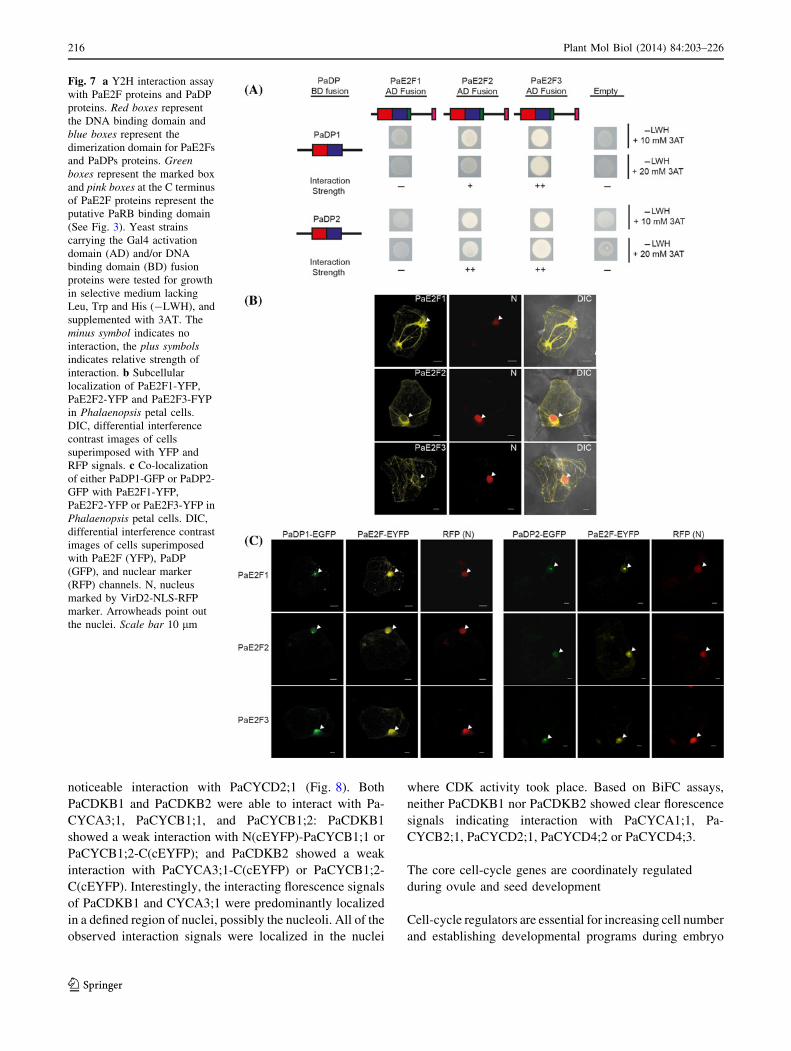
PaDPs interact with PaE2F1, PaE2F2, and PaE2F3,
and this interaction stimulates nuclear translocation
of PaE2Fs
Phalaenopsis E2F and DP have N-terminal DNA binding
and dimerization domains that are evolutionarily conserved
among their homologs derived from plants and animals
(Fig. 3b, d). We used a Y2H assay to test the interaction
Fig. 5 a Expression analysis of various cell-cycle genes in mature
leaves (leaf), 2 cm-long root tips (root tips), floral stalks (stalks),
floral buds, protocorms, and 1-month-old protocorm-like bodies
(PLB) by quantitative RT-PCR. Ubiquitin (PATC150470) was used
for normalization. The relative expression levels of PaCDKA1,
PaCYCA2;1, PaE2F2, and PaE2F3 are plotted on a liner scale. The
rest of the genes are plotted on a logarithmic scale. b In situ
hybridization with an anti-sense PaCDKB1 or PaCYCB1;1 probe
(labeled as AS) on longitudinal sections through the center of the
anterior end of 28-day-old protocorms. Sense probe of PaCDKB1 or
PaCYCB1;1 (labeled as S) was used as a negative control
214 Plant Mol Biol (2014) 84:203–226
123
between each PaE2F and each PaDP protein. PaDP1 or
PaDP2 protein was fused to the yeast Gal4 DNA binding
domain (DB) as a bait construct. PaE2F1, PaE2F2, or
PaE2F3 protein was fused to the yeast Gal4 activation
domain (AD) as a prey construct. The plasmids were then
transformed into a yeast strain that has HIS3 and URA3
under the control of GAL4 binding sites as reporters.
Neither co-transformation of PaDP-DB with empty AD
constructs nor PaE2F-AD with empty BD constructs
caused reporter activation in the Y2H assay (Fig. 7a,
Supplementary Fig. S3). On the other hand, co-expression
of the PaE2F2-AD or PaE2F3-AD construct with either the
PaDP1-DB or PaDP2-DB construct showed weak to strong
reporter activation (Fig. 7a). This indicates a specific
interaction between PaDP proteins, and PaE2F2 and
PaE2F3 proteins. There was no detectable reporter acti-
vation when PaE2F1-AD and PaDP1-DB or PaE2F1-AD
and PaDP2-DB constructs were co-transformed (Fig. 7a).
Interestingly, PaE2F3-DB bait construct exhibited auto-
activation activity, but PaDP1-DB did not in the Y2H
assay (Supplementary Fig. S3).
We next examined the subcellular localization of PaE2F
and PaDP1 protein in Phalaenopsis petal tissues using
bombardment-mediated transient expression assay (Hsu
et al. 2011b). Enhanced yellow fluorescent protein (EYFP)
was C-terminally fused to the full-length PaE2F1, PaE2F2,
or PaE2F3 cDNA. Enhanced green fluorescent protein
(EGFP) was C-terminally fused to the full-length PaDP1 or
PaDP2 cDNA. In the absence of the PaDP-EGFP construct,
the subcellular localization patterns of PaE2F1-EYFP,
PaE2F2-EYFP, and PaE2F3-EYFP proteins resembled
those of the EYFP protein control for most of the examined
cells (Fig. 7b). Occasionally, nuclear localization of
PaE2F1-EYFP, PaE2F2-EYFP, and PaE2F3-EYFP proteins
was observed (data not shown). The occasional nuclear
localization of single PaE2F1-EYFP, PaE2F2-EYFP, or
PaE2F3-EYFP protein is likely due to its interaction with the
endogenous PaDP proteins. We then examined the change in
the subcellular localization of PaE2F1-EYFP, PaE2F2-
EYFP, and PaE2F3-EYFP by co-bombardment with the
PaDP1-EGFP or PaDP2-EGFP construct. Expression of
the PaDP1-EGFP or PaDP2-EGFP protein greatly facili-
tated the nuclear translocation of PaE2F1-EYFP, PaE2F2-
EYFP, and PaE2F3-EYFP (Fig. 7c). These results indicate
that PaDP1 and PaDP2 proteins can interact with each
PaE2F proteins and this interaction stimulates nuclear
translocation of the PaE2Fs proteins. Taken together, our
data suggest that PaDP1 and PaDP2 proteins interact with
PaE2F1, PaE2F2, or PaE2F3 respectively and that this
interaction is crucial for nuclear translocation of the PaE2F
proteins in P. aphrodite.
Interaction map of the major CDKs and CYCs
of P. aphrodite
CDK activity requires interaction with CYC proteins. To
uncover specific CDK/CYC protein complexes that operate
during the cell-cycle transitions in P. aphrodite, we used
BiFC assays to generate an interaction map. We only tested
proteins whose full-length cDNA could be isolated. PaC-
DKA1, PaCDKB1, and PaCDKB2 proteins were N-termi-
nally tagged with the N terminal half of EYFP (nEYFP;
amino acids 1–174). PaCYCA1;1, PaCYCA3;1, Pa-
CYCB1;1, PaCYCB1;2, PaCYCB2;1, PaCYCD2;1, Pa-
CYCD4;2, and PaCYCD4;3 were N-terminally or
C-terminally tagged with the C-terminal half of EYFP
(cEYFP; amino acids 175–239). Only the N-terminally
tagging version was obtained for PaCYCB1;2 and Pa-
CYCD2;1 genes. The constructs were co-bombarded and
transiently expressed in the petal epidermal cells of Pha-
laenopsis. Protein–protein interactions were visualized
using confocal microscopy (Fig. 8). The construct contain-
ing the CDK gene co-bombarded with a construct containing
only the C-terminal half of EYFP was used as a negative
control. No fluorescence could be detected when only the
C-terminal half of the EYFP was co-bombarded with CDK
constructs (Supplementary Fig. S4). Positive interaction was
scored by the presence of yellow florescence from co-bom-
bardment of the CDK construct and either one of the tested
CYC constructs. Of the 24 interactions tested, ten interacting
pairs were identified. PaCDKA1 showed strong interaction
with PaCYCA3;1, N(cEYFP)-PaCYCB1;1, and Pa-
CYCD4;3 (Fig. 8). PaCDKA1 also showed a very weak but
Fig. 6 Complementation tests of PaCDKA1, PaCDKB1, and PaC-
DKB2 in the yeast cdc28-as1 mutant
Plant Mol Biol (2014) 84:203–226 215
123
noticeable interaction with PaCYCD2;1 (Fig. 8). Both
PaCDKB1 and PaCDKB2 were able to interact with Pa-
CYCA3;1, PaCYCB1;1, and PaCYCB1;2: PaCDKB1
showed a weak interaction with N(cEYFP)-PaCYCB1;1 or
PaCYCB1;2-C(cEYFP); and PaCDKB2 showed a weak
interaction with PaCYCA3;1-C(cEYFP) or PaCYCB1;2-
C(cEYFP). Interestingly, the interacting florescence signals
of PaCDKB1 and CYCA3;1 were predominantly localized
in a defined region of nuclei, possibly the nucleoli. All of the
observed interaction signals were localized in the nuclei
where CDK activity took place. Based on BiFC assays,
neither PaCDKB1 nor PaCDKB2 showed clear florescence
signals indicating interaction with PaCYCA1;1, Pa-
CYCB2;1, PaCYCD2;1, PaCYCD4;2 or PaCYCD4;3.
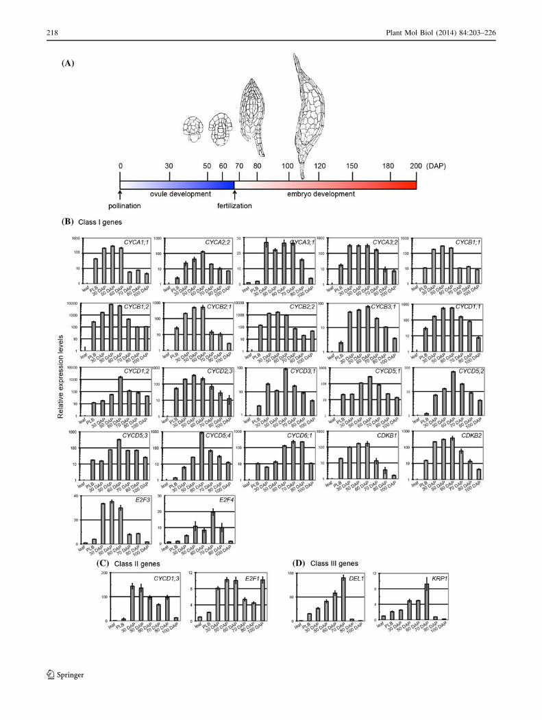
The core cell-cycle genes are coordinately regulated
during ovule and seed development
Cell-cycle regulators are essential for increasing cell number
and establishing developmental programs during embryo
Fig. 7 a Y2H interaction assay
with PaE2F proteins and PaDP
proteins. Red boxes represent
the DNA binding domain and
blue boxes represent the
dimerization domain for PaE2Fs
and PaDPs proteins. Green
boxes represent the marked box
and pink boxes at the C terminus
of PaE2F proteins represent the
putative PaRB binding domain
(See Fig. 3). Yeast strains
carrying the Gal4 activation
domain (AD) and/or DNA
binding domain (BD) fusion
proteins were tested for growth
in selective medium lacking
Leu, Trp and His (-LWH), and
supplemented with 3AT. The
minus symbol indicates no
interaction, the plus symbols
indicates relative strength of
interaction. b Subcellular
localization of PaE2F1-YFP,
PaE2F2-YFP and PaE2F3-FYP
in Phalaenopsis petal cells.
DIC, differential interference
contrast images of cells
superimposed with YFP and
RFP signals. c Co-localization
of either PaDP1-GFP or PaDP2-
GFP with PaE2F1-YFP,
PaE2F2-YFP or PaE2F3-YFP in
Phalaenopsis petal cells. DIC,
differential interference contrast
images of cells superimposed
with PaE2F (YFP), PaDP
(GFP), and nuclear marker
(RFP) channels. N, nucleus
marked by VirD2-NLS-RFP
marker. Arrowheads point out
the nuclei. Scale bar 10 lm
216 Plant Mol Biol (2014) 84:203–226
123
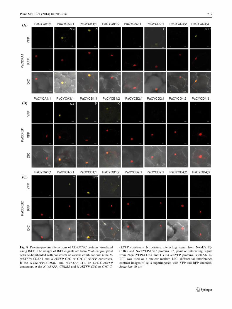
Fig. 8 Protein–protein interactions of CDK/CYC proteins visualized
using BiFC. The images of BiFC-signals are from Phalaenopsis petal
cells co-bombarded with constructs of various combinations: a the N-
(nEYFP)-CDKA1 and N-cEYFP-CYC or CYC-C-cEYFP constructs,
b the N-(nEYFP)-CDKB1 and N-cEYFP-CYC or CYC-C-cEYFP
constructs, c the N-(nEYFP)-CDKB2 and N-cEYFP-CYC or CYC-C-
cEYFP constructs. N, positive interacting signal from N-(nEYFP)-
CDKs and N-cEYFP-CYC proteins. C, positive interacting signal
from N-(nEYFP)-CDKs and CYC-C-cEYFP proteins. VirD2-NLS-
RFP was used as a nuclear marker. DIC, differential interference
contrast images of cells superimposed with YFP and RFP channels.
Scale bar 10 lm
Plant Mol Biol (2014) 84:203–226 217
123
218 Plant Mol Biol (2014) 84:203–226
123
development (Ebel et al. 2004; Gutzat et al. 2011; Eloy et al.
2012). Phalaenopsis orchids have a very unique ovule and
embryonic development program: pollination-induced cell
division in the placental ridge initiates ovule development,
and fertilization does not occur until 60–70 days after pol-
lination (Nadeau et al. 1996; O’Neill 1997; Lee et al. 2008).
This unique developmental program suggests that the cell-
cycle program is coordinated to accommodate this special
developmental decision. To gain insights into how cell-cycle
regulators are regulated during ovule and embryo develop-
ment in Phalaenopsis orchids, we analyzed the transcript
abundance of selected cell-cycle genes during reproductive
development after pollination (Fig. 9). As a comparison,
RNA samples from mature leaves were used as a baseline.
Because PLBs have been referred to as somatic embryos in
orchid species (Chang and Chang 1998; Ishii et al. 1998;
Chen et al. 1999), one-month-old PLB samples were also
included in this study. Cell-cycle regulators such as
PaCYCA1;1, PaCYCA2;2, PaCYCA3;1, PaCYCA3;2,
PaCYCB1;1, PaCYCB1;2, PaCYCB2;1, PaCYCB2;2,
PaCYCB3;1, PaCYCD1;1, PaCYCD1;2, PaCYCD2;3,
PaCYCD3;1, PaCYCD5;1, PaCYCD5;2, PaCYCD5;3,
PaCYCD5;4, PaCYCD6;1, PaCDKB1, PaCDKB2,
PaE2F3, and PaE2F4 accumulated after pollination as ovule
primordia started to develop and enlarge. Their levels
reached peaks at approximately 60–70 days after pollination
(DAP) when fertilization occurred and then decreased
gradually or sharply as embryo development initiated. The
core cell-cycle genes that shared this expression pattern were
categorized as class I genes. Similar to class I cell-cycle
genes, accumulation of PaCYCD1;3 and PaE2F1 mRNAs
also reached their first peak during ovule development;
however, their expression levels declined and then reached a
second peak at 80 or 100 DAP. Genes that shared this
expression pattern were categorized as class II cell-cycle
genes. Most of the cell-cycle regulators described above
were hardly detectable or expressed in low abundance in the
mature leaf tissues and showed relatively low to moderate
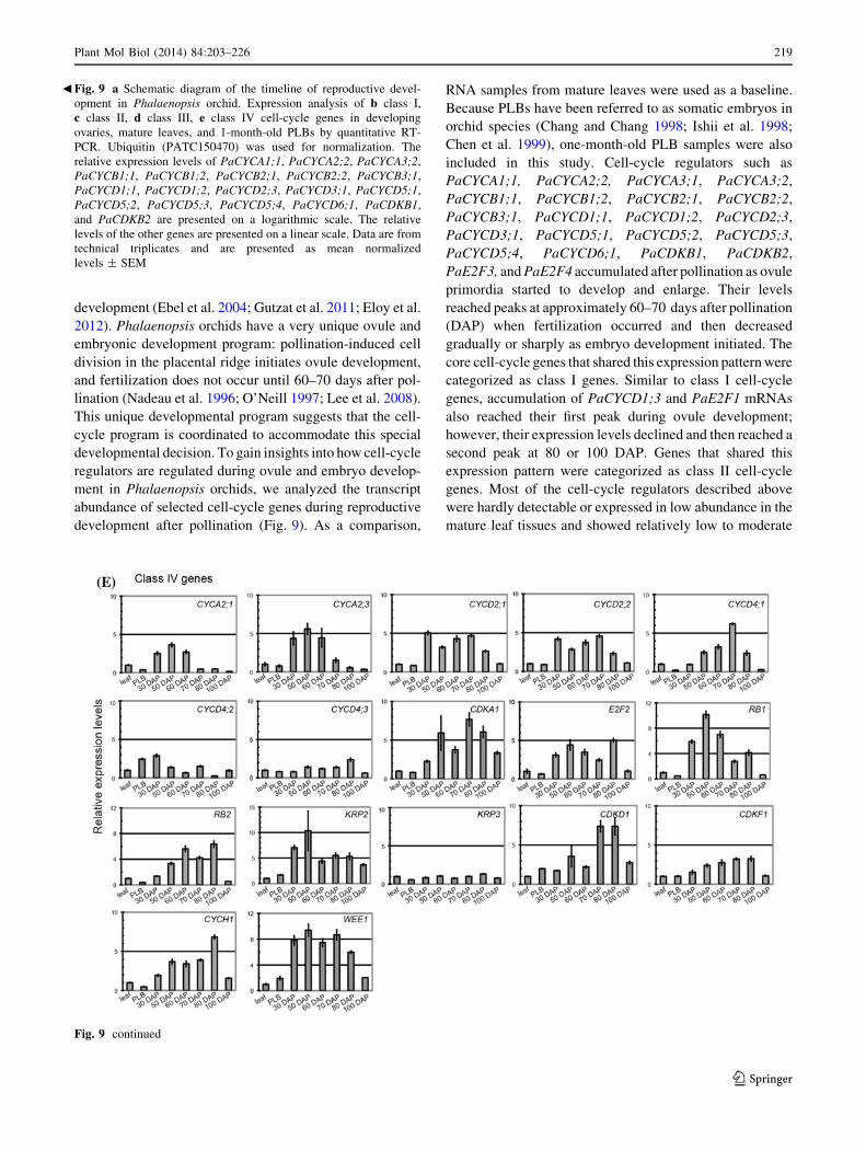
Fig. 9 a Schematic diagram of the timeline of reproductive devel-
opment in Phalaenopsis orchid. Expression analysis of b class I,
c class II, d class III, e class IV cell-cycle genes in developing
ovaries, mature leaves, and 1-month-old PLBs by quantitative RT-
PCR. Ubiquitin (PATC150470) was used for normalization. The
relative expression levels of PaCYCA1;1, PaCYCA2;2, PaCYCA3;2,
PaCYCB1;1, PaCYCB1;2, PaCYCB2;1, PaCYCB2;2, PaCYCB3;1,
PaCYCD1;1, PaCYCD1;2, PaCYCD2;3, PaCYCD3;1, PaCYCD5;1,
PaCYCD5;2, PaCYCD5;3, PaCYCD5;4, PaCYCD6;1, PaCDKB1,
and PaCDKB2 are presented on a logarithmic scale. The relative
levels of the other genes are presented on a linear scale. Data are from
technical triplicates and are presented as mean normalized
levels ± SEM
b
Fig. 9 continued
Plant Mol Biol (2014) 84:203–226 219
123
expression levels in one-month-old PLBs. The expression
patterns of a third class of cell-cycle regulators including
PaDEL1 and PaKRP1 resembled class I genes with the
expression plateauing at approximately 70 DAP; however,
unlike the class I genes, the expression peaks of these cell-
cycle regulators were followed by a steep drop in mRNA
abundance. These cell-cycle regulators were classified as class
III cell-cycle genes. PaCYCA2;1, PaCYCA2;3, PaCYCD2;1,
PaCYCD2;2, PaCYCD4;1, PaCYCD4;2, PaCYCD4;3,
PaCDKA1, PaE2F2, PaRB1, PaRB2, PaKRP2, PaKRP3,
PaCDKD1, PaCDKF1, PaCYCH1, and PaWEE1, on the
other hand, only showed steady to slight increases (less than
tenfold difference) in mRNA abundance during ovule and
embryo development. These genes were classified as class IV
cell-cycle genes. Among them, genes likely required for G1/S
transition such as PaCYCD2;1, PaCYCD2;2, PaCYCD4;1,
PaE2F2, and PaWEE1 showed slight, but significant induc-
tion at the transcript level. Taken together, the distinct
expression patterns of the cell-cycle genes define molecular
aspects of cell-cycle program during gametophyte and
embryo development in Phalaenopsis orchid.
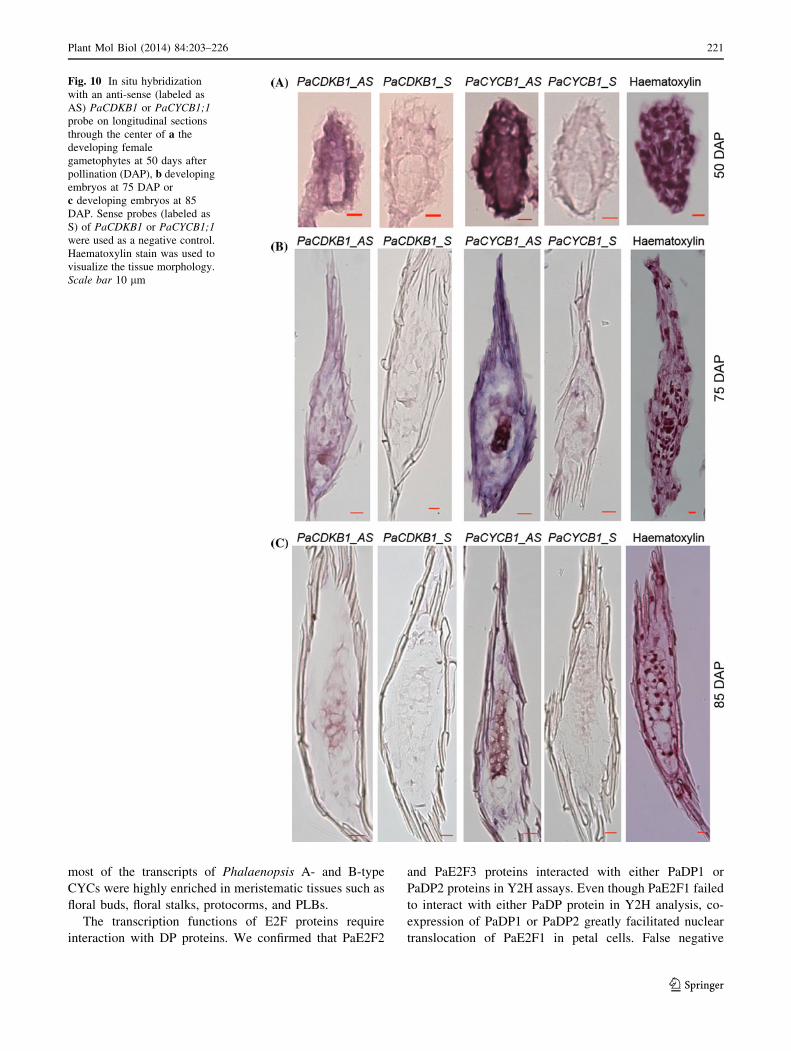
To gain further insights into temporal and spatial gene
expression patterns during mitotic cell-cycle activity in
reproductive development, we monitored mitotic cell
markers PaCYCB1 and PaCDKB1 (class I cell-cycle genes)
using in situ hybridization. Fifty days after pollination,
transcripts of PaCYCB1 and PaCDKB1 accumulated to
high level in cells of mature embryo sacs and integuments
(Fig. 10a). At the early stages of seed development (75
DAP), PaCYCB1 and PaCDKB1 mRNAs were concen-
trated in newly developed embryos and moderately
expressed in the surrounding tissues of the developing
embryos (Fig. 10b). As embryo development proceeded
(85 DAP), the transcript abundance of PaCYCB1 and
PaCDKB1 gradually diminished and was spatially restric-
ted to the developing embryos (Fig. 10c). Taken together
these results indicate that the core cell-cycle genes are
temporally and spatially regulated during reproductive
development in Phalaenopsis orchids.
Discussion
Cell-cycle control is an integral part of plant growth and
development. Despite its importance, knowledge about
how cell-cycle regulation modulates growth and develop-
ment, and how various developmental signals are coordi-
nated to regulate cell-cycle programs to achieve different
body plans is limited. Orchidaceae is one of the most
abundant families in the plant kingdom and exhibits
diverse and specialized developmental programs. Identifi-
cation and analysis of orchid cell-cycle genes will provide
the basis for understanding of how cell-cycle machinery is
integrated into the developmental program in Phalaenopsis
orchids. Here, we isolated and validated the cDNA
sequences of some of the core cell-cycle regulators in P.
aphrodite. Analysis of the Phalaenopsis core cell-cycle
genes revealed that the number of the cell-cycle genes in
each family category is similar to those in Arabidopsis and
rice (Supplementary Table S1). Since the Phalaenopsis
genome is not yet available, we cannot exclude the possi-
bility that there are also other, as yet unidentified, cell-
cycle genes whose expression is restricted to a defined
developmental window or that are induced in response to
specific environmental cues that are missing from available
databases. Nevertheless, this study is, to our knowledge,
the first comprehensive analysis of the cell-cycle genes in
orchid species.
Similar to Arabidopsis and rice, eight types of CDKs
(A-, B-, C-, D-, E-, F-, G-, and L-types) were found in
P. aphrodite. Protein sequence analysis showed consider-
able conservation in the catalytic core and specific motifs of
each type of CDK. A- and B-type CDKs are the master
regulators that control the cell-cycle transitions in plants.
One A-type CDK and two B-type CDKs were identified
from the P. aphrodite transcriptome database. PaCDKA1
was constitutively expressed in the examined tissues. This is
consistent with observations in other plant species. PaC-
DKA1 is evolutionarily conserved because it was able to
functionally substitute the cdc28 protein kinase in budding
yeast. Phalaenopsis B-type CDKs, on the other hand, could
not functionally complement the yeast cdc28 mutant. Sim-
ilarly, B-type CDKs from several plant species have been
shown to fail to functionally replace yeast cdc28 mutant
(Hirayama et al. 1991; Porceddu et al. 1999; Corellou et al.
2005). This result supports the notion that B-type CDKs are
plant specific and functionally diverged from A-type CDKs
(Joubes et al. 2000; Boudolf et al. 2001; Inze and De
Veylder 2006). Similar to the expression patterns found in
other plant B-type CDKs, PaCDKB1 and PaCDKB2 were
highly enriched in meristematic tissues with strong cell-
cycle activity (Menges et al. 2002; Guo et al. 2007). It is,
therefore, likely that Phalaenopsis B-type CDKs play a role
in regulating G2/M and M phases as demonstrated in other
plant species.
Five out of the nine types of CYCs found in rice and
Arabidopsis (Vandepoele et al. 2002; La et al. 2006) were
identified from the P. aphrodite transcriptome database.
Failure to identify members of the four other CYC families
suggests that they might be expressed at defined develop-
mental time points or be induced under specific conditions,
resulting in their lack of representation in the current
transcriptome database. Among the identified CYC fami-
lies, A-, B-, and D-type CYCs are the major cell-cycle
regulators that play crucial roles in regulating cell-cycle
transitions. Consistent with their roles during the cell cycle,
220 Plant Mol Biol (2014) 84:203–226
123
most of the transcripts of Phalaenopsis A- and B-type
CYCs were highly enriched in meristematic tissues such as
floral buds, floral stalks, protocorms, and PLBs.
The transcription functions of E2F proteins require
interaction with DP proteins. We confirmed that PaE2F2
and PaE2F3 proteins interacted with either PaDP1 or
PaDP2 proteins in Y2H assays. Even though PaE2F1 failed
to interact with either PaDP protein in Y2H analysis, co-
expression of PaDP1 or PaDP2 greatly facilitated nuclear
translocation of PaE2F1 in petal cells. False negative
Fig. 10 In situ hybridization
with an anti-sense (labeled as
AS) PaCDKB1 or PaCYCB1;1
probe on longitudinal sections
through the center of a the
developing female
gametophytes at 50 days after
pollination (DAP), b developing
embryos at 75 DAP or
c developing embryos at 85
DAP. Sense probes (labeled as
S) of PaCDKB1 or PaCYCB1;1
were used as a negative control.
Haematoxylin stain was used to
visualize the tissue morphology.
Scale bar 10 lm
Plant Mol Biol (2014) 84:203–226 221
123
results have been reported in Y2H assays (Walhout et al.
2000), probably due to stringent scoring criteria (Boruc
et al. 2010) or low sampling sensitivity (Venkatesan et al.
2009). Unlike constitutive nuclear localization of E2F-1,
-2, and -3 proteins in humans (Verona et al. 1997), PaE2F
proteins alone did not target exclusively into the nucleus.
The nuclear translocation of PaE2F proteins requires co-
expression of PaDP proteins (Fig. 7b, c). A similar sub-
cellular localization pattern has also been reported for
Arabidopsis E2F proteins (Kosugi and Ohashi 2002).
Taken together these findings suggest that the nuclear
localization of Phalaenopsis E2F proteins requires their
interaction with DP proteins.
Similar to Arabidopsis, CDKA1 in P. aphrodite behaved
as the most interconnected node in the CDK/CYC inter-
acting network (Boruc et al. 2010; Van Leene et al. 2007,
2010, 2011). In Arabidopsis, CDKA is able to interact with
D-, A-, and B-type CYCs. This shows that the functional
activity of CDKA is required throughout the G1/S to mid-
M phases (Porceddu et al. 2001; Sorrell et al. 2001; Boruc
et al. 2010). The protein–protein interaction studies based
on BiFC confirm that the functional units of CDKA protein
complexes are evolutionarily conserved in P. aphrodite.
However, PaCDKA1 seems to selectively interact with
members of the D-, A-, and B-type CYCs while Arabi-
dopsis CDKA1 is less selective for CYC binding. It is
possible that insufficient flexibility and steric hindrance of
the fusion proteins resulted in false negative results. It is
also possible that the stable interaction of the Phalaenopsis
CDKA/CYC pairs require interactor proteins, which are
lacking in the fully differentiated petal cells. Our protein–
protein interaction studies also confirmed that both PaC-
DKB1 and PaCDKB2 are able to interact with Pa-
CYCB1;1, PaCYCB1;2 and PaCYCA3;1. A similar
interaction network has been documented in Arabidopsis
(Boruc et al. 2010; Van Leene et al. 2011). Taken together,
our studies and studies from Arabidopsis suggest that the
functional CDK/CYC units are evolutionarily conserved in
plants.
In many orchid species, ovule development initiates after
pollination occurs (Nadeau et al. 1996). It has been sug-
gested that ovule development, redirection of pollen tube
growth, and subsequent fertilization require timely coordi-
nation of hormone regulation. Because ovule initiation and
subsequent fertilization are nearly synchronous and thou-
sands of ovules are present in each ovary (Nadeau et al.
1996), orchids provide an excellent system through which to
study the timely regulation of gene expression during ovule
and embryo development. In this study, we monitored and
categorized the expression patterns of the cell-cycle genes in
developing capsules into four categories. A subset of the
cell-cycle regulators controlling G1/S and S/M transitions
were grouped into the class I genes. They are: PaCYCA1;1,
PaCYCA2;2, PaCYCA3;1, PaCYCA3;2, PaCYCB1;1,
PaCYCB1;2, PaCYCB2;1, PaCYCB2;2, PaCYCB3;1,
PaCYCD1;1, PaCYCD1;2, PaCYCD2;3, PaCYCD3;1,
PaCYCD5;1, PaCYCD5;2, PaCYCD5;3, PaCYCA5;4,
PaCYCD6;1, PaCDKB1, PaCDKB2, PaE2F3, and PaE2F4.
The transcript levels of the class I cell-cycle regulators were
dramatically enriched during ovule development (30–60
DAP) and gradually declined as embryos started to develop
(70–100 DAP). This expression pattern suggests that cell-
cycle-dependent activity regulated by these genes is
important for development of the female gametophytes. The
biological implications of the decline in transcripts of class I
cell-cycle regulators as development switches from ovule
development to embryogenesis are not clear. One possible
reason is that accumulation of the class I cell-cycle proteins
during ovule development is sufficient to initiate embryo-
genesis. The other possibility is that the cellular requirement
of the cell-cycle transcripts is gradually restricted to defined
domains and specific time intervals. The spatial and tem-
poral expression patterns of cell-cycle genes during embryo
development have also been documented in Arabidopsis
(Collins et al. 2012; Belmonte et al. 2013).
Mirroring class I genes, class II genes were up-regulated
during ovule development and down-regulated after fer-
tilization. Intriguingly, however, the mRNA of the class II
cell-cycle regulators (PaE2F1 and PaCYCD1;3) reached a
second peak at 80–100 DAP as embryos entered the mat-
uration stage. The molecular basis of this expression pat-
tern is not clear. Class III cell-cycle genes (PaDEL1 and
PaKRP1) showed a distinct expression pattern with accu-
mulation of transcripts reaching a peak at 70 DAP (Fig. 9d)
during which ovule development ceased and fertilization
occurred, followed by a steep drop after onset of embryo-
genesis. In the future, it will be interesting to examine
whether the class II and/or the class III cell-cycle regulators
are involved in developmental processes that help to define
the fate transition between gametophyte and embryo
development.
In summary, we have identified and isolated the core
cell-cycle genes in Phalaenopsis orchid, and conducted a
comprehensive study of their dynamic expression patterns
during reproductive development. Protein sequence ana-
lysis showed that P. aphrodite cell-cycle regulators are
highly similar to their respective orthologs in other plant
species. We confirmed the functional units of the CDK/
CYC and E2F/DP by Y2H and BiFC analysis. Expression
patterns and subcellular localization studies indicate that
the cell-cycle regulators of P. aphrodite are involved in cell
proliferation as well as cell-cycle related developmental
processes. We observed that the expression patterns of the
cell-cycle genes are coordinately regulated as the repro-
ductive system proceeds from ovule development to
embryogenesis in P. aphrodite. These results establish the
222 Plant Mol Biol (2014) 84:203–226
123
first molecular signatures of the cell-cycle program during
pollination-induced reproductive development in Pha-
laenopsis orchid. Further studies of the molecular functions
of the core cell-cycle genes during seed development will
be important to provide clues about how the cell-cycle
program is regulated and incorporated into developmental
decisions.
Acknowledgments We are grateful to Dr. Ming-Tsair Chan for
providing Y2H and BiFC plasmids and orchid samples, to Dr. Rey-
Huei Chen and Dr. Ting-Fang Wang for the gift of yeast cdc28-as1
mutant, and to Dr. Wan-Sheng Lo for sharing the yeast AH109 strain.
We express our appreciation to Dr. Choun-Sea Lin and Mr. Chen-
Tran Hsu at the Plant Technology Core Facility of Academia Sinica
for their assistance in transient expression, to Ms. Shu-Chen Shen at
the Confocal Microscope Core facility of Academia Sinica for her
assistance on microscope work, to Mr. Min-Jeng Li for sharing his
rice cDNA, and to Ms. Miranda Loney for English editing. This work
was supported by the Development Program of Industrialization for
Agricultural Biotechnology grant (to S.-C.F.); and in part by a grant
(to S.-C.F.) from the Biotechnology Center in Southern Taiwan,
Academia Sinica.
Open Access This article is distributed under the terms of the
Creative Commons Attribution License which permits any use, dis-
tribution, and reproduction in any medium, provided the original
author(s) and the source are credited.
References
Adachi S, Minamisawa K, Okushima Y, Inagaki S, Yoshiyama K,
Kondou Y, Kaminuma E, Kawashima M, Toyoda T, Matsui M,
Kurihara D, Matsunaga S, Umeda M (2011) Programmed
induction of endoreduplication by DNA double-strand breaks
in Arabidopsis. Proc Natl Acad Sci USA 108(24):10004–10009.
doi:10.1073/pnas.1103584108
An FM, Hsiao SR, Chan MT (2011) Sequencing-based approaches
reveal low ambient temperature-responsive and tissue-specific
microRNAs in phalaenopsis orchid. PLoS One 6(5):e18937.
doi:10.1371/journal.pone.0018937
Belmonte MF, Kirkbride RC, Stone SL, Pelletier JM, Bui AQ, Yeung
EC, Hashimoto M, Fei J, Harada CM, Munoz MD, Le BH,
Drews GN, Brady SM, Goldberg RB, Harada JJ (2013)
Comprehensive developmental profiles of gene activity in
regions and subregions of the Arabidopsis seed. Proc Natl Acad
Sci USA 110(5):E435–E444. doi:10.1073/pnas.1222061110
Bi X, Khush GS, Bennett J (2005) The rice nucellin gene ortholog
OsAsp1 encodes an active aspartic protease without a plant-
specific insert and is strongly expressed in early embryo. Plant
Cell Physiol 46(1):87–98. doi:10.1093/pcp/pci002
Bishop AC, Ubersax JA, Petsch DT, Matheos DP, Gray NS, Blethrow
J, Shimizu E, Tsien JZ, Schultz PG, Rose MD, Wood JL,
Morgan DO, Shokat KM (2000) A chemical switch for inhibitor-
sensitive alleles of any protein kinase. Nature
407(6802):395–401. doi:10.1038/35030148
Boniotti MB, Gutierrez C (2001) A cell-cycle-regulated kinase activity
phosphorylates plant retinoblastoma protein and contains, in
Arabidopsis, a CDKA/cyclin D complex. Plant J 28(3):341–350
Booher RN, Deshaies RJ, Kirschner MW (1993) Properties of
Saccharomyces cerevisiae wee1 and its differential regulation of
p34CDC28 in response to G1 and G2 cyclins. EMBO J
12(9):3417–3426
Borghi L, Gutzat R, Futterer J, Laizet Y, Hennig L, Gruissem W (2010)
Arabidopsis RETINOBLASTOMA-RELATED is required for
stem cell maintenance, cell differentiation, and lateral organ
production. Plant Cell 22(6):1792–1811. doi:10.1105/tpc.110.
074591
Boruc J, Van den Daele H, Hollunder J, Rombauts S, Mylle E, Hilson P,
Inze D, De Veylder L, Russinova E (2010) Functional modules in
the Arabidopsis core cell cycle binary protein–protein interaction
network. Plant Cell 22(4):1264–1280. doi:10.1105/tpc.109.073635
Boudolf V, Rombauts S, Naudts M, Inze D, De Veylder L (2001)
Identification of novel cyclin-dependent kinases interacting with
the CKS1 protein of Arabidopsis. J Exp Bot 52(359):1381–1382
Bracken AP, Ciro M, Cocito A, Helin K (2004) E2F target genes:
unraveling the biology. Trends Biochem Sci 29(8):409–417
Breyne P, Dreesen R, Vandepoele K, De Veylder L, Van Breusegem
F, Callewaert L, Rombauts S, Raes J, Cannoot B, Engler G, Inze
D, Zabeau M (2002) Transcriptome analysis during cell division
in plants. Proc Natl Acad Sci USA 99(23):14825–14830. doi:10.
1073/pnas.222561199
Burssens S, Van Montagu M, Inze D (1998) The cell cycle in
Arabidopsis. Plant Physiol Biochem 36(1–2):9–19. doi:10.1016/
s0981-9428(98)80087-9
Chaboute ME, Clement B, Sekine M, Philipps G, Chaubet-Gigot N
(2000) Cell cycle regulation of the tobacco ribonucleotide
reductase small subunit gene is mediated by E2F-like elements.
Plant Cell 12(10):1987–2000
Chang C, Chang WC (1998) Plant regeneration from callus culture of
Cymbidium ensifolium var. misericors. Plant Cell Rep 17(4):251–255
Chen JT, Chang C, Chang WC (1999) Direct somatic embryogenesis
on leaf explants of Oncidium Gower Ramsey and subsequent
plant regeneration. Plant Cell Rep 19(2):143–149
Citovsky V, Lee LY, Vyas S, Glick E, Chen MH, Vainstein A, Gafni
Y, Gelvin SB, Tzfira T (2006) Subcellular localization of
interacting proteins by bimolecular fluorescence complementa-
tion in planta. J Mol Biol 362(5):1120–1131. doi:10.1016/j.jmb.
2006.08.017
Collins C, Dewitte W, Murray JA (2012) D-type cyclins control cell
division and developmental rate during Arabidopsis seed devel-
opment. J Exp Bot 63(10):3571–3586. doi:10.1093/jxb/ers015
Corellou F, Camasses A, Ligat L, Peaucellier G, Bouget FY (2005)
Atypical regulation of a green lineage-specific B-type cyclin-
dependent kinase. Plant Physiol 138(3):1627–1636. doi:10.1104/
pp.105.059626
Cruz-Ramirez A, Diaz-Trivino S, Blilou I, Grieneisen VA, Sozzani R,
Zamioudis C, Miskolczi P, Nieuwland J, Benjamins R, Dho-
nukshe P, Caballero-Perez J, Horvath B, Long Y, Mahonen AP,
Zhang H, Xu J, Murray JA, Benfey PN, Bako L, Maree AF,
Scheres B (2012) A bistable circuit involving SCARECROW-
RETINOBLASTOMA integrates cues to inform asymmetric
stem cell division. Cell 150(5):1002–1015. doi:10.1016/j.cell.
2012.07.017
De Schutter K, Joubes J, Cools T, Verkest A, Corellou F, Babiychuk
E, Van Der Schueren E, Beeckman T, Kushnir S, Inze D, De
Veylder L (2007) Arabidopsis WEE1 kinase controls cell cycle
arrest in response to activation of the DNA integrity checkpoint.
Plant Cell 19(1):211–225. doi:10.1105/tpc.106.045047
De Veylder L, Beemster GT, Beeckman T, Inze D (2001) CKS1At
overexpression in Arabidopsis thaliana inhibits growth byreducing meristem size and inhibiting cell-cycle progression.
Plant J 25(6):617–626
De Veylder L, Beeckman T, Beemster GT, de Almeida Engler J,
Ormenese S, Maes S, Naudts M, Van Der Schueren E, Jacqmard
A, Engler G, Inze D (2002) Control of proliferation, endoredu-
plication and differentiation by the Arabidopsis E2Fa-DPa
Plant Mol Biol (2014) 84:203–226 223
123
transcription factor. EMBO J 21(6):1360–1368. doi:10.1093/
emboj/21.6.1360
De Veylder L, Joubes J, Inze D (2003) Plant cell cycle transitions.
Curr Opin Plant Biol 6(6):536–543
De Veylder L, Beeckman T, Inze D (2007) The ins and outs of the
plant cell cycle. Nat Rev Mol Cell Biol 8(8):655–665. doi:10.
1038/nrm2227
del Pozo JC, Boniotti MB, Gutierrez C (2002) Arabidopsis E2Fc
functions in cell division and is degraded by the ubiquitin-
SCF(AtSKP2) pathway in response to light. Plant Cell
14(12):3057–3071
Dewitte W, Murray JA (2003) The plant cell cycle. Annu Rev Plant Biol
54:235–264. doi:10.1146/annurev.arplant.54.031902.134836
Di Stefano L, Jensen MR, Helin K (2003) E2F7, a novel E2F
featuring DP-independent repression of a subset of E2F-
regulated genes. EMBO J 22(23):6289–6298. doi:10.1093/
emboj/cdg613
Dresser R (1990) The orchids: natural history and classification.
Harvard University Press, Cambridge
Ebel C, Mariconti L, Gruissem W (2004) Plant retinoblastoma
homologues control nuclear proliferation in the female gameto-
phyte. Nature 429(6993):776–780. doi:10.1038/nature02637
Eloy NB, Gonzalez N, Van Leene J, Maleux K, Vanhaeren H, De
Milde L, Dhondt S, Vercruysse L, Witters E, Mercier R, Cromer
L, Beemster GT, Remaut H, Van Montagu MC, De Jaeger G,
Ferreira PC, Inze D (2012) SAMBA, a plant-specific anaphase-
promoting complex/cyclosome regulator is involved in early
development and A-type cyclin stabilization. Proc Natl Acad Sci
USA 109(34):13853–13858. doi:10.1073/pnas.1211418109
Endo M, Nakayama S, Umeda-Hara C, Ohtsuki N, Saika H, Umeda
M, Toki S (2011) CDKB2 is involved in mitosis and DNA
damage response in rice. Plant J. doi:10.1111/j.1365-313X.2011.
04847.x
Ferreira PC, Hemerly AS, Villarroel R, Van Montagu M, Inze D
(1991) The Arabidopsis functional homolog of the p34cdc2
protein kinase. Plant Cell 3(5):531–540
Fobert PR, Gaudin V, Lunness P, Coen ES, Doonan JH (1996)
Distinct classes of cdc2-related genes are differentially expressed
during the cell division cycle in plants. Plant Cell
8(9):1465–1476. doi:10.1105/tpc.8.9.1465
Fu CH, Chen YW, Hsiao YY, Pan ZJ, Liu ZJ, Huang YM, Tsai WC,
Chen HH (2011) OrchidBase: a collection of sequences of the
transcriptome derived from orchids. Plant Cell Physiol
52(2):238–243. doi:10.1093/pcp/pcq201
Gould KL, Nurse P (1989) Tyrosine phosphorylation of the fission
yeast cdc2? protein kinase regulates entry into mitosis. Nature
342(6245):39–45. doi:10.1038/342039a0
Guo J, Song J, Wang F, Zhang XS (2007) Genome-wide identification
and expression analysis of rice cell cycle genes. Plant Mol Biol
64(4):349–360. doi:10.1007/s11103-007-9154-y
Gutierrez C (1998) The retinoblastoma pathway in plant cell cycle
and development. Curr Opin Plant Biol 1(6):492–497
Gutierrez C (2005) Coupling cell proliferation and development in
plants. Nat Cell Biol 7(6):535–541. doi:10.1038/ncb0605-535
Gutierrez C, Ramirez-Parra E, Castellano MM, del Pozo JC (2002)
G(1) to S transition: more than a cell cycle engine switch. Curr
Opin Plant Biol 5(6):480–486
Gutzat R, Borghi L, Futterer J, Bischof S, Laizet Y, Hennig L, Feil R,
Lunn J, Gruissem W (2011) RETINOBLASTOMA-RELATED
PROTEIN controls the transition to autotrophic plant develop-
ment. Development 138(14):2977–2986. doi:10.1242/dev.
060830
Hall MN, Raff M, Thomas G (2004) Cell growth—control of cell
size. John Inglis, Cold Spring Harbor
Harbour JW, Dean DC (2000) The Rb/E2F pathway: expanding roles
and emerging paradigms. Genes Dev 14(19):2393–2409
Hirayama T, Imajuku Y, Anai T, Matsui M, Oka A (1991)
Identification of two cell-cycle-controlling cdc2 gene homologs
in Arabidopsis thaliana. Gene 105(2):159–165
Hsu CC, Chung YL, Chen TC, Lee YL, Kuo YT, Tsai WC, Hsiao
YY, Chen YW, Wu WL, Chen HH (2011a) An overview of the
Phalaenopsis orchid genome through BAC end sequence
analysis. BMC Plant Biol 11:3. doi:10.1186/1471-2229-11-3
Hsu CT, Liao DC, Wu FH, Liu NT, Shen SC, Chou SJ, Tung SY,
Yang CH, Chan MT, Lin CS (2011b) Integration of molecular
biology tools for identifying promoters and genes abundantly
expressed in flowers of Oncidium Gower Ramsey. BMC Plant
Biol 11:60. doi:10.1186/1471-2229-11-60
Imajuku Y, Hirayama T, Endoh H, Oka A (1992) Exon-intron
organization of the Arabidopsis thaliana protein kinase genes
CDC2a and CDC2b. FEBS Lett 304(1):73–77
Inagaki S, Umeda M (2011) Cell-cycle control and plant develop-
ment. Int Rev Cell Mol Biol 291:227–261. doi:10.1016/B978-0-
12-386035-4.00007-0
Inze D, De Veylder L (2006) Cell cycle regulation in plant
development. Annu Rev Genet 40:77–105. doi:10.1146/
annurev.genet.40.110405.090431
Ishii Y, Takamura T, Goi M, Tanaka M (1998) Callus induction and
somatic embryogenesis of Phalaenopsis. Plant Cell Rep
17(6):446–450. doi:10.1007/s002990050423
Jasinski S, Riou-Khamlichi C, Roche O, Perennes C, Bergounioux C,
Glab N (2002) The CDK inhibitor NtKIS1a is involved in plant
development, endoreduplication and restores normal develop-
ment of cyclin D3; 1-overexpressing plants. J Cell Sci 115(Pt
5):973–982
Jin P, Gu Y, Morgan DO (1996) Role of inhibitory CDC2
phosphorylation in radiation-induced G2 arrest in human cells.
J Cell Biol 134(4):963–970
Joubes J, Chevalier C, Dudits D, Heberle-Bors E, Inze D, Umeda M,
Renaudin JP (2000) CDK-related protein kinases in plants. Plant
Mol Biol 43(5–6):607–620
Kellogg DR (2003) Wee1-dependent mechanisms required for
coordination of cell growth and cell division. J Cell Sci 116(Pt
24):4883–4890. doi:10.1242/jcs.00908
Kitsios G, Alexiou KG, Bush M, Shaw P, Doonan JH (2008) A
cyclin-dependent protein kinase, CDKC2, colocalizes with and
modulates the distribution of spliceosomal components in
Arabidopsis. Plant J 54(2):220–235. doi:10.1111/j.1365-313X.
2008.03414.x
Koroleva OA, Tomlinson M, Parinyapong P, Sakvarelidze L, Leader
D, Shaw P, Doonan JH (2004) CycD1, a putative G1 cyclin from
Antirrhinum majus, accelerates the cell cycle in cultured tobacco
BY-2 cells by enhancing both G1/S entry and progression
through S and G2 phases. Plant Cell 16(9):2364–2379. doi:10.
1105/tpc.104.023754
Kosugi S, Ohashi Y (2002) Interaction of the Arabidopsis E2F and DP
proteins confers their concomitant nuclear translocation and
transactivation. Plant Physiol 128(3):833–843. doi:10.1104/pp.
010642
La H, Li J, Ji Z, Cheng Y, Li X, Jiang S, Venkatesh PN,
Ramachandran S (2006) Genome-wide analysis of cyclin family
in rice (Oryza sativa L.). Mol Genet Genomics 275(4):374–386.
doi:10.1007/s00438-005-0093-5
Lammens T, Boudolf V, Kheibarshekan L, Zalmas LP, Gaamouche T,
Maes S, Vanstraelen M, Kondorosi E, La Thangue NB, Govaerts
W, Inze D, De Veylder L (2008) Atypical E2F activity restrains
APC/CCCS52A2 function obligatory for endocycle onset. Proc
Natl Acad Sci USA 105(38):14721–14726. doi:10.1073/pnas.
0806510105
Lammens T, Li J, Leone G, De Veylder L (2009) Atypical E2Fs: new
players in the E2F transcription factor family. Trends Cell Biol
19(3):111–118. doi:10.1016/j.tcb.2009.01.002
224 Plant Mol Biol (2014) 84:203–226
123
Lauber MH, Waizenegger I, Steinmann T, Schwarz H, Mayer U, Hwang
I, Lukowitz W, Jurgens G (1997) The Arabidopsis KNOLLE protein
is a cytokinesis-specific syntaxin. J Cell Biol 139(6):1485–1493
Lee Y-I, Yeung EC, Lee N, Chung M-C (2008) Embryology of
Phalaenopsis amabilis var. formosa: embryo development. Bot
Stud 49:139–146
Liu J, Zhang Y, Qin G, Tsuge T, Sakaguchi N, Luo G, Sun K, Shi D,
Aki S, Zheng N, Aoyama T, Oka A, Yang W, Umeda M, Xie Q,
Gu H, Qu LJ (2008) Targeted degradation of the cyclin-
dependent kinase inhibitor ICK4/KRP6 by RING-type E3 ligases
is essential for mitotic cell cycle progression during Arabidopsis
gametogenesis. Plant Cell 20(6):1538–1554. doi:10.1105/tpc.
108.059741
Lui H, Wang H, Delong C, Fowke LC, Crosby WL, Fobert PR (2000)
The Arabidopsis Cdc2a-interacting protein ICK2 is structurally
related to ICK1 and is a potent inhibitor of cyclin-dependent
kinase activity in vitro. Plant J 21(4):379–385
Magyar Z, Meszaros T, Miskolczi P, Deak M, Feher A, Brown S,
Kondorosi E, Athanasiadis A, Pongor S, Bilgin M, Bako L,
Koncz C, Dudits D (1997) Cell cycle phase specificity of
putative cyclin-dependent kinase variants in synchronized alfalfa
cells. Plant Cell 9(2):223–235. doi:10.1105/tpc.9.2.223
Magyar Z, De Veylder L, Atanassova A, Bako L, Inze D, Bogre L
(2005) The role of the Arabidopsis E2FB transcription factor in
regulating auxin-dependent cell division. Plant Cell
17(9):2527–2541. doi:10.1105/tpc.105.033761
Magyar Z, Horvath B, Khan S, Mohammed B, Henriques R, De Veylder
L, Bako L, Scheres B, Bogre L (2012) Arabidopsis E2FA
stimulates proliferation and endocycle separately through RBR-
bound and RBR-free complexes. EMBO J 31(6):1480–1493.
doi:10.1038/emboj.2012.13
Mariconti L, Pellegrini B, Cantoni R, Stevens R, Bergounioux C,
Cella R, Albani D (2002) The E2F family of transcription factors
from Arabidopsis thaliana. Novel and conserved components of
the retinoblastoma/E2F pathway in plants. J Biol Chem
277(12):9911–9919. doi:10.1074/jbc.M110616200
Menges M, Hennig L, Gruissem W, Murray JA (2002) Cell cycle-
regulated gene expression in Arabidopsis. J Biol Chem
277(44):41987–42002. doi:10.1074/jbc.M207570200
Menges M, de Jager SM, Gruissem W, Murray JA (2005) Global analysis
of the core cell cycle regulators of Arabidopsis identifies novel
genes, reveals multiple and highly specific profiles of expression
and provides a coherent model for plant cell cycle control. Plant J
41(4):546–566. doi:10.1111/j.1365-313X.2004.02319.x
Meszaros T, Miskolczi P, Ayaydin F, Pettko-Szandtner A, Peres A,
Magyar Z, Horvath GV, Bako L, Feher A, Dudits D (2000)
Multiple cyclin-dependent kinase complexes and phosphatases
control G2/M progression in alfalfa cells. Plant Mol Biol
43(5–6):595–605
Mironov VV, De Veylder L, Van Montagu M, Inze D (1999) Cyclin-
dependent kinases and cell division in plants—the nexus. Plant
Cell 11(4):509–522
Morgan DO (2007) The cell cycle: principles of control. New
Science, London
Muller H, Helin K (2000) The E2F transcription factors: key regulators
of cell proliferation. Biochim Biophys Acta 1470(1):M1–M12
Nadeau JA, Zhang XS, Li J, O’Neill SD (1996) Ovule development:
identification of stage-specific and tissue-specific cDNAs. Plant
Cell 8(2):213–239. doi:10.1105/tpc.8.2.213
Nakagami H, Kawamura K, Sugisaka K, Sekine M, Shinmyo A
(2002) Phosphorylation of retinoblastoma-related protein by the
cyclin D/cyclin-dependent kinase complex is activated at the G1/
S-phase transition in tobacco. Plant Cell 14(8):1847–1857
Nieuwland J, Scofield S, Murray JA (2009) Control of division and
differentiation of plant stem cells and their derivatives. Semin Cell
Dev Biol 20(9):1134–1142. doi:10.1016/j.semcdb.2009.09.011
Nishimura G (1981) Comparative morphology of Cattleya and
Phalaenopsis (Orchidaceae) seedlings. Bot Gaz 142(3):360–365
O’Neill SD (1997) Pollination regulation of flower development.
Annu Rev Plant Physiol Plant Mol Biol 48:547–574. doi:10.
1146/annurev.arplant.48.1.547
Ormenese S, de Almeida Engler J, De Groodt R, De Veylder L, Inze
D, Jacqmard A (2004) Analysis of the spatial expression pattern
of seven Kip related proteins (KRPs) in the shoot apex of
Arabidopsis thaliana. Ann Bot 93(5):575–580. doi:10.1093/aob/
mch077
Perry JA, Kornbluth S (2007) Cdc25 and Wee1: analogous opposites?
Cell Div 2:12. doi:10.1186/1747-1028-2-12
Porceddu A, De Veylder L, Hayles J, Van Montagu M, Inze D, Mironov
V (1999) Mutational analysis of two Arabidopsis thaliana cyclin-
dependent kinases in fission yeast. FEBS Lett 446(1):182–188
Porceddu A, Stals H, Reichheld JP, Segers G, De Veylder L, Barroco
RP, Casteels P, Van Montagu M, Inze D, Mironov V (2001) A
plant-specific cyclin-dependent kinase is involved in the control of
G2/M progression in plants. J Biol Chem 276(39):36354–36360.
doi:10.1074/jbc.M011060200
Ramirez-Parra E, Lopez-Matas MA, Frundt C, Gutierrez C (2004) Role
of an atypical E2F transcription factor in the control of Arabidopsis
cell growth and differentiation. Plant Cell 16(9):2350–2363.
doi:10.1105/tpc.104.023978
Renaudin JP, Doonan JH, Freeman D, Hashimoto J, Hirt H, Inze D,
Jacobs T, Kouchi H, Rouze P, Sauter M, Savoure A, Sorrell DA,
Sundaresan V, Murray JA (1996) Plant cyclins: a unified
nomenclature for plant A-, B- and D-type cyclins based on
sequence organization. Plant Mol Biol 32(6):1003–1018
Rossignol P, Stevens R, Perennes C, Jasinski S, Cella R, Tremousaygue
D, Bergounioux C (2002) AtE2F-a and AtDP-a, members of the
E2F family of transcription factors, induce Arabidopsis leaf cells
to re-enter S phase. Mol Genet Genomics 266(6):995–1003.
doi:10.1007/s00438-001-0624-7
Russell P, Nurse P (1987) Negative regulation of mitosis by wee1?, a
gene encoding a protein kinase homolog. Cell 49(4):559–567
Schnittger A, Weinl C, Bouyer D, Schobinger U, Hulskamp M (2003)
Misexpression of the cyclin-dependent kinase inhibitor ICK1/
KRP1 in single-celled Arabidopsis trichomes reduces endoredu-
plication and cell size and induces cell death. Plant Cell
15(2):303–315
Shimotohno A, Matsubayashi S, Yamaguchi M, Uchimiya H, Umeda M
(2003) Differential phosphorylation activities of CDK-activating
kinases in Arabidopsis thaliana. FEBS Lett 534(1–3):69–74
Shimotohno A, Umeda-Hara C, Bisova K, Uchimiya H, Umeda M
(2004) The plant-specific kinase CDKF;1 is involved in activating
phosphorylation of cyclin-dependent kinase-activating kinases in
Arabidopsis. Plant Cell 16(11):2954–2966. doi:10.1105/tpc.104.
025601
Shimotohno A, Ohno R, Bisova K, Sakaguchi N, Huang J, Koncz C,
Uchimiya H, Umeda M (2006) Diverse phosphoregulatory
mechanisms controlling cyclin-dependent kinase-activating
kinases in Arabidopsis. Plant J 47(5):701–710. doi:10.1111/j.
1365-313X.2006.02820.x
Sorrell DA, Menges M, Healy JM, Deveaux Y, Amano C, Su Y,
Nakagami H, Shinmyo A, Doonan JH, Sekine M, Murray JA
(2001) Cell cycle regulation of cyclin-dependent kinases in
tobacco cultivar Bright Yellow-2 cells. Plant Physiol
126(3):1214–1223
Sorrell DA, Marchbank A, McMahon K, Dickinson JR, Rogers HJ,
Francis D (2002) A WEE1 homologue from Arabidopsis thaliana.
Planta 215(3):518–522. doi:10.1007/s00425-002-0815-4
Sozzani R, Maggio C, Varotto S, Canova S, Bergounioux C, Albani D,
Cella R (2006) Interplay between Arabidopsis activating factors
E2Fb and E2Fa in cell cycle progression and development. Plant
Physiol 140(4):1355–1366. doi:10.1104/pp.106.077990
Plant Mol Biol (2014) 84:203–226 225
123
Su CL, Chao YT, Alex Chang YC, Chen WC, Chen CY, Lee AY,
Hwa KT, Shih MC (2011) De novo assembly of expressed
transcripts and global analysis of the Phalaenopsis aphrodite
transcriptome. Plant Cell Physiol 52(9):1501–1514. doi:10.1093/
pcp/pcr097
Tamura K, Peterson D, Peterson N, Stecher G, Nei M, Kumar S
(2011) MEGA5: molecular evolutionary genetics analysis using
maximum likelihood, evolutionary distance, and maximum
parsimony methods. Mol Biol Evol 28(10):2731–2739. doi:10.
1093/molbev/msr121
Tsai WC, Fu CH, Hsiao YY, Huang YM, Chen LJ, Wang M, Liu ZJ,
Chen HH (2013) OrchidBase 2.0: comprehensive collection of
Orchidaceae floral transcriptomes. Plant Cell Physiol 54(2):e7.
doi:10.1093/pcp/pcs187
Tzfira T, Tian GW, Lacroix B, Vyas S, Li J, Leitner-Dagan Y,
Krichevsky A, Taylor T, Vainstein A, Citovsky V (2005) pSAT
vectors: a modular series of plasmids for autofluorescent protein
tagging and expression of multiple genes in plants. Plant Mol
Biol 57(4):503–516. doi:10.1007/s11103-005-0340-5
Umeda M, Bhalerao RP, Schell J, Uchimiya H, Koncz C (1998) A
distinct cyclin-dependent kinase-activating kinase of Arabi-
dopsis thaliana. Proc Natl Acad Sci USA 95(9):5021–5026
Umeda M, Umeda-Hara C, Yamaguchi M, Hashimoto J, Uchimiya H
(1999) Differential expression of genes for cyclin-dependent
protein kinases in rice plants. Plant Physiol 119(1):31–40
Van Leene J, Stals H, Eeckhout D, Persiau G, Van De Slijke E, Van
Isterdael G, De Clercq A, Bonnet E, Laukens K, Remmerie N,
Henderickx K, De Vijlder T, Abdelkrim A, Pharazyn A, Van
Onckelen H, Inze D, Witters E, De Jaeger G (2007) A tandem
affinity purification-based technology platform to study the cell
cycle interactome in Arabidopsis thaliana. Mol Cell Proteomics
6(7):1226–1238. doi:10.1074/mcp.M700078-MCP200
Van Leene J, Hollunder J, Eeckhout D, Persiau G, Van De Slijke E,
Stals H, Van Isterdael G, Verkest A, Neirynck S, Buffel Y, De
Bodt S, Maere S, Laukens K, Pharazyn A, Ferreira PC, Eloy N,
Renne C, Meyer C, Faure JD, Steinbrenner J, Beynon J, Larkin
JC, Van de Peer Y, Hilson P, Kuiper M, De Veylder L, Van
Onckelen H, Inze D, Witters E, De Jaeger G (2010) Targeted
interactomics reveals a complex core cell cycle machinery in
Arabidopsis thaliana. Mol Syst Biol 6:397. doi:10.1038/msb.
2010.53
Van Leene J, Boruc J, De Jaeger G, Russinova E, De Veylder L
(2011) A kaleidoscopic view of the Arabidopsis core cell cycle
interactome. Trends Plant Sci 16(3):141–150. doi:10.1016/j.
tplants.2010.12.004
Vandepoele K, Raes J, De Veylder L, Rouze P, Rombauts S, Inze D
(2002) Genome-wide analysis of core cell cycle genes in
Arabidopsis. Plant Cell 14(4):903–916
Vanneste S, Coppens F, Lee E, Donner TJ, Xie Z, Van Isterdael G,
Dhondt S, De Winter F, De Rybel B, Vuylsteke M, De Veylder
L, Friml J, Inze D, Grotewold E, Scarpella E, Sack F, Beemster
GT, Beeckman T (2011) Developmental regulation of CYCA2s
contributes to tissue-specific proliferation in Arabidopsis. EMBO
J 30(16):3430–3441. doi:10.1038/emboj.2011.240
Venkatesan K, Rual JF, Vazquez A, Stelzl U, Lemmens I, Hirozane-
Kishikawa T, Hao T, Zenkner M, Xin X, Goh KI, Yildirim MA,
Simonis N, Heinzmann K, Gebreab F, Sahalie JM, Cevik S,
Simon C, de Smet AS, Dann E, Smolyar A, Vinayagam A, Yu H,
Szeto D, Borick H, Dricot A, Klitgord N, Murray RR, Lin C,
Lalowski M, Timm J, Rau K, Boone C, Braun P, Cusick ME,
Roth FP, Hill DE, Tavernier J, Wanker EE, Barabasi AL, Vidal
M (2009) An empirical framework for binary interactome
mapping. Nat Methods 6(1):83–90. doi:10.1038/nmeth.1280
Verkest A, Manes CL, Vercruysse S, Maes S, Van Der Schueren E,
Beeckman T, Genschik P, Kuiper M, Inze D, De Veylder L
(2005a) The cyclin-dependent kinase inhibitor KRP2 controls
the onset of the endoreduplication cycle during Arabidopsis leaf
development through inhibition of mitotic CDKA;1 kinase
complexes. Plant Cell 17(6):1723–1736. doi:10.1105/tpc.105.
032383
Verkest A, Weinl C, Inze D, De Veylder L, Schnittger A (2005b)
Switching the cell cycle. Kip-related proteins in plant cell cycle
control. Plant Physiol 139(3):1099–1106. doi:10.1104/pp.105.
069906
Verona R, Moberg K, Estes S, Starz M, Vernon JP, Lees JA (1997)
E2F activity is regulated by cell cycle-dependent changes in
subcellular localization. Mol Cell Biol 17(12):7268–7282
Vlieghe K, Boudolf V, Beemster GT, Maes S, Magyar Z, Atanassova
A, de Almeida Engler J, De Groodt R, Inze D, De Veylder L
(2005) The DP-E2F-like gene DEL1 controls the endocycle in
Arabidopsis thaliana. Curr Biol 15(1):59–63. doi:10.1016/j.cub.
2004.12.038
Walhout AJ, Sordella R, Lu X, Hartley JL, Temple GF, Brasch MA,
Thierry-Mieg N, Vidal M (2000) Protein interaction mapping in
C. elegans using proteins involved in vulval development.
Science 287(5450):116–122
Wang H, Fowke LC, Crosby WL (1997) A plant cyclin-dependent
kinase inhibitor gene. Nature 386(6624):451–452. doi:10.1038/
386451a0
Wang H, Zhou Y, Gilmer S, Whitwill S, Fowke LC (2000)
Expression of the plant cyclin-dependent kinase inhibitor ICK1
affects cell division, plant growth and morphology. Plant J
24(5):613–623
Wang G, Kong H, Sun Y, Zhang X, Zhang W, Altman N,
DePamphilis CW, Ma H (2004) Genome-wide analysis of the
cyclin family in Arabidopsis and comparative phylogenetic
analysis of plant cyclin-like proteins. Plant Physiol
135(2):1084–1099. doi:10.1104/pp.104.040436
Weinberg RA (1995) The retinoblastoma protein and cell cycle
control. Cell 81(3):323–330
Wildwater M, Campilho A, Perez-Perez JM, Heidstra R, Blilou I,
Korthout H, Chatterjee J, Mariconti L, Gruissem W, Scheres B
(2005) The RETINOBLASTOMA-RELATED gene regulates
stem cell maintenance in Arabidopsis roots. Cell
123(7):1337–1349. doi:10.1016/j.cell.2005.09.042
Yamaguchi M, Fabian T, Sauter M, Bhalerao RP, Schrader J,
Sandberg G, Umeda M, Uchimiya H (2000) Activation of CDK-
activating kinase is dependent on interaction with H-type cyclins
in plants. Plant J 24(1):11–20
Zhou Y, Li G, Brandizzi F, Fowke LC, Wang H (2003) The plant
cyclin-dependent kinase inhibitor ICK1 has distinct functional
domains for in vivo kinase inhibition, protein instability and
nuclear localization. Plant J 35(4):476–489
226 Plant Mol Biol (2014) 84:203–226
123


![«Звезда Афродиты» Михаила Кузмина: опыт прочтения [A Reading of Mikhail Kuzmin’s “Aphrodite Star”]](https://img.pdfslide.net/doc/110x75/634f5f672630aed500086a20/zvezda-afroditi-mikhaila-kuzmina-opit-prochteniya.jpg)


