Embed Size (px)
Citation preview
HAL Id: tel-03008821https://tel.archives-ouvertes.fr/tel-03008821
Submitted on 17 Nov 2020
HAL is a multi-disciplinary open accessarchive for the deposit and dissemination of sci-entific research documents, whether they are pub-lished or not. The documents may come fromteaching and research institutions in France orabroad, or from public or private research centers.
L’archive ouverte pluridisciplinaire HAL, estdestinée au dépôt et à la diffusion de documentsscientifiques de niveau recherche, publiés ou non,émanant des établissements d’enseignement et derecherche français ou étrangers, des laboratoirespublics ou privés.
Glycolipid - and Lectin-Dependant Endocytosis studiesby a chemical biology Approch.
Joanna Zell
To cite this version:Joanna Zell. Glycolipid - and Lectin-Dependant Endocytosis studies by a chemical biology Approch..Other. Université Sorbonne Paris Cité, 2018. English. �NNT : 2018USPCC239�. �tel-03008821�
Thèse de doctorat
de l’Université Sorbonne Paris Cité
Préparée à l’Université Paris Diderot
Ecole doctorale Frontières du Vivant, CRI, Paris
UMR3666/U1143 Chemical Biology of Membranes and Therapeutic Delivery, Institut Curie
Glycolipid- and Lectin-Dependant Endocytosis
Studies by a Chemical Biology Approach
Joanna ZELL
Thèse de doctorat de chemobiologie
Dirigée par Ludger Johannes et Frédéric Schmidt
Présentée et soutenue publiquement à Paris, le 16 novembre 2018
Président du jury : Prof. Didier Dubreuil, Université de Nantes
Rapporteur : Prof. Christoph Thiele, Universität Bonn, Allemagne
Rapporteur : Dr. Boris Vauzeilles, CNRS ICSN/Université Paris Sud
Examinatrice : Dr. Lydia Danglot, Centre de Psychiatrie et Neurosciences/INSERM
Directeur de thèse : Dr. Ludger Johannes, Institut Curie/INSERM
Co-directeur de thèse : Dr. Frédéric Schmidt, Institut Curie/CNRS
Membre invité : Dr. Jean-Claude Florent, Institut Curie/CNRS
Joanna Zell, Doctoral thesis, 2018
2
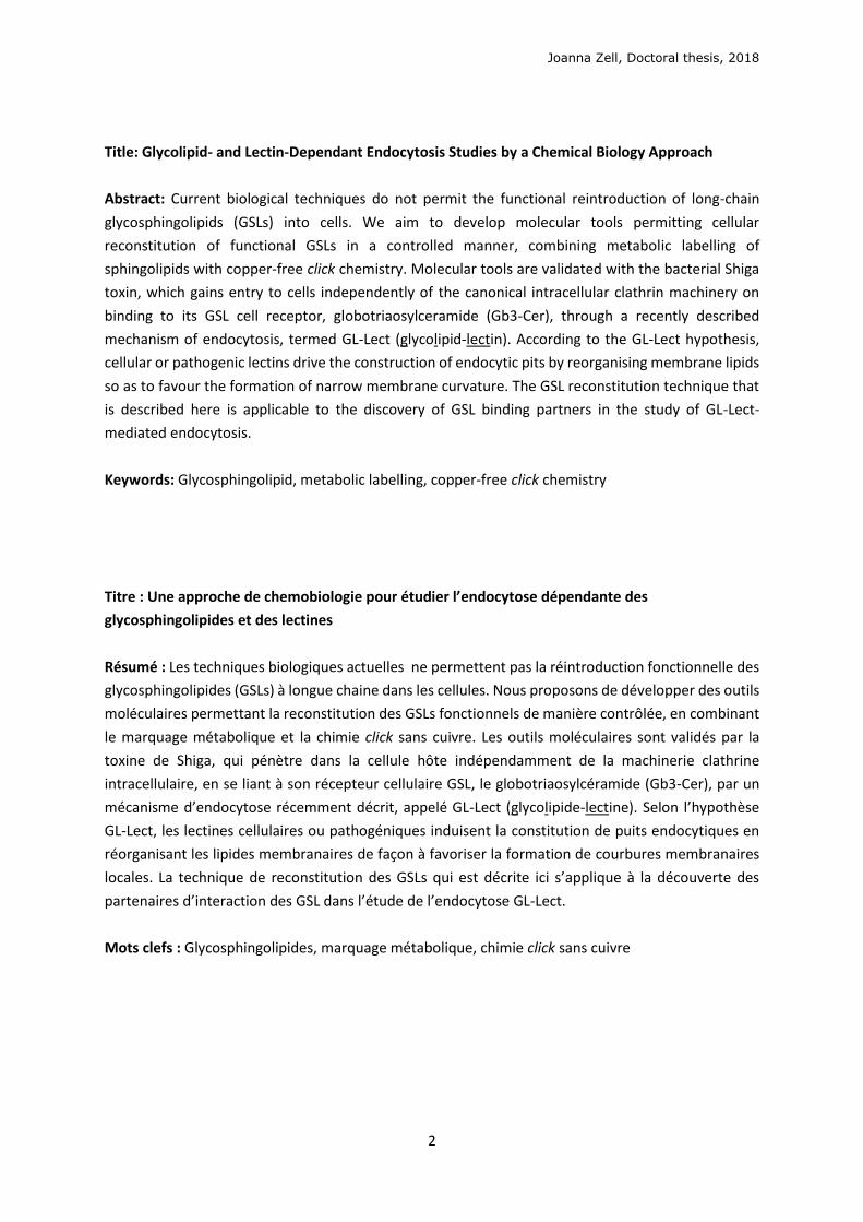
Title: Glycolipid- and Lectin-Dependant Endocytosis Studies by a Chemical Biology Approach
Abstract: Current biological techniques do not permit the functional reintroduction of long-chain
glycosphingolipids (GSLs) into cells. We aim to develop molecular tools permitting cellular
reconstitution of functional GSLs in a controlled manner, combining metabolic labelling of
sphingolipids with copper-free click chemistry. Molecular tools are validated with the bacterial Shiga
toxin, which gains entry to cells independently of the canonical intracellular clathrin machinery on
binding to its GSL cell receptor, globotriaosylceramide (Gb3-Cer), through a recently described
mechanism of endocytosis, termed GL-Lect (glycolipid-lectin). According to the GL-Lect hypothesis,
cellular or pathogenic lectins drive the construction of endocytic pits by reorganising membrane lipids
so as to favour the formation of narrow membrane curvature. The GSL reconstitution technique that
is described here is applicable to the discovery of GSL binding partners in the study of GL-Lect-
mediated endocytosis.
Keywords: Glycosphingolipid, metabolic labelling, copper-free click chemistry
Titre : Une approche de chemobiologie pour étudier l’endocytose dépendante des
glycosphingolipides et des lectines
Résumé : Les techniques biologiques actuelles ne permettent pas la réintroduction fonctionnelle des
glycosphingolipides (GSLs) à longue chaine dans les cellules. Nous proposons de développer des outils
moléculaires permettant la reconstitution des GSLs fonctionnels de manière contrôlée, en combinant
le marquage métabolique et la chimie click sans cuivre. Les outils moléculaires sont validés par la
toxine de Shiga, qui pénètre dans la cellule hôte indépendamment de la machinerie clathrine
intracellulaire, en se liant à son récepteur cellulaire GSL, le globotriaosylcéramide (Gb3-Cer), par un
mécanisme d’endocytose récemment décrit, appelé GL-Lect (glycolipide-lectine). Selon l’hypothèse
GL-Lect, les lectines cellulaires ou pathogéniques induisent la constitution de puits endocytiques en
réorganisant les lipides membranaires de façon à favoriser la formation de courbures membranaires
locales. La technique de reconstitution des GSLs qui est décrite ici s’applique à la découverte des
partenaires d’interaction des GSL dans l’étude de l’endocytose GL-Lect.
Mots clefs : Glycosphingolipides, marquage métabolique, chimie click sans cuivre

Joanna Zell, Doctoral thesis, 2018
3
Live is if you were to die tomorrow,
learn as if you were to live forever
Mahatma Gandhi
Joanna Zell, Doctoral thesis, 2018
4
Acknowledgements
Je suis reconnaissante d’avoir fait partie de l’unité UMR3666, qui est multidisciplinaire,
multiculturelle, étonnamment accueillante et ouverte à tous. À mes deux superviseurs, Ludger et
Frédéric, qui ont eu tant de patience à travers les échecs et les désaccords, et les réussites toujours
trop courtes ; je vous remercie de m’avoir donné cette opportunité de découvertes, défis et
fascinations qui m’ont énormément motivé tous les jours. Je remercie également Jean-Claude,
superviseur-ange gardien.
À mes camarades de labo, Steve, Stefan, Vesela, Haifei, Mélanie, Stéphanie, Alena, Carlos, Marco,
Fabien, Antoine, Seb, Anne, Anne, Thomas, Tati, Raph, Yannick, Christine, Sylvie, Michelle, Alison,
merci d’avoir travaillé avec moi et partagé ces expériences délicieuses, passionnantes et
inoubliables.
À Siau, qui m’a formé en bio avec Ludger et Christian, qui m’a soutenu, m’a écouté et m’a fait sourire
tous les jours.
À Raphaël G, qui m’a donné la confiance dont j’avais besoin pendant ces longues périodes de
syndrome de l’imposteur.
To Julio, who has driven this project with me, feeding all the creativity, imagination and dedication,
and with whom I can always share a great moment in and out of the lab.
I thank all my friends from CRI, who managed to laugh in the face of PhD stress with me, whilst
nourishing overpriced pints, whenever we managed to get out of the lab.
I thank the BIOPOL Innovative Training Network (European H2020 funding) that funded the project
and my international training stations, and grouped together 16 PhD students, making us spend
quality-science-time together across Europe. We had never felt so different, yet so alike.
After countless hours complaining on the phone, I retain some gems from my mum, who said:
“You don’t learn unless you’re suffering;”
and my dad, who said
“You’re doing a PhD because once you’re a Dr., you never have to say if you’re a bloody Miss or
Mrs., because you’ll be a Dr.!”
I would be nowhere without you.
Carolyn, I know one day your wisdom will come crashing down on me. I love you and M&M to the
end and back.
Joanna Zell, Doctoral thesis, 2018
5
Table of contents
Abbreviations……………………………………………………………………………………………………………..……………. 5
1 INTRODUCTION ................................................................................................................................ 9
1.1 Endocytosis mechanisms ........................................................................................................ 9
1.2 Glycolipid- and Lectin-dependent endocytosis ..................................................................... 11
1.3 GSL function studies .............................................................................................................. 15
1.4 GSL function and biosynthesis .............................................................................................. 22
1.5 Biomedical relevance ............................................................................................................ 27
1.6 Bioorthogonal conjugations and click chemistry .................................................................. 29
1.7 Metabolic labelling of glycans and lipids .............................................................................. 36
1.8 Project aims........................................................................................................................... 39
2 RESULTS ........................................................................................................................................ 42
2.1 Synthesis of biomolecule analogues ......................................................................................... 42
2.1.1 Reconstitutable globotriose phospholipid ........................................................................ 42
2.1.2 Clickable sphingolipid and globotriose ............................................................................. 46
2.1.3 Cyclooctyne-functionalised glycans .................................................................................. 51
2.2 Biological validation of analogues............................................................................................. 58
2.2.1 Model membrane validation ............................................................................................ 58
2.2.2 Cellular metabolism and localisation of AzSph ................................................................. 61
2.2.3 Glycan click ........................................................................................................................ 68
2.2.4 Changing the lipid precursor to change localisation ......................................................... 75
3 DISCUSSION .................................................................................................................................. 79
3.1 Phospholipid strategy ........................................................................................................... 79
3.2 Clickable sphingolipid synthetic strategy .............................................................................. 82
3.3 Model membrane testing ..................................................................................................... 89
3.4 Sphingolipid metabolism ...................................................................................................... 90
3.5 In cellulo click ........................................................................................................................ 96
3.6 Optimisation of lipid localisation ........................................................................................ 100
4 PERSPECTIVES ............................................................................................................................. 103
5 CONCLUSIONS ............................................................................................................................ 106
6 EXPERIMENTAL ........................................................................................................................... 107
6.1 Biological validation ............................................................................................................. 107
6.2 Chemical synthesis .............................................................................................................. 114
7 BIBLIOGRAPHY ............................................................................................................................ 146

Joanna Zell, Doctoral thesis, 2018
6
Abbreviations
A.U. Arbitrary units
Ac Acetyl
AzCer 1-Azidoceramide
AzPhy 1-Azidophytosphingosine
AzSph 1-Azidosphingosine
BCN Bicyclononyne
Boc Tert-butyloxycarbonyl
BSA Bovine serum albumine
Bz Benzoyl
CAN Ceric ammonium nitrate
Cer Ceramide
CerS Ceramide synthase
CES Carboxylesterase
CHO Chinese hamster ovary
Chol Cholesterol
CIE Clathrin-independent endocytosis
COP 2-Chloro[1,3,2]dioxaphospholene-2-oxide
CuAAC Copper(I)-catalysed azide-alkyne cycloaddition
DBU 1,8-Diazabicyclo(5.4.0)undec-7-ene
DCC N,N’-Dicyclohexylcarbodiimide
DIBO Dibenzocyclooctyne
DIFO Difluorocyclooctyne
DIPEA Diisopropylethylamine
DMAP 4-Dimethylaminopyridine
DMF Dimethylformamide
DMSO Dimethyl sulfoxide
DOPC 1,2-Dioleoyl-sn-glycero-3-phosphocholine
DOPE 1,2-Dioleoyl-sn-glycero-3-phosphoethanolamine
EA Ethanolamine
Joanna Zell, Doctoral thesis, 2018
7
EC50 Half maximal effective concentration
EDC 1-Ethyl-3-(3-dimethymainopropyl)carbodiimide
EG Ethylene glycol
ER Endoplasmic reticulum
ESI Electrospray ionisation
EtOAc Ethyl acetate
EWG Electron-withdrawing group
FSC Fluorescence scanning microscopy
FSL Function-spacer-lipid
Gb3 Globotriose
Gb3-Cer Globotriaosylceramide
GCS Glucosylceramide synthase
GL Glycolipid
GlcCer Glucosylceramide
Gly Glycine
GPI-AP Glycosylphosphatidylinositol-anchored protein
GSL Glycosphingolipid
GUV Giant unilamellar vesicle
HeLa Henrietta Lacks (cell line)
HOBt Hydroxybenzotriazole
HPTLC High performance thin layer chromatography
HRMS High resolution mass spectrometry
IF Immunofluorescence
IR Infrared
LacCer Lactosylceramide
Lo/Ld Liquid ordered/disordered
LUV Large unilamellar vesicle
MFCO Monofluorocyclooctyne
MS Mass spectrometry
Ms Mesylate
NIS N-Iodosuccinimide
PBS Phosphate-buffered saline
PC Phosphatidylcholine
Joanna Zell, Doctoral thesis, 2018
8
PE Phosphatidylethanolamine
PL Phospholipid
PM Plasma membrane
PS Phosphatidylserine
ROS Reactive oxygen species
rt Room temperature
S1P Sphingosine-1-phosphate
SIMS Secondary ion mass spectrometry
SM Sphingomyelin
SPAAC Strain-promoted azide-alkyne cycloaddition
TAMRA Tetramethyl-rhodamine
TBAF Tetra-n-butylammonium fluoride
TBDPSCl Tert-butyldiphenylsilyl chloride
TFA Trifluoroacetic acid
TGN Trans-Golgi network
THF Tetrahydrofuran
TLC Thin layer chromatography
TMSOTf Trimethylsilyl trifluoromethanesulfonate
TMSSCN Trimethylsilylisothiocyanate
UPLC-MS Ultra performation liquid chromatography - tandem mass spectrometry
w/o With or without
Joanna Zell, Doctoral thesis, 2018
9
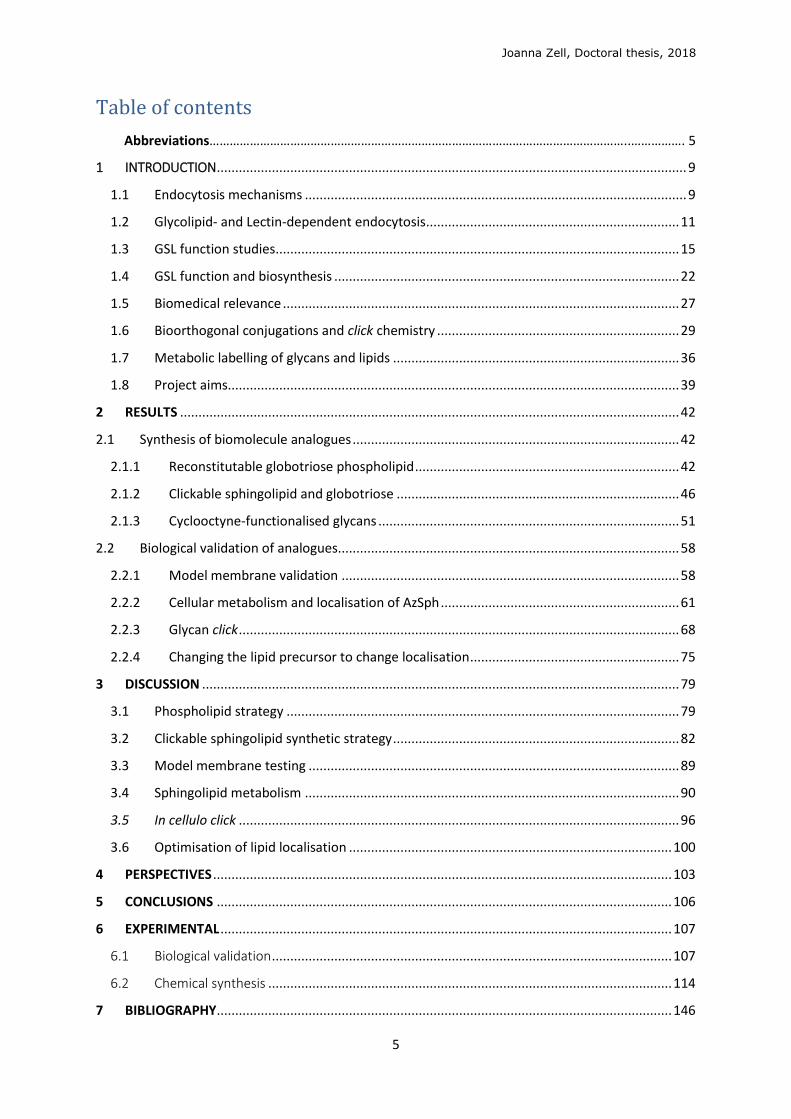
1 INTRODUCTION
1.1 Endocytosis mechanisms
Endocytosis encompasses the cellular uptake of diverse cargo, including
macromolecules, membrane components, nutrients and fluids, by their encapsulation in
membrane vesicles which bud inwards to be pinched off from the plasma membrane (PM).
From the PM, cargoes are trafficked through endosomes, where they can be (1) recycled back
to the PM, (2) sorted via the retrograde route to the Golgi apparatus and in some cases to the
endoplasmic reticulum (ER), or (3) targeted to the late endosomes and lysosomes for
degradation (Figure 1.1.1a). The best characterised mechanism of endocytosis is mediated by
clathrin, which coats the PM on binding to its adaptor proteins, forming a concave lattice
structure (Figure 1.1.1b)1 to bend and engulf portions of the membrane containing endocytic
cargoes. These clathrin-coated vesicles (70-150 nm)2 are eventually scissionned from the PM
with assistance from intracellular protein machinery including a GTPase, dynamin (Figure
1.1.2), recruited by curvature-inducing BAR domain proteins, which themselves bind to and
constrict negatively charged, curved membranes.
Figure 1.1.1: (a) Intracellular vesicular
trafficking pathways3 (b) Electron
microscopy image of clathrin-coated pits
in hypertonic fibroblasts, scale bar
100 nm1
(1)
(2)
(3)
Joanna Zell, Doctoral thesis, 2018
10
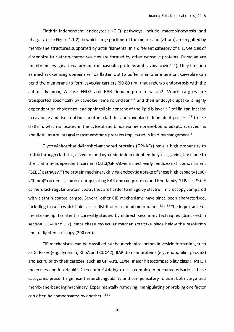
Clathrin-independent endocytosis (CIE) pathways include macropinocytosis and
phagocytosis (Figure 1.1.2), in which large portions of the membrane (>1 μm) are engulfed by
membrane structures supported by actin filaments. In a different category of CIE, vesicles of
closer size to clathrin-coated vesicles are formed by other cytosolic proteins. Caveolae are
membrane invaginations formed from caveolin proteins and cavins (cavin1-4). They function
as mechano-sensing domains which flatten out to buffer membrane tension. Caveolae can
bend the membrane to form caveolar carriers (50-80 nm) that undergo endocytosis with the
aid of dynamin, ATPase EHD2 and BAR domain protein pacsin2. Which cargoes are
transported specifically by caveolae remains unclear,4–6 and their endocytic uptake is highly
dependent on cholesterol and sphingolipid content of the lipid bilayer.7 Flotillin can localise
in caveolae and itself outlines another clathrin- and caveolae-independent process.4,5 Unlike
clathrin, which is located in the cytosol and binds via membrane-bound adaptors, caveolins
and flotillins are integral transmembrane proteins implicated in lipid rearrangement.4
Glycosylphosphatidylinositol-anchored proteins (GPI-ACs) have a high propensity to
traffic through clathrin-, caveolin- and dynamin-independent endocytosis, giving the name to
the clathrin-independent carrier (CLIC)/GPI-AC-enriched early endosomal compartment
(GEEC) pathway.8 The protein machinery driving endocytic uptake of these high capacity (100-
200 nm)9 carriers is complex, implicating BAR domain proteins and Rho family GTPases.10 CIE
carriers lack regular protein coats, thus are harder to image by electron microscopy compared
with clathrin-coated cargos. Several other CIE mechanisms have since been characterised,
including those in which lipids are redistributed to bend membranes.8,11–13 The importance of
membrane lipid content is currently studied by indirect, secondary techniques (discussed in
section 1.3-4 and 1.7), since these molecular mechanisms take place below the resolution
limit of light microscopy (200 nm).
CIE mechanisms can be classified by the mechanical actors in vesicle formation, such
as GTPases (e.g. dynamin, RhoA and CDC42), BAR domain proteins (e.g. endophilin, pacsin2)
and actin, or by their cargoes, such as GPI-APs, CD44, major histocompatibility class I (MHCI)
molecules and interleukin 2 receptor.9 Adding to this complexity in characterisation, these
categories present significant interchangeability and compensatory roles in both cargo and
membrane-bending machinery. Experimentally removing, manipulating or probing one factor
can often be compensated by another.14,15
Joanna Zell, Doctoral thesis, 2018
11
Figure 1.1.2: Mechanisms of endocytosis. Dynamin-dependent pathways (+) are typically associated with
small-scale circular carriers such as clathrin- or caveolin-mediated pits.5
1.2 Glycolipid- and Lectin-dependent endocytosis
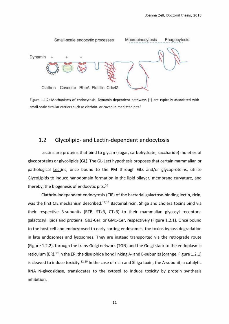
Lectins are proteins that bind to glycan (sugar, carbohydrate, saccharide) moieties of
glycoproteins or glycolipids (GL). The GL-Lect hypothesis proposes that certain mammalian or
pathological Lectins, once bound to the PM through GLs and/or glycoproteins, utilise
GlycoLipids to induce nanodomain formation in the lipid bilayer, membrane curvature, and
thereby, the biogenesis of endocytic pits.16
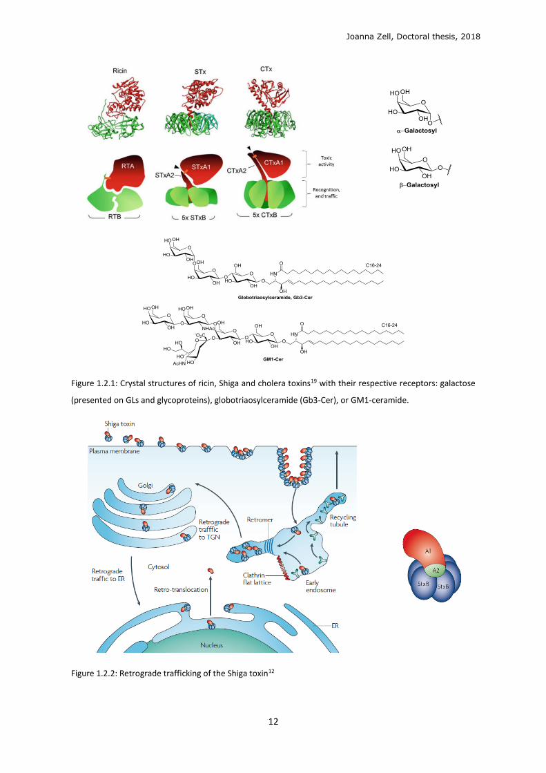
Clathrin-independent endocytosis (CIE) of the bacterial galactose-binding lectin, ricin,
was the first CIE mechanism described.17,18 Bacterial ricin, Shiga and cholera toxins bind via
their respective B-subunits (RTB, STxB, CTxB) to their mammalian glycosyl receptors:
galactosyl lipids and proteins, Gb3-Cer, or GM1-Cer, respectively (Figure 1.2.1). Once bound
to the host cell and endocytosed to early sorting endosomes, the toxins bypass degradation
in late endosomes and lysosomes. They are instead transported via the retrograde route
(Figure 1.2.2), through the trans-Golgi network (TGN) and the Golgi stack to the endoplasmic
reticulum (ER).19 In the ER, the disulphide bond linking A- and B-subunits (orange, Figure 1.2.1)
is cleaved to induce toxicity.12,20 In the case of ricin and Shiga toxin, the A-subunit, a catalytic
RNA N-glycosidase, translocates to the cytosol to induce toxicity by protein synthesis
inhibition.
Joanna Zell, Doctoral thesis, 2018
12
Figure 1.2.1: Crystal structures of ricin, Shiga and cholera toxins19 with their respective receptors: galactose
(presented on GLs and glycoproteins), globotriaosylceramide (Gb3-Cer), or GM1-ceramide.
Figure 1.2.2: Retrograde trafficking of the Shiga toxin12
Joanna Zell, Doctoral thesis, 2018
13
The homopentameric B-subunits of AB5 Shiga and cholera toxin, STxB, CTxB, as well as
the pentameric VP-1 capsid protein of Simian polyoma virus SV40, have flat, doughnut-like
structures (Figure 1.2.3a), where they dock onto the membrane via their glycosphingolipid
(GSL) receptors. The lectins’ polyvalent binding is only of small individual affinity (dissociation
constant Kd ~ 10-3 M for STxB) for each GSL, but combine to a large avidity (Kd ~ 10-9 M).21 STxB
contains three globotriose (Gb3) binding pockets per monomer, thus 15 total Gb3 binding
sites. CTxB and SV40 protein have one GM1 binding pocket per monomer, totalling five GM1
binding sites.22
Of note, these three lectins have no sequence similarity, and yet the glycan receptors
are all positioned around the edge of the protein in a seemingly overlapping geometry (Figure
1.2.3a), indicating a convergent evolution of structure. The glycan binding sites are of a
geometry in which the membrane must bend up around the lectin to fit the glycan part of the
GSL receptor molecules into their binding sites, thereby generating negative, inward-oriented
curvature. This was recently demonstrated in silico for STxB-Gb313-15 in all-atom molecular
dynamics studies (Figure 1.2.3b,d).23 Effects on the membrane underneath the docked STxB,
such as suppression of lipid fluctuations,24 lead to membrane-mediated clustering to induce
wider membrane invagination (Figure 1.2.3c). These early endocytic pits are then recognised
by cytosolic machinery, dynamin, endophilin and/or actin,15 for pit elongation and scission,
before transport to endosomes.
Joanna Zell, Doctoral thesis, 2018
14
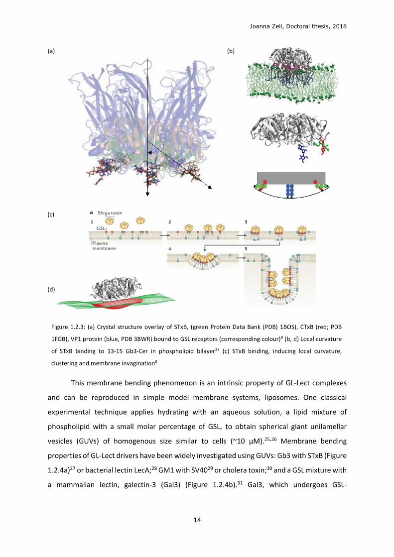
(a)
(b)
(c)
(d)
Figure 1.2.3: (a) Crystal structure overlay of STxB, (green Protein Data Bank (PDB) 1BOS), CTxB (red; PDB
1FGB), VP1 protein (blue, PDB 3BWR) bound to GSL receptors (corresponding colour)8 (b, d) Local curvature
of STxB binding to 13-15 Gb3-Cer in phospholipid bilayer23 (c) STxB binding, inducing local curvature,
clustering and membrane invagination8
This membrane bending phenomenon is an intrinsic property of GL-Lect complexes
and can be reproduced in simple model membrane systems, liposomes. One classical
experimental technique applies hydrating with an aqueous solution, a lipid mixture of
phospholipid with a small molar percentage of GSL, to obtain spherical giant unilamellar
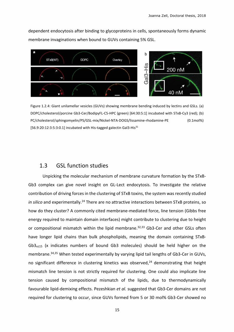
vesicles (GUVs) of homogenous size similar to cells (~10 μM).25,26 Membrane bending
properties of GL-Lect drivers have been widely investigated using GUVs: Gb3 with STxB (Figure
1.2.4a)27 or bacterial lectin LecA;28 GM1 with SV4029 or cholera toxin;30 and a GSL mixture with
a mammalian lectin, galectin-3 (Gal3) (Figure 1.2.4b).31 Gal3, which undergoes GSL-
Joanna Zell, Doctoral thesis, 2018
15
dependent endocytosis after binding to glycoproteins in cells, spontaneously forms dynamic
membrane invaginations when bound to GUVs containing 5% GSL.
Figure 1.2.4: Giant unilamellar vesicles (GUVs) showing membrane bending induced by lectins and GSLs. (a)
DOPC/cholesterol/porcine Gb3-Cer/BodipyFL-C5-HPC (green) [64:30:5:1] incubated with STxB-Cy3 (red); (b)
PC/cholesterol/sphingomyelin/PS/GSL-mix/Nickel-NTA-DOGS/lissamine-rhodamine-PE (0.1mol%)
[56.9:20:12:3:5:3:0.1] incubated with His-tagged galectin Gal3-His31
1.3 GSL function studies
Unpicking the molecular mechanism of membrane curvature formation by the STxB-
Gb3 complex can give novel insight on GL-Lect endocytosis. To investigate the relative
contribution of driving forces in the clustering of STxB toxins, the system was recently studied
in silico and experimentally.24 There are no attractive interactions between STxB proteins, so
how do they cluster? A commonly cited membrane-mediated force, line tension (Gibbs free
energy required to maintain domain interfaces) might contribute to clustering due to height
or compositional mismatch within the lipid membrane.32,33 Gb3-Cer and other GSLs often
have longer lipid chains than bulk phospholipids, meaning the domain containing STxB-
Gb3x≤15 (x indicates numbers of bound Gb3 molecules) should be held higher on the
membrane.34,35 When tested experimentally by varying lipid tail lengths of Gb3-Cer in GUVs,
no significant difference in clustering kinetics was observed,24 demonstrating that height
mismatch line tension is not strictly required for clustering. One could also implicate line
tension caused by compositional mismatch of the lipids, due to thermodynamically
favourable lipid-demixing effects. Pezeshkian et al. suggested that Gb3-Cer domains are not
required for clustering to occur, since GUVs formed from 5 or 30 mol% Gb3-Cer showed no
b
Joanna Zell, Doctoral thesis, 2018
16
significant difference in binding or invagination; 30 mol% being the Gb3 concentration that one
expects to find under STxB molecules.24 Solevyeva et al. showed that STxB binding induced
membrane lipid domain organisation in GUVs,36 but this reasons for the reverse causality;
that STxB clustering induces compositional mismatch.
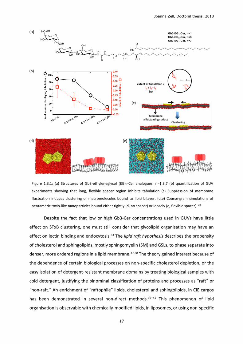
To elucidate the mechanism, synthetic Gb3-spacer-Cer analogues (Figure 1.3.1a) were
synthesised in the L. Johannes laboratory and tested in GUV experiments. The longer ethylene
glycol (EG) spacers between the Gb3 head and the ceramide tail increasingly inhibited STxB
clustering and invaginations in GUVs (n=3 or 7, Figure 1.3.1a, b). These experiments gave birth
to the fluctuation force hypothesis, proposing that a suppression of fluctuation of the
membrane is required for toxins to cluster (Figure 1.3.1c). This effect is commonly alluded to
being like drifting boats on the ocean, or cheerios on milk, which aggregate together. This
hypothesis was studied by course-grained molecular dynamics simulations, showing that two
macromolecules, with dimensions corresponding to the membrane-bound STxB-Gb3x≤15
complex, would randomly drift on a phospholipid bilayer. In the simulation, only the
macromolecules tightly bound to the bilayer (Figure 1.3.1d) would be attracted to each other
from a distance of several nanometres, whereas loosely bound macromolecules (Figure
1.3.1e) would experience no attraction force.24 Hence, the tight binding of the toxin to the
membrane via Gb3 receptors is sufficient to induce clustering. These results will in turn guide
this project, in the constraints on structure of Gb3 analogues proposed and synthesised,
which should retain natural lectin binding and membrane bending actively.
Joanna Zell, Doctoral thesis, 2018
17
(a)
(b)
(c)
(d)
(e)
Figure 1.3.1: (a) Structures of Gb3-ethyleneglycol (EG)n-Cer analogues, n=1,3,7 (b) quantification of GUV
experiments showing that long, flexible spacer region inhibits tabulation (c) Suppression of membrane
fluctuation induces clustering of macromolecules bound to lipid bilayer. (d,e) Course-grain simulations of
pentameric toxin-like nanoparticles bound either tightly (d, no spacer) or loosely (e, flexible spacer). 24
Despite the fact that low or high Gb3-Cer concentrations used in GUVs have little
effect on STxB clustering, one must still consider that glycolipid organisation may have an
effect on lectin binding and endocytosis.33 The lipid raft hypothesis describes the propensity
of cholesterol and sphingolipids, mostly sphingomyelin (SM) and GSLs, to phase separate into
denser, more ordered regions in a lipid membrane.37,38 The theory gained interest because of
the dependence of certain biological processes on non-specific cholesterol depletion, or the
easy isolation of detergent-resistant membrane domains by treating biological samples with
cold detergent, justifying the binominal classification of proteins and processes as “raft” or
“non-raft.” An enrichment of “raftophile” lipids, cholesterol and sphingolipids, in CIE cargos
has been demonstrated in several non-direct methods.39–41 This phenomenon of lipid
organisation is observable with chemically-modified lipids, in liposomes, or using non-specific
Joanna Zell, Doctoral thesis, 2018
18
extraction techniques, but the biological relevance is difficult to evaluate for natural, non-
fluorescent lipids in cellulo or in vivo.
Lipid organisation can be driven by a multitude of factors such as actin cytoskeleton
organisation,42 ectodomain association, and membrane pits, ruffles and contact sites.43
Specific lipid binding pockets in proteins and charge interactions also induce lipid
organisation. 20-30% of the PM area is covered by proteins, and lipid solvation rings around
proteins could account for the majority of the lipid area remaining.44 Eggeling et al. recently
used super resolution fluorescence scanning microscopy (FSC) to show that fluorescent
phospholipids or cholesterol presented no significant lateral heterogeneity, whilst fluorescent
SM showed transient trapping in ~80 nm domains, but hardly moved once trapped. This
observation does not correspond to lipid raft criteria, but infers specific lipid interactions with
membrane components.45,46
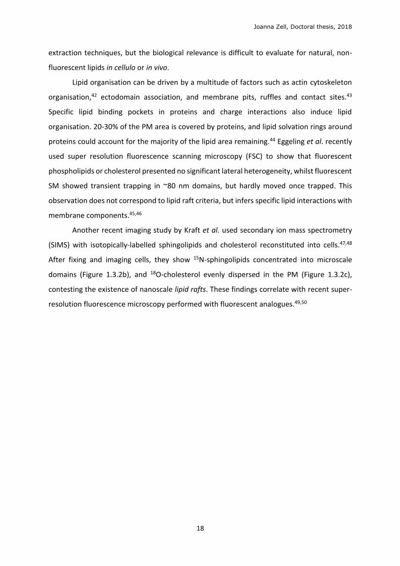
Another recent imaging study by Kraft et al. used secondary ion mass spectrometry
(SIMS) with isotopically-labelled sphingolipids and cholesterol reconstituted into cells.47,48
After fixing and imaging cells, they show 15N-sphingolipids concentrated into microscale
domains (Figure 1.3.2b), and 18O-cholesterol evenly dispersed in the PM (Figure 1.3.2c),
contesting the existence of nanoscale lipid rafts. These findings correlate with recent super-
resolution fluorescence microscopy performed with fluorescent analogues.49,50
Joanna Zell, Doctoral thesis, 2018
19
Figure 1.3.2: Scanning electron microscopy (a) and high-
resolution SIMS images (b,c) of a representative clone 15
fibroblast cell. (b) 15N-sphingolipids are enriched in
micron scale domains on the PM, (c) whereas 18O-
cholesterol showed no enrichment.
The high spatiotemporal resolution of fluorescence microscopy has promoted the
development of fluorescent GSLs, obtained by some complex chemical syntheses.51–55 Despite
progress in smaller fluorophores and faster imaging techniques, the study of these molecular
tools in GUVs and cells has highlighted the precariousness in studying lipid properties with
modified lipids. Lipids, including fluorophore-tagged lipids, can separate into ordered (Lo, e.g.
saturated lipids) or disordered domains (Ld, e.g. unsaturated lipids) easily visible in GUVs by
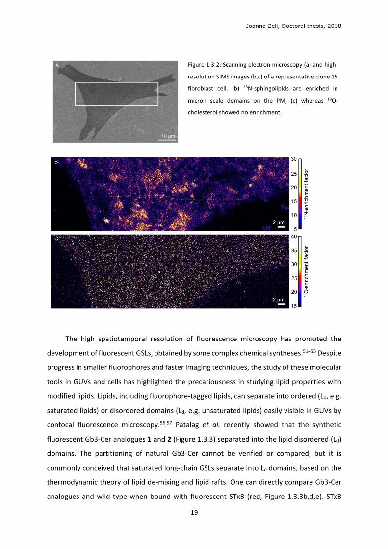
confocal fluorescence microscopy.56,57 Patalag et al. recently showed that the synthetic
fluorescent Gb3-Cer analogues 1 and 2 (Figure 1.3.3) separated into the lipid disordered (Ld)
domains. The partitioning of natural Gb3-Cer cannot be verified or compared, but it is
commonly conceived that saturated long-chain GSLs separate into Lo domains, based on the
thermodynamic theory of lipid de-mixing and lipid rafts. One can directly compare Gb3-Cer
analogues and wild type when bound with fluorescent STxB (red, Figure 1.3.3b,d,e). STxB
Joanna Zell, Doctoral thesis, 2018
20
localised to the Ld domains (green) in GUVs with 1 and 2 separately, whereas with natural
Gb3-Cer, STxB localised to Lo domains.58
Wild type
Figure 1.3.3: GUVs with 5mol% Gb3 analogue 1 (a,b) or 259 (c,d) or wild type Gb3-Cer (Figure 1.2.1)58 (e)
(DOPC/SM/Chol/Gb3-Cer [40:35:20:5] with <1% fluorescent phospholipid (green, Ld), incubated with STxB-
Cy3 (red).
Specific study of Gb3-Cer and other long-chain GSLs in cell culture is practically
unattainable since they cannot be added directly to cells in culture in a controlled manner.60
Chinnapen et al., when investigating GM1 and cholera toxin traffic in live cells, reincorporated
GM1 species bearing C18 chain length or shorter, despite specifying that the natural profile
contains mostly C24 and C16 acyl chains.30 Pedrosa et al. showed that short C8 acyl chain
GlcCer could be taken up with a yield of 20% in cancer cells, and 5% in normal cells, but do
not discuss longer chains.61 Fusogenic liposomes have been used to improve uptake,62,63
employing neutral and positively charged phospholipids to improve fusion of lipid vesicles
with the PM.
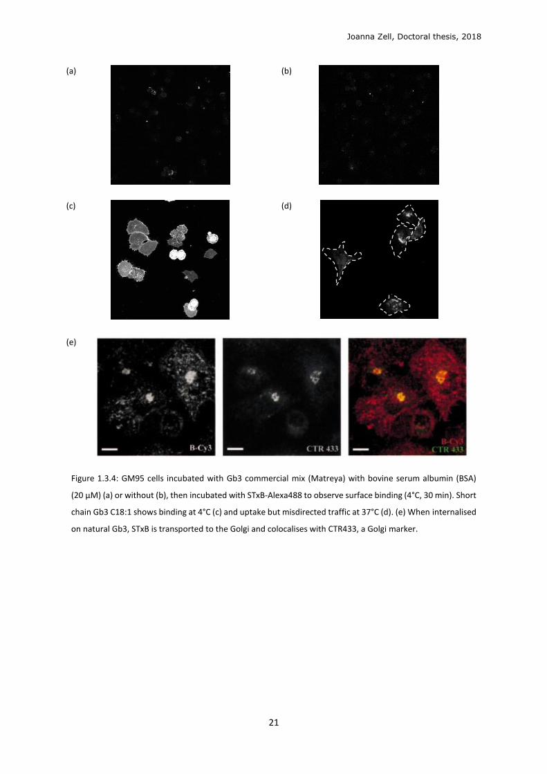
Our laboratory and others have never shown natural retrograde transport of STxB by
supplementing Gb3-Cer to cells not expressing Gb3-Cer natively. Attempted reconstitution of
commercial Gb3-Cer with long ceramide acyl chains (majority C22, C24 chains)64 into GSL-
negative (GSL-) GM95 cells showed negligible STxB cell-surface binding (Figure 1.3.4a) and
endocytosis. Gb3-Cer with shorter C18 acyl chain could allow STxB binding to cells, at 4°C,
before endocytosis (Figure 1. 3.4c), but its traffic is misdirected away from the Golgi (Figure
1. 3.4d). An example by Haicheur et al. shows the canonical retrograde transport of STxB to
the Golgi in Gb3-expressing D1 murine dendritic cells (Figure 3. 3.4e),65 which is reproducible
in most Gb3-expressing (Gb3+) cell lines.40 Even when two strains of bacterial STx, verotoxin
1 and 2, bind to distinct PM domains, they coalesce into retrograde traffic to the Golgi.66
E)
Joanna Zell, Doctoral thesis, 2018
21
(a)
(b)
(c)
(d)
(e)
Figure 1.3.4: GM95 cells incubated with Gb3 commercial mix (Matreya) with bovine serum albumin (BSA)
(20 μM) (a) or without (b), then incubated with STxB-Alexa488 to observe surface binding (4°C, 30 min). Short
chain Gb3 C18:1 shows binding at 4°C (c) and uptake but misdirected traffic at 37°C (d). (e) When internalised
on natural Gb3, STxB is transported to the Golgi and colocalises with CTR433, a Golgi marker.
Joanna Zell, Doctoral thesis, 2018
22
1.4 GSL function and biosynthesis
The biosynthesis of glycosphingolipids (GSLs) is well documented. However, GSL
function and their interactions is still mystifying scientists, apt to the etymology of the word
“sphingolipids” – named by J. L. W. Thudichum in 1884 after the enigmatic sphinx.34 Over 400
unique oligosaccharide head groups make up the enigmatic “glycolipidome” in mammals.67
GSLs constitute 2-10% of total lipids in membranes,68 where sphingomyelin (SM) is estimated
at 15-20% of the plasma membranes (PM).69 GSLs are found at higher concentrations at the
apical side of epithelial cells, and in certain cell types including oligodendrocytes and neurons.
Their fingerprint expression levels vary throughout development and pathogenesis, amongst
cell and tissue type.
Functions of groups of GSLs have been studied by genetic suppressions. Mice deficient
in all GSLs, obtained with a glucosylceramide synthase (GCS) gene knockout, do not survive
beyond gastrulation,70 similar to lactosylceramide synthase knockout mice.71 Genetic
suppression of the gala- and ganglio-series GSLs in mice shows phenotypes in nervous system
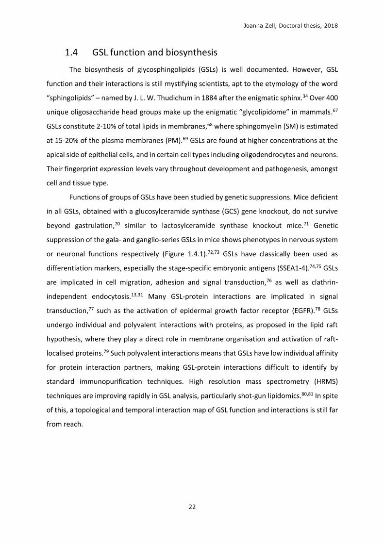
or neuronal functions respectively (Figure 1.4.1).72,73 GSLs have classically been used as
differentiation markers, especially the stage-specific embryonic antigens (SSEA1-4).74,75 GSLs
are implicated in cell migration, adhesion and signal transduction,76 as well as clathrin-
independent endocytosis.13,31 Many GSL-protein interactions are implicated in signal
transduction,77 such as the activation of epidermal growth factor receptor (EGFR).78 GLSs
undergo individual and polyvalent interactions with proteins, as proposed in the lipid raft
hypothesis, where they play a direct role in membrane organisation and activation of raft-
localised proteins.79 Such polyvalent interactions means that GSLs have low individual affinity
for protein interaction partners, making GSL-protein interactions difficult to identify by
standard immunopurification techniques. High resolution mass spectrometry (HRMS)
techniques are improving rapidly in GSL analysis, particularly shot-gun lipidomics.80,81 In spite
of this, a topological and temporal interaction map of GSL function and interactions is still far
from reach.
Joanna Zell, Doctoral thesis, 2018
23
Figure 1.4.1: Stepwise
synthesis of GSLs from
ceramide (Cer).
Knockout of pathways
shows phenotypes in
early development,
myelin (nervous
system) or neuronal
function. Modified
from D’Angelo et al.82
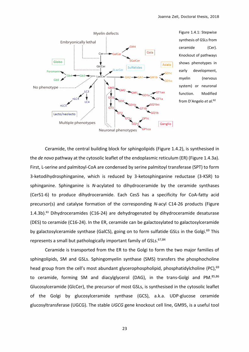
Ceramide, the central building block for sphingolipids (Figure 1.4.2), is synthesised in
the de novo pathway at the cytosolic leaflet of the endoplasmic reticulum (ER) (Figure 1.4.3a).
First, L-serine and palmitoyl-CoA are condensed by serine palmitoyl transferase (SPT) to form
3-ketodihydrosphinganine, which is reduced by 3-ketosphinganine reductase (3-KSR) to
sphinganine. Sphinganine is N-acylated to dihydroceramide by the ceramide synthases
(CerS1-6) to produce dihydroceramide. Each CesS has a specificity for CoA-fatty acid
precursor(s) and catalyse formation of the corresponding N-acyl C14-26 products (Figure
1.4.3b).83 Dihydroceramides (C16-24) are dehydrogenated by dihydroceramide desaturase
(DES) to ceramide (C16-24). In the ER, ceramide can be galactosylated to galactosylceramide
by galactosylceramide synthase (GalCS), going on to form sulfatide GSLs in the Golgi.69 This
represents a small but pathologically important family of GSLs.67,84
Ceramide is transported from the ER to the Golgi to form the two major families of
sphingolipids, SM and GSLs. Sphingomyelin synthase (SMS) transfers the phosphocholine
head group from the cell’s most abundant glycerophospholipid, phosphatidylcholine (PC),69
to ceramide, forming SM and diacylglycerol (DAG), in the trans-Golgi and PM.85,86
Glucosylceramide (GlcCer), the precursor of most GSLs, is synthesised in the cytosolic leaflet
of the Golgi by glucosylceramide synthase (GCS), a.k.a. UDP-glucose ceramide
glucosyltransferase (UGCG). The stable UGCG gene knockout cell line, GM95, is a useful tool
Joanna Zell, Doctoral thesis, 2018
24
in the study of GSL function.87 In lesser part, ceramide is phosphorylated to ceramide-1-
phosphate (C1P) by ceramide kinase (CK).88,89
The only irreversible degradation route leaving the sphingolipid network is the
catabolic pathway. Ceramide is reversibly hydrolysed to sphingosine by ceramidases (CDase),
which are organelle specific, pathway specific and substrate specific.85 Sphingosine is
phosphorylated to sphingosine-1-phosphate (S1P) by sphingosine kinase (SK), while the
reverse reaction is catalysed by S1P phosphatase (S1PP). S1P can then be irreversibly
degraded to ethanolamine-1-phosphate and C16 fatty acid by S1P lyase in the ER.
Ceramide is (re)formed in the salvage pathway, by reacylation of sphingosine,
predominantly by the CerS5 enzyme.83,90 This pool cycles back to form 50-90% of total
sphingolipid content.91 Ceramide is also reformed by the degradation of GSLs by
glycosylceramidase (GCS) or galactosylceramidase (Gal-CDase) in lysosomes, the degradation
of SM by sphingomyelinase (SMase), and of C1P by ceramide-1-phosphate phosphatase
(C1PPase).
Figure 1.4.2: Sphingolipid biosynthesis, modified from S Merscher et al.86
Joanna Zell, Doctoral thesis, 2018
25
B
Figure 1.4.3: (a) GSL synthesis and transport through cellular compartments.72 (b) Ceramide synthase (CerS)
specificities.83
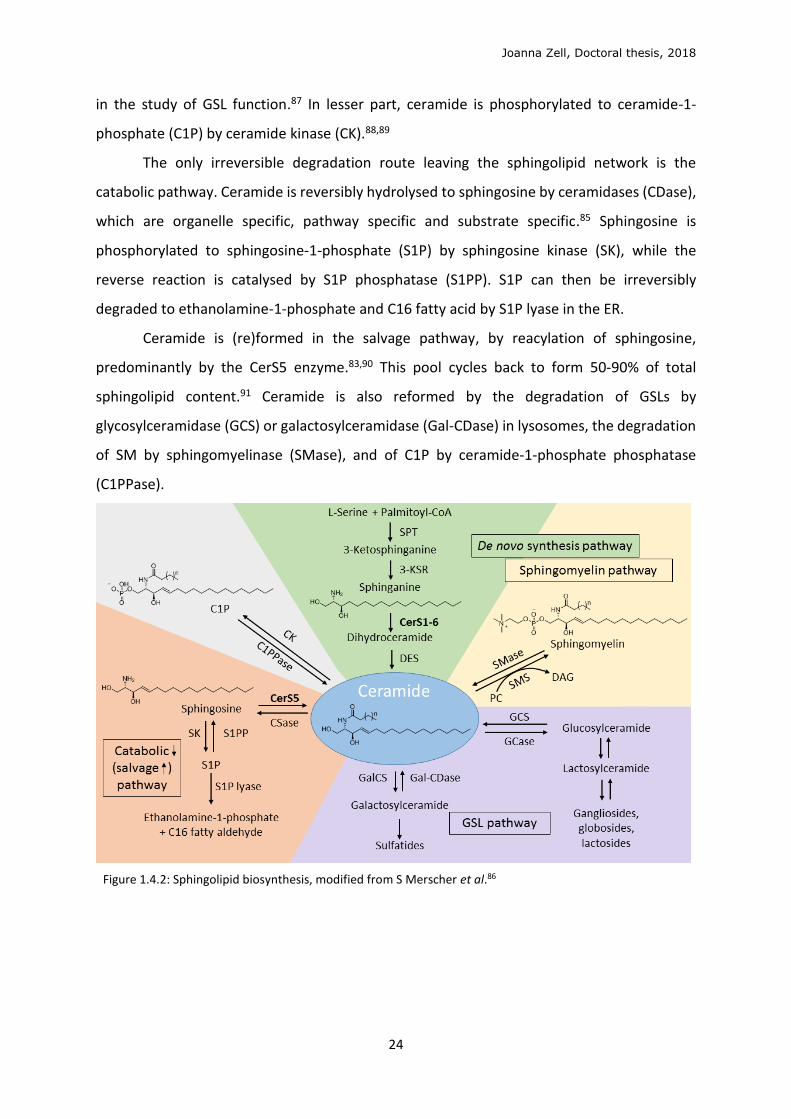
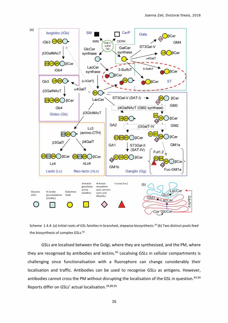
The stepwise biosynthesis of complex GSLs (Figure 1.4.4a) is fed by two distinct pools of
lactosylceramide (LacCer), both formed by LacCer synthase from GlcCer, differing in localisation and
glycosylation fate. The pool of GlcCer transported through the Golgi cisternae by vesicular transport
forms LacCer to be sialylated to GM3 and ensuing gangliosides (sialosides, sialic acid-containing GSLs)
(Figure 1.4.4b). Alternatively, GlcCer transported by the FAPP2 transfer protein to the trans-Golgi
network (TGN) feeds the pool of LacCer which is galactosylated by Gb3 synthase to form Gb3 and the
globo- and asialo-series.82 The biosynthesis of GSLs is mediated by glycosylation enzymes in the Golgi,
which can act on multiple GSL substrates. However, the glycosylations are not random. Twelve glycans
are used in the GSL “alphabet,” with sulfatation adding further complexity. However, only 400
different head groups have been characterised. D’Angelo et al. commented that, of the 144 linkages
possible from 12 glycans, only 29 unique covalent bonds are observed.72 SphinGOMAP, an open source
website compiled by the GSL research community (www.sphingomap.org), contains over 500
sphingolipid and GSL species arranged according to their biosynthesis, displaying nomenclature and
stereochemistry.
Joanna Zell, Doctoral thesis, 2018
26
(a)
Scheme 1.4.4: (a) Initial roots of GSL families in branched, stepwise biosynthesis.34 (b) Two distinct pools feed
the biosynthesis of complex GSLs.82
GSLs are localised between the Golgi, where they are synthesised, and the PM, where
they are recognised by antibodies and lectins.92 Localising GSLs in cellular compartments is
challenging since functionalisation with a fluorophore can change considerably their
localisation and traffic. Antibodies can be used to recognise GSLs as antigens. However,
antibodies cannot cross the PM without disrupting the localisation of the GSL in question.93,94
Reports differ on GSLs’ actual localisation.34,89,95
Joanna Zell, Doctoral thesis, 2018
27
1.5 Biomedical relevance
In this project, we develop tools for the investigation of GSLs in GL-Lect endocytosis.
GSLs are of medical interest for the investigation of GSL lysosomal storage diseases, caused
by mutations in sphingolipid degradation genes. Major examples include Gaucher disease,
(GlcCer accumulation), Fabry (Gb3-Cer), Tay–Sachs and Sandhoff (GM2) diseases.86,96 These
can be treated with substrate reduction therapy, such as the glucosyl-ceramide synthase
inhibitor N-butyldeoxyno-jirimycin,84 and enzyme replacement therapy at high cost.
Moreover, better comprehension of the molecular mechanism of toxins, which hijack
mammalian GSLs for cellular uptake via GL-Lect endocytosis, will aid the development of
treatment. Shiga toxin, the major virulence factor of Shigella dysenteriae, and Shiga-like
toxins, also known as verotoxins, secreted by enterohemorrhagic strains of Escherichia coli
bacteria, have a highly conserved structure.97 All bind specifically to Gb3-Cer, which is
expressed on kidneys and brain tissues,98 and some strains bind to Gb4-Cer with lower
affinity. Infection induces diarrhoea, and eventually haemolytic uremic syndrome (HUS) and
acute renal failure, with high infant fatality rate across the world, and complex cases can have
neurological symptons.20 Cholera toxin, secreted by Vibrio cholerae, causes cholera, a lethal
disease with high fatalities in the developing world, and outbreaks of disease in conflict zones
is regularly reported in standard news media around the world.99 Currently no curative
treatment is known for disease caused by the Shiga and cholera toxin-producing bacteria.20
Gb3 is expressed in several cell types of interest to cancer research: cancer cells and
cancer stem cells themselves,100,101 and antigen-presenting dendritic cells, which stimulate
the immune system. The interest in GSLs spans the development of cancer
chemotherapeutics, in which it is paramount that effective drug targets are cell-specific to
reduce non-specific toxicity. GSLs as cell biomarkers is under-explored territory in the state of
chemotherapeutic research, and yet their expression levels are known to change drastically
with pathogenesis.101,102 Furthermore, study of the GL-Lect mechanism will inform the
development of lectin- or STxB-conjugated drug-vectors; the rational for which aims at
following the non-degradative retrograde pathway, from which STxB benefits, to target and
invade host cells (Figure 1.5.1).98 This route is complementary to the standard pathway
followed by large antibody-drug conjugates (ADCs),103 which are normally taken up by
Joanna Zell, Doctoral thesis, 2018
28
clathrin-mediated endocytosis and sorted to lysosomes to be degraded. STxB-drug conjugates
have shown promise in targeting Gb3-overexpressing cancer cells.104–110
Endogenous lectins and their GL-Lect interactions with GSLs are coming to light.2,16 For
example, galectin-3, which is deregulated in human cancers, is endocytosed with CD44 and
β1-integrin in a clathrin-independent mechanism dependent on GSLs and N-glycosylation.31
Gb3-Cer is the cell receptor for the Shiga toxin, but no endogenous functions are currently
known. Although GSLs are not classically “druggable” compounds due to their undesirable
pharmacokinetic properties, research into their function and therapeutic role requires
methods to manipulate their expression in cells. Hence a robust and versatile reconstitution
method and related syntheses of these products are of great interest.
Figure 1.5.1: Cellular
incorporation and traffic of
antibody-drug conjugates
(ADCs) and STx and its
conjugates
In this project, we endeavour to reconstitute GSLs by conjugating in situ the two parts
of a GSL (glycan and lipid) with a spacer group of the smallest possible size. We employ specific
bioorthogonal chemistry, known as click chemistry, to achieve this in cell culture.
Joanna Zell, Doctoral thesis, 2018
29
1.6 Bioorthogonal conjugations and click chemistry
Chemical biology uses chemical techniques to effect or probe biological systems at the
molecular level. This includes the development of fluorophores, small-molecule protein
inhibitors/activators, non-natural amino acids and any other synthetically modified
biomolecule. Genetic tagging of proteins, for example, with green fluorescent protein (GFP),
has significantly promoted research in molecular biology. However, smaller chemical
modifications are needed for smaller metabolites, lipids, carbohydrates and post-
translational modifications. Although chemical synthesis may never attain the complexity of
nature’s highly regulated protein factory, progress is constantly being made in establishing
better, subtler and more specific manipulation and higher-resolution study of the cellular
system.
Current techniques for the study of lipids and GSLs are limited. Stable isotope labelling
should not disrupt the biochemical interactions of the molecule under study, but
incorporation is often low yielding, requiring technically complicated protocols for low
sensitivity and high costs, and topological imaging techniques able to localise stable isotopes
are still developmental (secondary ion mass spectrometry, SIMS, section 1.3). Radioisotope
labelling can be localised but is innately hazardous. Mass spectrometry (MS) coupled to liquid
chromatography (LC-MS) has traditionally been used for exact identification for the brute
structure of the lipidome, all of the lipids present in a biological sample. Liquid
chromatography separation of analyte species allows higher signal to noise ratio, but is not
quantifiable. Now, shotgun lipidomics is coming to dominate the field, and is applied in this
project. This rapidly developing technology permits identification and quantification of the
lipidome using high resolution mass spectrometry (HRMS) to access exact masses at a parts-
per-million (ppm, 10-6) mass unit accuracy, coupled with high-throughput data handling.
Highly adapted lipid extraction protocols are employed with direct infusion, without
chromatographic separation. Direct infusion allows exact quantification since a constant flow
of lipid sample of known concentration is analysed, with the use of appropriate internal
standards.111,112
To investiage biomolecule localisation and function, small chemical reporter groups,
such as fluorophores for fluorescence imaging, or affinity tags like biotin for purification, can
be incorporated in vitro, before their incorporation and study in cells or organisms. For in vitro
Joanna Zell, Doctoral thesis, 2018
30
tagging, selective or orthogonal chemical conjugations are required for specific tagging in a
non-perturbing site on the biomolecule. For small biomolecules such as lipids and
carbohydrates, where even the smallest fluorophores can perturb lipid function,57 the
reporter group can be added in cellulo by means of a minute, latently reactive tag or labelling
group. After cellular incorporation and biological function, the tag can be specifically
conjugated to a reporter group (e.g. fluorophore or biotin) for visualisation or isolation (Figure
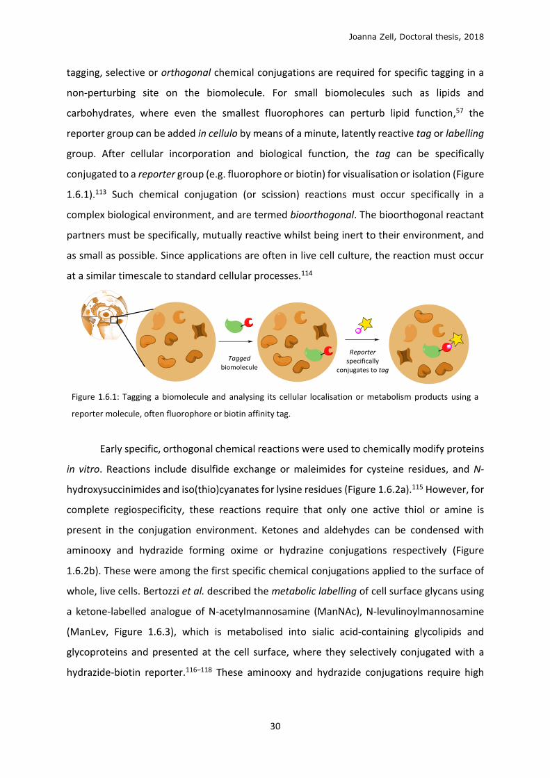
1.6.1).113 Such chemical conjugation (or scission) reactions must occur specifically in a
complex biological environment, and are termed bioorthogonal. The bioorthogonal reactant
partners must be specifically, mutually reactive whilst being inert to their environment, and
as small as possible. Since applications are often in live cell culture, the reaction must occur
at a similar timescale to standard cellular processes.114
Figure 1.6.1: Tagging a biomolecule and analysing its cellular localisation or metabolism products using a
reporter molecule, often fluorophore or biotin affinity tag.
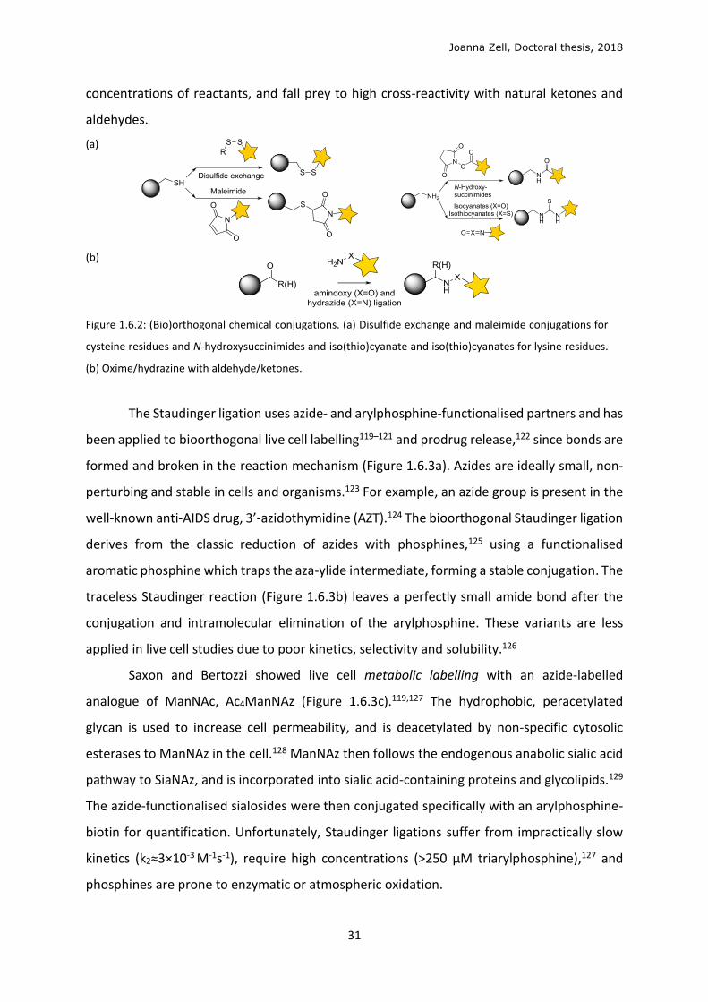
Early specific, orthogonal chemical reactions were used to chemically modify proteins
in vitro. Reactions include disulfide exchange or maleimides for cysteine residues, and N-
hydroxysuccinimides and iso(thio)cyanates for lysine residues (Figure 1.6.2a).115 However, for
complete regiospecificity, these reactions require that only one active thiol or amine is
present in the conjugation environment. Ketones and aldehydes can be condensed with
aminooxy and hydrazide forming oxime or hydrazine conjugations respectively (Figure
1.6.2b). These were among the first specific chemical conjugations applied to the surface of
whole, live cells. Bertozzi et al. described the metabolic labelling of cell surface glycans using
a ketone-labelled analogue of N-acetylmannosamine (ManNAc), N-levulinoylmannosamine
(ManLev, Figure 1.6.3), which is metabolised into sialic acid-containing glycolipids and
glycoproteins and presented at the cell surface, where they selectively conjugated with a
hydrazide-biotin reporter.116–118 These aminooxy and hydrazide conjugations require high
Joanna Zell, Doctoral thesis, 2018
31
concentrations of reactants, and fall prey to high cross-reactivity with natural ketones and
aldehydes.
(a)
(b)
Figure 1.6.2: (Bio)orthogonal chemical conjugations. (a) Disulfide exchange and maleimide conjugations for
cysteine residues and N-hydroxysuccinimides and iso(thio)cyanate and iso(thio)cyanates for lysine residues.
(b) Oxime/hydrazine with aldehyde/ketones.
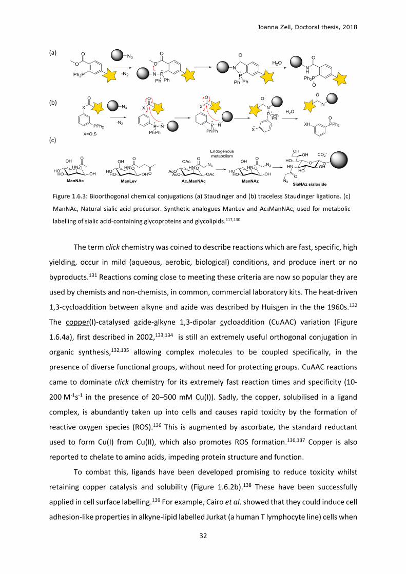
The Staudinger ligation uses azide- and arylphosphine-functionalised partners and has
been applied to bioorthogonal live cell labelling119–121 and prodrug release,122 since bonds are
formed and broken in the reaction mechanism (Figure 1.6.3a). Azides are ideally small, non-
perturbing and stable in cells and organisms.123 For example, an azide group is present in the
well-known anti-AIDS drug, 3’-azidothymidine (AZT).124 The bioorthogonal Staudinger ligation
derives from the classic reduction of azides with phosphines,125 using a functionalised
aromatic phosphine which traps the aza-ylide intermediate, forming a stable conjugation. The
traceless Staudinger reaction (Figure 1.6.3b) leaves a perfectly small amide bond after the
conjugation and intramolecular elimination of the arylphosphine. These variants are less
applied in live cell studies due to poor kinetics, selectivity and solubility.126
Saxon and Bertozzi showed live cell metabolic labelling with an azide-labelled
analogue of ManNAc, Ac4ManNAz (Figure 1.6.3c).119,127 The hydrophobic, peracetylated
glycan is used to increase cell permeability, and is deacetylated by non-specific cytosolic
esterases to ManNAz in the cell.128 ManNAz then follows the endogenous anabolic sialic acid
pathway to SiaNAz, and is incorporated into sialic acid-containing proteins and glycolipids.129
The azide-functionalised sialosides were then conjugated specifically with an arylphosphine-
biotin for quantification. Unfortunately, Staudinger ligations suffer from impractically slow
kinetics (k2≈3×10⁻³ M-1s-1), require high concentrations (>250 µM triarylphosphine),127 and
phosphines are prone to enzymatic or atmospheric oxidation.
Joanna Zell, Doctoral thesis, 2018
32
(a)
(b)
(c)
Figure 1.6.3: Bioorthogonal chemical conjugations (a) Staudinger and (b) traceless Staudinger ligations. (c)
ManNAc, Natural sialic acid precursor. Synthetic analogues ManLev and Ac4ManNAc, used for metabolic
labelling of sialic acid-containing glycoproteins and glycolipids.117,130
The term click chemistry was coined to describe reactions which are fast, specific, high
yielding, occur in mild (aqueous, aerobic, biological) conditions, and produce inert or no
byproducts.131 Reactions coming close to meeting these criteria are now so popular they are
used by chemists and non-chemists, in common, commercial laboratory kits. The heat-driven
1,3-cycloaddition between alkyne and azide was described by Huisgen in the the 1960s.132
The copper(I)-catalysed azide-alkyne 1,3-dipolar cycloaddition (CuAAC) variation (Figure
1.6.4a), first described in 2002,133,134 is still an extremely useful orthogonal conjugation in
organic synthesis,132,135 allowing complex molecules to be coupled specifically, in the
presence of diverse functional groups, without need for protecting groups. CuAAC reactions
came to dominate click chemistry for its extremely fast reaction times and specificity (10-
200 M-1s-1 in the presence of 20–500 mM Cu(I)). Sadly, the copper, solubilised in a ligand
complex, is abundantly taken up into cells and causes rapid toxicity by the formation of
reactive oxygen species (ROS).136 This is augmented by ascorbate, the standard reductant
used to form Cu(I) from Cu(II), which also promotes ROS formation.136,137 Copper is also
reported to chelate to amino acids, impeding protein structure and function.
To combat this, ligands have been developed promising to reduce toxicity whilst
retaining copper catalysis and solubility (Figure 1.6.2b).138 These have been successfully
applied in cell surface labelling.139 For example, Cairo et al. showed that they could induce cell
adhesion-like properties in alkyne-lipid labelled Jurkat (a human T lymphocyte line) cells when
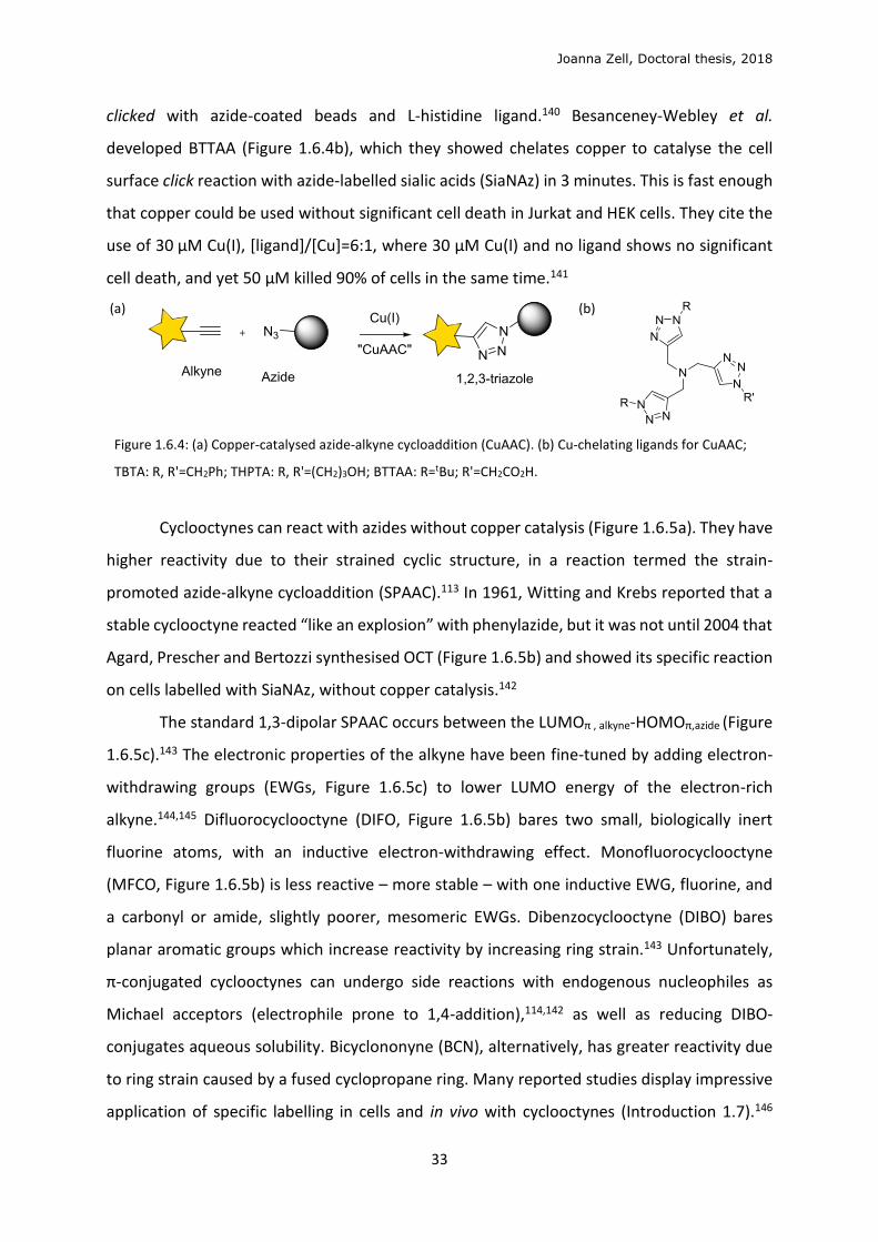
Joanna Zell, Doctoral thesis, 2018
33
clicked with azide-coated beads and L-histidine ligand.140 Besanceney-Webley et al.
developed BTTAA (Figure 1.6.4b), which they showed chelates copper to catalyse the cell
surface click reaction with azide-labelled sialic acids (SiaNAz) in 3 minutes. This is fast enough
that copper could be used without significant cell death in Jurkat and HEK cells. They cite the
use of 30 µM Cu(I), [ligand]/[Cu]=6:1, where 30 µM Cu(I) and no ligand shows no significant
cell death, and yet 50 µM killed 90% of cells in the same time.141
(a)
(b)
Figure 1.6.4: (a) Copper-catalysed azide-alkyne cycloaddition (CuAAC). (b) Cu-chelating ligands for CuAAC;
TBTA: R, R'=CH2Ph; THPTA: R, R'=(CH2)3OH; BTTAA: R=tBu; R'=CH2CO2H.
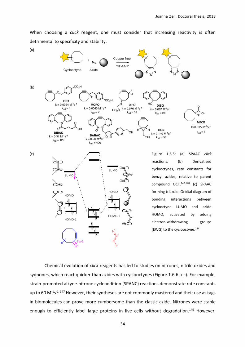
Cyclooctynes can react with azides without copper catalysis (Figure 1.6.5a). They have
higher reactivity due to their strained cyclic structure, in a reaction termed the strain-
promoted azide-alkyne cycloaddition (SPAAC).113 In 1961, Witting and Krebs reported that a
stable cyclooctyne reacted “like an explosion” with phenylazide, but it was not until 2004 that
Agard, Prescher and Bertozzi synthesised OCT (Figure 1.6.5b) and showed its specific reaction
on cells labelled with SiaNAz, without copper catalysis.142
The standard 1,3-dipolar SPAAC occurs between the LUMOπ , alkyne-HOMOπ,azide (Figure
1.6.5c).143 The electronic properties of the alkyne have been fine-tuned by adding electron-
withdrawing groups (EWGs, Figure 1.6.5c) to lower LUMO energy of the electron-rich
alkyne.144,145 Difluorocyclooctyne (DIFO, Figure 1.6.5b) bares two small, biologically inert
fluorine atoms, with an inductive electron-withdrawing effect. Monofluorocyclooctyne
(MFCO, Figure 1.6.5b) is less reactive – more stable – with one inductive EWG, fluorine, and
a carbonyl or amide, slightly poorer, mesomeric EWGs. Dibenzocyclooctyne (DIBO) bares
planar aromatic groups which increase reactivity by increasing ring strain.143 Unfortunately,
π-conjugated cyclooctynes can undergo side reactions with endogenous nucleophiles as
Michael acceptors (electrophile prone to 1,4-addition),114,142 as well as reducing DIBO-
conjugates aqueous solubility. Bicyclononyne (BCN), alternatively, has greater reactivity due
to ring strain caused by a fused cyclopropane ring. Many reported studies display impressive
application of specific labelling in cells and in vivo with cyclooctynes (Introduction 1.7).146
Joanna Zell, Doctoral thesis, 2018
34
When choosing a click reagent, one must consider that increasing reactivity is often
detrimental to specificity and stability.
(a)
(b)
(c)
Figure 1.6.5: (a) SPAAC click
reactions. (b) Derivatised
cyclooctynes, rate constants for
benzyl azides, relative to parent
compound OCT.147,148 (c) SPAAC
forming triazole. Orbital diagram of
bonding interactions between
cyclooctyne LUMO and azide
HOMO, activated by adding
electron-withdrawing groups
(EWG) to the cyclooctyne.144
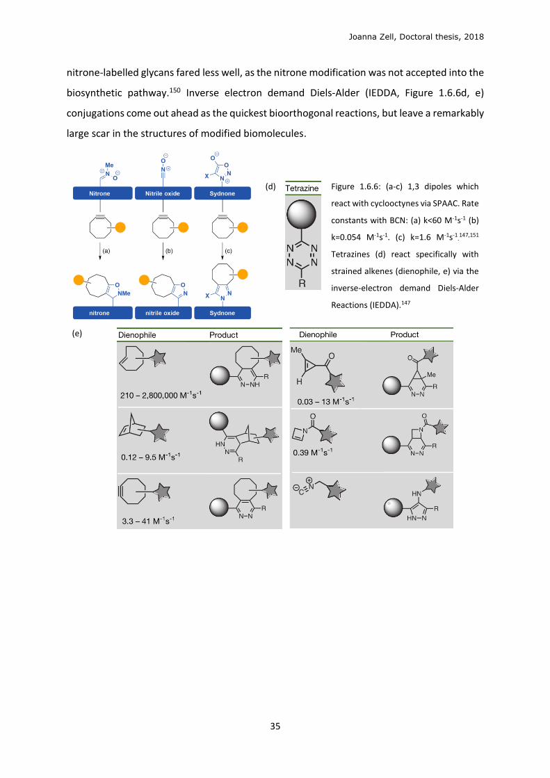
Chemical evolution of click reagents has led to studies on nitrones, nitrile oxides and
sydnones, which react quicker than azides with cyclooctynes (Figure 1.6.6 a-c). For example,
strain-promoted alkyne-nitrone cycloaddition (SPANC) reactions demonstrate rate constants
up to 60 M-1s-1.147 However, their syntheses are not commonly mastered and their use as tags
in biomolecules can prove more cumbersome than the classic azide. Nitrones were stable
enough to efficiently label large proteins in live cells without degradation.149 However,
Joanna Zell, Doctoral thesis, 2018
35
nitrone-labelled glycans fared less well, as the nitrone modification was not accepted into the
biosynthetic pathway.150 Inverse electron demand Diels-Alder (IEDDA, Figure 1.6.6d, e)
conjugations come out ahead as the quickest bioorthogonal reactions, but leave a remarkably
large scar in the structures of modified biomolecules.
(d)
Figure 1.6.6: (a-c) 1,3 dipoles which
react with cyclooctynes via SPAAC. Rate
constants with BCN: (a) k<60 M-1s-1 (b)
k=0.054 M-1s-1. (c) k=1.6 M-1s-1.147,151
Tetrazines (d) react specifically with
strained alkenes (dienophile, e) via the
inverse-electron demand Diels-Alder
Reactions (IEDDA).147
(e)
Joanna Zell, Doctoral thesis, 2018
36
1.7 Metabolic labelling of glycans and lipids
Click chemistry’s application in metabolic labelling has had great influence on several
advanced biological techniques. Genetic code expansion, using synthetically evolved
derivatives of tRNA synthetases, now allows site-specific genetic encoding of clickable amino
acids.152 With click chemistry, radiolabels can be added to proteins regioselectively under mild
conditions, for techniques such as positron emission tomography (PET) and radiotherapy, and
which show potential in animal models.153 This project has been inspired and guided by recent
innovations in metabolic labelling to study lipid and glycan interactions, briefly outlined in this
chapter.
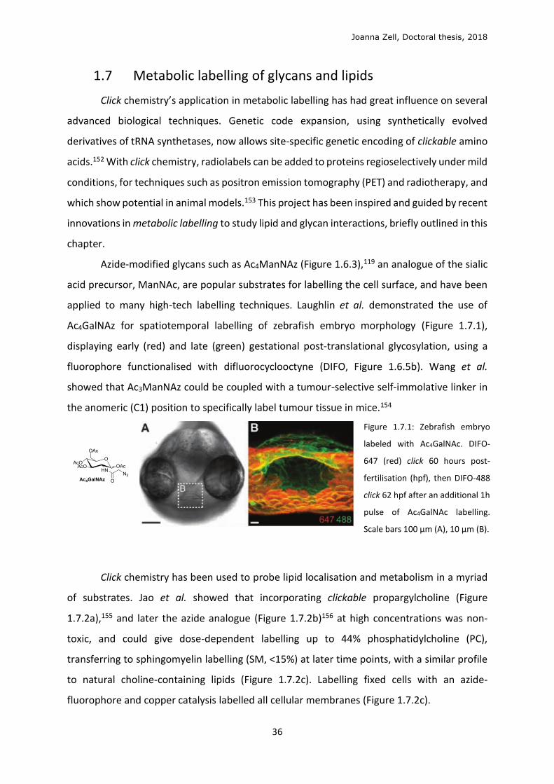
Azide-modified glycans such as Ac4ManNAz (Figure 1.6.3),119 an analogue of the sialic
acid precursor, ManNAc, are popular substrates for labelling the cell surface, and have been
applied to many high-tech labelling techniques. Laughlin et al. demonstrated the use of
Ac4GalNAz for spatiotemporal labelling of zebrafish embryo morphology (Figure 1.7.1),
displaying early (red) and late (green) gestational post-translational glycosylation, using a
fluorophore functionalised with difluorocyclooctyne (DIFO, Figure 1.6.5b). Wang et al.
showed that Ac3ManNAz could be coupled with a tumour-selective self-immolative linker in
the anomeric (C1) position to specifically label tumour tissue in mice.154
Figure 1.7.1: Zebrafish embryo
labeled with Ac4GalNAc. DIFO-
647 (red) click 60 hours post-
fertilisation (hpf), then DIFO-488
click 62 hpf after an additional 1h
pulse of Ac4GalNAc labelling.
Scale bars 100 μm (A), 10 μm (B).
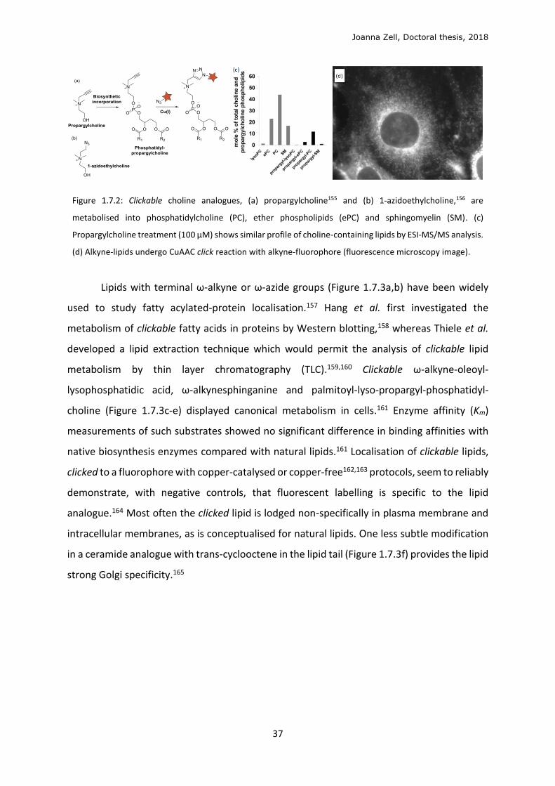
Click chemistry has been used to probe lipid localisation and metabolism in a myriad
of substrates. Jao et al. showed that incorporating clickable propargylcholine (Figure
1.7.2a),155 and later the azide analogue (Figure 1.7.2b)156 at high concentrations was non-
toxic, and could give dose-dependent labelling up to 44% phosphatidylcholine (PC),
transferring to sphingomyelin labelling (SM, <15%) at later time points, with a similar profile
to natural choline-containing lipids (Figure 1.7.2c). Labelling fixed cells with an azide-
fluorophore and copper catalysis labelled all cellular membranes (Figure 1.7.2c).
Joanna Zell, Doctoral thesis, 2018
37
Figure 1.7.2: Clickable choline analogues, (a) propargylcholine155 and (b) 1-azidoethylcholine,156 are
metabolised into phosphatidylcholine (PC), ether phospholipids (ePC) and sphingomyelin (SM). (c)
Propargylcholine treatment (100 μM) shows similar profile of choline-containing lipids by ESI-MS/MS analysis.
(d) Alkyne-lipids undergo CuAAC click reaction with alkyne-fluorophore (fluorescence microscopy image).
Lipids with terminal ω-alkyne or ω-azide groups (Figure 1.7.3a,b) have been widely
used to study fatty acylated-protein localisation.157 Hang et al. first investigated the
metabolism of clickable fatty acids in proteins by Western blotting,158 whereas Thiele et al.
developed a lipid extraction technique which would permit the analysis of clickable lipid
metabolism by thin layer chromatography (TLC).159,160 Clickable ω-alkyne-oleoyl-
lysophosphatidic acid, ω-alkynesphinganine and palmitoyl-lyso-propargyl-phosphatidyl-
choline (Figure 1.7.3c-e) displayed canonical metabolism in cells.161 Enzyme affinity (Km)
measurements of such substrates showed no significant difference in binding affinities with
native biosynthesis enzymes compared with natural lipids.161 Localisation of clickable lipids,
clicked to a fluorophore with copper-catalysed or copper-free162,163 protocols, seem to reliably
demonstrate, with negative controls, that fluorescent labelling is specific to the lipid
analogue.164 Most often the clicked lipid is lodged non-specifically in plasma membrane and
intracellular membranes, as is conceptualised for natural lipids. One less subtle modification
in a ceramide analogue with trans-cyclooctene in the lipid tail (Figure 1.7.3f) provides the lipid
strong Golgi specificity.165
Joanna Zell, Doctoral thesis, 2018
38
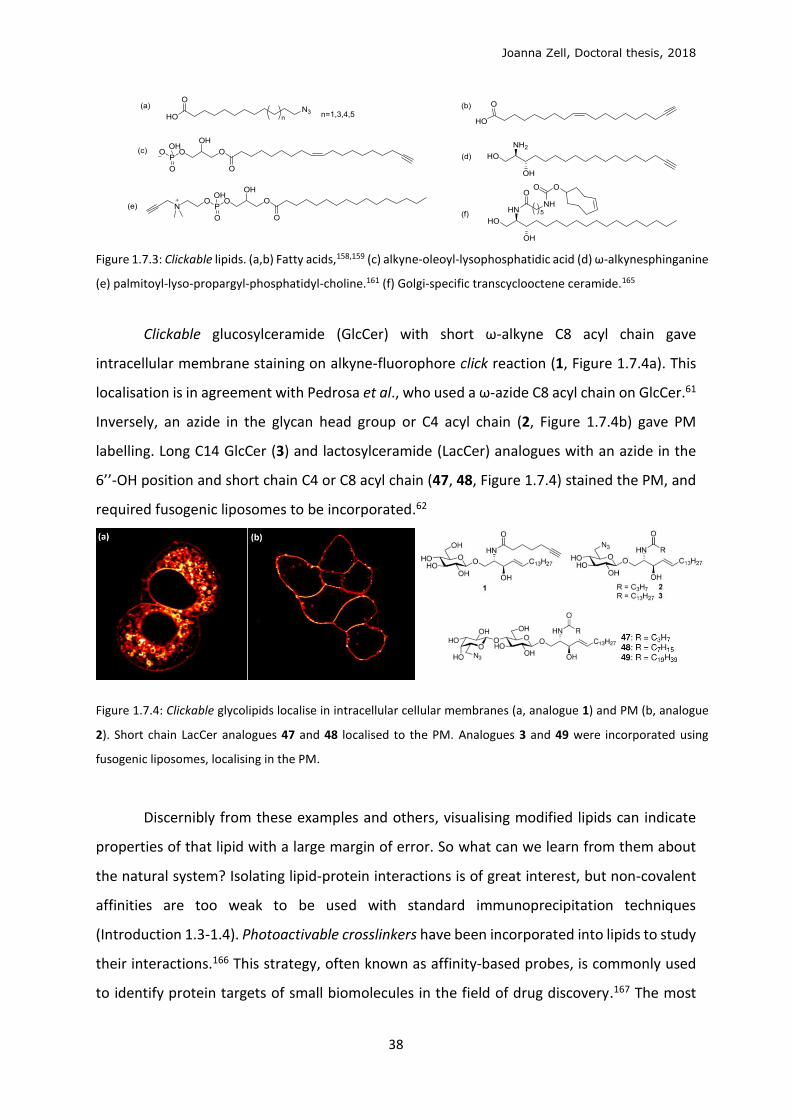
Figure 1.7.3: Clickable lipids. (a,b) Fatty acids,158,159 (c) alkyne-oleoyl-lysophosphatidic acid (d) ω-alkynesphinganine
(e) palmitoyl-lyso-propargyl-phosphatidyl-choline.161 (f) Golgi-specific transcyclooctene ceramide.165
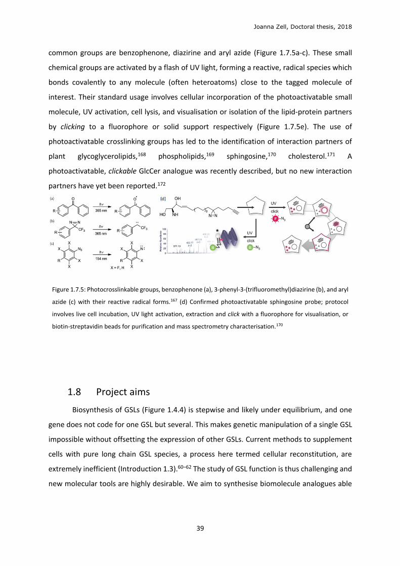
Clickable glucosylceramide (GlcCer) with short ω-alkyne C8 acyl chain gave
intracellular membrane staining on alkyne-fluorophore click reaction (1, Figure 1.7.4a). This
localisation is in agreement with Pedrosa et al., who used a ω-azide C8 acyl chain on GlcCer.61
Inversely, an azide in the glycan head group or C4 acyl chain (2, Figure 1.7.4b) gave PM
labelling. Long C14 GlcCer (3) and lactosylceramide (LacCer) analogues with an azide in the
6’’-OH position and short chain C4 or C8 acyl chain (47, 48, Figure 1.7.4) stained the PM, and
required fusogenic liposomes to be incorporated.62
Figure 1.7.4: Clickable glycolipids localise in intracellular cellular membranes (a, analogue 1) and PM (b, analogue
2). Short chain LacCer analogues 47 and 48 localised to the PM. Analogues 3 and 49 were incorporated using
fusogenic liposomes, localising in the PM.
Discernibly from these examples and others, visualising modified lipids can indicate
properties of that lipid with a large margin of error. So what can we learn from them about
the natural system? Isolating lipid-protein interactions is of great interest, but non-covalent
affinities are too weak to be used with standard immunoprecipitation techniques
(Introduction 1.3-1.4). Photoactivable crosslinkers have been incorporated into lipids to study
their interactions.166 This strategy, often known as affinity-based probes, is commonly used
to identify protein targets of small biomolecules in the field of drug discovery.167 The most
Joanna Zell, Doctoral thesis, 2018
39
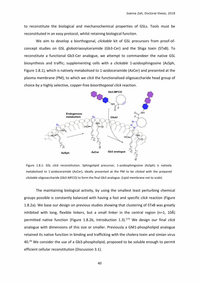
common groups are benzophenone, diazirine and aryl azide (Figure 1.7.5a-c). These small
chemical groups are activated by a flash of UV light, forming a reactive, radical species which
bonds covalently to any molecule (often heteroatoms) close to the tagged molecule of
interest. Their standard usage involves cellular incorporation of the photoactivatable small
molecule, UV activation, cell lysis, and visualisation or isolation of the lipid-protein partners
by clicking to a fluorophore or solid support respectively (Figure 1.7.5e). The use of
photoactivatable crosslinking groups has led to the identification of interaction partners of
plant glycoglycerolipids,168 phospholipids,169 sphingosine,170 cholesterol.171 A
photoactivatable, clickable GlcCer analogue was recently described, but no new interaction
partners have yet been reported.172
Figure 1.7.5: Photocrosslinkable groups, benzophenone (a), 3-phenyl-3-(trifluoromethyl)diazirine (b), and aryl
azide (c) with their reactive radical forms.167 (d) Confirmed photoactivatable sphingosine probe; protocol
involves live cell incubation, UV light activation, extraction and click with a fluorophore for visualisation, or
biotin-streptavidin beads for purification and mass spectrometry characterisation.170
1.8 Project aims
Biosynthesis of GSLs (Figure 1.4.4) is stepwise and likely under equilibrium, and one
gene does not code for one GSL but several. This makes genetic manipulation of a single GSL
impossible without offsetting the expression of other GSLs. Current methods to supplement
cells with pure long chain GSL species, a process here termed cellular reconstitution, are
extremely inefficient (Introduction 1.3).60–62 The study of GSL function is thus challenging and
new molecular tools are highly desirable. We aim to synthesise biomolecule analogues able
Joanna Zell, Doctoral thesis, 2018
40
to reconstitute the biological and mechanochemical properties of GSLs. Tools must be
reconstituted in an easy protocol, whilst retaining biological function.
We aim to develop a biorthogonal, clickable kit of GSL precursors from proof-of-
concept studies on GSL globotriaosylceramide (Gb3-Cer) and the Shiga toxin (STxB). To
reconstitute a functional Gb3-Cer analogue, we attempt to commandeer the native GSL
biosynthesis and traffic; supplementing cells with a clickable 1-azidosphingosine (AzSph,
Figure 1.8.1), which is natively metabolised to 1-azidoceramide (AzCer) and presented at the
plasma membrane (PM), to which we click the functionalised oligosaccharide head group of
choice by a highly selective, copper-free bioorthogonal click reaction.
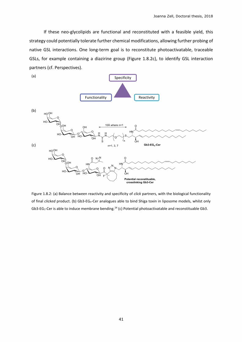
Figure 1.8.1: GSL click reconstitution. Sphingolipid precursor, 1-azidosphingosine (AzSph) is natively
metabolised to 1-azidoceramide (AzCer), ideally presented at the PM to be clicked with the prepared
clickable oligosaccharide (Gb3-MFCO) to form the final Gb3 analogue. (Lipid membrane not to scale)
The maintaining biological activity, by using the smallest least perturbing chemical
groups possible is constantly balanced with having a fast and specific click reaction (Figure
1.8.2a). We base our design on previous studies showing that clustering of STxB was greatly
inhibited with long, flexible linkers, but a small linker in the central region (n=1, 10Å)
permitted native function (Figure 1.8.2b, Introduction 1.3).173 We design our final click
analogue with dimensions of this size or smaller. Previously a GM1-phospholipid analogue
retained its native function in binding and trafficking with the cholera toxin and simian virus
40.29 We consider the use of a Gb3-phospholipid, proposed to be soluble enough to permit
efficient cellular reconstitution (Discussion 3.1).
Joanna Zell, Doctoral thesis, 2018
41
If these neo-glycolipids are functional and reconstituted with a feasible yield, this
strategy could potentially tolerate further chemical modifications, allowing further probing of
native GSL interactions. One long-term goal is to reconstitute photoactivatable, traceable
GSLs, for example containing a diazirine group (Figure 1.8.2c), to identify GSL interaction
partners (cf. Perspectives).
(a)
(b)
(c)
Figure 1.8.2: (a) Balance between reactivity and specificity of click partners, with the biological functionality
of final clicked product. (b) Gb3-EGn-Cer analogues able to bind Shiga toxin in liposome models, whilst only
Gb3-EG1-Cer is able to induce membrane bending.24 (c) Potential photoactivatable and reconstituable Gb3.
Specificity
Functionality Reactivity
Joanna Zell, Doctoral thesis, 2018
42
2 RESULTS
2.1 Synthesis of biomolecule analogues
2.1.1 Reconstitutable globotriose phospholipid
We aim to synthesise a Gb3 with retained native function that can be reintroduced
into cells in culture. Gb3-phospholipid analogues, or function-spacer-lipid (FSL) constructs,
have shown to retain similar biological activity as the natural Gb3-Cer with known binding
lectins: Shiga toxin (verotoxin1) and HIV-1.29,174,175 Crucially, phospholipids (PLs) are more
efficiently incorporated into cells in culture than GSLs (Introduction 1.3).60,62 Hence, we
proposed that a Gb3-PL would be a valuable tool. The spacer between glycan functional head
and lipid tail of the FSL must be biostable, meaning it must not be recognised and digested by
cellular clearance machinery. We require the spacer between the Gb3 head group and
phospholipid tail to be short to retain membrane bending properties (Introduction 1.3).24 This
contributes to the originality of this endeavour, since only long Gb3-phospholipids have been
synthesised in the literature.29,174,175
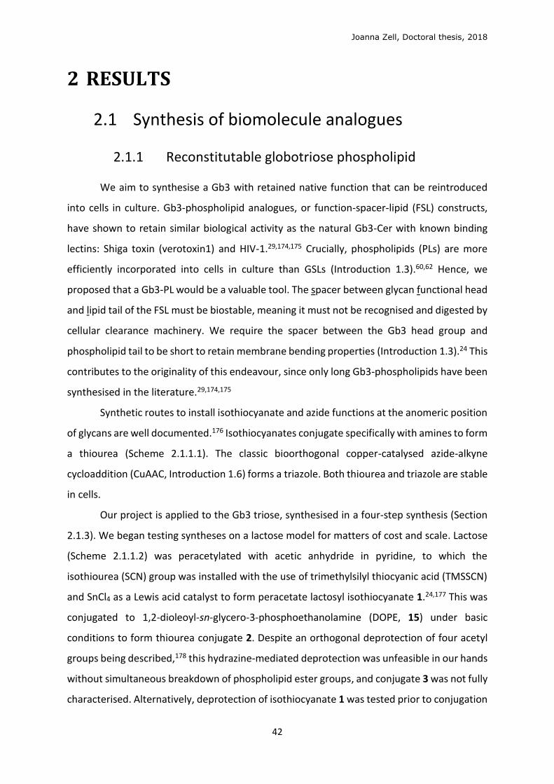
Synthetic routes to install isothiocyanate and azide functions at the anomeric position
of glycans are well documented.176 Isothiocyanates conjugate specifically with amines to form
a thiourea (Scheme 2.1.1.1). The classic bioorthogonal copper-catalysed azide-alkyne
cycloaddition (CuAAC, Introduction 1.6) forms a triazole. Both thiourea and triazole are stable
in cells.
Our project is applied to the Gb3 triose, synthesised in a four-step synthesis (Section
2.1.3). We began testing syntheses on a lactose model for matters of cost and scale. Lactose
(Scheme 2.1.1.2) was peracetylated with acetic anhydride in pyridine, to which the
isothiourea (SCN) group was installed with the use of trimethylsilyl thiocyanic acid (TMSSCN)
and SnCl4 as a Lewis acid catalyst to form peracetate lactosyl isothiocyanate 1.24,177 This was
conjugated to 1,2-dioleoyl-sn-glycero-3-phosphoethanolamine (DOPE, 15) under basic
conditions to form thiourea conjugate 2. Despite an orthogonal deprotection of four acetyl
groups being described,178 this hydrazine-mediated deprotection was unfeasible in our hands
without simultaneous breakdown of phospholipid ester groups, and conjugate 3 was not fully
characterised. Alternatively, deprotection of isothiocyanate 1 was tested prior to conjugation
Joanna Zell, Doctoral thesis, 2018
43
with DOPE, but isothiocyanate was unstable to methoxide conditions. It proved equally
impossible to install the isothiocyanate group without hydroxyl protecting groups.
Scheme 2.1.1.1: Biostable chemical conjugations
Scheme 2.1.1.2: Attempted synthesis of lactose-phospholipid analogue with isothiocyanate linkage.
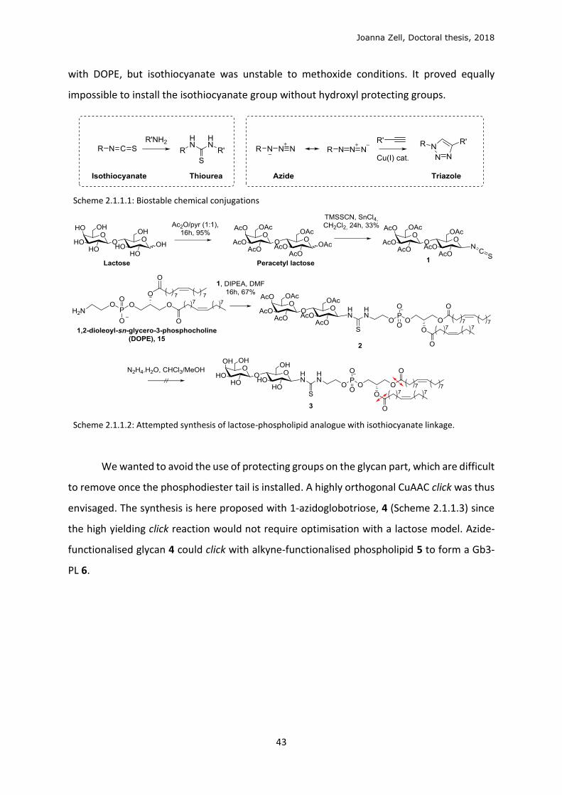
We wanted to avoid the use of protecting groups on the glycan part, which are difficult
to remove once the phosphodiester tail is installed. A highly orthogonal CuAAC click was thus
envisaged. The synthesis is here proposed with 1-azidoglobotriose, 4 (Scheme 2.1.1.3) since
the high yielding click reaction would not require optimisation with a lactose model. Azide-
functionalised glycan 4 could click with alkyne-functionalised phospholipid 5 to form a Gb3-
PL 6.
Joanna Zell, Doctoral thesis, 2018
44
Scheme 2.1.1.3: Proposed globotriose-phospholipid formed from click conjugation.
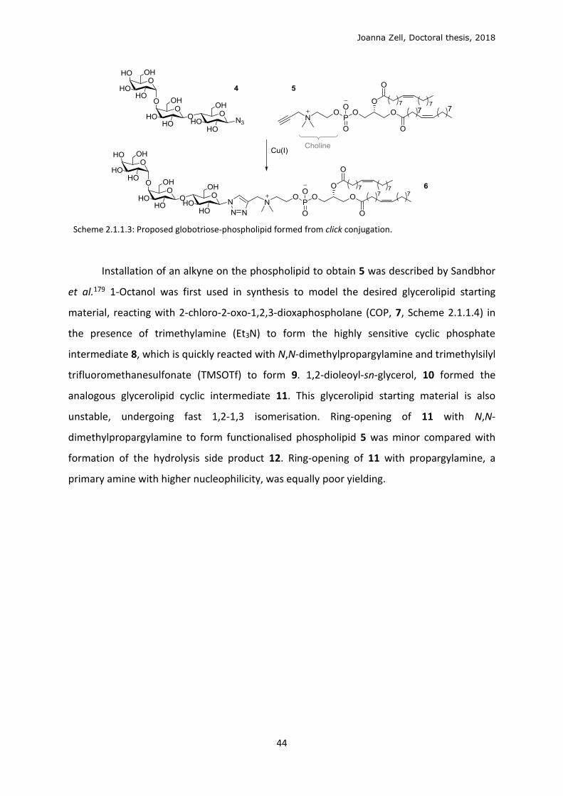
Installation of an alkyne on the phospholipid to obtain 5 was described by Sandbhor
et al.179 1-Octanol was first used in synthesis to model the desired glycerolipid starting
material, reacting with 2-chloro-2-oxo-1,2,3-dioxaphospholane (COP, 7, Scheme 2.1.1.4) in
the presence of trimethylamine (Et3N) to form the highly sensitive cyclic phosphate
intermediate 8, which is quickly reacted with N,N-dimethylpropargylamine and trimethylsilyl
trifluoromethanesulfonate (TMSOTf) to form 9. 1,2-dioleoyl-sn-glycerol, 10 formed the
analogous glycerolipid cyclic intermediate 11. This glycerolipid starting material is also
unstable, undergoing fast 1,2-1,3 isomerisation. Ring-opening of 11 with N,N-
dimethylpropargylamine to form functionalised phospholipid 5 was minor compared with
formation of the hydrolysis side product 12. Ring-opening of 11 with propargylamine, a
primary amine with higher nucleophilicity, was equally poor yielding.
Joanna Zell, Doctoral thesis, 2018
45
Scheme 2.1.1.4: Attempted synthesis of alkyne-functionalised phospholipid and major hydrolysis side
product.
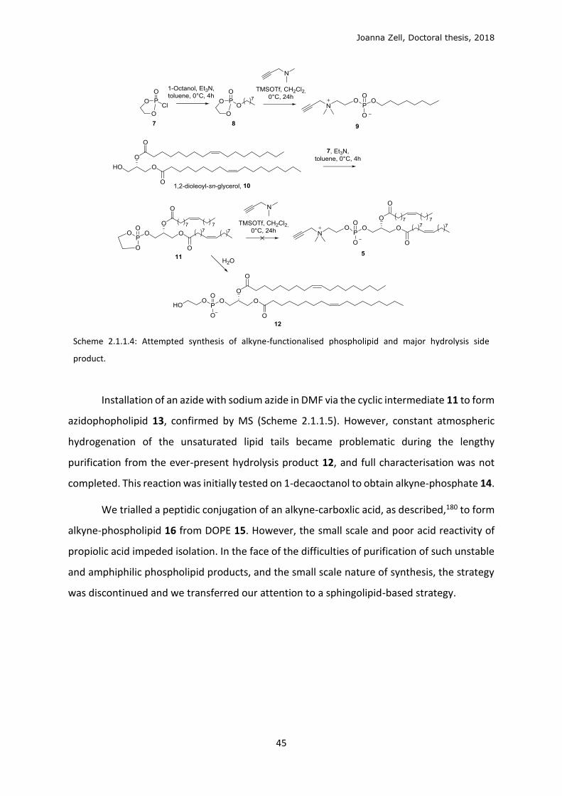
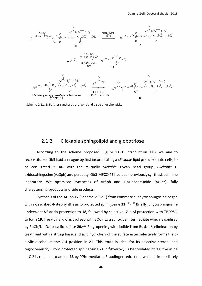
Installation of an azide with sodium azide in DMF via the cyclic intermediate 11 to form
azidophopholipid 13, confirmed by MS (Scheme 2.1.1.5). However, constant atmospheric
hydrogenation of the unsaturated lipid tails became problematic during the lengthy
purification from the ever-present hydrolysis product 12, and full characterisation was not
completed. This reaction was initially tested on 1-decaoctanol to obtain alkyne-phosphate 14.
We trialled a peptidic conjugation of an alkyne-carboxlic acid, as described,180 to form
alkyne-phospholipid 16 from DOPE 15. However, the small scale and poor acid reactivity of
propiolic acid impeded isolation. In the face of the difficulties of purification of such unstable
and amphiphilic phospholipid products, and the small scale nature of synthesis, the strategy
was discontinued and we transferred our attention to a sphingolipid-based strategy.
Joanna Zell, Doctoral thesis, 2018
46
Scheme 2.1.1.5: Further syntheses of alkyne and azide phospholipids.
2.1.2 Clickable sphingolipid and globotriose
According to the scheme proposed (Figure 1.8.1, Introduction 1.8), we aim to
reconstitute a Gb3 lipid analogue by first incorporating a clickable lipid precursor into cells, to
be conjugated in situ with the mutually clickable glycan head group. Clickable 1-
azidosphingosine (AzSph) and peracetyl Gb3-MFCO 47 had been previously synthesised in the
laboratory. We optimised syntheses of AzSph and 1-azidoceramide (AzCer), fully
characterising products and side products.
Synthesis of the AzSph 17 (Scheme 2.1.2.1) from commercial phytosphingosine began
with a described 4-step synthesis to protected sphingosine 21.181,182 Briefly, phytosphingosine
underwent N2-azido protection to 18, followed by selective O1-silyl protection with TBDPSCl
to form 19. The vicinal diol is cyclised with SOCl2 to a sulfoxide intermediate which is oxidised
by RuCl3/NaIO4 to cyclic sulfate 20.183 Ring-opening with iodide from Bu4NI, β-elimination by
treatment with a strong base, and acid hydrolysis of the sulfate ester selectively forms the E-
allylic alcohol at the C-4 position in 21. This route is ideal for its selective stereo- and
regiochemistry. From protected sphingosine 21, O3-hydroxyl is benzoylated to 22, the azide
at C-2 is reduced to amine 23 by PPh3-mediated Staudinger reduction, which is immediately
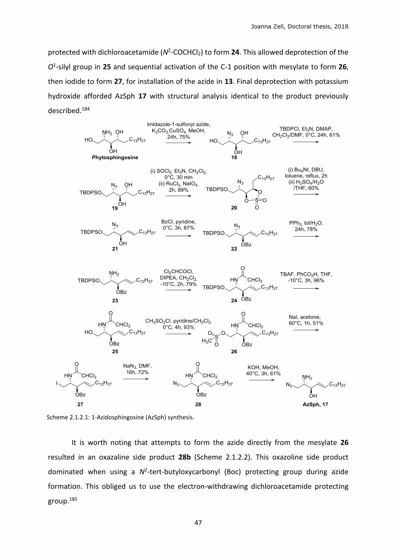
Joanna Zell, Doctoral thesis, 2018
47
protected with dichloroacetamide (N2-COCHCl2) to form 24. This allowed deprotection of the
O1-silyl group in 25 and sequential activation of the C-1 position with mesylate to form 26,
then iodide to form 27, for installation of the azide in 13. Final deprotection with potassium
hydroxide afforded AzSph 17 with structural analysis identical to the product previously
described.184
Scheme 2.1.2.1: 1-Azidosphingosine (AzSph) synthesis.
It is worth noting that attempts to form the azide directly from the mesylate 26
resulted in an oxazaline side product 28b (Scheme 2.1.2.2). This oxazoline side product
dominated when using a N2-tert-butyloxycarbonyl (Boc) protecting group during azide
formation. This obliged us to use the electron-withdrawing dichloroacetamide protecting
group.185
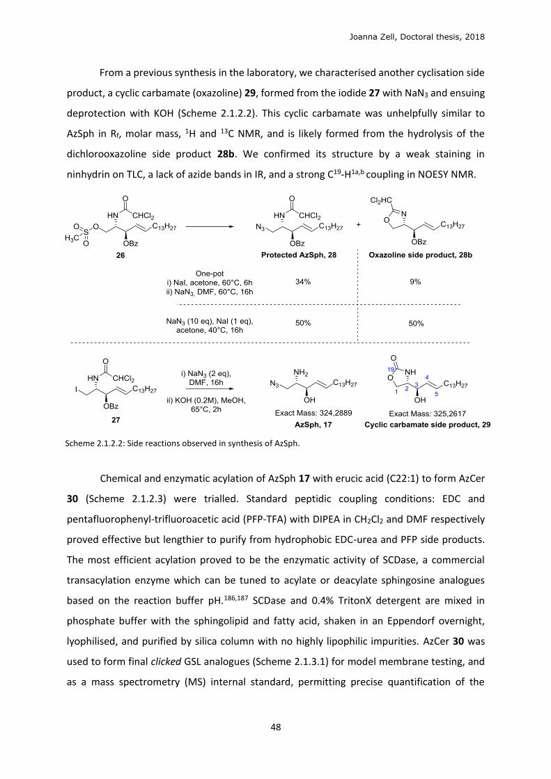
Joanna Zell, Doctoral thesis, 2018
48
From a previous synthesis in the laboratory, we characterised another cyclisation side
product, a cyclic carbamate (oxazoline) 29, formed from the iodide 27 with NaN3 and ensuing
deprotection with KOH (Scheme 2.1.2.2). This cyclic carbamate was unhelpfully similar to
AzSph in Rf, molar mass, 1H and 13C NMR, and is likely formed from the hydrolysis of the
dichlorooxazoline side product 28b. We confirmed its structure by a weak staining in
ninhydrin on TLC, a lack of azide bands in IR, and a strong C19-H1a,b coupling in NOESY NMR.
Scheme 2.1.2.2: Side reactions observed in synthesis of AzSph.
Chemical and enzymatic acylation of AzSph 17 with erucic acid (C22:1) to form AzCer
30 (Scheme 2.1.2.3) were trialled. Standard peptidic coupling conditions: EDC and
pentafluorophenyl-trifluoroacetic acid (PFP-TFA) with DIPEA in CH2Cl2 and DMF respectively
proved effective but lengthier to purify from hydrophobic EDC-urea and PFP side products.
The most efficient acylation proved to be the enzymatic activity of SCDase, a commercial
transacylation enzyme which can be tuned to acylate or deacylate sphingosine analogues
based on the reaction buffer pH.186,187 SCDase and 0.4% TritonX detergent are mixed in
phosphate buffer with the sphingolipid and fatty acid, shaken in an Eppendorf overnight,
lyophilised, and purified by silica column with no highly lipophilic impurities. AzCer 30 was
used to form final clicked GSL analogues (Scheme 2.1.3.1) for model membrane testing, and
as a mass spectrometry (MS) internal standard, permitting precise quantification of the
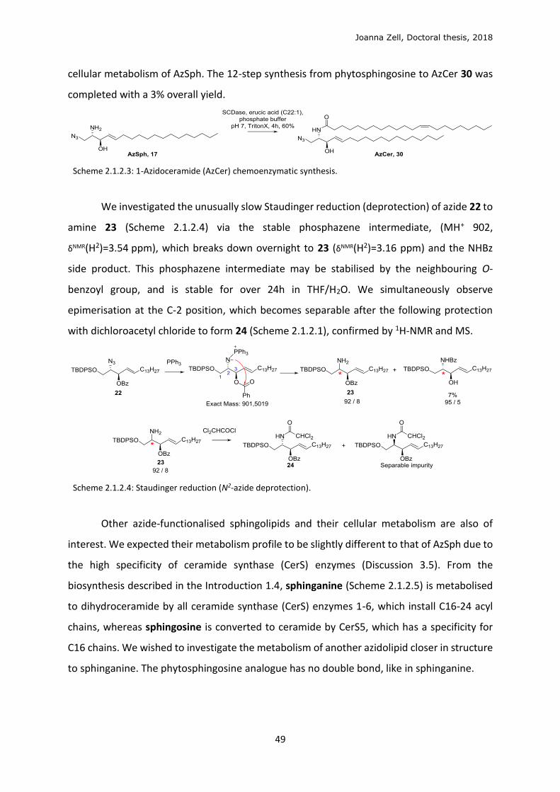
Joanna Zell, Doctoral thesis, 2018
49
cellular metabolism of AzSph. The 12-step synthesis from phytosphingosine to AzCer 30 was
completed with a 3% overall yield.
Scheme 2.1.2.3: 1-Azidoceramide (AzCer) chemoenzymatic synthesis.
We investigated the unusually slow Staudinger reduction (deprotection) of azide 22 to
amine 23 (Scheme 2.1.2.4) via the stable phosphazene intermediate, (MH+ 902,
δNMR(H2)=3.54 ppm), which breaks down overnight to 23 (δNMR(H2)=3.16 ppm) and the NHBz
side product. This phosphazene intermediate may be stabilised by the neighbouring O-
benzoyl group, and is stable for over 24h in THF/H2O. We simultaneously observe
epimerisation at the C-2 position, which becomes separable after the following protection
with dichloroacetyl chloride to form 24 (Scheme 2.1.2.1), confirmed by 1H-NMR and MS.
Scheme 2.1.2.4: Staudinger reduction (N2-azide deprotection).
Other azide-functionalised sphingolipids and their cellular metabolism are also of
interest. We expected their metabolism profile to be slightly different to that of AzSph due to
the high specificity of ceramide synthase (CerS) enzymes (Discussion 3.5). From the
biosynthesis described in the Introduction 1.4, sphinganine (Scheme 2.1.2.5) is metabolised
to dihydroceramide by all ceramide synthase (CerS) enzymes 1-6, which install C16-24 acyl
chains, whereas sphingosine is converted to ceramide by CerS5, which has a specificity for
C16 chains. We wished to investigate the metabolism of another azidolipid closer in structure
to sphinganine. The phytosphingosine analogue has no double bond, like in sphinganine.
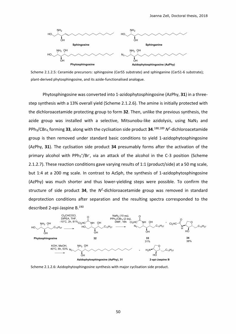
Joanna Zell, Doctoral thesis, 2018
50
Scheme 2.1.2.5: Ceramide precursors: sphingosine (CerS5 substrate) and sphinganine (CerS1-6 substrate);
plant-derived phytosphingosine, and its azide-functionalised analogue.
Phytosphingosine was converted into 1-azidophytosphingosine (AzPhy, 31) in a three-
step synthesis with a 13% overall yield (Scheme 2.1.2.6). The amine is initially protected with
the dichloroacetamide protecting group to form 32. Then, unlike the previous synthesis, the
azide group was installed with a selective, Mitsunobu-like azidolysis, using NaN3 and
PPh3/CBr4, forming 33, along with the cyclisation side product 34.188,189 N2-dichloroacetamide
group is then removed under standard basic conditions to yield 1-azidophytosphingosine
(AzPhy, 31). The cyclisation side product 34 presumably forms after the activation of the
primary alcohol with PPh3+/Br-, via an attack of the alcohol in the C-3 position (Scheme
2.1.2.7). These reaction conditions gave varying results of 1:1 (product/side) at a 50 mg scale,
but 1:4 at a 200 mg scale. In contrast to AzSph, the synthesis of 1-azidophytosphingosine
(AzPhy) was much shorter and thus lower-yielding steps were possible. To confirm the
structure of side product 34, the N2-dichloroacetamide group was removed in standard
deprotection conditions after separation and the resulting spectra corresponded to the
described 2-epi-Jaspine B.190
Scheme 2.1.2.6: Azidophytosphingosine synthesis with major cyclisation side product.
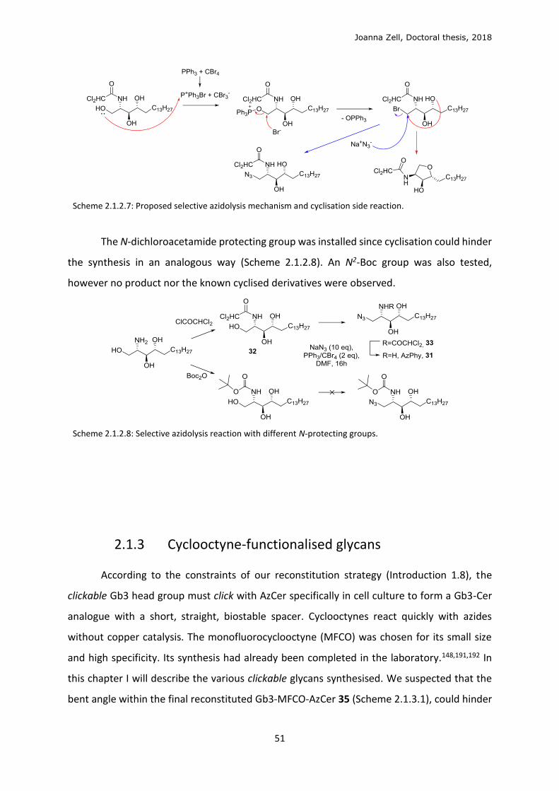
Joanna Zell, Doctoral thesis, 2018
51
Scheme 2.1.2.7: Proposed selective azidolysis mechanism and cyclisation side reaction.
The N-dichloroacetamide protecting group was installed since cyclisation could hinder
the synthesis in an analogous way (Scheme 2.1.2.8). An N2-Boc group was also tested,
however no product nor the known cyclised derivatives were observed.
Scheme 2.1.2.8: Selective azidolysis reaction with different N-protecting groups.
2.1.3 Cyclooctyne-functionalised glycans
According to the constraints of our reconstitution strategy (Introduction 1.8), the
clickable Gb3 head group must click with AzCer specifically in cell culture to form a Gb3-Cer
analogue with a short, straight, biostable spacer. Cyclooctynes react quickly with azides
without copper catalysis. The monofluorocyclooctyne (MFCO) was chosen for its small size
and high specificity. Its synthesis had already been completed in the laboratory.148,191,192 In
this chapter I will describe the various clickable glycans synthesised. We suspected that the
bent angle within the final reconstituted Gb3-MFCO-AzCer 35 (Scheme 2.1.3.1), could hinder
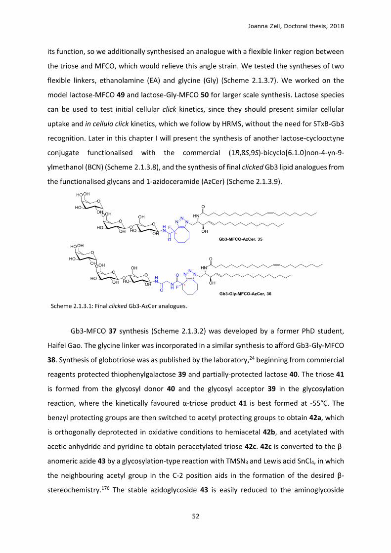
Joanna Zell, Doctoral thesis, 2018
52
its function, so we additionally synthesised an analogue with a flexible linker region between
the triose and MFCO, which would relieve this angle strain. We tested the syntheses of two
flexible linkers, ethanolamine (EA) and glycine (Gly) (Scheme 2.1.3.7). We worked on the
model lactose-MFCO 49 and lactose-Gly-MFCO 50 for larger scale synthesis. Lactose species
can be used to test initial cellular click kinetics, since they should present similar cellular
uptake and in cellulo click kinetics, which we follow by HRMS, without the need for STxB-Gb3
recognition. Later in this chapter I will present the synthesis of another lactose-cyclooctyne
conjugate functionalised with the commercial (1R,8S,9S)-bicyclo[6.1.0]non-4-yn-9-
ylmethanol (BCN) (Scheme 2.1.3.8), and the synthesis of final clicked Gb3 lipid analogues from
the functionalised glycans and 1-azidoceramide (AzCer) (Scheme 2.1.3.9).
Scheme 2.1.3.1: Final clicked Gb3-AzCer analogues.
Gb3-MFCO 37 synthesis (Scheme 2.1.3.2) was developed by a former PhD student,
Haifei Gao. The glycine linker was incorporated in a similar synthesis to afford Gb3-Gly-MFCO
38. Synthesis of globotriose was as published by the laboratory,24 beginning from commercial
reagents protected thiophenylgalactose 39 and partially-protected lactose 40. The triose 41
is formed from the glycosyl donor 40 and the glycosyl acceptor 39 in the glycosylation
reaction, where the kinetically favoured α-triose product 41 is best formed at -55°C. The
benzyl protecting groups are then switched to acetyl protecting groups to obtain 42a, which
is orthogonally deprotected in oxidative conditions to hemiacetal 42b, and acetylated with
acetic anhydride and pyridine to obtain peracetylated triose 42c. 42c is converted to the β-
anomeric azide 43 by a glycosylation-type reaction with TMSN3 and Lewis acid SnCl4, in which
the neighbouring acetyl group in the C-2 position aids in the formation of the desired β-
stereochemistry.176 The stable azidoglycoside 43 is easily reduced to the aminoglycoside
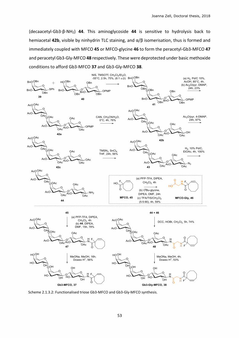
Joanna Zell, Doctoral thesis, 2018
53
(decaacetyl-Gb3-β-NH2) 44. This aminoglycoside 44 is sensitive to hydrolysis back to
hemiacetal 42b, visible by ninhydrin TLC staining, and α/β isomerisation, thus is formed and
immediately coupled with MFCO 45 or MFCO-glycine 46 to form the peracetyl-Gb3-MFCO 47
and peracetyl Gb3-Gly-MFCO 48 respectively. These were deprotected under basic methoxide
conditions to afford Gb3-MFCO 37 and Gb3-Gly-MFCO 38.
Scheme 2.1.3.2: Functionalised triose Gb3-MFCO and Gb3-Gly-MFCO synthesis.
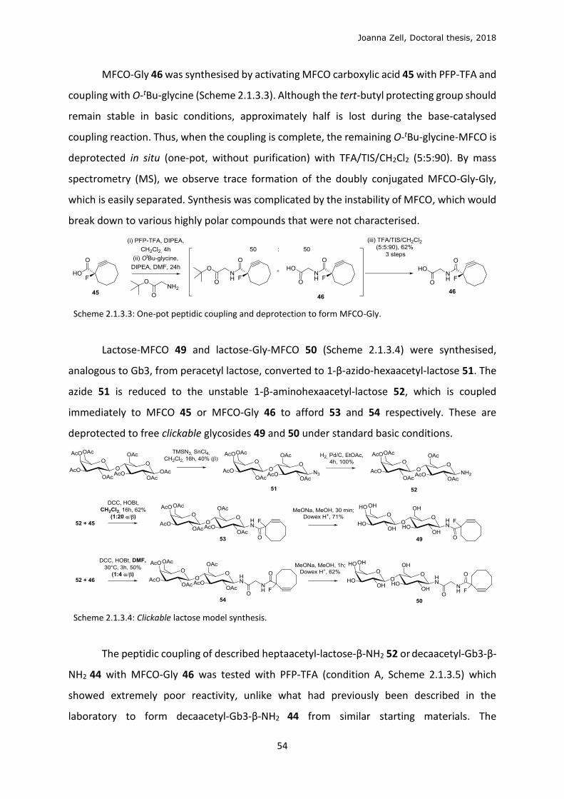
Joanna Zell, Doctoral thesis, 2018
54
MFCO-Gly 46 was synthesised by activating MFCO carboxylic acid 45 with PFP-TFA and
coupling with O-tBu-glycine (Scheme 2.1.3.3). Although the tert-butyl protecting group should
remain stable in basic conditions, approximately half is lost during the base-catalysed
coupling reaction. Thus, when the coupling is complete, the remaining O-tBu-glycine-MFCO is
deprotected in situ (one-pot, without purification) with TFA/TIS/CH2Cl2 (5:5:90). By mass
spectrometry (MS), we observe trace formation of the doubly conjugated MFCO-Gly-Gly,
which is easily separated. Synthesis was complicated by the instability of MFCO, which would
break down to various highly polar compounds that were not characterised.
Scheme 2.1.3.3: One-pot peptidic coupling and deprotection to form MFCO-Gly.
Lactose-MFCO 49 and lactose-Gly-MFCO 50 (Scheme 2.1.3.4) were synthesised,
analogous to Gb3, from peracetyl lactose, converted to 1-β-azido-hexaacetyl-lactose 51. The
azide 51 is reduced to the unstable 1-β-aminohexaacetyl-lactose 52, which is coupled
immediately to MFCO 45 or MFCO-Gly 46 to afford 53 and 54 respectively. These are
deprotected to free clickable glycosides 49 and 50 under standard basic conditions.
Scheme 2.1.3.4: Clickable lactose model synthesis.
The peptidic coupling of described heptaacetyl-lactose-β-NH2 52 or decaacetyl-Gb3-β-
NH2 44 with MFCO-Gly 46 was tested with PFP-TFA (condition A, Scheme 2.1.3.5) which
showed extremely poor reactivity, unlike what had previously been described in the
laboratory to form decaacetyl-Gb3-β-NH2 44 from similar starting materials. The
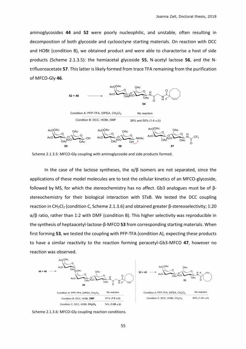
Joanna Zell, Doctoral thesis, 2018
55
aminoglycosides 44 and 52 were poorly nucleophilic, and unstable, often resulting in
decomposition of both glycoside and cyclooctyne starting materials. On reaction with DCC
and HOBt (condition B), we obtained product and were able to characterise a host of side
products (Scheme 2.1.3.5): the hemiacetal glycoside 55, N-acetyl lactose 56, and the N-
trifluoroacetate 57. This latter is likely formed from trace TFA remaining from the purification
of MFCO-Gly 46.
Scheme 2.1.3.5: MFCO-Gly coupling with aminoglycoside and side products formed.
In the case of the lactose syntheses, the α/β isomers are not separated, since the
applications of these model molecules are to test the cellular kinetics of an MFCO-glycoside,
followed by MS, for which the stereochemistry has no affect. Gb3 analogues must be of β-
stereochemistry for their biological interaction with STxB. We tested the DCC coupling
reaction in CH2Cl2 (condition C, Scheme 2.1.3.6) and obtained greater β-stereoselectivity; 1:20
α/β ratio, rather than 1:2 with DMF (condition B). This higher selectivity was reproducible in
the synthesis of heptaacetyl-lactose-β-MFCO 53 from corresponding starting materials. When
first forming 53, we tested the coupling with PFP-TFA (condition A), expecting these products
to have a similar reactivity to the reaction forming peracetyl-Gb3-MFCO 47, however no
reaction was observed.
Scheme 2.1.3.6: MFCO-Gly coupling reaction conditions.
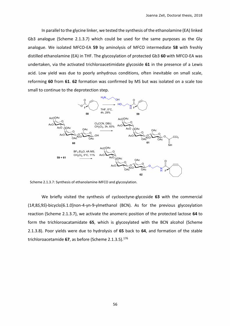
Joanna Zell, Doctoral thesis, 2018
56
In parallel to the glycine linker, we tested the synthesis of the ethanolamine (EA) linked
Gb3 analogue (Scheme 2.1.3.7) which could be used for the same purposes as the Gly
analogue. We isolated MFCO-EA 59 by aminolysis of MFCO intermediate 58 with freshly
distilled ethanolamine (EA) in THF. The glycosylation of protected Gb3 60 with MFCO-EA was
undertaken, via the activated trichloroacetimidate glycoside 61 in the presence of a Lewis
acid. Low yield was due to poorly anhydrous conditions, often inevitable on small scale,
reforming 60 from 61. 62 formation was confirmed by MS but was isolated on a scale too
small to continue to the deprotection step.
Scheme 2.1.3.7: Synthesis of ethanolamine-MFCO and glycosylation.
We briefly visited the synthesis of cyclooctyne-glycoside 63 with the commercial
(1R,8S,9S)-bicyclo[6.1.0]non-4-yn-9-ylmethanol (BCN). As for the previous glycosylation
reaction (Scheme 2.1.3.7), we activate the anomeric position of the protected lactose 64 to
form the trichloroacatamidate 65, which is glycosylated with the BCN alcohol (Scheme
2.1.3.8). Poor yields were due to hydrolysis of 65 back to 64, and formation of the stable
trichloroacetamide 67, as before (Scheme 2.1.3.5).176
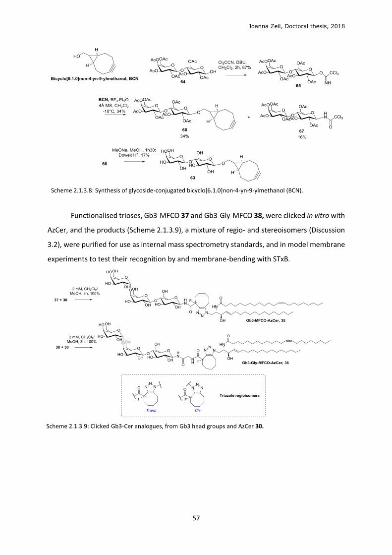
Joanna Zell, Doctoral thesis, 2018
57
Scheme 2.1.3.8: Synthesis of glycoside-conjugated bicyclo[6.1.0]non-4-yn-9-ylmethanol (BCN).
Functionalised trioses, Gb3-MFCO 37 and Gb3-Gly-MFCO 38, were clicked in vitro with
AzCer, and the products (Scheme 2.1.3.9), a mixture of regio- and stereoisomers (Discussion
3.2), were purified for use as internal mass spectrometry standards, and in model membrane
experiments to test their recognition by and membrane-bending with STxB.
Scheme 2.1.3.9: Clicked Gb3-Cer analogues, from Gb3 head groups and AzCer 30.

Joanna Zell, Doctoral thesis, 2018
58
2.2 Biological validation of analogues
2.2.1 Model membrane validation
We conducted preliminary validation of the biological function of final clicked Gb3
analogues by testing their binding and membrane bending properties with the B-subunit of
the Shiga toxin (STxB) (Introduction 1.2). These mechanochemical properties of the GSL
analogues are traditionally tested in model membrane systems called giant unilamellar
vesicles (GUVs), or liposomes. This robust technique shows spontaneous, dynamic tubule
formation of STxB bound to the natural Gb3 in the liposome bilayer, indicating that GL-Lect
dependant endocytic interactions could occur in a cellular environment. We use this
technique to test each final synthetic product. To follow the process by fluorescence
microscopy, we use STxB-Alexa488, where the green fluorophore (Alexa488) is conjugated via
a lysine-specific sulfodicholorphenol (SDP) functionality. A red fluorescent phospholipid,
Rhod-SDP-PE, is used to stain the liposome (Figure 2.2.1.1).
Figure 2.2.1.1: Canonical STxB-Cy3(wild type, red) binding and invagination of membrane in GUVs (64 mol%
DOPC; 30mol% chol; 5mol% porcine Gb3; 1mol% BodipyFL-C5-HPC (green)).27 Schematic structures of STxB bound
with 15 Gb3,193 STxB-Alexa488 with Gb3 binding sites, and Lissamine Rhodamine phosphoethanolamine
(Rhod-SDP-PE).
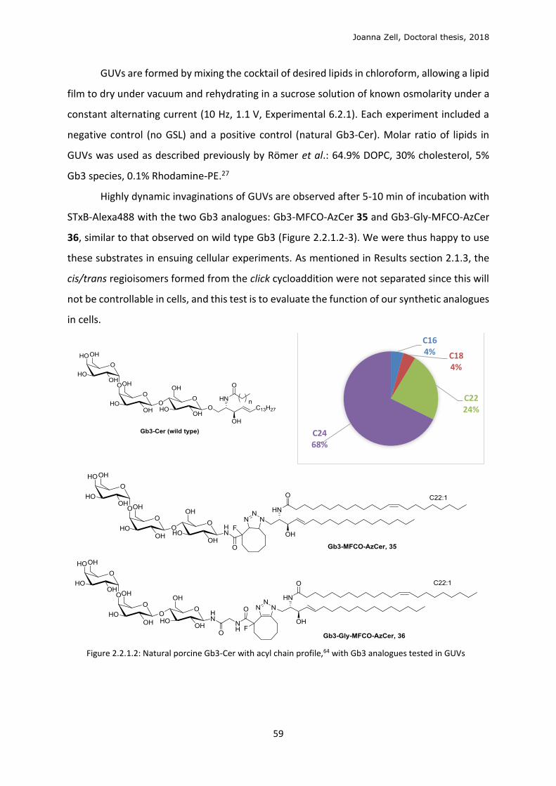
Joanna Zell, Doctoral thesis, 2018
59
GUVs are formed by mixing the cocktail of desired lipids in chloroform, allowing a lipid
film to dry under vacuum and rehydrating in a sucrose solution of known osmolarity under a
constant alternating current (10 Hz, 1.1 V, Experimental 6.2.1). Each experiment included a
negative control (no GSL) and a positive control (natural Gb3-Cer). Molar ratio of lipids in
GUVs was used as described previously by Römer et al.: 64.9% DOPC, 30% cholesterol, 5%
Gb3 species, 0.1% Rhodamine-PE.27
Highly dynamic invaginations of GUVs are observed after 5-10 min of incubation with
STxB-Alexa488 with the two Gb3 analogues: Gb3-MFCO-AzCer 35 and Gb3-Gly-MFCO-AzCer
36, similar to that observed on wild type Gb3 (Figure 2.2.1.2-3). We were thus happy to use
these substrates in ensuing cellular experiments. As mentioned in Results section 2.1.3, the
cis/trans regioisomers formed from the click cycloaddition were not separated since this will
not be controllable in cells, and this test is to evaluate the function of our synthetic analogues
in cells.
Figure 2.2.1.2: Natural porcine Gb3-Cer with acyl chain profile,64 with Gb3 analogues tested in GUVs
C164% C18
4%
C2224%
C2468%
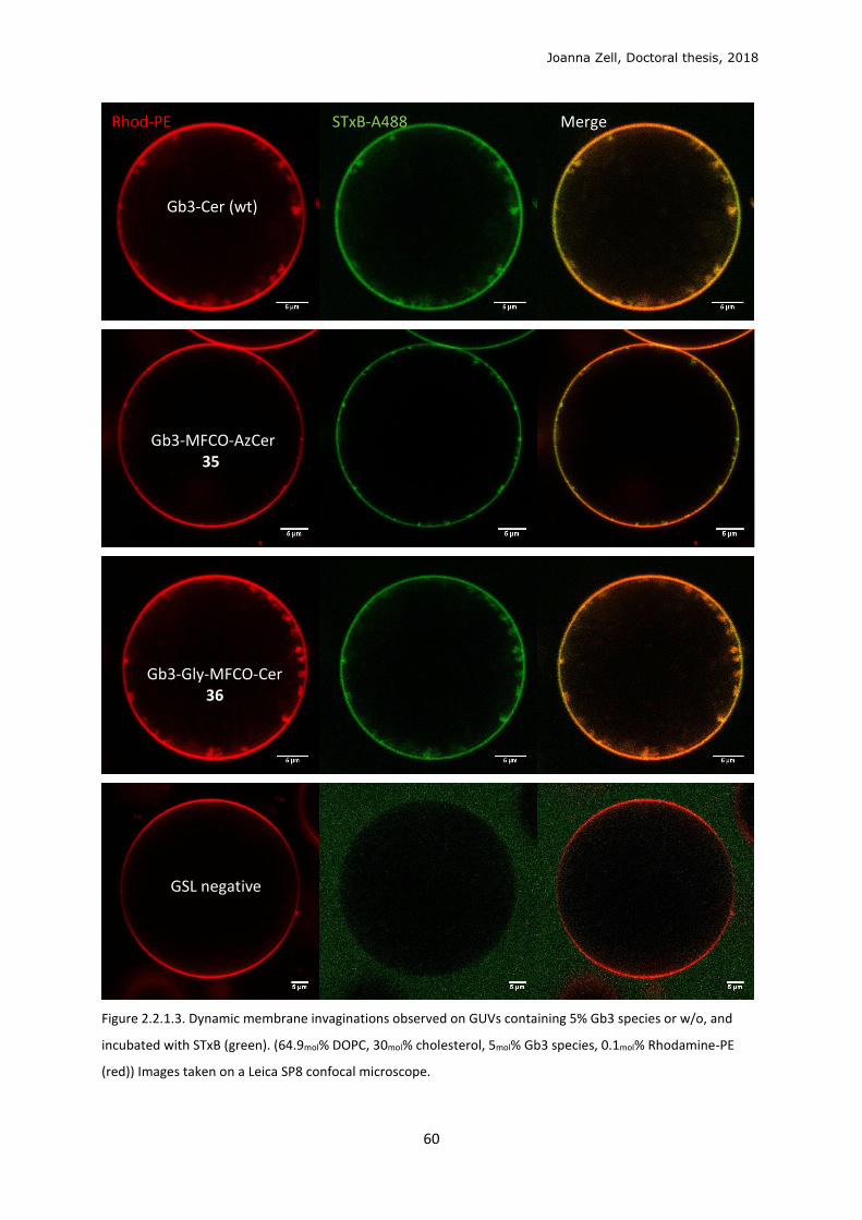
Joanna Zell, Doctoral thesis, 2018
60
Figure 2.2.1.3. Dynamic membrane invaginations observed on GUVs containing 5% Gb3 species or w/o, and
incubated with STxB (green). (64.9mol% DOPC, 30mol% cholesterol, 5mol% Gb3 species, 0.1mol% Rhodamine-PE
(red)) Images taken on a Leica SP8 confocal microscope.
Rhod-PE STxB-A488 Merge Gb3-Cer (wt) Gb3-MFCO-AzCer 35 Gb3-Gly-MFCO-Cer 36 GSL negative
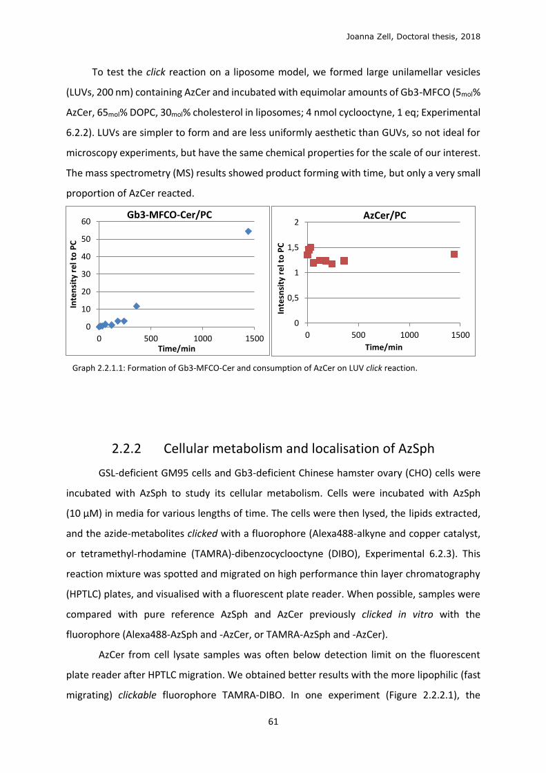
Joanna Zell, Doctoral thesis, 2018
61
To test the click reaction on a liposome model, we formed large unilamellar vesicles
(LUVs, 200 nm) containing AzCer and incubated with equimolar amounts of Gb3-MFCO (5mol%
AzCer, 65mol% DOPC, 30mol% cholesterol in liposomes; 4 nmol cyclooctyne, 1 eq; Experimental
6.2.2). LUVs are simpler to form and are less uniformly aesthetic than GUVs, so not ideal for
microscopy experiments, but have the same chemical properties for the scale of our interest.
The mass spectrometry (MS) results showed product forming with time, but only a very small
proportion of AzCer reacted.
Graph 2.2.1.1: Formation of Gb3-MFCO-Cer and consumption of AzCer on LUV click reaction.
2.2.2 Cellular metabolism and localisation of AzSph
GSL-deficient GM95 cells and Gb3-deficient Chinese hamster ovary (CHO) cells were
incubated with AzSph to study its cellular metabolism. Cells were incubated with AzSph
(10 μM) in media for various lengths of time. The cells were then lysed, the lipids extracted,
and the azide-metabolites clicked with a fluorophore (Alexa488-alkyne and copper catalyst,
or tetramethyl-rhodamine (TAMRA)-dibenzocyclooctyne (DIBO), Experimental 6.2.3). This
reaction mixture was spotted and migrated on high performance thin layer chromatography
(HPTLC) plates, and visualised with a fluorescent plate reader. When possible, samples were
compared with pure reference AzSph and AzCer previously clicked in vitro with the
fluorophore (Alexa488-AzSph and -AzCer, or TAMRA-AzSph and -AzCer).
AzCer from cell lysate samples was often below detection limit on the fluorescent
plate reader after HPTLC migration. We obtained better results with the more lipophilic (fast
migrating) clickable fluorophore TAMRA-DIBO. In one experiment (Figure 2.2.2.1), the
0
10
20
30
40
50
60
0 500 1000 1500
Inte
nsi
ty r
el t
o P
C
Time/min
Gb3-MFCO-Cer/PC
0
0,5
1
1,5
2
0 500 1000 1500
Inte
snsi
ty r
el t
o P
C
Time/min
AzCer/PC
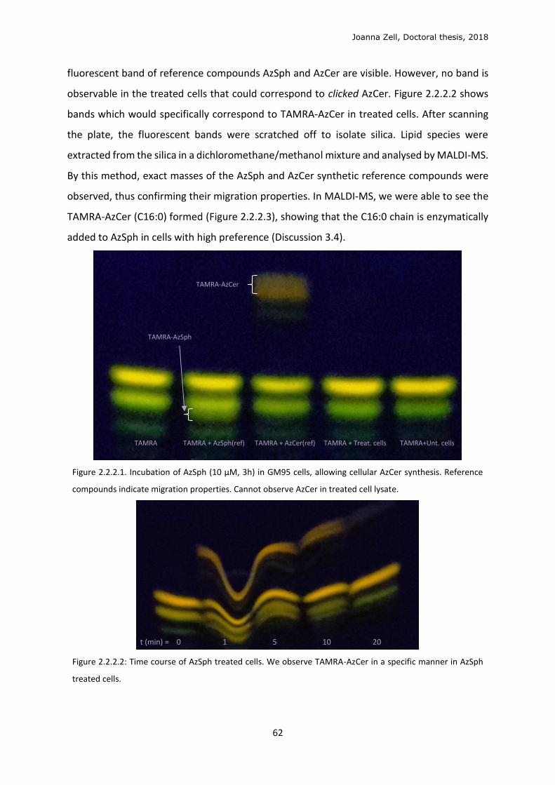
Joanna Zell, Doctoral thesis, 2018
62
fluorescent band of reference compounds AzSph and AzCer are visible. However, no band is
observable in the treated cells that could correspond to clicked AzCer. Figure 2.2.2.2 shows
bands which would specifically correspond to TAMRA-AzCer in treated cells. After scanning
the plate, the fluorescent bands were scratched off to isolate silica. Lipid species were
extracted from the silica in a dichloromethane/methanol mixture and analysed by MALDI-MS.
By this method, exact masses of the AzSph and AzCer synthetic reference compounds were
observed, thus confirming their migration properties. In MALDI-MS, we were able to see the
TAMRA-AzCer (C16:0) formed (Figure 2.2.2.3), showing that the C16:0 chain is enzymatically
added to AzSph in cells with high preference (Discussion 3.4).
Figure 2.2.2.1. Incubation of AzSph (10 μM, 3h) in GM95 cells, allowing cellular AzCer synthesis. Reference
compounds indicate migration properties. Cannot observe AzCer in treated cell lysate.
Figure 2.2.2.2: Time course of AzSph treated cells. We observe TAMRA-AzCer in a specific manner in AzSph
treated cells.
TAMRA TAMRA + AzSph(ref) TAMRA + AzCer(ref) TAMRA + Treat. cells TAMRA+Unt. cells
t (min) = 0 1 5 10 20
TAMRA-AzSph
TAMRA-AzCer
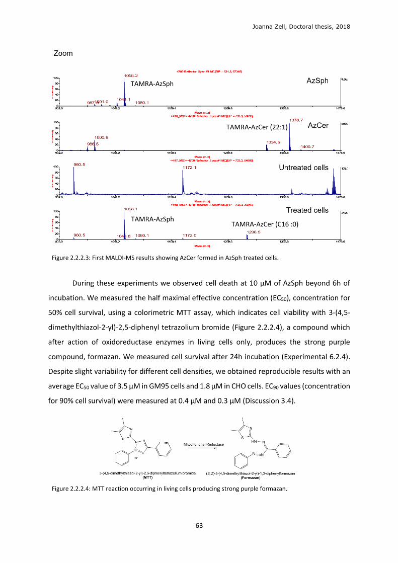
Joanna Zell, Doctoral thesis, 2018
63
Figure 2.2.2.3: First MALDI-MS results showing AzCer formed in AzSph treated cells.
During these experiments we observed cell death at 10 µM of AzSph beyond 6h of
incubation. We measured the half maximal effective concentration (EC50), concentration for
50% cell survival, using a colorimetric MTT assay, which indicates cell viability with 3-(4,5-
dimethylthiazol-2-yl)-2,5-diphenyl tetrazolium bromide (Figure 2.2.2.4), a compound which
after action of oxidoreductase enzymes in living cells only, produces the strong purple
compound, formazan. We measured cell survival after 24h incubation (Experimental 6.2.4).
Despite slight variability for different cell densities, we obtained reproducible results with an
average EC50 value of 3.5 μM in GM95 cells and 1.8 μM in CHO cells. EC90 values (concentration
for 90% cell survival) were measured at 0.4 μM and 0.3 μM (Discussion 3.4).
Figure 2.2.2.4: MTT reaction occurring in living cells producing strong purple formazan.
TAMRA-AzSph
TAMRA-AzCer (22:1)
TAMRA-AzSph TAMRA-AzCer (C16 :0)
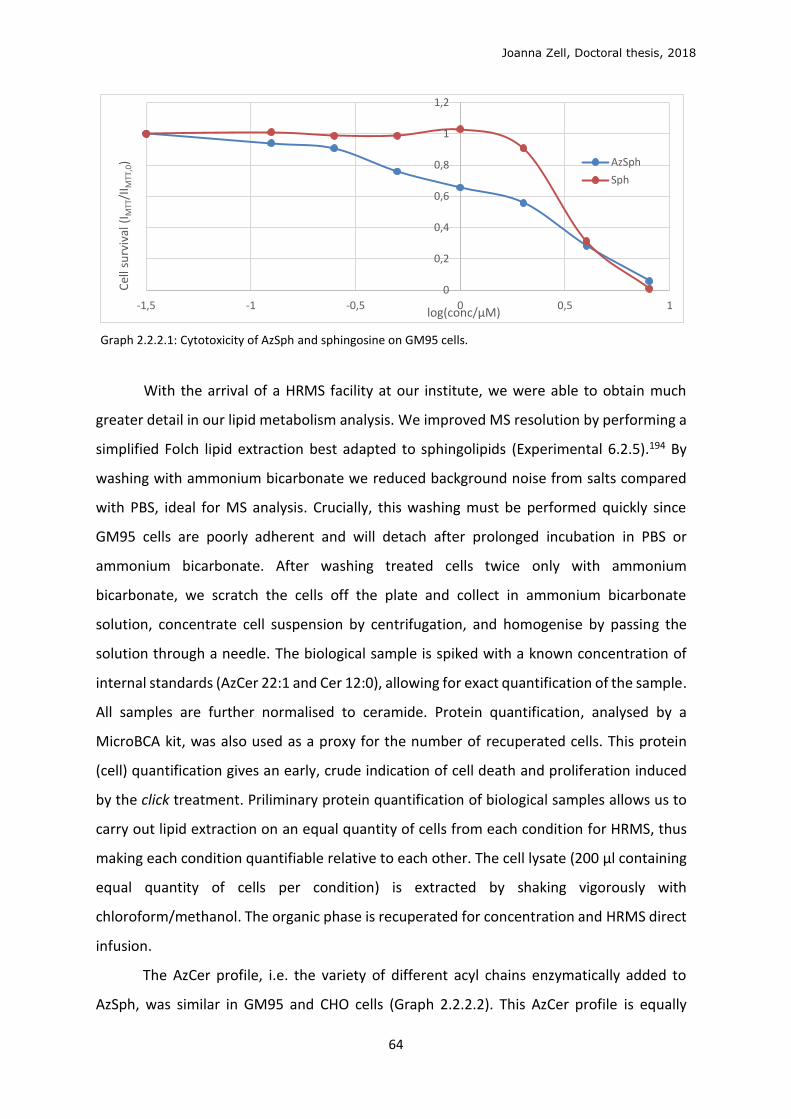
Joanna Zell, Doctoral thesis, 2018
64
Graph 2.2.2.1: Cytotoxicity of AzSph and sphingosine on GM95 cells.
With the arrival of a HRMS facility at our institute, we were able to obtain much
greater detail in our lipid metabolism analysis. We improved MS resolution by performing a
simplified Folch lipid extraction best adapted to sphingolipids (Experimental 6.2.5).194 By
washing with ammonium bicarbonate we reduced background noise from salts compared
with PBS, ideal for MS analysis. Crucially, this washing must be performed quickly since
GM95 cells are poorly adherent and will detach after prolonged incubation in PBS or
ammonium bicarbonate. After washing treated cells twice only with ammonium
bicarbonate, we scratch the cells off the plate and collect in ammonium bicarbonate
solution, concentrate cell suspension by centrifugation, and homogenise by passing the
solution through a needle. The biological sample is spiked with a known concentration of
internal standards (AzCer 22:1 and Cer 12:0), allowing for exact quantification of the sample.
All samples are further normalised to ceramide. Protein quantification, analysed by a
MicroBCA kit, was also used as a proxy for the number of recuperated cells. This protein
(cell) quantification gives an early, crude indication of cell death and proliferation induced
by the click treatment. Priliminary protein quantification of biological samples allows us to
carry out lipid extraction on an equal quantity of cells from each condition for HRMS, thus
making each condition quantifiable relative to each other. The cell lysate (200 μl containing
equal quantity of cells per condition) is extracted by shaking vigorously with
chloroform/methanol. The organic phase is recuperated for concentration and HRMS direct
infusion.
The AzCer profile, i.e. the variety of different acyl chains enzymatically added to
AzSph, was similar in GM95 and CHO cells (Graph 2.2.2.2). This AzCer profile is equally
0
0,2
0,4
0,6
0,8
1
1,2
-1,5 -1 -0,5 0 0,5 1
Cel
l su
rviv
al (
I MTT
/II M
TT,0
)
log(conc/μM)
AzSph
Sph
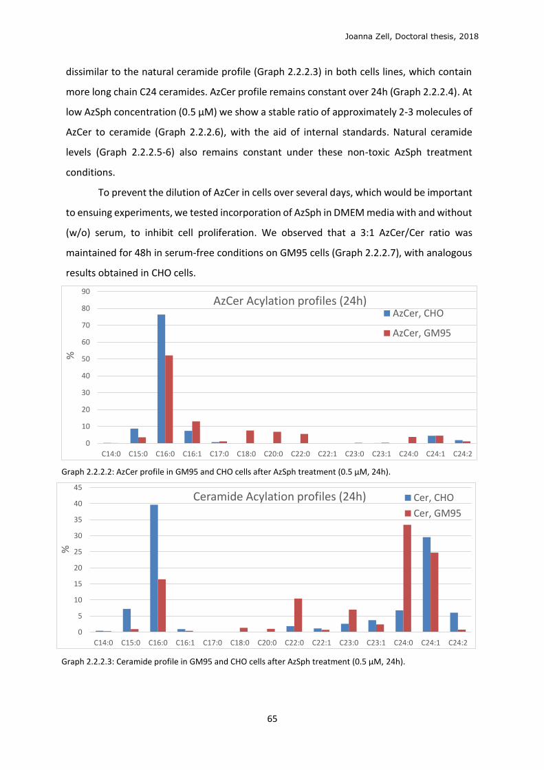
Joanna Zell, Doctoral thesis, 2018
65
dissimilar to the natural ceramide profile (Graph 2.2.2.3) in both cells lines, which contain
more long chain C24 ceramides. AzCer profile remains constant over 24h (Graph 2.2.2.4). At
low AzSph concentration (0.5 μM) we show a stable ratio of approximately 2-3 molecules of
AzCer to ceramide (Graph 2.2.2.6), with the aid of internal standards. Natural ceramide
levels (Graph 2.2.2.5-6) also remains constant under these non-toxic AzSph treatment
conditions.
To prevent the dilution of AzCer in cells over several days, which would be important
to ensuing experiments, we tested incorporation of AzSph in DMEM media with and without
(w/o) serum, to inhibit cell proliferation. We observed that a 3:1 AzCer/Cer ratio was
maintained for 48h in serum-free conditions on GM95 cells (Graph 2.2.2.7), with analogous
results obtained in CHO cells.
Graph 2.2.2.2: AzCer profile in GM95 and CHO cells after AzSph treatment (0.5 μM, 24h).
Graph 2.2.2.3: Ceramide profile in GM95 and CHO cells after AzSph treatment (0.5 μM, 24h).
0
10
20
30
40
50
60
70
80
90
C14:0 C15:0 C16:0 C16:1 C17:0 C18:0 C20:0 C22:0 C22:1 C23:0 C23:1 C24:0 C24:1 C24:2
%
AzCer Acylation profiles (24h)AzCer, CHO
AzCer, GM95
0
5
10
15
20
25
30
35
40
45
C14:0 C15:0 C16:0 C16:1 C17:0 C18:0 C20:0 C22:0 C22:1 C23:0 C23:1 C24:0 C24:1 C24:2
%
Ceramide Acylation profiles (24h) Cer, CHO
Cer, GM95
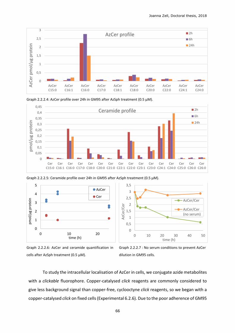
Joanna Zell, Doctoral thesis, 2018
66
Graph 2.2.2.4: AzCer profile over 24h in GM95 after AzSph treatment (0.5 μM).
Graph 2.2.2.5: Ceramide profile over 24h in GM95 after AzSph treatment (0.5 μM).
Graph 2.2.2.6: AzCer and ceramide quantification in
cells after AzSph treatment (0.5 μM).
Graph 2.2.2.7 : No serum conditions to prevent AzCer
dilution in GM95 cells.
To study the intracellular localisation of AzCer in cells, we conjugate azide metabolites
with a clickable fluorophore. Copper-catalysed click reagents are commonly considered to
give less background signal than copper-free, cyclooctyne click reagents, so we began with a
copper-catalysed click on fixed cells (Experimental 6.2.6). Due to the poor adherence of GM95
0
0,5
1
1,5
2
2,5
3
AzCerC15:0
AzCerC16:1
AzCerC16:0
AzCerC17:0
AzCerC18:1
AzCerC18:0
AzCerC20:0
AzCerC22:0
AzCerC24:1
AzCerC24:0
AzC
er p
mo
l/μ
g p
rote
inAzCer profile 2h
6h
24h
0
0,05
0,1
0,15
0,2
0,25
0,3
0,35
0,4
0,45
CerC15:0
CerC16:1
CerC16:0
CerC17:0
CerC18:0
CerC20:0
CerC21:0
CerC22:1
CerC22:0
CerC23:1
CerC23:0
CerC24:1
CerC24:0
CerC25:0
CerC26:0
CerC26:0
pm
ol/
μg
pro
tein
Ceramide profile 2h
6h
24h
0
1
2
3
4
5
0 10 20
pm
ol/
μg
pro
tein
time (h)
AzCer
Cer
0
0,5
1
1,5
2
2,5
3
3,5
0 10 20 30 40 50
AzC
er/C
er
time (h)
AzCer/Cer
AzCer/Cer(no serum)
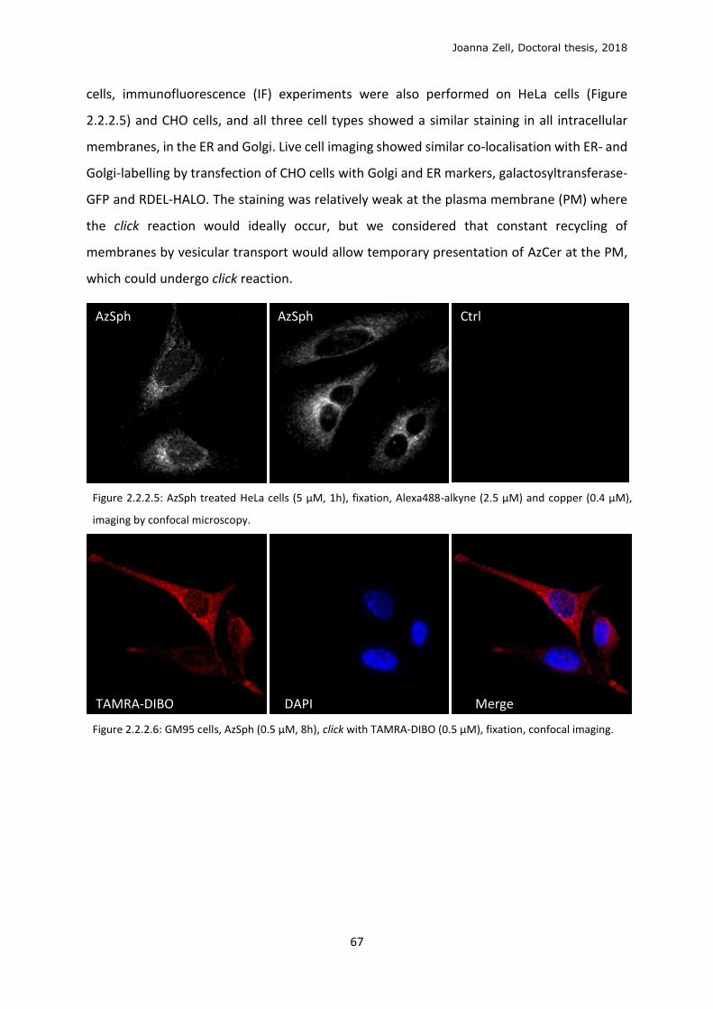
Joanna Zell, Doctoral thesis, 2018
67
cells, immunofluorescence (IF) experiments were also performed on HeLa cells (Figure
2.2.2.5) and CHO cells, and all three cell types showed a similar staining in all intracellular
membranes, in the ER and Golgi. Live cell imaging showed similar co-localisation with ER- and
Golgi-labelling by transfection of CHO cells with Golgi and ER markers, galactosyltransferase-
GFP and RDEL-HALO. The staining was relatively weak at the plasma membrane (PM) where
the click reaction would ideally occur, but we considered that constant recycling of
membranes by vesicular transport would allow temporary presentation of AzCer at the PM,
which could undergo click reaction.
Figure 2.2.2.5: AzSph treated HeLa cells (5 μM, 1h), fixation, Alexa488-alkyne (2.5 μM) and copper (0.4 μM),
imaging by confocal microscopy.
Figure 2.2.2.6: GM95 cells, AzSph (0.5 μM, 8h), click with TAMRA-DIBO (0.5 μM), fixation, confocal imaging.
AzSph AzSph Ctrl
TAMRA-DIBO DAPI Merge
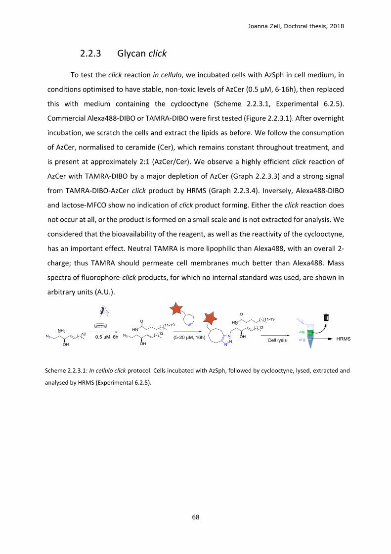
Joanna Zell, Doctoral thesis, 2018
68
2.2.3 Glycan click
To test the click reaction in cellulo, we incubated cells with AzSph in cell medium, in
conditions optimised to have stable, non-toxic levels of AzCer (0.5 μM, 6-16h), then replaced
this with medium containing the cyclooctyne (Scheme 2.2.3.1, Experimental 6.2.5).
Commercial Alexa488-DIBO or TAMRA-DIBO were first tested (Figure 2.2.3.1). After overnight
incubation, we scratch the cells and extract the lipids as before. We follow the consumption
of AzCer, normalised to ceramide (Cer), which remains constant throughout treatment, and
is present at approximately 2:1 (AzCer/Cer). We observe a highly efficient click reaction of
AzCer with TAMRA-DIBO by a major depletion of AzCer (Graph 2.2.3.3) and a strong signal
from TAMRA-DIBO-AzCer click product by HRMS (Graph 2.2.3.4). Inversely, Alexa488-DIBO
and lactose-MFCO show no indication of click product forming. Either the click reaction does
not occur at all, or the product is formed on a small scale and is not extracted for analysis. We
considered that the bioavailability of the reagent, as well as the reactivity of the cyclooctyne,
has an important effect. Neutral TAMRA is more lipophilic than Alexa488, with an overall 2-
charge; thus TAMRA should permeate cell membranes much better than Alexa488. Mass
spectra of fluorophore-click products, for which no internal standard was used, are shown in
arbitrary units (A.U.).
Scheme 2.2.3.1: In cellulo click protocol. Cells incubated with AzSph, followed by cyclooctyne, lysed, extracted and
analysed by HRMS (Experimental 6.2.5).
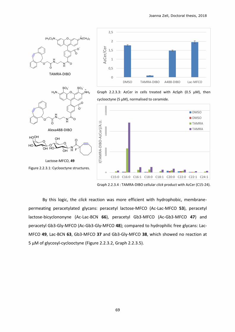
Joanna Zell, Doctoral thesis, 2018
69
TAMRA-DIBO
Alexa488-DIBO
Lactose-MFCO, 49
Figure 2.2.3.1: Cyclooctyne structures.
Graph 2.2.3.3: AzCer in cells treated with AzSph (0.5 μM), then
cyclooctyne (5 μM), normalised to ceramide.
Graph 2.2.3.4 : TAMRA-DIBO cellular click product with AzCer (C15-24).
By this logic, the click reaction was more efficient with hydrophobic, membrane-
permeating peracetylated glycans: peracetyl lactose-MFCO (Ac-Lac-MFCO 53), peracetyl
lactose-bicyclononyne (Ac-Lac-BCN 66), peracetyl Gb3-MFCO (Ac-Gb3-MFCO 47) and
peracetyl Gb3-Gly-MFCO (Ac-Gb3-Gly-MFCO 48); compared to hydrophilic free glycans: Lac-
MFCO 49, Lac-BCN 63, Gb3-MFCO 37 and Gb3-Gly-MFCO 38, which showed no reaction at
5 μM of glycosyl-cyclooctyne (Figure 2.2.3.2, Graph 2.2.3.5).
0
0,5
1
1,5
2
2,5
DMSO TAMRA-DIBO A488-DIBO Lac-MFCO
AzC
er/C
er0
50
00
00
010
000
000
1500
000
0
C15:0 C16:0 C16:1 C18:0 C18:1 C20:0 C22:0 C22:1 C24:1
I(TA
MR
A-D
IBO
-AzC
er)/
A.U
.
DMSO
DMSO
TAMRA
TAMRA
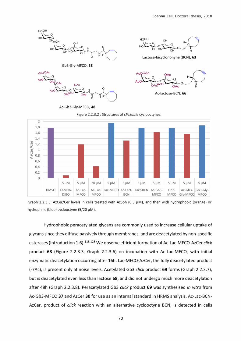
Joanna Zell, Doctoral thesis, 2018
70
Gb3-Gly-MFCO, 38
Ac-Gb3-Gly-MFCO, 48
Lactose-bicyclononyne (BCN), 63
Ac-lactose-BCN, 66
Figure 2.2.3.2 : Structures of clickable cyclooctynes.
Graph 2.2.3.5: AzCer/Cer levels in cells treated with AzSph (0.5 μM), and then with hydrophobic (orange) or
hydrophilic (blue) cyclooctyne (5/20 μM).
Hydrophobic peracetylated glycans are commonly used to increase cellular uptake of
glycans since they diffuse passively through membranes, and are deacetylated by non-specific
esterases (Introduction 1.6).118,128 We observe efficient formation of Ac-Lac-MFCO-AzCer click
product 68 (Figure 2.2.3.3, Graph 2.2.3.6) on incubation with Ac-Lac-MFCO, with initial
enzymatic deacetylation occurring after 16h. Lac-MFCO-AzCer, the fully deacetylated product
(-7Ac), is present only at noise levels. Acetylated Gb3 click product 69 forms (Graph 2.2.3.7),
but is deacetylated even less than lactose 68, and did not undergo much more deacetylation
after 48h (Graph 2.2.3.8). Peracetylated Gb3 click product 69 was synthesised in vitro from
Ac-Gb3-MFCO 37 and AzCer 30 for use as an internal standard in HRMS analysis. Ac-Lac-BCN-
AzCer, product of click reaction with an alternative cyclooctyne BCN, is detected in cells
0
0,2
0,4
0,6
0,8
1
1,2
1,4
1,6
1,8
2
5 µM 5 µM 20 µM 5 µM 5 µM 5 µM 5 µM 5 µM 5 µM 5 µM
DMSO TAMRA-DIBO
Ac-Lac-MFCO
Ac-Lac-MFCO
Lac-MFCO Ac-Lact-BCN
Lact-BCN Ac-Gb3-MFCO
Gb3-MFCO
Ac-Gb3-Gly-MFCO
Gb3-Gly-MFCO
AzC
er/C
er

Joanna Zell, Doctoral thesis, 2018
71
(Figure 2.2.3.4, Graph 2.2.3.9), but no deacetylation is observed. Conversely, the click product
of peracetylated Gb3-Gly-MFCO 48 was not observed. Potentially 48 is too large to permeate
the cell membrane, despite being hydrophobic.
Graph 2.2.3.6: Deacetylation profile of Ac-Lac-MFCO-AzCer product in GM95 cells treated consecutively with
AzSph (0.5 μM, 8h) and (Ac)-Gb3-MFCO (5 or 20 μM).
Graph 2.2.3.7: Ac-Gb3-MFCO-AzCer 69 deacetylation in
GM95 cells treated with AzSph (0.5 μM, 8h), then with (Ac)-
Gb3-MFCO (5 μM).
Figure 2.2.3.3: Ac-Lac-MFCO-AzCer 68 and
Ac-Gb3-MFCO-AzCer 69.
01
00
00
02
00
00
03
00
00
04
00
00
05
00
00
06
00
00
07
00
00
0
16 18 20 22 16 18 16 16 16 16 16 16
Ac7-Lac Ac7-Lac Ac7-Lac Ac7-Lac Ac6-Lac Ac6-Lac Ac5-Lac Ac4-Lac Ac3-Lac Ac2-Lac Ac1-Lac Lac
A.U
.
DMSO
AcO-Lac-MFCO 20 µM
AcO-Lac-MFCO 5 µM
Lact-MFCO
05
000
00
10
000
00
15
000
00
16:0 16:1 18:0 16:0
Ac10-Gb3-MFCO-AzCer
Ac10-Gb3-MFCO-AzCer
Ac10-Gb3-MFCO-AzCer
Ac9-Gb3-MFCO-AzCer
A.U
.
AcO-Gb3-MFCO
Gb3-MFCO
Peracetylated
-1Ac
-2Ac
-3Ac -4Ac -5Ac -6Ac -7Ac
Peracetylated
-1Ac
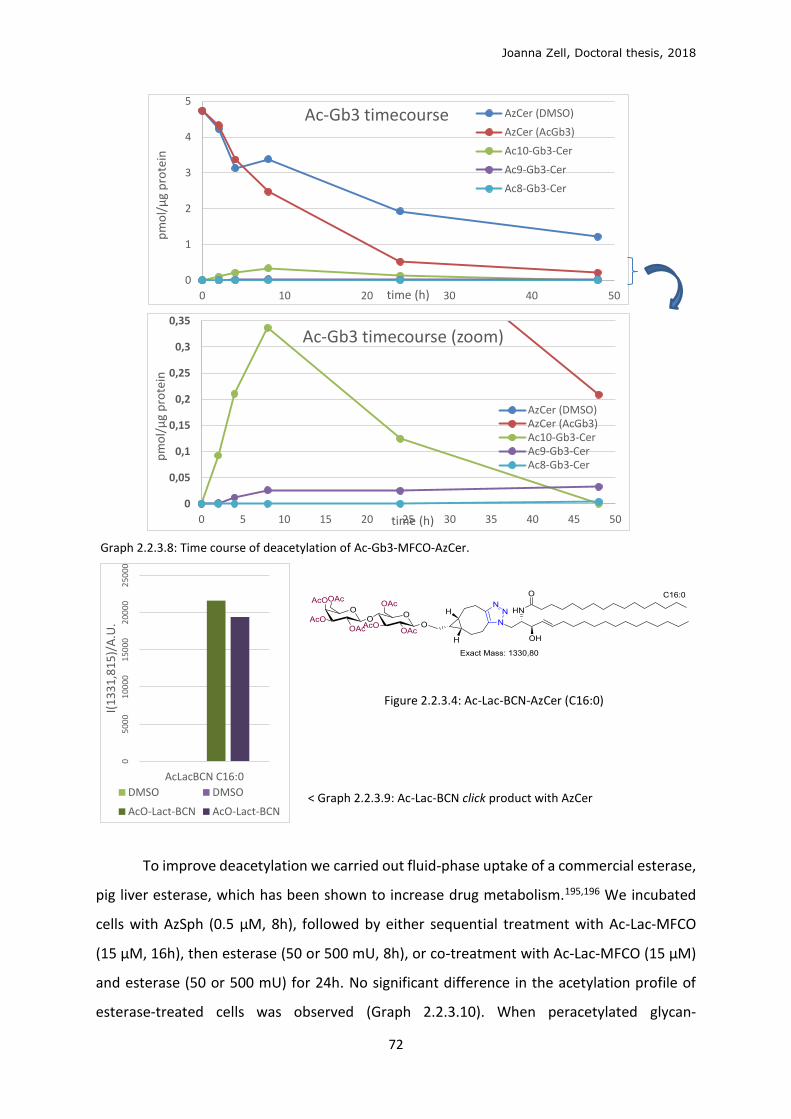
Joanna Zell, Doctoral thesis, 2018
72
Graph 2.2.3.8: Time course of deacetylation of Ac-Gb3-MFCO-AzCer.
Figure 2.2.3.4: Ac-Lac-BCN-AzCer (C16:0)
< Graph 2.2.3.9: Ac-Lac-BCN click product with AzCer
To improve deacetylation we carried out fluid-phase uptake of a commercial esterase,
pig liver esterase, which has been shown to increase drug metabolism.195,196 We incubated
cells with AzSph (0.5 μM, 8h), followed by either sequential treatment with Ac-Lac-MFCO
(15 μM, 16h), then esterase (50 or 500 mU, 8h), or co-treatment with Ac-Lac-MFCO (15 μM)
and esterase (50 or 500 mU) for 24h. No significant difference in the acetylation profile of
esterase-treated cells was observed (Graph 2.2.3.10). When peracetylated glycan-
0
1
2
3
4
5
0 10 20 30 40 50
pm
ol/
μg
pro
tein
time (h)
Ac-Gb3 timecourse AzCer (DMSO)
AzCer (AcGb3)
Ac10-Gb3-Cer
Ac9-Gb3-Cer
Ac8-Gb3-Cer
0
0,05
0,1
0,15
0,2
0,25
0,3
0,35
0 5 10 15 20 25 30 35 40 45 50
pm
ol/
μg
pro
tein
time (h)
Ac-Gb3 timecourse (zoom)
AzCer (DMSO)AzCer (AcGb3)Ac10-Gb3-CerAc9-Gb3-CerAc8-Gb3-Cer
05
000
10
000
150
002
0000
250
00
AcLacBCN C16:0
I(1
33
1,8
15
)/A
.U.
DMSO DMSO
AcO-Lact-BCN AcO-Lact-BCN
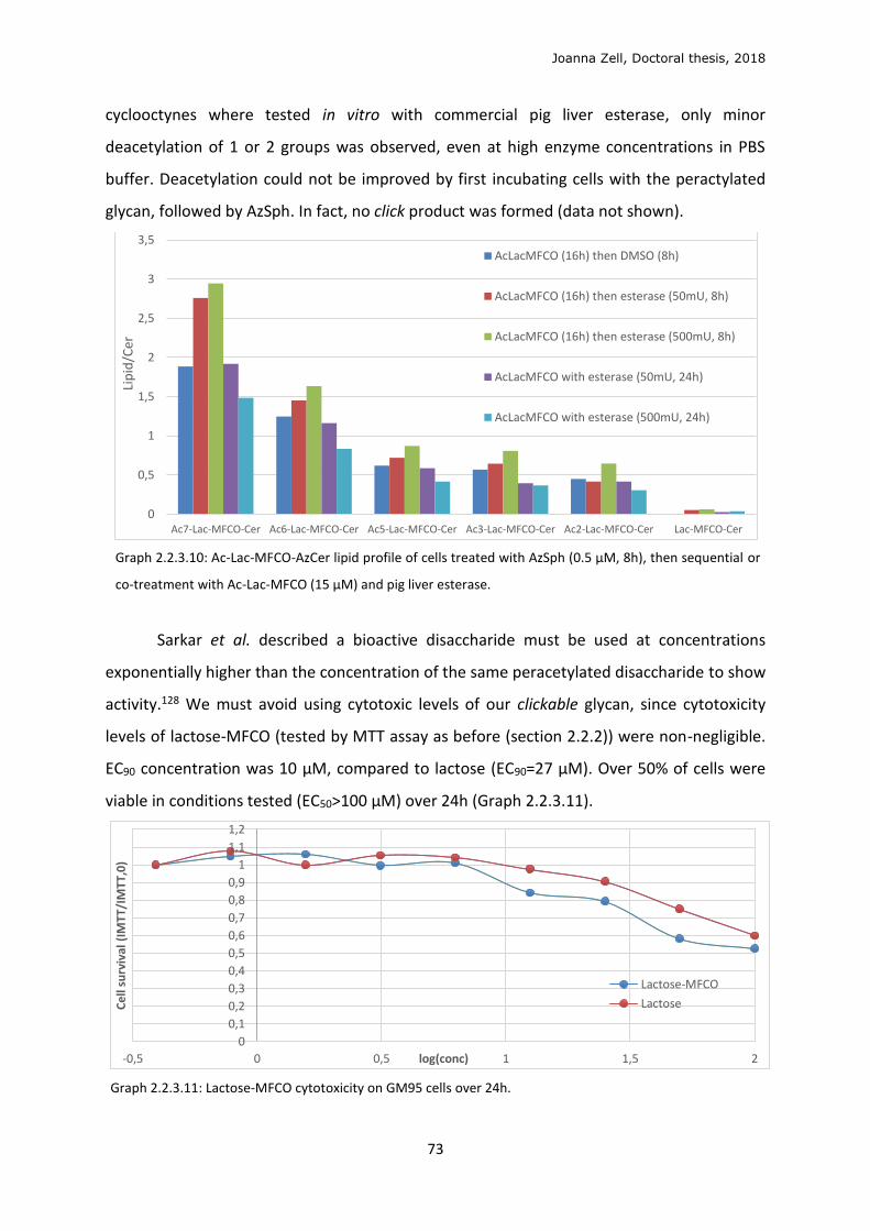
Joanna Zell, Doctoral thesis, 2018
73
cyclooctynes where tested in vitro with commercial pig liver esterase, only minor
deacetylation of 1 or 2 groups was observed, even at high enzyme concentrations in PBS
buffer. Deacetylation could not be improved by first incubating cells with the peractylated
glycan, followed by AzSph. In fact, no click product was formed (data not shown).
Graph 2.2.3.10: Ac-Lac-MFCO-AzCer lipid profile of cells treated with AzSph (0.5 μM, 8h), then sequential or
co-treatment with Ac-Lac-MFCO (15 μM) and pig liver esterase.
Sarkar et al. described a bioactive disaccharide must be used at concentrations
exponentially higher than the concentration of the same peracetylated disaccharide to show
activity.128 We must avoid using cytotoxic levels of our clickable glycan, since cytotoxicity
levels of lactose-MFCO (tested by MTT assay as before (section 2.2.2)) were non-negligible.
EC90 concentration was 10 μM, compared to lactose (EC90=27 μM). Over 50% of cells were
viable in conditions tested (EC50>100 μM) over 24h (Graph 2.2.3.11).
Graph 2.2.3.11: Lactose-MFCO cytotoxicity on GM95 cells over 24h.
0
0,5
1
1,5
2
2,5
3
3,5
Ac7-Lac-MFCO-Cer Ac6-Lac-MFCO-Cer Ac5-Lac-MFCO-Cer Ac3-Lac-MFCO-Cer Ac2-Lac-MFCO-Cer Lac-MFCO-Cer
Lip
id/C
er
AcLacMFCO (16h) then DMSO (8h)
AcLacMFCO (16h) then esterase (50mU, 8h)
AcLacMFCO (16h) then esterase (500mU, 8h)
AcLacMFCO with esterase (50mU, 24h)
AcLacMFCO with esterase (500mU, 24h)
0
0,1
0,2
0,3
0,4
0,5
0,6
0,7
0,8
0,9
1
1,1
1,2
-0,5 0 0,5 1 1,5 2
Ce
ll su
rviv
al (
IMTT
/IM
TT,0
)
log(conc)
Lactose-MFCO
Lactose
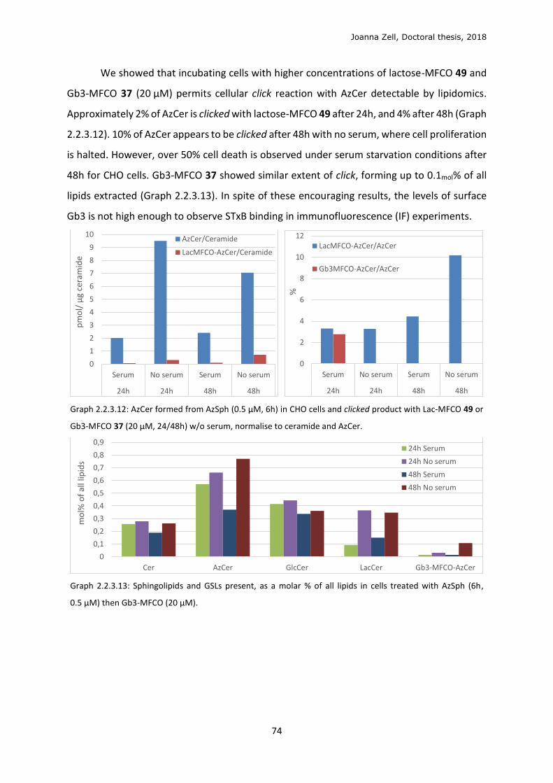
Joanna Zell, Doctoral thesis, 2018
74
We showed that incubating cells with higher concentrations of lactose-MFCO 49 and
Gb3-MFCO 37 (20 μM) permits cellular click reaction with AzCer detectable by lipidomics.
Approximately 2% of AzCer is clicked with lactose-MFCO 49 after 24h, and 4% after 48h (Graph
2.2.3.12). 10% of AzCer appears to be clicked after 48h with no serum, where cell proliferation
is halted. However, over 50% cell death is observed under serum starvation conditions after
48h for CHO cells. Gb3-MFCO 37 showed similar extent of click, forming up to 0.1mol% of all
lipids extracted (Graph 2.2.3.13). In spite of these encouraging results, the levels of surface
Gb3 is not high enough to observe STxB binding in immunofluorescence (IF) experiments.
Graph 2.2.3.12: AzCer formed from AzSph (0.5 μM, 6h) in CHO cells and clicked product with Lac-MFCO 49 or
Gb3-MFCO 37 (20 μM, 24/48h) w/o serum, normalise to ceramide and AzCer.
Graph 2.2.3.13: Sphingolipids and GSLs present, as a molar % of all lipids in cells treated with AzSph (6h,
0.5 μM) then Gb3-MFCO (20 μM).
0
1
2
3
4
5
6
7
8
9
10
Serum No serum Serum No serum
24h 24h 48h 48h
pm
ol/
μg
cera
mid
e
AzCer/Ceramide
LacMFCO-AzCer/Ceramide
0
2
4
6
8
10
12
Serum No serum Serum No serum
24h 24h 48h 48h
%
LacMFCO-AzCer/AzCer
Gb3MFCO-AzCer/AzCer
0
0,1
0,2
0,3
0,4
0,5
0,6
0,7
0,8
0,9
Cer AzCer GlcCer LacCer Gb3-MFCO-AzCer
mo
l% o
f al
l lip
ids
24h Serum
24h No serum
48h Serum
48h No serum
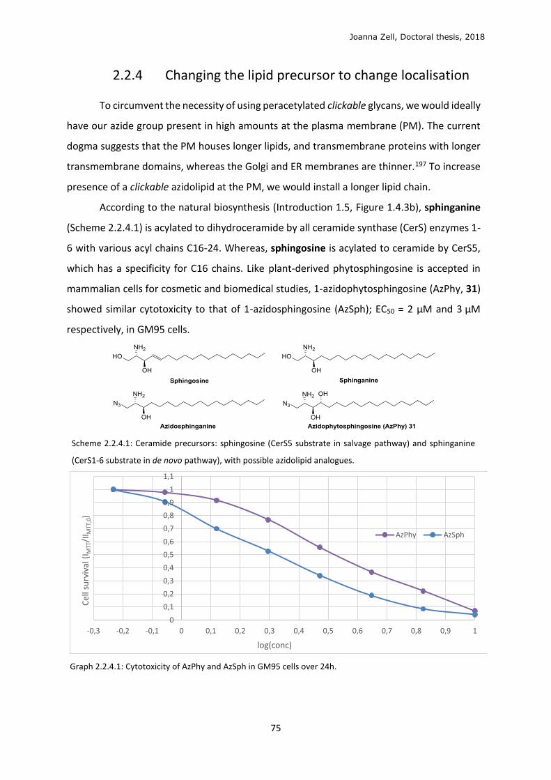
Joanna Zell, Doctoral thesis, 2018
75
2.2.4 Changing the lipid precursor to change localisation
To circumvent the necessity of using peracetylated clickable glycans, we would ideally
have our azide group present in high amounts at the plasma membrane (PM). The current
dogma suggests that the PM houses longer lipids, and transmembrane proteins with longer
transmembrane domains, whereas the Golgi and ER membranes are thinner.197 To increase
presence of a clickable azidolipid at the PM, we would install a longer lipid chain.
According to the natural biosynthesis (Introduction 1.5, Figure 1.4.3b), sphinganine
(Scheme 2.2.4.1) is acylated to dihydroceramide by all ceramide synthase (CerS) enzymes 1-
6 with various acyl chains C16-24. Whereas, sphingosine is acylated to ceramide by CerS5,
which has a specificity for C16 chains. Like plant-derived phytosphingosine is accepted in
mammalian cells for cosmetic and biomedical studies, 1-azidophytosphingosine (AzPhy, 31)
showed similar cytotoxicity to that of 1-azidosphingosine (AzSph); EC50 = 2 μM and 3 μM
respectively, in GM95 cells.
Scheme 2.2.4.1: Ceramide precursors: sphingosine (CerS5 substrate in salvage pathway) and sphinganine
(CerS1-6 substrate in de novo pathway), with possible azidolipid analogues.
Graph 2.2.4.1: Cytotoxicity of AzPhy and AzSph in GM95 cells over 24h.
0
0,1
0,2
0,3
0,4
0,5
0,6
0,7
0,8
0,9
1
1,1
-0,3 -0,2 -0,1 0 0,1 0,2 0,3 0,4 0,5 0,6 0,7 0,8 0,9 1
Cel
l su
rviv
al (
I MTT
/II M
TT,0
)
log(conc)
AzPhy AzSph
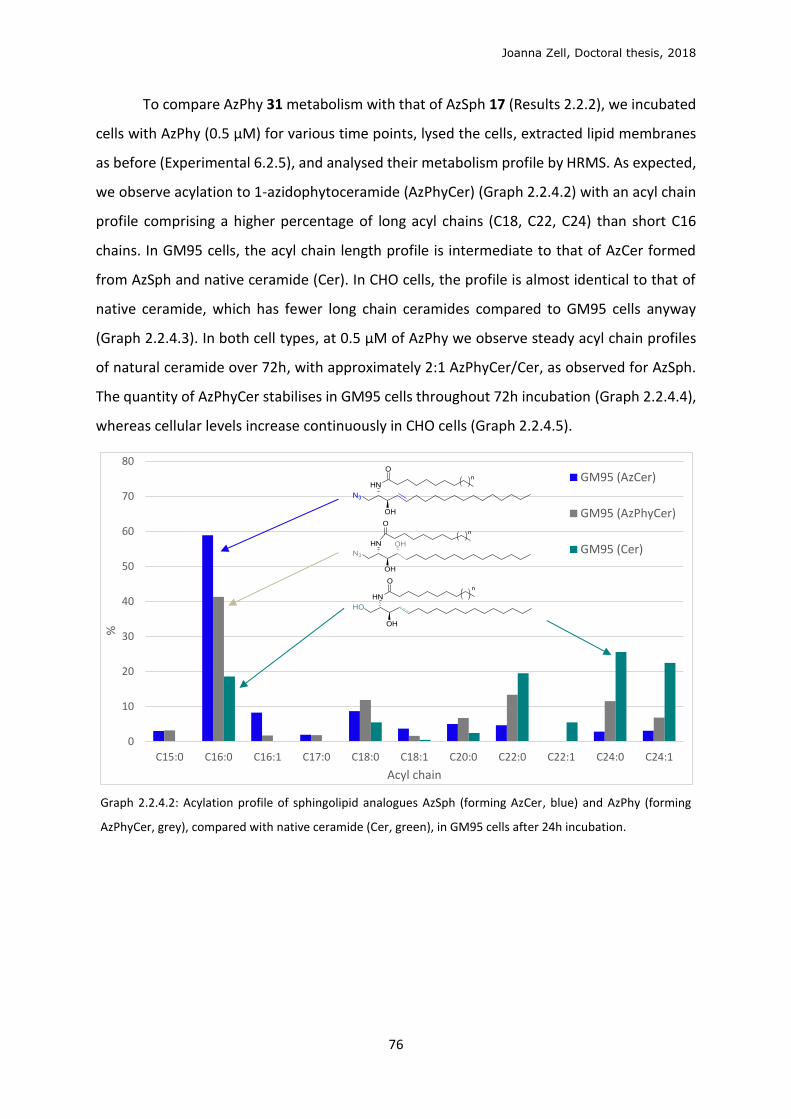
Joanna Zell, Doctoral thesis, 2018
76
To compare AzPhy 31 metabolism with that of AzSph 17 (Results 2.2.2), we incubated
cells with AzPhy (0.5 μM) for various time points, lysed the cells, extracted lipid membranes
as before (Experimental 6.2.5), and analysed their metabolism profile by HRMS. As expected,
we observe acylation to 1-azidophytoceramide (AzPhyCer) (Graph 2.2.4.2) with an acyl chain
profile comprising a higher percentage of long acyl chains (C18, C22, C24) than short C16
chains. In GM95 cells, the acyl chain length profile is intermediate to that of AzCer formed
from AzSph and native ceramide (Cer). In CHO cells, the profile is almost identical to that of
native ceramide, which has fewer long chain ceramides compared to GM95 cells anyway
(Graph 2.2.4.3). In both cell types, at 0.5 μM of AzPhy we observe steady acyl chain profiles
of natural ceramide over 72h, with approximately 2:1 AzPhyCer/Cer, as observed for AzSph.
The quantity of AzPhyCer stabilises in GM95 cells throughout 72h incubation (Graph 2.2.4.4),
whereas cellular levels increase continuously in CHO cells (Graph 2.2.4.5).
Graph 2.2.4.2: Acylation profile of sphingolipid analogues AzSph (forming AzCer, blue) and AzPhy (forming
AzPhyCer, grey), compared with native ceramide (Cer, green), in GM95 cells after 24h incubation.
0
10
20
30
40
50
60
70
80
C15:0 C16:0 C16:1 C17:0 C18:0 C18:1 C20:0 C22:0 C22:1 C24:0 C24:1
%
Acyl chain
GM95 (AzCer)
GM95 (AzPhyCer)
GM95 (Cer)
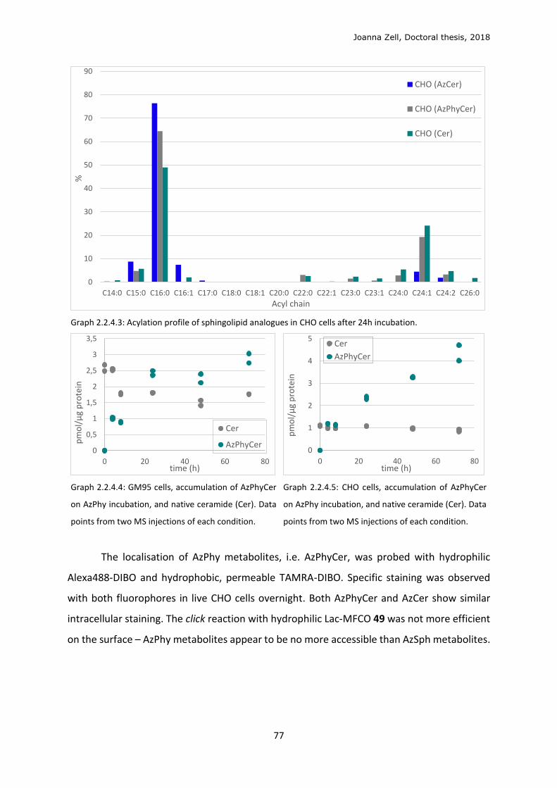
Joanna Zell, Doctoral thesis, 2018
77
Graph 2.2.4.3: Acylation profile of sphingolipid analogues in CHO cells after 24h incubation.
Graph 2.2.4.4: GM95 cells, accumulation of AzPhyCer
on AzPhy incubation, and native ceramide (Cer). Data
points from two MS injections of each condition.
Graph 2.2.4.5: CHO cells, accumulation of AzPhyCer
on AzPhy incubation, and native ceramide (Cer). Data
points from two MS injections of each condition.
The localisation of AzPhy metabolites, i.e. AzPhyCer, was probed with hydrophilic
Alexa488-DIBO and hydrophobic, permeable TAMRA-DIBO. Specific staining was observed
with both fluorophores in live CHO cells overnight. Both AzPhyCer and AzCer show similar
intracellular staining. The click reaction with hydrophilic Lac-MFCO 49 was not more efficient
on the surface – AzPhy metabolites appear to be no more accessible than AzSph metabolites.
0
10
20
30
40
50
60
70
80
90
C14:0 C15:0 C16:0 C16:1 C17:0 C18:0 C18:1 C20:0 C22:0 C22:1 C23:0 C23:1 C24:0 C24:1 C24:2 C26:0
%
Acyl chain
CHO (AzCer)
CHO (AzPhyCer)
CHO (Cer)
0
0,5
1
1,5
2
2,5
3
3,5
0 20 40 60 80
pm
ol/
μg
pro
tein
time (h)
Cer
AzPhyCer0
1
2
3
4
5
0 20 40 60 80
pm
ol/
μg
pro
tein
time (h)
Cer
AzPhyCer
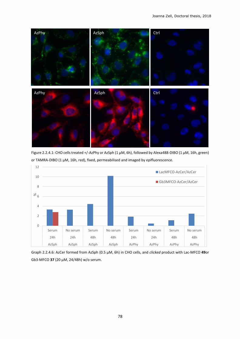
Joanna Zell, Doctoral thesis, 2018
78
Figure 2.2.4.1: CHO cells treated +/-AzPhy or AzSph (1 μM, 6h), followed by Alexa488-DIBO (1 μM, 16h, green)
or TAMRA-DIBO (1 μM, 16h, red), fixed, permeabilised and imaged by epifluorescence.
Graph 2.2.4.6: AzCer formed from AzSph (0.5 μM, 6h) in CHO cells, and clicked product with Lac-MFCO 49or
Gb3-MFCO 37 (20 μM, 24/48h) w/o serum.
0
2
4
6
8
10
12
Serum No serum Serum No serum Serum No serum Serum No serum
24h 24h 48h 48h 24h 24h 48h 48h
AzSph AzSph AzSph AzSph AzPhy AzPhy AzPhy AzPhy
%
LacMFCO-AzCer/AzCer
Gb3MFCO-AzCer/AzCer
AzPhy AzSph Ctrl
AzPhy AzSph Ctrl
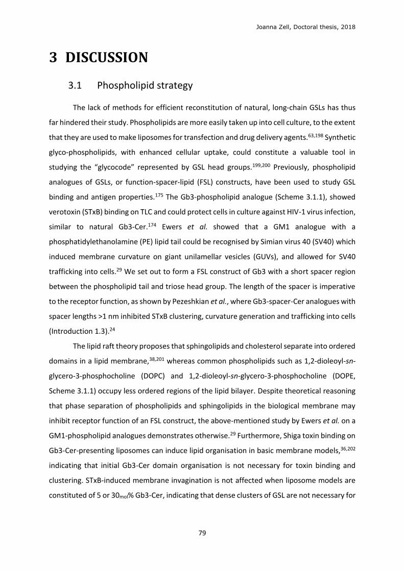
Joanna Zell, Doctoral thesis, 2018
79
3 DISCUSSION
3.1 Phospholipid strategy
The lack of methods for efficient reconstitution of natural, long-chain GSLs has thus
far hindered their study. Phospholipids are more easily taken up into cell culture, to the extent
that they are used to make liposomes for transfection and drug delivery agents.63,198 Synthetic
glyco-phospholipids, with enhanced cellular uptake, could constitute a valuable tool in
studying the “glycocode” represented by GSL head groups.199,200 Previously, phospholipid
analogues of GSLs, or function-spacer-lipid (FSL) constructs, have been used to study GSL
binding and antigen properties.175 The Gb3-phospholipid analogue (Scheme 3.1.1), showed
verotoxin (STxB) binding on TLC and could protect cells in culture against HIV-1 virus infection,
similar to natural Gb3-Cer.174 Ewers et al. showed that a GM1 analogue with a
phosphatidylethanolamine (PE) lipid tail could be recognised by Simian virus 40 (SV40) which
induced membrane curvature on giant unilamellar vesicles (GUVs), and allowed for SV40
trafficking into cells.29 We set out to form a FSL construct of Gb3 with a short spacer region
between the phospholipid tail and triose head group. The length of the spacer is imperative
to the receptor function, as shown by Pezeshkian et al., where Gb3-spacer-Cer analogues with
spacer lengths >1 nm inhibited STxB clustering, curvature generation and trafficking into cells
(Introduction 1.3).24
The lipid raft theory proposes that sphingolipids and cholesterol separate into ordered
domains in a lipid membrane,38,201 whereas common phospholipids such as 1,2-dioleoyl-sn-
glycero-3-phosphocholine (DOPC) and 1,2-dioleoyl-sn-glycero-3-phosphocholine (DOPE,
Scheme 3.1.1) occupy less ordered regions of the lipid bilayer. Despite theoretical reasoning
that phase separation of phospholipids and sphingolipids in the biological membrane may
inhibit receptor function of an FSL construct, the above-mentioned study by Ewers et al. on a
GM1-phospholipid analogues demonstrates otherwise.29 Furthermore, Shiga toxin binding on
Gb3-Cer-presenting liposomes can induce lipid organisation in basic membrane models,36,202
indicating that initial Gb3-Cer domain organisation is not necessary for toxin binding and
clustering. STxB-induced membrane invagination is not affected when liposome models are
constituted of 5 or 30mol% Gb3-Cer, indicating that dense clusters of GSL are not necessary for
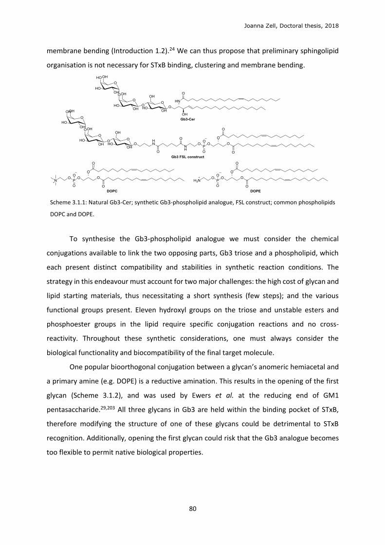
Joanna Zell, Doctoral thesis, 2018
80
membrane bending (Introduction 1.2).24 We can thus propose that preliminary sphingolipid
organisation is not necessary for STxB binding, clustering and membrane bending.
Scheme 3.1.1: Natural Gb3-Cer; synthetic Gb3-phospholipid analogue, FSL construct; common phospholipids
DOPC and DOPE.
To synthesise the Gb3-phospholipid analogue we must consider the chemical
conjugations available to link the two opposing parts, Gb3 triose and a phospholipid, which
each present distinct compatibility and stabilities in synthetic reaction conditions. The
strategy in this endeavour must account for two major challenges: the high cost of glycan and
lipid starting materials, thus necessitating a short synthesis (few steps); and the various
functional groups present. Eleven hydroxyl groups on the triose and unstable esters and
phosphoester groups in the lipid require specific conjugation reactions and no cross-
reactivity. Throughout these synthetic considerations, one must always consider the
biological functionality and biocompatibility of the final target molecule.
One popular bioorthogonal conjugation between a glycan’s anomeric hemiacetal and
a primary amine (e.g. DOPE) is a reductive amination. This results in the opening of the first
glycan (Scheme 3.1.2), and was used by Ewers et al. at the reducing end of GM1
pentasaccharide.29,203 All three glycans in Gb3 are held within the binding pocket of STxB,
therefore modifying the structure of one of these glycans could be detrimental to STxB
recognition. Additionally, opening the first glycan could risk that the Gb3 analogue becomes
too flexible to permit native biological properties.
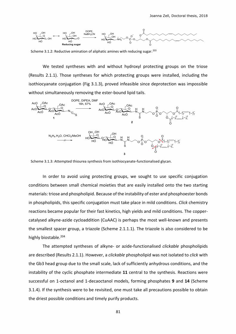
Joanna Zell, Doctoral thesis, 2018
81
Scheme 3.1.2: Reductive amination of aliphatic amines with reducing sugar.203
We tested syntheses with and without hydroxyl protecting groups on the triose
(Results 2.1.1). Those syntheses for which protecting groups were installed, including the
isothiocyanate conjugation (Fig 3.1.3), proved infeasible since deprotection was impossible
without simultaneously removing the ester-bound lipid tails.
Scheme 3.1.3: Attempted thiourea synthesis from isothiocyanate-functionalised glycan.
In order to avoid using protecting groups, we sought to use specific conjugation
conditions between small chemical moieties that are easily installed onto the two starting
materials: triose and phospholipid. Because of the instability of ester and phosphoester bonds
in phospholipids, this specific conjugation must take place in mild conditions. Click chemistry
reactions became popular for their fast kinetics, high yields and mild conditions. The copper-
catalysed alkyne-azide cycloaddition (CuAAC) is perhaps the most well-known and presents
the smallest spacer group, a triazole (Scheme 2.1.1.1). The triazole is also considered to be
highly biostable.204
The attempted syntheses of alkyne- or azide-functionalised clickable phospholipids
are described (Results 2.1.1). However, a clickable phospholipid was not isolated to click with
the Gb3 head group due to the small scale, lack of sufficiently anhydrous conditions, and the
instability of the cyclic phosphate intermediate 11 central to the synthesis. Reactions were
successful on 1-octanol and 1-decaoctanol models, forming phosphates 9 and 14 (Scheme
3.1.4). If the synthesis were to be revisited, one must take all precautions possible to obtain
the driest possible conditions and timely purify products.
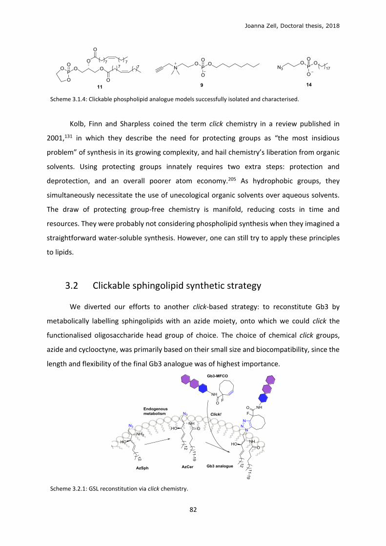
Joanna Zell, Doctoral thesis, 2018
82
Scheme 3.1.4: Clickable phospholipid analogue models successfully isolated and characterised.
Kolb, Finn and Sharpless coined the term click chemistry in a review published in
2001,131 in which they describe the need for protecting groups as “the most insidious
problem” of synthesis in its growing complexity, and hail chemistry’s liberation from organic
solvents. Using protecting groups innately requires two extra steps: protection and
deprotection, and an overall poorer atom economy.205 As hydrophobic groups, they
simultaneously necessitate the use of unecological organic solvents over aqueous solvents.
The draw of protecting group-free chemistry is manifold, reducing costs in time and
resources. They were probably not considering phospholipid synthesis when they imagined a
straightforward water-soluble synthesis. However, one can still try to apply these principles
to lipids.
3.2 Clickable sphingolipid synthetic strategy
We diverted our efforts to another click-based strategy: to reconstitute Gb3 by
metabolically labelling sphingolipids with an azide moiety, onto which we could click the
functionalised oligosaccharide head group of choice. The choice of chemical click groups,
azide and cyclooctyne, was primarily based on their small size and biocompatibility, since the
length and flexibility of the final Gb3 analogue was of highest importance.
Scheme 3.2.1: GSL reconstitution via click chemistry.
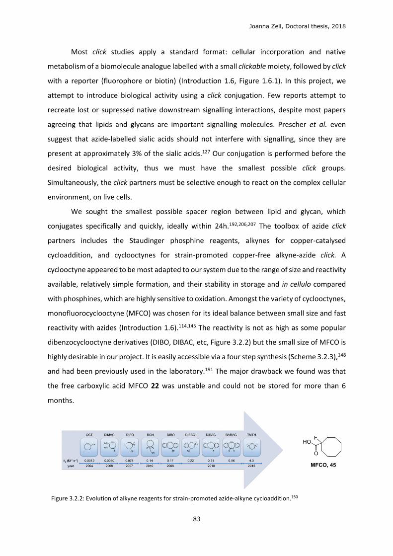
Joanna Zell, Doctoral thesis, 2018
83
Most click studies apply a standard format: cellular incorporation and native
metabolism of a biomolecule analogue labelled with a small clickable moiety, followed by click
with a reporter (fluorophore or biotin) (Introduction 1.6, Figure 1.6.1). In this project, we
attempt to introduce biological activity using a click conjugation. Few reports attempt to
recreate lost or supressed native downstream signalling interactions, despite most papers
agreeing that lipids and glycans are important signalling molecules. Prescher et al. even
suggest that azide-labelled sialic acids should not interfere with signalling, since they are
present at approximately 3% of the sialic acids.127 Our conjugation is performed before the
desired biological activity, thus we must have the smallest possible click groups.
Simultaneously, the click partners must be selective enough to react on the complex cellular
environment, on live cells.
We sought the smallest possible spacer region between lipid and glycan, which
conjugates specifically and quickly, ideally within 24h.192,206,207 The toolbox of azide click
partners includes the Staudinger phosphine reagents, alkynes for copper-catalysed
cycloaddition, and cyclooctynes for strain-promoted copper-free alkyne-azide click. A
cyclooctyne appeared to be most adapted to our system due to the range of size and reactivity
available, relatively simple formation, and their stability in storage and in cellulo compared
with phosphines, which are highly sensitive to oxidation. Amongst the variety of cyclooctynes,
monofluorocyclooctyne (MFCO) was chosen for its ideal balance between small size and fast
reactivity with azides (Introduction 1.6).114,145 The reactivity is not as high as some popular
dibenzocyclooctyne derivatives (DIBO, DIBAC, etc, Figure 3.2.2) but the small size of MFCO is
highly desirable in our project. It is easily accessible via a four step synthesis (Scheme 3.2.3),148
and had been previously used in the laboratory.191 The major drawback we found was that
the free carboxylic acid MFCO 22 was unstable and could not be stored for more than 6
months.
Figure 3.2.2: Evolution of alkyne reagents for strain-promoted azide-alkyne cycloaddition.150

Joanna Zell, Doctoral thesis, 2018
84
Azides are ideally small and stable in cells (Introduction 1.6).147 An azide was installed
at the head position (C-1) in the place of the natural hydroxyl group of sphingosine, since this
would form the most linear GSL analogue. The azide group is less hydrophilic than the
hydroxyl group, and may or may not be held within the hydrophobic layer of the lipid bilayer
when in a biological environment. Despite this, we were confident it would retain its reactivity
with a hydrophobic cyclcooctyne partner, as many literature reports suggest that click
reaction in cellulo and in liposomes can occur with lipids functionalised with a hydrophobic
moiety held within the membrane.170,208 Garrido et al. showed that 1-azidoceramide
incorporated into liposomes could undergo efficient copper-catalysed click reaction with an
alkyne-fluorophore.209
Even before the rise in click chemistry, the azide was a common functional group on
the synthetic route to amines, to which azides are easily reduced.210 The azide group is so
stable and convenient it is used as an amine protecting group,181 the latter being sensitive to
oxidation and carboxylation in long term storage. Phytosphingosine (Scheme 2.1.2.4) is an
ideal inexpensive starting material, with stereochemistry in the 2,3-positions identical to that
of sphingosine, and multiple syntheses to sphingosine have been reported.182,211,212 Synthesis
of sphingosine was mastered in the laboratory, to obtain natural Gb3-Cer for various
structure-function studies.24,27,35,54 We follow the synthesis described by Kim et al.181 to
sphingosine 6 (Scheme 3.2.2), installing an azide protecting group in the N-2 position for two
major advantages: the azide is non-nucleophilic, thus avoids possible cyclisation side
reactions (Scheme 2.1.2.2), and the azide cannot form a hydrogen bond with the terminal
hydroxyl group and the N-2 position, thus facilitating the ensuing functionalisation, in this
case the selective O1-silyl protection.182
After protection and installation of the double bond selectively at the C-4 position in
6 (Scheme 3.2.3), we protected the C-3 hydroxyl with a benzoyl (Bz) group as in 7, as has been
performed in the past for the natural Gb3-Cer synthesis, and because we considered this was
the most stable form for long-term storage of a large-scale synthesis. Once we obtained
sphingosine 22, we swapped the N2-azide for dichloroacetamide in 24 in order to eventually
install the N1-azide. One disadvantage in this route is that on deprotection of the azide, the
benzoyl can transfer to the amine, forming the thermodynamically more stable amide
product (NHBz) (Scheme 3.2.3). In hindsight, we could have avoided the parasitic NHBz
rearrangement by deprotecting the azide earlier, from compound 21 (Scheme 3.2.1),
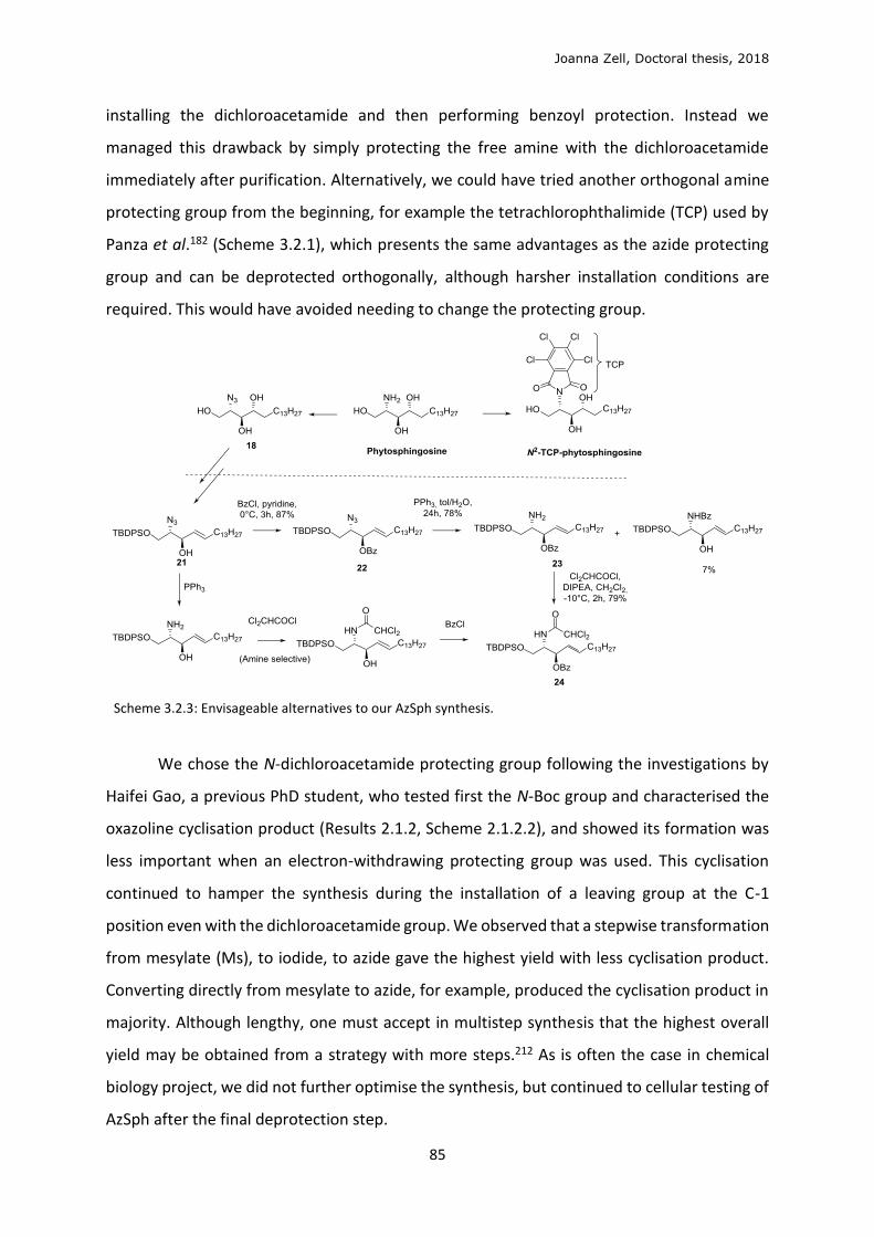
Joanna Zell, Doctoral thesis, 2018
85
installing the dichloroacetamide and then performing benzoyl protection. Instead we
managed this drawback by simply protecting the free amine with the dichloroacetamide
immediately after purification. Alternatively, we could have tried another orthogonal amine
protecting group from the beginning, for example the tetrachlorophthalimide (TCP) used by
Panza et al.182 (Scheme 3.2.1), which presents the same advantages as the azide protecting
group and can be deprotected orthogonally, although harsher installation conditions are
required. This would have avoided needing to change the protecting group.
Scheme 3.2.3: Envisageable alternatives to our AzSph synthesis.
We chose the N-dichloroacetamide protecting group following the investigations by
Haifei Gao, a previous PhD student, who tested first the N-Boc group and characterised the
oxazoline cyclisation product (Results 2.1.2, Scheme 2.1.2.2), and showed its formation was
less important when an electron-withdrawing protecting group was used. This cyclisation
continued to hamper the synthesis during the installation of a leaving group at the C-1
position even with the dichloroacetamide group. We observed that a stepwise transformation
from mesylate (Ms), to iodide, to azide gave the highest yield with less cyclisation product.
Converting directly from mesylate to azide, for example, produced the cyclisation product in
majority. Although lengthy, one must accept in multistep synthesis that the highest overall
yield may be obtained from a strategy with more steps.212 As is often the case in chemical
biology project, we did not further optimise the synthesis, but continued to cellular testing of
AzSph after the final deprotection step.
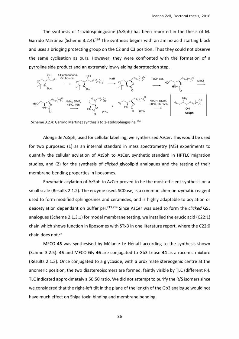
Joanna Zell, Doctoral thesis, 2018
86
The synthesis of 1-azidosphingosine (AzSph) has been reported in the thesis of M.
Garrido Martínez (Scheme 3.2.4).184 The synthesis begins with an amino acid starting block
and uses a bridging protecting group on the C2 and C3 position. Thus they could not observe
the same cyclisation as ours. However, they were confronted with the formation of a
pyrroline side product and an extremely low-yielding deprotection step.
Scheme 3.2.4: Garrido Martinez synthesis to 1-azidosphingosine.184
Alongside AzSph, used for cellular labelling, we synthesised AzCer. This would be used
for two purposes: (1) as an internal standard in mass spectrometry (MS) experiments to
quantify the cellular acylation of AzSph to AzCer, synthetic standard in HPTLC migration
studies, and (2) for the synthesis of clicked glycolipid analogues and the testing of their
membrane-bending properties in liposomes.
Enzymatic acylation of AzSph to AzCer proved to be the most efficient synthesis on a
small scale (Results 2.1.2). The enzyme used, SCDase, is a common chemoenzymatic reagent
used to form modified sphingosines and ceramides, and is highly adaptable to acylation or
deacetylation dependant on buffer pH.213,214 Since AzCer was used to form the clicked GSL
analogues (Scheme 2.1.3.1) for model membrane testing, we installed the erucic acid (C22:1)
chain which shows function in liposomes with STxB in one literature report, where the C22:0
chain does not.27
MFCO 45 was synthesised by Mélanie Le Hénaff according to the synthesis shown
(Schme 3.2.5). 45 and MFCO-Gly 46 are conjugated to Gb3 triose 44 as a racemic mixture
(Results 2.1.3). Once conjugated to a glycoside, with a proximate stereogenic centre at the
anomeric position, the two diastereoisomers are formed, faintly visible by TLC (different Rf).
TLC indicated approximately a 50:50 ratio. We did not attempt to purify the R/S isomers since
we considered that the right-left tilt in the plane of the length of the Gb3 analogue would not
have much effect on Shiga toxin binding and membrane bending.
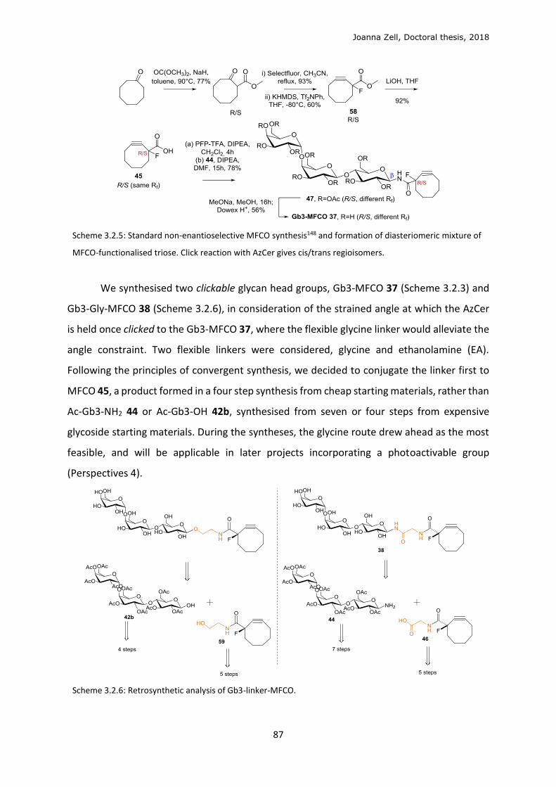
Joanna Zell, Doctoral thesis, 2018
87
Scheme 3.2.5: Standard non-enantioselective MFCO synthesis148 and formation of diasteriomeric mixture of
MFCO-functionalised triose. Click reaction with AzCer gives cis/trans regioisomers.
We synthesised two clickable glycan head groups, Gb3-MFCO 37 (Scheme 3.2.3) and
Gb3-Gly-MFCO 38 (Scheme 3.2.6), in consideration of the strained angle at which the AzCer
is held once clicked to the Gb3-MFCO 37, where the flexible glycine linker would alleviate the
angle constraint. Two flexible linkers were considered, glycine and ethanolamine (EA).
Following the principles of convergent synthesis, we decided to conjugate the linker first to
MFCO 45, a product formed in a four step synthesis from cheap starting materials, rather than
Ac-Gb3-NH2 44 or Ac-Gb3-OH 42b, synthesised from seven or four steps from expensive
glycoside starting materials. During the syntheses, the glycine route drew ahead as the most
feasible, and will be applicable in later projects incorporating a photoactivable group
(Perspectives 4).
Scheme 3.2.6: Retrosynthetic analysis of Gb3-linker-MFCO.
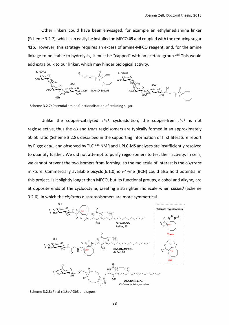
Joanna Zell, Doctoral thesis, 2018
88
Other linkers could have been envisaged, for example an ethylenediamine linker
(Scheme 3.2.7), which can easily be installed on MFCO 45 and coupled with the reducing sugar
42b. However, this strategy requires an excess of amine-MFCO reagent, and, for the amine
linkage to be stable to hydrolysis, it must be “capped” with an acetate group.215 This would
add extra bulk to our linker, which may hinder biological activity.
Scheme 3.2.7: Potential amine functionalisation of reducing sugar.
Unlike the copper-catalysed click cycloaddition, the copper-free click is not
regioselective, thus the cis and trans regioisomers are typically formed in an approximately
50:50 ratio (Scheme 3.2.8), described in the supporting information of first literature report
by Pigge et al., and observed by TLC.148 NMR and UPLC-MS analyses are insufficiently resolved
to quantify further. We did not attempt to purify regioisomers to test their activity. In cells,
we cannot prevent the two isomers from forming, so the molecule of interest is the cis/trans
mixture. Commercially available bicyclo[6.1.0]non-4-yne (BCN) could also hold potential in
this project. Is it slightly longer than MFCO, but its functional groups, alcohol and alkyne, are
at opposite ends of the cyclooctyne, creating a straighter molecule when clicked (Scheme
3.2.6), in which the cis/trans diastereoisomers are more symmetrical.
Scheme 3.2.8: Final clicked Gb3 analogues.
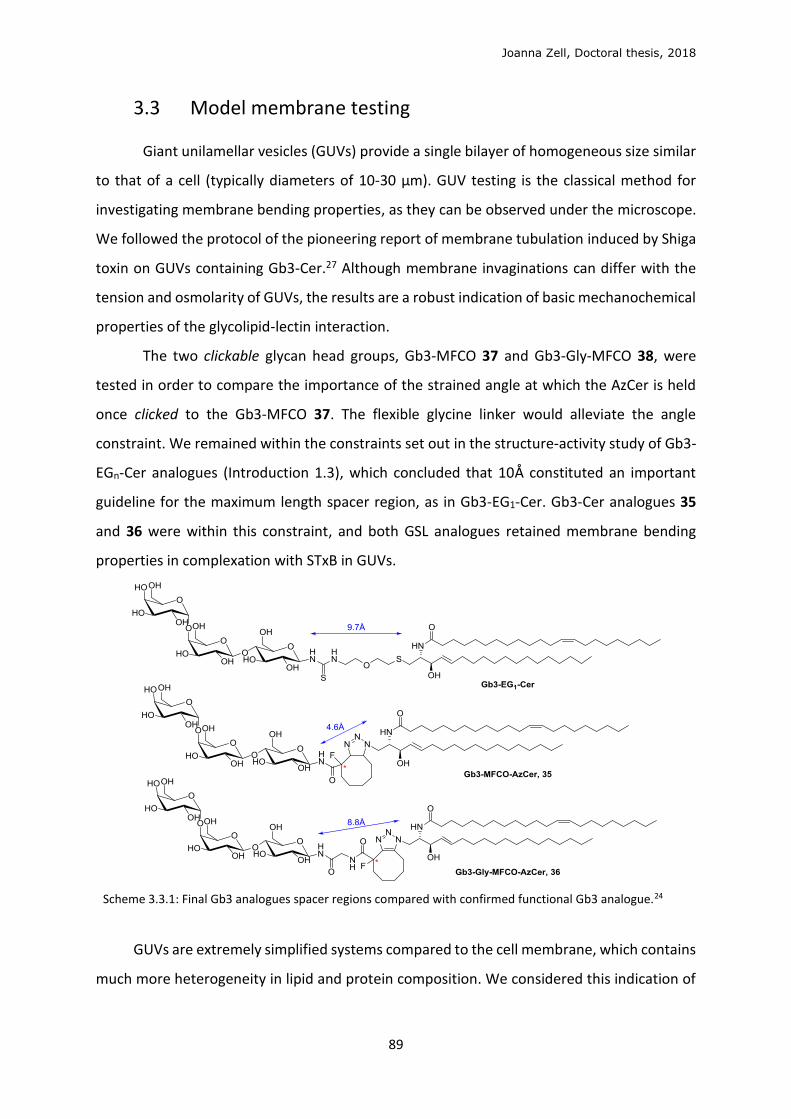
Joanna Zell, Doctoral thesis, 2018
89
3.3 Model membrane testing
Giant unilamellar vesicles (GUVs) provide a single bilayer of homogeneous size similar
to that of a cell (typically diameters of 10-30 µm). GUV testing is the classical method for
investigating membrane bending properties, as they can be observed under the microscope.
We followed the protocol of the pioneering report of membrane tubulation induced by Shiga
toxin on GUVs containing Gb3-Cer.27 Although membrane invaginations can differ with the
tension and osmolarity of GUVs, the results are a robust indication of basic mechanochemical
properties of the glycolipid-lectin interaction.
The two clickable glycan head groups, Gb3-MFCO 37 and Gb3-Gly-MFCO 38, were
tested in order to compare the importance of the strained angle at which the AzCer is held
once clicked to the Gb3-MFCO 37. The flexible glycine linker would alleviate the angle
constraint. We remained within the constraints set out in the structure-activity study of Gb3-
EGn-Cer analogues (Introduction 1.3), which concluded that 10Å constituted an important
guideline for the maximum length spacer region, as in Gb3-EG1-Cer. Gb3-Cer analogues 35
and 36 were within this constraint, and both GSL analogues retained membrane bending
properties in complexation with STxB in GUVs.
Scheme 3.3.1: Final Gb3 analogues spacer regions compared with confirmed functional Gb3 analogue.24
GUVs are extremely simplified systems compared to the cell membrane, which contains
much more heterogeneity in lipid and protein composition. We considered this indication of
Joanna Zell, Doctoral thesis, 2018
90
membrane curvature on GUVs sufficient to test the molecules in cells, despite the fact that
the cellular system is so chemically different.
We tested the click reaction on LUVs constituting 5% AzCer, since LUVs are simpler to
prepare and have similar chemical reactivity to GUVs, and are often used in the
literature.216,217 GUVs are typically used for microscopy observations, while LUVs can be
obtained in high quantities for biochemical studies. We observed very slow click kinetics. This
could be explained by the concentrations used, since click cycloadditions are extremely
dependent on concentration. Salomé et al. use similar LUV concentration (100 nM of lipid)
and observed 37% conversion after 24h with the solution phase reagent in large excess,
whereas we used 1 equivalent.218 Since this experiment required a large quantity of click
reagents, we did not optimise the experiment, but preferred to use the material for cellular
studies.
3.4 Sphingolipid metabolism
We began cellular testing on GM95 cells, a stable mutant cell line derived from mouse
melanoma MEB-4 cells, which are deficient in GSLs due to a lack of glucosylceramide synthase
(GCS) activity. Since we can envisage our reconstitution technique being applied to any
glycoside head group, for the functional reconstitution of any GSL analogue, not only Gb3, we
chose a system in which we could validate efficient reconstitution of any GSL. This cell line
has been used in many previous studies on GSL function.11,31,82 Chinese hamster ovary (CHO)
cells were used to validate our results further in a second cell line, since they express some
GSLs but lack the Gb3 synthase enzyme.
We followed the metabolism of 1-azidosphingosine (AzSph) into 1-azidoceramide
(AzCer) in cells via acylation by ceramide synthase (CesS) enzymes. We first analysed azide
metabolites by clicking with an alkyne-functionalised fluorophore in vitro, after cell treatment
and lysis. This method, pioneered by Thiele et al., involves separating the clicked biological
sample, based on their polarity, on high performance thin layer chromatography (HPTLC)
plates. This normally involves a purification step on the cell lysate; either protein
precipitation159 or lipid extraction,219 to purify lipid components from aqueous metabolites,
proteins and genetic molecules present in cells. We observe a specific band for AzCer clicked
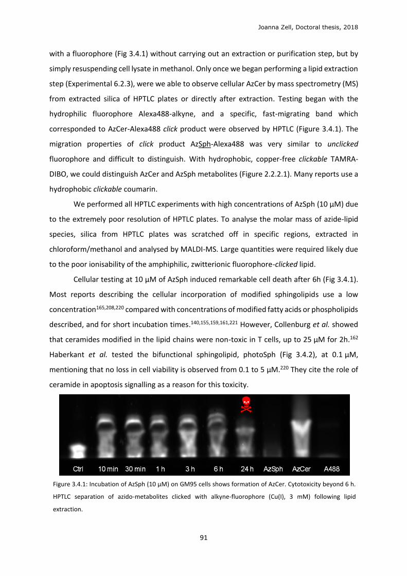
Joanna Zell, Doctoral thesis, 2018
91
with a fluorophore (Fig 3.4.1) without carrying out an extraction or purification step, but by
simply resuspending cell lysate in methanol. Only once we began performing a lipid extraction
step (Experimental 6.2.3), were we able to observe cellular AzCer by mass spectrometry (MS)
from extracted silica of HPTLC plates or directly after extraction. Testing began with the
hydrophilic fluorophore Alexa488-alkyne, and a specific, fast-migrating band which
corresponded to AzCer-Alexa488 click product were observed by HPTLC (Figure 3.4.1). The
migration properties of click product AzSph-Alexa488 was very similar to unclicked
fluorophore and difficult to distinguish. With hydrophobic, copper-free clickable TAMRA-
DIBO, we could distinguish AzCer and AzSph metabolites (Figure 2.2.2.1). Many reports use a
hydrophobic clickable coumarin.
We performed all HPTLC experiments with high concentrations of AzSph (10 μM) due
to the extremely poor resolution of HPTLC plates. To analyse the molar mass of azide-lipid
species, silica from HPTLC plates was scratched off in specific regions, extracted in
chloroform/methanol and analysed by MALDI-MS. Large quantities were required likely due
to the poor ionisability of the amphiphilic, zwitterionic fluorophore-clicked lipid.
Cellular testing at 10 μM of AzSph induced remarkable cell death after 6h (Fig 3.4.1).
Most reports describing the cellular incorporation of modified sphingolipids use a low
concentration165,208,220 compared with concentrations of modified fatty acids or phospholipids
described, and for short incubation times.140,155,159,161,221 However, Collenburg et al. showed
that ceramides modified in the lipid chains were non-toxic in T cells, up to 25 μM for 2h.162
Haberkant et al. tested the bifunctional sphingolipid, photoSph (Fig 3.4.2), at 0.1 μM,
mentioning that no loss in cell viability is observed from 0.1 to 5 μM.220 They cite the role of
ceramide in apoptosis signalling as a reason for this toxicity.
Figure 3.4.1: Incubation of AzSph (10 μM) on GM95 cells shows formation of AzCer. Cytotoxicity beyond 6 h.
HPTLC separation of azido-metabolites clicked with alkyne-fluorophore (Cu(I), 3 mM) following lipid
extraction.
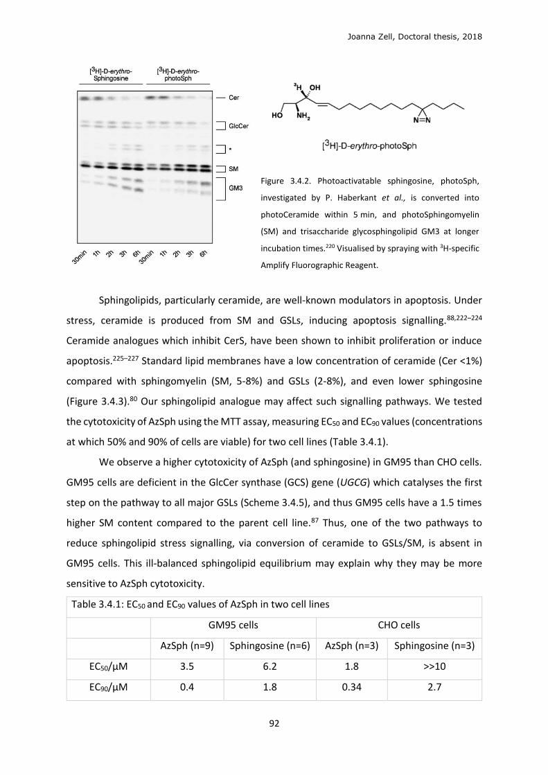
Joanna Zell, Doctoral thesis, 2018
92
Figure 3.4.2. Photoactivatable sphingosine, photoSph,
investigated by P. Haberkant et al., is converted into
photoCeramide within 5 min, and photoSphingomyelin
(SM) and trisaccharide glycosphingolipid GM3 at longer
incubation times.220 Visualised by spraying with 3H-specific
Amplify Fluorographic Reagent.
Sphingolipids, particularly ceramide, are well-known modulators in apoptosis. Under
stress, ceramide is produced from SM and GSLs, inducing apoptosis signalling.88,222–224
Ceramide analogues which inhibit CerS, have been shown to inhibit proliferation or induce
apoptosis.225–227 Standard lipid membranes have a low concentration of ceramide (Cer <1%)
compared with sphingomyelin (SM, 5-8%) and GSLs (2-8%), and even lower sphingosine
(Figure 3.4.3).80 Our sphingolipid analogue may affect such signalling pathways. We tested
the cytotoxicity of AzSph using the MTT assay, measuring EC50 and EC90 values (concentrations
at which 50% and 90% of cells are viable) for two cell lines (Table 3.4.1).
We observe a higher cytotoxicity of AzSph (and sphingosine) in GM95 than CHO cells.
GM95 cells are deficient in the GlcCer synthase (GCS) gene (UGCG) which catalyses the first
step on the pathway to all major GSLs (Scheme 3.4.5), and thus GM95 cells have a 1.5 times
higher SM content compared to the parent cell line.87 Thus, one of the two pathways to
reduce sphingolipid stress signalling, via conversion of ceramide to GSLs/SM, is absent in
GM95 cells. This ill-balanced sphingolipid equilibrium may explain why they may be more
sensitive to AzSph cytotoxicity.
Table 3.4.1: EC50 and EC90 values of AzSph in two cell lines
GM95 cells CHO cells
AzSph (n=9) Sphingosine (n=6) AzSph (n=3) Sphingosine (n=3)
EC50/μM 3.5 6.2 1.8 >>10
EC90/μM 0.4 1.8 0.34 2.7
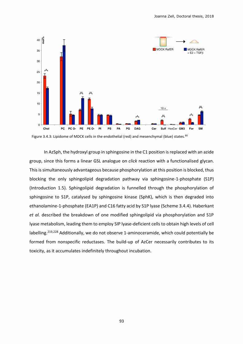
Joanna Zell, Doctoral thesis, 2018
93
Figure 3.4.3: Lipidome of MDCK cells in the endothelial (red) and mesenchymal (blue) states.80
In AzSph, the hydroxyl group in sphingosine in the C1 position is replaced with an azide
group, since this forms a linear GSL analogue on click reaction with a functionalised glycan.
This is simultaneously advantageous because phosphorylation at this position is blocked, thus
blocking the only sphingolipid degradation pathway via sphingosine-1-phosphate (S1P)
(Introduction 1.5). Sphingolipid degradation is funnelled through the phosphorylation of
sphingosine to S1P, catalysed by sphingosine kinase (SphK), which is then degraded into
ethanolamine-1-phosphate (EA1P) and C16 fatty acid by S1P lyase (Scheme 3.4.4). Haberkant
et al. described the breakdown of one modified sphingolipid via phosphorylation and S1P
lyase metabolism, leading them to employ SIP lyase-deficient cells to obtain high levels of cell
labelling.219,228 Additionally, we do not observe 1-aminoceramide, which could potentially be
formed from nonspecific reductases. The build-up of AzCer necessarily contributes to its
toxicity, as it accumulates indefinitely throughout incubation.
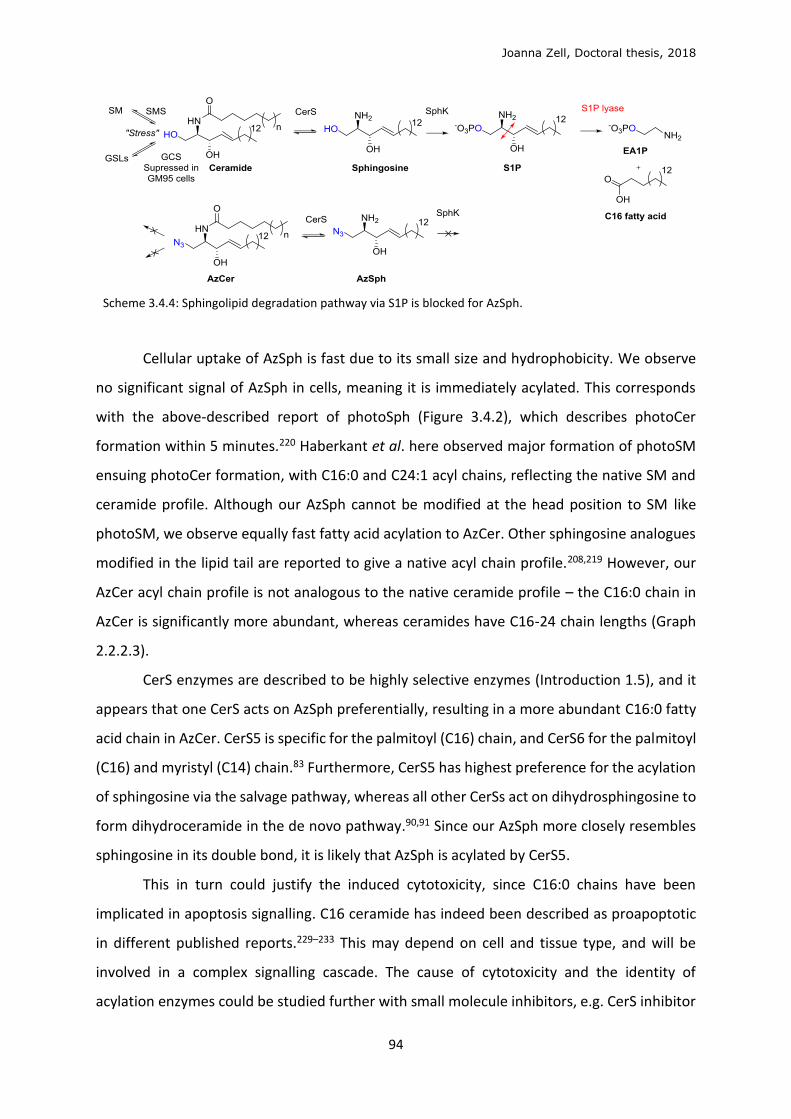
Joanna Zell, Doctoral thesis, 2018
94
Scheme 3.4.4: Sphingolipid degradation pathway via S1P is blocked for AzSph.
Cellular uptake of AzSph is fast due to its small size and hydrophobicity. We observe
no significant signal of AzSph in cells, meaning it is immediately acylated. This corresponds
with the above-described report of photoSph (Figure 3.4.2), which describes photoCer
formation within 5 minutes.220 Haberkant et al. here observed major formation of photoSM
ensuing photoCer formation, with C16:0 and C24:1 acyl chains, reflecting the native SM and
ceramide profile. Although our AzSph cannot be modified at the head position to SM like
photoSM, we observe equally fast fatty acid acylation to AzCer. Other sphingosine analogues
modified in the lipid tail are reported to give a native acyl chain profile.208,219 However, our
AzCer acyl chain profile is not analogous to the native ceramide profile – the C16:0 chain in
AzCer is significantly more abundant, whereas ceramides have C16-24 chain lengths (Graph
2.2.2.3).
CerS enzymes are described to be highly selective enzymes (Introduction 1.5), and it
appears that one CerS acts on AzSph preferentially, resulting in a more abundant C16:0 fatty
acid chain in AzCer. CerS5 is specific for the palmitoyl (C16) chain, and CerS6 for the palmitoyl
(C16) and myristyl (C14) chain.83 Furthermore, CerS5 has highest preference for the acylation
of sphingosine via the salvage pathway, whereas all other CerSs act on dihydrosphingosine to
form dihydroceramide in the de novo pathway.90,91 Since our AzSph more closely resembles
sphingosine in its double bond, it is likely that AzSph is acylated by CerS5.
This in turn could justify the induced cytotoxicity, since C16:0 chains have been
implicated in apoptosis signalling. C16 ceramide has indeed been described as proapoptotic
in different published reports.229–233 This may depend on cell and tissue type, and will be
involved in a complex signalling cascade. The cause of cytotoxicity and the identity of
acylation enzymes could be studied further with small molecule inhibitors, e.g. CerS inhibitor
Joanna Zell, Doctoral thesis, 2018
95
fumonisin B, manipulation of CerS enzyme expression, interference with apoptotic cascades
such as those depending on caspases, or broadening experiments to a further cell types.
Since each GSL is often present at a very small molar percentage of the PM (Figure
3.4.3), we considered that even a low concentration of our modified lipid at the PM should
be sufficient to observe lectin binding, this being our final validation. To avoid cytotoxic
effects of ceramide analogues, we incubated cells with a low AzSph concentration (0.5 μM)
to obtain a stable ratio of approximately 2-3 molecules of total AzCer to total ceramide (Graph
2.2.2.6), and a stable amount of ceramide over 24h, indicating that cells are not under
ceramide-signalling stress.
Attempts to halt cell proliferation, to thus reduce the dilution of AzCer labelling, serum
starvation conditions were tested. The presence of natural sphingolipids in serum should also
be noted. No serum conditions induced a higher AzCer/Cer ratio over 48h compared to serum
conditions. Eto et al. described an increase in cellular C16 ceramide in serum-free culture
conditions due to an onset of apoptosis.230 Despite this, we do not observe reduction in cell
viability at low AzSph concentration.
The lateral localisation of Gb3-Cer on the cell surface appears to be important to its
receptor activity (Introduction 1.3). In the natural system, Gb3-Cer is transported from the
Golgi to its functional position as a lectin receptor on the cell surface. Gb3-Cer negative (Gb3-
) cells incubated with external Gb3-Cer/BSA can permit low levels of binding of STxB at the
cell surface. However, the lectin’s traffic is always dysregulated from the natural retrograde
route (Figure 1.3.4). Externally supplemented Gb3-Cer with short acyl chains (natural long
chain species fail to be reconstituted) may function non-canonically because it does not
organise correctly within biological membranes. We hypothesised that AzCer with the natural
complement of acyl chains would be incorporated into native lipid transport of the cell,
allowing it to be recycled through a native pathway, to the PM, although temporarily, to
undergo click reaction and present Gb3-Cer at the surface.
We studied the intracellular localisation of AzCer by immunofluoresence (IF)
experiments. When cells were treated with AzSph, then stained with alkyne or cyclooctyne
fluorophores, AzSph metabolites (AzCer) appear to be localised in intracellular membranes,
possibly the ER, where CerS enzymes are known to be localised.83 One must consider a margin
of error in lipid localisation, since performing click reactions on AzSph with a fluorophore may
change its localisation. In general, fixation is poorly effective on lipids, which contain few
Joanna Zell, Doctoral thesis, 2018
96
nucleophilic, fixable groups, even after the most robust fixation techniques.93 A
permeabilisation step with Saponin was the cleanest method to wash cells of nonspecific
unclicked fluorophore. However, this treatment destabilises the membrane and adds even
greater possible error. Despite this, we see repeatedly specific staining compared to negative
controls, and similar staining in live cell imaging. AzCer localisation in all intracellular
membranes is rational since the substrate closely resembles ceramide, which is synthesised
in the ER and should have little intracellular specificity. Furthermore, short chain lipids are
considered to preferentially localise in the Golgi and ER, whereas long chain lipids would
preferentially localise in the PM, observed by differences in the thickness of transmembrane
domains of PM/Golgi/ER proteins.197
GM95 cells were too poorly adhesive to undertake lengthy IF experiments to study the
intracellular localisation of AzCer metabolites. GSLs have long been known as modulators of
cell adhesion.87,234,235 Jwa Hidari et al. showed that GM95 cells lost the ability to adhere to
standard extra-cellular matrix proteins when treated with sphingomyelinase, which removes
SM from the cell surface, whereas GSL-expressing MEB-4 cells, from which GM95 cells were
mutated, showed no loss in adhesion.87 An inhibitor GCS and major GSL synthesis, PDMP,
blocked adherence of macrophages during differentiation, further indicating GSLs are
implicated in cell adhesion.225 In the same way, PPMP treatment of naïve E14 mouse stem
cells sporadically reduced adhesion during differentiation (unpublished). Click protocols for
imaging the localisation of clickable lipids were optimised on CHO and HeLa cells
(Experimental 6.2.6).
3.5 In cellulo click
We synthesised Gb3-MFCO 37, Gb3-Gly-MFCO 38 (Scheme 2.1.3.2), lactose-MFCO
49, lactose-Gly-MFCO 50 and lactose-BCN 63 (Scheme 2.1.3.4, Results 2.1.3). Our final
validation of a functional GSL analogue would be via the recognition of the Gb3 head group
with its specific lectin binder, the Shiga toxin. However, to first study the click reaction via
high resolution mass spectrometry (HRMS) we tested the protocol with lactose-MFCO 49,
obtained on larger scale.
Joanna Zell, Doctoral thesis, 2018
97
We observed that the reaction with lactose-MFCO 49 was poorly efficient, since
there is no significant drop in cellular AzCer levels even when using an excess of cyclooctyne
reagent. In comparison, click reaction was efficient with hydrophobic clickable fluorophore
TAMRA-DIBO. This was observed by a major consumption of cellular AzCer and appearance
of the clicked product TAMRA-DIBO-AzCer. To confirm the lipid extraction of the clicked
glycolipid, we synthesised in vitro purified Gb3- and lactose-MFCO-AzCer (C22:1). These were
used as internal standards, spiked into biological samples before the lipid extraction, to
ensure that the click product is extracted and analysed by HRMS by our protocol. Since we
observe the Gb3- and lactose-MFCO-AzCer (C22:1) internal standards after extraction, we can
confirm that our extraction protocol is adapted to the desired glycolipid products, and that
cellular click reaction forming lactose-MFCO-AzCer is inefficient since no cellular product is
observed.
We had hypothesised that, although AzCer may be predominantly present in
intracellular membranes, it would cycle to the plasma membrane (PM) by vesicular trafficking
in order to undergo click reaction at the cell surface (Fig 3.5.1a). Since we observed no click
reaction with hydrophilic, non-permeating cyclooctynes (lactose-MFCO, Alexa488-DIBO), we
suggested that very little AzCer is present at the PM. This corresponds with the commonly
accepted localisation of natural ceramide, synthesised in the ER, glycosylated in the Golgi and
thus depleted at the PM.
Alexa488-DIBO is a commonly-used reagent for live-cell staining62,162,163, and is able
to specifically stain AzSph and AzPhy in IF experiments on live CHO cells (Fig 3.5.1b). Such
experiments were performed with overnight incubation of fluorophore, indicating that the
AzCer recycles to the PM overnight to click with extracellular fluorophore. Alternatively the
fluorophore could penetrate to the intracellular AzCer. Alexa488-DIBO click product was not
observed in HRMS experiments because it is too hydrophilic to be extracted in our lipid
extraction protocol, not because it was not taken up or clicked. After click at the surface, the
click product is endocytosed to intracellular membranes, as likely occurs to the small
percentage of the Gb3-MFCO-AzCer product. This would explain why no STxB binding is
observed.
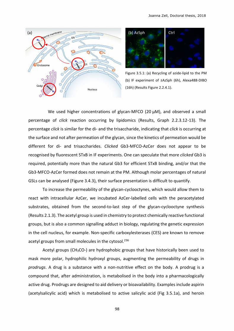
Joanna Zell, Doctoral thesis, 2018
98
Figure 3.5.1: (a) Recycling of azide-lipid to the PM
(b) IF experiment of ±AzSph (6h), Alexa488-DIBO
(16h) (Results Figure 2.2.4.1).
We used higher concentrations of glycan-MFCO (20 μM), and observed a small
percentage of click reaction occurring by lipidomics (Results, Graph 2.2.3.12-13). The
percentage click is similar for the di- and the trisaccharide, indicating that click is occurring at
the surface and not after permeation of the glycan, since the kinetics of permeation would be
different for di- and trisaccharides. Clicked Gb3-MFCO-AzCer does not appear to be
recognised by fluorescent STxB in IF experiments. One can speculate that more clicked Gb3 is
required, potentially more than the natural Gb3 for efficient STxB binding, and/or that the
Gb3-MFCO-AzCer formed does not remain at the PM. Although molar percentages of natural
GSLs can be analysed (Figure 3.4.3), their surface presentation is difficult to quantify.
To increase the permeability of the glycan-cyclooctynes, which would allow them to
react with intracellular AzCer, we incubated AzCer-labelled cells with the peracetylated
substrates, obtained from the second-to-last step of the glycan-cyclooctyne synthesis
(Results 2.1.3). The acetyl group is used in chemistry to protect chemically reactive functional
groups, but is also a common signalling adduct in biology, regulating the genetic expression
in the cell nucleus, for example. Non-specific carboxylesterases (CES) are known to remove
acetyl groups from small molecules in the cytosol.236
Acetyl groups (CH3CO-) are hydrophobic groups that have historically been used to
mask more polar, hydrophilic hydroxyl groups, augmenting the permeability of drugs in
prodrugs. A drug is a substance with a non-nutritive effect on the body. A prodrug is a
compound that, after administration, is metabolised in the body into a pharmacologically
active drug. Prodrugs are designed to aid delivery or bioavailability. Examples include aspirin
(acetylsalicylic acid) which is metabolised to active salicylic acid (Fig 3.5.1a), and heroin
(b) AzSph Ctrl (a)
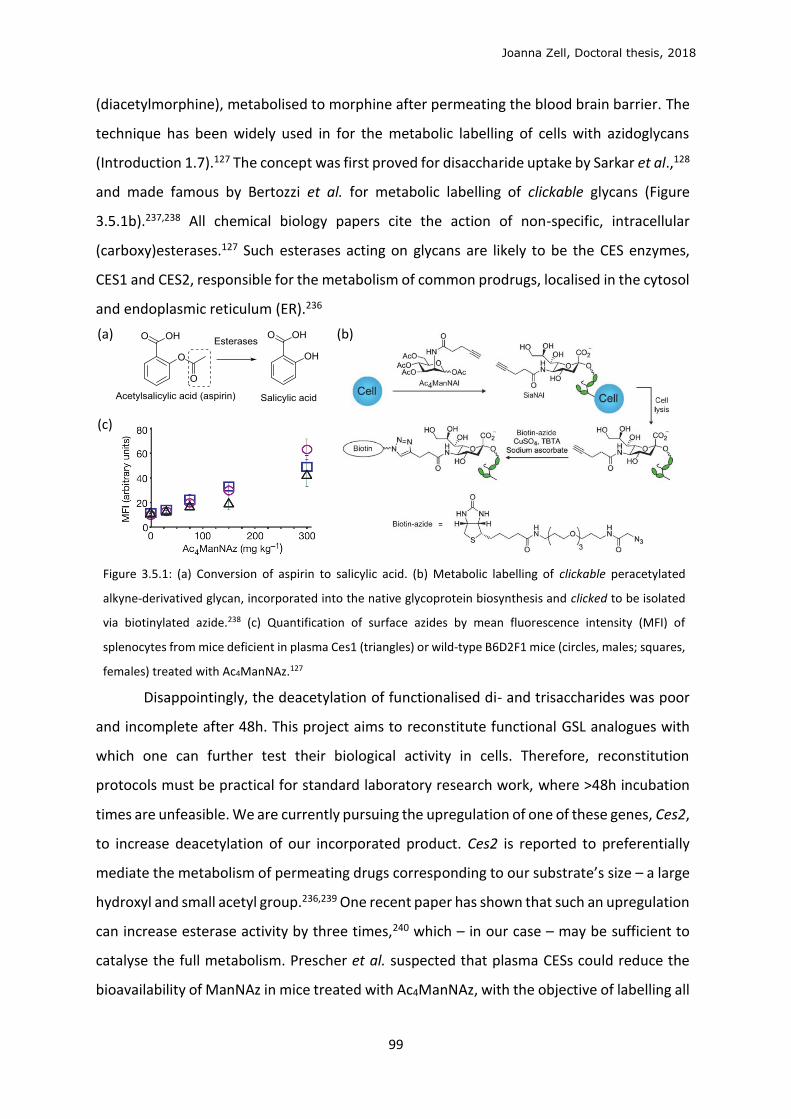
Joanna Zell, Doctoral thesis, 2018
99
(diacetylmorphine), metabolised to morphine after permeating the blood brain barrier. The
technique has been widely used in for the metabolic labelling of cells with azidoglycans
(Introduction 1.7).127 The concept was first proved for disaccharide uptake by Sarkar et al.,128
and made famous by Bertozzi et al. for metabolic labelling of clickable glycans (Figure
3.5.1b).237,238 All chemical biology papers cite the action of non-specific, intracellular
(carboxy)esterases.127 Such esterases acting on glycans are likely to be the CES enzymes,
CES1 and CES2, responsible for the metabolism of common prodrugs, localised in the cytosol
and endoplasmic reticulum (ER).236
Figure 3.5.1: (a) Conversion of aspirin to salicylic acid. (b) Metabolic labelling of clickable peracetylated
alkyne-derivatived glycan, incorporated into the native glycoprotein biosynthesis and clicked to be isolated
via biotinylated azide.238 (c) Quantification of surface azides by mean fluorescence intensity (MFI) of
splenocytes from mice deficient in plasma Ces1 (triangles) or wild-type B6D2F1 mice (circles, males; squares,
females) treated with Ac4ManNAz.127
Disappointingly, the deacetylation of functionalised di- and trisaccharides was poor
and incomplete after 48h. This project aims to reconstitute functional GSL analogues with
which one can further test their biological activity in cells. Therefore, reconstitution
protocols must be practical for standard laboratory research work, where >48h incubation
times are unfeasible. We are currently pursuing the upregulation of one of these genes, Ces2,
to increase deacetylation of our incorporated product. Ces2 is reported to preferentially
mediate the metabolism of permeating drugs corresponding to our substrate’s size – a large
hydroxyl and small acetyl group.236,239 One recent paper has shown that such an upregulation
can increase esterase activity by three times,240 which – in our case – may be sufficient to
catalyse the full metabolism. Prescher et al. suspected that plasma CESs could reduce the
bioavailability of ManNAz in mice treated with Ac4ManNAz, with the objective of labelling all
(a) (b)
(c)
Joanna Zell, Doctoral thesis, 2018
100
mouse tissues.127 They trialled the use of plasma Ces1-deficient mice to reduce deacetylation
in the plasma, but saw no difference in labelling of splenocyte (cells rich in sialiosides) (Figure
3.5.1c) and tissues compared with control mice. This indicates some compensatory effect of
multiple CES enzymes.
3.6 Optimisation of lipid localisation
To obtain an azide labelling at the plasma membrane (PM), we envisaged different
azide-lipid precursors, which might prove to have a different metabolite profile, and thus a
different cellular localisation. Longer chain lipids are thought to be preferentially housed in
the PM, whereas shorter lipids localise to the Golgi or ER. Although this theory is difficult to
verify, a study of protein transmembrane domains showed that longer hydrophobic domains
exist in PM-localised proteins than the ER- and Golgi-localised proteins.197
AzSph is acylated to AzCer (C16:0) by the CerS5 enzyme, which has high specificity for
sphingosine formed in the salvage pathway.91 Sphinganine, formed in the de novo synthesis
pathway, is metabolised by other CerSs, which have individual specificities for longer fatty
acid chains (Graph 3.6.1, Introduction 1.5). An azide analogue of sphinganine holds promise
as it may be metabolised like sphinganine, via the de novo pathway to long chain ceramides,
which should have a higher propensity to localise to the PM. Phytosphingosine is structurally
more similar to sphinganine than sphingosine as it has no double bond in the C4 position.
Phytosphingosine is a plant-derived starting material from which we begin the AzSph
synthesis. 1-azidophytosphingosine (AzPhy) was easily obtained synthetically (Results 2.1.2).
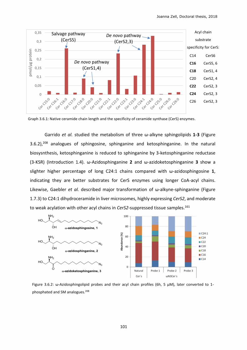
Joanna Zell, Doctoral thesis, 2018
101
Acyl chain
substrate
specificity for CerS:
C14
C16
C18
C20
C22
C24
C26
CerS6
CerS5, 6
CerS1, 4
CerS2, 4
CerS2, 3
CerS2, 3
CerS2, 3
Graph 3.6.1: Native ceramide chain length and the specificity of ceramide synthase (CerS) enzymes.
Garrido et al. studied the metabolism of three ω-alkyne sphingolipids 1-3 (Figure
3.6.2),208 analogues of sphingosine, sphinganine and ketosphinganine. In the natural
biosysnthesis, ketosphinganine is reduced to sphinganine by 3-ketosphinganine reductase
(3-KSR) (Introduction 1.4). ω-Azidosphinganine 2 and ω-azidoketosphinganine 3 show a
slighter higher percentage of long C24:1 chains compared with ω-azidosphingosine 1,
indicating they are better substrates for CerS enzymes using longer CoA-acyl chains.
Likewise, Gaebler et al. described major transformation of ω-alkyne-sphinganine (Figure
1.7.3) to C24:1 dihydroceramide in liver microsomes, highly expressing CerS2, and moderate
to weak acylation with other acyl chains in CerS2-suppressed tissue samples.161
Figure 3.6.2: ω-Azidosphingolipid probes and their acyl chain profiles (6h, 5 μM), later converted to 1-
phosphated and SM analogues.208
0
0,05
0,1
0,15
0,2
0,25
0,3
0,35p
mo
l/μ
g p
rote
inSalvage pathway
(CerS5) De novo pathway
(CerS2,3)
De novo pathway (CerS1,4)
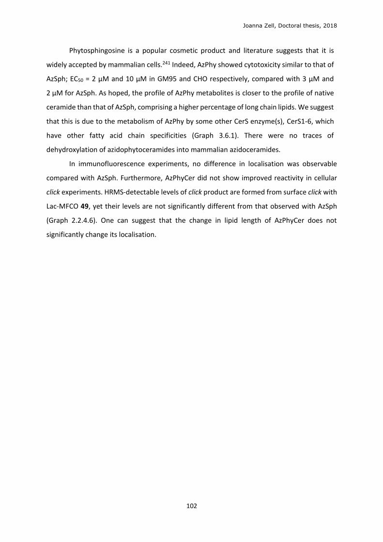
Joanna Zell, Doctoral thesis, 2018
102
Phytosphingosine is a popular cosmetic product and literature suggests that it is
widely accepted by mammalian cells.241 Indeed, AzPhy showed cytotoxicity similar to that of
AzSph; EC50 = 2 μM and 10 μM in GM95 and CHO respectively, compared with 3 μM and
2 μM for AzSph. As hoped, the profile of AzPhy metabolites is closer to the profile of native
ceramide than that of AzSph, comprising a higher percentage of long chain lipids. We suggest
that this is due to the metabolism of AzPhy by some other CerS enzyme(s), CerS1-6, which
have other fatty acid chain specificities (Graph 3.6.1). There were no traces of
dehydroxylation of azidophytoceramides into mammalian azidoceramides.
In immunofluorescence experiments, no difference in localisation was observable
compared with AzSph. Furthermore, AzPhyCer did not show improved reactivity in cellular
click experiments. HRMS-detectable levels of click product are formed from surface click with
Lac-MFCO 49, yet their levels are not significantly different from that observed with AzSph
(Graph 2.2.4.6). One can suggest that the change in lipid length of AzPhyCer does not
significantly change its localisation.
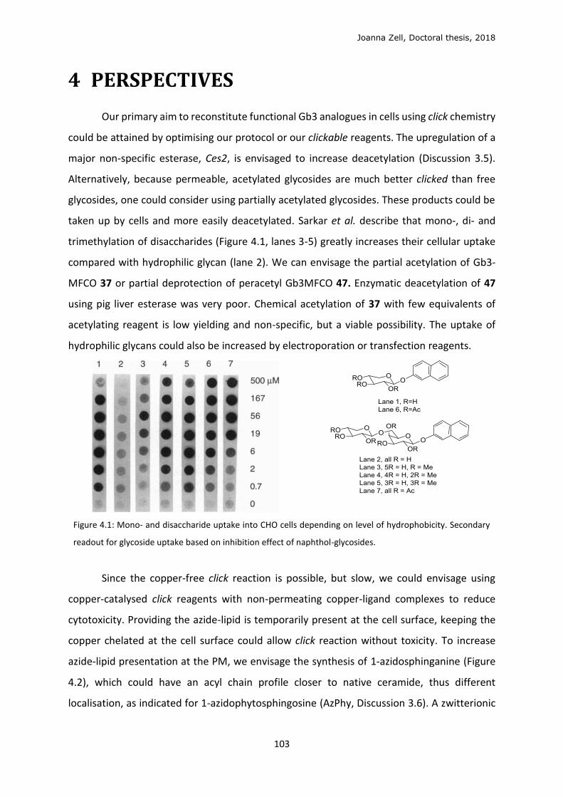
Joanna Zell, Doctoral thesis, 2018
103
4 PERSPECTIVES
Our primary aim to reconstitute functional Gb3 analogues in cells using click chemistry
could be attained by optimising our protocol or our clickable reagents. The upregulation of a
major non-specific esterase, Ces2, is envisaged to increase deacetylation (Discussion 3.5).
Alternatively, because permeable, acetylated glycosides are much better clicked than free
glycosides, one could consider using partially acetylated glycosides. These products could be
taken up by cells and more easily deacetylated. Sarkar et al. describe that mono-, di- and
trimethylation of disaccharides (Figure 4.1, lanes 3-5) greatly increases their cellular uptake
compared with hydrophilic glycan (lane 2). We can envisage the partial acetylation of Gb3-
MFCO 37 or partial deprotection of peracetyl Gb3MFCO 47. Enzymatic deacetylation of 47
using pig liver esterase was very poor. Chemical acetylation of 37 with few equivalents of
acetylating reagent is low yielding and non-specific, but a viable possibility. The uptake of
hydrophilic glycans could also be increased by electroporation or transfection reagents.
Figure 4.1: Mono- and disaccharide uptake into CHO cells depending on level of hydrophobicity. Secondary
readout for glycoside uptake based on inhibition effect of naphthol-glycosides.
Since the copper-free click reaction is possible, but slow, we could envisage using
copper-catalysed click reagents with non-permeating copper-ligand complexes to reduce
cytotoxicity. Providing the azide-lipid is temporarily present at the cell surface, keeping the
copper chelated at the cell surface could allow click reaction without toxicity. To increase
azide-lipid presentation at the PM, we envisage the synthesis of 1-azidosphinganine (Figure
4.2), which could have an acyl chain profile closer to native ceramide, thus different
localisation, as indicated for 1-azidophytosphingosine (AzPhy, Discussion 3.6). A zwitterionic
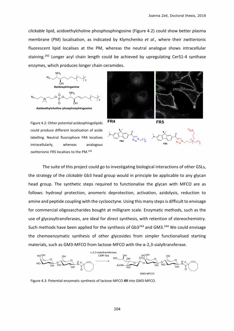
Joanna Zell, Doctoral thesis, 2018
104
clickable lipid, azidoethylcholine phosphosphingosine (Figure 4.2) could show better plasma
membrane (PM) localisation, as indicated by Klymchenko et al., where their zwitterionic
fluorescent lipid localises at the PM, whereas the neutral analogue shows intracellular
staining.242 Longer acyl chain length could be achieved by upregulating CerS1-4 synthase
enzymes, which produces longer chain ceramides.
Figure 4.2: Other potential azidosphingolipids
could produce different localisation of azide
labelling. Neutral fluorophore FR4 localises
intracellularly, whereas analogous
zwitterionic FR5 localises to the PM.242
The suite of this project could go to investigating biological interactions of other GSLs,
the strategy of the clickable Gb3 head group would in principle be applicable to any glycan
head group. The synthetic steps required to functionalise the glycan with MFCO are as
follows: hydroxyl protection, anomeric deprotection, activation, azidolysis, reduction to
amine and peptide coupling with the cyclooctyne. Using this many steps is difficult to envisage
for commercial oligosaccharides bought at milligram scale. Enzymatic methods, such as the
use of glycosyltransferases, are ideal for direct synthesis, with retention of stereochemistry.
Such methods have been applied for the synthesis of Gb3243 and GM3.244 We could envisage
the chemoenzymatic synthesis of other glycosides from simpler functionalised starting
materials, such as GM3-MFCO from lactose-MFCO with the α-2,3-sialyltransferase.
Figure 4.3: Potential enzymatic synthesis of lactose-MFCO 49 into GM3-MFCO.
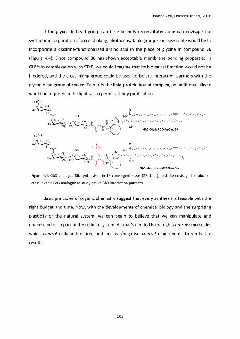
Joanna Zell, Doctoral thesis, 2018
105
If the glycoside head group can be efficiently reconstituted, one can envisage the
synthetic incorporation of a crosslinking, photoactivatable group. One easy route would be to
incorporate a diazirine-functionalised amino acid in the place of glycine in compound 36
(Figure 4.4). Since compound 36 has shown acceptable membrane bending properties in
GUVs in complexation with STxB, we could imagine that its biological function would not be
hindered, and the crosslinking group could be used to isolate interaction partners with the
glycan head group of choice. To purify the lipid-protein bound complex, an additional alkyne
would be required in the lipid tail to permit affinity purification.
Figure 4.4: Gb3 analogue 36, synthesised in 15 convergent steps (27 steps), and the envisageable photo-
crosslinkable Gb3 analogue to study native Gb3 interaction partners.
Basic principles of organic chemistry suggest that every synthesis is feasible with the
right budget and time. Now, with the developments of chemical biology and the surprising
plasticity of the natural system, we can begin to believe that we can manipulate and
understand each part of the cellular system. All that’s needed is the right controls: molecules
which control cellular function, and positive/negative control experiments to verify the
results!
Joanna Zell, Doctoral thesis, 2018
106
5 CONCLUSIONS
We have synthesised novel clickable biomolecule analogues and tested their validity
in model membranes and cell culture. Two different glycan-cyclooctyne head groups, Gb3-
MFCO and Gb3-Gly-MFCO, when clicked with 1-azidoceramide, show promising model
membrane activity when bound to fluorescent STxB. This indicates that the proposed
structural modification in the spacer region between glycan head and lipid tail is tolerated in
biological function. The synthesis of glycan-cyclooctynes can be applied to other glycans and
bioorthogonoal click reagents for the study of other GSLs. Similarly, the synthesis of 1-
azidosphingosine and 1-azidoceramide can be extended to other azido-sphingolipid
analogues for a varied metabolism profile.
We have shown efficient metabolic labelling of sphingolipids using clickable 1-
azidosphingosine, which is rapidly incorporated into cells and metabolised into 1-
azidoceramide. 1-azidoceramide localises in intracellular membranes, thus click reaction with
non-cell-permeating reagents such as glycan-cyclooctynes is poorly efficient. 1-
Azidoceramide undergoes specific and efficient bioorthogonal click reaction with permeable,
peracetylated glycan-cyclooctynes. We have quantified this in cellulo click reaction by HRMS
lipidomics. Endogenous deacetylation of clicked glycolipid analogues is inefficient in two cells
lines tested. Several pathways to change the localisation of lipid or glycan parts are available,
and should permit the reconstitution of a biologically functional glycolipid analogue into cells.
With this technique in hand, one can envisage a precise and efficient reconstitution of long
chain glycolipid analogues for the study of GL-Lect endocytosis.
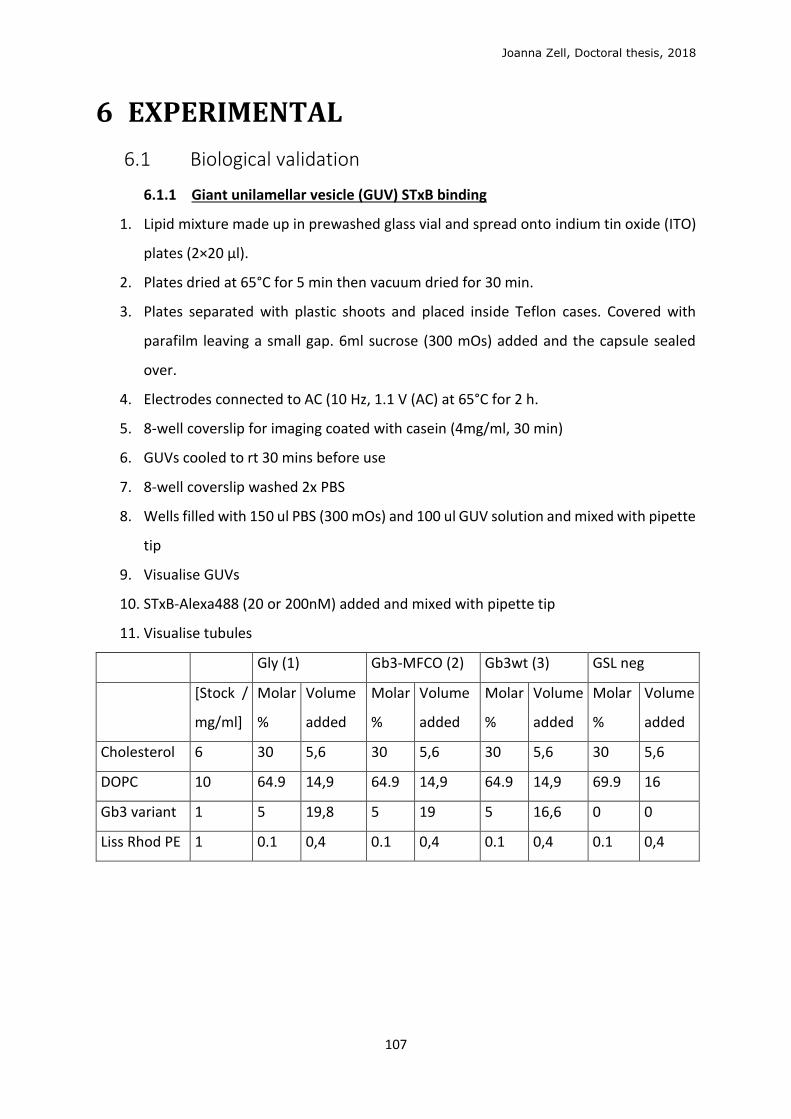
Joanna Zell, Doctoral thesis, 2018
107
6 EXPERIMENTAL
6.1 Biological validation
6.1.1 Giant unilamellar vesicle (GUV) STxB binding
1. Lipid mixture made up in prewashed glass vial and spread onto indium tin oxide (ITO)
plates (2×20 μl).
2. Plates dried at 65°C for 5 min then vacuum dried for 30 min.
3. Plates separated with plastic shoots and placed inside Teflon cases. Covered with
parafilm leaving a small gap. 6ml sucrose (300 mOs) added and the capsule sealed
over.
4. Electrodes connected to AC (10 Hz, 1.1 V (AC) at 65°C for 2 h.
5. 8-well coverslip for imaging coated with casein (4mg/ml, 30 min)
6. GUVs cooled to rt 30 mins before use
7. 8-well coverslip washed 2x PBS
8. Wells filled with 150 ul PBS (300 mOs) and 100 ul GUV solution and mixed with pipette
tip
9. Visualise GUVs
10. STxB-Alexa488 (20 or 200nM) added and mixed with pipette tip
11. Visualise tubules
Gly (1) Gb3-MFCO (2) Gb3wt (3) GSL neg
[Stock /
mg/ml]
Molar
%
Volume
added
Molar
%
Volume
added
Molar
%
Volume
added
Molar
%
Volume
added
Cholesterol 6 30 5,6 30 5,6 30 5,6 30 5,6
DOPC 10 64.9 14,9 64.9 14,9 64.9 14,9 69.9 16
Gb3 variant 1 5 19,8 5 19 5 16,6 0 0
Liss Rhod PE 1 0.1 0,4 0.1 0,4 0.1 0,4 0.1 0,4
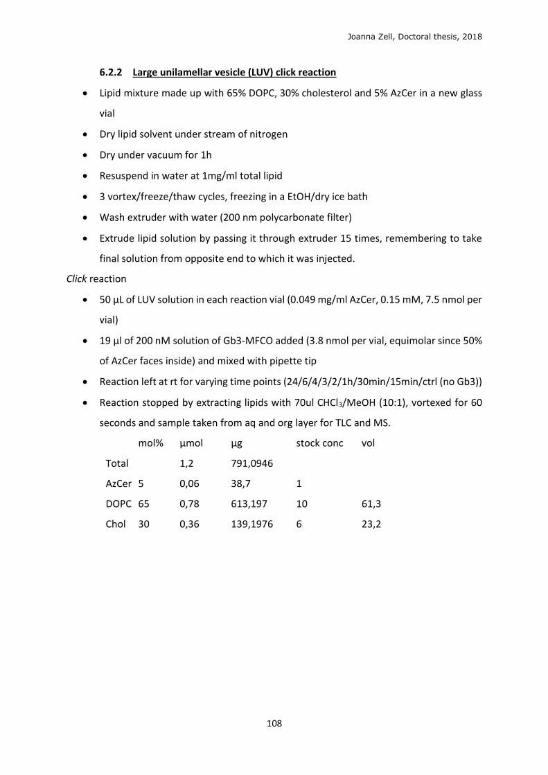
Joanna Zell, Doctoral thesis, 2018
108
6.2.2 Large unilamellar vesicle (LUV) click reaction
Lipid mixture made up with 65% DOPC, 30% cholesterol and 5% AzCer in a new glass
vial
Dry lipid solvent under stream of nitrogen
Dry under vacuum for 1h
Resuspend in water at 1mg/ml total lipid
3 vortex/freeze/thaw cycles, freezing in a EtOH/dry ice bath
Wash extruder with water (200 nm polycarbonate filter)
Extrude lipid solution by passing it through extruder 15 times, remembering to take
final solution from opposite end to which it was injected.
Click reaction
50 μL of LUV solution in each reaction vial (0.049 mg/ml AzCer, 0.15 mM, 7.5 nmol per
vial)
19 μl of 200 nM solution of Gb3-MFCO added (3.8 nmol per vial, equimolar since 50%
of AzCer faces inside) and mixed with pipette tip
Reaction left at rt for varying time points (24/6/4/3/2/1h/30min/15min/ctrl (no Gb3))
Reaction stopped by extracting lipids with 70ul CHCl3/MeOH (10:1), vortexed for 60
seconds and sample taken from aq and org layer for TLC and MS.
mol% μmol μg stock conc vol
Total
1,2 791,0946
AzCer 5 0,06 38,7 1
DOPC 65 0,78 613,197 10 61,3
Chol 30 0,36 139,1976 6 23,2
Joanna Zell, Doctoral thesis, 2018
109
6.2.3 Cellular metabolism of AzSph and click with fluorophore
The day before, 500,000 GM95 cells/well seeded in 6-well plate (2 ml DMEM++)
Wash 2x DMEM(++)
Add AzSph (10 µM, 1 ml) in DMEM(++)(Penicillin/streptomycin 5%, foetal bovine serum
10%)
Incubate 37 °C for various times
Wash 1x PBS and 2x ammonium bicarbonate solution (150 mM) at rt
Scratch cells in 1 mL ammonium bicarbonate solution at 4 °C (6 wells per condition)
Centrifuge 5 min at 4700 RPM at 4 °C
Remove supernatant
Lipid extraction
Resuspend pellet in 200 µL H2O
Add 1000 µL CHCl3/MeOH (10 :1)
Vortex 75 min
Centrifuge quickly in small benchtop centrifuge
Keep supernatant
Dry the solvent
Re-suspend in 100 μL of CHCl3/MeOH/H2O (50:40:5)
Click reaction (2h in Eppendorf shaker at 800 RPM, 25 °C)
1 – Cell lysate/reference (150 µL; references at 50 μM, 300 µL)
2 – Alexa/TAMRA (100 μM final, stock 1.25 mM)
3 – Cu(I) (2 mM) (Cu(BF4)) (100mM)
4 – 45 μL EtOH (volume total=100ul)
Spot supernatant mix (20 μL) on HPTLC plate (10 cm×10 cm) and dry 10 min under
vacuum or with heat gun
Elute TLC in cuvette (CHCl3/MeOH/H2O 50:40:10) and scan
Joanna Zell, Doctoral thesis, 2018
110
6.2.4 Cytotoxicity assay
1 The day before, 50,000 cells/well seeded (200 μl in DMEM++) in a 96-well plate
2 Wash 2x DMEM(++)
3 Add compound (AzSph/AzPhy/cyclooctyne, 100 µL at descending concentrations) in
DMEM(++), 3 wells per condition.
4 Incubate at 37 °C, 24 h
5 Remove medium
6 Wash cells with 200 µL virgin DMEM at 4 °C
7 Add 150 µL virgin DMEM and 20 µL MTT solution (5 mg.mL-1 in H2O, protected from the
light)
8 Incubate for at least 3h at 37°C in the dark
9 Gently remove medium
(At this step, the plate can be conserved at 4°C to read at a later date)
10 Add 100 µL DMSO
11 Shake for 20 min
12 Read plate on spectrophotometer at 570 nm
Joanna Zell, Doctoral thesis, 2018
111
6.2.5 Cell treatment of click reagents for lipidomics testing, optimised protocol
1. The day before, seed 250,000 GM95 cells/well in 6-well plate (2 ml)
2. Wash 2x DMEM (++)
3. Add AzSph/AzPhy (0.5 µM, 1 ml) in DMEM(++)
4. Incubate at 37 °C (6h to overnight)
5. Wash 2x DMEM(++)
6. Incubate w/o cyclooctyne reagent in DMEM(++) (varying time points)
7. Wash 2x with ammonium bicarbonate (150 mM) at 4°C
8. Add 1 ml ammonium bicarbonate and scratch cells at 4 °C
9. Centrifuge 5 min à 13.3K RPM à 4 °C
10. Throw away supernatant
11. Resuspend pellet in 300 µL ammonium bicarb
12. Homogenise with needle and syringe (18G, then 25G needle)
13. Take 50 μL for protein quantification (MicroBCA kit)
14. Snap freeze in N2(l)
15. Extract cell lysate solution (200 μl, quantity adjusted with water based on protein
quantification), spiked with internal standards (non-natural AzCer C22:1 and Cer C12:0)
Add CHCl3/MeOH (2.75:1, 1 ml)
Shake vigorously in Eppendorf shaker for 1h
Centrifuge briefly and collect lower organic layer
Dry solvent (rotary evaporator or SpeedVac)
For Gb3-MFCO-AzCer extraction, a two-step extraction is used: Extract cell lysate
solution (200 μl), spiked with internal standards (non-natural Gb3-MFCO-AzCer C22:1,
AzCer C22:1 and Cer C12:0) first with in CHCl3/MeOH (10:1, 1 ml), shaken for 2h. Organic
layer collected and remaining aqueous layer extracted with CHCl3/MeOH (2:1, 1 ml) as
before
16. Resuspend in 100 µL of MS mix (Propanol/MeOH/ CHCl3 [4:2:1] with ammonium acetate
7.5mM)
17. Direct infusion HRMS
Joanna Zell, Doctoral thesis, 2018
112
6.2.6 Immunofluorescence test to localise clickable lipid
1 The previous day, spread 200,000 GM95 cells or 50,000 CHO cells/well+coverslip (in 1 ml
DMEM++) in a 4-well plate with glass coverslips
2 Wash 2x with DMEM++
3 Add 500 μl AzSph in DMEM++ medium or DMEM++ medium only (control)
4 Incubate, 37°C
5 Wash 3x with DMEM++
6 Add clickable fluorophore (TAMRA- or Alexa-DIBO, Alexa488 alkyne) in DMEM++ and
incubate 1-2h, 37°C
7 Wash 3x with PBS++ and incubate 30 min, rt
8 Wash 1x with PBS++
9 Add 500 μl PFA (4%) and incubate 15 min, rt
10 Wash 3x with PBS++
11 Add NH4Cl (50mM) and incubate 30 min, rt
12 Wash 1x with PBS/BSA/Saponine 1X
13 Permeabilisation with PBS/BSA/Saponine 1X, rt, 30 minutes
14 Wash 1x with H2O then add H2O
15 Wash 3x with PBS++/1%FCS
16 Mount on a slide with Mowiol
Copper click solution (1 ml total, 0.4 mM Cu):
o 860 μL PBS
o 40 μL CuSO4 10 mM (0.4 mM final)
o 2 μL alkyne-A488 1.25 mM (2.5 μM final)
o 66 μL sodium ascorbate 150mM (10 mM final, dark - 20°C)
Joanna Zell, Doctoral thesis, 2018
113
6.2.7 Immunofluorescence test for clicked glycolipid with fluorescent STxB
1 The previous day, spread 50,000 CHO cells/well+coverslip (in 1 ml DMEM++) in a 4-well
plate with glass coverslips
2 Wash 2x with DMEM++
3 Add AzSph/AzPhy (0.5 μM, 500 μl) in DMEM++ medium or DMEM++ medium only
4 Incubate, 37°C
5 Wash 3x with DMEM++
6 Add Gb3-MFCO (20 μM, 500 μl), incubate 37°C
7 Wash 3x with DMEM++ at 4°C
8 Incubate at 4°C for 10 min
9 Add STxB-Cy3 in DMEM++, 4°C
10 Binding 30 min on ice, 4°C (Skip to step 19 to visualise STxB surface binding)
11 Add DMEM++ preheated to 37°C
12 Internalisation 45 min, 37°C
13 Wash 3x PBS++ (at 4°C for surface binding)
14 Fix with PFA 4% in PBS, 15 min, rt
15 Wash 1x NH4Cl 50 mM
16 Quench with NH4Cl 50 mM, 30 min
17 Wash 2x PBS++
18 Wash 1x H2O then add H2O
19 Mount on a slide with Mowiol+DAPI (Hoechst/Fluoromount 1:20000)
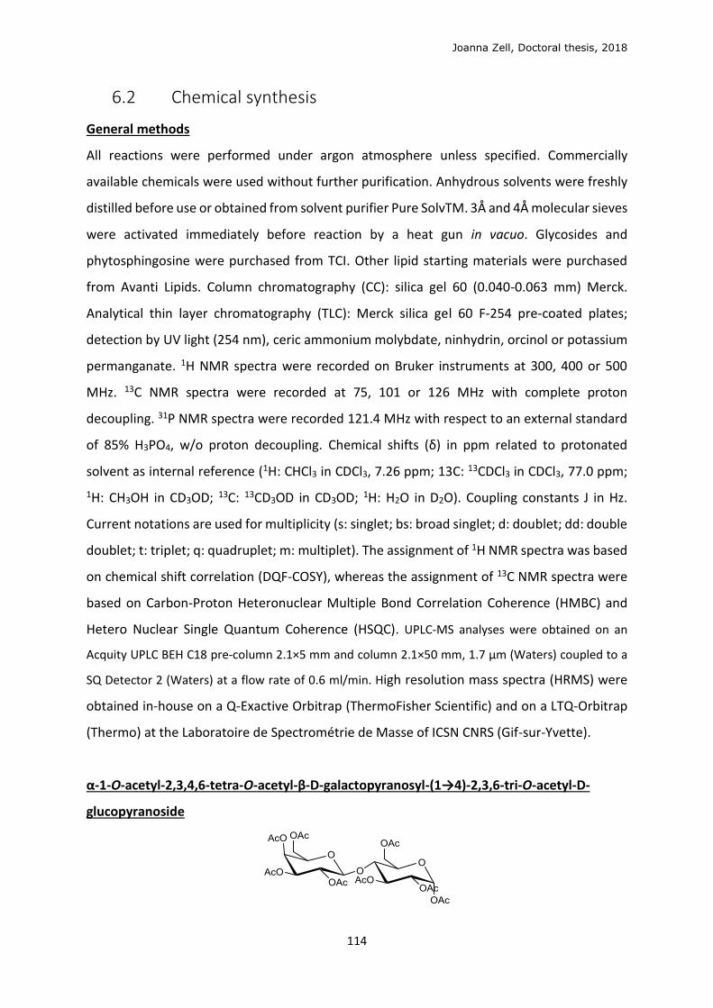
Joanna Zell, Doctoral thesis, 2018
114
6.2 Chemical synthesis
General methods
All reactions were performed under argon atmosphere unless specified. Commercially
available chemicals were used without further purification. Anhydrous solvents were freshly
distilled before use or obtained from solvent purifier Pure SolvTM. 3Å and 4Å molecular sieves
were activated immediately before reaction by a heat gun in vacuo. Glycosides and
phytosphingosine were purchased from TCI. Other lipid starting materials were purchased
from Avanti Lipids. Column chromatography (CC): silica gel 60 (0.040-0.063 mm) Merck.
Analytical thin layer chromatography (TLC): Merck silica gel 60 F-254 pre-coated plates;
detection by UV light (254 nm), ceric ammonium molybdate, ninhydrin, orcinol or potassium
permanganate. 1H NMR spectra were recorded on Bruker instruments at 300, 400 or 500
MHz. 13C NMR spectra were recorded at 75, 101 or 126 MHz with complete proton
decoupling. 31P NMR spectra were recorded 121.4 MHz with respect to an external standard
of 85% H3PO4, w/o proton decoupling. Chemical shifts (δ) in ppm related to protonated
solvent as internal reference (1H: CHCl3 in CDCl3, 7.26 ppm; 13C: 13CDCl3 in CDCl3, 77.0 ppm;
1H: CH3OH in CD3OD; 13C: 13CD3OD in CD3OD; 1H: H2O in D2O). Coupling constants J in Hz.
Current notations are used for multiplicity (s: singlet; bs: broad singlet; d: doublet; dd: double
doublet; t: triplet; q: quadruplet; m: multiplet). The assignment of 1H NMR spectra was based
on chemical shift correlation (DQF-COSY), whereas the assignment of 13C NMR spectra were
based on Carbon-Proton Heteronuclear Multiple Bond Correlation Coherence (HMBC) and
Hetero Nuclear Single Quantum Coherence (HSQC). UPLC-MS analyses were obtained on an
Acquity UPLC BEH C18 pre-column 2.1×5 mm and column 2.1×50 mm, 1.7 μm (Waters) coupled to a
SQ Detector 2 (Waters) at a flow rate of 0.6 ml/min. High resolution mass spectra (HRMS) were
obtained in-house on a Q-Exactive Orbitrap (ThermoFisher Scientific) and on a LTQ-Orbitrap
(Thermo) at the Laboratoire de Spectrométrie de Masse of ICSN CNRS (Gif-sur-Yvette).
α-1-O-acetyl-2,3,4,6-tetra-O-acetyl-β-D-galactopyranosyl-(1→4)-2,3,6-tri-O-acetyl-D-
glucopyranoside
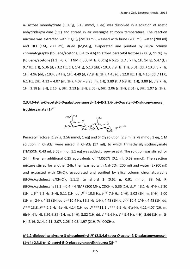
Joanna Zell, Doctoral thesis, 2018
115
α-Lactose monohydrate (1.09 g, 3.19 mmol, 1 eq) was dissolved in a solution of acetic
anhydride/pyridine (1:1) and stirred in air overnight at room temperature. The reaction
mixture was extracted with CH2Cl2 (2×100 ml), washed with brine (200 ml), water (200 ml)
and HCl (1M, 200 ml), dried (MgSO4), evaporated and purified by silica column
chromatography (toluene/acetone, 6:4 to 4:6) to afford peracetyl lactose (2.06 g, 95 %). Rf
(toluene/acetone [1:1])=0.7; 1H NMR (300 MHz, CDCl3) δ 6.26 (d, J 3.7 Hz, 1H, 1-Hα), 5.47 (t, J
9.7 Hz, 1H), 5.36 (d, J 3.2 Hz, 1H, 1’-Hα), 5.13 (dd, J 10.3, 7.9 Hz, 1H), 5.01 (dd, J 10.3, 3.7 Hz,
1H), 4.96 (dd, J 10.4, 3.4 Hz, 1H), 4.49 (d, J 7.8 Hz, 1H), 4.45 (d, J 12.0 Hz, 1H), 4.16 (dd, J 11.0,
6.1 Hz, 2H), 4.12 – 4.07 (m, 1H), 4.07 – 3.95 (m, 1H), 3.89 (t, J 6.8 Hz, 1H), 3.80 (d, J 9.7 Hz,
1H), 2.18 (s, 3H), 2.16 (s, 3H), 2.13 (s, 3H), 2.06 (s, 6H), 2.06 (s, 3H), 2.01 (s, 3H), 1.97 (s, 3H).
2,3,4,6-tetra-O-acetyl-β-D-galactopyranosyl-(1→4)-2,3,6-tri-O-acetyl-β-D-glucopyranosyl
isothiocyanate (1)177
Peracetyl lactose (1.87 g, 2.56 mmol, 1 eq) and SnCl4 solution (2.8 ml, 2.78 mmol, 1 eq, 1 M
solution in CH2Cl2) were mixed in CH2Cl2 (17 ml), to which trimethylsilylisothiocyanate
(TMSSCN, 0.43 ml, 3.06 mmol, 1.1 eq) was added dropwise at rt. The solution was stirred for
24 h, then an additional 0.25 equivalents of TMSSCN (0.1 ml, 0.69 mmol). The reaction
mixture stirred for another 24h, then washed with NaHCO3 (200 ml) and water (2×200 ml)
and extracted with CH2Cl2, evaporated and purified by silica column chromatography
(EtOAc/cyclohexane/CH2Cl2, 1:1:1) to afford 1 (0.62 g, 0.91 mmol, 33 %). Rf
(EtOAc/cyclohexane [1:1])=0.4; 1H NMR (300 MHz, CDCl3) δ 5.35 (1H, d, J4’-3’ 3.1 Hz, 4’-H), 5.20
(1H, t, J3-4 9.2 Hz, 3-H), 5.11 (1H, dd, J2’-1’ 10.3 Hz, J2’-3’ 7.9 Hz, 2’-H), 5.02 (1H, m, 3’-H), 5.00
(1H, m, 2-H), 4.95 (1H, dd, J1-2 10.4 Hz, J 3.3 Hz, 1-H), 4.48 (1H, d, J1’-2’ 10.4, 1’-H), 4.48 (1H, dd,
J6a-6b 13.8, J6a-5 2.2 Hz, 6a-H), 4.14 (1H, dd, J6’a-6’b 11.1, J6’a-5’ 6.5 Hz, 6’a-H), 4.11-4.07 (2H, m,
6b-H, 6’b-H), 3.91-3.85 (1H, m, 5’-H), 3.82 (1H, dd, J4-5 9.6 Hz, J4-3 9.4 Hz, 4-H), 3.66 (1H, m, 5-
H), 2.16, 2.14, 2.11, 2.07, 2.06, 2.05, 1.97 (21H, 7s, COCH3).
N-1,2-dioleoyl-sn-glycero-3-phosphoethyl-N’-(2,3,4,6-tetra-O-acetyl-β-D-galactopyranosyl-
(1→4)-2,3,6-tri-O-acetyl-β-D-glucopyranosyl)thiourea (2)177
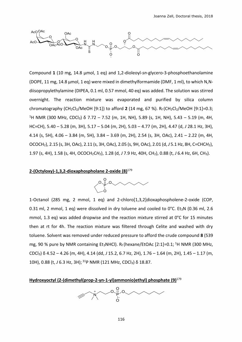
Joanna Zell, Doctoral thesis, 2018
116
Compound 1 (10 mg, 14.8 μmol, 1 eq) and 1,2-dioleoyl-sn-glycero-3-phosphoethanolamine
(DOPE, 11 mg, 14.8 μmol, 1 eq) were mixed in dimethylformamide (DMF, 1 ml), to which N,N-
diisopropylethylamine (DIPEA, 0.1 ml, 0.57 mmol, 40 eq) was added. The solution was stirred
overnight. The reaction mixture was evaporated and purified by silica column
chromatography (CH2Cl2/MeOH [9:1]) to afford 2 (14 mg, 67 %). Rf (CH2Cl2/MeOH [9:1]=0.3;
1H NMR (300 MHz, CDCl3) δ 7.72 – 7.52 (m, 1H, NH), 5.89 (s, 1H, NH), 5.43 – 5.19 (m, 4H,
HC=CH), 5.40 – 5.28 (m, 3H), 5.17 – 5.04 (m, 2H), 5.03 – 4.77 (m, 2H), 4.47 (d, J 28.1 Hz, 3H),
4.14 (s, 5H), 4.06 – 3.84 (m, 5H), 3.84 – 3.69 (m, 2H), 2.54 (s, 3H, OAc), 2.41 – 2.22 (m, 4H,
OCOCH2), 2.15 (s, 3H, OAc), 2.11 (s, 3H, OAc), 2.05 (s, 9H, OAc), 2.01 (d, J 5.1 Hz, 8H, C=CHCH2),
1.97 (s, 4H), 1.58 (s, 4H, OCOCH2CH2), 1.28 (d, J 7.9 Hz, 40H, CH2), 0.88 (t, J 6.4 Hz, 6H, CH3).
2-(Octyloxy)-1,3,2-dioxaphospholane 2-oxide (8)179
1-Octanol (285 mg, 2 mmol, 1 eq) and 2-chloro[1,3,2]dioxaphospholene-2-oxide (COP,
0.31 ml, 2 mmol, 1 eq) were dissolved in dry toluene and cooled to 0°C. Et3N (0.36 ml, 2.6
mmol, 1.3 eq) was added dropwise and the reaction mixture stirred at 0°C for 15 minutes
then at rt for 4h. The reaction mixture was filtered through Celite and washed with dry
toluene. Solvent was removed under reduced pressure to afford the crude compound 8 (539
mg, 90 % pure by NMR containing Et3NHCl). Rf (hexane/EtOAc [2:1]=0.1; 1H NMR (300 MHz,
CDCl3) δ 4.52 – 4.26 (m, 4H), 4.14 (dd, J 15.2, 6.7 Hz, 2H), 1.76 – 1.64 (m, 2H), 1.45 – 1.17 (m,
10H), 0.88 (t, J 6.3 Hz, 3H); 31P NMR (121 MHz, CDCl3) δ 18.87.
Hydroxyoctyl (2-(dimethyl(prop-2-yn-1-yl)ammonio)ethyl) phosphate (9)179
Joanna Zell, Doctoral thesis, 2018
117
Compound 8 (284 mg, 1.2 mmol, 1 eq) was dissolved in CH2Cl2, to which N,N-
dimethylpropargylamine (0.445 ml, 2.46 mmol, 2 eq) was added, then trimethylsilyl
trifluoromethanesulfonate (TMSOTf, 0.265 ml, 2.46 mmol, 2 eq) was added dropwise. The
reaction was stirred for 15 min at 0°C then at rt for 24 h. The reaction mixture was evaporated
and purified by silica column chromatography (CHCl3/MeOH/NH4OH [9:1:0 to 70:30:3]) to
afford 9 (129 mg, 34 % over two steps). Rf (CHCl3/MeOH/NH4OH [70:30:3])=0.2; 1H NMR
(300 MHz, MeOD) δ 4.46 (d, J 2.4 Hz, 2H, C≡CCH2N), 4.28 (m, 2H, NCH2CH2OPO3), 3.89 (q, J 6.5
Hz, 2H, PO3CH2), 3.80 – 3.71 (m, 2H, NCH2CH2OPO3), 3.58 (t, J 2.3 Hz, 1H, C≡CH), 3.29 (s, 6H,
N(CH3)2), 1.64 (dd, J 14.1, 6.7 Hz, 2H, PO3CH2CH2), 1.48 – 1.25 (m, 10H), 0.91 (d, J 6.9 Hz, 3H);
31P NMR (121 MHz, MeOD) δ 1.17; 13C NMR (75 MHz, MeOD) δ 83.21, 72.38, 66.81, 65.24,
60.00, 56.28, 51.85, 32.98, 31.78, 30.47, 26.90, 23.70, 14.28; ESI-MS 320.4 (MH+), 342.4
(MNH4+); with the major impurity formed from hydrolysis of 8 (178 mg, 58 %).
(Z)-(R)-3-((2-oxido-1,3,2-dioxaphospholan-2-yl)oxy)propane-1,2-diyl dioleate (11)179
1,2-dioleoyl-sn-glycerol 10 (80 mg, 129 μmol, 1 eq), COP (36 μl, 386 μmol, 3 eq) and 4-
dimethylaminopyridine (4-DMAP, 1.6 mg, 13 μmol, 0.1 eq) were dissolved in dry toluene and
cooled to 0°C. Et3N (540 μl, 10v% in toluene, 386 μmol, 3 eq) was added dropwise and the
reaction mixture stirred at 0°C for 15 minutes then at rt for 4h. The reaction mixture was
filtered through Celite and washed with dry toluene. Solvent was removed under reduced
pressure to afford the crude compound 11. Rf (cyclohexane/EtOAc [3:1])=0.1; 1H NMR (300
MHz, CDCl3) δ 5.41 – 5.29 (m, 4H, HC=CH), 5.29 – 5.18 (m, 1H, CH(CH2)2O), 4.42 (d, J 10.7 Hz,
4H, P(OCH2)2), 4.35 – 4.24 (m, 2H, CH2OPO3), 4.16 (dd, J 11.9, 5.7 Hz, 2H, CH2OCO), 2.34 (dt, J
11.4, 7.5 Hz, 4H, COCH2), 2.02 (s, J 5.3 Hz, 8H, HC=CHCH2), 1.61 (s, 4H, COCH2CH2), 1.28 (d, J
9.7 Hz, 40H, CH2), 0.88 (t, J 6.4 Hz, 6H, CH3); 31P NMR (121 MHz) δ 18.94 (s). ESI-MS 749.9
(MNa+), 764.5 (MK+).
1,2-dioleate-sn-glyceryl-3 (2-hydroxyethyl) phosphate (5), and 1,2-dioleate-sn-glyceryl-3
(2-(dimethyl(prop-2-yn-1-yl) ammonio)ethyl) phosphate (12)245
Joanna Zell, Doctoral thesis, 2018
118
With the intention of forming compound 5, compound 11 (284 mg, 1.2 mmol, 1 eq) was
dissolved in CH2Cl2, to which N,N-dimethylpropargylamine (0.445 ml, 2.46 mmol, 2 eq) was
added, then TMSOTf (265 μl, 2.46 mmol, 2 eq) was added dropwise. The reaction was stirred
for 15 min at 0°C then at rt for 24 h. The reaction mixture was evaporated and purified by
silica column chromatography (CHCl3/MeOH/NH4OH [9:1:0 to 70:30:3]) to afford the
hydrolysis impurity 12 (87 mg, 91 % over two steps). Rf (CHCl3/MeOH/NH4OH [70:30:3])=0.5;
1H NMR (300 MHz, MeOD) δ 5.40 – 5.28 (m, 4H, HC=CH), 5.27 – 5.19 (m, 1H, CH(CH2)O), 4.31
(ddd, J 18.8, 12.0, 4.9 Hz, 2H, CH2OCO), 3.99 (t, J 5.5 Hz, 2H, CH2OPO3), 3.90 (dd, J 11.1, 5.7
Hz, 2H, HOCH2CH2OPO3), 3.69 (t, J 4.9 Hz, 2H, HOCH2CH2OPO3), 2.32 (q, J 7.1 Hz, 4H, OCOCH2),
2.03 (d, J 5.4 Hz, 8H, HC=CHCH2), 1.61 (s, 4H, OCOCH2CH2), 1.46 – 1.21 (m, 40H, CH2), 0.90 (t,
J 6.6 Hz, 6H, CH3); 32P NMR (121 MHz, MeOD) δ 1.45; ESI-MS 744.0 (M-).
2-azidoethyl-(1,2-dioleate-sn-glyceryl-3)-phosphate (13)210
1,2-dioleoyl-sn-glycerol (12 mg, 20 μmol, 1 eq), COP (11 μl, 60 μmol, 3 eq) and 4-DMAP (0.25
mg, 2 μmol, cat.) were dissolved in dry toluene and cooled to 0°C. EtN3 (8 μl, 60 μmol, 3 eq)
was added dropwise and the reaction mixture stirred at 0°C for 15 minutes then at rt for 4h.
The reaction mixture was filtered through Celite and washed with dry toluene. Solvent was
removed under reduced pressure to afford the cyclic phosphate 11 (31P NMR δ 18.92 (s)). The
product was redissolved with NaN3 (7 mg, 110 μmol, 5 eq) in DMF (1.5 ml) and heated to 80°C
for 6h. The reaction mixture was dried and purified by silica column chromatography
(CH2Cl2/MeOH [95:5 to 85:15]) to afford the compound 13 (10 mg, 65 % over two steps). Rf
(CH2Cl2/MeOH [85:15])=0.5; 1H NMR (300 MHz, CDCl3) δ 6.75 – 5.21 (m, 5H, Hh, Hc), 4.44 –
Joanna Zell, Doctoral thesis, 2018
119
3.99 (m, 6H, Ha1, Hb, Hd), 3.47 (s, 2H, Ha1), 2.32 (dd, J 14.1, 6.7 Hz, 4H, He), 2.01 (d, J 5.9 Hz, 1H,
Hg), 1.61 (s, 4H, Hf’), 1.44 – 0.93 (m, 40H, Hf), 0.88 (t, J 6.0 Hz, 6H, Hi); 31P NMR (121 MHz) δ -
11.61 (s); ESI-MS: 769.0 (M-).
2-azidoethyl octadecyl phosphate (14)210
1-Octadecanol (100 mg, 370 μmol, 1 eq) was dissolved in dry toluene (3 ml), and cooled to
0°C. To this solution, COP (82 μl, 481 μmol, 1.3 eq) and EtN3 (67 μl, 481 μmol, 1.3 eq) was
added dropwise, to the apparition of white smoke. Reaction mixture stirred at 0°C for 30
minutes then at rt for 5h. The reaction mixture was filtered through Celite and washed with
dry toluene (2 ml). Solvent was removed under reduced pressure to afford the crude cyclic
intermediate; Rf (cyclohexane/EtOAc [2:1])=0.2; 1H NMR (300 MHz, CDCl3) δ 4.50 – 4.30 (m,
4 H), 4.15 (dd, J 15.2, 6.7 Hz, 2H), 1.77 – 1.64 (m, 2H), 1.26 (s, 30H), 0.88 (t, J 6.5 Hz, 3H). The
solvent was evaporated from the filtered reaction material overnight. To this dried material,
NaN3 (48 mg, 740 μmol, 2 eq) and DMF (4 ml) was added and the reaction mixture was heated
to 80°C. After 5h stirred, no starting material remained. The reaction was stopped and
extracted with EtOAc (6×5ml) and washed with water. The organic layers were combined,
dried and purified by silica column chromatography (CHCl3/MeOH [93:7 to 8:2]) to afford the
compound 14 (57 mg, 38 % over two steps). Rf (CH2Cl2/MeOH [9:1])=0.5; 1H NMR (400 MHz,
CDCl3/MeOD/D2O [50:25:25]) δ 3.99 (dd, J 10.4, 6.0 Hz, 2H, N3CH2CH2OP), 3.87 (q, J 6.6 Hz,
2H, POCH2), 3.45 (t, J 5.0 Hz, 2H, N3CH2), 1.67 – 1.56 (m, 2H, POCH2CH2), 1.38 – 1.17 (m, 30H),
0.86 (t, J 6.9 Hz, 3H, CH3); 31P NMR (121 MHz) δ 3.61 (s); 13C NMR (101 MHz, MeOD) δ 66.87,
64.93, 51.98, 32.52, 31.25, 31.17, 30.28, 30.01, 29.94, 26.34, 23.25, 14.51; IR/cm-1: 2915 (CH),
2849 (CH), 2109 (N3), 1082 (P=O); ESI-MS: 418.4 (M-).
(2S,3S,4R)-2-azido-1,3,4,-octadecanetriol (18)246
D-Ribo-phytosphingosine (1.04 g, 3.28 mmol, 1 eq), CuSO4.5H2O (0.81 g, 0.315 mmol, 1 eq)
and K2CO3 (0.76 g, 5.50 mmol, 1.7 eq) were dissolved in MeOH (20 ml). Imidazole-1-sulfonyl
Joanna Zell, Doctoral thesis, 2018
120
azide hydrochloride (0.87 g, 4.15 mmol, 1.2 eq) was added and the solution was stirred for 24
h. The solution was filtered and the filter cake washed with CH2Cl2/MeOH (10:1). The mother
liquor was evaporated and purified by silica column chromatography (9-20% MeOH in CH2Cl2)
to afford the described compound 18 as a white solid (0.85 g, 2.46 mmol, 75 %). Rf
(CH2Cl2/MeOH [10:1])=0.5; 1H NMR (300 MHz, MeOD) δ 3.92 (dd, J 11.5, 3.4 Hz, 1H, H1b), 3.75
(dd, J 11.6, 8.0 Hz, 1H, H1a), 3.63 – 3.55 (m, 1H, H2), 3.55 – 3.48 (m, J 4.4 Hz, 1H, H3, H4), 1.75
– 1.49 (m, 2H, CH2), 1.29 (s, 24H), 0.90 (t, J 6.7 Hz, 3H).
(2S,3S,4R)-2-azido-1-(tert-butyldiphenylsilyoxy)octadecane-3,4-triol (19)247
Compound 18 (0.85 g, 2.46 mmol, 1 eq), tert-butyldiphenylsilyl chloride (TBDPSCl, 0.81 g, 2.95
mmol, 1.2 eq), Et3N (0.86 ml, 6.15 mmol, 2.5 eq) and 4-DMAP (0.015 g, 0.123 mmol, 0.05 eq)
were dissolved in dry CH2Cl2/DMF (15/3 ml) at 0°C. The reaction mixture was stirred
overnight, then diluted with EtOAc, washed with brine (2×20 ml), dried with MgSO4 and
purified by silica column chromatography (10-20% EtOAc in cyclohexane) to afford 19 (0.84
g, 59 %). Rf (cyclohex/EtOAc [5:1])=0.4, 1H NMR (300 MHz, CDCl3) δ 7.76 – 7.65 (m, 4H), 7.54
– 7.34 (m, 6H), 4.03 (dd, J 10.9, 4.2 Hz, 1H), 3.91 (dd, J 10.9, 5.7 Hz, 1H), 3.75 – 3.65 (m, 2H),
3.56 (dd, J 9.9, 5.5 Hz, 1H), 2.51 (d, J 3.4 Hz, 1H), 1.99 (d, J 3.6 Hz, 1H), 1.63 – 1.46 (m, 2H),
1.26 (s, 24H), 1.08 (s, 9H), 0.88 (t, J 6.6 Hz, 3H); 13C NMR (75 MHz, CDCl3) δ 136.03, 136.00,
132.93, 132.85, 130.44, 74.52, 72.74, 64.55, 63.76, 32.34, 32.23, 30.09, 29.78, 27.15, 26.07,
23.11, 19.50, 14.55.
(4S,5R)-4-((S)-1-azido-2-((tert-butyldiphenylsilyl)oxy)ethyl)-5-tridecyl-1,3,2-dioxathiolane
2,2-dioxide (20)247
Compound 19 (0.84 g, 1.45 mmol, 1 eq) was dissolved in dry CH2Cl2 (8 ml), to which dry Et3N
(0.60 ml, 4.34 mmol, 3 eq) and SOCl2 (0.127 ml, 1.735 mmol, 1.2 eq) were added and stirred
at 0 °C. After 30 min the reaction mixture was washed with brine (2×50 ml) and extracted
Joanna Zell, Doctoral thesis, 2018
121
with EtOAc (2×50 ml), dried (MgSO4) and evaporated. The intermediate cyclic sulfite was
dissolved in CCl4/CH3CN/H2O (12 ml, 1:1: 1) and to the solution was added Ru(III)Cl3 (18 mg,
0.07 mmol, 0.05 eq) and NaIO4 (0.90 g, 4.22 mmol, 3 eq). The reaction mixture was stirred for
2 h, diluted with EtOAc (30 ml), washed with sat. NaHCO3 solution (2×20 ml), extracted with
EtOAc, dried (MgSO4), evaporated and purified by silica column chromatography (10% EtOAc
in cyclohexane) to afford 20 (0.83 g, 89 %). Rf (cyclohex:EtOAc [6:1])=0.8; 1H NMR (300 MHz,
CDCl3) δ 7.74 – 7.64 (m, 4H), 7.50 – 7.37 (m, 6H), 4.98 (ddd, J 10.2, 5.2, 2.6 Hz, 1H), 4.92 (dd,
J 9.9, 5.2 Hz, 1H), 4.04 (dd, J 11.3, 2.4 Hz, 1H), 3.89 (dd, J 11.3, 5.2 Hz, 1H), 3.69 (ddd, J 9.7,
5.1, 2.4 Hz, 1H), 2.00 – 1.66 (m, 2H), 1.41 – 1.30 (m, 2H), 1.27 (s, 22H), 1.09 (s, 9H), 0.88 (t, J
6.7 Hz, 3H); 13C NMR (75 MHz, CDCl3) δ 135.98, 132.60, 132.36, 130.56, 128.39, 86.85, 80.26,
63.97, 59.62, 32.33, 30.06, 29.86, 29.76, 29.70, 29.36, 28.53, 27.33, 27.14, 25.59, 23.10,
19.57, 14.52.
(2S,3R)-(E)-2-azido-1-(tert-butyldiphenylsilyoxy)octadec-4-en-3-ol (21)247
Compound 20 (772 mg, 1.20 mmol, 1 eq) was dissolved in dry toluene (8 ml), to which Bu4NI
(487 mg, 1.32 mmol, 1.1 eq) and 1,8-diazabicyclo(5.4.0)undec-7-ene (DBU, 274 mg,
1.80 mmol, 1.5 eq) was added. The reaction mixture was refluxed for 2 h, the solvent
evaporated and redissolved in a solution of concentrated H2SO4 (0.1 ml), H2O (0.8 ml) and
tetrahydrofuran (THF, 10 ml) and stirred for a further 1 h. Purification by silica column
chromatography (10% EtOAc in cyclohexane) afforded 21 (0.72 g, 60 %). Rf (cyclohex:EtOAc
[10:1])=0.4; 1H NMR (300 MHz, CDCl3) δ 7.74 – 7.65 (m, 4H), 7.49 – 7.33 (m, 6H), 5.74 (dtd, J
15.5, 6.5, 0.5 Hz, 1H), 5.43 (ddt, J 15.4, 7.1, 0.5 Hz, 1H), 4.22 (dd, J 11.8, 5.4 Hz, 1H), 3.82 (dd,
J 10.8, 6.3 Hz, 1H), 3.77 (dd, J 10.6, 4.6 Hz, 1H), 3.51 (dd, J 11.2, 4.9 Hz, 1H), 1.98 – 1.59 (m,
2H), 1.36 – 1.28 (m, 2H), 1.26 (s, 20H), 1.07 (s, 9H), 0.88 (t, J 6.7 Hz, 3H); 13C NMR (75 MHz,
CDCl3) δ 135.98, 135.85, 133.15, 130.31, 128.22, 73.23, 67.28, 64.53, 32.69, 32.33, 30.08,
29.88, 29.77, 29.59, 29.35, 27.12, 23.10, 19.51, 14.53.
(2S,3R)-(E)-2-azido-3-O-benzoyl-1-O-tert-butyldiphenylsilyl-4-octadecene-1,3-diol (22)248
Joanna Zell, Doctoral thesis, 2018
122
Compound 21 (1.95 g, 3.48 mmol, 1 eq) was dissolved in dry pyridine (12 ml) at 0 °C, to which
BzCl (960 µl, 8.29 mmol, 2.4 eq) was added dropwise and the reaction mixture was stirred for
3 h at 0 °C to rt, quenched with H2O (40 ml) and stirred for a further 20 min. The reaction
mixture was extracted with CH2Cl2 (3×50 ml), washed with HCl (1 M, 100 ml), saturated
NaHCO3 solution (100 ml) and brine (100 ml), dried (MgSO4), evaporated and purified by silica
column chromatography (20 – 10% EtOAc in cyclohexane) to afford 22 (2.01 g, 87 %). Rf
(cyclohex:EtOAc [10:1])=0.8; [α]22D=-9.0° (c 7.0, CHCl3); 1H NMR (300 MHz, CDCl3) δ 8.03 (d, J
7.5 Hz, 2H, Si(C6H5)2), 7.73– 7.56 (5H, m, Ph), 7.50-7.30 (8H, m, Si(C6H5)2), 5.91 (1H, ddd, J5-6
6.8 Hz, J5-6’ 6.8 Hz, J5-4 15.2 Hz, H5), 5.69 (1H, dd, J3-4 7.9, J3-2 4.7 Hz, H3), 5.52 (1H, dd, J4-5 15.4,
J4-3 8.0 Hz, H4), 3.85 (1H, m, H2), 3.75 (2H, m, H1a, H1b), 2.06 – 1.97 (2H, m, H6, H6’), 1.40 – 1.18
(22H, m, 11CH2), 1.09 (9H, s, tBu), 0.89 (3H, t, J 6.7 Hz, CH3); ESI-HRMS: Calcd for [MNa+]
690.4066. Found: m/z 690.4061.
(2S,3R)-(E)-2-amino-3-O-benzoyl-1-O-tert-butyldiphenylsilyl-4-octadecene-1,3-diol
(23)248,249
Compound 22 (685 mg, 1.03 mmol, 1 eq) was dissolved in toluene/water (20:1, 10 ml), to
which PPh3 (538 mg, 2.05 mmol, 2 eq) was added. The reaction mixture was stirred overnight,
heated to 60°C for 2h until all starting material and phosphorus intermediate had disappeared
from TLC. Reaction mixture was evaporated and purified by silica column chromatography
(0.5 to 10% EtOAc in cyclohexane) to afford 23 (494 mg, 78 %) as a mixture of inseparable
stereoisomers (2S,3R) and (2R,3R) (98:2). Rf (cyclohex:EtOAc [3:1]+1% Et3N)=0.1; 1H NMR
(300 MHz, CDCl3) δ 7.88 (d, J 7.2 Hz, 2H, Bz), 7.68 – 7.38 (m, 5H, TBDPS), 7.38 – 7.20 (m, 6H,
aromatic), 7.20 – 7.10 (m, 2H, Bz), 5.90 – 5.77 (m, 1H, H5), 5.48 – 5.34 (m, 2H, H3, H4), 3.72 –
3.59 (m, 2H, H1a, H1b), 3.10 (q, J 5.1 Hz, 1H, H2), 1.97 (q, J 6.9 Hz, 2H, H6), 1.29 – 1.07 (m, 22H,
11CH2), 0.99 (s, 9H, tBu), 0.80 (t, J 6.6 Hz, 3H, CH3); 13C NMR (75 MHz, CDCl3) δ 165.51 (CO
benzyl), 137.91 (C5), 135.75, 135.59, 135.59, 133.38, 133.22, 133.00), 130.71, 130.27, 130.23,
129.87, 129.81, 129.75, 128.79, 128.49, 128.08, 127.90, 127.82, 127.13, 125.05 (C4), 76.56
Joanna Zell, Doctoral thesis, 2018
123
(C3), 64.93 (C1), 55.56 (C2), 32.61 (C6), 32.11 (C16), 29.85, 29.77, 29.63, 29.55, 29.39, 29.09,
26.99 (tBu), 22.88 (tBu), 19.40 (C17), 14.32 (C18); ESI-MS 642.8 (MH+), 664.8 (MNa+), 1284.3
(2MH2+); ESI-HRMS: Calcd for [MNa+] 664.4161. Found: m/z 664.4156.
(2S,3R)-(E)-1-O-tert-butyldiphenylsilyl-2-(2,2-dichloroacetamido)-3-O-benzoyl-4-
octadecene-1,3-diol (24)250
Compound 23 (970 mg, 1.51 mmol, 1 eq) was dissolved in dry CH2Cl2 (20 ml) and cooled to -
15°C. DIPEA (385 μl, 2.26 mmol, 1.5 eq) was added dropwise, followed by dichloroacetyl
chloride (182 μl, 1.89 mmol, 1.25 eq) dropwise. After 15 minutes at -10°C, the reaction was
stirred 30 minutes at rt until complete conversion by TLC. The reaction was quenched with
water, extracted with CH2Cl2, washed with brine, dried (MgSO4), evaporated and purified by
silica column chromatography (1 – 10% EtOAc in cyclohexane) to afford 24 (918 mg, 81 %). Rf
(cyclohex:EtOAc [20:1])=0.3; [α]22D=+20.8° (c 5.0, CHCl3); 1H NMR (300 MHz, CDCl3) δ 7.95 –
7.89 (m, 2H, Bz), 7.60 – 7.45 (m, 5H, arom), 7.41 – 7.20 (m, 6H, arom), 7.12 (t, J 7.3 Hz, 2H,
Bz), 5.94 – 5.81 (m, 1H, H5), 5.84 (s, 1H, CHCl2), 5.66 (t, J3-2, 3-4 7.5 Hz, 1H, H3) 5.43 (dd, J4-5 15.4,
J4-3 7.5 Hz, 1H, H4), 4.27 (ddd, J2-3 7.2, J2-1a3.5, J2-1b4.0 Hz, 1H, H2), 3.84 (dd, J1a-b 10.6, J1a-2 3.0
Hz, 1H, H1a), 3.65 (dd, J1a-b 10.6, J1b-24.0 Hz, 1H, H1b), 1.95 (q, J6-5 6.9 Hz, 2H, H6), 1.32 – 1.09
(m, 22H), 0.99 (s, 9H), 0.81 (t, J 6.7 Hz, 3H); 13C NMR (75 MHz, CDCl3) δ 165.17 (COCHCl2),
163.57 (CO benzyl), 138.01 (C5), 135.53, 135.45, 133.10, 132.34, 132.18, 130.06, 130.00,
129.88, 129.71, 128.42, 127.91, 127.79, 124.13 (C4), 73.59 (C3), 66.57 (CHCl2), 61.72 (C1), 53.13
(C2), 32.37 (C6), 31.94 (C16), 29.68, 29.59, 29.47, 29.38, 29.25, 28.82, 26.77 (tBu), 22.71 (C17),
19.17 (tBu), 14.16 (C18); ESI-MS 752.7 (MH+); ESI-HRMS: Calcd for [MH+] 752.3668. Found: m/z
752.3663.
(2S,3R)-(E)-2-(2,2-dichloroacetamido)-3-O-benzoyl-4-octadecene-1,3-diol (25)
Joanna Zell, Doctoral thesis, 2018
124
To a solution of compound 24 (888 mg, 1.18 mmol, 1 eq) in THF (20 ml), benzoic acid (187 mg,
1.53 mmol, 1.3 eq) was added and the solution cooled to 0°C. To this, tetra-n-butylammonium
fluoride (TBAF, 1M solution in THF, 1.53 ml, 1.3 eq) was added dropwise over 3 minutes. The
reaction was warmed to rt over 10 minutes and stirred for 4 h before complete conversion.
The reaction was quenched with water (10 ml) and extracted with CH2Cl2 (2×10 ml), dried
(MgSO4), evaporated and purified by silica column chromatography (0.5 – 5% EtOH inCH2Cl2)
to afford 25 (582 mg, 96 %). Rf (EtOH/CH2Cl2 [1:99])=0.2; 1H NMR (300 MHz, CDCl3) δ 8.04 (d,
J 7.4 Hz, 2H, Bz), 7.53 (dt, J 41.5, 7.4 Hz, 3H, Bz), 7.10 (d, J 9.0 Hz, 1H, NH), 5.93 (s, 1H, CHCl2),
5.99 – 5.81 (m, 1H, H5), 5.68 – 5.50 (m, 2H, H4, H3), 4.23 (m, 1H, H2), 3.89 – 3.57 (m, 2H, H1a,b),
2.81 (br s, 1H, OH), 2.05 (dd, J6 14.1, J6-5 6.9 Hz, 2H, H6), 1.48 – 1.11 (m, 22H, CH2), 0.87 (t, J
6.6 Hz, 3H, CH3); 13C NMR (75 MHz, CDCl3) δ 166.51 (CO benzoyl), 164.08 (COCHCl2), 138.41
(C5), 133.61 (Bz), 129.87 (Bz), 128.57 (Bz), 123.93 (C4), 74.01 (C3), 66.44 (CHCl2), 60.79 (C2),
53.78 (C1), 32.32 (C6), 31.93 (C16), 29.67, 29.59, 29.45, 29.37, 29.24, 28.77, 22.71 (C17), 14.15
(C18); ESI-MS 512.4 (M-H-); ESI-HRMS: Calcd for [MH+] 514.2490. Found: m/z 514.2485.
(2S,3R)-(E)-1-((methylsulfonyl)oxy)-2-(2,2-dichloroacetamido)-3-O-benzoyl-4-octadecene-
1,3-diol (26)
To a solution of compound 25 (510 mg, 0.99 mmol, 1 eq) in pyridine (15 ml) at -10°C,
methanesulfonyl chloride (MsCl, 109 μl, 1.27 mmol, 1.4 eq) was added. The solution was
warmed to rt after 5 min and stirred for 3 h before co-evaporation with toluene. The product
was purified by silica column chromatography (10 – 50 % EtOAc in cyclohexane) to afford 26
(548 mg, 93 %). Rf (EtOH/CH2Cl2 [1:99])=0.5; 1H NMR (300 MHz, CDCl3) δ 8.07 – 7.99 (m, 2H),
7.60 (t, J 7.5 Hz, 1H), 7.47 (t, J 7.6 Hz, 2H), 6.93 (d, J 8.6 Hz, 1H, NH2), 6.07 – 5.94 (m, 1H, H5),
5.66 (t, J4-3 7.0 Hz, 1H, H3), 5.50 (dd, J 15.4, 7.4 Hz, 1H, H4), 4.54 (m, 1H, H2), 4.50 – 4.37 (m,
2H, H1a,b), 3.03 (s, 3H, Ms), 2.06 (dd, J6 14 Hz, J5 7.0 Hz, 2H, H6), 1.43 – 1.15 (m, 22H, CH2), 0.87
(t, J 6.6 Hz, 3H, CH3); 13C NMR (75 MHz, CDCl3) δ 165.31 (COCHCl2), 164.17 (COPh), 139.22 (C5),
133.57 (Bz), 129.74 (Bz), 129.43 (Bz), 128.62 (Bz), 123.06 (C4), 73.18 (C3), 66.57 (C1), 66.16
(CHCl2), 51.67 (C2), 37.65 (SO2CH3), 32.34 (C6), 31.93 (C16), 29.66, 29.57, 29.43, 29.37, 29.21,
Joanna Zell, Doctoral thesis, 2018
125
28.71, 22.70 (C17), 14.15 (C18); ESI-MS 590.5 (M-H-), 626.8 (MCl-); ESI-HRMS: Calcd for [MNa+]
614.2085. Found: m/z 614.2080.
(2S,3R)-(E)-2-(2,2-dichloroacetamido)-1-iodo-3-O-benzoyl-4-octadecene-3-ol (27)
To a solution of compound 26 (62 mg, 0.11 mmol, 1 eq) in distilled acetone (1 ml), sodium
iodide (146 mg, 0.30 mmol, 10 eq) was added and mixture was refluxed at 65°C for 5h. The
reaction was quenched with H2O, extracted with CH2Cl2, dried (MgSO4) and evaporated. The
product was purified by silica column chromatography (10 – 50 % EtOAc in cyclohexane) to
afford 27 (48 mg, 76 %). Rf (EtOAc/cyclohexane [9:1])=0.7; 1H NMR (300 MHz, CDCl3) δ 7.97
(d, J 7.3 Hz, 2H), 7.53 (m, 1H), 7.39 (t, J 7.6 Hz, 2H), 6.60 (d, JN-2 9.1 Hz, 1H, NH2), 6.02 – 5.88
(m, 1H, H1), 5.55 (t, J 6.9 Hz, 1H), 5.43 (dd, J 15.2, 7.5 Hz, 1H), 4.19 (td, J 11.1, 6.1 Hz, 1H), 3.35
(qd, J 10.8, 5.4 Hz, 1H), 1.99 (q, J 6.9 Hz, 1H), 1.40 – 1.04 (m, 12H), 0.81 (t, J 6.6 Hz, 2H); 13C
NMR (75 MHz, CDCl3) δ 165.07 (COCHCl2), 163.70 (COPh), 139.19 (C5), 133.42 (Bz), 129.75
(Bz), 129.59 (Bz), 128.55 (Bz),122.82 (C4), 75.43 (C3), 66.29 (CHCl2), 52.36 (C2), 32.40 (C6), 31.94
(C16), 29.67, 29.58, 29.43, 29.38, 29.21, 28.73, 22.72 (C17), 14.16 (C18), 5.17 (C1); ESI-MS 641.5
(MNH4+); ESI-HRMS: Calcd for [MNa+] 646.1327. Found: m/z 646.1322.
(2S,3R)-(E)- 1-azido-2-(2,2-dichloroacetamido)-3-O-benzoyl-4-octadecene-3-ol (28)
To a solution of compound 27 (380 mg, 0.61 mmol, 1 eq) in DMF (2.5 ml), sodium azide
(396 mg, 6.1 mmol, 10 eq) was added and the mixture stirred for 14 h. The reaction was
quenched with H2O and extracted with EtOAc, washed with brine, dried (MgSO4) and
evaporated. The product was purified by silica column chromatography (4 to 5 % EtOAc in
cyclohexane) to afford 28 (316 mg, 97 %). Rf (EtOAc/cyclohexane [9:1])=0.6; [α]22D=+11.7 (c
10.0, CHCl3); 1H NMR (300 MHz, CDCl3) δ 8.04 (d, J 7.3 Hz, 2H, Bz), 7.60 (t, J 7.4 Hz, 1H, Bz),
7.45 (m, 1H, Bz), 6.83 (d, JN-2 9.0 Hz, 1H, NH), 6.04 – 5.92 (m, 1H, H5), 5.61 (t, J3-4, 3-2 6.8 Hz, 1H,
Joanna Zell, Doctoral thesis, 2018
126
H3), 5.49 (dd, J4-5 15.3, J4-3 7.4 Hz, 1H, H4), 4.41 (m, 1H, H2), 3.70 – 3.54 (m, 2H, H1a,b), 2.06 (dd,
J6-5 6.9 Hz, J6-6’ 14 Hz, 2H, H6), 1.43 – 1.19 (m, 12H, CH2), 0.87 (t, J 6.6 Hz, 3H, CH3). 13C NMR
(75 MHz, CDCl3) δ 165.37 (COPh), 163.97 (COCHCl2), 138.70 (C5), 133.48 (Bz), 129.76 (Bz),
129.55 (Bz), 128.57 (Bz), 123.22 (C4), 73.93 (C3), 66.25 (CHCl2), 51.92 (C2), 50.58 (C1), 32.37
(C6), 31.94 (C16), 29.67, 29.59, 29.44, 29.38, 29.21, 28.76, 22.72 (C17), 14.16 (C18). IR, ν 3292
(NH), 2923 (C-H), 2853 (C=C), 2103 (N3), 1721, 1679, 1262, 710 cm-1; ESI-MS 561.5 (MNa+),
537.5 (M-H-); ESI-HRMS: Calcd for [MNa+] 561.2375. Found: m/z 561.2370.
(2S,3R)-(E)-2-amino-1-azido-4-octadecen-3-ol (1-azidosphingosine) (17)184
Compound 28 (30 mg, 0.056 mmol, 1 eq) was stirred in a solution of KOH (0.5M) in H2O/MeOH
(3:7) at 40°C for 4 h. The reaction mixture was evaporated and the product was purified by
silica column chromatography (cyclohexane/EtOAc/NH4OH [97:3:0.3]) to afford 17 (11 mg, 61
%). Rf (cyclohexane/EtOAc/NH4OH [97:3:0.3])=0.3; [α]22D=+9.8 (c 0.5, MeOH); 1H NMR (300
MHz, CDCl3) δ 5.87 – 5.68 (m, 1H, H5), 5.43 (dd, J4-5 15.4, J4-3 7.2 Hz, 1H, H4), 4.00 (t, J3-4, 3-2
6.1 Hz, 1H, H3), 3.48 (dd, J1a-b 12.2 Hz, J1-2 3.9 Hz, 1H, H1a), 3.32 (dd, J1a-b 12.1, 7.6 Hz, 1H, H1b),
2.95 (bs, 1H, H2), 2.06 (q, J 6.9 Hz, 2H, H6), 1.61 (bs, 2H, NH2), 1.44 – 1.11 (m, 23H, CH2), 1.06
(bs, 1H, OH), 0.87 (t, J 6.5 Hz, 3H, CH3); 13C NMR (75 MHz, CDCl3) δ 135.53 (C5), 128.24 (C4),
74.22 (C3), 54.93 (C2), 54.26 (C1), 32.38 (C6), 31.93 (C16), 29.68, 29.62, 29.48, 29.37, 29.23,
29.11, 22.71 (C17), 14.15 (C18); IR ν 3354 (NH), 3287 (NH), 3154 (OH, C=C-H), 2916 (C-H), 2848
(C-H), 2133 (N3); ESI-MS 5325.5 (MH+), 307.4 (MH-H2O+); ESI-HRMS: Calcd for [MH+] 325.2967.
Found: m/z 325.2962.
1-azidoceramide (C22:1) (30)209
Compound 17 (3×1.67 mg, 15.5 μmol, 1 eq) was mixed in 3 separate Eppendorf tubes with
erucic acid (3×2 mg, 5.9 μmol, 1.2 eq) in a phosphate buffer solution (pH 7.4, NaH2PO4 (0.1M),
Joanna Zell, Doctoral thesis, 2018
127
Na2HPO4 (0.1M), H3PO4 (0.1M)) with 0.4% Triton X-100 detergent. To this, SCDase enzyme
(10 μl, 50 mU) was added to each Eppendorf, and the mixture was shaken at 37°C overnight.
The reaction mixture was lyophilised and purified by silica column chromatography (0.4%
NH4OH in CH2Cl2) to afford 30 (6.3 mg, 64%). Rf (CH2Cl2/MeOH [95:5])=0.8; [α]22D=+1.6 (c 0.5,
CHCl3); 1H NMR (400 MHz, CDCl3) δ 5.82 (d, JNH-2 8.0 Hz, 1H, NH), 5.76 (dd, J5-4 15.0, J5-6 7.0 Hz,
1H, H5), 5.46 (dd, J4-5 15.4, J4-3 6.6 Hz, 1H, H4), 5.35 (t, Je-d 4.7 Hz, 2H, He), 4.18 (q, J3-4, 3-2 5.6
Hz, 1H, H3), 4.05 (td, J2-1 8.9, J2-3 4.9 Hz, 1H, H2), 3.58 (ddd, J1a-b 16.5, J1a-212.6, J1b-2 4.5 Hz, 2H,
H1a, H1b), 2.40 (d, JOH-1 5.6 Hz, 1H, OH), 2.20 (t, Ja-b 7.5 Hz, 2H, Ha), 2.12 – 1.95 (m, 6H, H6, Hd),
1.69 – 1.57 (m, 2H, Hb), 1.26 (s, 44H, H7, Hc), 0.88 (t, J 6.8 Hz, 6H, H8); 13C NMR (101 MHz,
CDCl3) δ 173.48 (NHCO), 135.05 (C5), 130.00 (Ce), 128.52 (C4), 73.51 (C3), 52.79 (C2), 50.96 (C1),
36.91 (Ca), 32.35 (C6), 32.01, 29.87, 29.78, 29.72, 29.66, 29.59, 29.45, 29.42, 29.36, 29.31,
29.18, 27.30, 25.75 (Cb), 22.78 (CH2CH3), 14.21 (C8); ESI-HRMS: Calcd for [MH+] 645.6046.
Found: m/z 645.6041.
(S)-4-((R,E)-1-hydroxyhexadec-2-en-1-yl)oxazolidin-2-one (29)
Rf (cyclohexane/EtOAc/NH4OH [97:3:0.3])=0.3; 1H NMR (300 MHz, CDCl3) δ 5.79 (dt, J4-5 15.4
Hz, J5-6 6.7 Hz, 1H, H5), 5.34 (dd, J4-5 15.4 Hz, J4-3 7.5 Hz, 1H, H4), 5.07 (bs, 1H, NH), 4.38 (t, J1a-
b, 1a-2 8.8 Hz, 1H, H1a), 4.25 (dd, J1a-b 8.9, J1b-2 5.0 Hz, 1H, H1b), 3.99 (t, J3-2, 3-4 6.6 Hz, 1H, H3),
3.76 (dt, J2-1a 8.6, J2-1b, 2-3 5.4 Hz, 1H, H2), 2.00 (q, J 6.8 Hz, 2H, H6), 1.59 (bs, 1H, OH), 1.39 –
1.25 (m, 2H, CH2), 1.19 (s, 20H, CH2), 0.81 (t, J 6.7 Hz, 3H, CH3); 13C NMR (75 MHz, CDCl3) δ
159.69 (C19), 137.30 (C5), 126.69 (C4), 73.93 (C3), 66.70 (C1), 55.99 (C2), 32.33 (C6), 31.91 (C7),
29.56, 29.43, 29.34, 29.19, 28.95, 22.67 (C17), 14.09 (C18). ; IR/cm-1: 3286 (OH), 2917 (CH),
2849 (CH), 16832 (C=O); ESI-MS: 326.7 [MH+], 343.5 [MNH4+], 348.5 [MNa+], 324.5 [M-H-]
(2S,3S,4R)-2-(N-2,2-dichloroacetamido)-1,3,4,-octadecanetriol (32)
Joanna Zell, Doctoral thesis, 2018
128
Phytosphingosine (1.1 g, 3.46 mmol, 1 eq) was suspended in THF (40 ml), to which DIPEA (0.8
ml, 4.34 mmol, 1.3 eq) was added and the mixture was cooled to -10°C. Dichloroacetyl
chloride (367 μl, 3.82 mmol, 1.1 eq) was added dropwise and the reaction was warmed to rt
after 15 min, then stirred at rt for 3h, evaporated and purified by silica column
chromatography (2.5 to 7% MeOH in CH2Cl2) to afford 32 as a white crystalline solid (1.41 g,
81 %). Rf (MeOH/CH2Cl2 [5:95]) =0.3; [α]22D=+7.9 (c 3.3, CHCl3/MeOH [1:1]); 1H NMR (500 MHz,
CDCl3) δ 7.60 (d, J 8.4 Hz, 1H, NH), 5.97 (s, 1H, CHCl2), 4.07 (dt, J 5.0, 3.2 Hz, 1H, H2), 3.84 (dd,
J 11.6, 3.4 Hz, 1H, H1a), 3.70 (dd, J 11.6, 5.4 Hz, 1H, H1b), 3.64 – 3.55 (m, 2H, H4, H3), 1.68 –
1.35 (m, 4H, H5, H6), 1.35 – 1.13 (m, 22H), 0.84 (t, J 7.0 Hz, 3H); 13C NMR (126 MHz, CDCl3) δ
164.56, 75.08 (C3), 72.59 (C4), 66.33 (CHCl2), 60.57 (C1), 52.30 (C2), 32.86 (C5), 31.89 (C6), 29.66,
29.62, 29.33, 25.80, 22.66, 14.07; ESI-HRMS: Calcd for [MH+] 428.2329. Found: m/z 428.2332.
(2S,3S,4R)-1-azido-2-(N-2,2-dichloroacetamido)-3,4,-octadecanediol (3)
N-Protected phytosphingosine 32 (50 mg, 0.117 mmol, 1 eq) and NaN3 (76 mg, 1.17 mmol,
10 eq) were suspended in DMF (1 ml), to which PPh3 (61 mg, 0.23 mmol, 2 eq) and then CBr4
(77 mg, 0.23 mmol, 2 eq) in DMF (0.5 ml) was added and the mixture stirred at rt overnight.
The reaction was quenched with MeOH, evaporated, diluted with CH2Cl2, washed with H2O,
and purified by silica column chromatography (10 to 25% EtOAc in n-hexane) to afford 33 as
a crystalline white solid (16 mg, 31 %). Rf (EtOAc/n-hexane [4:6]) =0.7; [α]22D=+14.3 (c 0.7,
CHCl3/MeOH [9:1]); 1H NMR (500 MHz, CDCl3) δ 7.10 (d, J 8.5 Hz, 1H, NH), 5.97 (s, 1H, CHCl2),
4.23 (td, J 8.9, 4.6 Hz, 1H, H2), 3.84 (dd, J 12.7, 4.8 Hz, 1H, H1a), 3.74 – 3.66 (m, 3H, H1b, H3, H4),
1.75 – 1.42 (m, 4H, H5, H6), 1.42 – 1.19 (m, 22H), 0.90 (t, J 6.9 Hz, 3H); 13C NMR (101 MHz,
CDCl3) δ 164.22, 74.35 (C3 or C4), 72.82 (C3 or C4), 66.27 (CHCl2), 51.01 (C1), 50.65 (C2), 32.74
(C5), 31.93 (C6), 29.69, 29.59, 29.56, 29.52, 29.37, 25.68, 22.70, 14.14; ESI-HRMS: Calcd for
[MH+] 453.2394. Found: m/z 453.2395.
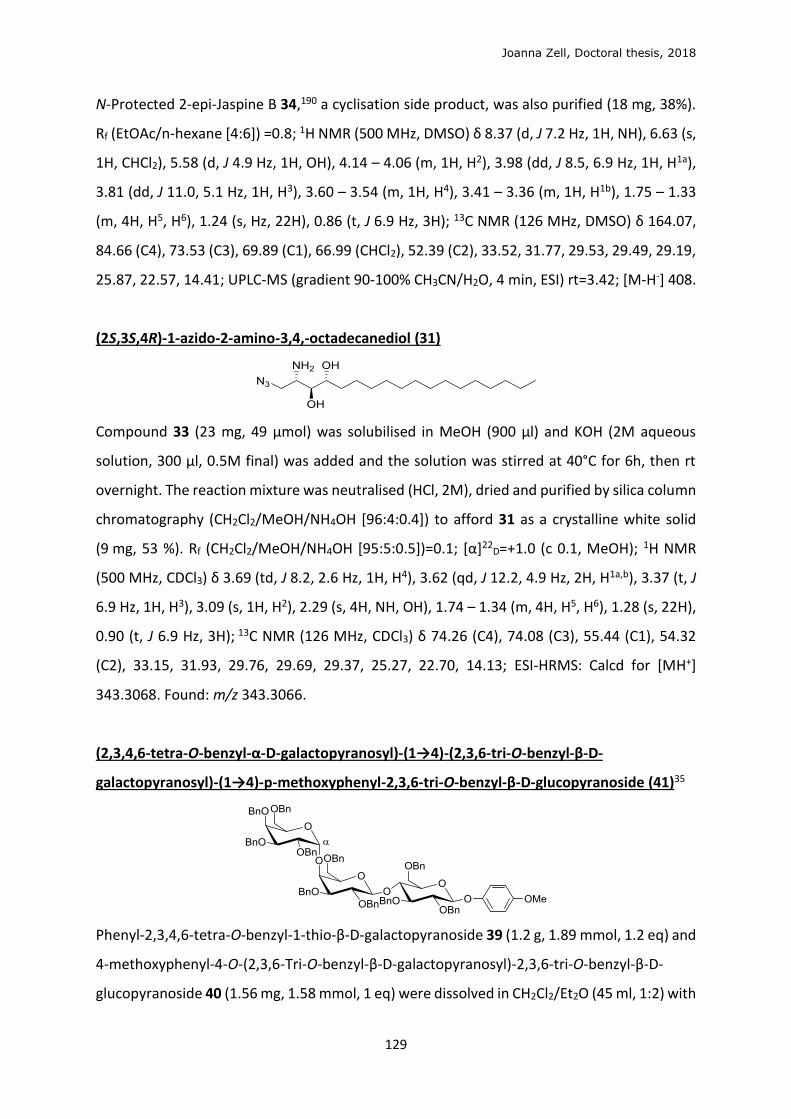
Joanna Zell, Doctoral thesis, 2018
129
N-Protected 2-epi-Jaspine B 34,190 a cyclisation side product, was also purified (18 mg, 38%).
Rf (EtOAc/n-hexane [4:6]) =0.8; 1H NMR (500 MHz, DMSO) δ 8.37 (d, J 7.2 Hz, 1H, NH), 6.63 (s,
1H, CHCl2), 5.58 (d, J 4.9 Hz, 1H, OH), 4.14 – 4.06 (m, 1H, H2), 3.98 (dd, J 8.5, 6.9 Hz, 1H, H1a),
3.81 (dd, J 11.0, 5.1 Hz, 1H, H3), 3.60 – 3.54 (m, 1H, H4), 3.41 – 3.36 (m, 1H, H1b), 1.75 – 1.33
(m, 4H, H5, H6), 1.24 (s, Hz, 22H), 0.86 (t, J 6.9 Hz, 3H); 13C NMR (126 MHz, DMSO) δ 164.07,
84.66 (C4), 73.53 (C3), 69.89 (C1), 66.99 (CHCl2), 52.39 (C2), 33.52, 31.77, 29.53, 29.49, 29.19,
25.87, 22.57, 14.41; UPLC-MS (gradient 90-100% CH3CN/H2O, 4 min, ESI) rt=3.42; [M-H-] 408.
(2S,3S,4R)-1-azido-2-amino-3,4,-octadecanediol (31)
Compound 33 (23 mg, 49 μmol) was solubilised in MeOH (900 μl) and KOH (2M aqueous
solution, 300 μl, 0.5M final) was added and the solution was stirred at 40°C for 6h, then rt
overnight. The reaction mixture was neutralised (HCl, 2M), dried and purified by silica column
chromatography (CH2Cl2/MeOH/NH4OH [96:4:0.4]) to afford 31 as a crystalline white solid
(9 mg, 53 %). Rf (CH2Cl2/MeOH/NH4OH [95:5:0.5])=0.1; [α]22D=+1.0 (c 0.1, MeOH); 1H NMR
(500 MHz, CDCl3) δ 3.69 (td, J 8.2, 2.6 Hz, 1H, H4), 3.62 (qd, J 12.2, 4.9 Hz, 2H, H1a,b), 3.37 (t, J
6.9 Hz, 1H, H3), 3.09 (s, 1H, H2), 2.29 (s, 4H, NH, OH), 1.74 – 1.34 (m, 4H, H5, H6), 1.28 (s, 22H),
0.90 (t, J 6.9 Hz, 3H); 13C NMR (126 MHz, CDCl3) δ 74.26 (C4), 74.08 (C3), 55.44 (C1), 54.32
(C2), 33.15, 31.93, 29.76, 29.69, 29.37, 25.27, 22.70, 14.13; ESI-HRMS: Calcd for [MH+]
343.3068. Found: m/z 343.3066.
(2,3,4,6-tetra-O-benzyl-α-D-galactopyranosyl)-(1→4)-(2,3,6-tri-O-benzyl-β-D-
galactopyranosyl)-(1→4)-p-methoxyphenyl-2,3,6-tri-O-benzyl-β-D-glucopyranoside (41)35
Phenyl-2,3,4,6-tetra-O-benzyl-1-thio-β-D-galactopyranoside 39 (1.2 g, 1.89 mmol, 1.2 eq) and
4-methoxyphenyl-4-O-(2,3,6-Tri-O-benzyl-β-D-galactopyranosyl)-2,3,6-tri-O-benzyl-β-D-
glucopyranoside 40 (1.56 mg, 1.58 mmol, 1 eq) were dissolved in CH2Cl2/Et2O (45 ml, 1:2) with
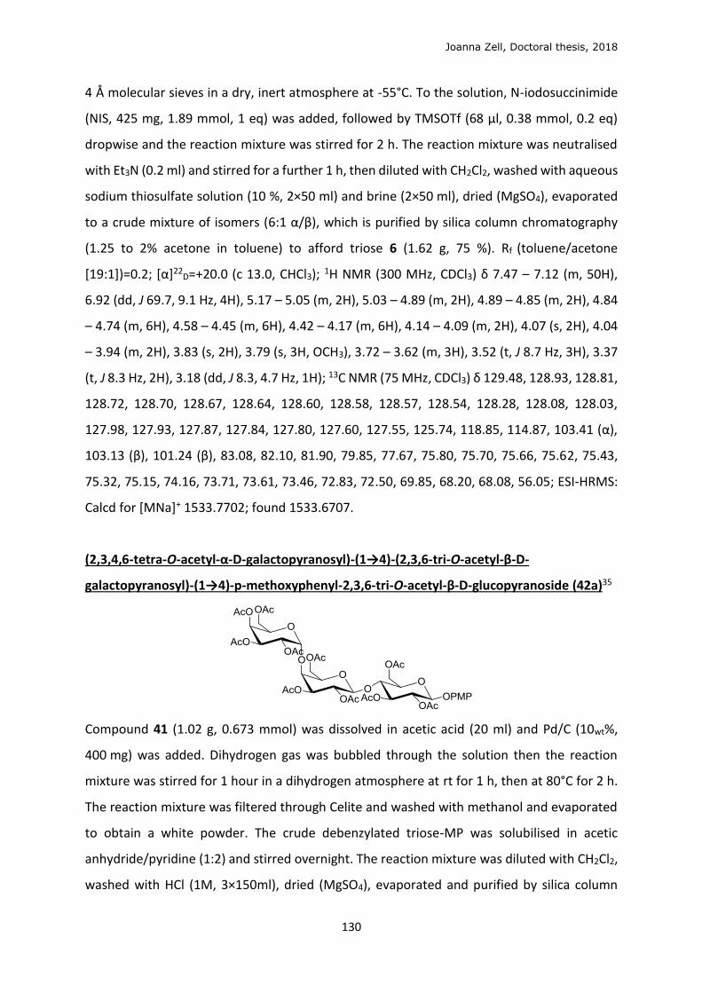
Joanna Zell, Doctoral thesis, 2018
130
4 Å molecular sieves in a dry, inert atmosphere at -55°C. To the solution, N-iodosuccinimide
(NIS, 425 mg, 1.89 mmol, 1 eq) was added, followed by TMSOTf (68 µl, 0.38 mmol, 0.2 eq)
dropwise and the reaction mixture was stirred for 2 h. The reaction mixture was neutralised
with Et3N (0.2 ml) and stirred for a further 1 h, then diluted with CH2Cl2, washed with aqueous
sodium thiosulfate solution (10 %, 2×50 ml) and brine (2×50 ml), dried (MgSO4), evaporated
to a crude mixture of isomers (6:1 α/β), which is purified by silica column chromatography
(1.25 to 2% acetone in toluene) to afford triose 6 (1.62 g, 75 %). Rf (toluene/acetone
[19:1])=0.2; [α]22D=+20.0 (c 13.0, CHCl3); 1H NMR (300 MHz, CDCl3) δ 7.47 – 7.12 (m, 50H),
6.92 (dd, J 69.7, 9.1 Hz, 4H), 5.17 – 5.05 (m, 2H), 5.03 – 4.89 (m, 2H), 4.89 – 4.85 (m, 2H), 4.84
– 4.74 (m, 6H), 4.58 – 4.45 (m, 6H), 4.42 – 4.17 (m, 6H), 4.14 – 4.09 (m, 2H), 4.07 (s, 2H), 4.04
– 3.94 (m, 2H), 3.83 (s, 2H), 3.79 (s, 3H, OCH3), 3.72 – 3.62 (m, 3H), 3.52 (t, J 8.7 Hz, 3H), 3.37
(t, J 8.3 Hz, 2H), 3.18 (dd, J 8.3, 4.7 Hz, 1H); 13C NMR (75 MHz, CDCl3) δ 129.48, 128.93, 128.81,
128.72, 128.70, 128.67, 128.64, 128.60, 128.58, 128.57, 128.54, 128.28, 128.08, 128.03,
127.98, 127.93, 127.87, 127.84, 127.80, 127.60, 127.55, 125.74, 118.85, 114.87, 103.41 (α),
103.13 (β), 101.24 (β), 83.08, 82.10, 81.90, 79.85, 77.67, 75.80, 75.70, 75.66, 75.62, 75.43,
75.32, 75.15, 74.16, 73.71, 73.61, 73.46, 72.83, 72.50, 69.85, 68.20, 68.08, 56.05; ESI-HRMS:
Calcd for [MNa]+ 1533.7702; found 1533.6707.
(2,3,4,6-tetra-O-acetyl-α-D-galactopyranosyl)-(1→4)-(2,3,6-tri-O-acetyl-β-D-
galactopyranosyl)-(1→4)-p-methoxyphenyl-2,3,6-tri-O-acetyl-β-D-glucopyranoside (42a)35
Compound 41 (1.02 g, 0.673 mmol) was dissolved in acetic acid (20 ml) and Pd/C (10wt%,
400 mg) was added. Dihydrogen gas was bubbled through the solution then the reaction
mixture was stirred for 1 hour in a dihydrogen atmosphere at rt for 1 h, then at 80°C for 2 h.
The reaction mixture was filtered through Celite and washed with methanol and evaporated
to obtain a white powder. The crude debenzylated triose-MP was solubilised in acetic
anhydride/pyridine (1:2) and stirred overnight. The reaction mixture was diluted with CH2Cl2,
washed with HCl (1M, 3×150ml), dried (MgSO4), evaporated and purified by silica column
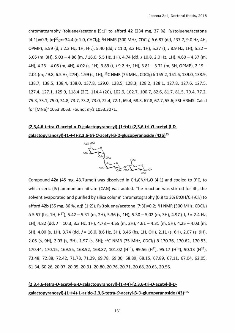
Joanna Zell, Doctoral thesis, 2018
131
chromatography (toluene/acetone [5:1] to afford 42 (234 mg, 37 %). Rf (toluene/acetone
[4:1])=0.3; [α]22D=+34.4 (c 1.0, CHCl3); 1H NMR (300 MHz, CDCl3) δ 6.87 (dd, J 37.7, 9.0 Hz, 4H,
OPMP), 5.59 (d, J 2.3 Hz, 1H, H1a), 5.40 (dd, J 11.0, 3.2 Hz, 1H), 5.27 (t, J 8.9 Hz, 1H), 5.22 –
5.05 (m, 3H), 5.03 – 4.86 (m, J 16.0, 5.5 Hz, 1H), 4.74 (dd, J 10.8, 2.0 Hz, 1H), 4.60 – 4.37 (m,
4H), 4.23 – 4.05 (m, 4H), 4.02 (s, 1H), 3.89 (t, J 9.2 Hz, 1H), 3.81 – 3.71 (m, 3H, OPMP), 2.19 –
2.01 (m, J 9.8, 6.5 Hz, 27H), 1.99 (s, 1H); 13C NMR (75 MHz, CDCl3) δ 155.2, 151.6, 139.0, 138.9,
138.7, 138.5, 138.4, 138.0, 137.8, 129.0, 128.5, 128.3, 128.2, 128.1, 127.8, 127.6, 127.5,
127.4, 127.1, 125.9, 118.4 (2C), 114.4 (2C), 102.9, 102.7, 100.7, 82.6, 81.7, 81.5, 79.4, 77.2,
75.3, 75.1, 75.0, 74.8, 73.7, 73.2, 73.0, 72.4, 72.1, 69.4, 68.3, 67.8, 67.7, 55.6; ESI-HRMS: Calcd
for [MNa]+ 1053.3063. Found: m/z 1053.3071.
(2,3,4,6-tetra-O-acetyl-α-D-galactopyranosyl)-(1→4)-(2,3,6-tri-O-acetyl-β-D-
galactopyranosyl)-(1→4)-2,3,6-tri-O-acetyl-β-D-glucopyranoside (42b)35
Compound 42a (45 mg, 43.7μmol) was dissolved in CH3CN/H2O (4:1) and cooled to 0°C, to
which ceric (IV) ammonium nitrate (CAN) was added. The reaction was stirred for 4h, the
solvent evaporated and purified by silica column chromatography (0.8 to 3% EtOH/CH2Cl2) to
afford 42b (35 mg, 86 %, α:β (1:2)). Rf (toluene/acetone [7:3])=0.2; 1H NMR (300 MHz, CDCl3)
δ 5.57 (bs, 1H, H1’’), 5.42 – 5.31 (m, 2H), 5.36 (s, 1H), 5.30 – 5.02 (m, 3H), 4.97 (d, J = 2.4 Hz,
1H), 4.82 (dd, J = 10.3, 3.3 Hz, 1H), 4.78 – 4.65 (m, 2H), 4.61 – 4.31 (m, 5H), 4.25 – 4.03 (m,
5H), 4.00 (s, 1H), 3.74 (dd, J = 16.0, 8.6 Hz, 3H), 3.46 (bs, 1H, OH), 2.11 (s, 6H), 2.07 (s, 9H),
2.05 (s, 9H), 2.03 (s, 3H), 1.97 (s, 3H); 13C NMR (75 MHz, CDCl3) δ 170.76, 170.62, 170.53,
170.44, 170.15, 169.55, 168.92, 168.87, 101.02 (H1’’), 99.56 (H1’), 95.17 (H1α), 90.13 (H1β),
73.48, 72.88, 72.42, 71.78, 71.29, 69.78, 69.00, 68.89, 68.15, 67.89, 67.11, 67.04, 62.05,
61.34, 60.26, 20.97, 20.95, 20.91, 20.80, 20.76, 20.71, 20.68, 20.63, 20.56.
(2,3,4,6-tetra-O-acetyl-α-D-galactopyranosyl)-(1→4)-(2,3,6-tri-O-acetyl-β-D-
galactopyranosyl)-(1→4)-1-azido-2,3,6-tetra-O-acetyl-β-D-glucopyranoside (43)185
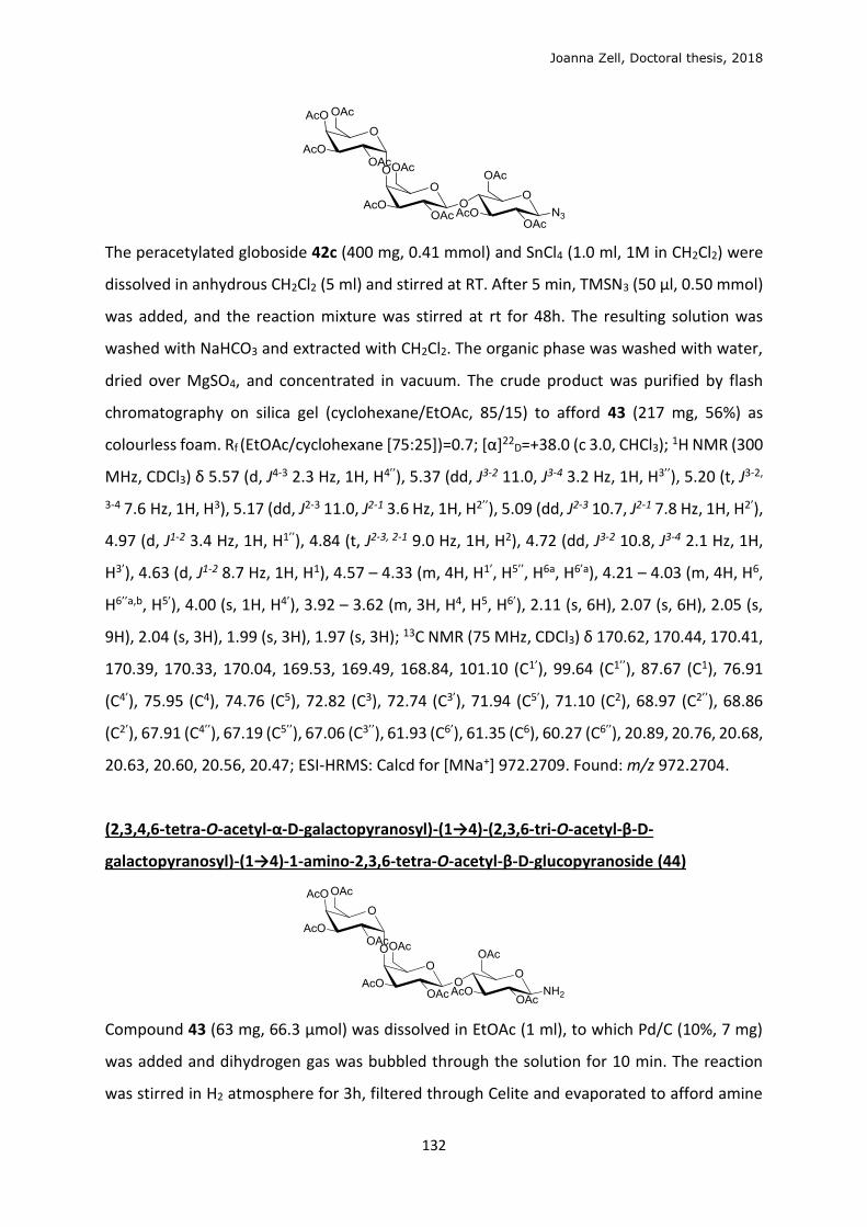
Joanna Zell, Doctoral thesis, 2018
132
The peracetylated globoside 42c (400 mg, 0.41 mmol) and SnCl4 (1.0 ml, 1M in CH2Cl2) were
dissolved in anhydrous CH2Cl2 (5 ml) and stirred at RT. After 5 min, TMSN3 (50 μl, 0.50 mmol)
was added, and the reaction mixture was stirred at rt for 48h. The resulting solution was
washed with NaHCO3 and extracted with CH2Cl2. The organic phase was washed with water,
dried over MgSO4, and concentrated in vacuum. The crude product was purified by flash
chromatography on silica gel (cyclohexane/EtOAc, 85/15) to afford 43 (217 mg, 56%) as
colourless foam. Rf (EtOAc/cyclohexane [75:25])=0.7; [α]22D=+38.0 (c 3.0, CHCl3); 1H NMR (300
MHz, CDCl3) δ 5.57 (d, J4-3 2.3 Hz, 1H, H4’’), 5.37 (dd, J3-2 11.0, J3-4 3.2 Hz, 1H, H3’’), 5.20 (t, J3-2,
3-4 7.6 Hz, 1H, H3), 5.17 (dd, J2-3 11.0, J2-1 3.6 Hz, 1H, H2’’), 5.09 (dd, J2-3 10.7, J2-1 7.8 Hz, 1H, H2’),
4.97 (d, J1-2 3.4 Hz, 1H, H1’’), 4.84 (t, J2-3, 2-1 9.0 Hz, 1H, H2), 4.72 (dd, J3-2 10.8, J3-4 2.1 Hz, 1H,
H3’), 4.63 (d, J1-2 8.7 Hz, 1H, H1), 4.57 – 4.33 (m, 4H, H1’, H5’’, H6a, H6’a), 4.21 – 4.03 (m, 4H, H6,
H6’’a,b, H5’), 4.00 (s, 1H, H4’), 3.92 – 3.62 (m, 3H, H4, H5, H6’), 2.11 (s, 6H), 2.07 (s, 6H), 2.05 (s,
9H), 2.04 (s, 3H), 1.99 (s, 3H), 1.97 (s, 3H); 13C NMR (75 MHz, CDCl3) δ 170.62, 170.44, 170.41,
170.39, 170.33, 170.04, 169.53, 169.49, 168.84, 101.10 (C1’), 99.64 (C1’’), 87.67 (C1), 76.91
(C4’), 75.95 (C4), 74.76 (C5), 72.82 (C3), 72.74 (C3’), 71.94 (C5’), 71.10 (C2), 68.97 (C2’’), 68.86
(C2’), 67.91 (C4’’), 67.19 (C5’’), 67.06 (C3’’), 61.93 (C6’), 61.35 (C6), 60.27 (C6’’), 20.89, 20.76, 20.68,
20.63, 20.60, 20.56, 20.47; ESI-HRMS: Calcd for [MNa+] 972.2709. Found: m/z 972.2704.
(2,3,4,6-tetra-O-acetyl-α-D-galactopyranosyl)-(1→4)-(2,3,6-tri-O-acetyl-β-D-
galactopyranosyl)-(1→4)-1-amino-2,3,6-tetra-O-acetyl-β-D-glucopyranoside (44)
Compound 43 (63 mg, 66.3 μmol) was dissolved in EtOAc (1 ml), to which Pd/C (10%, 7 mg)
was added and dihydrogen gas was bubbled through the solution for 10 min. The reaction
was stirred in H2 atmosphere for 3h, filtered through Celite and evaporated to afford amine
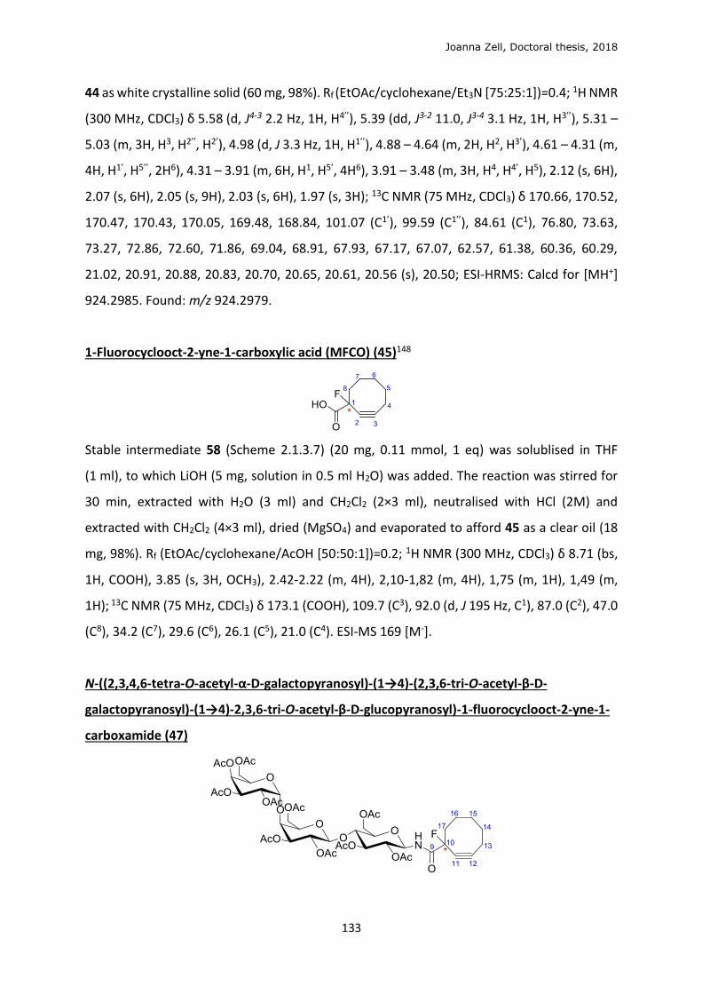
Joanna Zell, Doctoral thesis, 2018
133
44 as white crystalline solid (60 mg, 98%). Rf (EtOAc/cyclohexane/Et3N [75:25:1])=0.4; 1H NMR
(300 MHz, CDCl3) δ 5.58 (d, J4-3 2.2 Hz, 1H, H4’’), 5.39 (dd, J3-2 11.0, J3-4 3.1 Hz, 1H, H3’’), 5.31 –
5.03 (m, 3H, H3, H2’’, H2’), 4.98 (d, J 3.3 Hz, 1H, H1’’), 4.88 – 4.64 (m, 2H, H2, H3’), 4.61 – 4.31 (m,
4H, H1’, H5’’, 2H6), 4.31 – 3.91 (m, 6H, H1, H5’, 4H6), 3.91 – 3.48 (m, 3H, H4, H4’, H5), 2.12 (s, 6H),
2.07 (s, 6H), 2.05 (s, 9H), 2.03 (s, 6H), 1.97 (s, 3H); 13C NMR (75 MHz, CDCl3) δ 170.66, 170.52,
170.47, 170.43, 170.05, 169.48, 168.84, 101.07 (C1’), 99.59 (C1’’), 84.61 (C1), 76.80, 73.63,
73.27, 72.86, 72.60, 71.86, 69.04, 68.91, 67.93, 67.17, 67.07, 62.57, 61.38, 60.36, 60.29,
21.02, 20.91, 20.88, 20.83, 20.70, 20.65, 20.61, 20.56 (s), 20.50; ESI-HRMS: Calcd for [MH+]
924.2985. Found: m/z 924.2979.
1-Fluorocyclooct-2-yne-1-carboxylic acid (MFCO) (45)148
Stable intermediate 58 (Scheme 2.1.3.7) (20 mg, 0.11 mmol, 1 eq) was solublised in THF
(1 ml), to which LiOH (5 mg, solution in 0.5 ml H2O) was added. The reaction was stirred for
30 min, extracted with H2O (3 ml) and CH2Cl2 (2×3 ml), neutralised with HCl (2M) and
extracted with CH2Cl2 (4×3 ml), dried (MgSO4) and evaporated to afford 45 as a clear oil (18
mg, 98%). Rf (EtOAc/cyclohexane/AcOH [50:50:1])=0.2; 1H NMR (300 MHz, CDCl3) δ 8.71 (bs,
1H, COOH), 3.85 (s, 3H, OCH3), 2.42-2.22 (m, 4H), 2,10-1,82 (m, 4H), 1,75 (m, 1H), 1,49 (m,
1H); 13C NMR (75 MHz, CDCl3) δ 173.1 (COOH), 109.7 (C3), 92.0 (d, J 195 Hz, C1), 87.0 (C2), 47.0
(C8), 34.2 (C7), 29.6 (C6), 26.1 (C5), 21.0 (C4). ESI-MS 169 [M-].
N-((2,3,4,6-tetra-O-acetyl-α-D-galactopyranosyl)-(1→4)-(2,3,6-tri-O-acetyl-β-D-
galactopyranosyl)-(1→4)-2,3,6-tri-O-acetyl-β-D-glucopyranosyl)-1-fluorocyclooct-2-yne-1-
carboxamide (47)
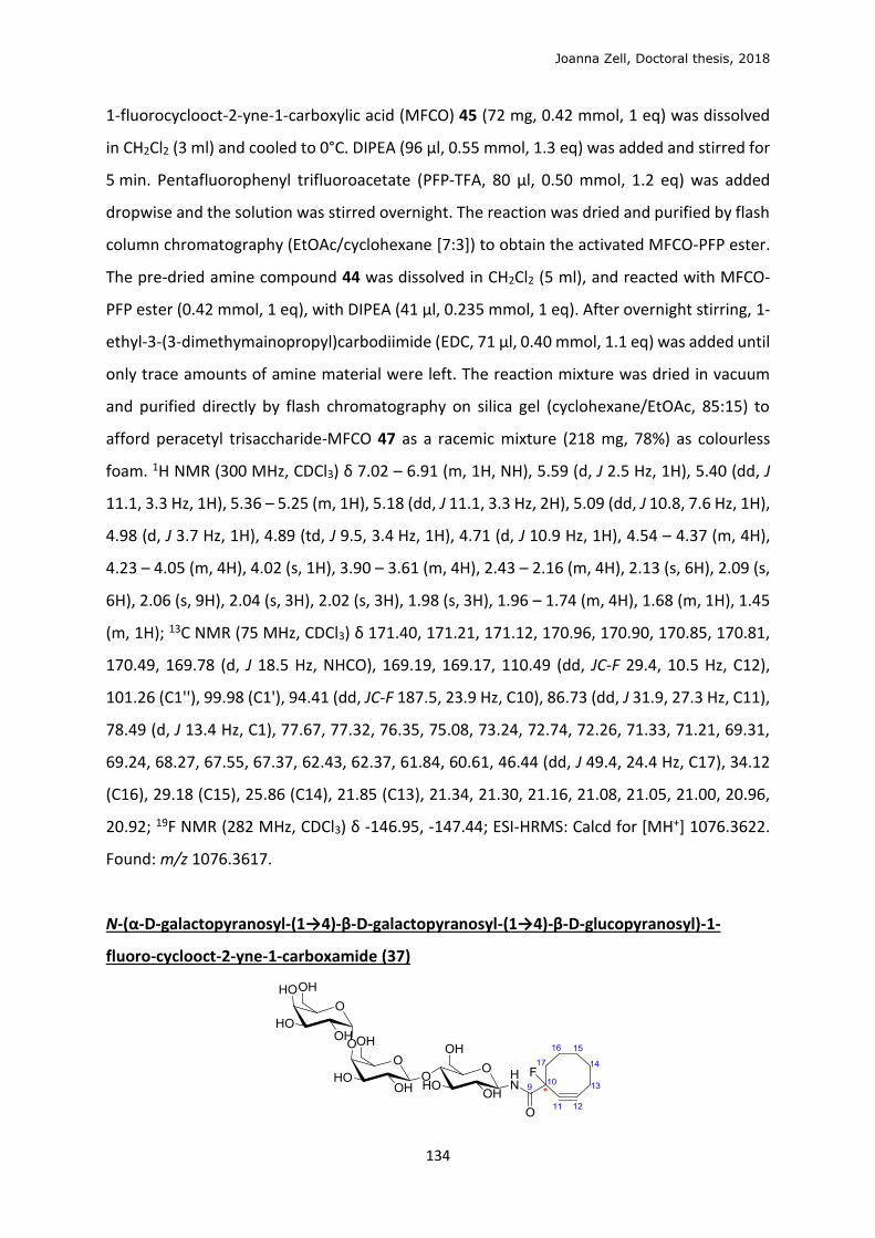
Joanna Zell, Doctoral thesis, 2018
134
1-fluorocyclooct-2-yne-1-carboxylic acid (MFCO) 45 (72 mg, 0.42 mmol, 1 eq) was dissolved
in CH2Cl2 (3 ml) and cooled to 0°C. DIPEA (96 μl, 0.55 mmol, 1.3 eq) was added and stirred for
5 min. Pentafluorophenyl trifluoroacetate (PFP-TFA, 80 μl, 0.50 mmol, 1.2 eq) was added
dropwise and the solution was stirred overnight. The reaction was dried and purified by flash
column chromatography (EtOAc/cyclohexane [7:3]) to obtain the activated MFCO-PFP ester.
The pre-dried amine compound 44 was dissolved in CH2Cl2 (5 ml), and reacted with MFCO-
PFP ester (0.42 mmol, 1 eq), with DIPEA (41 μl, 0.235 mmol, 1 eq). After overnight stirring, 1-
ethyl-3-(3-dimethymainopropyl)carbodiimide (EDC, 71 μl, 0.40 mmol, 1.1 eq) was added until
only trace amounts of amine material were left. The reaction mixture was dried in vacuum
and purified directly by flash chromatography on silica gel (cyclohexane/EtOAc, 85:15) to
afford peracetyl trisaccharide-MFCO 47 as a racemic mixture (218 mg, 78%) as colourless
foam. 1H NMR (300 MHz, CDCl3) δ 7.02 – 6.91 (m, 1H, NH), 5.59 (d, J 2.5 Hz, 1H), 5.40 (dd, J
11.1, 3.3 Hz, 1H), 5.36 – 5.25 (m, 1H), 5.18 (dd, J 11.1, 3.3 Hz, 2H), 5.09 (dd, J 10.8, 7.6 Hz, 1H),
4.98 (d, J 3.7 Hz, 1H), 4.89 (td, J 9.5, 3.4 Hz, 1H), 4.71 (d, J 10.9 Hz, 1H), 4.54 – 4.37 (m, 4H),
4.23 – 4.05 (m, 4H), 4.02 (s, 1H), 3.90 – 3.61 (m, 4H), 2.43 – 2.16 (m, 4H), 2.13 (s, 6H), 2.09 (s,
6H), 2.06 (s, 9H), 2.04 (s, 3H), 2.02 (s, 3H), 1.98 (s, 3H), 1.96 – 1.74 (m, 4H), 1.68 (m, 1H), 1.45
(m, 1H); 13C NMR (75 MHz, CDCl3) δ 171.40, 171.21, 171.12, 170.96, 170.90, 170.85, 170.81,
170.49, 169.78 (d, J 18.5 Hz, NHCO), 169.19, 169.17, 110.49 (dd, JC-F 29.4, 10.5 Hz, C12),
101.26 (C1''), 99.98 (C1'), 94.41 (dd, JC-F 187.5, 23.9 Hz, C10), 86.73 (dd, J 31.9, 27.3 Hz, C11),
78.49 (d, J 13.4 Hz, C1), 77.67, 77.32, 76.35, 75.08, 73.24, 72.74, 72.26, 71.33, 71.21, 69.31,
69.24, 68.27, 67.55, 67.37, 62.43, 62.37, 61.84, 60.61, 46.44 (dd, J 49.4, 24.4 Hz, C17), 34.12
(C16), 29.18 (C15), 25.86 (C14), 21.85 (C13), 21.34, 21.30, 21.16, 21.08, 21.05, 21.00, 20.96,
20.92; 19F NMR (282 MHz, CDCl3) δ -146.95, -147.44; ESI-HRMS: Calcd for [MH+] 1076.3622.
Found: m/z 1076.3617.
N-(α-D-galactopyranosyl-(1→4)-β-D-galactopyranosyl-(1→4)-β-D-glucopyranosyl)-1-
fluoro-cyclooct-2-yne-1-carboxamide (37)

Joanna Zell, Doctoral thesis, 2018
135
Compound 47 (24 mg, 22.3 μmol, 1 eq) was dissolved in dry MeOH (1.6 ml) and CH3ONa
(0.5 M solution in MeOH, 0.45 ml, 0.223 mmol, 10 eq) was added. The reaction was stirred
overnight, quenched with Dowex50WX8-200, filtered, evaporated and purified by reverse
phase C18 silica column chromatography (10 to 20% MeOH in H2O) to obtain the clickable
trisaccharide 37 (8 mg, 56 %) as a pure crystalline solid. Rf (nBuOH/EtOH/H2O [2:2:1])=0.5; 1H
NMR (400 MHz, MeOD) δ 4.96 – 4.91 (m, 2H, H1, H1’), 4.59 (bs, 1H, NH), 4.47 – 4.37 (m, 1H),
4.27 (t, J5-6 6.4 Hz, 1H), 3.99 (s, 1H), 3.94 – 3.82 (m, 5H), 3.81 (d, J 3.6 Hz, 1H), 3.79 (d, J 3.0 Hz,
1H), 3.77 – 3.63 (m, 3H), 3.63 – 3.51 (m, 4H), 3.51 – 3.41 (m, 2H), 2.43 – 2.20 (m, 4H), 2.19 –
1.78 (m, 4H), 1.75 – 1.58 (m, 1H), 1.58 – 1.37 (m, 1H). 13C NMR (101 MHz, MeOD) δ 172.04 (d,
J 25.8 Hz, C9), 110.20 (d, J 10.3 Hz, C12), 105.32 (C1’’), 102.65 (C1’), 95.23 (dd, J 187.6, 18.6 Hz,
C10), 88.13 (d, J 31.7 Hz, C11), 81.15 (d, J 5.6 Hz, C1), 80.42, 79.71, 78.36, 77.27, 76.55, 74.61,
73.20, 72.80, 72.65, 71.28, 71.03, 70.54, 62.65, 61.58, 61.49, 47.59 (d, J 24.8 Hz, C17), 34.96
(C16), 30.11 (C15), 26.72 (C14), 21.01 (C13). 19F NMR (282 MHz, MeOD) δ -149.05, -149.18. ESI-
HRMS: Calcd for [MH+] 656.2566. Found: m/z 656.2560.
N-(2,3,4,6-tetra-O-acetyl-β-D-galactopyranosyl-(1→4)-2,3,6-tri-O-acetyl-β-D-
glucopyranosyl) 1-fluoro-cyclooct-2-yne-1-carboxamide (53)
To a solution of MFCO 45 (18 mg, 106 μmol, 1 eq), the heptaacetyl-lactose-β-NH2 52 (85 mg,
120 μmol, 1.1 eq) and hydroxybenzotriazole (HOBt, 19 mg, 127 μmol, 1.2 eq) in CH2Cl2 (1 ml)
at 0°C, N,N’-dicyclohexylcarbodiimide (DCC, 44 mg, 212 μmol, 2 eq) was added at the reaction
mixture was warmed to rt after 10 min, and stirred overnight. The crude reaction mixture
(α/β≈1:11) was evaporated and purified by silica column chromatography (40 to 90 % EtOAc
in cyclohexane) containing some dicyclohexylurea impurity. This was removed by
precipitation in Et2O to afford 53 (52 mg (β), 62 %). Rf (EtOAc/cyclcohexane [1:1])=0.4. 1H NMR
(500 MHz, CDCl3) δ 6.94 (ddd, J 26.6, 9.4, 4.8 Hz, 1H, NH), 5.35 (d, J 3.1 Hz, 1H, H4’), 5.31 (td, J
9.2 Hz, 3.9 Hz, H3), 5.17 (qd, J 9.5 Hz, 1.2 Hz, 1H, H1), 5.11 (dd, J 10.4, 7.9 Hz, 1H, H2’), 4.94 (dd,
J 10.4, 3.5 Hz, 1H, H3’), 4.88 (td, J 9.5, 6.9 Hz, 1H, H2), 4.46 (dd, J 7.8, 1.1 Hz, 1H, H1’), 4.41-4.45
(m, 1H, H6), 4.10-4.17 (m, 2H, H6, H6’), 4.08 (dd, J 11.1, 7.2 Hz, 1H, H6’), 3.87 (t, J 6.8 Hz, 1H,
H5’), 3.78 (td, J 8.8 Hz, 2.3 Hz, 1H, H4), 3.73 (dt, J 10.0, 2.2 Hz, 1H, H5), 2.47 – 2.16 (m, 4H, H11,
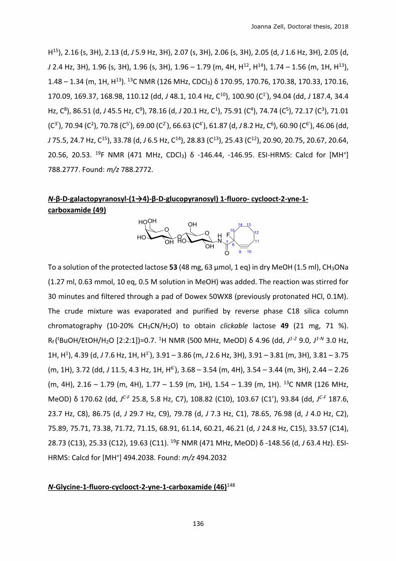
Joanna Zell, Doctoral thesis, 2018
136
H15), 2.16 (s, 3H), 2.13 (d, J 5.9 Hz, 3H), 2.07 (s, 3H), 2.06 (s, 3H), 2.05 (d, J 1.6 Hz, 3H), 2.05 (d,
J 2.4 Hz, 3H), 1.96 (s, 3H), 1.96 (s, 3H), 1.96 – 1.79 (m, 4H, H12, H14), 1.74 – 1.56 (m, 1H, H13),
1.48 – 1.34 (m, 1H, H13). 13C NMR (126 MHz, CDCl3) δ 170.95, 170.76, 170.38, 170.33, 170.16,
170.09, 169.37, 168.98, 110.12 (dd, J 48.1, 10.4 Hz, C10), 100.90 (C1’), 94.04 (dd, J 187.4, 34.4
Hz, C8), 86.51 (d, J 45.5 Hz, C9), 78.16 (d, J 20.1 Hz, C1), 75.91 (C4), 74.74 (C5), 72.17 (C3), 71.01
(C3’), 70.94 (C2), 70.78 (C5’), 69.00 (C2’), 66.63 (C4’), 61.87 (d, J 8.2 Hz, C6), 60.90 (C6’), 46.06 (dd,
J 75.5, 24.7 Hz, C15), 33.78 (d, J 6.5 Hz, C14), 28.83 (C13), 25.43 (C12), 20.90, 20.75, 20.67, 20.64,
20.56, 20.53. 19F NMR (471 MHz, CDCl3) δ -146.44, -146.95. ESI-HRMS: Calcd for [MH+]
788.2777. Found: m/z 788.2772.
N-β-D-galactopyranosyl-(1→4)-β-D-glucopyranosyl) 1-fluoro- cyclooct-2-yne-1-
carboxamide (49)
To a solution of the protected lactose 53 (48 mg, 63 μmol, 1 eq) in dry MeOH (1.5 ml), CH3ONa
(1.27 ml, 0.63 mmol, 10 eq, 0.5 M solution in MeOH) was added. The reaction was stirred for
30 minutes and filtered through a pad of Dowex 50WX8 (previously protonated HCl, 0.1M).
The crude mixture was evaporated and purified by reverse phase C18 silica column
chromatography (10-20% CH3CN/H2O) to obtain clickable lactose 49 (21 mg, 71 %).
Rf (tBuOH/EtOH/H2O [2:2:1])=0.7. 1H NMR (500 MHz, MeOD) δ 4.96 (dd, J1-2 9.0, J1-N 3.0 Hz,
1H, H1), 4.39 (d, J 7.6 Hz, 1H, H1’), 3.91 – 3.86 (m, J 2.6 Hz, 3H), 3.91 – 3.81 (m, 3H), 3.81 – 3.75
(m, 1H), 3.72 (dd, J 11.5, 4.3 Hz, 1H, H6’), 3.68 – 3.54 (m, 4H), 3.54 – 3.44 (m, 3H), 2.44 – 2.26
(m, 4H), 2.16 – 1.79 (m, 4H), 1.77 – 1.59 (m, 1H), 1.54 – 1.39 (m, 1H). 13C NMR (126 MHz,
MeOD) δ 170.62 (dd, JC-F 25.8, 5.8 Hz, C7), 108.82 (C10), 103.67 (C1’), 93.84 (dd, JC-F 187.6,
23.7 Hz, C8), 86.75 (d, J 29.7 Hz, C9), 79.78 (d, J 7.3 Hz, C1), 78.65, 76.98 (d, J 4.0 Hz, C2),
75.89, 75.71, 73.38, 71.72, 71.15, 68.91, 61.14, 60.21, 46.21 (d, J 24.8 Hz, C15), 33.57 (C14),
28.73 (C13), 25.33 (C12), 19.63 (C11). 19F NMR (471 MHz, MeOD) δ -148.56 (d, J 63.4 Hz). ESI-
HRMS: Calcd for [MH+] 494.2038. Found: m/z 494.2032
N-Glycine-1-fluoro-cyclooct-2-yne-1-carboxamide (46)148
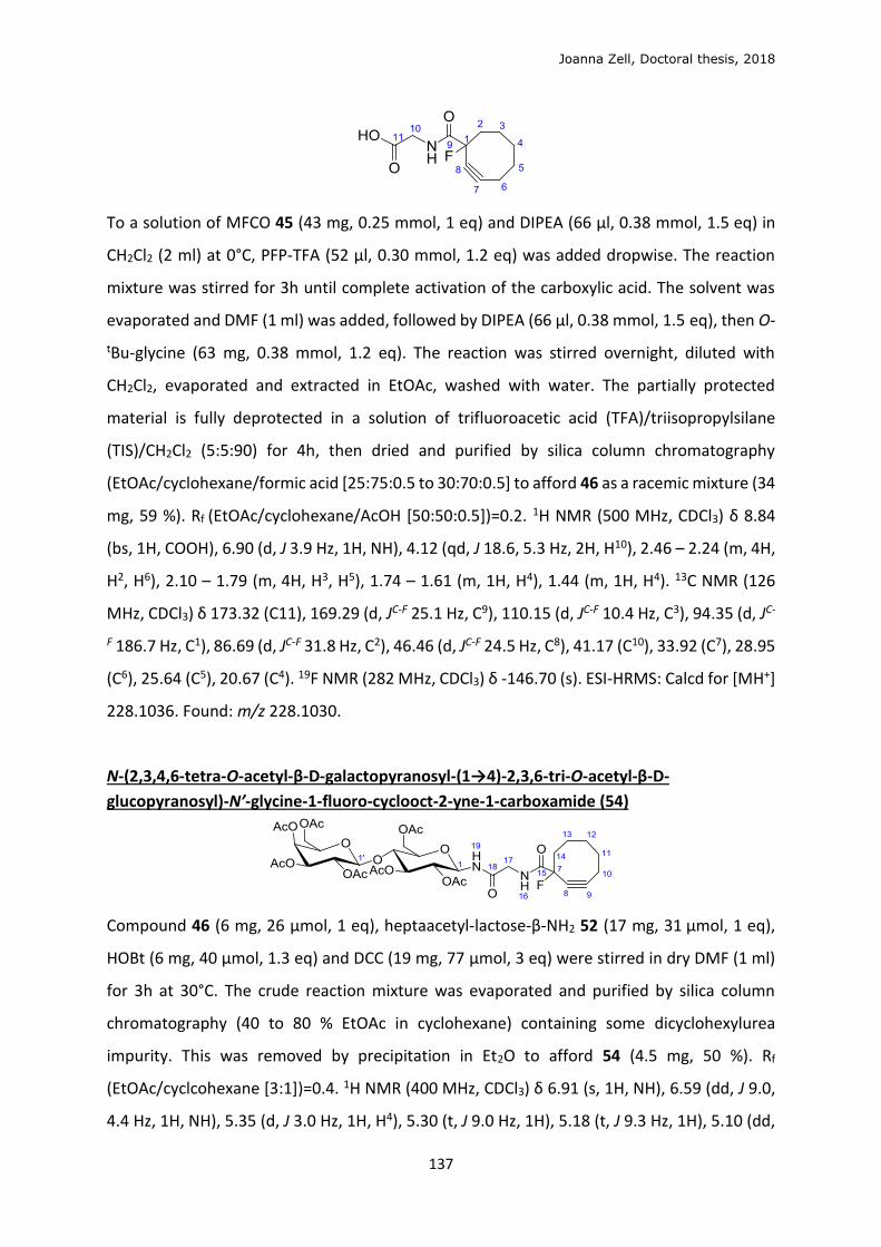
Joanna Zell, Doctoral thesis, 2018
137
To a solution of MFCO 45 (43 mg, 0.25 mmol, 1 eq) and DIPEA (66 μl, 0.38 mmol, 1.5 eq) in
CH2Cl2 (2 ml) at 0°C, PFP-TFA (52 μl, 0.30 mmol, 1.2 eq) was added dropwise. The reaction
mixture was stirred for 3h until complete activation of the carboxylic acid. The solvent was
evaporated and DMF (1 ml) was added, followed by DIPEA (66 μl, 0.38 mmol, 1.5 eq), then O-
tBu-glycine (63 mg, 0.38 mmol, 1.2 eq). The reaction was stirred overnight, diluted with
CH2Cl2, evaporated and extracted in EtOAc, washed with water. The partially protected
material is fully deprotected in a solution of trifluoroacetic acid (TFA)/triisopropylsilane
(TIS)/CH2Cl2 (5:5:90) for 4h, then dried and purified by silica column chromatography
(EtOAc/cyclohexane/formic acid [25:75:0.5 to 30:70:0.5] to afford 46 as a racemic mixture (34
mg, 59 %). Rf (EtOAc/cyclohexane/AcOH [50:50:0.5])=0.2. 1H NMR (500 MHz, CDCl3) δ 8.84
(bs, 1H, COOH), 6.90 (d, J 3.9 Hz, 1H, NH), 4.12 (qd, J 18.6, 5.3 Hz, 2H, H10), 2.46 – 2.24 (m, 4H,
H2, H6), 2.10 – 1.79 (m, 4H, H3, H5), 1.74 – 1.61 (m, 1H, H4), 1.44 (m, 1H, H4). 13C NMR (126
MHz, CDCl3) δ 173.32 (C11), 169.29 (d, JC-F 25.1 Hz, C9), 110.15 (d, JC-F 10.4 Hz, C3), 94.35 (d, JC-
F 186.7 Hz, C1), 86.69 (d, JC-F 31.8 Hz, C2), 46.46 (d, JC-F 24.5 Hz, C8), 41.17 (C10), 33.92 (C7), 28.95
(C6), 25.64 (C5), 20.67 (C4). 19F NMR (282 MHz, CDCl3) δ -146.70 (s). ESI-HRMS: Calcd for [MH+]
228.1036. Found: m/z 228.1030.
N-(2,3,4,6-tetra-O-acetyl-β-D-galactopyranosyl-(1→4)-2,3,6-tri-O-acetyl-β-D-
glucopyranosyl)-N’-glycine-1-fluoro-cyclooct-2-yne-1-carboxamide (54)
Compound 46 (6 mg, 26 μmol, 1 eq), heptaacetyl-lactose-β-NH2 52 (17 mg, 31 μmol, 1 eq),
HOBt (6 mg, 40 μmol, 1.3 eq) and DCC (19 mg, 77 μmol, 3 eq) were stirred in dry DMF (1 ml)
for 3h at 30°C. The crude reaction mixture was evaporated and purified by silica column
chromatography (40 to 80 % EtOAc in cyclohexane) containing some dicyclohexylurea
impurity. This was removed by precipitation in Et2O to afford 54 (4.5 mg, 50 %). Rf
(EtOAc/cyclcohexane [3:1])=0.4. 1H NMR (400 MHz, CDCl3) δ 6.91 (s, 1H, NH), 6.59 (dd, J 9.0,
4.4 Hz, 1H, NH), 5.35 (d, J 3.0 Hz, 1H, H4), 5.30 (t, J 9.0 Hz, 1H), 5.18 (t, J 9.3 Hz, 1H), 5.10 (dd,
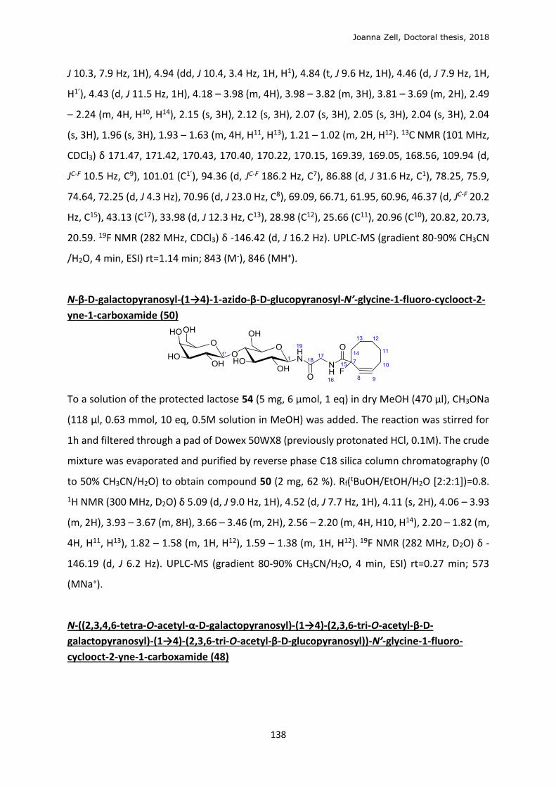
Joanna Zell, Doctoral thesis, 2018
138
J 10.3, 7.9 Hz, 1H), 4.94 (dd, J 10.4, 3.4 Hz, 1H, H1), 4.84 (t, J 9.6 Hz, 1H), 4.46 (d, J 7.9 Hz, 1H,
H1’), 4.43 (d, J 11.5 Hz, 1H), 4.18 – 3.98 (m, 4H), 3.98 – 3.82 (m, 3H), 3.81 – 3.69 (m, 2H), 2.49
– 2.24 (m, 4H, H10, H14), 2.15 (s, 3H), 2.12 (s, 3H), 2.07 (s, 3H), 2.05 (s, 3H), 2.04 (s, 3H), 2.04
(s, 3H), 1.96 (s, 3H), 1.93 – 1.63 (m, 4H, H11, H13), 1.21 – 1.02 (m, 2H, H12). 13C NMR (101 MHz,
CDCl3) δ 171.47, 171.42, 170.43, 170.40, 170.22, 170.15, 169.39, 169.05, 168.56, 109.94 (d,
JC-F 10.5 Hz, C9), 101.01 (C1’), 94.36 (d, JC-F 186.2 Hz, C7), 86.88 (d, J 31.6 Hz, C1), 78.25, 75.9,
74.64, 72.25 (d, J 4.3 Hz), 70.96 (d, J 23.0 Hz, C8), 69.09, 66.71, 61.95, 60.96, 46.37 (d, JC-F 20.2
Hz, C15), 43.13 (C17), 33.98 (d, J 12.3 Hz, C13), 28.98 (C12), 25.66 (C11), 20.96 (C10), 20.82, 20.73,
20.59. 19F NMR (282 MHz, CDCl3) δ -146.42 (d, J 16.2 Hz). UPLC-MS (gradient 80-90% CH3CN
/H2O, 4 min, ESI) rt=1.14 min; 843 (M-), 846 (MH+).
N-β-D-galactopyranosyl-(1→4)-1-azido-β-D-glucopyranosyl-N’-glycine-1-fluoro-cyclooct-2-
yne-1-carboxamide (50)
To a solution of the protected lactose 54 (5 mg, 6 μmol, 1 eq) in dry MeOH (470 μl), CH3ONa
(118 μl, 0.63 mmol, 10 eq, 0.5M solution in MeOH) was added. The reaction was stirred for
1h and filtered through a pad of Dowex 50WX8 (previously protonated HCl, 0.1M). The crude
mixture was evaporated and purified by reverse phase C18 silica column chromatography (0
to 50% CH3CN/H2O) to obtain compound 50 (2 mg, 62 %). Rf(tBuOH/EtOH/H2O [2:2:1])=0.8.
1H NMR (300 MHz, D2O) δ 5.09 (d, J 9.0 Hz, 1H), 4.52 (d, J 7.7 Hz, 1H), 4.11 (s, 2H), 4.06 – 3.93
(m, 2H), 3.93 – 3.67 (m, 8H), 3.66 – 3.46 (m, 2H), 2.56 – 2.20 (m, 4H, H10, H14), 2.20 – 1.82 (m,
4H, H11, H13), 1.82 – 1.58 (m, 1H, H12), 1.59 – 1.38 (m, 1H, H12). 19F NMR (282 MHz, D2O) δ -
146.19 (d, J 6.2 Hz). UPLC-MS (gradient 80-90% CH3CN/H2O, 4 min, ESI) rt=0.27 min; 573
(MNa+).
N-((2,3,4,6-tetra-O-acetyl-α-D-galactopyranosyl)-(1→4)-(2,3,6-tri-O-acetyl-β-D-
galactopyranosyl)-(1→4)-(2,3,6-tri-O-acetyl-β-D-glucopyranosyl))-N’-glycine-1-fluoro-
cyclooct-2-yne-1-carboxamide (48)
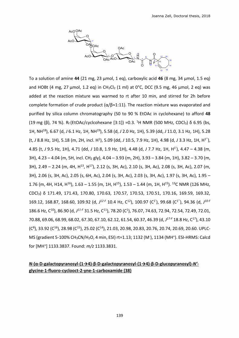
Joanna Zell, Doctoral thesis, 2018
139
To a solution of amine 44 (21 mg, 23 μmol, 1 eq), carboxylic acid 46 (8 mg, 34 μmol, 1.5 eq)
and HOBt (4 mg, 27 μmol, 1.2 eq) in CH2Cl2 (1 ml) at 0°C, DCC (9.5 mg, 46 μmol, 2 eq) was
added at the reaction mixture was warmed to rt after 10 min, and stirred for 2h before
complete formation of crude product (α/β≈1:11). The reaction mixture was evaporated and
purified by silica column chromatography (50 to 90 % EtOAc in cyclohexane) to afford 48
(19 mg (β), 74 %). Rf (EtOAc/cyclcohexane [3:1]) =0.3. 1H NMR (500 MHz, CDCl3) δ 6.95 (bs,
1H, NH18), 6.67 (d, J 6.1 Hz, 1H, NH19), 5.58 (d, J 2.0 Hz, 1H), 5.39 (dd, J 11.0, 3.1 Hz, 1H), 5.28
(t, J 8.8 Hz, 1H), 5.18 (m, 2H, incl. H1), 5.09 (dd, J 10.5, 7.9 Hz, 1H), 4.98 (d, J 3.3 Hz, 1H, H1’’),
4.85 (t, J 9.5 Hz, 1H), 4.71 (dd, J 10.8, 1.9 Hz, 1H), 4.48 (d, J 7.7 Hz, 1H, H1’), 4.47 – 4.38 (m,
3H), 4.23 – 4.04 (m, 5H, incl. CH2 gly), 4.04 – 3.93 (m, 2H), 3.93 – 3.84 (m, 1H), 3.82 – 3.70 (m,
3H), 2.49 – 2.24 (m, 4H, H13, H17), 2.12 (s, 3H, Ac), 2.10 (s, 3H, Ac), 2.08 (s, 3H, Ac), 2.07 (m,
3H), 2.06 (s, 3H, Ac), 2.05 (s, 6H, Ac), 2.04 (s, 3H, Ac), 2.03 (s, 3H, Ac), 1.97 (s, 3H, Ac), 1.95 –
1.76 (m, 4H, H14, H16), 1.63 – 1.55 (m, 1H, H15), 1.53 – 1.44 (m, 1H, H15). 13C NMR (126 MHz,
CDCl3) δ 171.49, 171.43, 170.80, 170.63, 170.57, 170.53, 170.51, 170.16, 169.59, 169.32,
169.12, 168.87, 168.60, 109.92 (d, J12-F 10.4 Hz, C12), 100.97 (C1’), 99.68 (C1’’), 94.36 (d, J10-F
186.6 Hz, C10), 86.90 (d, J11-F 31.5 Hz, C11), 78.20 (C1), 76.07, 74.63, 72.94, 72.54, 72.49, 72.01,
70.88, 69.06, 68.99, 68.02, 67.30, 67.10, 62.12, 61.54, 60.37, 46.39 (d, J17-F 18.8 Hz, C17), 43.10
(C8), 33.92 (C16), 28.98 (C15), 25.02 (C14), 21.03, 20.98, 20.83, 20.76, 20.74, 20.69, 20.60. UPLC-
MS (gradient 5-100% CH3CN/H2O, 4 min, ESI) rt=1.13; 1132 (M-), 1134 (MH+). ESI-HRMS: Calcd
for [MH+] 1133.3837. Found: m/z 1133.3831.
N-(α-D-galactopyranosyl-(1→4)-β-D-galactopyranosyl-(1→4)-β-D-glucopyranosyl)-N’-
glycine-1-fluoro-cyclooct-2-yne-1-carboxamide (38)
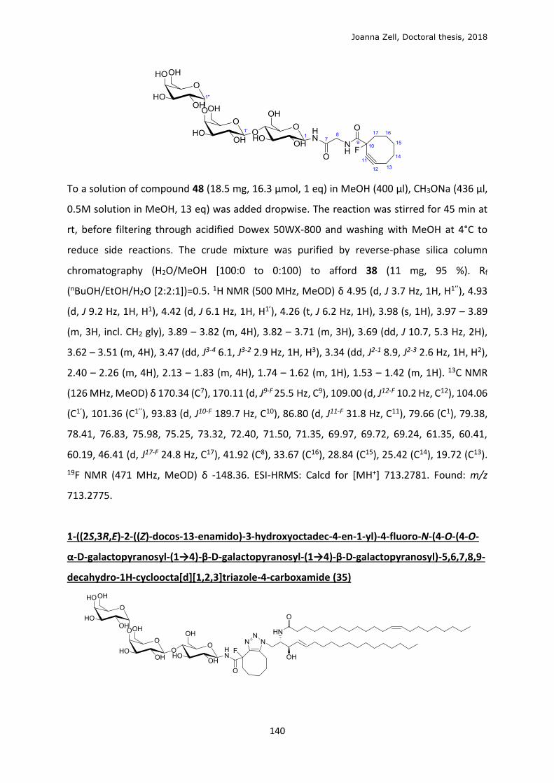
Joanna Zell, Doctoral thesis, 2018
140
To a solution of compound 48 (18.5 mg, 16.3 μmol, 1 eq) in MeOH (400 μl), CH3ONa (436 μl,
0.5M solution in MeOH, 13 eq) was added dropwise. The reaction was stirred for 45 min at
rt, before filtering through acidified Dowex 50WX-800 and washing with MeOH at 4°C to
reduce side reactions. The crude mixture was purified by reverse-phase silica column
chromatography (H2O/MeOH [100:0 to 0:100) to afford 38 (11 mg, 95 %). Rf
(nBuOH/EtOH/H2O [2:2:1])=0.5. 1H NMR (500 MHz, MeOD) δ 4.95 (d, J 3.7 Hz, 1H, H1’’), 4.93
(d, J 9.2 Hz, 1H, H1), 4.42 (d, J 6.1 Hz, 1H, H1’), 4.26 (t, J 6.2 Hz, 1H), 3.98 (s, 1H), 3.97 – 3.89
(m, 3H, incl. CH2 gly), 3.89 – 3.82 (m, 4H), 3.82 – 3.71 (m, 3H), 3.69 (dd, J 10.7, 5.3 Hz, 2H),
3.62 – 3.51 (m, 4H), 3.47 (dd, J3-4 6.1, J3-2 2.9 Hz, 1H, H3), 3.34 (dd, J2-1 8.9, J2-3 2.6 Hz, 1H, H2),
2.40 – 2.26 (m, 4H), 2.13 – 1.83 (m, 4H), 1.74 – 1.62 (m, 1H), 1.53 – 1.42 (m, 1H). 13C NMR
(126 MHz, MeOD) δ 170.34 (C7), 170.11 (d, J9-F 25.5 Hz, C9), 109.00 (d, J12-F 10.2 Hz, C12), 104.06
(C1’), 101.36 (C1’’), 93.83 (d, J10-F 189.7 Hz, C10), 86.80 (d, J11-F 31.8 Hz, C11), 79.66 (C1), 79.38,
78.41, 76.83, 75.98, 75.25, 73.32, 72.40, 71.50, 71.35, 69.97, 69.72, 69.24, 61.35, 60.41,
60.19, 46.41 (d, J17-F 24.8 Hz, C17), 41.92 (C8), 33.67 (C16), 28.84 (C15), 25.42 (C14), 19.72 (C13).
19F NMR (471 MHz, MeOD) δ -148.36. ESI-HRMS: Calcd for [MH+] 713.2781. Found: m/z
713.2775.
1-((2S,3R,E)-2-((Z)-docos-13-enamido)-3-hydroxyoctadec-4-en-1-yl)-4-fluoro-N-(4-O-(4-O-
α-D-galactopyranosyl-(1→4)-β-D-galactopyranosyl-(1→4)-β-D-galactopyranosyl)-5,6,7,8,9-
decahydro-1H-cycloocta[d][1,2,3]triazole-4-carboxamide (35)
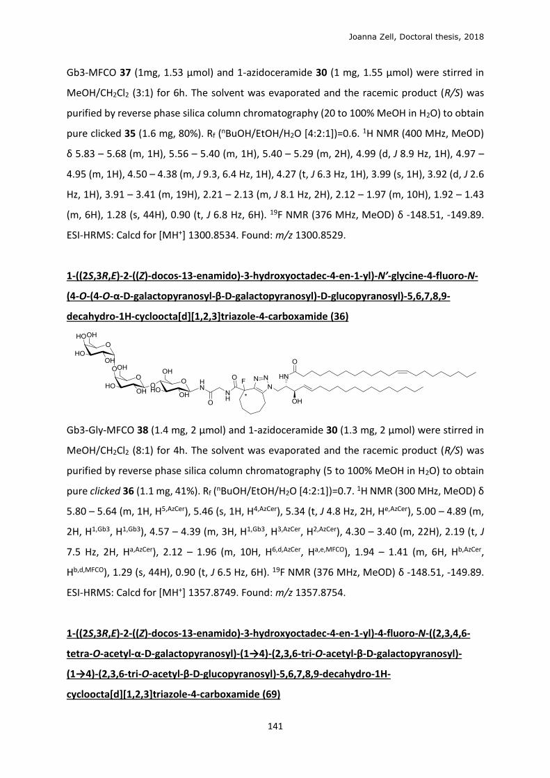
Joanna Zell, Doctoral thesis, 2018
141
Gb3-MFCO 37 (1mg, 1.53 μmol) and 1-azidoceramide 30 (1 mg, 1.55 μmol) were stirred in
MeOH/CH2Cl2 (3:1) for 6h. The solvent was evaporated and the racemic product (R/S) was
purified by reverse phase silica column chromatography (20 to 100% MeOH in H2O) to obtain
pure clicked 35 (1.6 mg, 80%). Rf (nBuOH/EtOH/H2O [4:2:1])=0.6. 1H NMR (400 MHz, MeOD)
δ 5.83 – 5.68 (m, 1H), 5.56 – 5.40 (m, 1H), 5.40 – 5.29 (m, 2H), 4.99 (d, J 8.9 Hz, 1H), 4.97 –
4.95 (m, 1H), 4.50 – 4.38 (m, J 9.3, 6.4 Hz, 1H), 4.27 (t, J 6.3 Hz, 1H), 3.99 (s, 1H), 3.92 (d, J 2.6
Hz, 1H), 3.91 – 3.41 (m, 19H), 2.21 – 2.13 (m, J 8.1 Hz, 2H), 2.12 – 1.97 (m, 10H), 1.92 – 1.43
(m, 6H), 1.28 (s, 44H), 0.90 (t, J 6.8 Hz, 6H). 19F NMR (376 MHz, MeOD) δ -148.51, -149.89.
ESI-HRMS: Calcd for [MH+] 1300.8534. Found: m/z 1300.8529.
1-((2S,3R,E)-2-((Z)-docos-13-enamido)-3-hydroxyoctadec-4-en-1-yl)-N’-glycine-4-fluoro-N-
(4-O-(4-O-α-D-galactopyranosyl-β-D-galactopyranosyl)-D-glucopyranosyl)-5,6,7,8,9-
decahydro-1H-cycloocta[d][1,2,3]triazole-4-carboxamide (36)
Gb3-Gly-MFCO 38 (1.4 mg, 2 μmol) and 1-azidoceramide 30 (1.3 mg, 2 μmol) were stirred in
MeOH/CH2Cl2 (8:1) for 4h. The solvent was evaporated and the racemic product (R/S) was
purified by reverse phase silica column chromatography (5 to 100% MeOH in H2O) to obtain
pure clicked 36 (1.1 mg, 41%). Rf (nBuOH/EtOH/H2O [4:2:1])=0.7. 1H NMR (300 MHz, MeOD) δ
5.80 – 5.64 (m, 1H, H5,AzCer), 5.46 (s, 1H, H4,AzCer), 5.34 (t, J 4.8 Hz, 2H, He,AzCer), 5.00 – 4.89 (m,
2H, H1,Gb3, H1,Gb3), 4.57 – 4.39 (m, 3H, H1,Gb3, H3,AzCer, H2,AzCer), 4.30 – 3.40 (m, 22H), 2.19 (t, J
7.5 Hz, 2H, Ha,AzCer), 2.12 – 1.96 (m, 10H, H6,d,AzCer, Ha,e,MFCO), 1.94 – 1.41 (m, 6H, Hb,AzCer,
Hb,d,MFCO), 1.29 (s, 44H), 0.90 (t, J 6.5 Hz, 6H). 19F NMR (376 MHz, MeOD) δ -148.51, -149.89.
ESI-HRMS: Calcd for [MH+] 1357.8749. Found: m/z 1357.8754.
1-((2S,3R,E)-2-((Z)-docos-13-enamido)-3-hydroxyoctadec-4-en-1-yl)-4-fluoro-N-((2,3,4,6-
tetra-O-acetyl-α-D-galactopyranosyl)-(1→4)-(2,3,6-tri-O-acetyl-β-D-galactopyranosyl)-
(1→4)-(2,3,6-tri-O-acetyl-β-D-glucopyranosyl)-5,6,7,8,9-decahydro-1H-
cycloocta[d][1,2,3]triazole-4-carboxamide (69)
Joanna Zell, Doctoral thesis, 2018
142
Peracteyl Gb3-MFCO 47 (0.8 mg, 0.7 μmol) and 1-azidoceramide 30 (0.5 mg, 0.78 μmol) were
stirred in CH2Cl2 for 48h. The solvent was evaporated and the racemic product (R/S) was
purified by silica column chromatography (0 to 2.5% MeOH in CH2Cl2) to obtain the isomeric
clicked 69 (1 mg, 94%) and 1-N-MFCO-Gb3 (0.1 mg) recuperated. Rf (2.5% MeOH/CH2Cl2)=0.3.
ESI-HRMS: Calcd for [M+H+]+ 1720.9585, [M+H++NH4+]+2 869.4962. Found: m/z 1720.9589 and
869.4965.
N-2-hydroxyethyl-1-fluoro-cyclooct-2-yne-1-carboxamide (59)
Stable intermediate 58 (Scheme 2.1.3.7) (9 mg, 49 μmol, 1 eq) was dissolved in THF (240 μl)
and cooled to 0°C, to which freshly distilled ethanolamine in solution with THF (50 μl, 1:3) was
added dropwise. The reaction mixture was stirred for 4h, evaporated and purified by silica
column chromatography (EtOAc/cyclohexane [1:1]) to afford 59 as a racemic mixture (3 mg,
29%) Rf (EtOAc)=0.5. 1H NMR (300 MHz, CDCl3) δ 6.80 (s, 1H, OH), 3.77 (t, J 4.9 Hz, 2H, H11),
3.48 (dd, J 10.1, 5.2 Hz, 2H, H10), 2.52 – 2.18 (m, 4H, H6, H2), 2.18 – 1.77 (m, 4H, H3, H5), 1.78
– 1.57 (m, 1H, H4), 1.56 – 1.37 (m, 1H, H4). 13C NMR (101 MHz, CDCl3) δ 169.56 (d, J 24.2 Hz,
C9), 109.68 (d, J 10.5 Hz, C3), 94.57 (d, J 186.6 Hz, C1), 87.11 (d, J 31.8 Hz, C2), 62.05 (C11), 46.48
(d, J 24.5 Hz, C8), 42.39 (C10), 33.93 (C7), 28.95 (C6), 25.71 (C5), 20.67 (C4). 19F NMR (376 MHz,
CDCl3) δ -146.53. UPLC-MS (gradient 5-100% CH3CN/H2O, 4 min, ESI) rt=1.75; 214 [MH+].
β-1-O-trichloroacetamidate-2,3,4,6-tetra-O-acetyl-(1→4)-β-D-galactopyranosyl-2,3,6-tri-O-
acetyl-β-D-glucopyranose (65)

Joanna Zell, Doctoral thesis, 2018
143
To a solution of peracetyl lactose (2×100 mg, 0.157 mmol, 1 eq) in CH2Cl2 (1 ml) was added
trichloroacetonitrile (Cl3CCN, 258 μl, 2.56 mmol, 20 eq) and DBU (65 μl, 0.384 mmol, 2.5 eq).
The reaction was stirred overnight, evaporated and by silica column chromatography (12%-
14% acetone in toluene + 0.5% Et3N) to afford 65 (100 mg (1:3 α/β), 41% %). Rf
(acetone/toluene/Et3N [20:80:0.5])=0.4. 1H NMR (300 MHz, CDCl3) δ 8.65 (s, 1H, NH), 6.48 (d,
J 3.7 Hz, 1H, H1), 5.55 (t, J3-2, 3-4 9.7 Hz, 1H, H3), 5.35 (d, J4-3 3.0 Hz, 1H, H4’), 5.12 (dd, J2-3 10.3,
J2-1 7.9 Hz, 1H, H2’), 5.05 (dd, J2-3 10.1, J2-1 3.8 Hz, 1H, H2), 4.95 (dd, J3-2 10.3, J3-4 3.3 Hz, 2H,
H3’), 4.57 – 4.40 (m, 2H, H1’, H6), 4.20 – 4.02 (m, 4H, H5, H6, 2H6’), 3.92 – 3.77 (m, 2H, H4, H5’),
2.15 (s, 3H), 2.10 (s, 3H), 2.06 (s, 6H), 2.03 (s, 3H), 2.00 (s, 3H), 1.96 (s, 3H). 13C NMR (75 MHz,
CDCl3) δ 170.27, 170.18, 170.08, 170.01, 169.29, 169.01, 160.98 (C=NH), 101.20 (C1’), 92.91
(C1), 90.70 (CCl3), 75.89 (C4), 71.12 (C3’), 70.93 (C5), 70.73 (C5’), 69.97 (C2), 69.58 (C3), 69.16
(C2’), 66.61 (C4’), 61.51 (C6), 60.78 (C6’), 20.83, 20.76, 20.60, 20.46, 20.42. ν/cm-1 3311 (NH),
1744 (C=O), 1676 (C=N), 1368 (C-H), 1213 (C-O), 1044 (C-O), 732 (C-Cl). ESI-HRMS: Calcd for
[MNH4+] 797.1336. Found 797.1334.
(1R,8S,9S)-Bicyclo[6.1.0]non-4-yn-9-yl-methyl-2,3,4,6-tetra-O-acetyl-(1→4)-β-D-
galactopyranosyl-2,3,6-tri-O-acetyl-β-D-glucopyranoside (66)
To a solution of trichloroacetamidate 65 (95 mg, 0.122 mmol, 1 eq) and (1R,8S,9s)-
bicyclo[6.1.0]non-4-yn-9-ylmethanol (BCN, 22 mg, 0.146 mmol, 1.2 eq) with activated
molecular sieves (4Å) in freshly distilled CH2Cl2 (2 ml), after cooling to -25°C, TMSOTf (11 μl,
61 μmol, 0.5 eq) was added. The reaction was stirred at for 4h and quenched with Et3N,
evaporated and purified by silica column chromatography (10 % acetone in toluene) to afford
66 (32 mg (β pure), 34 %). Rf (acetone/toluene [3:7]) =0.6. 1H NMR (500 MHz, CDCl3) δ 5.34
(d, J4-3 3.3 Hz, 1H, H4’), 5.19 (t, J3-4,3-2 9.3 Hz, 1H, H3), 5.10 (dd, J2-3 10.3 Hz, J2-1 7.9 Hz, 1H, H2’),
4.95 (dd, J3-2 10.4 Hz, J3-4 3.4 Hz, 1H, H3’), 4.88 (dd, J2-3 9.5 Hz, J2-1 8.1 Hz, 1H, H2), 4.53 – 4.45
(m, 3H, H1, H1’, H6), 4.17 – 4.02 (m, 3H, H6, H6’), 3.92 (dd, J7a-7b 10.6, J7-8 7.3 Hz, 1H, H7a), 3.86
(t, J 6.9 Hz, 1H, H5’), 3.78 (t, J 9.5 Hz, 1H, H4), 3.60 – 3.57 (m, 1H, H5), 3.54 (dd, J7a-7b 10.6, J7-8
8.6 Hz, 1H, H7b), 2.35 – 2.15 (m, 6H, H10, H11), 2.14 (s, 3H), 2.10 (s, 3H), 2.05 (s, 3H), 2.04 (s,
Joanna Zell, Doctoral thesis, 2018
144
6H), 2.02 (s, 3H), 1.96 (s, 3H), 1.57 – 1.39 (m, 2H, H10), 1.35 – 1.21 (m, 1H, H8), 0.96 – 0.83 (m,
2H, H9). 13C NMR (126 MHz, CDCl3) δ 170.37, 170.16, 170.09, 169.84, 169.59, 169.11, 101.13
(C1β), 99.90 (C1’β), 98.92 (C12a), 98.71 (C12b), 76.37 (C4), 72.93 (C3), 72.58 (C5), 71.78 (C2), 70.99
(C3’), 70.68 (C5’), 69.13 (C2’), 66.97 (C7), 66.59 (C4’), 61.99 (C6), 60.77 (C6’), 29.17 (C10a), 29.10
(C10b), 21.42 (C11), 20.87, 20.83, 20.75, 20.65, 20.52, 20.01, 19.93 (C9), 18.06 (C8). ESI-HRMS:
Calcd for [MNH4+] 786.3179. Found: m/z 768.3173.
β-1-O-trichloroacetamido-2,3,4,6-tetra-O-acetyl-(1→4)-β-D-galactopyranosyl-2,3,6-tri-O-
acetyl-β-D-glucopyranose (67)
Amide decomposition impurity 67 (15 mg, 16%) closely followed 66 in chromatography
purification. (Rf (acetone/toluene [3:7]) =0.5. 1H NMR (500 MHz, CDCl3) δ 8.44 (s, 1H, NH),
6.48 (d, J1-2 3.7 Hz, 1H, H1), 5.56 (t, J3-2, 3-4 9.8 Hz, 1H, H3), 5.27 (d, J4-3 2.7 Hz, 1H, H4’), 5.05 (dd,
J2-3 10.4, J2-1 7.9 Hz, 1H, H2’), 5.00 (dd, J2-3 10.3, J2-1 3.8 Hz, 1H, H2), 4.88 (dd, J 10.4, 3.4 Hz, 1H,
H3’), 4.40 (d, J2-1 7.9 Hz, 2H, H1’), 4.36 (m, 1H, H6), 4.12 – 4.03 (m, 3H, H6, 2H6’, H5), 4.03 – 3.92
(m, 2H, H4, H5’), 3.84 – 3.73 (m, 3H), 2.08 (s, 3H), 2.06 (s, 3H), 2.05 (s, 3H), 1.98 (s, 3H), 1.96 (s,
3H), 1.95 (s, 3H), 1.88 (s, 3H). 13C NMR (126 MHz, CDCl3) δ 170.43, 170.38, 170.27, 170.17,
169.21, 169.15, 168.85, 101.26 (C1’), 90.54 (CCl3), 87.61 (C1), 75.88, 73.85, 71.12, 70.80, 70.78,
69.35, 69.21, 66.66, 61.63, 60.88, 20.96, 20.77, 20.61. UPLC-MS (gradient 90-100%
CH3CN/H2O, 4 min, ESI) rt=1.14; 780 [MH+].
(1R,8S,9S)-Bicyclo[6.1.0]non-4-yn-9-methyl-(1→4)-β-D-galactopyranosyl-β-D-
glucopyranoside (63)
Peracteyl lactose-1-O-bicyclo[6.1.0]nonyne 66 (29 mg, 38 μmol) was solubilised in dry MeOH
(1 ml) and CH3ONa (530 μl, 0.5M in MeOH) was added dropwise. The reaction was stirred for
1h30 before Dowex H+ was added, the reaction mixture filtered, concentrated and purified
Joanna Zell, Doctoral thesis, 2018
145
by reverse phase column chromatography to afford 63 (3 mg, 17 %). Rf (nBuOH/EtOH/H2O
[2:2:1])=0.7. 1H NMR (500 MHz, MeOD) δ 4.36 (d, J1-2 7.6 Hz, 1H, H1’), 4.32 (d, J1-2 7.8 Hz, 1H,
H1), 4.00 (dd, J7a-b 10.6, J7-8 7.4 Hz, 1H, H7a), 3.90 (dd, J6 12.1, J6-5 2.5 Hz, 1H, H6), 3.83 (dd, J6
12.1, J6-5 4.4 Hz, 1H, H6), 3.81 (dd, J4-3 3.2, J4-5 0.8 Hz, 1H, H4’), 3.78 (dd, J6’ 11.4, J6-5 7.5 Hz, 1H,
H6’), 3.70 (dd, J6’ 11.5, J6-5 4.6 Hz, 1H, H6’), 3.66 (dd, J7a-b 10.6, J7-8 8.0 Hz, 1H, H7b), 3.61 – 3.50
(m, 4H, H5’, H4, H2’, H3), 3.48 (dd, J3-2 9.8, J3-4 3.3 Hz, 1H, H3’), 3.40 (ddd, J4-5 9.4, 5-6 4.3, 5-62.6
Hz, 1H, H5), 3.25 (dd, J2-3 8.9, J2-1 7.9 Hz, 1H, H2), 2.30 – 2.20 (m, 4H, H10, H11), 2.20 – 2.11 (m,
2H, H11), 1.69 – 1.54 (m, 2H, H10), 1.37 (ddd, J 16.9, 9.1, 7.8 Hz, 1H, H8), 0.98 – 0.86 (m, 2H,
H9). 13C NMR (126 MHz, MeOD) δ 105.11 (C1’), 103.85 (C1), 99.59 (C12), 80.74 (C4), 77.10 (C5’),
76.51 (C5 or C3), 76.46 (C5 or C3), 74.83 (C2 or C3’), 74.80 (C2 or C3’), 72.57 (C2’), 70.31 (C4’),
67.77 (C7), 62.50 (C6), 62.00 (C6’), 30.26 (C10), 30.14 (C10), 21.96 (C11), 21.24 (C9), 21.11 (C9),
19.52 (C8). UPLC-MS (gradient 5-100% CH3CN/H2O, 4 min, ESI) rt=1.55; 473 [M-], 519 [M+FA-
H-], 475 [MH+]. ESI-HRMS: Calcd for [MNa+] 497.1993. Found: m/z 497.1995.
Joanna Zell, Doctoral thesis, 2018
146
7 BIBLIOGRAPHY 1. Heuser, J. E. & Anderson, R. G. W. Hypertonic media inhibit receptor-mediated endocytosis by blocking
clathrin-coated pit formation. J. Cell Biol. 108, 389–400 (1989).
2. McMahon, H. T. & Boucrot, E. Molecular mechanism and physiological functions of clathrin-mediated endocytosis. Nat. Rev. Mol. Cell Biol. 12, 517–533 (2011).
3. Connerly, P. L. How Do Proteins Move Through the Golgi Apparatus? Nat. Educ. 3, 60 (2010).
4. Mayor, S. & Pagano, R. E. Pathways of clathrin-independent endocytosis. Nat. Rev. Mol. Cell Biol. 603–612 (2007).
5. Mayor, S., Parton, R. G. & Donaldson, J. G. Clathrin-independent pathways of endocytosis. Cold Spring Harb. Perspect. Biol. 6, 1–20 (2014).
6. Echarri, A. & Del Pozo, M. A. Caveolae. Curr. Biol. 22, 114–116 (2012).
7. Sonnino, S. & Prinetti, A. Sphingolipids and membrane environments for caveolin. FEBS Lett. 583, 597–606 (2009).
8. Johannes, L., Parton, R. G., Bassereau, P. & Mayor, S. Building endocytic pits without clathrin. Nat. Rev. Mol. Cell Biol. 16, 311–321 (2015).
9. Howes, M. T., Mayor, S. & Parton, R. G. Molecules, mechanisms, and cellular roles of clathrin-independent endocytosis. Curr. Opin. Cell Biol. 22, 519–527 (2010).
10. Hinze, C. & Boucrot, E. Local actin polymerization during endocytic carrier formation. Biochem. Soc. Trans. 167, 1–12 (2018).
11. Ewers, H. & Helenius, A. Lipid-Mediated Endocytosis. Cold Spring Harb. Perspect. Biol. 3, 4721–4721 (2011).
12. Johannes, L. & Römer, W. Shiga toxins - from cell biology to biomedical applications. Nat. Rev. Microbiol. 8, 105–116 (2010).
13. Cheng, Z.-J. et al. Distinct Mechanisms of Clathrin-independent Endocytosis Have Unique Sphingolipid Requirements. Mol. Biol. Cell 17, 3197–3210 (2006).
14. Ferreira, A. P. A. & Boucrot, E. Mechanisms of Carrier Formation during Clathrin-Independent Endocytosis. Trends Cell Biol. 28, 188–200 (2017).
15. Renard, H.-F. et al. Endophilin-A2 functions in membrane scission in clathrin-independent endocytosis. Nature 517, 493–6 (2014).
16. Johannes, L., Wunder, C. & Shafaq-Zadah, M. Glycolipids and Lectins in Endocytic Uptake Processes. J. Mol. Biol. 428, 4792–4818 (2016).
17. Moya, M., Dautry-Varsat, A., Goud, B., Louvard, D. & Boquet, P. Inhibition of coated pit formation in hep2 cells blocks the cytotoxicity of diphtheria toxin but not that of ricin toxin. J. Cell Biol. 101, 548–559 (1985).
18. Schmid, S. L., Sorkin, A. & Zerial, M. Endocytosis: Past, Present, and Future. Cold Spring Harb. Perspect. Biol. 6, 1–10 (2014).
19. Spooner, R. A. & Lord, J. M. How Ricin and Shiga Toxin Reach the Cytosol of Target Cells: Retrotranslocation from the Endoplasmis Reticulum. Curr. Top. Microbiol. Immunol. 357, 19–40 (2012).
20. Bergan, J., Dyve Lingelem, A. B., Simm, R., Skotland, T. & Sandvig, K. Shiga toxins. Toxicon 60, 1085–1107 (2012).
21. Pina, D. G. & Johannes, L. Cholera and Shiga toxin B-subunits: Thermodynamic and structural considerations for function and biomedical applications. Toxicon 45, 389–393 (2005).
22. Merritt, E. A. et al. Crystal structure of cholera toxin B-pentamer bound to receptor GM1 pentasaccharide. Protein Sci. 3, 166–75 (1994).
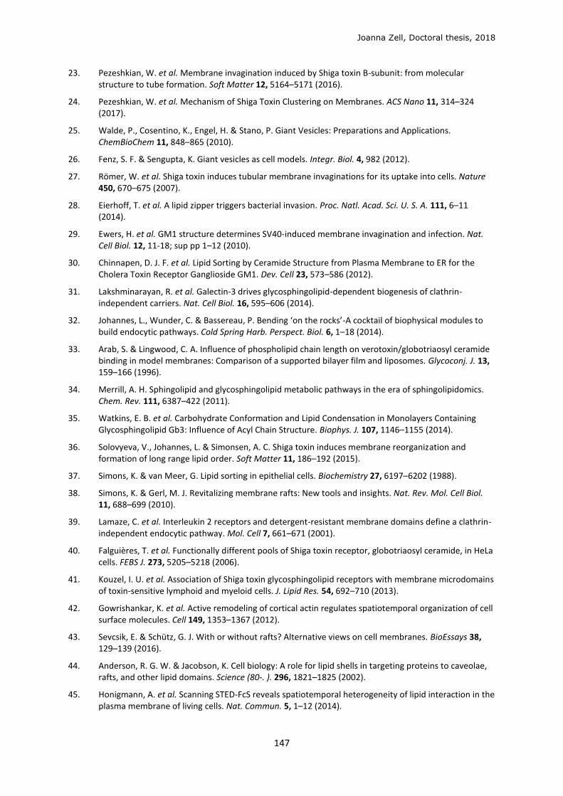
Joanna Zell, Doctoral thesis, 2018
147
23. Pezeshkian, W. et al. Membrane invagination induced by Shiga toxin B-subunit: from molecular structure to tube formation. Soft Matter 12, 5164–5171 (2016).
24. Pezeshkian, W. et al. Mechanism of Shiga Toxin Clustering on Membranes. ACS Nano 11, 314–324 (2017).
25. Walde, P., Cosentino, K., Engel, H. & Stano, P. Giant Vesicles: Preparations and Applications. ChemBioChem 11, 848–865 (2010).
26. Fenz, S. F. & Sengupta, K. Giant vesicles as cell models. Integr. Biol. 4, 982 (2012).
27. Römer, W. et al. Shiga toxin induces tubular membrane invaginations for its uptake into cells. Nature 450, 670–675 (2007).
28. Eierhoff, T. et al. A lipid zipper triggers bacterial invasion. Proc. Natl. Acad. Sci. U. S. A. 111, 6–11 (2014).
29. Ewers, H. et al. GM1 structure determines SV40-induced membrane invagination and infection. Nat. Cell Biol. 12, 11-18; sup pp 1–12 (2010).
30. Chinnapen, D. J. F. et al. Lipid Sorting by Ceramide Structure from Plasma Membrane to ER for the Cholera Toxin Receptor Ganglioside GM1. Dev. Cell 23, 573–586 (2012).
31. Lakshminarayan, R. et al. Galectin-3 drives glycosphingolipid-dependent biogenesis of clathrin-independent carriers. Nat. Cell Biol. 16, 595–606 (2014).
32. Johannes, L., Wunder, C. & Bassereau, P. Bending ‘on the rocks’-A cocktail of biophysical modules to build endocytic pathways. Cold Spring Harb. Perspect. Biol. 6, 1–18 (2014).
33. Arab, S. & Lingwood, C. A. Influence of phospholipid chain length on verotoxin/globotriaosyl ceramide binding in model membranes: Comparison of a supported bilayer film and liposomes. Glycoconj. J. 13, 159–166 (1996).
34. Merrill, A. H. Sphingolipid and glycosphingolipid metabolic pathways in the era of sphingolipidomics. Chem. Rev. 111, 6387–422 (2011).
35. Watkins, E. B. et al. Carbohydrate Conformation and Lipid Condensation in Monolayers Containing Glycosphingolipid Gb3: Influence of Acyl Chain Structure. Biophys. J. 107, 1146–1155 (2014).
36. Solovyeva, V., Johannes, L. & Simonsen, A. C. Shiga toxin induces membrane reorganization and formation of long range lipid order. Soft Matter 11, 186–192 (2015).
37. Simons, K. & van Meer, G. Lipid sorting in epithelial cells. Biochemistry 27, 6197–6202 (1988).
38. Simons, K. & Gerl, M. J. Revitalizing membrane rafts: New tools and insights. Nat. Rev. Mol. Cell Biol. 11, 688–699 (2010).
39. Lamaze, C. et al. Interleukin 2 receptors and detergent-resistant membrane domains define a clathrin-independent endocytic pathway. Mol. Cell 7, 661–671 (2001).
40. Falguières, T. et al. Functionally different pools of Shiga toxin receptor, globotriaosyl ceramide, in HeLa cells. FEBS J. 273, 5205–5218 (2006).
41. Kouzel, I. U. et al. Association of Shiga toxin glycosphingolipid receptors with membrane microdomains of toxin-sensitive lymphoid and myeloid cells. J. Lipid Res. 54, 692–710 (2013).
42. Gowrishankar, K. et al. Active remodeling of cortical actin regulates spatiotemporal organization of cell surface molecules. Cell 149, 1353–1367 (2012).
43. Sevcsik, E. & Schütz, G. J. With or without rafts? Alternative views on cell membranes. BioEssays 38, 129–139 (2016).
44. Anderson, R. G. W. & Jacobson, K. Cell biology: A role for lipid shells in targeting proteins to caveolae, rafts, and other lipid domains. Science (80-. ). 296, 1821–1825 (2002).
45. Honigmann, A. et al. Scanning STED-FcS reveals spatiotemporal heterogeneity of lipid interaction in the plasma membrane of living cells. Nat. Commun. 5, 1–12 (2014).
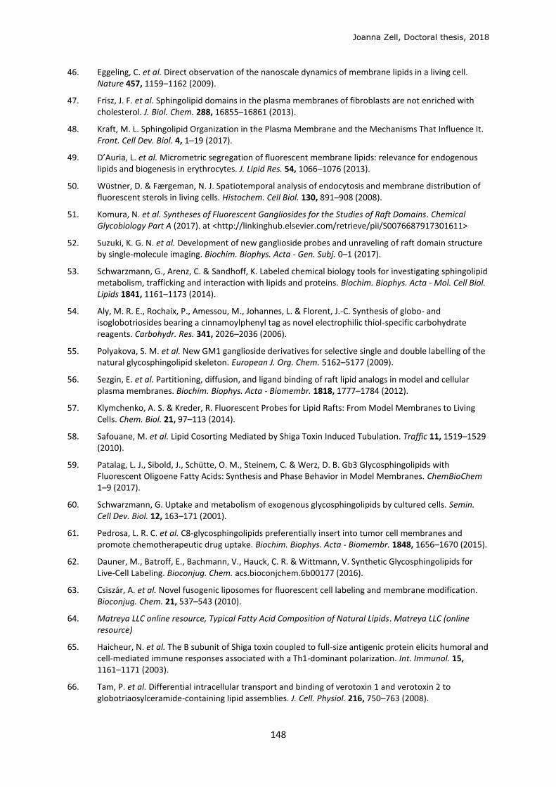
Joanna Zell, Doctoral thesis, 2018
148
46. Eggeling, C. et al. Direct observation of the nanoscale dynamics of membrane lipids in a living cell. Nature 457, 1159–1162 (2009).
47. Frisz, J. F. et al. Sphingolipid domains in the plasma membranes of fibroblasts are not enriched with cholesterol. J. Biol. Chem. 288, 16855–16861 (2013).
48. Kraft, M. L. Sphingolipid Organization in the Plasma Membrane and the Mechanisms That Influence It. Front. Cell Dev. Biol. 4, 1–19 (2017).
49. D’Auria, L. et al. Micrometric segregation of fluorescent membrane lipids: relevance for endogenous lipids and biogenesis in erythrocytes. J. Lipid Res. 54, 1066–1076 (2013).
50. Wüstner, D. & Færgeman, N. J. Spatiotemporal analysis of endocytosis and membrane distribution of fluorescent sterols in living cells. Histochem. Cell Biol. 130, 891–908 (2008).
51. Komura, N. et al. Syntheses of Fluorescent Gangliosides for the Studies of Raft Domains. Chemical Glycobiology Part A (2017). at <http://linkinghub.elsevier.com/retrieve/pii/S0076687917301611>
52. Suzuki, K. G. N. et al. Development of new ganglioside probes and unraveling of raft domain structure by single-molecule imaging. Biochim. Biophys. Acta - Gen. Subj. 0–1 (2017).
53. Schwarzmann, G., Arenz, C. & Sandhoff, K. Labeled chemical biology tools for investigating sphingolipid metabolism, trafficking and interaction with lipids and proteins. Biochim. Biophys. Acta - Mol. Cell Biol. Lipids 1841, 1161–1173 (2014).
54. Aly, M. R. E., Rochaix, P., Amessou, M., Johannes, L. & Florent, J.-C. Synthesis of globo- and isoglobotriosides bearing a cinnamoylphenyl tag as novel electrophilic thiol-specific carbohydrate reagents. Carbohydr. Res. 341, 2026–2036 (2006).
55. Polyakova, S. M. et al. New GM1 ganglioside derivatives for selective single and double labelling of the natural glycosphingolipid skeleton. European J. Org. Chem. 5162–5177 (2009).
56. Sezgin, E. et al. Partitioning, diffusion, and ligand binding of raft lipid analogs in model and cellular plasma membranes. Biochim. Biophys. Acta - Biomembr. 1818, 1777–1784 (2012).
57. Klymchenko, A. S. & Kreder, R. Fluorescent Probes for Lipid Rafts: From Model Membranes to Living Cells. Chem. Biol. 21, 97–113 (2014).
58. Safouane, M. et al. Lipid Cosorting Mediated by Shiga Toxin Induced Tubulation. Traffic 11, 1519–1529 (2010).
59. Patalag, L. J., Sibold, J., Schütte, O. M., Steinem, C. & Werz, D. B. Gb3 Glycosphingolipids with Fluorescent Oligoene Fatty Acids: Synthesis and Phase Behavior in Model Membranes. ChemBioChem 1–9 (2017).
60. Schwarzmann, G. Uptake and metabolism of exogenous glycosphingolipids by cultured cells. Semin. Cell Dev. Biol. 12, 163–171 (2001).
61. Pedrosa, L. R. C. et al. C8-glycosphingolipids preferentially insert into tumor cell membranes and promote chemotherapeutic drug uptake. Biochim. Biophys. Acta - Biomembr. 1848, 1656–1670 (2015).
62. Dauner, M., Batroff, E., Bachmann, V., Hauck, C. R. & Wittmann, V. Synthetic Glycosphingolipids for Live-Cell Labeling. Bioconjug. Chem. acs.bioconjchem.6b00177 (2016).
63. Csiszár, A. et al. Novel fusogenic liposomes for fluorescent cell labeling and membrane modification. Bioconjug. Chem. 21, 537–543 (2010).
64. Matreya LLC online resource, Typical Fatty Acid Composition of Natural Lipids. Matreya LLC (online resource)
65. Haicheur, N. et al. The B subunit of Shiga toxin coupled to full-size antigenic protein elicits humoral and cell-mediated immune responses associated with a Th1-dominant polarization. Int. Immunol. 15, 1161–1171 (2003).
66. Tam, P. et al. Differential intracellular transport and binding of verotoxin 1 and verotoxin 2 to globotriaosylceramide-containing lipid assemblies. J. Cell. Physiol. 216, 750–763 (2008).
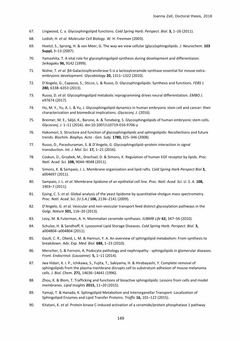
Joanna Zell, Doctoral thesis, 2018
149
67. Lingwood, C. a. Glycosphingolipid functions. Cold Spring Harb. Perspect. Biol. 3, 1–26 (2011).
68. Lodish, H. et al. Molecular Cell Biology. W. H. Freeman (2003).
69. Hoetzl, S., Sprong, H. & van Meer, G. The way we view cellular (glyco)sphingolipids. J. Neurochem. 103 Suppl, 3–13 (2007).
70. Yamashita, T. A vital role for glycosphingolipid synthesis during development and differentiaion. Seikagaku 96, 9142 (1999).
71. Nishie, T. et al. β4-Galactosyltransferase-5 is a lactosylceramide synthase essential for mouse extra-embryonic development. Glycobiology 20, 1311–1322 (2010).
72. D’Angelo, G., Capasso, S., Sticco, L. & Russo, D. Glycosphingolipids: Synthesis and functions. FEBS J. 280, 6338–6353 (2013).
73. Russo, D. et al. Glycosphingolipid metabolic reprogramming drives neural differentiation. EMBO J. e97674 (2017).
74. Ho, M. Y., Yu, A. L. & Yu, J. Glycosphingolipid dynamics in human embryonic stem cell and cancer: their characterization and biomedical implications. Glycoconj. J. (2016).
75. Breimer, M. E., Säljö, K., Barone, A. & Teneberg, S. Glycosphingolipids of human embryonic stem cells. Glycoconj. J. 1–11 (2016). doi:10.1007/s10719-016-9706-y
76. Hakomori, S. Structure and function of glycosphingolipids and sphingolipids: Recollections and future trends. Biochim. Biophys. Acta - Gen. Subj. 1780, 325–346 (2008).
77. Russo, D., Parashuraman, S. & D’Angelo, G. Glycosphingolipid–protein interaction in signal transduction. Int. J. Mol. Sci. 17, 1–21 (2016).
78. Coskun, Ü., Grzybek, M., Drechsel, D. & Simons, K. Regulation of human EGF receptor by lipids. Proc. Natl. Acad. Sci. 108, 9044–9048 (2011).
79. Simons, K. & Sampaio, J. L. Membrane organization and lipid rafts. Cold Spring Harb Perspect Biol 3, a004697 (2011).
80. Sampaio, J. L. et al. Membrane lipidome of an epithelial cell line. Proc. Natl. Acad. Sci. U. S. A. 108, 1903–7 (2011).
81. Ejsing, C. S. et al. Global analysis of the yeast lipidome by quantitative shotgun mass spectrometry. Proc. Natl. Acad. Sci. {U.S.A.} 106, 2136–2141 (2009).
82. D’Angelo, G. et al. Vesicular and non-vesicular transport feed distinct glycosylation pathways in the Golgi. Nature 501, 116–20 (2013).
83. Levy, M. & Futerman, A. H. Mammalian ceramide synthases. IUBMB Life 62, 347–56 (2010).
84. Schulze, H. & Sandhoff, K. Lysosomal Lipid Storage Diseases. Cold Spring Harb. Perspect. Biol. 3, a004804–a004804 (2011).
85. Gault, C. R., Obeid, L. M. & Hannun, Y. A. An overview of sphingolipid metabolism: From synthesis to breakdown. Adv. Exp. Med. Biol. 688, 1–23 (2010).
86. Merscher, S. & Fornoni, A. Podocyte pathology and nephropathy - sphingolipids in glomerular diseases. Front. Endocrinol. (Lausanne). 5, 1–11 (2014).
87. Jwa Hidari, K. I. P., Ichikawa, S., Fujita, T., Sakiyama, H. & Hirabayashi, Y. Complete removal of sphingolipids from the plasma membrane disrupts cell to substratum adhesion of mouse melanoma cells. J. Biol. Chem. 271, 14636–14641 (1996).
88. Zhou, K. & Blom, T. Trafficking and functions of bioactive sphingolipids: Lessons from cells and model membranes. Lipid Insights 2015, 11–20 (2015).
89. Yamaji, T. & Hanada, K. Sphingolipid Metabolism and Interorganellar Transport: Localization of Sphingolipid Enzymes and Lipid Transfer Proteins. Traffic 16, 101–122 (2015).
90. Kitatani, K. et al. Protein kinase C-induced activation of a ceramide/protein phosphatase 1 pathway
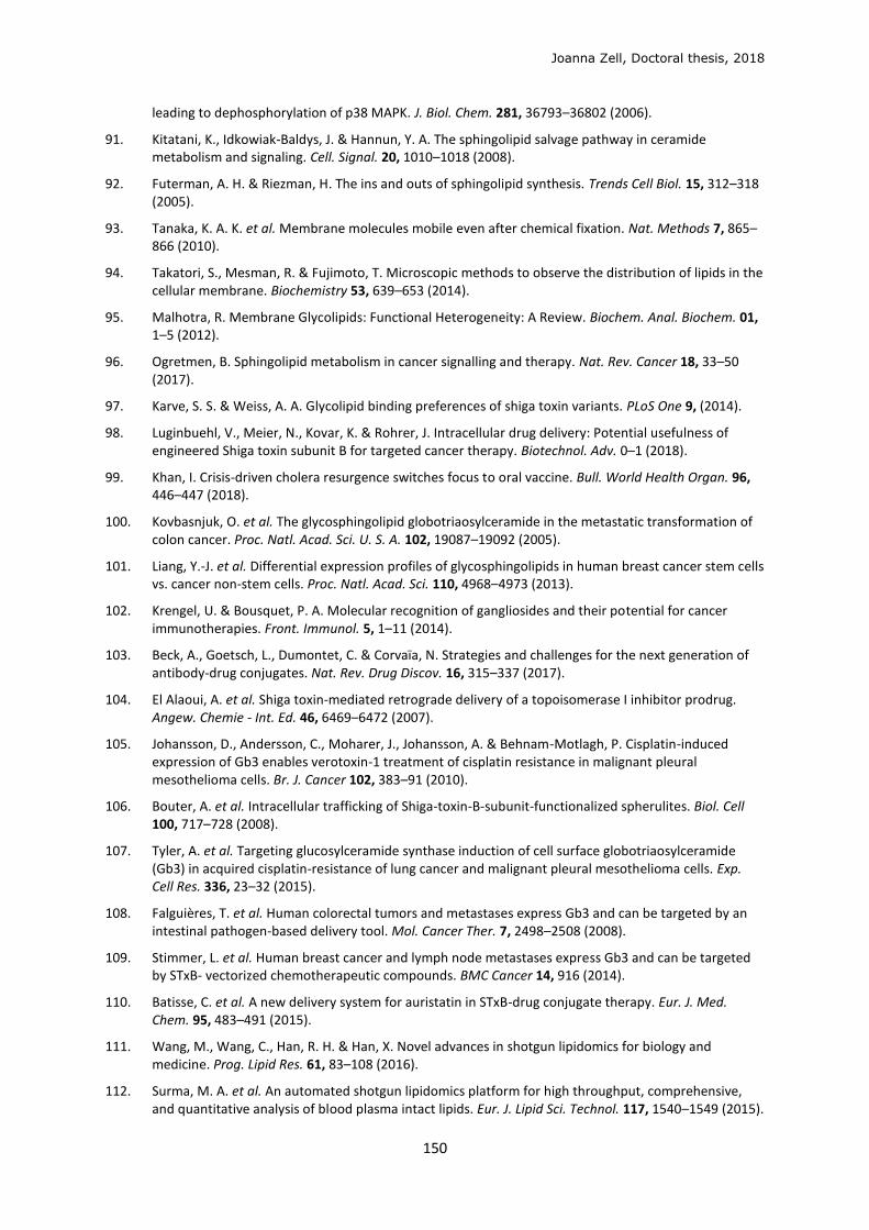
Joanna Zell, Doctoral thesis, 2018
150
leading to dephosphorylation of p38 MAPK. J. Biol. Chem. 281, 36793–36802 (2006).
91. Kitatani, K., Idkowiak-Baldys, J. & Hannun, Y. A. The sphingolipid salvage pathway in ceramide metabolism and signaling. Cell. Signal. 20, 1010–1018 (2008).
92. Futerman, A. H. & Riezman, H. The ins and outs of sphingolipid synthesis. Trends Cell Biol. 15, 312–318 (2005).
93. Tanaka, K. A. K. et al. Membrane molecules mobile even after chemical fixation. Nat. Methods 7, 865–866 (2010).
94. Takatori, S., Mesman, R. & Fujimoto, T. Microscopic methods to observe the distribution of lipids in the cellular membrane. Biochemistry 53, 639–653 (2014).
95. Malhotra, R. Membrane Glycolipids: Functional Heterogeneity: A Review. Biochem. Anal. Biochem. 01, 1–5 (2012).
96. Ogretmen, B. Sphingolipid metabolism in cancer signalling and therapy. Nat. Rev. Cancer 18, 33–50 (2017).
97. Karve, S. S. & Weiss, A. A. Glycolipid binding preferences of shiga toxin variants. PLoS One 9, (2014).
98. Luginbuehl, V., Meier, N., Kovar, K. & Rohrer, J. Intracellular drug delivery: Potential usefulness of engineered Shiga toxin subunit B for targeted cancer therapy. Biotechnol. Adv. 0–1 (2018).
99. Khan, I. Crisis-driven cholera resurgence switches focus to oral vaccine. Bull. World Health Organ. 96, 446–447 (2018).
100. Kovbasnjuk, O. et al. The glycosphingolipid globotriaosylceramide in the metastatic transformation of colon cancer. Proc. Natl. Acad. Sci. U. S. A. 102, 19087–19092 (2005).
101. Liang, Y.-J. et al. Differential expression profiles of glycosphingolipids in human breast cancer stem cells vs. cancer non-stem cells. Proc. Natl. Acad. Sci. 110, 4968–4973 (2013).
102. Krengel, U. & Bousquet, P. A. Molecular recognition of gangliosides and their potential for cancer immunotherapies. Front. Immunol. 5, 1–11 (2014).
103. Beck, A., Goetsch, L., Dumontet, C. & Corvaïa, N. Strategies and challenges for the next generation of antibody-drug conjugates. Nat. Rev. Drug Discov. 16, 315–337 (2017).
104. El Alaoui, A. et al. Shiga toxin-mediated retrograde delivery of a topoisomerase I inhibitor prodrug. Angew. Chemie - Int. Ed. 46, 6469–6472 (2007).
105. Johansson, D., Andersson, C., Moharer, J., Johansson, A. & Behnam-Motlagh, P. Cisplatin-induced expression of Gb3 enables verotoxin-1 treatment of cisplatin resistance in malignant pleural mesothelioma cells. Br. J. Cancer 102, 383–91 (2010).
106. Bouter, A. et al. Intracellular trafficking of Shiga-toxin-B-subunit-functionalized spherulites. Biol. Cell 100, 717–728 (2008).
107. Tyler, A. et al. Targeting glucosylceramide synthase induction of cell surface globotriaosylceramide (Gb3) in acquired cisplatin-resistance of lung cancer and malignant pleural mesothelioma cells. Exp. Cell Res. 336, 23–32 (2015).
108. Falguières, T. et al. Human colorectal tumors and metastases express Gb3 and can be targeted by an intestinal pathogen-based delivery tool. Mol. Cancer Ther. 7, 2498–2508 (2008).
109. Stimmer, L. et al. Human breast cancer and lymph node metastases express Gb3 and can be targeted by STxB- vectorized chemotherapeutic compounds. BMC Cancer 14, 916 (2014).
110. Batisse, C. et al. A new delivery system for auristatin in STxB-drug conjugate therapy. Eur. J. Med. Chem. 95, 483–491 (2015).
111. Wang, M., Wang, C., Han, R. H. & Han, X. Novel advances in shotgun lipidomics for biology and medicine. Prog. Lipid Res. 61, 83–108 (2016).
112. Surma, M. A. et al. An automated shotgun lipidomics platform for high throughput, comprehensive, and quantitative analysis of blood plasma intact lipids. Eur. J. Lipid Sci. Technol. 117, 1540–1549 (2015).
Joanna Zell, Doctoral thesis, 2018
151
113. Patterson, D. M., Nazarova, L. A. & Prescher, J. A. Finding the Right (Bioorthogonal) Chemistry. ACS Chem. Biol. 9, 592–605 (2014).
114. Baskin, J. M. et al. Copper-free click chemistry for dynamic in vivo imaging. Proc. Natl. Acad. Sci. U. S. A. 104, 16793–16797 (2007).
115. Sletten, E. M. & Bertozzi, C. R. Bioorthogonal chemistry: Fishing for selectivity in a sea of functionality. Angew. Chemie - Int. Ed. 48, 6974–6998 (2009).
116. Hang, H. C. & Bertozzi, C. R. Ketone isosteres of 2-N-acetamidosugars as substrates for metabolic cell surface engineering [7]. J. Am. Chem. Soc. 123, 1242–1243 (2001).
117. Yarema, K. J., Mahal, L. K., Bruehl, R. E., Rodriguez, E. C. & Bertozzi, C. R. Metabolic delivery of ketone groups to sialic acid residues. Application to cell surface glycoform engineering. J. Biol. Chem. 273, 31168–31179 (1998).
118. Jacobs, C. L. et al. Metabolic labeling of glycoproteins with chemical tags through unnatural sialic acid biosynthesis. Methods Enzymol. 327, 260–275 (2000).
119. Saxon, E. & Bertozzi, C. R. Cell Surface Engineering by a Modified Staudinger Reaction. Science (80-. ). 287, 2007–2010 (2000).
120. Saxon, E. et al. Investigating Cellular Metabolism of Synthetic Azidosugars with the Staudinger Ligation. J. Am. Chem. Soc. 124, 14893–14902 (2002).
121. Rampoldi, F., Sandhoff, R., Owen, R. W., Gröne, H.-J. & Porubsky, S. A new, robust, and nonradioactive approach for exploring N-myristoylation. J. Lipid Res. 53, 2459–68 (2012).
122. Azoulay, M., Tuffin, G., Sallem, W. & Florent, J.-C. A new drug-release method using the Staudinger ligation. Bioorganic Med. Chem. Lett. 16, 3147–3149 (2006).
123. Prescher, J. a & Bertozzi, C. R. Chemistry in living systems. Nat. Chem. Biol. 1, 13–21 (2005).
124. Clayden, N. G., Warren, S. & Wothers, P. D. Organic Chemistry. (Oxford University Press, 2001).
125. Nilsson, B. L., Kiessling, L. L. & Raines, R. T. Staudinger ligation: a peptide from a thioester and azide. Org. Lett. 2, 1939–1941 (2000).
126. Wang, Z.-P. A., Tian, C.-L. & Zheng, J.-S. The recent developments and applications of the traceless-Staudinger reaction in chemical biology study. RSC Adv. 5, 107192–107199 (2015).
127. Prescher, J. A., Dube, D. H. & Bertozzi, C. R. Chemical remodelling of cell surfaces in living animals. Nature 430, 873–878 (2004).
128. Sarkar, A. K., Fritz, T. A., Taylor, W. H. & Esko, J. D. Disaccharide uptake and priming in animal cells: inhibition of sialyl Lewis X by acetylated Gal beta 1-->4GlcNAc beta-O-naphthalenemethanol. Proc. Natl. Acad. Sci. U. S. A. 92, 3323–7 (1995).
129. Whitman, C. M., Yang, F. & Kohler, J. J. Modified GM3 gangliosides produced by metabolic oligosaccharide engineering. Bioorganic Med. Chem. Lett. 21, 5006–5010 (2011).
130. Luchansky, S. J. et al. Constructing azide-labeled cell surfaces using polysaccharide biosynthetic pathways. Methods Enzymol. 362, 249–272 (2003).
131. Kolb, H. C., Finn, M. G. & Sharpless, K. B. Click Chemistry: Diverse Chemical Function from a Few Good Reactions. Angew. Chemie - Int. Ed. 40, 2004–2021 (2001).
132. Huisgen, R. 1,3-Dipolar Cycloadditions. Proc. Chem. Soc 357–396 (1961).
133. Rostovtsev, V., Green, L., Fokin, V. V. & Sharpless, K. B. A Stepwise Huisgen Cycloaddition Process Catalyzed by Copper (I): Regioselective Ligation of Azides and Terminal Alkynes. Angew. Chem. Int. Ed. 41, 2596–2599 (2002).
134. Tornøe, C. W., Christensen, C. & Meldal, M. Peptidotriazoles on solid phase: [1,2,3]-Triazoles by regiospecific copper(I)-catalyzed 1,3-dipolar cycloadditions of terminal alkynes to azides. J. Org. Chem. 67, 3057–3064 (2002).
135. Huisgen, R. 1,3-Dipolar Cycloadditions; Past and Future. Angew. Chem. Int. Ed. 2, 565–598 (1963).
Joanna Zell, Doctoral thesis, 2018
152
136. Kennedy, D. C. et al. Cellular consequences of copper complexes used to catalyze bioorthogonal click reactions. J. Am. Chem. Soc. 133, 17993–18001 (2011).
137. Hong, V., Presolski, S. I., Ma, C. & Finn, M. G. Analysis and Optimization of Copper-Catalysed Azide-Alkyne Cycloaddition for Bioconjugation. Angew. Chem., Int. Ed. Engl. 48, 9879–9883 (2009).
138. Li, S. et al. Copper-catalyzed click reaction on/in live cells. Chem. Sci. 21, 1075–1101 (2016).
139. Hong, V., Steinmetz, N. F., Manchester, M. & Finn, M. G. Labeling live cells by copper-catalyzed alkyne-azide click chemistry. Bioconjug. Chem. 21, 1912–1916 (2010).
140. Li, C. et al. Practical labeling methodology for choline-derived lipids and applications in live cell fluorescence imaging. Photochem. Photobiol. 90, 686–695 (2014).
141. Besanceney-Webler, C. et al. Increasing the efficacy of bioorthogonal click reactions for bioconjugation: A comparative study. Angew. Chemie - Int. Ed. 50, 8051–8056 (2011).
142. Agard, N. J., Prescher, J. a & Bertozzi, C. R. A Strain-Promoted [3 + 2] Azide-Alkyne Cycloaddition for Covalent Modification of Biomolecules in Living Systems. J. Am. Chem. Soc. 126, 15046–15047 (2004).
143. Dommerholt, J., Rutjes, F. P. J. T. & van Delft, F. L. Strain-Promoted 1,3-Dipolar Cycloaddition of Cycloalkynes and Organic Azides. Top. Curr. Chem. 374, 1–20 (2016).
144. Dommerholt, J. et al. Highly accelerated inverse electron-demand cycloaddition of electron-deficient azides with aliphatic cyclooctynes. Nat. Commun. 5, 1–7 (2014).
145. Codelli, J. a, Baskin, J. M., Agard, N. J. & Bertozzi, C. R. Second-Generation Difluorinated Cyclooctynes for Copper-Free Click Chemistry. J. Am. Chem. Soc. 130, 11486–11493 (2008).
146. Borrmann, A. & Van Hest, J. C. M. Bioorthogonal chemistry in living organisms. Chem. Sci. 5, 2123–2134 (2014).
147. McKay, C. S. & Finn, M. G. Click chemistry in complex mixtures: Bioorthogonal bioconjugation. Chem. Biol. 21, 1075–1101 (2014).
148. Schultz, M. K., Parameswarappa, S. G. & Pigge, F. C. Synthesis of a DOTA-biotin conjugate for radionuclide chelation via Cu-free click chemistry. Org. Lett. 12, 2398–2401 (2010).
149. McKay, C. S., Blake, J. A., Cheng, J., Danielson, D. C. & Pezacki, J. P. Strain-promoted cycloadditions of cyclic nitrones with cyclooctynes for labeling human cancer cells. Chem. Commun. 47, 10040–10042 (2011).
150. Ramil, C. P. & Lin, Q. Bioorthogonal chemistry: Strategies and recent developments. Chem. Commun. 49, 11007–11022 (2013).
151. Boutureira, O. & Bernardes, G. J. L. Advances in Chemical Protein Modification. Chem. Rev. 115, 2174–2195 (2015).
152. Lang, K. et al. Genetically encoded norbornene directs site-specific cellular protein labelling via a rapid bioorthogonal reaction. Nat. Chem. 4, 298–304 (2012).
153. Meyer, J. P., Adumeau, P., Lewis, J. S. & Zeglis, B. M. Click Chemistry and Radiochemistry: The First 10 Years. Bioconjug. Chem. 27, 2791–2807 (2016).
154. Wang, H. et al. Selective in vivo metabolic cell-labeling-mediated cancer targeting. Nat. Chem. Biol. 13, 415–424 (2017).
155. Jao, C. Y., Roth, M., Welti, R. & Salic, A. Metabolic labeling and direct imaging of choline phospholipids in vivo. Proc. Natl. Acad. Sci. U. S. A. 106, 15332–15337 (2009).
156. Jao, C. Y., Roth, M., Welti, R. & Salic, A. Biosynthetic labeling and two-color imaging of phospholipids in cells. ChemBioChem 16, 472–476 (2015).
157. Hannoush, R. N. & Sun, J. The chemical toolbox for monitoring protein fatty acylation and prenylation. Nat. Chem. Biol. 6, 498–506 (2010).
158. Hang, H. C. et al. Chemical probes for the rapid detection of fatty-acylated proteins in mammalian cells. J. Am. Chem. Soc. 129, 2744–2745 (2007).
Joanna Zell, Doctoral thesis, 2018
153
159. Thiele, C. et al. Tracing fatty acid metabolism by click chemistry. ACS Chem. Biol. 7, 2004–2011 (2012).
160. Kuerschner, L. & Thiele, C. Multiple bonds for the lipid interest. Biochim. Biophys. Acta - Mol. Cell Biol. Lipids 1841, 1031–1037 (2014).
161. Gaebler, A. et al. Alkyne lipids as substrates for click chemistry-based in vitro enzymatic assays. J. Lipid Res. 54, 2282–90 (2013).
162. Collenburg, L. et al. A Functionalized Sphingolipid Analogue for Studying Redistribution during Activation in Living T Cells. J. Immunol. 196, 3951–3962 (2016).
163. Walter, T. et al. Incorporation and visualization of azido-functionalized N-oleoyl serinol in Jurkat cells, mouse brain astrocytes, 3T3 fibroblasts and human brain microvascular endothelial cells. Chem. Commun. 52, 8612–8614 (2016).
164. Fink, J. & Seibel, J. Click reactions with functional sphingolipids. Biol. Chem. 0, (2018).
165. Erdmann, R. S. et al. Super-Resolution Imaging of the Golgi in Live Cells with a Bioorthogonal Ceramide Probe. Angew. Chemie - Int. Ed. 53, 10242–10246 (2014).
166. MacKinnon, A. L. & Taunton, J. Target Identification by Diazirine Photo-Cross-linking and Click Chemistry. Curr. Protoc. Chem. Biol. 1, 55–73 (2009).
167. Yang, P. & Liu, K. Activity-based protein profiling: Recent advances in probe development and applications. ChemBioChem 16, 712–724 (2015).
168. Liu, X., Dong, T., Zhou, Y., Huang, N. & Lei, X. Exploring the Binding Proteins of Glycolipids with Bifunctional Chemical Probes. Angew. Chemie - Int. Ed. 55, 14330–14334 (2016).
169. Gubbens, J. et al. Photocrosslinking and Click Chemistry Enable the Specific Detection of Proteins Interacting with Phospholipids at the Membrane Interface. Chem. Biol. 16, 3–14 (2009).
170. Haberkant, P. & Holthuis, J. C. M. Fat & fabulous: Bifunctional lipids in the spotlight. Biochim. Biophys. Acta - Mol. Cell Biol. Lipids 1841, 1022–1030 (2014).
171. Hulce, J. J., Cognetta, A. B., Niphakis, M. J., Tully, S. E. & Cravatt, B. F. Proteome-wide Mapping of Cholesterol Interacting Proteins in Mammalian Cells. Nat. Methods 10, 259–264 (2013).
172. Bockelmann, S. et al. A search for ceramide binding proteins using bifunctional lipid analogs yields CERT-related protein StarD7. J. Lipid Res. 59, 515–530 (2018).
173. Pezeshkian, W. et al. Fluctuation-driven clustering of nanoparticles on lipid membranes. Submitted 1–13
174. Harrison, A. L. et al. A synthetic globotriaosylceramide analogue inhibits HIV-1 infection in vitro by two mechanisms. Glycoconj. J. 27, 515–524 (2010).
175. Blake, D. A., Bovin, N. V, Bess, D. & Henry, S. M. FSL constructs: a simple method for modifying cell/virion surfaces with a range of biological markers without affecting their viability. J. Vis. Exp. 1–9 (2011).
176. Boons, G.-J. (University of Birmingham, U. Carbohydrate Chemistry. (Blackie A&P, 1998).
177. Kuehne, M., Gyoergydeak, Z. & Lindhorst, T. K. A simple method for the preparation of glycosyl isothiocyanates. Synthesis (Stuttg). 949–951 (2006).
178. Greimel, P. & Ito, Y. First synthesis of natural phosphatidyl-B-d-glucoside. Tetrahedron Lett. 49, 3562–3566 (2008).
179. Sandbhor, M. S., Key, J. a., Strelkov, I. S. & Cairo, C. W. A modular synthesis of alkynyl-phosphocholine headgroups for labeling sphingomyelin and phosphatidylcholine. J. Org. Chem. 74, 8669–8674 (2009).
180. Cavalli, S., Overhand, M. & Kros, A. Assembly into beta-sheet structures upon peptide-liposome conjugation through copper(I)-catalyzed [3+2] azide-alkyne cycloaddition. Chempluschem 79, 564–568 (2014).
181. Kim, S., Lee, S., Lee, T., Ko, H. & Kim, D. Efficient Synthesis of D-erythro-Sphingosine and D-erythro-Azidosphingosine from D-ribo-Phytosphingosine via a Cyclic Sulfate Intermediate. J. Org. Chem. 71,
Joanna Zell, Doctoral thesis, 2018
154
8661–8664 (2006).
182. Di Benedetto, R. et al. Protected sphingosine from phytosphingosine as an efficient acceptor in glycosylation reaction. Org. Lett. 16, 952–955 (2014).
183. Gao, Y. & Sharpless, K. B. Vicinal Diol Cyclic Sulfates: Like Epoxides Only More Reactive. J. Am. Chem. Soc. 110, 7538–7539 (1988).
184. Garrido Martinez, M. New sphingolipid probes for metabolism and trafficking studies. (Thesis from Universitat de Barcelona, IQAC-CSIC, 2012).
185. Gao, H. Chemical biology approach to study toxin clustering and lipids reorganization in Shiga toxin endocytosis. (2015).
186. Kita, K., Kurita, T. & Ito, M. Characterization of the reversible nature of the reaction catalyzed by sphingolipid ceramide N-deacylase: A novel form of reverse hydrolysis reaction. Eur. J. Biochem. 268, 592–602 (2001).
187. Gantner, M., Schwarzmann, G., Sandhoff, K. & Kolter, T. Partial synthesis of ganglioside and lysoganglioside lipoforms as internal standards for MS quantification. J. Lipid Res. 55, 2692–2704 (2014).
188. Gouin, S. G. & Kovensky, J. Direct azidation of unprotected carbohydrates with PPh3/CBr4/NaN3. Modulation of the degree of substitution. Tetrahedron Lett. 48, 2875–2879 (2007).
189. Wakiyama, Y. et al. Synthesis and structure-activity relationships of novel lincomycin derivatives. Part 4: Synthesis of novel lincomycin analogs modified at the 6- and 7-positions and their potent antibacterial activities. J. Antibiot. (Tokyo). 70, 888–906 (2017).
190. Canals, D. et al. Synthesis and biological properties of Pachastrissamine (jaspine B) and diastereoisomeric jaspines. Bioorganic Med. Chem. 17, 235–241 (2009).
191. Kostova, V. et al. Targeted Shiga toxin – drug conjugates prepared via Cu-free click chemistry. Bioorg. Med. Chem. 23, 7150–7157 (2015).
192. Chang, T. C. et al. A photo-cleavable biotin affinity tag for the facile release of a photo-crosslinked carbohydrate-binding protein. Bioorganic Med. Chem. 24, 1216–1224 (2016).
193. Ling, H. et al. Structure of the Shiga-like toxin I B-pentamer complexed with an analogue of its receptor Gb3. Biochemistry 37, 1777–1788 (1998).
194. Carvalho, M. et al. Effects of diet and development on the Drosophila lipidome. Mol. Syst. Biol. 8, 1–17 (2012).
195. Ogasawara, H. et al. Intracellular Carboxyl esterase activity is a determinant of cellular sensitivity to the antineoplastic agent KW-2189 in cell lines resistant to Cisplatin and CPT-11. Jpn. J. Cancer Res. 86, 124–129 (1995).
196. Ogasawara, H. et al. A novel antitumor antibiotic, KW-2189 is activated by carboxyl esterase and induces DNA strand breaks in human small cell lung cancer cells. Jpn. J. Cancer Res. 85, 418 (1994).
197. Sharpe, H. J., Stevens, T. J. & Munro, S. A Comprehensive Comparison of Transmembrane Domains Reveals Organelle-Specific Properties. Cell 142, 158–169 (2010).
198. Li, J. et al. A review on phospholipids and their main applications in drug delivery systems. Asian J. Pharm. Sci. 10, 81–98 (2014).
199. Korchagina, E. Y. & Henry, S. M. Synthetic Glycolipid Like Constructs as Tools for Glycobiology Research, Diagnostics, and as Potential Therapeutics. Biochem. 80, (2015).
200. Korchagina, E. et al. Toward creating cell membrane glyco-landscapes with glycan lipid constructs. Carbohydr. Res. 356, 238–246 (2012).
201. Simons, K. & Ikonen, E. Functional rafts in cell membranes. Nature 387, 569–72 (1997).
202. Windschieg, B. et al. Lipid reorganization induced by Shiga toxin clustering on planar membranes. PLoS One 4, 1–11 (2009).
Joanna Zell, Doctoral thesis, 2018
155
203. Chai, W., Stoll, M. S., Galustian, C., Lawson, A. M. & Feizi, T. Neoglycolipid technology: deciphering information content of glycome. Methods Enzymol. 362, 160–195 (2003).
204. Bock, V. D., Speijer, D., Hiemstra, H. & van Maarseveen, J. H. 1,2,3-Triazoles as peptide bond isosteres: synthesis and biological evaluation of cyclotetrapeptide mimics. Org. Biomol. Chem. 5, 971–975 (2007).
205. Young, I. S. & Baran, P. S. Protecting-group-free synthesis as an opportunity for invention. Nat. Chem. 1, 193–205 (2009).
206. Hörner, A., Hagendorn, T., Schepers, U. & Bräse, S. Photophysical properties and synthesis of new dye-cyclooctyne conjugates for multicolor and advanced microscopy. Bioconjug. Chem. 26, 718–724 (2015).
207. Rong, L. et al. Long-term thiol monitoring in living cells using bioorthogonal chemistry. Chem. Commun. (Camb). 51, 388–90 (2015).
208. Garrido, M., Abad, J. L., Fabriàs, G., Casas, J. & Delgado, A. Azide-tagged sphingolipids: New tools for metabolic flux analysis. ChemBioChem 16, 641–650 (2015).
209. Garrido, M. et al. In situ synthesis of fluorescent membrane lipids (ceramides) using click chemistry. J. Chem. Biol. 5, 119–123 (2012).
210. Song, Y., Yuan, W., Luo, Y. & Lu, W. A new synthetic approach to phosphatidylethanolamine. Chinese Chem. Lett. 23, 154–156 (2012).
211. Lee, Y. M. et al. A practical and cost-effective synthesis of d-erythro-sphingosine from d-ribo-phytosphingosine via a cyclic sulfate intermediate. Synthesis (Stuttg). 867–872 (2011).
212. Van Den Berg, R. J. B. H. N. et al. A rapid and efficient synthesis of D-erythro-sphingosine from D-ribo-phytosphingosine. European J. Org. Chem. 6685–6689 (2011).
213. Huang, F.-T., Han, Y.-B., Feng, Y. & Yang, G.-Y. A facile method for controlling the reaction equilibrium of sphingolipid ceramide N-deacylase for lyso-glycosphingolipid production. J. Lipid Res. 56, 1836–1842 (2015).
214. Ito, M., Kurita, T. & Kita, K. A novel enzyme that cleaves the N-acyl linkage of ceramides in various glycosphingolipids as well as sphingomyelin to produce their lyso forms. J. Biol. Chem. 270, 24370–24374 (1995).
215. Chwalek, M., Auzély, R. & Fort, S. Synthesis and biological evaluation of multivalent carbohydrate ligands obtained by click assembly of pseudo-rotaxanes. Org. Biomol. Chem. 7, 1680–1688 (2009).
216. Feldborg, L. N., Jølck, R. I. & Andresen, T. L. Quantitative evaluation of bioorthogonal chemistries for surface functionalization of nanoparticles. Bioconjug. Chem. 23, 2444–2450 (2012).
217. Oude Blenke, E. et al. Liposome functionalization with copper-free ‘click chemistry’. J. Control. Release 202, 14–20 (2015).
218. Salomé, C. et al. Smart tools and orthogonal click-like reactions onto small unilamellar vesicles: Additional molecular data. Chem. Phys. Lipids J. 188, 27–36 (2015).
219. Haberkant, P. et al. Bifunctional Sphingosine for Cell-Based Analysis of Protein-Sphingolipid Interactions. ACS Chem. Biol. 11, doi: 10.1021/acschembio.5b00810 (2015).
220. Haberkant, P. et al. Protein-sphingolipid interactions within cellular membranes. J. Lipid Res 49, 251–262 (2008).
221. Hofmann, K. et al. A novel alkyne cholesterol to trace cellular cholesterol metabolism and localization. J. Lipid Res. 55, 583–91 (2014).
222. Boppana, N. B. et al. Ceramide synthase inhibitor fumonisin B1 inhibits apoptotic cell death in SCC17B human head and neck squamous carcinoma cells after Pc4 photosensitization. Photochem. Photobiol. Sci. 13, 1621–1627 (2014).
223. Young, M. M., Kester, M. & Wang, H.-G. Sphingolipids: regulators of crosstalk between apoptosis and autophagy. J. Lipid Res. 54, 5–19 (2013).
224. Woodcock, J. Sphingosine and ceramide signalling in apoptosis. IUBMB Life (International Union
Joanna Zell, Doctoral thesis, 2018
156
Biochem. Mol. Biol. Life) 58, 462–466 (2006).
225. Kan, C. C. & Kolesnick, R. N. A synthetic ceramide analog, D-threo-1-phenyl-2-decanoylamino-3- morpholino-1-propanol, selectively inhibits adherence during macrophage differentiation of human leukemia cells. J Biol Chem 267, 9663–9667 (1992).
226. Sugiki, H. et al. C2-Ceramide induces apoptosis in a human squamous cell carcinoma cell line. Br. J. Dermatol. 143, 1154–1163 (2000).
227. Desai, K. et al. Fumonisins and fumonisin analogs as inhibitors of ceramide synthase and inducers of apoptosis. Biochim. Biophys. Acta - Mol. Cell Biol. Lipids 1585, 188–192 (2002).
228. Gerl, M. J. et al. Sphingosine-1-phosphate lyase deficient cells as a tool to study protein lipid interactions. PLoS One 11, 1–25 (2016).
229. Hartmann, D. et al. Long chain ceramides and very long chain ceramides have opposite effects on human breast and colon cancer cell growth. Int. J. Biochem. Cell Biol. 44, 620–628 (2012).
230. Osawa, Y. et al. Roles for C16-ceramide and sphingosine 1-phosphate in regulating hepatocyte apoptosis in response to tumor necrosis factor-α. J. Biol. Chem. 280, 27879–27887 (2005).
231. Eto, M., Bennouna, J., Hunter, O. C., Lotze, M. T. & Amoscato, A. A. Importance of C16 ceramide accumulation during apoptosis in prostate cancer cells. Int. J. Urol. 13, 148–156 (2006).
232. Hannun, Y. A. & Obeid, L. M. Sphingolipids and their metabolism in physiology and disease. Nat. Rev. Mol. Cell Biol. 19, 175–191 (2018).
233. Stiban, J. & Perera, M. Very long chain ceramides interfere with C16-ceramide-induced channel formation: A plausible mechanism for regulating the initiation of intrinsic apoptosis. Biochim. Biophys. Acta - Biomembr. 1848, 561–567 (2015).
234. Regina Todeschini, A. & Hakomori, S. Functional role of glycosphingolipids and gangliosides in control of cell adhesion, motility, and growth, through glycosynaptic microdomains. Biochim. Biophys. Acta - Gen. Subj. 1780, 421–433 (2008).
235. Mathow, D. et al. Zeb1 affects epithelial cell adhesion by diverting glycosphingolipid metabolism. EMBO Rep. 16, 321–331 (2015).
236. Laizure, S. C., Herring, V., Hu, Z., Witbrodt, K. & Parker, R. B. The role of human carboxylesterases in drug metabolism: have we overlooked their importance? Pharmacotherapy. 33, 210–222 (2013).
237. Laughlin, S. T. & Bertozzi, C. R. Metabolic labeling of glycans with azido sugars and subsequent glycan-profiling and visualization via staudinger ligation. Nat. Protoc. 2, 2930–2944 (2007).
238. Chang, P. V. et al. Metabolie labeling of sialic acids in living animals with alkynyl sugars. Angew. Chemie - Int. Ed. 48, 4030–4033 (2009).
239. Lian, J., Nelson, R. & Lehner, R. Carboxylesterases in lipid metabolism: from mouse to human. Protein Cell 9, 178–195 (2017).
240. Takumi, S. et al. Overexpression of carboxylesterase contributes to the attenuation of cyanotoxin microcystin-LR toxicity. Comp. Biochem. Physiol. Part - C Toxicol. Pharmacol. 194, 22–27 (2017).
241. Blauvelt, M. L. et al. α-S-GalCer: Synthesis and evaluation for iNKT cell stimulation. Bioorganic Med. Chem. Lett. 18, 6374–6376 (2008).
242. Shaya, J. et al. Turn-on Fluorene Push-Pull Probes with High Brightness and Photostability for Visualizing Lipid Order in Biomembranes. ACS Chem. Biol. 12, 3022–3030 (2017).
243. Adlercreutz, D. et al. Enzymatic synthesis of Gb3 and iGb3 ceramides. Carbohydr. Res. 345, 1384–1388 (2010).
244. Earle, M. a., Manku, S., Hultin, P. G., Li, H. & Palcic, M. M. Chemoenzymatic synthesis of a trimeric ganglioside GM3 analogue. Carbohydr. Res. 301, 1–4 (1997).
245. Roodsari, F. S., Wu, D., Pum, G. S. & Hajdu, J. A new approach to the stereospecific synthesis of phospholipids. The use of L-glyceric acid for the preparation of diacylglycerols, phosphatidylcholines,
Joanna Zell, Doctoral thesis, 2018
157
and related derivatives. J. Org. Chem. 64, 7727–7737 (1999).
246. Goddard-Borger, E. D. & Stick, R. V. An Efficient , Inexpensive , and Shelf-Stable Diazotransfer Reagent: Imidazole-1-sulfonyl Azide Hydrochloride. Org. Lett. 9, 3797–3800 (2007).
247. Kim, S., Lee, S., Lee, T., Ko, H. & Kim, D. Efficient Synthesis of D - erythro -Sphingosine and D - erythro -Azidosphingosine from D - ribo -Phytosphingosine via a Cyclic Sulfate Intermediate The synthesis of naturally occurring D - erythro -sphingosine and synthetically useful D - erythro -2-azido. J. Org. Chem. 71, 8661–8664 (2006).
248. Ito, Y., Kiso, M. & Hasegawa, A. Studies on the Thioglycosides of N- Acetylneuraminic Acid. 6: Synthesis of Ganglioside GM4 Analogs. J. Carbohydr. Chem. 8, 285–294 (1989).
249. Xing, G. W., Chen, L. & Liang, F. F. Facile synthesis of tumor-associated carbohydrate antigen ganglioside GM3 from sialic acid, lactose, and serine. European J. Org. Chem. 5963–5970 (2009).
250. Albrecht, B., Pütz, U. & Schwarzmann, G. Synthesis of fluorescent and radioactive analogues of two lactosylceramides and glucosylceramide containing β-thioglycosidic bonds that are resistant to enzymatic degradation. Carbohydr. Res. 276, 289–308 (1995).


