Embed Size (px)
Citation preview

Intraoperative Flow Measurement in Coronary Artery Surgery
Giuseppe D’Ancona
Intraoperative Flow M
easurement in C
oronary Artery Surgery
Giuseppe D
’Ancona


Intraoperative Flow Measurement in Coronary Artery Surgery
Giuseppe D’Ancona
Giuseppe Text alt.indd 1Giuseppe Text alt.indd 1 08-Sep-09 14:31:34 PM08-Sep-09 14:31:34 PM

isbn: 978-90-8559-531-1
2009 Giuseppe D’Ancona
All rights reserved. No part of this thesis may be reproduced or transmitted in any form or by any means, electronically or mechanical, including photocopying, recording or by any other information storage or retrieval system, without written permission from the author.
Layout and printing: Optima Grafi sche Communicatie, Rotterdam, Th e Nether lands
Th e printing of this thesis has been possible thanks to the fi nancial support of Medi-Stim AS.
Giuseppe Text alt.indd 2Giuseppe Text alt.indd 2 08-Sep-09 14:31:36 PM08-Sep-09 14:31:36 PM
Intraoperative Flow Measurement in Coronary Artery Surgery:
Present Applications and Future Perspectives
Meten van bloedstroom tijdens coronaire bypass chirurgie:Huidige toepassingen en toekomstige ontwikkelingen
Proefschrift
ter verkrijging van de graad van doctor aan deErasmus Universiteit Rotterdam op gezag van de
rector magnifi cus
Prof.dr. H.G. Schmidt
en volgens besluit van het College voor Promoties.
De openbare verdediging zal plaatsvinden op woensdag 28 oktober 2009 om 15.30 uur
door
Giuseppe Francesco Maria D’Ancona, MDgeboren te Milaan, Italië
Giuseppe Text alt.indd 3Giuseppe Text alt.indd 3 08-Sep-09 14:31:36 PM08-Sep-09 14:31:36 PM

PROMOTIECOMMISSIE
Promotor: Prof.dr. A.J.J.C. Bogers
Overige leden: Prof.dr. D.J.G.M. Duncker Prof.dr. P.J. de Feijter Dr. J.J.M. Takkenberg Copromotor: Dr. A.P. Kappetein
Financial support by the Netherlands Heart Foundation for the publication of this thesis is gratefully acknowledged.
Giuseppe Text alt.indd 4Giuseppe Text alt.indd 4 08-Sep-09 14:31:39 PM08-Sep-09 14:31:39 PM

Contents
General Introduction 7
Chapter 1 Graft patency verifi cation in coronary artery bypass grafting: principles and clinical applications of transit time fl ow measurement.Angiology 51 (2000) 725-731
13
Chapter 2 Flow measurement in coronary surgeryHeart Surg Forum 2 (1999) 121-124
25
Chapter 3 Is intraoperative measurement of coronary blood fl ow a good predictor of graft patency? (letter to the editor)Eur J Cardiothor Surg 20 (2001) 1075-1076
37
Chapter 4 Graft revision after transit time fl ow measurement in off -pump coronary artery bypass grafting.Eur J Cardiothor Surg 17 (2000) 287-293
43
Chapter 5 Coronary grafts fl ow and cardiac pacing modalities: how to improve perioperative myocardial perfusion.Eur J Cardiothor Surg 26 (2004) 85-88
57
Chapter 6 Preoperative angiography and intraoperative transit time fl ow measurement to detect coronary graft patency in reoperations: an integrated approach, a case report.Angiology 51 (2000) 777-780
67
Chapter 7 Heparin dose, transfusion rates, and intraoperative graft patency in minimally invasive direct coronary artery bypass.Heart Surgery Forum 6 (2003) 176-180
75
Chapter 8 Epicardial coronary artery Doppler: validation in an animal model.Interact Cardiovasc Th orac Surg 7 (2008) 634-637
87
Giuseppe Text alt.indd 5Giuseppe Text alt.indd 5 08-Sep-09 14:31:39 PM08-Sep-09 14:31:39 PM

6
Cont
ents
Chapter 9 Intraoperative validation of a new system for invasive continuous cardiac output measurementIntensive Care Med. 2009 May;35(5):943-7
99
Chapter 10 Use of Nitinol-U clips and fl ow characteristics of LIMA-LAD anastomosis.Interact CardioVasc Th orac Surg 2 (2003) 237-240
109
Chapter 11 Intraoperative graft patency verifi cation: should you trust your fi ngertips?Heart Surg Forum 3 (2000) 99-102
119
Chapter 12 Intraoperative graft patency verifi cation in coronary surgery: modern diagnostic tools.J Cardiothorac Vasc Anesth. 2009 Apr;23(2):232-8
129
Chapter 13 Is hybrid coronary revascularization favoured by cardiologists or cardiac surgeons?Heart Surgery Forum 5 (2002) 393-395
147
Chapter 14 Ischemic mitral valve regurgitation: Th e new challenge for magnetic resonance imagingEur J Cardiothorac Surg 32 (2007) 475-480
155
Chapter 15 Ischemic mitral valve regurgitation in patients with depressed ventricular function: cardiac geometrical and myocardial perfusion evaluation with magnetic resonance imaging.Eur J Cardiothor Surg 34 (2008) 964-948
171
Chapter 16 Discussion 185
Chapter 17 Summary• English• DutchCurriculum VitaeList of publicationsAcknowledgementsPortfolio
193197201203219221
Giuseppe Text alt.indd 6Giuseppe Text alt.indd 6 08-Sep-09 14:31:39 PM08-Sep-09 14:31:39 PM

G eneral Introduction
“Wir müssen wissen, wir werden wissen.”(“We must know, we shall know.”)
David Hilbert (German mathematician, 1862–1943)
Giuseppe Text alt.indd 7Giuseppe Text alt.indd 7 08-Sep-09 14:31:39 PM08-Sep-09 14:31:39 PM

Giuseppe Text alt.indd 8Giuseppe Text alt.indd 8 08-Sep-09 14:31:39 PM08-Sep-09 14:31:39 PM

General Introduction 9
INTRODUCTION
Although most cardiac surgeons perform coronary surgery on microscopic structures, not often any sort of direct quality control method to test patency of the constructed anastomoses is applied.
In most cases, simple evaluation of the immediate perioperative hemody-namic performance is considered as an acceptable marker of operative success.
In this regard, it is very provocative that in surgical myocardial revasculariza-tion, electrocardiographic and hemodynamic parameters may remain postopera-tively unchanged, even with a malfunctioning coronary graft.
In the light of the new transitions in cardiac surgery, we must concentrate on optimizing the quality of care we are giving to our patients.
Every medical procedure requires a quality control method, and this is particularly true for complex procedures such as heart operations. Th is may be diffi cult to accept as it requires maturity and self-judgment.
Th e present thesis summarizes clinical experience with intraoperative graft patency verifi cation in cardiac surgery.
Intraoperative graft patency verifi cation after coronary surgery has been proposed since the early 60’s, when coronary surgery was at its infancy. Th e fi rst fl ow measurement devices were based on electromagnetic technology and were soon abandoned for their many limitations.
As experience with coronary surgery grew, surgeons felt less and less obliged to document the quality of their coronary anastomoses and, until recently, coronary graft testing was seldom adopted.
Th anks to the technological improvements, ultrasound based fl ow meters (mainly Doppler) were available on the market in the early 80’s.
Although these devices off ered many advantages when compared to the electromagnetic technology, they still had many limitations.
In the late 90’s, the introduction and popularization of beating heart “off -pump” coronary surgery stimulated interest upon intraoperatively documenting patency of coronary grafts.
At the same time, a further technological achievement was available with the introduction of transit time fl ow meters (TTFM) that off ered a reproducible and simple method for testing coronary graft fl ow.
Construction of the anastomoses on the beating heart was supposedly techni-cally more challenging than on the arrested bloodless heart and, for this reason, surgeons, at least some of them, felt the urgency to guarantee and proof the
Giuseppe Text alt.indd 9Giuseppe Text alt.indd 9 08-Sep-09 14:31:39 PM08-Sep-09 14:31:39 PM

10
Gen
eral
Intr
oduc
tion
technical feasibility of this innovative procedure and its good outcome in terms of quality of coronary grafting.
Th e present research focused on understanding and documenting the clinical applicability of this technology and its possible benefi ts. All the initial impetus was mainly focused at justifying the applicability of beating heart coronary surgery by proofi ng that patent anastomoses could be constructed even on the beating heart.
As experience with TTFM grew, a number of pitfalls in coronary grafting became clear and a number of technical mistakes that would have remained otherwise missed were detected.
After over 2 years of clinical experience, it was very evident that coronary bypass fl ow was a complex and multifactorial entity that was dependent on many other variables.
When looking at TTFM of coronary grafts, certain parameters can be defi ned, such as fl ow curve shape, percentage of diastolic fl ow, pulsatility index, and absolute fl ow value that, when simultaneously all accounted for, could help in better defi ning the quality of the anastomoses.
Interestingly, the absolute fl ow value was not, “per se”, a good indicator of graft quality.
Although the correct interpretation of TTFM fi nding is complex, it is easy to discern completely occluded grafts from fully patent ones but there will be no detectable modifi cations in the fl ow parameters until the stenosis of the anasto-mosis has reached a hemodynamically signifi cant critical level of 75.
Although at this stage TTF has resulted a sensitive tool for intraoperative quality assessment of newly constructed grafts, its application can not be extended to the evaluation of coronary targets.
Transit time probes are formed by two piezoelectric crystals and one metal refl ector placed on the opposite side of the probe itself. Th e vessel under evalu-ation is placed within the probe and interposed between the crystals and the refl ector. For this reason, accurate dissection of the vessel is required before applying the TTFM probe.
Diff erently from TTFM probes, Doppler epi-coronary probes are formed by a single crystal and do not require dissecting and encircling the target artery under study and, therefore, are more easily applicable to test the status of the native coronaries and their blood fl ow before and after the revascularization has been performed.
Giuseppe Text alt.indd 10Giuseppe Text alt.indd 10 08-Sep-09 14:31:39 PM08-Sep-09 14:31:39 PM

General Introduction 11
Furthermore, TTF allows for a purely functional analysis of the graft-anas-tomosis unit and does not add any anatomical information about the degree of patency of the anastomosis.
As we understand more about coronary grafts fl ow patterns, we feel the neces-sity to better investigate intraoperatively the rheology of the coronary arteries via newly designed epicardial Doppler micro-probes.
Intraoperative epicardial coronary ultrasound has been proposed since the 1980’s as a possible tool to guide cardiac surgeons on selection of appropriate coronary targets and to assess coronary anastomosis quality.
In spite of initial enthusiasm, this technology was soon abandoned due to its limitations in terms of technological designing and interpretation of fi ndings.
Very recently, Doppler technology has resurged as a valuable intraoperative armamentarium to help cardiac surgeons selecting adequate coronary targets for revascularization and depicting both anatomical and functional features of newly constructed anastomosis.
Moreover, micro-probe Doppler has allowed for safe graft vessel harvesting (left internal mammary artery) and for selection of optimal anastomotic target site.
A newly designed Doppler micro-probe allows for recording of purely func-tional values (fl ow velocity) at the level of the anastomosis and directly on the coronary vessel.
PROSPECTS
As technology evolves, and research continues, we hope that multiple features will be included in the same vascular fl ow meter device to allow for simultaneous coronary graft fl ow measurement, epicardial coronary blood fl ow velocity and fl ow direction detection, and ultrasound imaging reconstruction of coronary targets and coronary anastomosis.
At that stage, cardiac surgeons will be able to demonstrate consistently and intraoperatively the quality of their surgical eff orts.
Giuseppe Text alt.indd 11Giuseppe Text alt.indd 11 08-Sep-09 14:31:39 PM08-Sep-09 14:31:39 PM

Giuseppe Text alt.indd 12Giuseppe Text alt.indd 12 08-Sep-09 14:31:39 PM08-Sep-09 14:31:39 PM

Chapter 1
G raft Patency Verifi cation in Coronary Artery
Bypass Grafting: Principles and Clinical
Applications of Transit Time Flow Measurement
Giuseppe D’Ancona, MDHratch L. Karamanoukian, MDMarco Ricci, MDJacob Bergsland, MDTomas A. Salerno, MD
Center for Minimally Invasive Cardiothoracic Surgery, Kaleida Health Systems and the State
University of New York at Buff alo, Buff alo, New York.
Angiology 51(2000)725-31
Giuseppe Text alt.indd 13Giuseppe Text alt.indd 13 08-Sep-09 14:31:39 PM08-Sep-09 14:31:39 PM

Chap
ter 1
14
ABSTRACT
Th e increasing popularity of beating-heart coronary surgery has raised concerns and doubts about the quality of the coronary anastomoses performed. Intraop-erative graft patency verifi cation methods are not commonly used after coronary surgery and, most of the cardiac surgeons rely on the simple clinical signs (electrocardiogram tracings and hemodynamic stability) to make a diagnosis of coronary graft occlusion. New transit time ultrasound based methods for graft-patency verifi cation have been adopted in many centers during beating-heart and traditional bypass grafting. Although the results are very encouraging, correct interpretation of the fl ow fi ndings may prove diffi cult if specifi c rules are not properly followed. Flow curves, pulsatility index, and fl ow values should always be considered simultaneously before revising a coronary graft. Measure-ments should also be always performed with and without a proximal coronary snare. Th is article summarizes the main features of fl owmetry and provides some technical pitfalls and suggestions to achieve an adequate intraoperative fl ow measurement adopting the transit time method.
Giuseppe Text alt.indd 14Giuseppe Text alt.indd 14 08-Sep-09 14:31:39 PM08-Sep-09 14:31:39 PM

Graft Patency Verifi cation in Coronary Artery Bypass Grafting 15
INTRODUCTION
Th e main aim of coronary artery bypass grafting (CABG) is to increase blood fl ow to ischemic myocardium. Although this procedure is successfully performed in more than several hundred thousand patients a year in the United States, graft fl ow measurements are rarely performed in most centers. It is assumed that grafts are patent at the end of the operation, especially if the patient has no hemody-namic demise and, if cardiopulmonary bypass (CPB) was used, separation from CPB was successful.
Th e need to measure coronary graft fl ows intraoperative has not been clearly defi ned. Electromagnetic fl ow meters were fi rst used in the early 1960s (1, 2).
In the past decade, measurement of coronary graft fl ow has been almost abandoned due to limitations of the electromagnetic technique.1. Th e increasing popularity of CABG performed on a beating heart without CPB, with the recent introduction of ultrasound-based fl ow meters (Doppler and transit time fl ow measurement [TTFM]), has revived interest and concerns about intraoperative evaluation of graft patency. In this review article, the basis of intraoperative fl ow measurement in coronary surgery is reviewed with particular emphasis on modern technologies available for graft patency verifi cation.
Electromagnetic, Doppler and TTFM Devices
Regardless of the type of fl ow measurement device used, general principles for measurement of coronary blood fl ow must be met. Th e measurement must be stable, reproducible, and representative of the real blood fl ow within the con-structed graft. Flow probes should be user friendly and easy to calibrate. Th e recorded data should be stored in the fl ow meter for future analysis.
Electromagnetic devices measure the intensity of the electromagnetic fi eld generated by the electrically charged red blood cells (iron bound to haemoglo-bin) that fl ow within the vessel. Actual blood fl ow value is derived from, and is directly proportional to, the intensity of the electromagnetic fi eld generated. Th is technology has been abandoned due to the many sources of error that can be introduced during graft fl ow measurements. Th e probe must be placed perfectly perpendicular to the vessel and careful calibration should be obtained before and during each measurement. Th e measured values are infl uenced by the hematocrit and the thickness of the vessel wall. Th e recorded blood fl ow may be falsely increased when compared to the real blood fl ow in the vessel. Ultrasound technology has recently replaced the electromagnetic devices. It includes two diff erent methods: the Doppler and TTFM. Although the Doppler method has
Giuseppe Text alt.indd 15Giuseppe Text alt.indd 15 08-Sep-09 14:31:39 PM08-Sep-09 14:31:39 PM

Chap
ter 1
16
shown good reliability both in vivo and in vitro (3), the TTFM technology is the most accurate for intraoperative verifi cation of coronary graft patency (4).
Th e transit-time technique off ers many advantages: measurements are theo-retically independent of internal or external vessel diameter, vessel shape, and Doppler angle; and it is insensitive to the alignment between probe and vessel. Th e probe does not have to be in direct contact with the vessel and calibration is not necessary. Th e recordings are stable and data storage and analysis are rou-tinely done. Many of these features are not off ered by the Doppler technology. For this reason, TTFM has become the most widely used device for accurate intraoperative interpretation of graft patency.
Principles of Transit Time Flow meters
Th e fi rst transit time fl ow meter was described in 1962 (5) and was never used clinically because of technologic limitations. In 1978, Drost et al (6) presented the theoretical basis for volume fl ow measurement based on the transit time principle. Initial limitations had been eliminated and the fl ow meter became commercially available in 1983. Th e device is available with 2- to 32-mm fl ow probes. Th e fl ow probe consists of two small piezoelectric crystals, one upstream and one downstream, mounted on the same side of the vessel. Opposite to the crystals, there is a small metallic refl ector. Each crystal produces a wide pulsed ultrasound beam covering the entire vessel width. Th e area of the transducers and the distance the beam has to travel between the two transducers are known.
Although diff erent probes are available for a wide range of vessel sizes, the most frequently used in cardiac surgery is between 2 and 3.5 mm (frequency emitted, 3.7 MHz). Th e probe is connected to a computer that has more than 200 MB memory and is programmed with software in Microsoft Windows format. Th e necessary time for an ultrasound beam, emitted from the up-stream crystal, to arrive at the downstream crystal after being refl ected, and for a signal from the downstream crystal to reach the upstream crystal, is measured. Since ultrasound travels faster if transmitted in the same direction as fl ow, a small time diff erence between the two beams is calculated as the transit time of fl ow. Th us, the fl ow is proportional to the transit time. All calculations are made automati-cally by the fl owmeter and are displayed as millilitres per minute. Th e level of acoustical coupling is expressed by a colour-coded square and as a percentage of the optimal contact.
Measurements are not dependent on the angle between vessel and probe. Th e two crystals are mounted in a fi xed position and, an increase in the angle between the upstream probe and the vessel, will always be compensated by a corresponding decrease of the angle between the downstream probe and the
Giuseppe Text alt.indd 16Giuseppe Text alt.indd 16 08-Sep-09 14:31:39 PM08-Sep-09 14:31:39 PM

Graft Patency Verifi cation in Coronary Artery Bypass Grafting 17
vessel and vice versa. Homogenous distribution of fl ow within the vessel is not necessary because all fl ow components are detected across the vessel diameter by the wide ultrasound beam. As previously mentioned, measurements are also independent of the hematocrit level, heart rate, and thickness of the vessel wall. Flow curves, together with fl ow and pulsatile index (PI) values, are visualized in real time on a video screen. Up to four simultaneous fl ow measurements can be performed. A copy can be saved in the hard disk and can be printed through a parallel port.
Flow Curves and Pulsatile Index
To correctly interpret TTFM, fl ow curves, PI, and mean fl ow values should be evaluated simultaneously. In a patent coronary graft, the hemodynamics are similar to those physiologically observed in the coronary circulation: blood fl ow should be mainly diastolic with minimal systolic peaks taking place during the isovolumetric ventricular contraction (QRS complex) (Figure 1). To have a cor-rect interpretation of blood fl ow patterns, curves should always be coupled with the ECG tracing to diff erentiate the systolic from the diastolic component.
Th e PI, expressed as an absolute number, is a good indicator of the blood fl ow pattern and, consequently, of the quality of the anastomosis. Th is number is obtained by dividing the diff erence between the maximum and the minimum fl ow by the value of the mean fl ow. In our experience, optimum PI should be between 1 and 5. Th e possibility of a technical error in the anastomosis increases for higher PI values (7). Mean fl ow is expressed as millilitres per minute; its value is not, per se, a good indicator of the quality of the anastomosis and is dependent
Figure 1. Normal appearing TTFM curve.
Giuseppe Text alt.indd 17Giuseppe Text alt.indd 17 08-Sep-09 14:31:39 PM08-Sep-09 14:31:39 PM

Chap
ter 1
18
on the quality of the native coronary artery. Low fl ow values can be expected in fully patent anastomoses whenever the target territory has poor run-off (7).
Transit Time Flow meters in Cardiac Surgery: Technique and Pitfalls
By avoiding CPB in the construction of coronary grafts, surgeons have had to document graft patency intraoperatively. We adopted the transit time technol-ogy to depict patency of more then 1,000 coronary grafts performed without CPB (7).
In our experience, fl ow values and fl ow curves were obtained using the TTFM device (Medistim BF 2004; Medistim; Oslo, Norway) at the end of every single anastomosis. A standard technique of measurement has been developed to avoid erroneous results.
Th e TTFM probe is perfectly fi tted around the graft. Diff erent probe sizes are available to avoid distortion or compression of the graft. Skeletonization of a small segment of the mammary artery is necessary to reduce the quantity of tissue interposed between the vessel and the probe. No dissection is necessary for the venous grafts. Aqueous gel is used to improve probe contact. Transit time fl ow measurements have to be evaluated with and without proximal snaring of the native coronary artery to detect any possible imperfection localized at the toe of the anastomosis and to exclude fl ow competition from the native vessel. Before making any measurements, adequate de-airing of the grafts is performed using a 25 G needle. Adequate systemic blood pressure is maintained and traction on the pericardium is released to allow the heart to return to its anatomic position. Transit time fl owmetry should be repeated before chest closure after protamine administration to confi rm graft patency and to depict any possible graft kinking or compression. Th e TTFM fi ndings are stored in the fl owmeter hard disk with data concerning size and quality of the grafts and revascularized vessels.
Clinical Interpretation of Findings
A large series of comparative and validation studies have been performed in vitro and in vivo before accepting the clinical applicability of TTFM in vascular and cardiac surgery. Lundell and co-workers were the fi rst to document the small variability and error of measurement of the transit time fl owmeter (8).
Laustsen and associates validated the clinical use of TTFMs to depict blood fl ow in venous and arterial grafts after CABG (9). Comparison studies between TTFM and Doppler ultrasound methods have helped underline the superiority and increase the popularity of TTFM in cardiac surgery (4). We began using TTFM routinely in off -CPB coronary surgery in 1996. After 3 years of clinical experience, we believe that this technology is eff ective in the depiction of highly
Giuseppe Text alt.indd 18Giuseppe Text alt.indd 18 08-Sep-09 14:31:39 PM08-Sep-09 14:31:39 PM

Graft Patency Verifi cation in Coronary Artery Bypass Grafting 19
stenotic coronary anastomoses. Data concerning the specifi city and sensitivity of TTFM have never been published and, at the moment, the ability of TTFM to detect less than critical stenosis has not been clearly defi ned.
A neural network pattern recognition analysis of graft fl ow characteristics has been proposed by Cerrito and coworkers (10) to improve TTFM depiction of anastomotic errors. After a complex mathematical analysis of the fl ow curves, it is possible to detect stenoses causing a 50 or greater narrowing of the anasto-moses. Less than critical stenoses cannot be detected with TTFM due to the fact that no modifi cations in the hemodynamic performances of the grafts happen at this level. Another limit of TTFM, which will be possibly solved with ever-increasing clinical experience, is the lack of standard or nominal curves and fl ow values for diff erent types of grafts and revascularized vessels. Standardization of the TTFM fi ndings is diffi cult due to large biologic variability between diff erent subjects and within the same subject. Interpretation of fl ow curves and TTFM fi ndings is still empirical and dependent on the surgeon’s personal experience. Spence and associates (11) have tested the ability of 19 international surgeons to detect anastomotic errors by evaluating mean blood fl ow and fl ow waveform morphology. More than 70 of the surgeons accepted anastomoses with severe stenoses but all of them were able to detect highly stenotic anastomoses (>90 stenoses).
We believe the ability to correctly interpret TTFM fi ndings needs to be acquired with clinical and experimental experience. Surgeons who have not been exposed to this technology cannot assign exact importance to the TTFM patterns. To improve the applicability of TTFM, fl ow patterns, PI values, fl ow values, and clinical fi ndings (for example, ECG tracing, hemodynamic values) should always be evaluated simultaneously. Specifi c features should always be recognized in a fl ow curve. In patent grafts, fl ow curves should have a diastolic pattern with a small component of negative systolic fl ow. As previously men-tioned, the fl ow in the coronary grafts follows the same hemodynamic rules as the fl ow in the native coronary arteries. During diastole, blood fl ows into the graft and is directed to the coronary artery; during systole, the coronary artery is compressed and retrograde blood fl ow is detected in the graft (Figure 1). If the anastomosis is stenotic, the fl ow curve becomes spiky and mainly systolic (Figure 2). In this situation the only fl ow through the graft is negative systolic fl ow since there is no perfusion of the coronary artery during diastole. Th e right coronary system follows diff erent rules: a good quantity of blood fl ows in the right coronary during systole due to less compression of the epicardial vessels during right ventricular contraction. For this reason, whenever testing patent grafts to the right coronary system, a larger component of positive systolic fl ow
Giuseppe Text alt.indd 19Giuseppe Text alt.indd 19 08-Sep-09 14:31:39 PM08-Sep-09 14:31:39 PM

Chap
ter 1
20
may be recorded (7). Ironically, clinical experience has shown that absolute fl ow value per se is not a good indicator of the quality of the anastomosis and cannot justify graft revision. Th ere are too many variables infl uencing absolute fl ow, including size of the graft and quality of the coronary artery revascularized. Moreover, coronary fl ow reserve can better delineate anastomotic imperfections than absolute fl ow. Walpoth and co-workers (12) documented that the quality of an anastomosis can be better defi ned by testing its dynamic ability to increase graft fl ow whenever myocardial oxygen demands are increased during infusion of adenosine. In our experience (7), PI values are good indicators of the quality of the anastomosis. High PI values are suggestive of anastomotic imperfections; therefore, the PI alone could justify coronary graft revision (7). Even though an absolute PI value has not been defi ned, we have empirically selected the limit of 5 on the basis of our clinical experience with TTFM. Di Giammarco and associ-ates proposed a value of 2.5 as the limit PI above which an anastomosis should be revised. Again this value was derived from their clinical experience (13).
A standardized method of fl ow measurement should be used to minimize the errors of TTFM. Proximal snaring of the native coronary is important to obtain reliable TTFMs. Flow pattern remains unchanged when the coronary artery is proximally snared and increases in the unsnared artery. If lesions at the level of the toe of the anastomosis are present, a decrease in absolute fl ow will be recorded when the native coronary artery is snared proximal to the anastomosis.
Postoperative angiography is considered the gold standard in the evaluation of coronary graft patency. Findings with TTFM have often been compared with those of postoperative angiography. We believe a comparative study between postoperative angiography and intraoperative TTFM has limitations since
Figure 2. Spiked appearance of TTFM curve in a severely stenotic coronary graft.
Giuseppe Text alt.indd 20Giuseppe Text alt.indd 20 08-Sep-09 14:31:39 PM08-Sep-09 14:31:39 PM

Graft Patency Verifi cation in Coronary Artery Bypass Grafting 21
angiography provides a limited biplanar view of the coronary arteries and the coronary grafts, without providing specifi c information about the hemodynamic parameters of the anastomoses. Confl icting results between angiography and intraoperative TTFM have been reported (14).
TTFM is useful for the intraoperative detection of coronary graft stenoses, its ability to help predict midterm postoperative graft lesions needs to be better defi ned. Louagie and co-workers (15) reported that intraoperative hemodynamic assessment, via pulsed Doppler fl owmeter, can have a satisfactory predictive value for midterm graft occlusion. On the contrary, the same hemodynamic parameters are useless in the prediction of midterm graft stenoses. Th is can be explained by the fact that midterm stenoses development is a dynamic process related to scar tissue formation and degeneration of the graft. For this reason, it cannot be detected at surgery. If the comparison between TTFM and angiography is diffi cult, encouraging results have been obtained when using other techniques of postoperative graft patency verifi cation that, diff erently from angiography, can provide more precise information on hemodynamic characteristics of the grafts. Walpoth and associates (16) for example, have shown a signifi cant correlation between intraoperative TTFMs and postoperative magnetic resonance imaging results.
CONCLUSION
Transit time fl owmetry is a useful tool in the depiction of coronary graft imperfections intraoperative. As compared with angiography, this method is minimally invasive, easy to use, and provides real time information about the hemodynamic characteristics regarding constructed grafts. Compared to tradi-tional fl ow meters, TTFM shows a higher standard of technology and reliability. Although it is evident that the sensitivity and specifi city of TTFM remain to be understood (10, 11), correct interpretation of fl ow curves, mean fl ows, and PI values can reduce the number of undetected technical errors (7). Th ese values should always be considered together with the clinical fi ndings. Mean fl ow value is of minor importance when evaluating a coronary graft; on the contrary, PI values and fl ow patterns are good indicators of the quality of the anastomoses. Acceptable fl ow values with abnormal fl ow patterns and high PIs may underline highly stenotic lesions of the distal anastomoses (7). On the contrary, fl ows with good curve patterns may occur when the revascularized territory has poor run off (7). Even if interpretation of TTFM fi ndings is still based on personal experience and empirical values, many researchers are focusing their attention on trying to
Giuseppe Text alt.indd 21Giuseppe Text alt.indd 21 08-Sep-09 14:31:40 PM08-Sep-09 14:31:40 PM

Chap
ter 1
22
develop nominal TTFM curves and objective mathematical values to defi ne the applicability of this new technology (10, 13). Regarding the ability of TTFM to help predict mid- and long-term patency rates, comparative studies with other diagnostic tools are necessary.
Giuseppe Text alt.indd 22Giuseppe Text alt.indd 22 08-Sep-09 14:31:40 PM08-Sep-09 14:31:40 PM

Graft Patency Verifi cation in Coronary Artery Bypass Grafting 23
REFERENCES
1. Kolin A, Ross G, Gaal P, et al: Simultaneous electromagnetic measurement of blood fl ow in the major coronary arteries. Nature 203: 148-53, 1964.
2. Canver CC, Cooler SD, Murray EL, et al: Clinical importance of measuring coronary graft fl ows in the revascularized heart. Ultrasonic or electromagnetic? J Cardiovasc Surg 38: 211-215, 1997.
3. Segadal LK, Matre H, Engedal H, et al: Estimation of fl ow in aortocoronary grafts with a pulsed ultra-sound Doppler meter. Th orac Cardiovasc Surg 30: 265-268, 1982.
4. Matre K, Birkeland S, Hessevik I, et al: Comparison of transit time and Doppler ultrasound methods for measurements of fl ow in aortocoronary bypass grafts during cardiac surgery. Th orac Cardiovasc Surg 42: 170-174, 1994.
5. Franklin DL, Ellis RS, Rushmir RF: Ultrasonic transit time fl owmeter. IRE Trans Biomed Engl 9: 44-49, 1962.
6. Drost CJ: Vessel diameter independent fl ow volume measurements using ultrasound. Proc San Diego Biomed Symp 17: 299-302, 1962.
7. D’Ancona G, Karamanoukian H, Ricci M, et al: Graft revision after transit time fl ow measure-ments in off -pump coronary artery bypass grafting. Eur J Cardiothorac Surg (in press).
8. Lundell A, Bergqvist D, Mattsson E, et al: Volume blood fl ow measurements with a transit time fl owmeter: An in vivo and in vitro variability and validation study. Clin Physiol 13: 547-557, 1993.
9. Laustsen J, Pedersen EM, Terp K, et al: Validation of a new transit time ultrasound fl owmeter in man. Eur J Vast Endovasc Surg 12: 91-96, 1996.
10. Cerrito PB, Koenig SC, Koenig SC, et al: Neural network pattern recognition analysis of graft fl ow characteristics improves intraoperative anastomotic error detection in minimally invasive CABG. Eur J Cardiothorac Surg 16: 88-93, 1999.
11. Jaber SF, Koenig SC, Bhasker Rao B, et al: Can visual assessment of fl ow waveform morphology detect anastomotic error in off pump coronary artery bypass grafting? Eur J Cardiothorac Surg 14: 476-479, 1998.
12. Walpoth BH, Bosshard A, Kipfer B, et al: Failed coronary artery bypass anastomosis detected by intraoperative coronary fl ow measurement. Eur J Cardiothorac Surg 10: 1064-1070, 1996.
13. Di Giammarco G: Myocardial revascularization without cardiopulmonary bypass. Presented at State of the Art in Emerging Coronary Revascularization; EACTS. Glasgow, Scotland; Sept 4, 1999.
14. D’Ancona G, Karamanoukian HL, Ricci M, et al: Preoperative angiography and intraoperative transit time fl ow measurement to detect coronary graft patency in reoperations: An integrated approach. Angiology (in press).
15. Louagie YAG, Brockmann CE, Jamarat J, et al: Pulsed Doppler intraoperative fl ow assessment and midterm coronary graft patency. Ann Th orac Surg 66: 1282-1288, 1998.
16. Walpoth BH, Muller MF, Genyk I, et al: Evaluation of coronary bypass fl ow with colour-Doppler and magnetic resonance imaging techniques: Comparison with intraoperative fl ow measure-ments. Eur J Cardiothorac Surg 15: 795-802, 1999.
Giuseppe Text alt.indd 23Giuseppe Text alt.indd 23 08-Sep-09 14:31:40 PM08-Sep-09 14:31:40 PM

Giuseppe Text alt.indd 24Giuseppe Text alt.indd 24 08-Sep-09 14:31:40 PM08-Sep-09 14:31:40 PM

Chapter 2
F low Measurement in Coronary Surgery
Giuseppe D’Ancona, MDHratch L. Karamanoukian, MDTomas A. Salerno, MDSue Schmid, RNJacob Bergsland, MD
The Center for Minimally Invasive Cardiac Surgery, Buff alo General Hospital and State University
of New York at Buff alo, New York, NY
Heart Surgery Forum 2 (1999)121–124
Giuseppe Text alt.indd 25Giuseppe Text alt.indd 25 08-Sep-09 14:31:40 PM08-Sep-09 14:31:40 PM

Chap
ter 2
26
ABSTRACT
Background
Many of the modern less invasive approaches to coronary artery bypass grafting (CABG) are performed without the use of the heart lung machine and cardiac asystole. Even after the introduction of mechanical stabilizers, the ability to achieve a technically perfect anastomosis is less certain in beating heart bypass surgery. Our group has begun to assess the surgical results of beating heart CABG using Transit Time Flow Measurement (TTFM). Our experience indicates that a meticulous and controlled method of assessing the results of intraoperative fl ow measurements can improve the quality of information and increases the accuracy of diagnosing technical problems with newly constructed bypass grafts. For this reason, we developed a standard algorithm for using and interpreting intraoperative TTFM.
Methods
From January to August of 1998, 161 patients underwent off -pump CABG with a total of 323 distal anastomoses (2.0 grafts per patient). All completed grafts were tested intraoperative with TTFM and the decision to accept or revise any individual graft was based on a decision nomogram using key values readily available from the TTFM output.
Results
Th irty-two grafts (9.9) were surgically revised based on unsatisfactory fl ow curves, the Pulsatile Index, or both. All revised grafts were found to have a sig-nifi cant technical error, such as an intimal fl ap, thrombus, conduit kinking, or dissection. Th ere were no major complications, myocardial infarctions, or deaths in the entire series of patients.
Conclusions
Based on our favourable use of TTFM, we strongly recommend that patency of every graft be assessed whether the operation is performed off pump or on cardiopulmonary bypass. Guidelines for performing and interpreting TTFM ensure a high degree of confi dence in the completed graft. Th e decision to revise a graft can be made based on simple parameters easily acquired from the TTFM device. Any concern about quality or quantity of fl ow should prompt immediate revision.
Giuseppe Text alt.indd 26Giuseppe Text alt.indd 26 08-Sep-09 14:31:40 PM08-Sep-09 14:31:40 PM

Flow Measurement in Coronary Surgery 27
INTRODUCTION
Our group has established a new standard for performing coronary artery bypass grafting without the use of heart lung support in the majority of our current patient referral group (1). We have previously reported on the results of 505 patients undergoing transsternal multivessel off -pump coronary artery bypass (OPCAB) as compared to a historical control group of 2,869 patients oper-ated with traditional heart-lung bypass and cardioplegia (1). Our initial results indicated that complications were fewer in the OPCAB group despite a greater number of redos, calcifi ed ascending aortas, and immunocompromised patients when compared with the traditional group.
To ensure quality of the anastomosis in beating heart surgery, we have inves-tigated the use of a specifi c fl ow-measurement device and a set of information parameters yielded by analysis of the fl ow curves. Transit time fl ow measurement (TTFM) is a new ultrasound based technology that improves the accuracy of graft fl ow measurement and yields real-time waveforms of graft fl ow (2). Th e use of the MediStim BF2004 transit-time fl ow meter has improved our surgical results by early detection of graft problems allowing immediate intraoperative revision.
Our experience with this technology in several hundred patients confi rms our premise that off pump coronary revascularization can be performed safely with excellent intraoperative patency rates. Th is manuscript describes our intraopera-tive experience with TTFM in our most recent cohort of OPCAB patients.
MATERIALS AND METHODS
From January 1998 to August 1998, 161 patients underwent coronary revascu-larization without the use of the heart lung machine. A total of 323 distal anas-tomoses were created using saphenous vein and left internal mammary artery (LIMA) conduits, for a mean of 2.0 grafts per patient. Th ere were 183 grafts to the anterior wall (left anterior descending and diagonal), 60 to the lateral wall (circumfl ex or marginal), 75 to the posterior wall (right coronary artery or posterior descending) and 5 to other minor coronary branches.
Th e MediStim Butterfl y Model BF 2004 transit time fl ow meter (MediStim AS, Oslo, Norway) was used for assessment of every graft prior to closure of the chest. A standard algorithm for performance of the measurement, for quantita-tive analysis, and for waveform interpretation was developed and applied to each case.
Giuseppe Text alt.indd 27Giuseppe Text alt.indd 27 08-Sep-09 14:31:40 PM08-Sep-09 14:31:40 PM

Chap
ter 2
28
SURGICAL TECHNIQUE
Once the target vessel was identifi ed, a 4-0 pledgetted Prolene® suture is used to snare the coronary artery proximally. After 3 minutes of ischemic precon-ditioning, the snare was released and the coronary stabilizer put into place at the target site chosen for revascularization. After the arteriotomy was made, an intracoronary shunt (Cardiothoracic System, Cupertino, CA) was positioned into the vessel lumen and the anastomosis was completed using continuous 7-0 Prolene® suture. Th e stabilizer was then removed and TTFMs obtained with the snare on and off . Flow values and fl ow curves were recorded for every graft. All measurements were repeated again prior to chest closure.
RESULTS
We developed a standard algorithm for utilizing intra-operative TTFM data. Since normal fl ow measurement patterns have not been published, we revised the anastomosis if there was any doubt about its integrity.
Th e total number of revised grafts was 32 (9.9): 17 LAD grafts, 7 circum-fl ex or marginal grafts, 7 PDA grafts, and one to an acute marginal branch. At graft revision, six of the 32 explored grafts (18.8) were found to be completely obstructed. Another nine grafts (28.1) had minimal stenosis, 12 (37.5) had an intimal fl ap or a clot in the native coronary. In fi ve cases (15.6), the conduit was kinked or a dissection of the LIMA was found.
All patients recovered without acute myocardial infarction. Th ere were no major postoperative complications and no postoperative deaths. All patients are currently alive and symptom free at follow-up.
DISCUSSION
Flow assessment has been used in the past as a method of determining acute intraoperative graft failure. Foxworthy et al. published the classic paper on the decision to revise grafts based on intraoperative fl ow measurement (3). His group used an electromagnetic fl ow meter to assess every graft by performing maximal vasodilation and recruitment of maximal fl ow using intra-graft injections of 20 mg of papaverine. Th e average increase in post-papaverine fl ow was 115 (3). Grafts, which did not demonstrate at least a two-fold increase in electromagnetic fl ow value after papaverine injection, were subject to mechanical probing of the
Giuseppe Text alt.indd 28Giuseppe Text alt.indd 28 08-Sep-09 14:31:40 PM08-Sep-09 14:31:40 PM

Flow Measurement in Coronary Surgery 29
anastomosis. Of the 32 grafts which were probed, 26 improved immediately (to 126 of baseline fl ow) indicating a minor but correctable problem with the distal anastomosis. If the fl ow still did not increase, then the grafts were revised.
Most devices used in past decades have been based on electromagnetics. Th ese devices measure the defl ection of the magnetic force created by the movement of the iron atoms in the hemoglobin complex. However, many variables aff ect the accuracy of electromagnetic fl ows.
Transit time fl ow measurement is based on the transit time principle. Lausten et al. verifi ed improved accuracy with TTFM when compared with electromag-netic measurements (2). Other authors have documented the use of the TTFM and Doppler methods in cardiac surgery (4-7).
Our group has also developed a performance algorithm that tests the reactiv-ity of the graft and the distal bed. Th e proximal snare also allows for detection of technical mistakes. For example, if a stenosis exists at the toe of the anastomosis, fl ow through the graft will be mainly retrograde and will decrease drastically whenever the proximal coronary snare is applied. If the native coronary has a non-critical lesion, absolute fl ow measurement may drop after the release of the snare due competition with native vessel fl ow.
Our practice is to perform TTFMs immediately after the anastomosis is completed and then again, several more times thereafter to detect spasm result-ing from manipulation. TTFM readings should be made after removing the stabilizer and releasing all pericardial traction sutures since direct compression or distortion of the coronaries can result in false measurements. Close monitoring of the systemic pressure is also necessary when obtaining readings, especially when arterial grafts are used. Low systemic pressure and manipulation can cause spasm of the graft resulting in decreased absolute fl ow.
Competition between diff erent grafts may also play a role in TTFM. Com-petitive fl ow from adjacent grafts supplying the same territory may aff ect the TTFM results. For this reason, whenever inadequate fl ow is measured, we test repeatedly while adjacent grafts are momentarily clamped to eliminate this eff ect. When venous grafts are used, measurement immediately before chest closure may reveal possible graft kinking.
Th e size of the probe used to measure fl ow is important. Only good contact with the fl ow probe can guarantee an accurate measurement. We utilize three diff erent probe sizes for each case: 2, 2.5 and 3 millimeters. Th e fl ow probe should be applied so that the vessel lies within the sensing window and care should be taken not to compress the vessel. In addition, the probe should be exactly perpendicular to a straight, non-curved portion of the graft.
Giuseppe Text alt.indd 29Giuseppe Text alt.indd 29 08-Sep-09 14:31:40 PM08-Sep-09 14:31:40 PM

Chap
ter 2
30
Turbulence will decrease the sensitivity of the measurement. For this reason, it is recommended not to place the probe close to a stenosis, side branch or curved segments of the vessel.
For a pedicled internal mammary artery graft, a section should be skeletonized so that the acoustic window of the probe fi ts exactly over the IMA to ensure optimal contact. An aqueous gel is used to decrease the space between the probe transducer and the vessel wall, thereby improving contact and reducing interference.
Th e importance of TTFM for evaluating coronary artery bypass grafts lies in the interpretation of the data. A low value of mean fl ow is not “per se” an indicator of an inadequate anastomosis. Grafts placed to small or diff usely diseased target vessels may yield low values even with a technically perfect anastomosis. Th is obligates the surgeon to understand the characteristics of the TTFM waveforms and the meaning of the derived values. When interpreting a TTFM curve, there are some crucial numerical values derived from the fl ow tracing which are displayed on the accompanying monitor. Th e mean fl ow (Q), expressed as millilitres per minute, and Th e Pulsatile Index (PI) expressed as an absolute number.
Th e Pulsatile index (PI) is a dynamic parameter obtained by dividing the diff erence between the maximum and minimum fl ow by the value of the mean fl ow. In our experience, the PI should be between one and fi ve. Th e probability of a technical error in the anastomosis increases for higher PI values.
Understanding the fl ow curves is also essential to correctly interpreting the clinical signifi cance of these numerical values. Th e TTFM curve is pulsatile with
Figure 1: Normal appearing TTFM curve
Giuseppe Text alt.indd 30Giuseppe Text alt.indd 30 08-Sep-09 14:31:40 PM08-Sep-09 14:31:40 PM

Flow Measurement in Coronary Surgery 31
a maximum, minimum and a mean fl ow value (Figure 1). Th e minimum fl ow is the early systolic negative peak normally followed by a large positive diastolic peak (diastolic fi lling). Typically, fl ow in a patent coronary graft occurs in dias-tole. Th erefore, only variations from the standard fl ow curve, together with high PI values and low fl ow, justify revision.
For example, in Figure 2, the fl ow curve of a LIMA-LAD graft changes drasti-cally after coronary snaring. Flow is reduced to almost zero, the PI increases, and the fl ow curve shows mainly systolic fl ow. As the proximal snare is reapplied, retrograde fl ow is eliminated unmasking a problem with the outfl ow in the graft. Exploration of the anastomosis revealed a stenosis at the toe.
In Figure 3, a saphenous - RCA graft has a fl ow of only 6ml/min with a PI of 29. Th e fl ow curve, with positive systolic peak, is suggestive of a technical
Figure 2: TTFM fl ow curve of LIMA to left anterior descending: note the mainly systolic fl ow. Revision of the graft revealed a stenosis at the toe of the anastomosis
Figure 3: TTFM fl ow curve of a kinked SVG to the right coronary artery
Giuseppe Text alt.indd 31Giuseppe Text alt.indd 31 08-Sep-09 14:31:40 PM08-Sep-09 14:31:40 PM

Chap
ter 2
32
mistake. At re-exploration, the graft was found to be kinked. Figure 4 shows the curve after graft revision.
As noted above, sometimes low-fl ows are present despite a perfect anastomo-ses. Figure 5 shows a TTFM curve obtained from a LIMA - LAD graft where the mean fl ow is only seven ml/min but the PI and curve morphologies are perfect. Th is graft was not revised because both the mammary artery and the native coronary artery were very small in calibre.
Figure 4: TTFM fl ow curve of the revised SVG to the right coronary artery
Figure 5: TTFM fl ow curve of a perfectly patent LIMA to the left anterior descending: note how the fl ow is only 7 ml/min but PI and curve shape are perfect. Lima and coronary were found to be very small in caliber
Giuseppe Text alt.indd 32Giuseppe Text alt.indd 32 08-Sep-09 14:31:43 PM08-Sep-09 14:31:43 PM

Flow Measurement in Coronary Surgery 33
In conclusion, we have routinely used TTFM in coronary artery bypass sur-gery to verify patency of every graft prior to chest closure. We have used the same fl ow equipment for hundreds of patients and developed a standard protocol for testing the completed graft with and without contribution of fl ow. Interpreta-tion of the values obtained has allowed us to reach a decision whether or not to revise a graft. Based on our fi ndings at revision, we believe this technology accurately diagnoses technical problems with a newly constructed bypass grafts. It should be used to assess every graft; even those created on the arrested heart with the assistance of the heart-lung machine.
In our experience, this device has proven to be very sensitive in detecting highly stenotic anastomoses. At this time, we can not make any comment about its ability to detect lower grade anastomotic stenosis. We believe that improve-ments in the technology of fl ow measurement will permit even more sensitive and specifi c fl ow-measurement devices. Further investigation and strict follow up studies are strongly encouraged by the present positive results.
Giuseppe Text alt.indd 33Giuseppe Text alt.indd 33 08-Sep-09 14:31:44 PM08-Sep-09 14:31:44 PM

Chap
ter 2
34
REFERENCES
1. Bergsland J, Schmid S, Yanulevish J, Hasnain S, Lajos TZ, Salerno TA. Coronary artery bypass grafting (CABG) without cardiopulmonary bypass (CPB): a strategy for improving results in surgical revascularization. Heart Surgery Forum 1998-1593; 1(2): 107–10, 1998.
2. Lausten J, Pedersen EM, Terp K, Steinbruchel D, Kure HH. Paulsen PK, Jorgensen H and. Paaske WP. Validation of a New Transit Time Ultrasound Flowmeter in Man. Euro J Endovasc Surg 12: 91–96, 1996.
3. Foxworthy JV, Monro JL, Lewis B. Th e response to papaver¬ine in coronary artery bypass graft fl ows. J Cardiovasc Surg 26: 439–442, 1985.
4. van Son JA, Skotnicki ST, Peters MB, Pijls NH, Noyez L, van Asten WNJ. Non invasive hemo-dynamic assesment of the internal mammary artery in myocardial revascularization. Ann Th orac Surg 55: 404–09, 1993.
5. Canver CC, Dame NA. Ultrasonic assessment of Internal Th oracic Artery Graft Flow in the Revascularized Heart. Ann Th orac Surg 58: 135–8, 1994.
6. Louagie YA, Haxhe JP, Jamart J, Buche M, Schoevaerdts JC. Doppler fl ow measurements in coronary artery bypass grafts and early postoperative clinical outcome. Th orac. Cardiovasc. Surg 42: 175–81, 1994.
7. Walpoth BH, Mohadjer A, Gersbach P, Rogulenko R, Walpoth BN, Althaus U. Intraoperative internal mammary artery transit-time fl ow measurements: comparative evaluation of two surgi-cal pedicle preparation techniques. Eur J Cardio-thorac Surg 10: 1064–70, 1996.
Giuseppe Text alt.indd 34Giuseppe Text alt.indd 34 08-Sep-09 14:31:44 PM08-Sep-09 14:31:44 PM

Giuseppe Text alt.indd 35Giuseppe Text alt.indd 35 08-Sep-09 14:31:44 PM08-Sep-09 14:31:44 PM

Giuseppe Text alt.indd 36Giuseppe Text alt.indd 36 08-Sep-09 14:31:44 PM08-Sep-09 14:31:44 PM

Chapter 3
I s intraoperative measurement of coronary blood
fl ow a good predictor of graft patency?
Giuseppe D’Ancona, MDHratch L. Karamanoukian, MDJacob Bergsland, MD
Center for Minimally Invasive Cardiothoracic Surgery, Kaleida Health Systems and the State
University of New York at Buff alo, Buff alo, New York.
Eur J Cardiothor Surg 20 (2001) 1075–1076
Giuseppe Text alt.indd 37Giuseppe Text alt.indd 37 08-Sep-09 14:31:44 PM08-Sep-09 14:31:44 PM

Giuseppe Text alt.indd 38Giuseppe Text alt.indd 38 08-Sep-09 14:31:45 PM08-Sep-09 14:31:45 PM

Is intraoperative measurement of coronary blood fl ow a good predictor of graft patency? 39
We read with interest Hirotani et al. manuscript (1) about intraoperative graft patency verifi cation using transit time fl ow measurement (TTFM). Th e authors report their results with intraoperative TTFMs and postoperative angiography concluding that intraoperative coronary bypass fl ow is not, per se, a good predic-tor of graft stenosis.
We have been using TTFM technology since 1996 (2) to evaluate graft patency in more than 1200 patients. In our opinion strict protocols should be followed to correctly interpret TTFM fi ndings.
1. Measurements should always be done with and without proximal occlusion of the revascularized coronary artery to detect any stenosis localized at the toe of the anastomosis and to exclude fl ow competition from the native vessel (2). High level of retrograde blood fl ow may exist in spite of stenosis at the toe of the anastomosis; in this case drastic reduction in absolute fl ow is observed after proximal snaring of the coronary artery (2). On the contrary, low fl ow status may be detected in perfectly patent anastomoses, whenever competition is present from less than critically stenosed coronary arteries. In this case, after placement of proximal snare, an increase in absolute graft fl ow can be observed (2).
2. Graft patency evaluation on the only basis of absolute fl ow value should be discouraged. Blood fl ow is directly proportional to blood pressure and inversely proportional to vascular resistance (Q = P/R, Q = Blood fl ow, P = Blood pres-sure, R = vascular resistance). Vascular resistance is the real limit to blood fl ow and is dependent on many variables including blood viscosity, length of the vascular conduits, and the fourth power of the vessel’s radius (R = 8ηL/πr4, R = vascular resistance, η = blood viscosity, L = conduit length, r = vessel’s radius). For this reason, absolute blood fl ow is not a good predictor of anastomotic quality because high vascular resistances may exist in spite of fully patent anastomoses.
3. To correctly address TTFM fi ndings, fl ow curves, pulsatile index (PI) and mean fl ow values should be evaluated simultaneously. Th e curves are coupled with the EKG tracing to correctly diff erentiate systolic from the diastolic fl ow. In a patent coronary graft, the hemodynamics are similar to those physiologi-cally observed in the coronary circulation: blood fl ows mainly during diastole with minimal systolic peaks taking place during the isovolumetric ventricular contraction (QRS complex).
Giuseppe Text alt.indd 39Giuseppe Text alt.indd 39 08-Sep-09 14:31:45 PM08-Sep-09 14:31:45 PM
Chap
ter 3
40
Th e PI, expressed as an absolute number, is a good indicator of the fl ow pattern and, consequently, of the quality of the anastomosis. Th is number is obtained by dividing the diff erence between the maximum and the minimum fl ow by the value of the mean fl ow. In our experience, the PI should be between 1 and 5. Th e possibility of a technical error in the anastomosis increases for higher PI values (2, 3).
In conclusion we agree that mean graft fl ow, being very dependent by the quality of the revascularized coronary artery, is not per se a good indicator of the quality of the anastomosis. On the contrary, TTFM technology may be very useful if mean fl ow values are interpreted together with TTF curves and PI values. Although there is still necessity to defi ne the sensitivity of TTFM in detecting less then critical stenosis, correct and simultaneous interpretation of fl ow curves, mean fl ows, and PI values is crucial to reduce the number of undetected technical errors.
Giuseppe Text alt.indd 40Giuseppe Text alt.indd 40 08-Sep-09 14:31:45 PM08-Sep-09 14:31:45 PM

Is intraoperative measurement of coronary blood fl ow a good predictor of graft patency? 41
REFERENCES
1. Hirotani T, Kameda T, Shirota S, Nakao Y. An evaluation of the intraoperative transit time measurements of coronary bypass fl ow. Eur J Cardio-thorac Surg 2001; 19: 848–852.
2. D’Ancona G, Karamanoukian H, Ricci M, Schmid S, Bergsland J, Salerno T. Graft revision after transit time fl ow measurement in off pump coronary artery bypass grafting. Eur J Cardio-thorac Surg 2000; 17: 287–293
3. D’Ancona G, Karamanoukian H, Ricci M, Salerno T, Bergsland J.Intraoperative graft patency verifi cation in cardiac and vascular surgery. Futura Publishing Company Inc, Armonk, NY, USA, 2001.
Giuseppe Text alt.indd 41Giuseppe Text alt.indd 41 08-Sep-09 14:31:45 PM08-Sep-09 14:31:45 PM

Giuseppe Text alt.indd 42Giuseppe Text alt.indd 42 08-Sep-09 14:31:45 PM08-Sep-09 14:31:45 PM

Chapter 4
G raft revision after transit time fl ow measurement
in off -pump coronary artery bypass grafting
Giuseppe D’Ancona, MDHratch L. Karamanoukian, MDMarco Ricci, MDSusan Schmid, RNJacob Bergsland, MDTomas A. Salerno, MD
Center for Minimally Invasive Cardio-thoracic Surgery, Kaleida Health Systems and the State
University of New York at Buff alo, Buff alo, NY, USA
Eur J Cardiothor Surg 17 (2000) 287-293
Presented at the 13th Annual Meeting of the European Association for Cardio-thoracic Surgery,
Glasgow, Scotland, UK, September 5-8, 1999.
Giuseppe Text alt.indd 43Giuseppe Text alt.indd 43 08-Sep-09 14:31:45 PM08-Sep-09 14:31:45 PM

Chap
ter 4
44
ABSTRACT
Objective
To determine whether coronary graft patency can be predicted by transit time fl ow measurement (TTFM).
Methods
From May 1 1997 to December 31 1998, TTFM was prospectively evaluated in 409 patients undergoing coronary artery bypass grafting (CABG) without cardiopulmonary bypass (CPB). All grafts (1145) were tested with TTFM.
Results
Th irty-seven out of 1145 grafts (3.2) were revised in 33 patients (7.6). In six cases (18.1) use of CPB was necessary during revision due to hemodynamic instability. Th e remaining patients underwent revision off -pump. Th irty-four grafts (91.9) were revised for both low fl ow and abnormal fl ow curve patterns. Findings at revision included: thrombosis of the anastomosis (n . 6), stenosis at the toe or heel of the anastomosis (n . 8), coronary fl ap or dissection (n . 5), dissec-tion of the internal mammary artery (n . 5), graft kinking (n . 4), fl ap at proximal anastomosis (n . 1), coronary stenosis distal to the graft (n . 3), and no fi ndings (n . 2). After revision all fl ow values and fl ow patterns improved. Although three additional grafts (8.1) were revised for low fl ow (7 ml/min) despite normal fl ow patterns, there were no fi ndings at revision and fl ow values and curves remained unchanged after revision. Postoperatively, one patient developed a stroke (3), one had an acute myocardial infarction (MI) (3), one had a sternal wound infection (3), and one required prolonged ventilatory support (3).
Conclusion
Evaluation of TTFM is valuable in determining the status of a coronary graft after CABG. Correct interpretation of fl ow patterns allows for correction of abnormalities prior to chest closure.
Giuseppe Text alt.indd 44Giuseppe Text alt.indd 44 08-Sep-09 14:31:45 PM08-Sep-09 14:31:45 PM
Graft revision after transit time fl ow measurement in off -pump coronary artery bypass grafting 45
INTRODUCTION
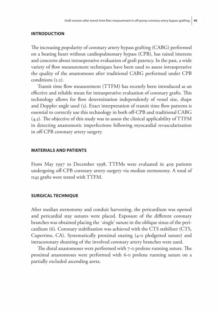
Th e increasing popularity of coronary artery bypass grafting (CABG) performed on a beating heart without cardiopulmonary bypass (CPB), has raised interests and concerns about intraoperative evaluation of graft patency. In the past, a wide variety of fl ow measurement techniques have been used to assess intraoperative the quality of the anastomoses after traditional CABG performed under CPB conditions (1,2).
Transit time fl ow measurement (TTFM) has recently been introduced as an eff ective and reliable mean for intraoperative evaluation of coronary grafts. Th is technology allows for fl ow determination independently of vessel size, shape and Doppler angle used (3). Exact interpretation of transit time fl ow patterns is essential to correctly use this technology in both off -CPB and traditional CABG (4,5). Th e objective of this study was to assess the clinical applicability of TTFM in detecting anastomotic imperfections following myocardial revascularization in off -CPB coronary artery surgery.
MATERIALS AND PATIENTS
From May 1997 to December 1998, TTFMs were evaluated in 409 patients undergoing off -CPB coronary artery surgery via median sternotomy. A total of 1145 grafts were tested with TTFM.
SURGICAL TECHNIQUE
After median sternotomy and conduit harvesting, the pericardium was opened and pericardial stay sutures were placed. Exposure of the diff erent coronary branches was obtained placing the ̀ single’ suture in the oblique sinus of the peri-cardium (6). Coronary stabilization was achieved with the CTS stabilizer (CTS, Cupertino, CA). Systematically proximal snaring (4-0 pledgetted suture) and intracoronary shunting of the involved coronary artery branches were used.
Th e distal anastomoses were performed with 7-0 prolene running suture. Th e proximal anastomoses were performed with 6-0 prolene running suture on a partially excluded ascending aorta.
Giuseppe Text alt.indd 45Giuseppe Text alt.indd 45 08-Sep-09 14:31:45 PM08-Sep-09 14:31:45 PM

Chap
ter 4
46
TTFM TECHNIQUE
At the end of every single anastomosis, fl ow values and fl ow curves were obtained using the TTFM device (MediStim BF 2004, MediStim, Oslo, Norway).
Th e TTFM probe was perfectly fi tted around the graft. Diff erent probe sizes were available to avoid distortion or compression of the graft. Skeletonization of a small segment of the mammary artery was necessary to reduce the quantity of tissue interposed between the vessel and the probe.
Aqueous gel was used to improve probe contact. TTFM was evaluated both with and without proximal snaring of the native coronary artery to detect any possible imperfection localized at the toe of the anastomosis and to exclude fl ow competition from the native vessel.
Before making any measurement, adequate de-airing of the grafts was performed, adequate systemic blood pressure was maintained, traction on the pericardium was released and the stabilizer was removed from the epicardial surface to allow for the heart to return to its anatomical position. TTFM was repeated before chest closure to confi rm graft patency and to detect any possible graft kinking or compression.
CURVE INTERPRETATION
During our clinical experience we developed a progressive expertise in TTFM fi ndings interpretation. To correctly address the TTFM fi ndings, fl ow curves, pulsatile index (PI) and mean fl ow values are evaluated. Th e curves should always be coupled with the EKG tracing to correctly diff erentiate the systolic from the diastolic fl ow. In a patent coronary graft, the hemodynamics are similar to those physiologically observed in the coronary circulation: blood fl ows mainly dur-ing diastole with minimal systolic peaks taking place during the isovolumetric ventricular contraction (QRS complex) (Fig. 1).
Th e PI, expressed as an absolute number, is a good indicator of the fl ow pattern and, consequently, of the quality of the anastomosis. Th is number is obtained by dividing the diff erence between the maximum and the minimum fl ow by the value of the mean fl ow. In our experience, the PI should be included between 1 and 5. Th e possibility of a technical error in the anastomosis increases for higher PI values. Th e mean fl ow is expressed as ml/min and, being very dependent by the quality of the revascularized coronary artery, is not a good indicator of the quality of the anastomosis.
Giuseppe Text alt.indd 46Giuseppe Text alt.indd 46 08-Sep-09 14:31:45 PM08-Sep-09 14:31:45 PM

Graft revision after transit time fl ow measurement in off -pump coronary artery bypass grafting 47
Mean fl ow values should always be interpreted together with TTF curves and PI values.
RESULTS
Forty-one grafts (41/1145) were revised in 33 patients. In three patients, four fl ow curves and fl ow values were not properly stored in the TTFM device hardware and for this reason have not been included in this study.
A total of 37 grafts are included: 18 to the left anterior descending coronary (LAD) and diagonal branches, ten to the circumfl ex system and nine to the right coronary artery system (RCA) (Table 1).
A total of six patients (18.1) underwent graft revision on CPB. TTFM fi nd-ings before revision are summarized in Table 1.
Curve patterns, fl ow and PI values remained unchanged after topical use of vasodilators (papaverine and nitrates). Twenty-nine grafts (78.37) were revised for abnormal (systolic) fl ow patterns, high PIs and low fl ow values. In fi ve cases (13.51), despite abnormal fl ow curves (systolic spikes) and high PIs, fl ow values were on average greater than 15 ml/min. Findings at revision of these 34 grafts included: thrombosis of the anastomosis (six patients), stenosis at the toe or heel of the anastomosis (eight patients), intimal fl ap or dissection in the native coronary artery (fi ve patients), dissection of the internal mammary artery (fi ve patients), graft kinking (four patients), fl ap at proximal anastomosis (one
Figure 1: Normal TTFM curve
Giuseppe Text alt.indd 47Giuseppe Text alt.indd 47 08-Sep-09 14:31:45 PM08-Sep-09 14:31:45 PM

Chap
ter 4
48
patient), coronary stenosis distal to the graft (three patients) and no fi ndings (two patients). After revision, all fl ow patterns improved (diastolic fl ows) and mean fl ow values increased from a mean value of 3.85±4.63 to 32.47±28.59 ml/min with proximal snare (P<0.0001) and from 6.58±6.00 to 36.29±26.91 ml/min without snare (P<0.0001). PI values also improved from 38.45 ± 56.56 to 3.03 ±1.6 with snare and from 24.44±46.51 to 2.80±1.68 without snare (P<0.0001). TTFM fi ndings after revision are summarized in Table 2. In three additional grafts (8.1) revision was performed on the basis of low mean fl ow values (7.3 ±2.51 ml/min with snare and 6±1 ml/min without snare) despite normal fl ow curves (diastolic) and PI values (1.86±2.20 with and 3.06 ±1.66 without snare). Th ere were no fi ndings at revision and curves, fl ow and PI values remained unchanged after revision (Tables 1 and 2). Postoperatively one patient developed a stroke (3), one had an acute myocardial infarction (AMI) (3), one required reoperation for bleeding (3), one had a sternal wound infection and one
Table 1: TTFM fi ndings in 37 grafts before revision.
Bold characters indicate grafts revised on the basis of low fl ow values despite normal fl ow patterns. Italic characters indicate grafts revised on the basis of abnormal fl ow patterns despite fl ow values greater than 15 ml/min on average.LIMA, left internal mammary artery; SVG, saphenous vein graft; RIMA, right mammary artery; LAD, left anterior descending; D, diagonal; RCA, right coronary artery; CX, circumfl ex coronary artery; OM, obtuse marginal.
Giuseppe Text alt.indd 48Giuseppe Text alt.indd 48 08-Sep-09 14:31:45 PM08-Sep-09 14:31:45 PM

Graft revision after transit time fl ow measurement in off -pump coronary artery bypass grafting 49
required prolonged ventilatory support (3). All patients were discharged after a mean hospital stay of 8.15 days.
DISCUSSION
Several techniques have been used in the past to test coronary graft fl ow intraop-erative: electromagnetic fl ow meters, initially adopted in coronary surgery, have been recently replaced by ultrasonic technology (Doppler and TTFM). Many authors have demonstrated the superiority of TTFM over Doppler systems in direct real time detection of fl ow independently of vessel diameter and Doppler angle (2,3).
Increasing interest in intraoperative evaluation of graft fl ows has followed the advent of CABG without CPB. Intraoperative fl ow measurements together with post-operative angiographic follow-up are important methods aimed at
Table 2: TTFM fi ndings in 37 grafts after revision
LIMA, left internal mammary artery; SVG, saphenous vein graft; RIMA, right mammary artery; LAD, left anterior descending; D, diagonal; RCA, right coronary artery; CX circumfl ex coronary artery; OM, obtuse marginal.
Giuseppe Text alt.indd 49Giuseppe Text alt.indd 49 08-Sep-09 14:31:46 PM08-Sep-09 14:31:46 PM

Chap
ter 4
50
documenting the feasibility of this operation. We began using TTFM routinely in off CPB coronary surgery since 1996. After 3 years of clinical experience we believe that this technology is eff ective in detecting highly stenotic coronary anastomoses. Th e sensitivity of TTFM in detecting less than critical stenoses remains to be defi ned. Cerrito et al. (7) indicated that neural network pattern recognition analysis of graft fl ow characteristics, can improve detection of anastomotic errors with intra-operative TTFM. After a complex mathematical analysis of the fl ow curves is possible to detect stenoses causing a 50 or greater narrowing of the anastomoses. It is evident that less than critical stenoses can not be detected by TTFM due to the fact that no modifi cations in the hemody-namic performances of the grafts happen at this level. At the present, standard or nominal curves and fl ow values for diff erent type of grafts and revascularized vessels have not been described and the variability between diff erent subjects and within subjects is extremely large.
In an interesting survey, Spence et al. (8) tested the ability of 19 interna-tional surgeons to detect anastomotic errors by evaluating mean fl ow and fl ow waveforms. More than 70 of the surgeons accepted anastomoses with severe stenoses but, all of them, were able to detect highly stenotic anastomoses (.90 stenosis).
It is important to emphasize the fact that the ability to correctly interpret TTFM fi ndings is slowly acquired with clinical and experimental experience.
Even if we understand there is a limit in TTFM fi ndings interpretation, our clinical experience with more than 1000 grafts tested, has showed that early detection of stenotic grafts can be achieved by the surgeons simultaneous evalu-ation of fl ow patterns, PI values, fl ow values and clinical fi ndings (i.e. EKG trac-ing, hemodynamic values). Flow curves in patent grafts have a mainly diastolic pattern with a small component of negative systolic fl ow. Th e diastolic fl ow is the actual fl ow that at every diastole fl ows from the graft in to the coronary through the anastomosis, the systolic component is retrograde fl ow that cannot fl ow in to the anastomosis during systole and goes backwards in to the graft (Fig. 1). In the case of stenotic anastomosis the fl ow curve becomes spiky and mainly systolic (Fig. 2): in this situation the main fl ow through the graft is systolic and there is minimal perfusion of the anastomosis during diastole. Even if these rules apply generally to all vessels, we have noticed some diff erences whenever testing grafts anastomosed to the right coronary system. A good component of blood fl ow in to the right coronary takes place during systole simply due to a minor compression of the epicardial vessels during right ventricular contraction. For this reason, a larger component of systolic positive fl ow can be observed in patent anastomoses to the RCA. As mentioned, we do not have nominal fl ow and PI values to suggest
Giuseppe Text alt.indd 50Giuseppe Text alt.indd 50 08-Sep-09 14:31:47 PM08-Sep-09 14:31:47 PM

Graft revision after transit time fl ow measurement in off -pump coronary artery bypass grafting 51
for a correct interpretation of TTFM fi ndings and our statements are based on the simple visual assessment of fl ow curve morphology and clinical fi ndings. Di Giammarco et al. (9) have analyzed the diff erences in TTFM patterns in diff er-ent coronary grafts but, at present, no standard values have been reported. We are convinced that fl ow value per se is not a good indicator of the quality of the anastomosis and can not justify graft revision: absolute fl ow is infl uenced by too many variables including type and size of the graft, and quality of the coronary artery distal territory. In our experience graft revision was erroneously performed in three cases only on the basis of low fl ow values despite satisfactory fl ow pat-terns and low PI values: at surgical inspection no anastomotic lesions were found and the fl ow values remained unchanged after revision (Tables 1 and 2). Use of vasodilating agents (i.e. papaverine and nitro-glycerine) did not improve the fl ow values and the small calibre of the revascularized coronary arteries (,1.5 mm) was responsible for our fi ndings. Coronary fl ow reserve can help in correctly diagnose anastomotic imperfections and, as described by Walpoth et al. (10), the quality of the anastomosis can be tested by recording the modifi cations of fl ow during infusion of adenosine. We do not have experience with calcula-tion of fl ow reserve but we believe that a more precise detection of anastomotic imperfections with TTFM could be achieved by testing the dynamic ability of the anastomoses to increase the blood fl ow whenever the oxygen requests of the myocardium are increased. If mean fl ow values are not good predictors of grafts’ quality, on the contrary PI values are, per se, very suggestive of the actual status of the anastomoses. As mentioned in the results, we correctly revised fi ve grafts on the basis of abnormal fl ow curves and high PI values (22.04 ±21.17 with snare
Figure 2: TTF curve in a severely stenotic saphenous vein graft to the RCA.
Giuseppe Text alt.indd 51Giuseppe Text alt.indd 51 08-Sep-09 14:31:47 PM08-Sep-09 14:31:47 PM

Chap
ter 4
52
and 13.94 ± 19.77 without snare) despite fl ow was on average higher than 15 ml/min. At surgical inspection all fi ve anastomoses resulted to be severely stenotic and after revision fl ow patterns and PIs were improved (2.64 ±1.07 with snare and 2.82±1.01 without snare) (Tables 1 and 2).
To our knowledge, an absolute PI value has never been offi cially proposed and we empirically decided the limit of 5 on the basis of our clinical experience. Di Giammarco et al.(9) proposed a value of 2.5 as the limit PI above which an anastomosis should be revised but, again, this value seems to be derived by per-sonal clinical experience. Flow curves, PI and mean fl ow values should always be evaluated with and without occlusion of the native coronary arteries: proximal snaring of the native coronary is, in our opinion, important in order to achieve a reliable interpretation of TTFM fi ndings. Th e shape of the curve should remain unchanged when snaring the coronary proximally and an increase in absolute fl ow should be recorded if competition from the native coronary was present with the unsnared coronary. Th e proximal snare will also permit to detect lesions at the level of the toe of the anastomosis: in this situation whenever the coro-nary is snared the absolute fl ow will drastically decrease documenting lack of ante-grade fl ow through the anastomosis. Verifi cation of intraoperative TTFM fi ndings can be obtained with immediate postoperative angiographic studies. Even if we do not have a systematic angiographic follow up for our revised grafts, we believe our immediate postoperative clinical results being satisfactory and somehow confi rming intraoperative TTFM fi ndings. Angiography, per se, gives a limited bidimensional view of the coronary arteries and the coronary grafts without giving any specifi c information about the hemodynamic parameters of the anastomoses; for this reason a comparative study between postopera-tive angiography and intraoperative TTFM may result diffi cult. In reoperative CABG for example, we found confl icting results when comparing preoperative angiography and intraoperative TTFM; in one particular case angiographic documentation of anastomotic sub-occlusion of an old saphenous graft, was not confi rmed during surgical revision and intraoperative TTFM.
In an interesting study, Louagie et al. (11) reported that intraoperative hemodynamic assessment, via pulsed Doppler fl owmeter, can have a satisfactory predictive value for midterm graft occlusion, on the contrary the same hemody-namic parameters are useless for prediction of midterm graft stenosis. Together with Dr Louagie we feel that midterm stenosis development is a dynamic process related to scar tissue formation and degeneration of the graft and, for this reason, can not be detected at the time of the procedure. TTFM has also been compared with other techniques of postoperative graft patency verifi cation that, diff erently from angiography, can give more precise information about the hemodynamic
Giuseppe Text alt.indd 52Giuseppe Text alt.indd 52 08-Sep-09 14:31:48 PM08-Sep-09 14:31:48 PM

Graft revision after transit time fl ow measurement in off -pump coronary artery bypass grafting 53
characteristics of the grafts. In a series of 22 patients, Walpoth et al. (12) have shown a signifi cant correlation between intraoperative TTFM and post-operative magnetic resonance fi ndings of the internal mammary artery grafts.
In conclusion we may say that there are certainly some limits in the inter-pretation of TTFMs fi ndings and there is still necessity to defi ne the sensitivity of TTFM in detecting less then critical stenosis (7, 8). Correct interpretation of fl ow curves, mean fl ows and PI values are crucial in reducing the number of undetected technical errors and in decreasing the number of grafts erroneously revised. Th e mean fl ow value per se is not a good indicator of the quality of the anastomosis.
Acceptable fl ow values with abnormal fl ow patterns and high PIs may under-line highly stenotic lesions of the anastomoses (fi ve cases in our experience). On the contrary, we observed low fl ow conditions with good fl ow curves in three anastomoses which, at revision, resulted in fully patent anastomoses. Th is situation may occur whenever the revascularized territory has poor run off .
TTFM is reliable in detecting technical errors after CABG without CPB. Graft revision should be promptly performed whenever fl ow curves, mean fl ows, and PI values are abnormal. In this situation, revision of the distal anastomoses leads to improvement in fl ow patterns. Postoperative outcome can be improved by a meticulous use and understanding of TTFM in patients undergoing coro-nary artery surgery with and without CPB.
Giuseppe Text alt.indd 53Giuseppe Text alt.indd 53 08-Sep-09 14:31:48 PM08-Sep-09 14:31:48 PM

Chap
ter 4
54
REFERENCES
1. Louagie YAG, Haxhe JP, Jamarat J, Buch M, Schoevaerdts JC. Intraoperative assessment of coro-nary artery bypass grafts using a pulsed doppler fl owmeter. Ann Th orac Surg 1994; 58: 742±749.
2. Canver CC, Dame N. Ultrasonic assessment of internal thoracic artery graft fl ow in the revascu-larized heart. Ann Th orac Surg 1994; 58: 135-138.
3. Canver CC, Cooler SD, Murray EL, et al. Clinical importance of measuring coronary graft fl ows in the revascularized heart. Ultrasonic or electromagnetic? J Cardiovasc Surg 1997; 38: 211±215.
4. Walpoth BH, Mohadjer A, Gersbach P, Rogulenko R, Walpoth BN, Althaus U. Intraoperative internal mammary artery transit-time fl ow measurements: comparative evaluation of two surgi-cal pedicle preparation techniques. Eur J Cardio-thorac Surg 1996; 10(12): 1064±1070.
5. Jaber SF, Koenig SC, BhaskerRao B, VanHimbergen DJ, Ceriito PB, Ewert DJ, Gray Jr LA, Spence PA. Role of graft fl ow measurement technique in anastomotic quality assessment in minimally invasive CABG. Ann Th orac Surg 1998; 66: 1087±1092.
6. Bergsland J, Karamanoukian HL, Soltoski PR, Salerno TA. “Single suture” for circumfl ex expo-sure in off -pump coronary artery bypass grafting. Ann Th orac Surg 1999; 68(4): 1428±1430.
7. Cerrito PB, Koenig SC, Koenig SC, Van Himbergen DJ, Jaber SF, Ewert DL, Spence PA. Neural network pattern recognition analysis of graft fl ow characteristics improves intra-operative anas-tomotic error detection in minimally invasive CABG. Eur J Cardio-thorac Surg 1999; 16: 88±93.
8. Jaber SF, Koenig SC, BhaskerRao B, Van Himbergen DJ, Spence PA. Can visual assessment of fl ow waveform morphology detedt anastomotic error in off pump coronary artery bypass grafting? Eur J Cardiothorac Surg 1998; 14: 476±479.
9. Di Giammarco G. Myocardial revascularization without cardiopulmonary bypass. Presented at the symposium: State of the art in emerging coronary revascularization; EACTS, Glasgow, Scotland Sept 4, 1999.
10. Walpoth BH, Bosshard A, Genyk I, Kipfer B, Berdat PA, Hess OM, Althaus U, Carrel TP. Transit time fl ow measurement for detection of early graft failure during myocardial revascularization. Ann Th orac Surg 1998; 66(3): 1097±1100.
11. Louagie YAG, Brockmann CE, Jamart J, Schroeder E, Buche M, Eucher PM, Schoevaerdts JC. Pulsed Doppler intraoperative fl ow assessment and midterm coronary graft patency. Ann Th orac Surg 1998; 66(4): 1282±1288.
12. Walpoth BH, Muller MF, Genyk I, Aeschbacher B, Althaus U, Carrel TP. Evaluation of coronary bypass fl ow with color-Doppler and magnetic resonance imaging techniques: comparison with intraoperative fl ow measurements. Eur J Cardio-thorac Surg 1999; 15: 795±802.
Giuseppe Text alt.indd 54Giuseppe Text alt.indd 54 08-Sep-09 14:31:48 PM08-Sep-09 14:31:48 PM

Graft revision after transit time fl ow measurement in off -pump coronary artery bypass grafting 55
APPENDIX A. CONFERENCE DISCUSSION
Dr. A. Royse (Victoria, Australia): You certainly have a very large series. I think you have shown us fairly well from your data that a high PI value and abnormal fl ow curves was correlated with a critical conduit of anastomotic stenosis, but what you haven’t shown us is what is the denominator?
How many of those who had a normal or slightly abnormal fl ow curve actu-ally had a critical stenosis but you missed it or you didn’t revise it? In other words, you are only telling us of the critical stenoses that you actually revised, not how many theoretically there were in those that you didn’t revise.
My question is, have you done any other tests such, as, perhaps angiography to try and establish if there is a critical stenosis in those who you didn’t revise?
Dr. D’Ancona: No, we didn’t make any angiographic study. I outlined in the conclusion, we really are not able to say how critical should be the stenosis to be detected by the TTFM. Dr Paul Spence’s group in Louisville, Kentucky did make some experimental settings showing that TTFM is able to detect stenosis that at angiography result higher than 50.
Dr. J. Melo (Carnaxide, Portugal): When we measurements low fl ows should we take precautions like a late angiography?
Dr. D’Ancona: In our experience, if we are sure that the size of the vessel and the territory revascularized were very poor, we are not really very much concerned when we have low fl ows if we have good curves and good PI values, so we don’t do anything, and as a matter of fact, we didn’t have any clinical postoperative implication using this strategy. So I think you shouldn’t be worried about any angiographical study in those patients. Th is is our personal opinion.
Giuseppe Text alt.indd 55Giuseppe Text alt.indd 55 08-Sep-09 14:31:49 PM08-Sep-09 14:31:49 PM

Giuseppe Text alt.indd 56Giuseppe Text alt.indd 56 08-Sep-09 14:31:49 PM08-Sep-09 14:31:49 PM

Chapter 5
C oronary grafts fl ow and cardiac pacing modalities:
how to improve perioperative myocardial perfusion
Giuseppe D’Ancona, MDMartin Hargrove, RNJohn Hinchion, MDB.C. Ramesh, MDJehan Zeb Chughtai, MDMuhammad Nadeem Anjum, MDAonghus O’Donnell, MDTom Aherne, MD
Department of Cardiac Surgery, Cork University Hospital, Cork, Ireland
Eur J Cardiothor Surg 26 (2004) 85–88
Presented at the 13th Annual Meeting of the European Association for Cardio-thoracic Surgery,
Glasgow, Scotland, UK, September 5-8, 1999.
Giuseppe Text alt.indd 57Giuseppe Text alt.indd 57 08-Sep-09 14:31:49 PM08-Sep-09 14:31:49 PM

Chap
ter 5
58
ABSTRACT
Objective
Aim of this study was to investigate modifi cations of coronary grafts fl ow during diff erent pacing modalities after CABG.
Materials and Methods
Two separate prospective studies were conducted in patients undergoing CABG and requiring intraoperative epicardial pacing. In a fi rst study (22 patients) coro-nary grafts fl ows were measured during dual chamber pacing (DDD) and during ventricular pacing (VVI). In a second study (10 patients) fl ows were measured during DDD pacing at diff erent atrio-ventricular (A-V) delay periods. A-V delay was adjusted in 25 ms increments from 25 to 250 ms and fl ow measurements were performed for each A-V delay increment. A transit time fl owmeter was used for the measurements.
Results
An average of 3.4 grafts/patient were performed. In the fi rst study, average coronary graft fl ow was 47.4 ± 20.8 ml/min during DDD pacing and 41.8 ± 18.2 ml/min during VVI pacing (P = 0.0004): Furthermore average systolic pressure was 94.3 ± 10.1 mmHg during DDD pacing and 89.6 ± 12.2 mmHg during VVV pacing (P = 0.0007): No signifi cant diff erences in diastolic pressure were recorded during the two diff erent pacing modalities. In the second study, maximal fl ows were achieved during DDD pacing with an A-V delay of 175 ms (54 ± 9.6 ml/min) and minimal fl ows were detected at 25 ms A-V delay (38.1 ± 4.7 ml/min) (P = ns). No signifi cant diff erences in systolic or diastolic blood pressure were noticed during the diff erent A-V delays.
Conclusion
Grafts fl owmetry provides an extra tool to direct supportive measures such as cardiac pacing after CABG. DDD mode with A-V delay around 175 ms. should be preferred to allow for maximal myocardial perfusion via the grafts.
Giuseppe Text alt.indd 58Giuseppe Text alt.indd 58 08-Sep-09 14:31:49 PM08-Sep-09 14:31:49 PM

Coronary grafts fl ow and cardiac pacing modalities: how to improve perioperative myocardial perfusion 59
INTRODUCTION
Th e main aim of coronary artery bypass grafting (CABG) is to increase blood fl ow to ischemic myocardium. Although this procedure is successfully performed in more than several hundred thousand patients per year in Europe, intraopera-tive graft patency verifi cation is still considered optional in most of the cardiac surgery centers. Grafts are assumed to be patent at the end of the operation, especially if the patient has no hemodynamic compromise and if weaning from cardiopulmonary bypass (CPB) is successful. After an initial attempt to expand graft fl owmetry in standard cardiac surgery practice, in the early 1960s mea-surement of coronary graft fl ow has been almost abandoned due to the many limitations of the obsolete electromagnetic fl owmeters adopted at that time (1)
Th e recent popularization of off -pump CABG, together with the amelioration of the technology available for accurate graft fl ow measurement, have revived interests and concerns about the focal importance of intraoperative graft patency verifi cation and documentation (2). Th e introduction of ultrasound-based fl ow meters such as Doppler and Transit Time Flow Measurement (TTFM) systems, is presently giving a tremendous scientifi c and technological impact in the fi eld of rheology and fl ow measurement.
In our experience intraoperative graft patency verifi cation, if properly performed, may not only inform the surgeon about the status of the newly constructed coronary anastomoses, but could also guide him in modifying his perioperative conduct and strategy to achieve an ideal hemodynamic perfor-mance and, consequently, a maximal myocardial perfusion through the bypass conduits (3).
In the present manuscript we summarize our experience with intraoperative cardiac pacing and simultaneous coronary graft fl ow measurement trying to identify which are the pacing strategies that allow for an optimal coronary graft rheology.
MATERIAL AND METHODS
Over a one-year period, two separate prospective studies were conducted at the Cork University Hospital in order to evaluate coronary grafts fl ow modifi cations in patients requiring epicardial pacing following primary CABG.
Patients were enrolled following preoperative informed consent.All patients were operated on CPB and cardiac arrest was achieved via
antegrade intermittent crystalloid cold cardioplegia. Distal anastomoses were
Giuseppe Text alt.indd 59Giuseppe Text alt.indd 59 08-Sep-09 14:31:49 PM08-Sep-09 14:31:49 PM

Chap
ter 5
60
performed with 7-0 polypropilene running sutures. Proximal anastomoses were constructed on partial aortic clamping with 5-0 polypropylene running sutures.
After aortic declamping, epicardial pacing was initiated due to nodal rhythm, atrio-ventricular dissociation, or sinus bradycardia.
Two epicardial atrial and two ventricular pacing wires were placed in each patient.
In a fi rst study including 22 patients, coronary grafts fl ows were measured after weaning from CPB initially during dual chamber pacing (DDD) and secondly during ventricular pacing (VVI).
In a second study including 10 patients, fl ows were measured during DDD pacing at diff erent atrio-ventricular (A-V) delay periods. A-V delay was adjusted in 25 milliseconds increments from 25 to 250 ms and fl ow measurements were performed for each A-V delay increment.
Although cardiac indexes were not routinely recorded, fl ow in the new pacing modality was detected only after allowing two minutes of hemodynamic stabili-zation aiming at systolic arterial blood pressure of 100 mmHg and diastolic of 60 mmHg to standardize measurements. Measurements were performed twice per each graft in the same pacing modality and averages were calculated.
A TTFM device was used to test the grafts. Flow probes have a size ranging from 2.5 to 3 mm and can be easily placed around the constructed grafts. A small amount of aqueous gel is placed between the probe and the conduit to increase the contact. Th e probe consists of two small piezoelectric crystals, one upstream and one downstream, mounted on the same side of the vessel. Opposite to the crystals, there is a small metallic refl ector. Each crystal produces a wide pulsed ultrasound beam covering the entire vessel width. Both the amount of time nec-essary for an ultrasound beam emitted from the upstream crystal to arrive at the downstream crystal after being refl ected, and for a signal from the down-stream crystal to reach the upstream crystal are measured. Since ultrasound travels faster if transmitted in the same direction as fl ow, a small time diff erence between the two beams is calculated as the transit time of fl ow and thus, the actual fl ow is proportional to the transit time (5). All calculations are made automatically by the fl ow meter and are displayed as ml/min.
Flow fi ndings were recorded together with hemodynamic values. No pulsatil-ity indexes were recorded. Data were stored in a database and diff erences were statistically tested with the paired Student t-test (DDD-VVI and A-V delay study), and with the one-way ANOVA test (A-V delay study).
Diff erences in mean fl ows between arterial and venous conduits at diff erent pacing modalities, correlations between mean fl ow changes and conduits size, fl ow variations for grafts to diff erent myocardial areas and after use of diff erent
Giuseppe Text alt.indd 60Giuseppe Text alt.indd 60 08-Sep-09 14:31:49 PM08-Sep-09 14:31:49 PM

Coronary grafts fl ow and cardiac pacing modalities: how to improve perioperative myocardial perfusion 61
myocardial protection techniques were not investigated in the present study and are object of analysis in two further ongoing larger studies.
RESULTS
Study 1
An average of 3.4 grafts/patient were performed. All 22 patients received a LIMA graft to the LAD. No further arterial grafts were adopted. Average coronary graft fl ow as measured in the 22 patients included in the study was 47.4 ± 20.8 ml/min during DDD pacing and 41.8 ± 18.2 ml/min during VVI pacing (P = 0.0004). Furthermore average systolic pressure was 94.3 ± 10.1 mmHg during DDD pacing and 89.6 ± 12.2 mmHg during VVV pacing (P = 0.0007): No signifi cant diff erences in diastolic pressure were recorded during the 2 diff erent pacing modalities
Study 2
An average of 3.4 grafts/patient were performed. All 10 patients received a LIMA graft to the LAD. No further arterial grafts were adopted. Although maximal coronary grafts fl ows were achieved during DDD pacing with an A-V delay of 175 ms (mean 54 ± 9.6 ml/min) and minimal fl ows were detected at 25 ms A-V delay (mean 38.1 ± 4.7 ml/min), no statistically signifi cant diff erences were reported (P = ns). Furthermore, no signifi cant diff erences in systolic or diastolic blood pressure were noticed during the diff erent A-V delays. Mean fl ow values as recorded for each patient at diff erent A-V delays are reported in Fig. 1.
All patients involved in study 1 and 2 remained hemodynamically stable dur-ing the procedure, and upon transfer to the ICU. No perioperative AMIs were reported.
No further mortality or morbidity were noticed. No angiographic studies were conducted.
DISCUSSION
Intraoperative graft patency verifi cation should be considered as a precious technology to include in the armamentarium already available to the modern cardiac surgeon.
Giuseppe Text alt.indd 61Giuseppe Text alt.indd 61 08-Sep-09 14:31:49 PM08-Sep-09 14:31:49 PM

Chap
ter 5
62
Unfortunately scepticism and misinformation persist in the surgical com-munity and the large majority of cardiac surgeons are still reliant on the tactile sense of their fi ngertips to evaluate quality and fl ow of their grafting (6).
Although performed in a limited group of patients, our study has emphasized the importance of intraoperative fl ow measurement not only in testing patency of coronary anastomosis but also in correctly guiding perioperative management (i.e. cardiac pacing) to achieve ideal hemodynamics and consequently maximal grafts fl ows.
Electrical conduction disturbances may frequently occur after CABG as a result of mechanical problems or ischemic/reperfusion injury. To optimize hemody-namics, epicardial pacing may be required following CABG. Bicameral pacing has been proved to be the most physiological pacing mode (7). Although it has been demonstrated that A-V sequential pacing may increase atrial priming of the left ventricular end diastolic pressure and consequently allow for an increase in cardiac output up to 25 (8), benefi ts in terms of increased bypass grafts performance have never been investigated.
As shown in our fi rst study, improvement in systemic hemodynamics during DDD versus VVV pacing does also permit to achieve optimal myocardial perfu-sion via the newly constructed grafts. Increment in graft fl ow averages 6 ml/min and can arrive to a maximum of 20 ml/min.
Figure 1: Mean grafts fl ows in 10 patients at diff erent A-V delays.
Giuseppe Text alt.indd 62Giuseppe Text alt.indd 62 08-Sep-09 14:31:49 PM08-Sep-09 14:31:49 PM

Coronary grafts fl ow and cardiac pacing modalities: how to improve perioperative myocardial perfusion 63
On the basis of these fi ndings, DDD mode should be preferred in patients requiring pacing immediately after CABG in order to allow for maximal reper-fusion of the previously ischemic myocardium via the grafts.
Moreover, an ideal or optimal A-V delay during perioperative DDD pacing has never been proposed. In our second study we have tried to defi ne the most appropriate A-V delay by using conduit fl ow as a hemodynamic index in patients requiring sequential pacing post CABG.
Interestingly, although systemic hemodynamics do not seem to be infl uenced by the A-V delay value, coronary grafts fl ows achieve an optimal level at 175 ms. of A-V delay and a minimum level towards 25 ms. of A-V delay. Although these fi ndings are limited to a small number of patients and diff erences did not achieve statistical signifi cance, it could be suggested that, in patients requiring DDD pacing, maximal coronary grafts fl ow may be obtained when maintaining an A-V delay in the 175 ms range.
As already emphasized, the importance of intraoperative coronary grafts fl ows fi ndings is continuously increasing as a result of the technological improvements in the fl ow measurement technology. Th e introduction of ultrasound-based systems has revolutionized the fl ow measurement fi eld. Th e term ultrasound has a generic defi nition that includes two diff erent methods: Doppler and TTFM. Th e two systems rely on diff erent properties of the ultrasound waves and, although the Doppler methods have shown good reliability both in vivo and in vitro (4), the TTFM technology off ers many important advantages and is the most accurate system for intraoperative verifi cation of coronary graft patency (9–11). In the present studies we have adopted a TTFM device whose principles of functioning have been already described above. In our experience, the TTFM device is very easy to use and requires no more than 30 s per measurement. No complications resulted from the use of this fl owmeter device. Th e fl owmeter provides not only an absolute value expressed as ml/min but gives also a fl ow curve that summarizes the variations of graft fl ow during the diff erent phases of the cardiac cycle. Because coronary graft fl ow is mainly diastolic, it is important to adopt an adequate modality of pacing that could allow for good diastolic fi lling and pressure without compromising the systolic performance.
CONCLUSION
Intraoperative graft patency verifi cation and coronary grafts fl ow detection should be used to testify for a successful intraoperative myocardial revasculariza-tion and to guide a safe perioperative hemodynamic performance. Graft fl ow
Giuseppe Text alt.indd 63Giuseppe Text alt.indd 63 08-Sep-09 14:31:49 PM08-Sep-09 14:31:49 PM

Chap
ter 5
64
fi ndings are not solely an anastomosis status indicator, but also a useful index for the patient hemodynamic condition. Appropriate intraoperative evaluation of coronary grafts fl ows may provide an extra tool to correctly direct supportive measures such as cardiac pacing modalities after CABG.
Giuseppe Text alt.indd 64Giuseppe Text alt.indd 64 08-Sep-09 14:31:49 PM08-Sep-09 14:31:49 PM

Coronary grafts fl ow and cardiac pacing modalities: how to improve perioperative myocardial perfusion 65
REFERENCES
1. Kolin A, Ross G, Gaal P, Austin S. Simultaneous electromagnetic measurement of blood fl ow in the major coronary arteries. Nature 1964; 203: 148–50.
2. Canver CC, Cooler SD, Murray EL, Nichols RD, Heisey DM. Clinical importance of measur-ing coronary graft fl ows in the revascularized heart. Ultrasonic or electromagnetic? Cardiovasc Surg 1997; 38(3): 211–5.
3. Flynn MJ, Winters D, Breen P, O’Sullivan G, Shorten G, O’Connell D, O’Donnell A, Aherne T. Dopexamine increases internal mammary artery blood fl ow following coronary artery bypass grafting. Eur J Cardiothorac Surg 2003; 24(4): 547–51.
4. Laustsen J. Transit time fl ow measurement: principles and clinical applications. In: D’Ancona G, editor. Intraoperative graft patency verifi cation in cardiac and vascular surgery. Armonk, NY: Futura Publishing Company; 2001. p. 65–70.
5. D’Ancona G, Karamanoukian HL, Ricci M, Schmid S, Spanu I, Apfel L, Bergsland J, Salerno TA. Intraoperative graft patency verifi cation: should you trust your fi ngertips? Heart Surg Forum 2000; 3(2): 99–102.
6. Hartzler GO, Maloney JD, Curtis JJ, Barnhorst DA. Hemodynamic benefi ts of atrioventricular sequential pacing after cardiac surgery. Am J Cardiol 1997; 40(2): 232–6.
7. Durbin Jr CG, Kopel RF. Optimal atrioventricular (AV) pacing interval during temporary AV sequential pacing after cardiac surgery. J Cardiothorac Vasc Anesth 1993; 7(3): 316–20.
8. Segadal L, Matre K, Engedal H, Resch F, Grip A. Estimation of fl ow in aortocoronary grafts with a pulsed ultrasound Doppler meter. Th orac Cardiovasc Surg 1982; 30(5): 265–8.
9. Laustsen J, Pedersen EM, Terp K, Steinbruchel D, Kure HH, Paulsen PK, Jorgensen H, Paaske WP. Validation of a new transit time ultrasound fl ow meter in man. Eur J Vasc Endovasc Surg 1996; 12: 91–6.
10. Lundell A, Bergqvist D, Mattsson E, Nilsson B. Volume blood fl ow measurements with a transit time fl ow meter: an in vivo and in vitro variability and validation study. Clin Physiol 1993; 13: 547–57.
11. Matre K, Birkeland S, Hessevik I, Segadal L. Comparison of transit time and Doppler ultrasound methods for measurements of fl ow in aortocoronary bypass grafts during cardiac surgery. Th orac Cardio¬vasc Surg 1994; 42: 170–4.
Giuseppe Text alt.indd 65Giuseppe Text alt.indd 65 08-Sep-09 14:31:49 PM08-Sep-09 14:31:49 PM

Giuseppe Text alt.indd 66Giuseppe Text alt.indd 66 08-Sep-09 14:31:49 PM08-Sep-09 14:31:49 PM

Chapter 6
P reoperative Angiography and Intraoperative Transit
Time Flow Measurement to Detect Coronary Graft
Patency in Reoperations: An Integrated Approach
Giuseppe D’Ancona, MDHratch L. Karamanoukian, MDMarco Ricci, MDJacob Bergsland, MDTomas A. Salerno, MD
Center for Minimally Invasive Cardiothoracic Surgery, Kaleida Health Systems and the State
University of New York at Buff alo, Buff alo, New York.
Angiology 51 (2000) 777-780
Giuseppe Text alt.indd 67Giuseppe Text alt.indd 67 08-Sep-09 14:31:49 PM08-Sep-09 14:31:49 PM

Chap
ter 6
68
ABSTRACT
Coronary angiography is the gold standard for preoperative diagnosis of coronary artery disease. In reoperative coronary artery surgery the physiologic signifi cance of suspect lesions by angiography can be confi rmed by direct intraoperative measurement of blood fl ow in the old grafts; such interpretation can prevent unnecessary graft revisions.
Giuseppe Text alt.indd 68Giuseppe Text alt.indd 68 08-Sep-09 14:31:49 PM08-Sep-09 14:31:49 PM

Preoperative Angiography to Detect Coronary Graft Patency in Reoperations 69
INTRODUCTION
Coronary angiography provides a two-dimensional image of epicardial vessels and coronary grafts and remains the “gold standard” for the diagnosis of native coronary artery disease and graft stenosis. Methods of intraoperative detection of graft fl ow (eg, transit time fl ow measurement or TTFM) provide additional dynamic physiologic information regarding fl ow in coronary grafts including absolute graft fl ow, diastolic fl ow pattern, and coronary fl ow reserve. In reopera-tions, this information may be useful in patients with questionable graft lesions observed by pre-operative coronary angiography. We herein describe a case in which the clinical and anatomic signifi cance of what was viewed as a critical angiographic lesion in a vein graft was questioned by intraoperative TTFM.
CASE-REPORT
A 73-year-old woman with a history positive for coronary artery disease, previous anterior myocardial infarction, unstable angina (CCS IV), and congestive heart failure (CHF), presented with non-Q wave myocardial infarction. Coronary angiography demonstrated depressed left ventricular function (ejection fraction 25) with critical three-vessel coronary artery disease. She underwent coronary artery bypass grafting (CABG) without cardiopulmonary bypass (CPB) using the left internal mammary artery to the left anterior descending (LAD), and separate saphenous vein grafting of the right and circumfl ex coronary arteries. Graft fl ows were measured intraoperatively via transit time fl ow measurement (TTFM) and were satisfactory. She had an uneventful post-operative course and was discharged home.
She remained asymptomatic and had good functional recovery at 3 and 6 months follow-up. She was readmitted 9 months later for an episode of sud-den cardiac death. An electrophysiologic study was performed and an internal cardioverter defi brillator was implanted for sustained ventricular tachycardia. Angiography documented sub-occlusion of the saphenous graft to the right coronary artery (Figure 1) with patent grafts to the LAD and circumfl ex arteries. Via lower ministernotomy, the saphenous vein graft to the right coronary artery (RCA) was identifi ed and a 3 mm TTFM probe was positioned around it and excellent fl ow values and curves were recorded documenting patency of this graft (Figure 2). Th e graft was temporarily clamped, and after the coronary distal to the anastomosis was opened, good antegrade fl ow was noticed whenever the clamp on the vein graft was released. Although an anatomic lesion was not found
Giuseppe Text alt.indd 69Giuseppe Text alt.indd 69 08-Sep-09 14:31:49 PM08-Sep-09 14:31:49 PM

Chap
ter 6
70
Figure 1. Angiography showing subocclusion of right coronary graft.
Figure 2. TTFM fi ndings of old right coronary graft.
Figure 3. TTFM fi ndings of new right coronary graft.
Giuseppe Text alt.indd 70Giuseppe Text alt.indd 70 08-Sep-09 14:31:49 PM08-Sep-09 14:31:49 PM

Preoperative Angiography to Detect Coronary Graft Patency in Reoperations 71
to explain the angiographic stenosis, a new saphenous graft was anastomosed to the distal RCA on the beating heart; proximal anastomosis was performed to the lateral wall of the old RCA graft. TTFM was obtained and fl ow curves were satisfactory; interestingly, absolute fl ow in the new graft was lower than that recorded in the old graft (Figure 3). Th e operation was completed and recovery was uneventful with discharge home on the fourth postoperative day.
DISCUSSION
Although coronary angiography is the “gold standard” for the evaluation of coronary artery stenosis, its interpretation can occasionally be misleading (1). As in this case, coronary angiography can falsely diagnose coronary graft stenosis, resulting in an unnecessary operative intervention. Angiography gives a limited biplanar view of the coronary bed without a real measurement of the blood fl ow; on the contrary there are other methods that can be used to gain hemodynamic data about native coronary and coronary grafts. Intracoronary translesional fl ow velocity measurements using the Doppler fl ow wire (2) for example, provide pre-stenotic and post-stenotic fl ow values, aiding in the discrimination of physiologically signifi cant coronary stenoses. Less accurate techniques include phase diff erence magnetic resonance fl ow (3), which is a non-invasive method of assessing physiologically signifi cant coronary artery stenosis.
In redo coronary surgery, intraoperative TTFM can be used to gain physio-logic information about fl ow in all grafts, especially when their degree of stenosis is suspect and in situations where the above mentioned diagnostic modalities are not available preoperatively. Intraoperative TTFM to document graft patency after CABG has recently been introduced. Th is technology is ultrasound based and off ers many advantages when compared to the traditional methods of intra-operative fl ow measurement, ie, electromagnetic and Doppler. TTFM fi ndings are independent of hematocrit level, Doppler angle, and size of the graft tested; the fl ow values recorded result from a real-time direct measurement and are not derived from mathematical calculations. Together with mean fl ow values, fl ow patterns, pulsatile index, and vascular resistance values can also be assessed by TTFM. We have used this technology to detect patency in more than 1,000 anastomoses performed off CPB, systematically revising grafts with abnormal TTFM patterns (4). Graft patency is always tested with and without proximal occlusion of the revascularized coronary in order to exclude competitive fl ow from the native vessels and to assess patency at both toe and heel of the anasto-moses.
Giuseppe Text alt.indd 71Giuseppe Text alt.indd 71 08-Sep-09 14:31:52 PM08-Sep-09 14:31:52 PM

Chap
ter 6
72
On the basis of our clinical experience, we believe TTFM is a good modal-ity that can reliably interpret the functional status of coronary anastomoses. In experimental studies TTFM has shown a good sensitivity, and lesions that at angiography appeared equal to or greater than 50 were easily detected by correct interpretation of TTFM fi ndings (5).
To our knowledge this is the fi rst report of graft patency documented by intraoperative TTFM after being incorrectly interpreted as critically stenosed by angiography. Surgical strategy is normally directed by angiographic fi ndings, and for this reason we decided to perform a new bypass distal to the old one despite satisfactory TTFM fi ndings. We believe that unnecessary revision of patent grafts can be avoided by integrating dynamic intraoperative studies, i.e., TTFM, with preoperative angiographic studies; with this integrated approach patency of old grafts, in reoperative coronary surgery, can be confi rmed and assessed by direct intraoperative evaluation of graft hemodynamics.
Giuseppe Text alt.indd 72Giuseppe Text alt.indd 72 08-Sep-09 14:31:52 PM08-Sep-09 14:31:52 PM

Preoperative Angiography to Detect Coronary Graft Patency in Reoperations 73
REFERENCES
1. White CW, Wright CB, Doty DB, et al: Does visual interpretation of the coronary arteriogram predict the physiologic importance of a coronary stenosis? N Engl J Med 310: 819, 1984.
2. Bach RG, Kern MJ: Practical coronary physiology. Clinical application of the Doppler fl ow velocity. Cardiology Clinics. 15: 77-99, 1997.
3. Molinari G, Sardanelli F, Zandrino F, et al: Magnetic resonance assessment of coronary artery bypass grafts. Rays 24: 131-139, 1999.
4. D’Ancona G, Karamanoukian H, Salerno T, et al: Graft revision after transit time fl ow measure-ment in off -pump coronary artery bypass grafting. Presented at the European Association of Cardiothoracic Surgery, September 1999, Glasgow, Scotland.
5. Cerrito PB, Koenig SC, Koenig SC, et al: Neural network pattern recognition analysis of graft fl ow characteristics improves intraoperative anastomotic error detection in minimally invasive CABG. Eur J Cardiothorac Surg 16: 88-89, 1999.
Giuseppe Text alt.indd 73Giuseppe Text alt.indd 73 08-Sep-09 14:31:52 PM08-Sep-09 14:31:52 PM

Giuseppe Text alt.indd 74Giuseppe Text alt.indd 74 08-Sep-09 14:31:52 PM08-Sep-09 14:31:52 PM

Chapter 7
H eparin Dose, Transfusion Rates, and
Intraoperative Graft Patency in Minimally
Invasive Direct Coronary Artery Bypass
Harry W. Donias, MD³Giuseppe D’Ancona, MD¹Ravi U. Pande, MD¹Dennis Schimpf, MD³Akira T. Kawaguchi, MD, PhD¹Hratch L. Karamanoukian, MD¹ ² ³
¹ Center for Less Invasive Cardiac Surgery and Robotic Heart Surgery, Buff alo General Hospital at
Kaleida Health
² Division of Cardiothoracic Surgery
³ Department of Surgery, State University of New York at Buff alo, Buff alo, New York, USA
The Heart Surgery Forum #2002-40102 6 (3), 2003
Giuseppe Text alt.indd 75Giuseppe Text alt.indd 75 08-Sep-09 14:31:52 PM08-Sep-09 14:31:52 PM

Chap
ter 7
76
ABSTRACT
Background
Many investigators have demonstrated the short-term and midterm effi cacy of minimally invasive direct coronary artery bypass (MIDCAB). However, the infl uence of heparin dosing during MIDCAB on postoperative and immedi-ate graft patency is less well defi ned. Th is report outlines our experience with MIDCAB employing a variety of heparinization protocols.
Methods
Th e traditional MIDCAB approach was used in 152 patients who underwent single-vessel off -pump coronary artery bypass. Before the left internal mammary artery was divided, a 150-U/kg bolus of heparin sodium was given to 76 patients (group 1), and 300 U/kg was given to another 76 patients (group 2). Additional heparin was given during the procedures to maintain an activated clotting time of greater than 300 seconds for group 1 and greater than 400 seconds for group 2.
Results
On average, patients in group 1 required more boluses of heparin during treat-ment than patients in group 2. A larger standard deviation from the mean was observed for the activated clotting time in group 1 at any time during treatment than for patients in group 2. Th e number of revised grafts was smaller in group 2 (1/76, 1.3) than in group 1 (4/76, 5.2). All of these revisions revealed thrombus at the site of anastomosis. In addition, noncoronary thrombotic complications were seen in 5 patients in group 1, and none were seen in group 2.
Conclusion
Coronary artery surgery without cardiopulmonary bypass does not trigger the systemic infl ammatory response, but surgical tissue trauma remains a constant. Th e preserved hemostasis theoretically may lead to a procoagulant state. Th is study demonstrates that insuffi cient anticoagulation therapy can lead to intra-coronary thrombosis following MIDCAB as well as increased noncoronary thrombotic complications.
Giuseppe Text alt.indd 76Giuseppe Text alt.indd 76 08-Sep-09 14:31:52 PM08-Sep-09 14:31:52 PM

Heparin Dose, Transfusion Rates, and Intraoperative Graft Patency in Minimally Invasive Direct Coronary Artery Bypass 77
INTRODUCTION
Coronary artery bypass grafting (CABG) without cardiopulmonary bypass (CPB) has been popularized as an alternative to conventional myocardial revas-cularization in the treatment of coronary artery disease [1]. Long-term studies of off -pump CABG (OPCAB) are pending; shortterm and midterm effi cacy of both minimally invasive direct coronary artery bypass (MIDCAB) and OPCAB have been demonstrated by many investigators [2-5]. Technical refi nements have improved hemodynamics, target vessel exposure, and mechanical epicardial stabilization, enabling both MIDCAB and OPCAB to be performed by the vast majority of cardiac surgeons. Despite these enabling technologies, hemorrhagic complications and the need for allogeneic transfusions are still major problems after MIDCAB and OPCAB [6,7]. Investigators have demonstrated not only reduced bleeding following MIDCAB and OPCAB but also reduced require-ments in the postoperative period for transfusions of packed red blood cells, fresh frozen plasma, and platelet-rich plasma [8-10]. However, the infl uence of heparin dosing during MIDCAB and OPCAB on postoperative bleeding and immediate graft patency is less well defi ned [11]. Th e present report outlines our experience with MIDCAB employing two diff erent anticoagulation regimes.
MATERIALS AND METHODS
From May 1999 to July 2001, 152 patients underwent single vessel OPCAB using the traditional MIDCAB approach as described by Calafi ore and colleagues. All patients requiring MIDCAB during this period were included in this series except patients who underwent a redo procedure and patients who were taking platelet glycoprotein IIb/IIIa inhibitors or clopidogrel at the time of the opera-tion. Th is series also excluded 53 patients who underwent single-vessel OPCAB via the endoscopic atraumatic coronary artery bypass procedure, which used the AESOP robotic system (Computer Motion, Goleta, CA, USA) to harvest the left internal mammary artery (LIMA) via minimal access techniques [12]. Th e clinical characteristics of the MIDCAB patients are summarized in Table 1. A standard MIDCAB incision (8 cm) was used in these 152 patients. Th ree patients (3/152, 2) required rib resection to enable complete harvesting of the LIMA. Before the left internal mammary artery was divided, a 150-U/kg bolus of heparin sodium was given to 76 patients (group 1), and 300 U/kg was given to another 76 patients (group 2), with the patients assigned at random to groups by medical record number. Additional heparin was given during the procedure to
Giuseppe Text alt.indd 77Giuseppe Text alt.indd 77 08-Sep-09 14:31:52 PM08-Sep-09 14:31:52 PM

Chap
ter 7
78
maintain an activated clotting time (ACT) greater than 300 seconds for group 1 or greater than 400 seconds for group 2. A mechanical stabilizer (CardioTh oracic Systems, Cupertino, CA, USA) was used for stabilization of the left anterior descending coronary artery target. A humidifi ed, sterile, carbon dioxide blower was used to clear the surgical fi eld of blood from the arteriotomy (Medtronic DLP, Grand Rapids, MI, USA). Th e coronary target was occluded proximally with a pledgetted 4-0 polypropylene (Prolene) snare in all cases unless the native left anterior descending coronary was occluded proximally. All targets were shunted locally via intracoronary shunts (CardioTh oracic Systems) during creation of the anastomosis. Intraoperative graft patency was verifi ed in all cases with the transit time fl ow measurement technique and the Medi-Stim Butterfl y Flowmeter (Model BF2004; Medi-Stim AS, Oslo, Norway), as described by D’Ancona and colleagues [13]. All shed blood was recycled with a Cell Saver (Continuous AutoTransfusion System; Fresenius, Schweinfurt, Germany). Sys-temic heparinization was reversed completely with protamine in all patients. All patients were administered aspirin before surgery and in the intensive care unit the day following surgery.
RESULTS
Th ere was no operative or 30-day mortality in this series of MIDCAB patients. Postoperative myocardial infarctions as assessed by cardiac enzyme levels (cre-atine phosphokinase and troponin) or electrocardiogram occurred in 2 patients, and none were transmural. One myocardial infarction occurred in group 1, and another occurred in group 2. Overall, there were no neurologic complications in these patients. Th e average length of stay in the intensive care unit was 24 hours for group 1 and 26 hours for group 2, and the total postoperative length of stay was 3.8 days for group 1 and 4.3 days for group 2 (Table 2). Th ere were no diff erences in age, sex, or elective/urgent status between the groups. Preoperative risk factors (stroke, hypertension, previous myocardial infarction, diabetes, and congestive heart failure) were identical in the two groups (Table 1). Following harvest of the LIMA, group 1 patients received 150 U/kg of heparin as a bolus, and group 2 patients received 300 U/kg of heparin as a bolus. Group 1 patients received both a smaller bolus of heparin and a smaller total heparin dose (28,000 U) to maintain ACT levels above 300 seconds during the procedure. Th is value is lower than the total heparin dose (32,000 U, bolus and maintenance) required to maintain the a CT above 400 seconds for group 2 patients during the MIDCAB procedure. On average, patients in group 1 required more boluses of heparin (5000 units)
Giuseppe Text alt.indd 78Giuseppe Text alt.indd 78 08-Sep-09 14:31:52 PM08-Sep-09 14:31:52 PM

Heparin Dose, Transfusion Rates, and Intraoperative Graft Patency in Minimally Invasive Direct Coronary Artery Bypass 79
during the procedure than patients in group 2 (2.8 versus 1.5; P < .05). A larger standard deviation from the mean value for ACT was observed for patients in group 1 at any time during the procedure than for patients in group 2 (95 seconds versus 55 seconds). Th us, the ACT was more variable in group 1 than in group 2 at any time during the MIDCAB procedure. Th is observation remained true at the end of the procedure before reversal with protamine, and therefore the mean dose of protamine required to reverse the heparin therapy and to obtain the baseline values of ACT at the end of grafting was slightly higher (1.3 times) for patients in group 1 than for patients in group 2. Th e number of revised grafts was smaller in group 2 (1/76, 1.3) than in group 1 (4/76, 5.2; P < .05). All of these revisions revealed thrombus at the site of anastomosis without any other techni-cal problems (eg, dissection, “back-walling,” conduit failure, and so on). Th e mean volume of shed blood harvested by the Cell Saver was 800 mL for group 1 and 650 mL for group 2. Blood transfusion was required for 34 of the patients in group 1 and 28 of the patients in group 2. On average, patients in group 1 who required transfusion received 1.4 units of packed red blood cells (range, 1-3 units), and patients in group 2 who required transfusion received 1.2 units of
Table 1. Patient Demographics
Table 2. Postoperative Data
Giuseppe Text alt.indd 79Giuseppe Text alt.indd 79 08-Sep-09 14:31:52 PM08-Sep-09 14:31:52 PM

Chap
ter 7
80
packed red blood cells (range, 1-2 units) (Table 2). Th ere were no diff erences in the frequencies of transfusions of fresh frozen plasma or platelets between the groups, because the frequencies of transfusions for these products were very small (less than 5). Nine patients (6) developed postoperative complications (Table 3) within the fi rst 30 days of surgery. Th ese complications included respiratory insuffi ciency (5 patients), renal insuffi ciency (1 patient), superfi cial wound infec-tion (2 patients), bleeding requiring reexploration (1 patient), nontransmural myocardial infarction (2 patients), and noncoronary thrombotic complications (5 patients). Table 4 shows that respiratory insuffi ciency was due to atelectasis (3 patients), pulmonary embolism (1 patient), and pneumonia (1 patient). Of
Table 3. Postoperative Complications
Table 4. Respiratory Insuffi ciency
Table 5. Noncoronary Thrombotic Complications*
Giuseppe Text alt.indd 80Giuseppe Text alt.indd 80 08-Sep-09 14:31:53 PM08-Sep-09 14:31:53 PM

Heparin Dose, Transfusion Rates, and Intraoperative Graft Patency in Minimally Invasive Direct Coronary Artery Bypass 81
the entire study group, 3 patients (3/152, 2) were readmitted during the fi rst 30 days after discharge. Two patients (group 1) had deep vein thrombosis requir-ing anticoagulation therapy, and 1 patient experienced a pulmonary embolism. Non-coronary thrombotic complications were seen in 5 patients in group 1, and none were seen in group 2 (Table 5). Postoperative atrial fi brillation rates were nearly identical in the two groups (15 for group 1 and 18 for group 2).
COMMENT
Surgical coronary revascularization using CPB remains an important determi-nant of morbidity, which includes bleeding, thromboembolism, and temporary or permanent organ dysfunction [14-16]. Avoidance of CPB has recently emerged as an eff ective strategy to further reduce the complications encountered during single-vessel CABG, especially in patients with major preoperative risk factors who may benefi t to a greater extent [17]. Signifi cant improvements in technology and technique have rendered it possible to perform MIDCAB in a large proportion of the patients with single-vessel disease requiring surgical revascularization and have resulted in excellent angiographic patency rates [10]. Although postoperative bleeding following OPCAB and MIDCAB is reduced compared with CABG using CPB, questions and concerns have arisen about the need, type, and amount of antiplatelet therapy before and after OPCAB and about the amount of heparinization required to perform these procedures safely [11]. A recent study performed by our group has shown that the practice pattern of cardiac surgeons with regard to the use of both antiplatelet therapy and heparinization for MIDCAB and OPCAB is highly variable [11]. Unlike conventional surgery on CPB, OPCAB surgery does not trigger the systemic infl ammatory response; however, surgical tissue trauma remains a constant between the two techniques. Th e preserved hemostasis theoretically achieved by using the MIDCAB or OPCAB technique may lead to a procoagulant state, as has been reported for major general surgery. On the contrary, whenever hemo-stasis is impaired, as occurs during CABG with CPB, the chances of microvas-cular thrombosis are reduced. As a consequence, it is reasonable to conclude that the risk of anastomotic thrombosis unrelated to technical mistakes should be greater in MIDCAB and OPCAB patients. In our experience, anastomotic thrombosis has been diagnosed more frequently during MIDCAB/OPCAB than during CABG using CPB [13]. Th e current study demonstrates this fact and provides further support for the practice of maintaining high ACT levels (exceeding 400 seconds) during MIDCAB. Higher ACT levels were achieved
Giuseppe Text alt.indd 81Giuseppe Text alt.indd 81 08-Sep-09 14:31:56 PM08-Sep-09 14:31:56 PM
Chap
ter 7
82
in group 2 patients, and the number of revised grafts was smaller (1/76, 1.3) than in group 1 (4/76, 5.2). All of these revisions revealed thrombus at the site of anastomosis without other technical problems (eg, dissection, back-walling, conduit failure, and so forth). Th e higher dose of protamine required to reverse the heparin eff ect in group 1 cannot alone account for the 4-fold increase in graft thrombus formation compared with patients in group 2. Intraoperative graft fl ow measurements are very useful in diagnosing anastomotic thrombosis follow-ing CABG. Surgeons who do not use any method of graft patency verifi cation may not be aware of the actual graft patency rates at the end of the MIDCAB procedure [18]. Th e current study demonstrates that insuffi cient anticoagulation therapy can lead to increased intracoronary thrombosis following MIDCAB. Mariani and colleagues [19] have reported intracoronary thrombosis and pul-monary embolism in OPCAB. Th ey tested the procoagulant activity in a series of patients undergoing OPCAB. Prothrombin F1+2, factor VII, and fi brinolysis degradation products were sampled to test inherent coagulation activity. Proco-agulant activity as represented by prothrombin F1+2 levels increased signifi cantly 24 hours after surgery. A depletion of the coagulation factors in the extrinsic pathway was indicated by a signifi cant decrease in factor VII levels 24 hours after surgery. Fibrinolysis was also activated, as indicated by an increase in the level of degradation products at postoperative day 1. Our current study leads one to believe that a procoagulant eff ect in MIDCAB can be reduced intraoperatively by maintaining higher ACT levels. It is surmised that the further procoagulant activity of MIDCAB and OPCAB at 24 hours following surgery, as described by Mariani and colleagues, may be attenuated by an aggressive anticoagulation protocol during surgical coronary revascularization without CPB. Pulmonary embolism may have been underdiagnosed in our patients, and we were able to confi rm pulmonary embolism for only 1 patient in this series of 152 patients who underwent MIDCAB. Th is embolism occurred in a female patient with a previous history of deep vein thrombosis who was given a bolus with full-dose heparin (group 2). Two patients in group 1 had femoral deep vein thrombosis and were readmitted within 30 days of discharge, and 2 other patients in group 1 had upper extremity deep vein thrombosis that was treated with outpatient anticoagulation therapy. Th ese observations are further corroborative evidence to suggest that proper anticoagulation therapy may be benefi cial in attenuat-ing the procoagulant state associated with coronary revascularization without CPB. Th is study supports the basic science work presented by Mariani and colleagues [19]. Despite the smaller dose in the heparin bolus and the smaller total heparin dose used during MIDCAB in group 1, patients in this group shed more blood than the patients in group 2 whose anticoagulation therapy
Giuseppe Text alt.indd 82Giuseppe Text alt.indd 82 08-Sep-09 14:31:56 PM08-Sep-09 14:31:56 PM

Heparin Dose, Transfusion Rates, and Intraoperative Graft Patency in Minimally Invasive Direct Coronary Artery Bypass 83
consisted of a full heparin dose (800 mL versus 650 mL). Th e higher rate of bleeding resulted in a higher frequency of transfusions in group 1 patients. As this study has demonstrated, a larger bolus of heparin during the harvesting of the LIMA conduit enables a higher baseline ACT (exceeding 400 seconds) and reduces the need for excessive bolus administrations of heparin. Also, this prac-tice avoids a larger standard deviation from a mean value for a therapeutic ACT. Such swings in ACT values may create a prothrombotic state and may explain in part the increased frequency of graft thrombosis following MIDCAB and the larger amounts of blood shed despite a smaller total dose of heparin for a given MIDCAB procedure. ACT levels were more variable in group 1 than in group 2 at any time during the MIDCAB procedure. Th is observation remained true at the end of the procedure, i.e., before reversal with protamine. Th us, the mean dose of protamine required to reverse heparin therapy and to obtain baseline values of ACT at the end of grafting was slightly higher in group 1 patients than in the patients of group 2. Protamine dosing at the termination of the MIDCAB procedure may also be another important factor in the rates of graft thrombosis and transfusion needs following MIDCAB. A larger study investigating this variable in beating heart coronary artery surgery is in progress. Th e improved intraoperative patency rates documented by transit time fl ow measurements and reduced transfusion rates in MIDCAB cases treated with the full heparinization protocols presented here are encouraging. Th ese data are clear evidence that careful surgical technique, enabling stabilization technologies, and meticulous blood recovery techniques using Cell Saver permit reproducible and precise con-struction of coronary bypass anastomoses on the beating heart. However, only a large, prospective, randomized longitudinal comparison of graft patency using angiographic techniques after MIDCAB and OPCAB procedures can validate the safety, effi cacy, and superiority of full-dose heparinization during beating heart coronary artery bypass surgery.
Giuseppe Text alt.indd 83Giuseppe Text alt.indd 83 08-Sep-09 14:31:56 PM08-Sep-09 14:31:56 PM

Chap
ter 7
84
REFERENCES
1. Mack MJ. 2001. Minimally invasive and robotic surgery. JAMA 285: 568-72.
2. Calafi ore AM, DiMauro M, Contini M, et al. 2001. Myocardial revascularization with and without cardiopulmonary bypass in multivessel disease: impact of the strategy on early outcome. Ann Th orac Surg 72: 456-62.
3. Mack M, Damiano R, Matheny R, Reichenspurner H, Carpentier A. 1999. Inertia of success: a response to minimally invasive coronary artery bypass. Circulation 99: 1404-6.
4. Karamanoukian HL, Kawaguchi AT, Karamanoukian RL, Spanu I, D’Ancona G, Bergsland J. 2001. Mid-term results after off -pump coronary artery bypass grafting. Heart Surg Forum 4: S76.
5. Stamou SC, Jablonski KA, Pfi ster AJ, et al. 2001. Coronary revascularization without cardio-pulmonary bypass is associated with a lower hospital mortality than the conventional approach. Heart Surg Forum 4: S77.
6. Bergsland J, Karamanoukian HL, Soltoski PR, Salerno TA. 1999. “Single suture” for circumfl ex exposure in off -pump coronary artery bypass grafting. Ann Th orac Surg 68: 1428-30.
7. Bergsland J, D’Ancona G, Karamanoukian HL, Ricci M, Schmid S, Salerno TA. 2000. Technical tips and pitfalls in OPCAB surgery: the Buff alo experience. Heart Surg Forum 3: 189-93.
8. Nader N, Khadra W, Reich N, Bacon D, Salerno T, Panos T. 1999. Blood product use in cardiac revascularization: comparison of on- and off -pump techniques. Ann Th orac Surg 68: 1640-3.
9. Lancey R, Soller BR, Vander Salm TJ. 2000. Off -pump versus on-pump coronary artery bypass surgery: a case-matched comparison of clinical outcomes and costs. Heart Surg Forum 3: 277-81.
10. Puskas JD, Wright CE, Ronson RS, Brown WM III, Gott JP, Guyton RA. 1999. Clinical out-comes and angiographic patency in 125 consecutive off -pump coronary bypass patients. Heart Surg Forum 2: 216-21.
11. D’Ancona G, Donias HW, Karamanoukian RL, Bergsland J, Karamanoukian HL. 2001. OPCAB therapy survey: off -pump clopidogrel, aspirin, or both therapy survey. Heart Surg Forum 4: 354-8.
12. Vassiliades TA, Rogers EW, Nielsen JL, Lonquist JL. 2000. Minimally invasive direct coronary artery bypass grafting: intermediate-term results. Ann Th orac Surg 70: 1063-5.
13. D’Ancona G, Karamanoukian HL, Bergsland J. 2001. Is intraoperative measurement of coronary blood fl ow a good predictor of graft patency? Eur J Cardiothorac Surg 20: 1075-7.
14. Butler J, Rocker GM, Westaby S. 1993. Infl ammatory response to cardiopulmonary bypass. Ann Th orac Surg 55: 552-9.
15. Edmunds LH. 1998. Infl ammatory response to cardiopulmonary bypass. Ann Th orac Surg 66: S12-6.
16. Ohata T, Sawa Y, Kaboda K, Masai T, Ichikawa H, Matsuda H. 1997. Eff ect of cardiopulmonary bypass under tepid temperature on the infl ammatory reaction. Ann Th orac Surg 64: 124-8.
17. Contini M, Vitolla G, D’Addario GD, Calafi ore AM. 2001. Th e left anterior small thoracotomy operation. In: Salerno TA, Ricci M, Karamanoukian HL, D’Ancona G, Bergsland J, editors. Beating heart coronary artery surgery. Armonk, New York: Futura Publishing Company. p 105-16.
Giuseppe Text alt.indd 84Giuseppe Text alt.indd 84 08-Sep-09 14:31:56 PM08-Sep-09 14:31:56 PM

Heparin Dose, Transfusion Rates, and Intraoperative Graft Patency in Minimally Invasive Direct Coronary Artery Bypass 85
18. D’Ancona G, Karamanoukian HL, Ricci M, et al. 2000. Intraoperative graft patency verifi cation: should you trust your fi ngertips? Heart Surg Forum 3: 99-102.
19. Mariani M, Gu YJ, Boonstra PW, Grandjean JG, van Oeveren W, Ebels T. 1999. Procoagulant activity after off -pump coronary operations: is the current anticoagulation adequate? Ann Th orac Surg 67: 1370-5.
Giuseppe Text alt.indd 85Giuseppe Text alt.indd 85 08-Sep-09 14:31:56 PM08-Sep-09 14:31:56 PM

Giuseppe Text alt.indd 86Giuseppe Text alt.indd 86 08-Sep-09 14:31:56 PM08-Sep-09 14:31:56 PM

Chapter 8
E picardial coronary artery doppler:
validation in the animal model
Giuseppe D’Ancona, MD ¹ ²Joost M. Hartman, MD PhD¹Fabio Bartolozzi, MD¹Elza van Deel, Research Analyst¹Dirk J.G.M. Duncker, MD PhD¹Ad J.J.C. Bogers, MD PhD¹Michele Pilato, MD²Arie P. Kappetein, MD PhD¹
¹ Thoraxcenter, Erasmus MC, Rotterdam, the Netherlands
² Department of Cardiothoracic Surgery, ISMETT @ University of Pittsburgh Medical Center,
Palermo, Italy
Interact Cardiovasc Thorac Surg 7(2008)634-637
Giuseppe Text alt.indd 87Giuseppe Text alt.indd 87 08-Sep-09 14:31:56 PM08-Sep-09 14:31:56 PM

Chap
ter 8
88
ABSTRACT
Aim of the study was to validate a newly-designed epicardial coronary artery Doppler probe and test its detection of changes in coronary blood fl ow veloc-ity.
Left anterior descending (LAD) coronary blood fl ow and fl ow-velocity were evaluated in 4 pigs with a pericoronary transit time fl ow (TTF) probe and a newly-designed epicardial Doppler micro-probe.
Four consecutive measurements were taken for each of the following condi-tion: basal, partial stenosis, occlusion, and reperfusion of the LAD.
Mean TTF value (ml/min) was 23.2±6.6 in basal condition, 16.2±5.7 after partial LAD stenosis, 0.1±0.3 during LAD occlusion, and 67.4±23.3 at reperfu-sion (p<0.001). Similar patterns were recorded in terms of Doppler velocity (cm/sec) with values of 4.0±1.9 in basal condition, 3.5±2.3 after partial LAD stenosis, 0.5±1.4 during LAD occlusion, and 11.1±5.5 at reperfusion (p<0.001).
No signifi cant diff erences in both TTF and Doppler velocity were detected between basal condition and partial LAD stenosis (p=ns).
Epicardial coronary arterial Doppler represents a valuable tool to detect coro-nary arterial fl ow velocity in basal condition. Although changes in fl ow velocity are easily recorded after coronary occlusion and reperfusion, modifi cations after partial coronary stenosis are not clearly defi ned.
Giuseppe Text alt.indd 88Giuseppe Text alt.indd 88 08-Sep-09 14:31:56 PM08-Sep-09 14:31:56 PM

Epicardial coronary artery doppler: validation in the animal model 89
INTRODUCTION
On selection of appropriate coronary targets and assessment of coronary anas-tomosis quality (1, 2) some authors have dedicated their researches to epicardial coronary Doppler performed with custom-made probes in both porcine models and in the ex-vivo human beating heart (3-5). Although this technology could be routinely adopted in the operating room, the lack of specifi cally-designed micro-probes and paucity of comparative studies with other functional evalu-ation means such as transit time fl ow-measurement (TTFM), have limited a wider application of epicoronary Doppler.
Diff erently from the existing tools for intraoperative coronary anastomoses evaluation, and thanks to modern micro-probes designing, Doppler technology will provide both functional and anatomical information on the target vessel, the graft used for revascularization, and the newly constructed anastomosis.
We herein report our initial experience, in a porcine model, with a newly-designed epicardial coronary Doppler micro-probe (X-plore®, MediStim, Oslo, Norway) and discuss the possible future applications of this technology.
MATERIALS AND METHODS
Four adult pigs (50 kg) were sedated with ketamine (30 mg kg−1i.m.), anaesthe-tized with thiopental (10 mg kg−1i.v.), intubated and ventilated. Anaesthesia was maintained with midazolam (2 mg kg−1+ 1 mg kg−1 h−1i.v.) and fentanyl (10 μg kg−1 h−1i.v.).
Animals were treated in compliance with the current ethical regulations on animal experimentation.
Median sternotomy and pericardiotomy were performed.Th e left anterior descending (LAD) coronary artery was isolated and a
pericoronary snare placed proximally around the vessel. LAD absolute fl ow (ml/min) and fl ow velocity (cm/sec) values were simultaneously measured using respectively a 2-mm pericoronary TTFM probe (Transonic Systems Inc.®, Ithaca, New York, USA) and a newly-designed epi-coronary Doppler probe (X-plore®, MediStim, Oslo, Norway). Th e Doppler probe used has a 7,5Mhz -3 by 6 mm unfocused crystal.
Th e Doppler probe is connected to a fl owmeter device (Veri-Q, MediStim, Oslo, Norway) for data measuring and storage.
Th e device applies a pulsed Doppler, allowing the user to control the depth from where the velocity should be measured.
Giuseppe Text alt.indd 89Giuseppe Text alt.indd 89 08-Sep-09 14:31:56 PM08-Sep-09 14:31:56 PM

Chap
ter 8
90
Additionally, sample volume is adjustable and selected as a range around the depth setting. Th e default settings are a depth of 5 mm, and a volume of 6 mm.
Th ese settings allow for sampling fl ow velocities at depths ranging 2 to 8 mm from the probe surface. Th e Veri-Q will display the Doppler spectrum at the default 5 seconds sweep rate as soon as a probe is connected. Th e probe crystal is arranged in a 45° angle when the probe is held perpendicularly to the vessel.
Th e velocity scales are compensated for the same 45° angle (Fig. 1-2).Th e TTF probe was placed immediately distally to the snare and the epi-
coronary Doppler probe placed more distally on the LAD and away from the point of stenosis to avoid extreme velocity peaks secondary to a condition of vorticosity.
Adequate contact between probe and vessel was achieved by means of aque-ous gel. Simultaneous measurements of TTF and Doppler fl ow velocity were recorded during 4 diff erent phases: in basal condition, after creating a stenosis of the LAD with the proximal snare, during coronary occlusion, and during coronary reperfusion.
Figure 1: 7.5 MhZ X-plore® Doppler probe. Note the 45˚ orientation of the unfocused crystal and the default 6mm sample volume and 5mm at the center of the probe.
Giuseppe Text alt.indd 90Giuseppe Text alt.indd 90 08-Sep-09 14:31:56 PM08-Sep-09 14:31:56 PM

Epicardial coronary artery doppler: validation in the animal model 91
Four consecutive measurements were recorded during each phase of the experiment. Similarly, invasive blood pressure (BP), heart rate (HR), and left ventricular end diastolic pressure (LVEDP) were recorded.
Data were stored and analyzed. Data were expressed in terms of means ± standard deviation. ANOVA was used in the analysis to evaluate signifi cant diff erences in TTF, Doppler fl ow velocity, BP, HR, and LVEDP between the four diff erent stages of the experiment (basal, partial stenosis of LAD, occlusion LAD, and reperfusion). Whenever signifi cant diff erences between the groups were reported, multiple range testing was adopted within coupled groups. Pear-son’s correlation coeffi cient was calculated to test relationship between coronary fl ow and coronary fl ow velocity values.
Figure 2: Suction type epicardial coronary Doppler probe
Giuseppe Text alt.indd 91Giuseppe Text alt.indd 91 08-Sep-09 14:31:57 PM08-Sep-09 14:31:57 PM

Chap
ter 8
92
Statistical signifi cance was stated for P values < than 0.05.All experiments were performed and funded by the Erasmus University
Hospital in Rotterdam, NL.At the end of the experiments, the animals were used for other investigations
and eventually sacrifi ced in the operating room.
RESULTS
Blood pressure, heart rate, and left ventricular end diastolic pressure were recorded during the four diff erent phases of the experiment (table 1) and no statistically signifi cant diff erences were detected.
Th ere were statistically signifi cant diff erences when comparing TTFM and Doppler fl ow velocity measurements during the four stages of the experiment. Doppler Velocity in cm/sec was 4.0±1.9 in basal condition, 3.5±2.2 during partial coronary stenosis, 0.50±1.4 at occlusion, and 11.0±5.5 during reperfusion (p<0.0001) (Table 2). Similarly, TTF in ml/min was 23.1±6.6 in basal condition, 16.2±5.7 during partial coronary stenosis, 0.0±0.2 at occlusion, and 67.4±23.2 during reperfusion (p<0.0001) (Table2).
Furthermore, no diff erences were found within groups, in TTF and Doppler fl ow velocity within the basal condition and partial LAD stenosis (p=ns).
A strong correlation was found between mean coronary fl ow and mean Dop-pler velocity values (P coeffi cient 0.99; p<0.001) (Figure 3).
Table 1: Hemodynamic values during the 4 experimental phasesBP mmHg HR bpm LVEDP mmHg
Baseline 69.1±8.7 98.8±19.7 22.1±24.5Partial Coronary Stenosis 64.1±2.0 86.9±23.5 31.2±28.4Coronary Occlusion 64.2±6.0 87.8±24.6 20.8±22.6Reperfusion 64.9±8.6 88.4±25.0 23.5±25.5P-value 0.2 0.4 0.8
Table 2: Coronary blood fl ow and blood velocity values during the four diff erent experimental phasesMean TT Flow (cc/min) Mean Doppler Flow Velocity
(cm/sec)
Baseline 23.2±6.6* 4.0±1.9*
Partial Coronary Stenosis 16.2±5.7* 3.5±2.2*
Coronary Occlusion 0.1±0.2 0.50±1.4
Reperfusion 67.4±23.2 11.0±5.5
P-value <0.0001* ns
<0.0001* ns
Giuseppe Text alt.indd 92Giuseppe Text alt.indd 92 08-Sep-09 14:31:58 PM08-Sep-09 14:31:58 PM

Epicardial coronary artery doppler: validation in the animal model 93
DISCUSSION
Th e current referral pattern for coronary artery bypass grafting (CABG) has changed including patients with more complex coronary pathology and anatomy, and associated comorbidities. In the light of this, there has been a revived interest in methods for intraoperative coronary graft patency verifi cation and coronary target selection. Although TTF has been widely demonstrated as a sensitive tool for intraoperative quality assessment of newly constructed grafts (6-9), its application cannot be extended to the evaluation of coronary targets due to limitations intrinsic to transit time technology. Transit time probes are formed by two piezoelectric crystals and one metal refl ector placed on the opposite side of the probe itself. Th e vessel under evaluation is placed within the probe and interposed between the crystals and the refl ector. For this reason, accurate dissection of the vessel is required before applying the TTFM probe.
Diff erently from TTFM probes, Doppler epi-coronary probes are formed by a single crystal and do not require dissecting and encircling the target artery under study and, therefore, are more easily applicable to test the status of the native coronaries and their blood fl ow before and after the revascularization has been performed.
0
10
20
30
40
50
60
70
80
0 2 4 6 8 10 12
Coronary Flow Velocity cm/sec
Coro
nary
Flo
w V
alue
cc/
sec
Series1
Figure 3: Correlation between mean coronary blood fl ow and velocity during the four diff erent experimental phases
Giuseppe Text alt.indd 93Giuseppe Text alt.indd 93 08-Sep-09 14:31:58 PM08-Sep-09 14:31:58 PM

Chap
ter 8
94
In this regard, Doppler technology has recently resurged as a valuable intraop-erative armamentarium to help cardiac surgeons select adequate coronary targets for revascularization and depicting both anatomical and functional features of newly constructed anastomosis.
Potential applications of a custom-made 13 Mhz epicardial coronary Dop-pler probe were previously investigated demonstrating its ability to successfully visualize and assess coronary arteries and anastomoses on all sides of the heart in both the animal model and ex-vivo in humans (4).
Moreover, micro-probe Doppler has allowed for safe graft vessel harvesting (left internal mammary artery) and for selection of optimal anastomotic target sites (5).
Although the X-plore® probe technical features are based on the same specifi -cations of the most commonly available Doppler probes with a similar frequency – i.e., a center frequency of 7.5 Mhz and a wide bandwidth of +/− 30 –, some small adaptations have been performed:
1) Th e angle of incidence between ultrasound and blood fl ow direction is crucial for accurate measurement of velocity. Standard Doppler probes must be angled correctly by the operator, while the X-plore® probe has an inbuilt angle of 45º allowing the probe to be placed perpendicularly to the vessel.2) In the X-plore® probe a rectangular 3 by 6 mm crystal has been used allowing the ultrasound beam to cover the full cross section of the vessel. Diff erently, standard probes have round crystals with a focused beam that measures only part of the vessel’s cross section.3) Th e above mentioned two features allow for a direct placement of the X-plore® probe on the coronary vessel with minimal wall compression.4) Lastly, the X-plore® probe has been adapted with a suction system to stabilize the device on the myocardium avoiding Doppler noise caused by heart move-ments.
In our analysis the X-plore® probe seemed to immediately detect adequate Dop-pler signals. Eventual disturbances in the signal wave were automatically fi ltered by the system.
A more advanced model of this micro-probe includes a suction system that maintains the probe on top of the vessel in order to reduce any signal disturbance (Fig.2).
In basal condition, Doppler velocity values were easily obtained and showed consistence during the four consecutive measurements. Interestingly, both fl ow velocity, as detected by the epicardial Doppler probe, and absolute fl ow values,
Giuseppe Text alt.indd 94Giuseppe Text alt.indd 94 08-Sep-09 14:31:58 PM08-Sep-09 14:31:58 PM

Epicardial coronary artery doppler: validation in the animal model 95
as detected by the pericoronary TTF probe, failed to show signifi cant change after partial snaring of the LAD, confi rming the fact that changes in coronary rheology may not occur until levels of vessel sub-occlusion are achieved.
As demonstrated in the occlusion phase of the experiment, zero fl ow veloc-ity may be easily detected with epicardial coronary Doppler technology and represents a total coronary occlusion situation. Furthermore, in the reperfusion phase, the Doppler micro-probe detected brisk increases in blood fl ow velocity documenting the occurrence of signifi cant increases in coronary absolute fl ow values. To translate these fi ndings clinically, if epicardial coronary Doppler tech-nology is used as a sole means to record fl ow velocity, some valuable conclusions may be deducted concerning the coronary status, location, and the successful reperfusion after graft anastomosis.
Despite the initial indication for the X-plore® probe to identify intra-myocar-dial coronary targets we believe its application should be enlarged.
When searching for intramuscular vessels, the surgeon places the probe in the approximate area of the vessel, and listens for the audible Doppler signal. Th e operator should select an appropriate volume setting that enables the surgeon to hear the signal, and possibly help diff erentiate arterial fl ow (pulsatile waveform as in fi gure 4) or venous fl ow (continuous fl ow) from acoustic disturbances related to probe movements.
When looking for a stenosis, the surgeon fi rst needs to measure a normal, patent segment of the vessel. When a stable curve is displayed, the operator
Figure 4: Coronary fl ow velocity as measured by epicoronary X-plore® Doppler probe: on the left side of the panel the velocity is measured before the stenosis; on the right, the fl ow velocity immediately at the level of the stenosis where vorticosity is increased and peak velocity is 55% higher than the reference value.
Giuseppe Text alt.indd 95Giuseppe Text alt.indd 95 08-Sep-09 14:31:58 PM08-Sep-09 14:31:58 PM

Chap
ter 8
96
should memorize the fi nding in the Veri-Q system. Th e system will display a reference line, demonstrating the recorded reference peak velocity. When the surgeon repositions the probe on the target vessel, the measured peak velocity will be compared with the reference line. Th e system will display the change in peak velocity as a percent of stenosis (Figure 4).
Th e velocity scale may also need to be modifi ed when the probe is above a narrow stenosis, causing the peak velocity to increase by four times (Figure 3).
As the probe is moved further down the stenosis, the velocity will go back down again and become much lower than the reference value.
Epicardial Doppler technology could also be applied in dubious situations to distinguish between arterial and venous coronary branches and guide adequate anastomotic targeting.
As demonstrated in our study, reperfusion after coronary revascularization may be easily detected with Doppler devices. In this context, we suggest using a combined approach by associating peri-graft TTF measurements and epicardial coronary Doppler to record simultaneously coronary graft absolute fl ow and coronary fl ow velocity. As previously described, TTF measurements may show faulty values if proximal coronary snaring is not applied in the experimental setting (9). As a matter of fact, almost normal TTF fi ndings are documented in some cases where a stenosis at the toe of the anastomosis is present together with a perfectly patent anastomotic heel. In this particular situation, the absolute values as detected with the perivascular TTF probe are representative of the sole fl ow going towards the proximal part of the coronary. In a similar situation, the proximal snaring would imply signifi cant reduction of the TTF values and document the anastomotic failure. Snare omission may be obviated by the routine use of epicardial coronary Doppler and documentation of fl ow velocity direction and its increase proximally and distally to the anastomosis. In addition, improvements in coronary perfusion may be selectively and specifi cally identi-fi ed even in sequential grafts where measurement of TTF values at the level of the main graft may not be fully representative of the status of the diff erent sequential anastomosis.
Giuseppe Text alt.indd 96Giuseppe Text alt.indd 96 08-Sep-09 14:31:58 PM08-Sep-09 14:31:58 PM

Epicardial coronary artery doppler: validation in the animal model 97
REFERENCES
1. Hiratzka LF, McPherson DD, Lamberth WC Jr, Brandt B 3rd, Armstrong ML, Schröder E, Hunt M, Kieso R, Megan MD, Tompkins PK. Intraoperative evaluation of coronary artery bypass graft anastomoseswith high-frequency epicardial echocardiography: experimental validation and initial patient studies. Circulation 1986; 73: 1199 –205.
2. McPherson DD, Hiratzka LF, Lamberth WC, Brandt B, Hunt M, Kieso RA, Marcus ML, Kerber RE. Delineation of the extent of coronary atherosclerosis by highfrequency epicardial echocardiography. N Engl J Med 1987; 316: 304 –9.
3. Suematsu Y, Takamoto S, Ohtsuka T. Intraoperative echocardiographic imaging of coronary arteries and graft anastomoses during coronary artery bypass grafting without cardiopulmonary bypass. J Th orac Cardiovasc Surg 2001; 122: 1147-54.
4. Budde RP, Bakker PF, Meijer R, Borst C, Gründeman PF. Ultrasound Mini-Transducer with Malleable Handle for Coronary Artery Surgery. Ann Th orac Surg 2006; 81: 322– 6.
5. Stein H, Smith JM, Robinson JR, Katz MR. Target Vessel Detection and Coronary Anastomosis Assessment by Intraoperative 12-MHz Ultrasound Ann Th orac Surg 2006; 82: 1078–84.
6. Beldi G, Bosshard A, Hess OM, Althaus U, Walpoth BH.. Transit time fl ow measurement: experimental validation and comparison of three diff erent systems. Ann Th orac Surg. 2000 Jul; 70(1): 212-7.
7. Di Giammarco G, Pano M, Cirmeni S, Pelini P, Vitolla G, Di Mauro M. Predictive value of intraoperative transit-time fl ow measurement for short-term graft patency in coronary surgery. J Th orac Cardiovasc Surg. 2006 Sep; 132(3): 468-74.
8. Kim KB, Kang CH, Lim C. Prediction of graft fl ow impairment by intraoperative transit time fl ow measurement in off -pump coronary artery bypass using arterial grafts. Ann Th orac Surg. 2005 Aug; 80(2): 594-8.
9. D’Ancona G, Karamanoukian HL, Ricci M, Schmid S, Bergsland J, Salerno TA. Graft revi-sion after transit time fl ow measurement in off -pump coronary artery bypass grafting. Eur J Cardiothorac Surg. 2000 Mar; 17(3): 287-93
Giuseppe Text alt.indd 97Giuseppe Text alt.indd 97 08-Sep-09 14:31:58 PM08-Sep-09 14:31:58 PM

Giuseppe Text alt.indd 98Giuseppe Text alt.indd 98 08-Sep-09 14:31:58 PM08-Sep-09 14:31:58 PM

Chapter 9
I ntraoperative validation of a new system for
invasive continuous cardiac output measurement
Giuseppe D’Ancona, MD 1 2
Matteo Parrinello, MD 3
Gianluca Santise, MD 1
Domenico Biondo, MS 4
Francesco Pirone, MD 1
Sergio Sciacca, MD 1
Marco Turrisi, MD 1
Antonio Arcadipane, MD 3
Michele Pilato, MD 1
1 Department of CT Surgery, ISMETT@ University of Pittsburgh Medical Center, Palermo, Italy2 Thoraxcenter, Erasmus MC, Rotterdam, the Netherlands3 Department of Anesthesia and Critical Care, ISMETT@ University of Pittsburgh Medical Center,
Palermo, Italy4 Offi ce of Research and Biomedical Sciences ISMETT@ University of Pittsburgh Medical Center,
Palermo, Italy
Intensive Care Med. 2009 May;35(5):943-7
Giuseppe Text alt.indd 99Giuseppe Text alt.indd 99 08-Sep-09 14:31:58 PM08-Sep-09 14:31:58 PM

Chap
ter 9
100
Objective
Although bolus thermodilution technique for cardiac output (CO) measurement has widespread acceptance, new systems are currently available. We evaluated a continuous CO system (TruCCOMS, Aortech International, Inc) that operates on the thermal conservation principle and we compared it with the reference standard transit time fl ow measurement (TTFM).
Materials and Methods
Nine consecutive cardiac surgery patients were evaluated. After general anes-thesia and intubation, a TruCCOMS catheter was percutaneously placed in the pulmonary artery (PA).
After median sternotomy and pericardiotomy, a TTFM probe was placed around the main PA. Right ventricular (RV) CO measurements were recorded with both the TruCCOMS and TTFM at diff erent times: before cardiopulmo-nary bypass (CPB) (T0), during weaning from CPB (T1), and prior to sternal closure (T2). Data analysis included paired student t-test, Pearson correlation test, and Bland-Altman plotting.
Results
TruCCOMS CO values were signifi cantly lower at T0 (TruCCOMS 4.0±1.0 vs. TTFM 4.5±1.0 L/min; p<0.0001) and T1 (TruCCOMS 3.6±0.5 vs. TTFM 4.2±0.7 L/min; p<0.0001), and comparable at T2 (TruCCOMS 4.5±0.7 vs. TTFM 4.6±0.8 L/min; p=0.4).
Pearson test showed a signifi cant correlation between TruCCOMS and TTFM CO measurements (RT0=0.9, RT1=0.8, RT2=0.6; p<0.0001).
Bland-Altmann plotting showed a bias of -0.53±0.43 L (-12) at T0, -0.64±0.43 L (-14.5) at T1, and -0.1±0.66 L (-0.8) at T2.
Conclusion
Although TruCCOMS may signifi cantly underestimate CO, measurement trends correlate with TTFM. For this reason, a negative trend in RV output should trigger more specifi c diagnostic procedures.
Giuseppe Text alt.indd 100Giuseppe Text alt.indd 100 08-Sep-09 14:31:58 PM08-Sep-09 14:31:58 PM

Intraoperative validation of a new system for invasive continuous cardiac output measurement 101
INTRODUCTION
Th e recent change in the referral pattern for cardiac procedures has increased the number of patients with complex comorbidity profi les that are evaluated for surgical intervention.
In a similar context, perioperative anesthesia monitoring of cardiac function should guarantee appropriate continuous hemodynamic surveillance particularly in patients with compromised ventricular function.
Although at the present stage the “gold standard” for continuous cardiac out-put (CCO) monitoring is based upon the heat thermodilution technique, some limitations of this method should be emphasized. In fact, the main disadvantage of the current CCO systems is that measurement are not recorded as “real time” beat-to-beat values but are the result of averaging sample values actually taken at scheduled time intervals (approximately every minute).
In this study we have evaluated the performance of a CCO intrapulmonary catheter (truCCOMS system, Aortech Critical Care, Scotland, UK), that works on the principle of energy conservation and gives real time continuous measurements. We focused primarily on the right ventricular CCO monitoring performed with an intrapulmonary catheter and, as a reference method, we used a peri-pulmonary artery transit time fl ow probe (TTF) (Veri-Q, Medistim, Oslo, Norway) during diff erent phases of cardiac surgery procedures.
MATERIALS AND METHODS
After signing a written consent, 9 consecutive patients undergoing cardiac sur-gery were included in this prospective study.
In the operating room, in addition to continuous EKG, pulse oximetry, and noninvasive blood pressure monitoring, a 20-G radial artery catheter was placed under local anesthesia. General anesthesia was induced with midazolam (0.1 mg/kg), fentanyl (0.01 mg/kg), and propofol (1mg/kg). Myorelaxation was achieved with cisatracurium (0.2 mg/kg). After tracheal intubation, anesthesia was main-tained with sevofl urane 1-2 (propofol 5 mg/kg/hr during cardiopulmonary by-pass).
Additional fentanyl and cisatracurium were administered, when necessary.
After tracheal intubation, the lungs were mechanically ventilated with a mixture of oxygen and air. A right internal jugular introducer (AVA HF Triple Lumen, Edwards Lifesciences, LLC) plus a TRUCCOMS pulmonary artery catheter
Giuseppe Text alt.indd 101Giuseppe Text alt.indd 101 08-Sep-09 14:31:58 PM08-Sep-09 14:31:58 PM

Chap
ter 9
102
were placed and correct positioning at the bifurcation of the main pulmonary trunk (with the tip of the catheter pointing towards the right pulmonary artery) was confi rmed with trans-esophageal echocardiography (TEE). Th e TruCCO catheter allowed for routine monitoring of central venous pressures (proximal port) and pulmonary artery pressures (distal port) that were measured with a standard invasive pressure monitoring set (Edwards Lifesciences). Data were shown on the operating room monitor.
Th e TruCCO pulmonary catheter was also connected to a separete TruCCO monitor, which recorded CO measured with the CCO system. Th e measure-ment of the CCO is based on the principle of thermodynamic heat transfer. Th is catheter includes a thermistor and a heat transfer device cooled by the circulating blood. Th e thermistor measures the blood temperature and the heat transfer device maintains its temperature 1°C above blood temperature. Th e CO is continuously calculated from the amount of energy required to maintain that temperature gradient at a constant level.
TEE was performed as part of our standard monitoring during cardiac sur-gery.
After median sternotomy and pericardial opening, a TTF probe was placed around the main pulmonary artery. We deliberately decided to use TTF as refer-ence method as this technology gives a real time, continuous, and true evalua-tion of cardiovascular rheology that includes blood fl ow, fl ow curve (systolic and diastolic), and pulsatility index (Maximal fl ow-Minimal fl ow/Mean Flow) [1].
TTFM probes work on the transit-time principle. Th e probe is placed around the vessel, generating a uniform ultrasound fi eld across the vessel lumen. Ultra-sound pulses are transmitted from two separate piezoelectric crystals located on the same side of the probe (and so of the vessel). On the opposite side of the probe, and at the same distance from the 2 crystals, there is a refl ector. Ultrasound beams cross the vessel and are refl ected back to the crystals. Th e time spent by the ultrasound wave to go from one crystal across the vessel and then to be refl ected (by the refl ector on the opposite side of the probe) and reach the other crystal, is called transit time. Ultrasound waves that travel upstream the fl ow will travel slower than those going downstream. Th e diff erence in transit time between the pulse going upstream and the one going downstream is proportional to the volume of fl ow passing through the probe (Q≈ t2 - t1).
TTFMs remain stable independently by the measurement angle (position of the probe on the vessel), blood temperature, and hematocrit level.
During the diff erent stages of the operation, a total of 9 CO measurements were simultaneously recorded with both the TruCCOMS and TTFM at sched-uled times. Th ree measurements were taken before initiation of cardiopulmonary
Giuseppe Text alt.indd 102Giuseppe Text alt.indd 102 08-Sep-09 14:31:58 PM08-Sep-09 14:31:58 PM

Intraoperative validation of a new system for invasive continuous cardiac output measurement 103
bypass (CPB) (T0), 3 during weaning from CPB (T1), and 3 before sternal closure after achieving normothermia, discontinuing CPB, and administering protamine (T2).
To guarantee a steady hemodynamic state, heart and great vessels manipula-tion was avoided while taking the measurements. Furthermore, measurements were taken only whenever a stable systemic blood pressure trace was achieved and while maintaining stable perfusion fl ows during CPB. To reduce artifacts on the TruCCO monitor, electric cautering was discontinued during the mea-surements. Furthermore, close body temperature monitoring was adopted to maintain normothermia during the diff erent phases of the operation (including while on CPB) and to guarantee for stable TruCCO catheter readings. Data were recorded and analyzed. Normality was tested by means of Wilk-Shapiro test. Paired student t-test was used to compare measurements taken with the TruCCOMS and TTFM. Pearson correlation test was used to evaluate correla-tion between measurements taken with the two methods. Moreover, correlation of changes from one time point to the other was tested to evaluate concordance in time. ANOVA was used to test for reproducibility of measurements in short time intervals per patient. Bland-Altman plotting test was used to calculate the bias between the TruCCOMS and the reference measurement (TTF). A p-value<0.05 was considered statistically signifi cant. Data are presented as mean ± standard deviation.
RESULTS
At T0 (before starting CPB) TruCCOMS average values were signifi cantly lower (TruCCOMS 4.0±1.0 vs. TTFM 4.5±1.0 L/min; p<0.0001). Similarly, while weaning off -CPB (T1), TruCCOMS underestimated the CCO reference value (TruCCOMS 3.6±0.5 vs. TTFM 4.2±0.7 L/min; p<0.0001).
At T2, CCO values were comparable (TruCCOMS 4.5±0.7 vs. TTFM 4.6±0.8 L/min; p=0.4).
Pearson test showed a signifi cant correlation between TruCCOMS and TTFM CO measurements at the 3 diff erent time stages (RT0=0.9, RT1=0.8, RT2=0.6; p<0.0001).
Correlation was also present when comparing the diff erences of the measure-ments taken at the diff erent time points (T0, T1, T2) with the TruCCOMS and TTFM (RT0-T1=0.9, RT1-T2=0.8, RT0-T2=0.9; p<0.0001).
Giuseppe Text alt.indd 103Giuseppe Text alt.indd 103 08-Sep-09 14:31:58 PM08-Sep-09 14:31:58 PM

Chap
ter 9
104
Per each patient we tested reproducibility of measurements taken with TruCCOMS at T0, T1, and T2. ANOVA confi rmed that, per each of the 9 patients, TruCCOMS measurements were reproducible and values taken in the same patient within short time frames were not signifi cantly diff erent (p>0.05).
Bland-Altmann plotting showed a bias of -0.53±0.43 L (-12) at T0, -0.64±0.43 L (-14.5) at T1, and -0.1±0.66 L (-0.8) at T2. Precision was 0.43 at T0, 0.43
Figure 1: Bland Altman diagram of cardiac output (CO) measurements with TruCCOMScompared to Transit Time Flow (TTF) at T0 (prior to cannulation).
Figure 2: Bland Altman diagram of cardiac output (CO) measurements with TruCCOMScompared to Transit Time Flow (TTF) at T1 (during weaning from cardio-pulmonary bypass).
Giuseppe Text alt.indd 104Giuseppe Text alt.indd 104 08-Sep-09 14:31:58 PM08-Sep-09 14:31:58 PM

Intraoperative validation of a new system for invasive continuous cardiac output measurement 105
at T1, and 0.66 at T2. Lower limits of agreement were -1.40 (-31.7) at T0, -1.50 (-32.5) at T1, and -1.41 (-31.9) at T2. Upper limits of agreement were 0.34 (7.6) at T0, 0.21 (3.5) at T2, and 1.2 (30.3) at T2 (Fig. 1-2-3). Th ose limits defi ned 95 confi dence intervals for T0, T1, and T2.
DISCUSSION
Adequate CO measurement acquires major importance when dealing with patients with impaired ventricular function.
Many authors have compared bolus with automatic thermodilution CO measurement methods, concluding that the latter ones provide accurate values [2-5].
Th e TruCCO system is based on the principle of thermodynamic heat transfer and, despite off ering the advantage of continuous monitoring, its accuracy when compared to the standard thermodilution methods remains controversial.
Leather et al. have compared TruCCO measurements with triplicate cold bolus thermodilution as the standard reference in a cardiac surgery setting. Th ey concluded that TruCCO underestimates CO at higher values and overestimates low COs. Most importantly, TruCCO failed to accurately detect CO drug-induced changes (dobutamine) [6].
To our knowledge, TruCCOMS has been compared to other continuous CO techniques only in a limited number of studies.
Figure 3: Bland Altman diagram of cardiac output (CO) measurements with TruCCOMScompared to Transit Time Flow (TTF) at T2 (prior to sternal closure)
Giuseppe Text alt.indd 105Giuseppe Text alt.indd 105 08-Sep-09 14:32:00 PM08-Sep-09 14:32:00 PM

Chap
ter 9
106
Padua et al. have evaluated in 16 cardiac surgery patients the performance of TRUCCOMS, pulsion continuous CO (PiCCO), and echocardiography using traditional thermodilution as standard method of reference (7). Within the 3 methods, TRUCCOMS showed the lowest mean diff erence (bias) with the standard reference [7].
As previously suggested by Th ierry et al. [8], we used peri-vascular TTF as reference standard for CO evaluation. We believe the “gold standard” for CO measurement should be a method providing a more direct and real time blood fl ow monitoring unbiased by sudden variations in hemodynamics, hema-tocrit, and/or blood temperature that may take place in the clinical context. TTF technology fully responds to such requirements and its accuracy has been already tested in even more complex rheological conditions such as those often encountered within the coronary anatomy [1].
Th ierry et al. highlighted how continuous CO measurement by means of the TruCCO catheter was in agreement with continuous CO measurement using a peri-aortic TTF probe, and with bolus CO measurement with the conventional pulmonary artery catheter. In the operating room, the coeffi cient of correlation between CO measured by the TTF probe and the TruCCO system was 0.79 and the bias was +0.11 l/min (i.e., TruCCO slightly under-estimated CO). In the intensive care unit the coeffi cient of correlation between CO measured by thermodilution and the TruCCO system was 0.56 and the bias was 0.07 l/min [8].
In our experimental set-up, we decided to compare TruCCO values with those obtained with the TTF probe placed around the pulmonary artery as we were interested in specifi cally evaluating variations in right ventricular function at baseline conditions and during weaning from CPB. We designed this experi-ment based on the fact that in our clinical experience continuous evaluation of residual right ventricular output by means of intrapulmonary catheters becomes very crucial and cumbersome in patients placed on diff erent forms of complex ventricular assistance (either extracorporeal membrane oxygenator or simple left ventricular assist devices).
Our fi ndings are somewhat in agreement with those obtained by Th ierry et al. as we also noticed a considerable statistical correlation between TruCCO and TTF measurements during the three phases of the experiment. Correlation per-sisted when comparing the diff erences of the measurements taken with TruCCO and TTFM at the diff erent time points. In this context, we may conclude that the TruCCO system gives an indication of the hemodynamic trends in diff erent clinical conditions.
Giuseppe Text alt.indd 106Giuseppe Text alt.indd 106 08-Sep-09 14:32:00 PM08-Sep-09 14:32:00 PM

Intraoperative validation of a new system for invasive continuous cardiac output measurement 107
More specifi cally, we noted that a signifi cant underestimation of CO may result by TruCCO, particularly in conditions of sudden hemodynamic or blood temperature variations such as those observed in the initial phases of heart sur-gery, after sternotomy and pericardial opening, during heart manipulation for cannulation, and while weaning the patient off CPB. In the latter condition, for example, TruCCO has an average negative bias of 640 ml. that accounts for up to 14.5 of the total CO.
Once the hemodynamic status and the patient temperature have been sta-bilized at the end of the procedure, TruCCO readings are comparable to TTF probe ones with a negative bias of only 100ccs.
We suggest that, for this reason, TruCCOMS should have a more appropriate applicability in the ICU continuous CO monitoring rather than in the intraop-erative settings where clinical conditions may undergo sudden changes.
Moreover, TruCCO catheter position within the pulmonary vasculature may, theoretically, further infl uence the readings. Although we did not specifi cally investigate the diff erences in CO measurements when moving the catheter within diff erent pulmonary artery branches, we may state that positioning at the level of the main pulmonary trunk bifurcation gives stable and reproducible measurements.
In our experience, TruCCOMS may easily detect variations in CO, although exact quantifi cation of CO in the clinical setting remains a limit.
In a meta-analysis of studies using bias and precision statistics to compare CO measurement techniques, Critcheley et al. have shown that a new technique of CO monitoring should rely on limits of agreement between the new and ref-erence technique of up to ± 30 [9]. Th ose limits were proposed by the Authors after an attempt analysis of the literature where thermodilution was taken as the reference method against which compare new methods of CO monitoring. In our analysis we achieved limits of agreement slightly higher (-32.5 lowest of the lower limits and 30.3 highest of the upper limits) than those proposed by Critcheley. We should emphasize that we took a much more sensitive technique (TTFM) as reference method and, for this reason, an extrapolation of ± 30 limits, suggested when using thermodilution as reference, may be inappropri-ate.
In this context, we could argue that more emphasis should be placed on the device’s reliability in reporting trends in CO than in defi ning its absolute values.
For this reason, a negative trend in ventricular output should trigger perfor-mance of more specifi c diagnostic procedures such as echocardiography.
Giuseppe Text alt.indd 107Giuseppe Text alt.indd 107 08-Sep-09 14:32:00 PM08-Sep-09 14:32:00 PM

Chap
ter 9
108
REFERENCES
1. D’Ancona G, Karamanoukian H, Ricci M, Salerno T, Bergsland J (2001). Intraoperative graft patency verifi cation in cardiac and vascular surgery. Futura Publishing Inc., New York
2. Yelderman ML, Ramsay MA, Quinn MD (1992). Continuous thermodilution cardiac output measurement in intensive care unit patients. J Cardiothorac Vasc Anesth 6: 270–274
3. Haller M, Zöllner C, Briegel J, Forst H (1995). Evaluation of a new continuous thermodilution cardiac output monitor in critically ill patients: a prospective criterion standard study. Crit Care Med 23: 860–866
4. Boldt J, Menges T, Wollbrück M (1994). Is continuous cardiac output measurement using thermodilution reliable in the critically ill patient? Crit Care Med 22: 1913–1918
5. Jacquet L, Hanique G, Glorieux D (1996). Analysis of the accuracy of continuous thermodilu-tion cardiac output measurement. Intensive Care Med 22: 1125–1129
6. Leather HA, Vuylsteke A, Bert C, M’Fam W, Segers P, Sergeant P, Vandermeersch E, Wouters PF (2004). Evaluation of a new continuous cardiac output monitor in off -pump coronary artery surgery. Anaesthesia 59(4): 385-9
7. Padua G, Canestrelli G, Pala G, Sechi D, Spanu MC (2003).Original insight into continuous cardiac output monitoring: “TruCCOMS”. Correlation with other methods. Minerva Anestesiol 69(7-8): 617-24
8. Th ierry S, Th ebert D, Brocas E, Razzaghi F, Van De Louw A, Loisance D, Teboul JL (2003). Evaluation of a new invasive continuous cardiac output monitoring system: the truCCOMS system. Intensive Care Med 29(11): 2096-9.
9. Critchley LAH, Critchley JAJH (1999). A meta-analysis of studies using bias and precision statistics to compare cardiac output measurement techniques. J Clin Monit Comput 15: 85-91
Giuseppe Text alt.indd 108Giuseppe Text alt.indd 108 08-Sep-09 14:32:00 PM08-Sep-09 14:32:00 PM

Chapter 10
U se of nitinol-U clips and fl ow characteristics
of LIMA-LAD anastomoses
Giuseppe D’Ancona, MDTomas A. Salerno, MDPierre S. Aoukar, MDHratch Karamanoukian, MD
Center for Minimally Invasive Cardiothoracic Surgery, Kaleida Health Systems and the State
University of New York at Buff alo, Buff alo, New York.
Interactive Cardiovascular and Thoracic Surgery 240 2 (2003) 237–240
Giuseppe Text alt.indd 109Giuseppe Text alt.indd 109 08-Sep-09 14:32:00 PM08-Sep-09 14:32:00 PM

Chap
ter 1
0
110
ABSTRACT
Th e aim of this study was to compare fl ow characteristics of LIMA-LAD anas-tomoses constructed using two diff erent techniques. Th irty patients underwent MIDCAB using either simple continuous suture (20) or separate nitinol U-clips (10). Intraoperative transit time fl ow measurements were recorded. Preoperative variables were similar in the two groups. Anastomosis time was signifi cantly higher in the U-clipped group (9.9 ± 1.5 min. vs.11.4± 1.5 min; P= 0.016). Average fl ow value in the 20 continuous suture LIMA-LAD anastomoses was 26.7 ± 8.8 ml/min versus 36.3 ± 10.6 ml/min in the 10 U-clipped grafts (P=0.014). Pulsatil-ity Index value was signifi cantly lower in the U-clipped group (3.1 ± 0.9 vs. 1.8 ± 0.3, P=0.0001) and diastolic fl ow index was signifi cantly higher (0.7 ± 0.04 vs. 0.8 ± 0.03, P=0.046). Perioperative results were similar in the two groups (P = ns). No mortality was reported. Postoperative trans-thoracic Doppler confi rmed patency of the LIMA in all 30 patients. At short-term clinical follow-up (60 ± 4 days), all patients were in CCSI. Nitinol U-clips can be used to construct LIMA-LAD anastomoses on the beating heart. Flow dynamics of these anastomoses are superior to those of grafts constructed using continuous suture technique. Particularly, total fl ow is higher and diastolic fl ow may reach values of 80 of the total measured fl ow.
Giuseppe Text alt.indd 110Giuseppe Text alt.indd 110 08-Sep-09 14:32:00 PM08-Sep-09 14:32:00 PM

Use of nitinol-U clips and fl ow characteristics of LIMA-LAD anastomoses 111
INTRODUCTION
Th e present technique in constructing vascular anastomoses with continuous sutures as described by Alexis Carrel at the beginning of the 20th century [1] has been proven to be reproducible and eff ective in millions of patients operated upon for both vascular and cardiac surgery procedures. Although satisfactory long-term results have been achieved when using this technique in conventional coronary artery bypass grafting (CABG) surgery, imperative questions regard-ing technical factors that may directly infl uence anastomotic patency rates and operative outcomes remain uninvestigated. In this regard diff erences between continuous and interrupted suture technique as applied to CABG have been poorly investigated. Th e limited number of studies showing defi nite advantage of the interrupted suture technique [2,3,4,5] has contributed to a broader adop-tion of the easier and faster continuous suture technique. On the other hand, the recent introduction and popularization of minimally invasive and endoscopic robotically enhanced coronary surgery [6] has refocused attention on new tech-nologies that may ease construction of coronary anastomoses maintaining good operative results. Many diff erent surgical devices have been investigated and among them, the coalescent U-clip (Coalescent Surgical. Inc. Sunnyvale, CA) has been safely adopted in animal and human studies [7, 8]. Th is device reproduces the theoretical advantages of the interrupted sutures anastomoses technique in a self-closing stitch that does not need to be manually tied. Although short-term angiographic patency rates of coronary anastomoses constructed with U-clips have been already addressed [7, 8], limited information is available concerning the blood fl ow patterns in U-clipped coronary artery grafts [8].
Furthermore, diff erences in graft fl ow characteristics of U-clipped versus conventionally sutured coronary anastomoses have never been investigated.
In this manuscript we report our results in a prospective comparison between intraoperative transit time fl ow measurement (TTFM) fi ndings of convention-ally constructed (continuous polypropylene suture) versus Uclipped left internal mammary artery (LIMA) to the left anterior descending (LAD) coronary artery anastomoses.
PATIENTS AND METHODS
Between October 2001 and May 2002, 30 patients were enrolled in this study at the Buff alo General Hospital. Minimally invasive direct coronary artery bypass grafting (MIDCAB) was performed in all 30 patients. Twenty consecutive proce-dures using continuous suture were done followed by 10 consecutive procedures
Giuseppe Text alt.indd 111Giuseppe Text alt.indd 111 08-Sep-09 14:32:00 PM08-Sep-09 14:32:00 PM

Chap
ter 1
0
112
using U-clip. Assignment to one of the two groups was mainly done on the basis of U-clip availability. All operations were performed by the same surgeon. Th e selection criteria used were: requirement for a single vessel LIMA-LAD anasto-mosis approachable by MIDCAB, and LAD diameter > 1.75 mm. Patients with calcifi ed, intramural, and < 1.75 mm LAD were excluded from the study. Th ese are the criteria that we normally apply to MIDCAB patients. Use of U-clip technique did not add any further exclusion criterion to the study. LIMA was harvested through a left anterior small thoracotomy in the 4th intercostal space, and the anterior pericardium was opened to expose the LAD. Coronary stabili-zation was achieved by means of pressure type stabilizer. A 1.75-mm shunt was routinely used and allowed for satisfactory hemostasis. Anastomosis of the LIMA to the LAD was performed in 20 (66.6) patients on the beating heart using a traditional technique with 7-0 polypropylene simple running suture. In 10 patients (33.3) LIMA to LAD anastomoses were performed on the beating heart using nitinol U-clip single armed sutures (Coalescent Surgical, Inc, Sunnyvale, CA). Th e clip is made of titanium and nickel and has shape-memory properties. Th e clip is maintained in its U-shape confi guration and is connected to a fl exible wire with a releasing mechanism in between. Th e 3-cm long wire has a needle at its other extremity. Th e needle is passed through the graft and the coronary as in conventional suturing. When the two tissues are properly approximated and the U-clip is placed in the desired position, the clip is deployed by compressing the 1-mm-long release mechanism located between the wire and the clip. At this point, the released clip returns to its original shape as a closed ring connecting the LIMA to the LAD. At the end of each anastomosis intraoperative graft patency verifi cation was performed by means of transit time technology (Medistim, Oslo, Norway). Measurements were performed with the proximal snare applied in order to exclude any possible competition of fl ow from the native coronary artery. Flow curves, total fl ow, pulsatility index (PI) (Maximum Flow-Minimum Flow/Mean Flow), and diastolic index values (mean diastolic/mean total fl ow) were recorded for each graft after protamine infusion. Cost of continuous suture was approximately $60 (one continuous suture + one additional, in case of placement of hemostatic sutures at $30 each), while the cost of U-clip sutures was $165 (11 U-clips at $15 each). Demographic and perioperative data were prospectively collected for the two groups (continuous suture vs. U-clip) and statistically compared. Diff erences between variables were considered signifi cant with a two-tailed P value <0.05. Continuous variables were compared using the unpaired t-test. Discrete variables were compared by using the chi-square test or the Fisher’s exact test. Welch t-test was used instead of the unpaired t-test when diff erences between standard deviations were signifi cant.
Giuseppe Text alt.indd 112Giuseppe Text alt.indd 112 08-Sep-09 14:32:00 PM08-Sep-09 14:32:00 PM
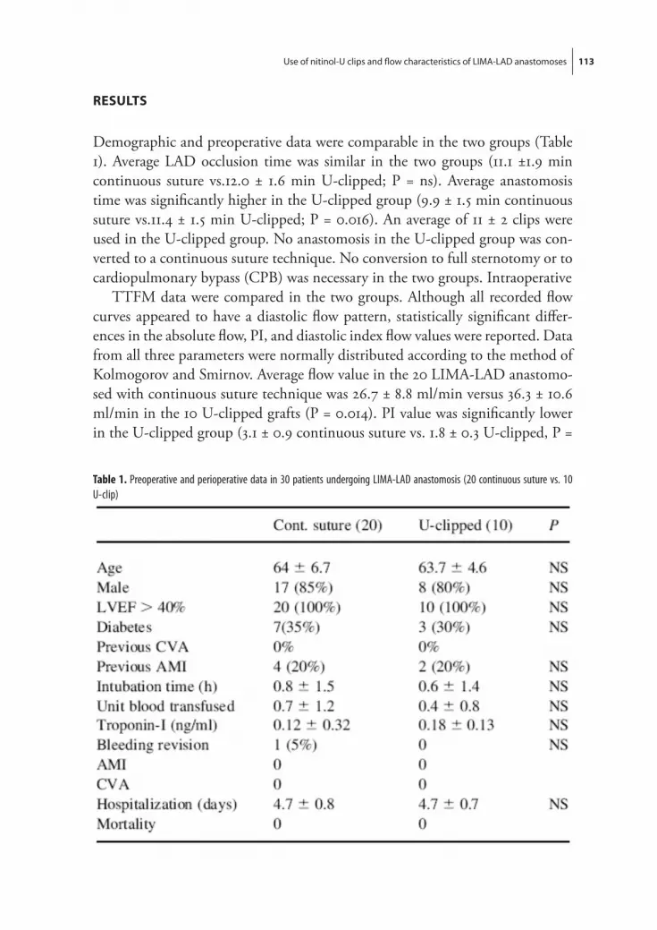
Use of nitinol-U clips and fl ow characteristics of LIMA-LAD anastomoses 113
RESULTS
Demographic and preoperative data were comparable in the two groups (Table 1). Average LAD occlusion time was similar in the two groups (11.1 ±1.9 min continuous suture vs.12.0 ± 1.6 min U-clipped; P = ns). Average anastomosis time was signifi cantly higher in the U-clipped group (9.9 ± 1.5 min continuous suture vs.11.4 ± 1.5 min U-clipped; P = 0.016). An average of 11 ± 2 clips were used in the U-clipped group. No anastomosis in the U-clipped group was con-verted to a continuous suture technique. No conversion to full sternotomy or to cardiopulmonary bypass (CPB) was necessary in the two groups. Intraoperative
TTFM data were compared in the two groups. Although all recorded fl ow curves appeared to have a diastolic fl ow pattern, statistically signifi cant diff er-ences in the absolute fl ow, PI, and diastolic index fl ow values were reported. Data from all three parameters were normally distributed according to the method of Kolmogorov and Smirnov. Average fl ow value in the 20 LIMA-LAD anastomo-sed with continuous suture technique was 26.7 ± 8.8 ml/min versus 36.3 ± 10.6 ml/min in the 10 U-clipped grafts (P = 0.014). PI value was signifi cantly lower in the U-clipped group (3.1 ± 0.9 continuous suture vs. 1.8 ± 0.3 U-clipped, P =
Table 1. Preoperative and perioperative data in 30 patients undergoing LIMA-LAD anastomosis (20 continuous suture vs. 10 U-clip)
Giuseppe Text alt.indd 113Giuseppe Text alt.indd 113 08-Sep-09 14:32:00 PM08-Sep-09 14:32:00 PM

Chap
ter 1
0
114
0.0001). Standard deviations between the two groups for PI were found to be extremely statistically signifi cant (P<0.0006) using the unpaired t-test. Th us the Welch t-test was conducted, yielding a P value of 0.0001. Diastolic fl ow index was signifi cantly higher in the U-clipped group (0.7 ±0.04 continuous suture vs. 0.8 ± 0.03 U-clipped, P=0.046). Perioperative data are reported in Table 1.
Postoperative trans-thoracic Doppler confi rmed patency of the LIMA with diastolic fl ow pattern in all 30 patients. At clinical follow-up (60 ± 4 days) all patients were free from angina and EKG stress tests performed 1 month after surgery did not show signs of myocardial ischemia.
DISCUSSION
More than three decades have passed since the basic techniques adopted for CABG were fi rstly described [9,10,11]. Although many changes and advancements have been introduced through the years to improve outcomes of the operation, considerable issues regarding physiological and technical factors involved in the construction of coronary anastomoses remain unanswered. In this regard only a limited number of comparative studies have objectively addressed diff erences between anastomoses constructed with continuous or interrupted suture tech-nique [2,3,4].
Using cast models, Young et al. [2] reproduced the internal confi guration of saphenous vein to coronary artery anastomoses and examined the eff ects of diff erent technical factors. Th e authors observed that, although the external appearance of the anastomosis was not a reliable indicator of its internal con-fi guration, interrupted suturing consistently produced an internal confi guration of the anastomosis with minimal deformities [2]. Shioi et al. [4] investigated the internal confi gurations of sequential anastomosis constructed with interrupted and continuous suturing technique. Interrupted sutures produced a larger anas-tomosed area for the incised length than other suture techniques. Furthermore, fi ne suturing spacing appeared to be less critical when interrupted sutures were used and an average of six to eight sutures per side appeared to produce well formed anastomoses. In a more recent experimental study, Baumgartner et al. [3] compared the eff ects of continuous vs. interrupted polypropylene (stiff ) and polybutester (compliant) sutures on the luminal dimension and compliance of vascular anastomoses. End-to-end anastomoses constructed with continuous polypropylene, continuous polybutester, and interrupted sutures had respec-tively cross-sectional areas of 70, 90, and 107 of that of the native coronary (P< 0.05). Th e authors concluded that interrupted suture line should be used
Giuseppe Text alt.indd 114Giuseppe Text alt.indd 114 08-Sep-09 14:32:00 PM08-Sep-09 14:32:00 PM

Use of nitinol-U clips and fl ow characteristics of LIMA-LAD anastomoses 115
when constructing end-to-end anastomoses in small vessels. In spite of the theo-retical superiority of the interrupted sutures technique, construction of vascular anastomoses with continuous simple sutures has increasingly gained popularity and has become standard practice in coronary surgery thanks to its advantages in terms of ease and speed of use. Th e recent popularization of minimally invasive coronary surgery and particularly the introduction of endoscopic robotically enhanced CABG [6] has focused interests in rethinking the technique of vascu-lar anastomoses construction. In an attempt at eliminating long suture handling and knot tying, that may became a real challenge especially during endoscopic surgery, and with the aim of speeding anastomosis construction on the beat-ing heart, many alternative ingenious techniques and technologies have been proposed [7,8,12,13,14,15]. Use of non-penetrating titanium clips to construct coronary anastomoses has been proposed by Nataf et al. [12]. Successful applica-tion of the clip requires eversion of the coronary and graft tissues and cannot be easily performed with calcifi ed vessels [12]. A one-shot anastomotic clip device has been proposed by Heijmen et al. Accurate removal of the periadventitial tissue of the LITA should be performed to allow for appropriate release of the clips [13]. Gundry et al. [14] and Buijsrogge et al. [15] have successfully used biologic adhesives to construct coronary anastomoses in animal models. Th e nitinol U-clip device has been successfully used to perform vascular anastomosis in animal [7] and human [8] studies. Hill et al. successfully created right internal mammary to coronary anastomosis on the beating heart and repaired carotid arteriotomies in 13 consecutive calves. Patency was confi rmed at angiography and neointimal resurfacing of the anastomoses was found at histopathologic evaluation [6]. More recently, Ono et al. reported successful U-clip construction of LIMA-LAD anastomoses in 14 patients operated upon with or without CPB [7]. In the present study we focused our attention on the comparison between intraoperative TTFM fi ndings of LIMA-LAD grafts performed with continuous suture versus interrupted U-clips. Although physiological and anatomical dif-ferences between anastomoses constructed with interrupted versus continuous sutures have been partially addressed [2,3,4], functional studies investigating diff erences in coronary graft fl ow characteristics are lacking. TTFM fi ndings in the present study confi rm that U-clipped anastomoses on the beating heart can be safely performed, considerably improving fl ow dynamics of the newly constructed grafts when compared to the traditional single continuous suture technique. Performances of U-clipped anastomoses in terms of fl ow dynamics are satisfactory and diastolic fl ow averages 80 of the total fl ow value (diastolic fl ow index 0.8 ± 0.03). On the other hand, although continuous single sutur-ing technique for LIMA-LAD anastomoses construction has also demonstrated
Giuseppe Text alt.indd 115Giuseppe Text alt.indd 115 08-Sep-09 14:32:00 PM08-Sep-09 14:32:00 PM

Chap
ter 1
0
116
satisfactory clinical results, its fl ow dynamics are signifi cantly diff erent when compared to the U-clip technique. Total fl ow value is signifi cantly less when using the continuous suturing technique (26.7 ± 8.8 ml/min continuous suture vs. 36.3 ± 10.6 ml/min U-clip, P = 0.014) and more importantly the percentage of diastolic fl ow is signifi cantly lower (0.7 ± 0.04 continuous suture vs. 0.8 ± 0.03 U-clip, P= 0.046). As confi rmation of the superior hemodynamic perfor-mance of the U-clip group, mean PI value was signifi cantly lower than in the continuous suture anastomoses (3.1 ±0.9 continuous suture vs. 1.8 ± 0.3 U-clip, P =0.0001).
CONCLUSION
Superior hemodynamic performances of the U-clipped anastomoses have been reported in our study with a particular emphasis on the superior values of total diastolic fl ow and diastolic fl ow ratio. Although these fi ndings are interesting and confi rm at least the theoretical advantages off ered by separated suture over continuous suture anastomoses, there has not been any clear diff erence in clini-cal outcome when using U-clips. Lack of angiographic controls and adequate mid-term follow-up are present limitations that will be specifi cally addressed in the future developments of this study.
ACKNOWLEDGEMENTS
We thank Mrs Judy Bassini for her precious help in collecting and analyzing the data.
Giuseppe Text alt.indd 116Giuseppe Text alt.indd 116 08-Sep-09 14:32:00 PM08-Sep-09 14:32:00 PM

Use of nitinol-U clips and fl ow characteristics of LIMA-LAD anastomoses 117
REFERENCES
1. Carrel A. LA technique operatoire des anastomoses vasculaires et la transplantation des visceres. Lyon Med 1902; 98: 859.
2. Young JN, MacMillan JC, May IA, Iverson LIG, Ecker RR. Internal confi guration of saphenous-coronary anastomoses as studied by the cast-injection technique. J Th orac Cardiovasc Surg 1978; 75: 179–85.
3. Baumgartner N, Dobrin PB, Morasch M, Dong QS, Mrkvicka R. J Th orac Cardiovasc Surg 1996; 111: 1063–72.
4. Shioi K, Washizu T, Kawamura M, Abe T, Iyomasa Y. A study of sequential anasyomoses in aortocoronary bypass surgery-internal confi guration by the casting injection technique. Th orac Cardiovasc Surg 1984; 32: 18–22.
5. Lytle BW. Anastomotic techniques. Operative techniques. Th orac Cardiovasc Surg 2000; 5: 222–30.
6. Falk V, Diegler A, Walther T, Banusch J, Brucerius J, Raumans J, Autschbach R, Mohr FW. Total endoscopic computer enhanced artery bypass grafting. Eur J Cardiothorac Surg. 2000; 17: 38–45.
7. Hill AC, Maroney TP, Virmani R. Facilitated coronary anastomosis using a nitinol u-clip device: bovine model. J Th orac Cardiovasc Surg 2001; 121: 859–70.
8. Ono M, Wolf RK, Angouras D, Schneeberger EW. Early experience of coronary artery bypass grafting with a new self-closing clip device. J Th orac Cardiovasc Surg 2002; 123: 783–7.
9. Johnson WD, Lepley Jr D. An aggressive surgical approach to coronary disease. J Th orac Cardio-vasc Surg 1970; 59: 128–38.
10. Kolessov VI. Mammary artery-coronary artery anastomosis as a method of treatment for angina pectoris. J Th orac Cardiovasc Surg 1967; 54: 535–44.
11. Favaloro RG. Saphenous vein graft in the surgical treatment of coronary artery disease. Operative technique. J Th orac Cardiovasc Surg 1969; 48: 178–85.
12. Nataf P, Kirsch W, Hill AC, Anton T, Zhu YH, Ramadan R, Lima L, Pavie A, Cabrol C, Gand-jbakhch I. Nonpenetrating clips for coronary anastomosis. Ann Th orac Surg 1997; 63: S135–7.
13. Heijmen RH, Hinchliff e P, Borst C, Verlaan CWJ, Moues CM, van der Helm YJ, Manzo S, Jansen EW, Grundeman PF. A novel one-shot anastomotic stapler prototype for coronary bypass grafting on the beating heart: feasibility in the pig. J Th orac Cardiovasc Surg 1999; 117–25.
14. Gundry SR, Black KB, Izutani H. Sutureless coronary artery bypass with biologic glue anasto-mosis: preliminary in vivo and in vitro results. J Th orac Cardiovasc Surg 2000; 120: 473–7.
15. Buijsrogge MP, Verlaan CWJ, van Rijen MHP, Grundeman PF, Borst C. Coronary end-to-side sleeve anastomosis using adhesive in off -pump bypass grafting in the pig. Ann Th orac Surg 2002; 73: 1451–6.
Giuseppe Text alt.indd 117Giuseppe Text alt.indd 117 08-Sep-09 14:32:00 PM08-Sep-09 14:32:00 PM

Giuseppe Text alt.indd 118Giuseppe Text alt.indd 118 08-Sep-09 14:32:00 PM08-Sep-09 14:32:00 PM

Chapter 11
I ntraoperative Graft Patency Verifi cation:
Should You Trust Your Fingertips?
Giuseppe D’Ancona, MD
The Center for Less Invasive Cardiac Surgery and
Robotic Heart Surgery, the State University of New York at
Buff alo, Buff alo, NY, USA
Editorial: Heart Surg Forum 3 (2000)99-102
Giuseppe Text alt.indd 119Giuseppe Text alt.indd 119 08-Sep-09 14:32:00 PM08-Sep-09 14:32:00 PM

Giuseppe Text alt.indd 120Giuseppe Text alt.indd 120 08-Sep-09 14:32:00 PM08-Sep-09 14:32:00 PM

Intraoperative Graft Patency Verifi cation: Should You Trust Your Fingertips? 121
Th e main aim of coronary artery bypass grafting (CABG) is to increase blood fl ow to ischemic myocardium. Although this procedure is successfully performed in more than several hundred thousand patients per year in the United States, intraoperative graft patency verifi cation is still considered optional in most centers. Grafts are assumed to be patent at the end of the operation, especially if the patient has no hemodynamic compromise and, if cardiopulmonary bypass (CPB) was used, that weaning from it is successful.
In the last decade, measurement of coronary graft fl ow has been almost aban-doned due to the many limitations of this obsolete electromagnetic technique (1). Th e increasing popularity of CABG performed on the beating heart without CPB, together with the introduction and improvement of ultrasound-based fl ow meters such as Doppler and Transit Time Flow Measurement (TTFM; MediStim, Oslo, Norway), has revived some interests and concerns about the importance of intraoperative documentation of graft patency (2). Despite the tremendous improvements off ered by the new technology in the fi eld of rheology and fl ow measurement, a large number of cardiac surgeons are still very sceptical and often misinformed about the applicability and limitations of modern fl ow meters.
A recent survey conducted on a limited sample of 100 cardiac surgeons and sponsored by an internationally known medical company (Genzyme Surgical Products, Cambridge, MA), has shown some interesting fi ndings about the cur-rent graft patency verifi cation practice in the United States. Th e majority (68.1) of those interviewed stated that manual palpation of the grafts is their current method to detect graft patency after CABG. More than 70 of the surgeons included had never used a fl ow meter in their practice. When questioned about the reasons for not using a graft patency verifi cation system, most of the surgeons interviewed answered that fl ow meters are often diffi cult to use, interpretation of the fi ndings are unclear, and revision rate is low enough that graft patency verifi cation seems unnecessary. Although most of the surgeons declared that a fl ow meter is not routinely necessary, interestingly 86 of them stated that it is important to have a system available in the operating room if, in some selected cases, it is necessary. More than 20 of those interviewed recognized the com-pelling importance of intraoperative graft patency verifi cation. More than 60 of the surgeons recognized the applicability of this technology in both off -pump and on-pump procedures. Th irty-seven percent of those interviewed stated that this technology is only applicable during off -pump procedures because, on pump, the chance of anastomotic mistakes is very low.
Giuseppe Text alt.indd 121Giuseppe Text alt.indd 121 08-Sep-09 14:32:00 PM08-Sep-09 14:32:00 PM

Chap
ter 1
1
122
Th e surgeons were then questioned about the action that they would have taken in case of abnormal fi ndings during intraoperative coronary graft-fl ow measure-ments. More than 50 of those interviewed stated they would revise the grafts in question. Th e remaining would either wait or use another method of graft patency verifi cation. When asked about their normal graft revision rate during CABG, 88 of the surgeons declared an error rate of 4 or less with an average rate of 3.1. Th e majority of the surgeons stated that poor native vessels were the most common cause of graft revision.
Finally, surgeons placed a dollar value on the ability to accurately measure graft patency intraoperative. In U.S. dollars, the responses ranged from $0 to $1,000. Although this questionnaire was limited to a small number of surgeons, the fi ndings are very interesting and probably applicable to all cardiac surgeons in the United States.
We began our experience with intraoperative graft patency verifi cation in 1996 to document the feasibility of off -pump CABG. To date, more than 800 patients who have had off -pump surgeries have been tested using intraoperative TTFM and we feel very confi dent with the features off ered by TTFM technology. We understand that graft testing with syringes, fi ngertips, and direct probing of the anastomoses seem to be more immediate than interpreting a fl ow curve, but this is not always the case. We believe the information given by fl ow meters may be misleading, especially when the operator does not fully understand the advantages and limitations of the present technology. Often the manufacturers are more interested in selling their product than clarifying its real applicability. Surgeons prefer, for this reason, to rely on their “accurate” tactile sense.
When choosing a fl ow meter, special features should be evaluated. Th e mea-surement must be stable, reproducible, and representative of the real fl ow within the constructed graft. Flow probes should be user friendly and easy to calibrate. Th e recorded data should be stored in the fl ow meter for future analysis and included in the patients’ charts for documentation. Much of the scepticism about intraoperative graft patency verifi cation is also based on failure of the elec-tromagnetic technology. Electromagnetic devices measure the intensity of the electromagnetic fi eld generated by the electrically charged red cells (iron bound to hemoglobin) that fl ow within a vessel. Actual blood fl ow value is derived from, and is directly proportional to, the intensity of the electromagnetic fi eld generated. Th is technology has been abandoned and has been recently replaced by ultrasound devices.
Th e term ultrasound has a generic defi nition that includes two diff erent methods: Doppler and TTFM. Th e two systems rely on diff erent properties of the ultrasound waves and, although the Doppler methods have shown good
Giuseppe Text alt.indd 122Giuseppe Text alt.indd 122 08-Sep-09 14:32:00 PM08-Sep-09 14:32:00 PM

Intraoperative Graft Patency Verifi cation: Should You Trust Your Fingertips? 123
reliability both in vivo and in vitro (3), the TTFM technology off ers many important advantages and is the most accurate system for intraoperative veri-fi cation of coronary graft patency (4-6). TTFM measurements are theoretically independent of internal or external vessel diameter, vessel shape, and Doppler angle. TTFM is also insensitive to the alignment between probe and vessel. Th e probe does not have to be in direct contact with the vessel and calibration is not necessary. Th e recordings are stable and data storage and analysis are routinely done. Many of these features are not off ered by Doppler technology.
Th e TTFM device is very easy to use and requires no more than 30 seconds per measurement. Flow-probe size varies from 2 to 32 mm and the size of the fl ow probes used most frequently in cardiac surgery ranges between 2 and 3.5 mm. Th e fl ow probe consists of two small piezoelectric crystals, one upstream and one downstream, mounted on the same side of the vessel. Opposite to the crystals, there is a small metallic refl ector. Each crystal produces a wide pulsed ultrasound beam covering the entire vessel width. Th e probe is connected to a computer that has more than 200 MB of memory and is programmed with software in Microsoft WINDOWS format. Both the amount of time neces-sary for an ultrasound beam emitted from the upstream crystal to arrive at the downstream crystal after being refl ected, and for a signal from the down-stream crystal to reach the upstream crystal are measured. Since ultrasound travels faster if transmitted in the same direction as fl ow, a small time diff erence between the two beams is calculated as the transit time of fl ow and thus, the actual fl ow is proportional to the transit time. All calculations are made automatically by the fl ow meter and are displayed, as ml/min. Measurements are not dependent on the angle between vessel and probe. Th e two crystals are mounted in a fi xed position. An increase in the angle between the upstream probe and the vessel will always be compensated by a corresponding decrease of the angle between the downstream probe and the vessel and vice versa. As mentioned above, measure-ments are also independent of hematocrit level, heart rate, and thickness of the vessel wall. Flow curves, together with fl ow and Pulsatile Index (PI) values, are visualized in real time on a video screen, can be saved in the hard drive, and can be printed through a parallel port.
Correct interpretation of TTFM fi ndings may be diffi cult if this technology is not routinely applied and if established protocols and rules for fl ow mea-surement are not followed. Th e fl ow meter is not a magical device that can be forgotten in the operating room only to provide an exact answer whenever we have doubts about the quality of the anastomosis. Th e operator should interpret fl ow curves and, although most of the TTFM fi ndings have an immediate interpretation, there is a learning curve for the more diffi cult cases. Confi dence
Giuseppe Text alt.indd 123Giuseppe Text alt.indd 123 08-Sep-09 14:32:01 PM08-Sep-09 14:32:01 PM

Chap
ter 1
1
124
with the fl owmeter increases with the number of cases in which this technology is applied. For TTFM to be correctly interpreted, fl ow curves, PI, and mean fl ow values should all be evaluated simultaneously. In a patent coronary graft, the hemodynamics are similar to those physiologically observed in the coronary circulation: blood fl ow should be mainly diastolic with minimal systolic peaks taking place during the isovolumetric ventricular con-traction (QRS complex). To correctly interpret fl ow patterns, curves should always be coupled with the ECG tracing to diff erentiate the systolic from the diastolic component. Th e PI is a good indicator of the fl ow pattern and, consequently, of the quality of the anastomosis. Th is number is obtained by dividing the diff erence between the maximum and the minimum fl ow by the value of the mean fl ow. In our experi-ence, the PI value should be between 1 and 5; the possibility of a technical error in the anastomosis increases for higher PI values (6, 7). Mean fl ow is expressed as mL/min and its value is not necessarily a good indicator of the quality of the anastomosis. Mean fl ow is very dependent on the quality of the native coronary artery and low fl ow values can be expected in fully patent anastomoses (6) when-ever the target territory has poor run-off .
We believe that intraoperative graft patency verifi cation should be routinely adopted in all cases and not exclusively in patients operated on without CPB. Today, the modern techniques of exposure and stabilization of the diff erent coronary artery branches can, in the majority of the off -CPB cases, provide very stable conditions and excellent surgical exposure comparable to the cases using CPB. In spite of that, surgical mistakes are still possible and, most of the time, diffi cult to admit. Our revision rate off -CPB has recently decreased from 8 to 4 and, in most of the cases, a technical error was found at revision (6). Flow abnormalities related to poor quality of the revascularized territory may be easily detected if a standard technique of measurement is correctly adopted. Th e TTFM probe should be perfectly fi tted around the graft. Skeletonization of a small segment of the mammary artery is necessary to reduce the quantity of tis-sue interposed between the vessel and the probe. Aqueous gel is used to improve probe contact. TTFM has to be evaluated both with and without proximal snar-ing of the native coronary artery to detect any possible imperfection localized at the toe of the anastomosis and to exclude fl ow competition from the native vessel. Before making any measurements, adequate de-airing of the grafts is per-formed using a 25-gauge needle. Adequate systemic blood pressure is maintained and traction on the pericardium is released to allow the heart to return to its anatomical position. TTFM should be repeated before chest closure and after protamine administration to confi rm graft patency and to detect any possible graft kinking or compression. We believe that prompt graft revision may very
Giuseppe Text alt.indd 124Giuseppe Text alt.indd 124 08-Sep-09 14:32:01 PM08-Sep-09 14:32:01 PM

Intraoperative Graft Patency Verifi cation: Should You Trust Your Fingertips? 125
well be necessary whenever abnormalities in fl ow curves and values are found. Although it is very hard to admit technical mistakes, most of the time errors are not visually detectable, at least while performing the anastomosis. It is also true that this technology has been proven eff ective in detecting highly stenotic coronary anastomoses, and that data concerning the specifi city and sensitivity of TTFM have never been published. A neural network pattern recognition analy-sis of graft-fl ow characteristics has been proposed by Cerrito et al. (8) to improve TTFM detection of anastomotic errors. After a complex mathematical analysis of the fl ow curves, it is possible to detect stenoses that causes 50 or greater narrowing of the anastomoses. Less than critical stenoses cannot be detected by TTFM because no modifi cations in the hemodynamic performances of the grafts occur at this level.
Another limit of TTFM that will be possibly solved with increasing clinical experience is the lack of standard or nominal curves and fl ow values for diff erent types of grafts and revascularized vessels. Standardization of the TTFM fi ndings is diffi cult due to large biological variability between diff erent subjects, as well as within the same subject. Interpretation of fl ow curves and TTFM fi ndings is still empirical and is dependent on the surgeon’s personal experience. Jaber et al. (9) have tested the ability of 19 international surgeons to detect anastomotic errors by evaluating mean fl ow and fl ow wave-form morphology. We believe that the abil-ity to correctly interpret TTFM fi ndings develops with clinical and experimental experience and, for this reason, surgeons who have not been exposed to this type of technology cannot easily give it the proper level of importance. Flow patterns, PI values, fl ow values, and clinical fi ndings (i.e., ECG tracing, hemodynamic values) should always be evaluated simultaneously to improve the applicability of TTFM. Absolute fl ow value does not necessarily refl ect anastomosis quality because there are too many variables infl uencing absolute fl ow, including size of the graft and quality of the revascularized coronary artery. Instead, coronary fl ow reserve can better help to correctly diagnose anastomotic imperfections. Wal-poth et al. (10) have documented that quality of the anastomosis can be better defi ned by testing its dynamic ability to increase graft fl ow whenever myocardial oxygen requests are increased during infusion of adenosine. PI values are good indicators of the quality of the anastomoses. In our experience, high PI values are suggestive of anastomotic imperfections and the high PI values alone could justify coronary graft revision (6,7). Even though an absolute PI value has not been defi ned, we have empirically selected the limit of 5 based on our clinical experience with TTFM.
Giuseppe Text alt.indd 125Giuseppe Text alt.indd 125 08-Sep-09 14:32:01 PM08-Sep-09 14:32:01 PM
Chap
ter 1
1
126
Di Giammarco et al. (11) proposed a value, derived from their clinical experience, of 2.5 as the limit of the PI value above which an anastomosis should be revised (11). Finally, the price for this technology cannot be defi ned with economical parameters. Even if interpretation of graft fl ows is still based on personal experi-ence and empirical values, these are not good excuses for avoiding intraoperative graft patency verifi cation. Th e fl ow meter should be used routinely as a surgical armamentarium to improve patient care and surgical results independently by the surgical technique adopted. Ability to interpret fl ow data will improve with use of the fl ow meter. Surgeons should not feel questioned or intimidated by the possible negative judgments of the fl ow meter. We understand the fi nancial limitations imposed by hospital administrators but we believe that, in the future, surgeons that do not routinely adopt methods of intraoperative graft patency verifi cation may be legally prosecuted in cases of perioperative complications. Th e use of fi ngertips to detect coronary graft patency after CABG will be seen, in years to come, in the same way as using the bare ear to detect paravalvular leaks after valve replacement.
ACKNOWLEDGMENTS
Th e authors thank Douglas Hutchinson of Genzyme for his cooperation in providing the market research data mentioned in this article.
Giuseppe Text alt.indd 126Giuseppe Text alt.indd 126 08-Sep-09 14:32:01 PM08-Sep-09 14:32:01 PM

Intraoperative Graft Patency Verifi cation: Should You Trust Your Fingertips? 127
REFERENCES
1. Kolin A, Ross G, Gaal P, Austin S. Simultaneous electromagnetic measurement of blood fl ow in the major coronary arteries. Nature. 203: 148–53, 1964.1.
2. Canver CC, Cooler SD, Murray EL, et al. Clinical importance of measuring coronary graft fl ows in the revascularized heart. Ultrasonic or electromagnetic? J Cardiovasc Surg 38: 211–5, 1997.
3. Segadal LK, Matre H, Engedal H, et al. Estimation of fl ow in aortocoronary grafts with a pulsed ultrasound Doppler meter. Th orac Cardiovasc Surgeon 30: 265–8, 1982.2.
4. Lundell A, Bergqvist D, Mattsson E, Nilsson B. Volume blood fl ow measurements with a transit time fl ow meter: an in vivo and in vitro variability and validation study. Clin Physi¬ol 13: 547–57, 1993.
5. Matre K, Birkeland S, Hessevik I, et al. Comparison of transit time and Doppler ultrasound methods for measurements of fl ow in aortocoronary bypass grafts during cardiac surgery. Th orac Cardiovasc Surg 42: 170–4, 1994.
6. Laustsen J, Pedersen EM, Terp K, et al. Validation of a new transit time ultrasound fl ow meter in man. Eur J Vasc Endovasc Surg 12: 91–6, 1996..
7. D’Ancona G, Karamanoukian H, Ricci M, Schmid S, Bergsland J, Salerno T. Graft revision after Transit Time Flow Measure¬ments in off -pump coronary artery bypass grafting. Eur J Cardiothorac Surg 17: 287–93, 2000.3.
8. Cerrito PB, Koenig SC, Koenig SC, Van Himbergen DJ, Jaber SF, Ewert DL, Spence PA. Neural network pattern recognition analysis of graft-fl ow characteristics improves intra-operative anas-tomotic error detection in minimally invasive CABG. Eur J Cardiothorac Surg 16: 88–93, 1999
9. Jaber SF, Koenig SC, BhaskerRao B, Van Himbergen DJ, Spence PA. Can visual assessment of fl ow waveform morphology detedt anastomotic error in off pump coronary artery bypass grafting? Eur J Cardiothorac Surg 14: 476–9, 1998.
10. Walpoth BH, Bosshard A, Kipfer B, et al. Failed coronary artery bypass anastomosis detected by intraoperative coronary fl ow measurement. Eur J Cardiothorac Surg 10: 1064–70, 1996.
11. Di Giammarco G. Myocardial revascularization without cardiopulmonary bypass. Presented at the symposium: State of the art in emerging coronary revascularization; EACTS. Glasgow, Scotland September 4, 1999.
Giuseppe Text alt.indd 127Giuseppe Text alt.indd 127 08-Sep-09 14:32:01 PM08-Sep-09 14:32:01 PM

Giuseppe Text alt.indd 128Giuseppe Text alt.indd 128 08-Sep-09 14:32:01 PM08-Sep-09 14:32:01 PM

Chapter 12
I ntraoperative graft patency verifi cation in
coronary surgery: Modern diagnostic tools
Giuseppe D’Ancona, MD ¹ ²Fabio Bartolozzi MD ¹Ad J.J.C. Bogers, MD PhD ¹Michele Pilato MD ²Matteo Parrinello MD ³Arie P. Kappetein, MD PhD ¹
¹ Thoraxcenter, Erasmus MC, Rotterdam, The Netherlands
² Department of Cardiothoracic Surgery, ISMETT @ University of Pittsburgh Medical Center,
Palermo, Italy
³ Department of Anesthesia, ISMETT @ University of Pittsburgh Medical Center, Palermo, Italy
J Cardiothorac Vasc Anesth. 2009 Apr;23(2):232-8
Giuseppe Text alt.indd 129Giuseppe Text alt.indd 129 08-Sep-09 14:32:01 PM08-Sep-09 14:32:01 PM
Chap
ter 1
2
130
ABSTRACT
Nowadays medical procedures require a quality control method, and this is espe-cially true for complex procedures such as heart operations. Various intraopera-tive tools are currently available to test coronary graft patency during coronary bypass grafting.
A distinction should be made between imaging techniques and fl ow assess-ment devices. Both methods have clear limitations, and we should consider using the two diff erent technologies simultaneously. Th is paper reviews the means currently available to intraoperative test and document the status of newly constructed coronary anastomoses.
Giuseppe Text alt.indd 130Giuseppe Text alt.indd 130 08-Sep-09 14:32:01 PM08-Sep-09 14:32:01 PM

Intraoperative graft patency verifi cation in coronary surgery: Modern diagnostic tools 131
INTRODUCTION
Th e standard of care in modern medicine is supported by many quality control tools and intraoperative graft patency testing after coronary surgery can be proposed, in this context, as part of the operation documenting the success of the procedure itself.
Th e aim of coronary artery bypass grafting (CABG) is to increase blood fl ow to ischemic myocardium. Th is statement acquires further impact if we realize that 5-20 of all grafts performed fail before discharge from the hospital and up to 30 before 1 year (1, 2) without any objective evidence that these grafts were actually patent in the operating room.
Many of these early failures are probably secondary to technical issues that could be promptly solved if adequately diagnosed intraoperatively.
Although there are no data available in literature documenting the actual uti-lization rate of intraoperative coronary graft patency verifi cation tools, a survey conducted on a limited sample of cardiac surgeons has shown some interesting fi gures (3). Th e majority (68.1) of those interviewed stated that manual palpa-tion of the grafts is their current method to detect graft patency after CABG; 70 of the surgeons included had never used an intraoperative graft patency verifi cation tool in their practice. Most of the surgeons interviewed thought that graft fl ow meters are often diffi cult to use, interpretation of the fi ndings is unclear, and revision rates are low enough that graft patency verifi cation seems unnecessary. Although the majority declared that a fl owmeter is not routinely necessary, 86 of them interestingly stated that it is important to have a system available in the operating room (3).
Being complementary to percutaneous coronary intervention, cardiac surgery has lost its supremacy in the treatment of coronary artery disease, and cardiac surgeons should concentrate on how to optimize the quality of care they are providing.
Th is paper reviews the methods that are currently available to intraoperative test and document the status of newly constructed coronary anastomoses.
A clear distinction is made between imaging techniques and fl ow assessment devices.
Giuseppe Text alt.indd 131Giuseppe Text alt.indd 131 08-Sep-09 14:32:01 PM08-Sep-09 14:32:01 PM
Chap
ter 1
2
132
IMAGING TECHNIQUES
Standard Angiography
Although coronary angiography is a purely anatomical evaluation of native ves-sels, constructed bypass grafts, and anastomoses, for historical, economic, and logistical reasons it is considered “the gold standard” and the benchmark which every new method of investigation of graft patency should be compared with.
Angiographic images however, have little value in testing the hemodynamic function of the coronary grafts. No information is obtained concerning the fl ow reserve and fl ow response to stress. Angiography is per se only a bi-dimensional representation without any direct functional information.
Intraoperative coronary angiography has logistic limitations that have pre-vented most surgeons from its routine adoption to test coronary graft patency.
Integration of high-end digital angiographic imaging equipment in the operating room may off er several opportunities (4). In addition to allowing a hybrid approach to coronary disease, on-table assessment of graft patency can be performed with cardiac catheter lab quality images. Intraoperative angiograms should be performed after sternal closure as chest closure may cause graft distor-tion.
Using fi xed angiographic equipment in a hybrid operating theater, Hol and colleagues have reported an intraoperative graft failure rate of 4.2 (5). A team from the catheter lab performed the on-table angiography ensuring acquisition of high image quality, maintenance of low complication rate, and limitation of procedural time (30 extra minutes) (5).
Although many technical improvements have been introduced such as pulsed fl uoroscopic systems with dynamic acquisition, the extra budgetary and person-nel demands have limited broad application of conventional angiography in the operating room. With the advent of combined open surgical and catheter based procedures the availability of angiographic systems in the operating room will possibly increase in the very next future. Th is will eventually off er a broader spectrum of treatments in case of early detection of graft malfunction. Possibly, with the device available in the operating room, angiography should be antici-pated before sternal closure and performed immediately after weaning off car-diopulmonary bypass. Prompt graft revision (either on or off -cardiopulmonary bypass) should be performed whenever necessary and possible. Alternatively, percutaneous intervention could be suggested for particularly complicated cases where further prolongation of the operative time could lead to deleterious complications.
Giuseppe Text alt.indd 132Giuseppe Text alt.indd 132 08-Sep-09 14:32:01 PM08-Sep-09 14:32:01 PM
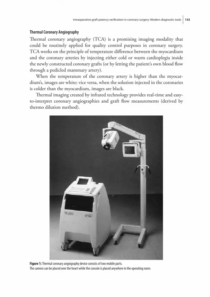
Intraoperative graft patency verifi cation in coronary surgery: Modern diagnostic tools 133
Thermal Coronary Angiography
Th ermal coronary angiography (TCA) is a promising imaging modality that could be routinely applied for quality control purposes in coronary surgery. TCA works on the principle of temperature diff erence between the myocardium and the coronary arteries by injecting either cold or warm cardioplegia inside the newly constructed coronary grafts (or by letting the patient’s own blood fl ow through a pedicled mammary artery).
When the temperature of the coronary artery is higher than the myocar-dium’s, images are white; vice versa, when the solution injected in the coronaries is colder than the myocardium, images are black.
Th ermal imaging created by infrared technology provides real-time and easy-to-interpret coronary angiographies and graft fl ow measurements (derived by thermo dilution method).
Figure 1: Thermal coronary angiography device consists of two mobile parts.The camera can be placed over the heart while the console is placed anywhere in the operating room.
Giuseppe Text alt.indd 133Giuseppe Text alt.indd 133 08-Sep-09 14:32:01 PM08-Sep-09 14:32:01 PM

Chap
ter 1
2
134
Th e TCA device consists of two mobile parts, namely a camera and a console. Th e console contains a video recorder, a video printer, a monitor, a wired remote control and an image-processing unit. Th e camera unit contains an infrared imager, a video camera and a monitor on a post for the surgeon’s view (Fig. 1). Th e camera unit is easily placed over the heart from the anesthesia side and it can be easily pointed at the area of interest by the surgeon using sterile handles. Th e console can be placed anywhere in the operating room. Th e coronary arteries, either on the front or the back of the heart, can be visualized
Reports on TCA date back to the late 1980’s (6). More recently, Falk and co-workers applied TCA to visualize a total of 693 vein grafts. In 9.4 of the measurements, TCA failed to produce usable images. In the remaining 628 grafts, TCA revealed intraoperative patency in 98.8. (7).
In a more recent and larger experience, Sonmez et al. investigated 4105 bypass grafts in 1401 patients. TCA guided the decision to perform some changes in the surgical strategy in 23 patients (adding another graft, or performing coronary endarterectomy) (8).
Furthermore, Iwahashi and colleagues compared the fi ndings of immediate postoperative standard angiography with a new generation intraoperative TCA
Figure 2: Saphenous vein grafting (SVG) to the left anterior descending (LAD) coronary artery as visualized by intraoperative new generation thermal coronary angiography.This picture was published in Iwahashi H, Tashiro T, Morishige N, et al. New method of thermal coronary angiography for intraop-erative patency control in off -pump and on-pump coronary artery bypass grafting.Ann Thorac Surg 84(5): 1504-7, 2007
Giuseppe Text alt.indd 134Giuseppe Text alt.indd 134 08-Sep-09 14:32:02 PM08-Sep-09 14:32:02 PM

Intraoperative graft patency verifi cation in coronary surgery: Modern diagnostic tools 135
(9). Th e graft patency rate as assessed with TCA was 96.3. By contrast, the postoperative patency rate for standard contrasted angiography was 95.3. Tem-perature diff erences of only 0.1°C between the injectant and the epi-myocardium were necessary to obtain high contrast imaging (fi g. 2).
As technology improves, TCA may allow for measuring blood fl ow and obtain-ing imaging information simultaneously.
Intraoperative Fluorescence Imaging
Intraoperative fl uorescence imaging is a vascular imaging technique based on the fl uorescent properties of indocyanine green (ICG) dye. After intravenous injection, ICG binds to plasma proteins and, when illuminated with a laser light (near infrared), it emits light with a wavelength of 830 nm. Th is fl uorescence is captured on a charged couple device video camera. Practically, the camera is positioned 30 cm above the heart. At completion of the distal anastomosis, 1 cc of dye can be injected either in the ascending aorta or in the central venous pressure catheter (if the operation is performed off -cardiopulmonary bypass) or directly into the oxygenator in on-pump surgery.
Immediately after intravenous dye injection (or after a 5-second delay if the dye is injected into the oxygenator), the laser power is activated. Images are recorded on the computer and the appearance of fl uorescent ICG dye passing inside bypass grafts and perfusing the coronary tree confi rms graft patency (fi g. 3). Th e procedure takes approximately 3 minutes per graft.
First experience with intraoperative fl uorescence imaging in coronary surgery was reported by Rubens et al. in 2002 (10). To date, at least 5 major centers have
Figure 3: A, The image shows in situ left internal thoracic artery (LITA) graft to the left anterior descending (LAD) artery. A composite radial artery (RA) graft from the LITA is anastomosed to the obtuse marginal (OM) artery. As fl uorescence is not seen within the RA, a technical problem is suspected and the anastomosis is revised. B, This image was taken after revision of the LITA-to-RA anastomosis. Fluorescence is now seen in the RA and OM coronary artery.This picture was published in Balacumaraswami L, Abu-Omar Y, Anastasiadis K, Choudhary B, Pigott D, Yeong SK, Taggart DP.Does off -pump total arterial grafting increase the incidence of intraoperative graft failure?J Thorac Cardiovasc Surg. 2004 Aug; 128(2): 238-44.
Giuseppe Text alt.indd 135Giuseppe Text alt.indd 135 08-Sep-09 14:32:02 PM08-Sep-09 14:32:02 PM
Chap
ter 1
2
136
reported their experience with this technology and over 1,400 coronary grafts have been investigated (11).
Results are very much consistent showing an overall graft revision rate of 1.7 in approximately 5 of patients.
Furthermore, Desai et al. (12) have very recently evaluated, in a prospective randomized study, sensitivity and specifi city of intraoperative coronary fl uores-cence imaging to detect grafts that at postoperative standard angiography had resulted to be abnormal (>50 stenosis) or occluded. Th e sensitivity and speci-fi city of indocyanine green angiography to detect stenosis or occlusion greater than 50 was 83.3 and 100, respectively (12).
Although this technique has promising features, some limitations have to be outlined.
As any purely anatomical study, fl uorescence imaging does not produce pre-cise measurements of fl ow in patent grafts but gives a rather semi-quantitative and monoplane assessment of graft patency.
Furthermore, in conditions where the native fl ow creates competition, fl uorescence imaging may lead to erroneous interpretation of the graft status. In this situation, snaring of the native coronary proximally to the anastomosis may clarify dubious fi ndings.
Another limitation is that as a result of limited tissue penetration and image acquisition from directly above the anastomoses, fl uorescence imaging cannot provide precise details on anastomotic quality and mainly detects fl uorescing contrast distribution within the grafts and the coronary tree.
Moreover, the laser light source may detect only a portion of the graft and, whenever an abnormality (a compression, twist or bend) occurs in the graft in a point far away from the laser light source, the fi nding may be erroneously interpreted as a graft stenosis on a still image capture.
Finally it may be argued that, to standardize the fi ndings of this technique and to optimize its diagnostic potential, the indocyanine dye should be injected only once the heart is completely reperfused, the eff ects of cardioplegia are com-pletely resolved, and the cardiopulmonary bypass has been weaned off . Although at present there is no scientifi c evidence, it seems reasonable to state that grafts fl ow dynamics should be tested in physiologic conditions such as those achieved in a full beating heart and with pulsatile arterial pressure.
Giuseppe Text alt.indd 136Giuseppe Text alt.indd 136 08-Sep-09 14:32:03 PM08-Sep-09 14:32:03 PM

Intraoperative graft patency verifi cation in coronary surgery: Modern diagnostic tools 137
FLOWMETRY
Diff erently from imaging tools, fl ow measuring devices give us the possibilities to intraoperative detect the fl ow dynamics of newly constructed coronary grafts.
Th e functional status of a vascular anastomosis is an indicator of the diff erent anatomical parameters of graft, anastomosis, and revascularized territory.
Although fl ow meters can provide us with a good representation of how a coronary graft performs and responds to the blood requirements of the revascu-larized myocardial territory, correct interpretation of fl ow measurement fi ndings requires a good knowledge of the diff erent variables involved in coronary rheol-ogy.
At present, ultrasound based fl ow meters are those most commonly used as intraoperative diagnostic tools during coronary surgery. Among the ultrasound fl ow meters, we should make a distinction between transit time and Doppler based fl ow meters.
Transit Time Flow Measurement (TTFM)
TTFM probes work on the transit-time principle. Th e probe is placed around the vessel, generating a uniform ultrasound fi eld across the vessel lumen. Ultrasound pulses are transmitted from two separate piezoelectric crystals located on the same side of the probe (and so of the vessel). On the opposite side of the probe, and at the same distance from the 2 crystals, there is a refl ector. Ultrasound beams cross the vessel and are refl ected back to the crystals. Th e time spent by the ultrasound wave to go from one crystal across the vessel and then to be refl ected (by the refl ector on the opposite side of the probe) and reach the other crystal, is called transit time. Ultrasound waves that travel upstream the fl ow will travel slower than those going downstream. Th e diff erence in transit time between the pulse going upstream and the one going downstream is proportional to the volume of fl ow passing through the probe (Q ≈ t2 - t1) (Fig.4).
A range of diff erent probes are available for vessel diameters from 1.5 mm up to 35 mm. Probes are connected to a computerized systems that allows for fl ow curve and fl ow parameters recording, printing, and retrieval (Fig.5).
TTFM has been recently popularized with the resurgence of beating heart coronary surgery (13). Many authors have tried to interpret intraoperative TTFM fi ndings and their sensitivity and specifi city.
In summary, TTFM fi ndings include: mean fl ow value, fl ow curve pattern, and pulsatility index. Mean fl ow depends on many factors, including mean arterial pressure and graft-coronary resistance. Th e value of vascular resistance is summarized in the formula R = 8ηL/πr4 where η refers to blood viscosity, L to
Giuseppe Text alt.indd 137Giuseppe Text alt.indd 137 08-Sep-09 14:32:03 PM08-Sep-09 14:32:03 PM

Chap
ter 1
2
138
Figure 4: Transit Time Principle: Ultrasound pulses are transmitted from two separate piezoelectric crystals located on the same side of the probe. Ultrasound beams cross the vessel and are refl ected back to the crystals. The time spent by the ultrasound wave to go from one crystal across the vessel and then reach the other crystal, is called transit time. The diff erence in transit time between the pulse going upstream and the one going downstream is proportional to the volume of fl ow passing through the probe (Q≈ t2 - t1).
Figure 5: New generation transit time fl ow measurement device
Giuseppe Text alt.indd 138Giuseppe Text alt.indd 138 08-Sep-09 14:32:03 PM08-Sep-09 14:32:03 PM

Intraoperative graft patency verifi cation in coronary surgery: Modern diagnostic tools 139
graft length, and r to the radius of the graft. From this mathematical approxima-tion, it is quite clear that the quantity of fl ow may change even if the quality of the anastomosis remains excellent as often happens when the coronary has a very poor run-off . Th erefore, mean fl ow should not be considered, per se, as a good indicator of graft patency and its value should always be interpreted taking the other parameters into consideration (13).
Th e fl ow curve (Fig.6) is the graphic representation of the hemodynamic function of the graft. Within the fl ow curve itself, we can distinguish percentage of systolic fl ow, percentage of diastolic fl ow, and total areas under the curve for systolic and diastolic fl ow. Th e curves should always be coupled with the ECG tracing to correctly diff erentiate the systolic from the diastolic fl ow. Diastolic fi lling percentage (DF ) indicates the percentage of coronary fi lling in diastole. By using ECG synchronization, the DF is the blood volume fi lling in diastole divided by the total blood volume in one heart cycle. DF is especially important because it indicates the diastolic forward fl ow that, during diastole, goes within the coronary while the muscle is not contracting. Some authors have classifi ed the fl ow curve pattern according to dominancy: systolic dominant when peak systolic fl ow exceeds peak diastolic fl ow by 10; diastolic dominant or balanced when peak diastolic fl ow exceeds peak systolic fl ow by 10 (14). Diff erent pat-terns may be related to the target vessel: grafts on the left coronary system will have a higher diastolic fl ow when compared to grafts on the right coronary (where the right ventricular muscle’s squeezing eff ect is weaker).
Figure 6: Transit-time Flow Curve
Giuseppe Text alt.indd 139Giuseppe Text alt.indd 139 08-Sep-09 14:32:05 PM08-Sep-09 14:32:05 PM

Chap
ter 1
2
140
Another parameter that can be detected from the fl ow curve is the percentage of backward fl ow (BF), i.e., the percentage of fl ow that, through the anasto-mosis, is directed backward within the graft (area below zero) compared with the total forward fl ow (area above zero) of the same cardiac cycle. As we will discuss in the next paragraphs, the lower the rate of backward fl ow, the better the quality of the grafting.
Th e pulsatility index (PI) is summarized in the formula Qmax – Qmin/Qmean where mean fl ow is calculated across fi ve cardiac cycles (Qmean), maximum fl ow is recorded in one cardiac cycle (Qmax), and minimum fl ow is recorded through one cardiac cycle (Qmin). Th e Qmin is a negative number if there is backward fl ow (14). Th e PI is proportional to the vascular resistance and therefore a high PI is an indicator of poor graft or anastomosis quality.
Many authors have tried to defi ne threshold values for the diff erent TTFM fi ndings and to distinguish patent from occluded grafts.
A mean fl ow of 15 mL/min or less, a PI of 5 or higher, and a BF of 4 or higher seem to be the optimal cut-off criteria to predict early graft failure (14-16).
For what concerns fl ow curves, a systolic dominant fl ow curve pattern is a risk factor only in grafts to the left coronary artery (16).
When comparing TTFM results with intraoperative fl uorescence imaging, Balacumaraswami reported that TTFM alone may prompt unnecessary graft revision (17).
In a similar comparison between intraoperative fl uorescence imaging and TTFM where the reference standard was coronary angiography, Desai et al. found that the sensitivity and specifi city of transit-time ultrasonic fl ow mea-surement to detect greater than 50 stenosis or occlusion was 25 and 98.4 respectively (12).
In our opinion, correct interpretation of TTFM fi ndings should take into account other variables such as confi dence in the anastomosis performed, qual-ity of target vessel and conduit, EKG, and hemodynamic fi ndings. In addition, multiple measurements should be taken when doubts occur including snaring of the native coronary proximally to the anastomosis, and simulation of stress testing by increasing blood pressure with inotropic agents.
Although TTFM technology can clearly show diff erences between hemody-namically normal and abnormal grafts, correct interpretation is limited by our inability to analyze the fl ow curves in their diff erent components.
Th is feature should be provided within an automated analysis of fl ow curves.
Giuseppe Text alt.indd 140Giuseppe Text alt.indd 140 08-Sep-09 14:32:05 PM08-Sep-09 14:32:05 PM

Intraoperative graft patency verifi cation in coronary surgery: Modern diagnostic tools 141
Further information may be derived from a more complex analysis of the fl ow curves with fast Fourier transformation (FFT). Th e FFT is a mathematical transformation of the fl ow waveform (18) that allows for a numerical representa-tion of fl ow curves. Th e analysis is based upon the principal that all periodic waveforms (such as fl ow curves) can be broken down into a series of pure sine waves or harmonics.
Th e pulsatile waveform of graft fl ow in CABG can be considered to have a fundamental frequency (i.e., the patient’s heart rate) and its power is defi ned as F0. Th e power of the following harmonics in which the original waveform is broken down, through the FFT analysis, can be represented with the letter H followed by a number (H1 for the fi rst harmonic, H2 for the second and so on).
Patent grafts seem to have an FFT ratio (F0/H1) greater than 1.0 (18) and a signifi cantly lower power of the remaining harmonics (19).
Although FFT analysis could be, in the future, a good base for automated fl ow curves interpretation, at the present there are no cut off values to guide graft revision.
Doppler Technology and Epicardial Ultrasound
Although ultrasound based Doppler technology was initially proposed to moni-tor anastomosis quality after surgical myocardial revascularization (20), its wide application has been limited by the introduction of TTFM technology on the market.
Recently, it has been demonstrated that the performance of a newly designed dual beam Doppler fl owmeter compared favorably to TTFM (21).
Doppler technology is limited to the investigation of fl ow parameters, and epicardial ultrasound may add anatomical information and very suggestive iconographic reproductions of the coronary anastomosis.
In the future epicardial ultrasound may become the gold standard for intra-operative coronary graft patency verifi cation due to its dual ability to provide both morphological and functional information.
In this regard, some authors have recently started to consistently dedicate their research on epicardial coronary Doppler performed with custom-made Doppler probes in both porcine models and in the ex-vivo human beating heart (22-25).
Th e possible applications of a custom-made 13 MHz epicardial coronary Doppler probe were investigated demonstrating its ability to successfully visual-ize and assess coronary arteries and anastomoses on all sides of the heart in both animal models and ex-vivo in humans (23, 24). (Fig.7)
Giuseppe Text alt.indd 141Giuseppe Text alt.indd 141 08-Sep-09 14:32:05 PM08-Sep-09 14:32:05 PM

Chap
ter 1
2
142
Moreover, micro-probe Doppler has allowed for safe graft vessel harvesting (left internal mammary artery) and for selection of optimal anastomotic target site (25).
Very recently, Hol et al. (26) have compared epicardial ultrasonography with TTFM and intraoperative angiography.
It seems clear that epicardial ultrasonography is a promising method in graft quality assessment, but needs further evaluation in comparative studies.
Outcome data of intraoperative graft patency verifi cation
Although the intraoperative benefi ts of graft patency verifi cation have been described theoretically, to our knowledge there are no prospective randomized studies where the impact of intraoperative graft verifi cation tools has been tested in a case/control fashion. Although such trials could further validate a wider adoption of the described technologies, it is also true that for surgeons seeing the daily benefi ts of intraoperative graft patency verifi cation tools is diffi cult to refrain from their use for research purposes in an eventual control group.
For this reason, results should be compared with historical cohorts of patients where no graft verifi cation was adopted.
For what concerns prediction of long-term graft patency, it should be said that the process for the development of fi brous intimal hyperplasia or atherosclerosis could be simply due to patient characteristics, and it cannot be completely pre-dicted by intraoperative measurements alone. However, in TTFM the combina-tion of the 3 major parameters (mean fl ow, pulsatility index, and percentage of backward fl ow) results in the chance to predict a graft failure (either anatomic or functional) within the fi rst postoperative year. Di Giammarco et al.and Tokuda et al. (15, 16) have shown that the intraoperative TTF parameters are predictive
Figure 7: Suture crossover anastomosis: panels from left to right, transverse and longitudinal ultrasonographic images, angio-gram, and angioscopic image taken from ITA toward outfl ow corner. Note in the longitudinal ultrasonographic image the suture traversing the anastomotic orifi ce twice (arrow). The transverse image was taken at the level of the overcrossing suture. The anastomosis appeared normal on the angiogram. Dist: Distal;Prox: proximal. This picture was published in Budde RP, Meijer R, Dessing TC, et al. Detection of construction errors in ex vivo coronary artery anastomoses by 13-MHz epicardial ultrasonography.J Thorac Cardiovasc Surg129(5): 1078-83, 2005
Giuseppe Text alt.indd 142Giuseppe Text alt.indd 142 08-Sep-09 14:32:05 PM08-Sep-09 14:32:05 PM

Intraoperative graft patency verifi cation in coronary surgery: Modern diagnostic tools 143
of mid-term follow-up angiographic fi ndings. Particularly, lower mean fl ow and higher pulsatility index and percentage of backward fl ow values are independent risks of midterm angiographic failure.
To our knowledge, there are no further studies that have evaluated the predic-tive value of the above mentioned intraoperative tools for long-term coronary graft status.
CONCLUSION
Every medical procedure requires a quality control method, and this is particularly true for complex procedures such as heart operations. Medical practice is urging us towards quality control for scientifi c, clinical, ethical, as well as legal purposes. For these reasons, all the available armamentaria should be applied to provide evidence of good clinical practice. When new technologies are introduced on the market, they do not enter into clinical practice most often for lack of economic resources, lack of a clear advantage with respect to traditional methods, and lack of evidence-based information to support their routine use. It should be our aim, as medical care providers, to evaluate and eventually document the clinical applicability of these technologies and justify, this way, the economic burden required for their acquisition.
Th e intraoperative tools that surgeons presently have to test coronary graft patency include imaging techniques and fl ow assessment devices. Most surgeons use none of these technologies possibly for lack of knowledge, lack of economic resources, lack of interpretation guidelines or simply for overconfi dence in their operative skills.
Correct use of graft patency verifi cation tools and interpretation of their intraoperative fi ndings is not always immediate and has a learning curve. In this regard we believe that these techniques should be part of a routine examination that will help surgeons in familiarizing with the technology. A selected use only in the dubious and more complicated cases could actually lead to erroneous interpretations and further aggravate the clinical picture.
Most often imaging modalities do not clearly demonstrate the functional capacity and reserve of the anastomoses and, on the other side, fl ow assessment devices do not clearly address the degree of patency or stenosis of the anastomoses. As both methods have clear limitations, in the near future we should consider adopting the two diff erent technologies simultaneously. Moreover, intraoperative trans-esophageal echocardiography (TEE) could be used as an added precious tool to early detect new wall motion abnormalities that can result from graft
Giuseppe Text alt.indd 143Giuseppe Text alt.indd 143 08-Sep-09 14:32:06 PM08-Sep-09 14:32:06 PM

Chap
ter 1
2
144
malfunction. In this context, TEE may help in the interpretation of graft imag-ing and fl ow measurements and guide a correct graft revision process.
Furthermore, manufacturing companies should focus on the development of a device that could allow surgeons to simultaneously identify anatomical and functional parameters of the newly constructed coronary anastomoses.
In conclusion, the authors would like to emphasize that most of the existing intraoperative coronary graft patency verifi cation tools have been tested using angiography as reference standard. In this regard, the biplane anatomical evalua-tion obtained with standard angiography is not always a good indicator of graft function. For this reason, MRI has been previously proposed to be an adequate postoperative control for the existing intraoperative graft patency verifi cation tools (27).
We auspicate that in the future postoperative graft patency and graft fl ow verifi cation (at rest and under stress) will be performed using MR angiography and MR fl ow measurements and this technology will be used as the most appro-priate reference standard to test the diff erent intraoperative graft quality control tools (28).
Furthermore, we auspicate that large controlled outcome studies will be undertaken to determine the long term outcome effi cacy of adopting one or more of these technologies into routine practice.
Giuseppe Text alt.indd 144Giuseppe Text alt.indd 144 08-Sep-09 14:32:06 PM08-Sep-09 14:32:06 PM

Intraoperative graft patency verifi cation in coronary surgery: Modern diagnostic tools 145
REFERENCES
1. Widimsky P, Straka Z, Stros P, et al.One-year coronary bypass graft patency: a randomized comparison between off -pump and on-pump surgery angiographic results of the PRAGUE-4 trial. Circulation 30; 110(22): 3418-23, 2004
2. Alexander JH, Hafl ey G, Harrington RA, et al; PREVENT IV Investigators. Effi cacy and safety of edifoligide, an E2F transcription factor decoy, for prevention of vein graft failure following coronary artery bypass graft surgery: PREVENT IV: a randomized controlled trial. JAMA 16; 294(19): 2446-54, 2005
3. D’Ancona G, Karamanoukian HL, Ricci M, et al. Intraoperative graft patency verifi cation: should you trust your fi ngertips? Heart Surg Forum 3(2): 99-102, 2000
4. ten Cate G, Fosse E, Hol PK, et al. Integrating surgery and radiology in one suite: a multicenter study. J Vasc Surg 40(3): 494-9, 2004
5. Hol PK, Lingaas PS, Lundblad R, et al. Intraoperative angiography leads to graft revision in coronary artery bypass surgery.Ann Th orac Surg 78(2): 502-5, 2004
6. Mohr FW, Matloff J, Grundfest W, et al. Th ermal coronary angiography: a method for assessing graft patency and coronary anatomy in coronary bypass surgery.Ann Th orac Surg 47(3): 441-9, 1989
7. Falk V, Walther T, Philippi A, et al. Th ermal coronary angiography for intraoperative patency control of arterial and saphenous vein coronary artery bypass grafts: results in 370 patients.J Card Surg 10(2): 147-60, 1995
8. Sönmez B, Arbatli H, Tansal S, et al. Real-time patency control with thermal coronary angiogra-phy in 1401 coronary artery bypass grafting patients.Eur J Cardiothorac Surg 24(6): 961-6, 2003
9. Iwahashi H, Tashiro T, Morishige N, et al. New method of thermal coronary angiography for intraoperative patency control in off -pump and on-pump coronary artery bypass grafting.Ann Th orac Surg 84(5): 1504-7, 2007
10. Rubens FD, Ruel M, Fremes SE. A new and simplifi ed method for coronary and graft imaging during CABG. Heart Surg Forum 5: 141– 4, 2002
11. Balacumaraswami L, Taggart DP. Intraoperative imaging techniques to assess coronary artery bypass graft patency. Ann Th orac Surg 83(6): 2251-7, 2007
12. Desai ND, Miwa S, Kodama D, et al. A randomized comparison of intraoperative green angiog-raphy and transit-time fl ow measurement to detect technical errors in coronary bypass grafts.J Th orac Cardiovasc Surg 132(3): 585-94, 2006
13. D’Ancona G, Karamanoukian HL, Ricci M, et al. Graft revision after transit time fl ow measure-ment in off -pump coronary artery bypass grafting. Eur J Cardiothorac Surg 17: 287–93, 2000
14. Kim KB, Kang CH, Lim C. Prediction of graft fl ow impairment by intraoperative transit time fl ow measurement in off -pump coronary artery bypass using arterial grafts.Ann Th orac Surg 80(2): 594-8, 2005
15. Di Giammarco G, Pano M, Cirmeni S, et al. Predictive value of intraoperative transit-time fl ow measurement for short-term graft patency in coronary surgery. J Th orac Cardiovasc Surg 132(3): 468-74, 2006
Giuseppe Text alt.indd 145Giuseppe Text alt.indd 145 08-Sep-09 14:32:07 PM08-Sep-09 14:32:07 PM

Chap
ter 1
2
146
16. Tokuda Y, Song MH, Ueda Y, et al. Predicting early coronary artery bypass graft ailure by intra-operative transit time fl ow measurement.Ann Th orac Surg 84(6): 1928-33, 2007
17. Balacumaraswami L, Abu-Omar Y, Choudhary B, et al. A comparison of transit-time fl owmetry and intraoperative fl uorescence imaging for assessing coronary artery bypass graft patency. J Th orac Cardiovasc Surg 130(2): 315-20, 2005
18. Takami Y, Ina H. Relation of intraoperative fl ow measurement with postoperative quantitative angiographic assessment of coronary artery bypass grafting. Ann Th orac Surg 72: 1270–4, 2001
19. Hatada A, Yoshimasu T, Kaneko M, et al. Relation of waveform of transit-time fl ow measurement and graft patency in coronary artery bypass grafting. J Th orac Cardiovasc Surg134(3): 789-91, 2007
20. Louagie YAG, Haxhe J-P, Jamart J, et al. Intraoperative assessment of coronary bypass grafts using a pulsed Doppler fl owmeter. Ann Th orac Surg 58: 742–9, 1994
21. Cikirikcioglu M, Cikirikcioglu YB, Khabiri E, et al.Pre-clinical validation of a new intra-operative “dual beam Doppler” blood fl owmeter in an artifi cial circuit.Heart Surg Forum. 9(1): E499-505, 2006
22. Suematsu Y, Takamoto S, Ohtsuka T. Intraoperative echocardiographic imaging of coronary arteries and graft anastomoses during coronary artery bypass grafting without cardiopulmonary bypass. J Th orac Cardiovasc Surg 122: 1147-54, 2001
23. Budde RP, Bakker PF, Meijer R, et al. Ultrasound Mini-Transducer with Malleable Handle for Coronary Artery Surgery. Ann Th orac Surg 81: 322– 6, 2006
24. Budde RP, Meijer R, Dessing TC, et al. Detection of construction errors in ex vivo coronary artery anastomoses by 13-MHz epicardial ultrasonography.J Th orac Cardiovasc Surg129(5): 1078-83, 2005
25. Stein H, Smith JM, Robinson JR, et al. Target Vessel Detection and Coronary Anastomosis Assessment by Intraoperative 12-MHz Ultrasound Ann Th orac Surg 82: 1078–84, 2006
26. Hol PK, Andersen K, Skulstad H, et al. Epicardial ultrasonography: a potential method for intraoperative quality assessment of coronary bypass anastomoses?Ann Th orac Surg. 84(3): 801-7, 2007
27. Walpoth BH, Müller MF, Genyk I, et al. Evaluation of coronary bypass fl ow with color-Doppler and magnetic resonance imaging techniques: comparison with intraoperative fl ow measurements.Eur J Cardiothorac Surg. 15(6): 795-802, 1999
28. Stauder NI, Klumpp B, Stauder H, et al. Assessment of coronary artery bypass grafts by magnetic resonance imaging.Br J Radiol80(960): 975-83, 2007
Giuseppe Text alt.indd 146Giuseppe Text alt.indd 146 08-Sep-09 14:32:07 PM08-Sep-09 14:32:07 PM

Chapter 13
I s Hybrid Coronary Revascularization Favored
by Cardiologists or Cardiac Surgeons?
Giuseppe D’Ancona, MD 1 2
Thomas A. Vassiliades, MD 3
W. Douglas Boyd, MD 4
Harry W. Donias, MD 1
Kenneth D. Stahl, MD 4
Hratch Karamanoukian, MD 1
1 Center for Less Invasive Cardiac Surgery and Robotic Heart Surgery, Buff alo General Hospital,
Buff alo, New York, USA2 Quebec Heart Institute, Ste-Foy, Quebec, Canada3 Pensacola Heart Institute, Pensacola, Florida; 4Cleveland Clinic Florida, Weston, Florida, USA4 Cleveland Clinic Florida, Weston, Florida, USA
Heart Surgery Forum; 5(2002)393-395
Giuseppe Text alt.indd 147Giuseppe Text alt.indd 147 08-Sep-09 14:32:07 PM08-Sep-09 14:32:07 PM

Chap
ter 1
3
148
ABSTRACT
We investigated the present use of integrated coronary revascularization (ICR) by interviewing a sample of United States invasive cardiologists and cardiac surgeons. Both groups still favor left internal mammary artery (LIMA) graft-ing to revascularize the left anterior descending (LAD) coronary artery. Th ere remains a lack of exposure to and acceptance of ICR, especially for surgeons. We report the fi ndings of this national survey of 180 cardiologists and 160 surgeons, as they may serve as an indicator of the current opinions about ICR and its future applicability as a standard method of coronary artery revascularization. We discuss the limited popularity of minimally invasive hybrid procedures and the importance of further exposing cardiologists and surgeons to ICR.
Giuseppe Text alt.indd 148Giuseppe Text alt.indd 148 08-Sep-09 14:32:07 PM08-Sep-09 14:32:07 PM

Is Hybrid Coronary Revascularization Favored by Cardiologists or Cardiac Surgeons? 149
INTRODUCTION
Since its introduction in the early 1990s, minimally invasive direct coronary artery bypass grafting (MIDCAB) via a left anterior small thoracotomy (LAST) without cardiopulmonary bypass (CPB) has become increasingly accepted as a procedure [1]. Th e eff ectiveness of this operation in the treatment of coronary artery disease (CAD) limited to the left anterior descending (LAD) coronary artery has been proven at midterm clinical and angiographic followup [2,3]. Fur-thermore, MIDCAB, in combination with percutaneous transluminal coronary angioplasty (PTCA) or coronary artery stenting, has been advocated recently to achieve complete myocardial revascularization in patients with multivessel CAD [4, 5]. Although the conceptual framework of the “hybrid” approach to CAD is of interest, its large scale application remains limited. Th erefore, we have investigated the present use of integrated coronary revascularization (ICR) by interviewing a sample of United States invasive cardiologists and cardiac surgeons. We report the fi ndings of this national survey, as they may serve as an indicator of the current opinions about ICR and its future applicability as a standard method of coronary artery revascularization.
METHODS
Semistructured surveys were mailed to 250 interventional cardiologists and 250 cardiac surgeons working in academic and community hospitals in the United States.
Th e aim of the questionnaires was to determine attitudes and perceived benefi ts of hybrid procedures. Surgeons received one set of questions, and cardiologists another. Surgeons were asked about their experience with MID-CAB procedure, their strategy and conduits of choice to revascularize LAD or non-LAD coronary targets, and their opinion about percutaneous interventions (PCI). Cardiologists were asked their opinion regarding MIDACB procedure, their favored revascularization conduit for non-LAD targets, their favored thera-peutic option for non-LAD targets (saphenous vein graft [SVG] versus PTCA versus stent), and their actual experience with ICR.
Giuseppe Text alt.indd 149Giuseppe Text alt.indd 149 08-Sep-09 14:32:07 PM08-Sep-09 14:32:07 PM

Chap
ter 1
3
150
RESULTS
One hundred sixty surgeons (64) completed the survey. For 152 (95) of them, left internal mammary artery (LIMA) graft is the graft of choice to revascularize the LAD. Of the 160 respondents, 128 surgeons (80) perform <5 MIDCAB/year. SV to non-LAD targets is the conduit of choice for 120 surgeons (75). Opinions about PCI and ICR are more homogeneous. Th e majority of the surgeons (140, 87.5) object to PCI, reporting that “stents have poor patency rates.” Only 16 surgeons (10) believe that longterm patency of non-LAD targets treated with PCI is better than patency of SV grafts to non-LAD targets. Only 16 surgeons (10) are in favor of ICR. Cardiologists’ participation was higher, with 180 (72) of them returning the completed survey. Surprisingly, only 2 cardiologists have referred patients for MIDCAB or robotically assisted minimal access procedures with ICR. Th e large majority of the respondents (178, 99) chose LIMA as the graft of choice to revascularize the LAD. One hundred forty cardiologists (78) believe that non-LAD targets should be revascularized with arterial conduits during coronary artery bypass grafting (CABG). Half of the respondents (90, 50) believe in ICR and 122 (68) believe that current stents have better patency rates than SV grafts to non-LAD targets. Although only
Table 1. The Hybrid Revascularization Survey: Opinions of 160 Interviewed Surgeons
Table 2. The Hybrid Revascularization Survey: Opinions of 180 Interviewed Cardiologists
Giuseppe Text alt.indd 150Giuseppe Text alt.indd 150 08-Sep-09 14:32:07 PM08-Sep-09 14:32:07 PM

Is Hybrid Coronary Revascularization Favored by Cardiologists or Cardiac Surgeons? 151
2 of the cardiologists have already used ICR, 90 (50) of them are looking forward to ICR procedures using coated stents.
Results of the surgeons’ and cardiologists’ surveys are summarized in Tables 1 and 2.
DISCUSSION
Th e recent introduction and popularization of off -pump coronary surgery has galvanized interest in developing new strategies to achieve complete myocardial revascularization using alternative and less invasive surgical approaches. Th e concept of ICR, as fi rst introduced by Angelini et al [4], seems to fully satisfy the issues of completeness of revascularization and minimal invasiveness. In this regard, PTCA and coronary stenting are eff ective treatments for multivessel CAD. In spite of a nonnegligible rate of restenosis observed especially in LAD interventions, PCI has become standard treatment for multivessel CAD, thanks to its relative eff ectiveness in decreasing symptoms, maintaining low morbid-ity rates, and maintaining costs when compared to traditional CABG [6]. No survival benefi ts of CABG over PCI exist at midterm follow-up [6].
Th e major benefi t of surgical intervention lies in the longterm patency of the LIMA to the LAD coronary artery. Although the MIDCAB operation theoretically provides long-lasting revascularization of the LAD with the LIMA, its application cannot be extended to the majority of surgical candidates with multivessel CAD. MIDCAB and PTCA (or stenting) have major limitations if used alone but may acquire higher therapeutic potential when adopted simul-taneously. Our survey fi nds that surgeons present the major obstacles to wider application and acceptance of ICR procedures. Two particular surgical issues are highlighted by our survey results. First, only a small number of surgeons are exposed to MIDCAB procedures and may not acquire enough surgical exper-tise to easily perform this operation. Second, the majority of surgeons (90) remain skeptical toward PTCA or stenting patency rates and believe vein grafts are still the best treatment for non-LAD revascularization. Although results of pioneering groups using MIDCAB are encouraging [2, 3], this operation has a steep learning curve. In MIDCAB surgery, suturing may be hampered by limited space of the surgical fi eld, and mobilization of the LIMA graft may be technically demanding. Th ese diffi culties may be aggravated in the presence of intramyocardial, tortuous, calcifi ed, and diff usely atherosclerotic LADs. Good candidates for MIDCAB may be diffi cult to fi nd, and in most instances, patients with good LAD targets are treated with PCI. It has been demonstrated that for
Giuseppe Text alt.indd 151Giuseppe Text alt.indd 151 08-Sep-09 14:32:08 PM08-Sep-09 14:32:08 PM
Chap
ter 1
3
152
limited type-C lesions of the LAD, 1-year survival and major adverse clinical events are similar with MIDCAB and PTCA [7]. Th ose patients refused for inva-sive cardiac treatment may not be good candidates for MIDCAB because of an inadequacy of coronary targets. As shown in our survey, 80 of the interviewed surgeons perform fewer than 5 MIDCABs per year. Th is data refl ects the lack of adequate referral for MIDCAB. How Can Surgeons Gain Broader Exposure to MIDCAB? How Can Th ey Master Th is Challenging Technique? ICR may be the answer. Th e popularity of hybrid procedures is limited most strongly by the recent success of off -pump coronary artery bypass (OPCAB). Patients with multivessel CAD who are presently being treated with OPCAB could be treated, in the majority of cases, using a diff erent approach—a hybrid procedure. By applying this alternative approach, surgeons would have the opportunity to master MIDCAB and broaden its application. But for those who do not believe in the long-term patency rate of PCI, OPCAB remains a strong competitor. How Can We Convince 90 of the Represented Surgeons Th at PCI May Lead to acceptable Long-term Patency Rates? Th e results of randomized trials have clearly shown the superiority of CABG versus PTCA in reducing recurrency of angina and reintervention rates [6, 8]. Th ese comparisons were done before the introduction of stents, which have improved long-term results compared to routine PTCA [9]. Th e ERAC II randomized trial has shown that multivessel stenting has a signifi cantly lower incidence of major procedural adverse cardiac events when compared with conventional CABG (1.8 in PTCA patients versus 11.4 in CABG patients). However, the 6-month incidence of target lesion revascularization was 13.7 in the stent group and 4.8 in the CABG group [10]. But the recent introduction of coated stents and vascular radiation therapy will further improve the long-term outcome of multivessel CAD patients under-going percutaneous revascularization. In this regard, a 100 1-year patency rate was reported using sirolimus-eluting stents in a small noncontrolled registry [11]. Th ese results are also confi rmed in a larger double-blinded randomized trial in Europe and Latin America (RAVEL), which found at 6 months that restenosis was reduced from 26 in patients receiving placebo to 0 in those receiving sirolimus-eluting stents [12]. Various medicated stents are currently in clinical trials, particularly new “intelligent” polymeric-coated stents that may release drug combinations at diff erent rates and on diff erent timelines. If the present data continue to be supported by ongoing placebo-controlled randomized tri-als, cardiologists will soon be able to off er to all patients a minimally invasive and durable revascularization technique. Some of the classic contraindications to PCI, such as multivessel CAD, left main stenosis, small diameter coronary arteries, and long atherosclerotic lesions will be easily approached and treated
Giuseppe Text alt.indd 152Giuseppe Text alt.indd 152 08-Sep-09 14:32:08 PM08-Sep-09 14:32:08 PM

Is Hybrid Coronary Revascularization Favored by Cardiologists or Cardiac Surgeons? 153
with drug-eluting stents with minimal restenosis rates. Th e threshold for surgical intervention will be lowered further, and surgeons will fi nd themselves able to treat the sickest patients who have been refused for PCI.
CONCLUSION
Present changes in the referral pattern for CABG are forcing us to reshape our therapeutic strategies. Although the long-term benefi ts of complete surgical myocardial revascularization are well known, an increasing number of patients of advanced age and multiple comorbidities are simply not good candidates for this operation. MIDCAB combined with catheter-based therapies could be used in the future to reduce therapeutic invasiveness, achieving at the same time a durable myocardial revascularization. Although ICR presently provides a less invasive approach for select patients with multivessel CAD, its future applicabil-ity could be extended to a majority of surgical candidates. Th e mid to long-term results of ICR should be evaluated with the new stent technology available (coated stents), and until then, MIDCAB procedures must be standardized for wider applicability. Innovations in the surgical fi eld should be evaluated in order to facilitate MIDCAB and ICR procedures. In this regard, new technologies such as robotic endoscopic surgery and mechanical anastomotic devices could be used in the future to further ease the performance and reduce surgical trauma of MIDCAB. Th e amounts of chest wall retraction, rib and costal cartilage sac-rifi ce, and incisional pain remain important issues in MIDCAB surgery. Recent advancements in robotic endoscopic surgery allow left internal thoracic artery (LITA) mobilization via 3-mm and 5-mm port access with minimal chest wall trauma. Th e fi nal goal is the adoption of totally endoscopic robotic closed-chest coronary bypass that will be the least invasive surgical procedure to revascularize the LAD with the LITA [13].
Giuseppe Text alt.indd 153Giuseppe Text alt.indd 153 08-Sep-09 14:32:08 PM08-Sep-09 14:32:08 PM

Chap
ter 1
3
154
REFERENCES
1. Calafi ore AM, Giammarco GD, Teodori G, et al. 1996. Left anterior descending coronary artery grafting via left anterior small thoracotomy without cardiopulmonary bypass. Ann Th orac Surg 61(6): 1658-63; discussion 1664-5.
2. Calafi ore AM, Vitolla G, Iovino T, Iaco AL, Mazzei V, Commodo M. 1998. Left anterior small thoracotomy (LAST): mid-term results in single vessel disease. J Card Surg 13(4): 306-9.
3. Diegeler A, Matin M, Kayser S, et al. 1999. Angiographic results after minimally invasive coro-nary bypass grafting using the minimally invasive direct coronary bypass grafting (MIDCAB) approach. Eur J Cardiothorac Surg. 15(5): 680-4.
4. Angelini GD, Wilde P, Salerno TA, Bosco G, Calafi ore AM. 1996. Integrated left small thoraco-tomy and angioplasty for multivessel coronary artery revascularization. Lancet 347: 757-8.
5. Izzat MB, Yim APC, Mehta D, et al. 1997. Staged minimally invasive direct coronary artery bypass and percutaneous angioplasty for multivessel coronary artery disease. Int J Cardiol 62(suppl I): S105-9.
6. Th e Bypass Angioplasty Revascularization Investigation (BARI) Investigators. 1996. Comparison of coronary bypass surgery with angioplasty in patients with multivessel disease. N Engl J Med 335: 217-25.
7. Mariani MA, Boonstra PW, Grandjean JG, et al. 1997. Minimally invasive coronary artery bypass grafting versus coronary angioplasty for isolated type C stenosis of the left anterior descending artery. J Th orac Cardiovasc Surg 114: 434-9.
8. Hlatky MA, Rogers WJ, Johnstone I, et al. 1997. Medical care costs and quality of life after randomization to coronary angioplasty or coronary bypass surgery. Bypass angioplasty revascu-larization investigation (BARI) investigators. N Engl J Med 336: 92-9.
9. Serruys PW, de Jaegere P, Kiemeneij F, et al. 1994. A comparison of balloon-expandable-stent implantation with balloon angioplasty in patients with coronary artery disease. N Engl J Med 331: 489-95.
10. Rodriquez AE, Santaera O, Grinfeld L, et al. 1999. Argentine randomized study optimal coronary balloon angioplasty and stenting versus coronary bypass surgery in multiple vessel disease (ERAC II): acute and mid term outcome. Eur Heart J 20(suppl): 137.
11. Sousa JE, Costa MA, Abizaid AC, et al. 2001. Sustained suppression of neointimal prolifera-tion by sirolimus-eluting stents: one-year angiographic and intravascular ultrasound follow-up. Circulation 104: 2007-11.
12. Teirstein PS. 2001. Living the dream of no restenosis. Circulation 104: 1996
13. Stahl KO, Boyd WD, Vassilides TA, Karamanoukian HL. Hybrid robotic coronary artery surgery and angioplasty in multiverse coronary artery surgery. Ann Th or Surg 2002; 74(suppl): 1358-62.
Giuseppe Text alt.indd 154Giuseppe Text alt.indd 154 08-Sep-09 14:32:08 PM08-Sep-09 14:32:08 PM

Chapter 14
I schemic mitral valve regurgitation: the new
challenge for magnetic resonance imaging
Giuseppe D’Ancona, MD 1
Giuseppe Mamone, MD 2
Gianluca Marrone, MD 2
Francesco Pirone, MD 1
Gianluca Santise, MD 1
Sergio Sciacca, MD 1
Michele Pilato, MD 1
1 Department of CT Surgery, ISMETT@ University of Pittsburgh Medical Center, Palermo, Italy2 Department of Radiology, ISMETT@ University of Pittsburgh Medical Center, Palermo, Italy
Eur J Cardiothorac Surg. 2007 Sep; 32(3): 475-80
Giuseppe Text alt.indd 155Giuseppe Text alt.indd 155 08-Sep-09 14:32:08 PM08-Sep-09 14:32:08 PM

Chap
ter 1
4
156
SUMMARY
Ischemic mitral valve regurgitation (IMVR) refers to mitral regurgitation in patients with ischemic heart disease (IHD) in the presence of a structurally normal mitral valve.
IMVR contributes signifi cantly to morbidity and mortality in patients with IHD.
Th e thresholds for clinical management, surgical intervention, and the choice of surgical procedure continue to evolve and independent determinants for surgical success in the pre and post-operative evaluation of IMVR are still controversial.
Although echocardiography has been valued as the gold standard in the evalu-ation of IMVR, new technologies such as magnetic resonance imaging (MRI) may reveal as applicable to the investigation of this complex pathology.
MRI may allow for detection of parameters that could help clinicians and surgeons to better assess IMVR and eventually guide appropriate treatment whenever necessary.
Th e present manuscript discusses the main parameters that should be routinely investigated while adopting MRI technology to assess patients with IMVR.
Th e review is the result of a multidisciplinary approach to this complex etio-pathogenic entity and involves expertise spanning from radiology, cardiology, and cardiac surgery.
Giuseppe Text alt.indd 156Giuseppe Text alt.indd 156 08-Sep-09 14:32:08 PM08-Sep-09 14:32:08 PM

Ischemic mitral valve regurgitation: the new challenge for magnetic resonance imaging 157
INTRODUCTION
Ischemic mitral valve regurgitation (IMVR) is a condition with a complex and controversial diagnostic-therapeutic pattern and, for this reason, is often associ-ated with uncertain evolution and prognosis. Th e clinical defi nition of IMVR implies a situation where MV insuffi ciency occurs after myocardial infarction (MI) in the presence of structurally normal MV leafl ets and subvalvular appara-tus (1, 2). Th is state develops in up to 15 of patients after AMI and is associated with excess cardiac morbidity and mortality. Although attention has been very recently focused on the importance of regional (posterior) mitro-annular enlarge-ment and posterior MV leafl et tethering, the etiology and pathogenesis of IMVR are more convoluted and involve interactions of several anatomic structures and their functional status. As the morphology and function of these structures is often diffi cult to determine simultaneously, choice of the therapeutic approach to IMVR remains controversial.
In this regard, the ability to predict whether a given patient’s IMVR will improve after surgical correction is critical in guiding treatment and determin-ing prognosis.
Because the surgical approach to IMVR may simultaneously address the diseased coronary targets (with bypass surgery) and the faulty MV (with MV repair), the preoperative diagnostic methodology adopted should routinely evaluate aspects concerning the ventricular muscle, its perfusion and contractil-ity, as well as the MV morphology and function.
Ideally, a comprehensive imaging modality should address all information needed in a single imaging session. Although echocardiography is, at the present, the main armamentarium routinely accepted to guide therapeutic decision mak-ing in IMVR, its status could be challenged by more updated diagnostic tools such as cardiac magnetic resonance imaging (MRI).
Preoperative cardiac MRI Evaluation in IMVR
Although the standard approach to IMVR includes an undersized MV annulo-plasty, recurrent IMVR has been reported in more than 20 of patients under-going this procedure (2). Th ese fi ndings could suggest that a good proportion of patients with IMVR should be treated diff erently rather than with simple MV annuloplasty. In reality, only a limited number of preoperative variables have been proposed as focal to guide the decision making process for the surgical treatment of IMVR. Furthermore, the possible applications of MRI in defi ning the geometric and functional variables associated with IMVR and eventually in guiding surgical decision making, are poorly represented in the existing literature
Giuseppe Text alt.indd 157Giuseppe Text alt.indd 157 08-Sep-09 14:32:08 PM08-Sep-09 14:32:08 PM

Chap
ter 1
4
158
(Table 1). In spite of this, we believe that cardiac MRI, if available within the hospital premises, should be routinely performed in any case suspect for IMVR unless contraindicated for other reasons.
In our Institute, typical cardiac MRI sequences include:a) Basal acquisitions:- Steady state free-precession sequences (Fiesta sequences), on short axis and
long axis view, 2-3 and 4 chambers views. Th ese are useful to study heart kine-sis and function, to visualize the degree of MVR (qualitative measurement), and to obtain data on the geometric parameters of the left atrio-ventricular plane (such as tenting area, interpapillary distance, coaptation depth, length of the anterior mitral valve leafl et, and annular dimensions).
- Black blood sequences (Double and Triple IR), used to defi ne the morphol-ogy of the ventricular walls.
Table 1: Relevant papers evaluating ischemic mitral valve regurgitation (IMVR) with MRI and Echocardiography.MRI ECHO
Yu HY et al. J Thorac Cardiovasc Surg 2004; 128: 543-51
correlation between LVESV, PMs distance to MV annulus, MAD, and functional IMVR correlation between LVEF and LVESV
Kaji S et al. Circulation 2005; 112; 409-414
correlation between LVESV, PMs distance to MV annulus, MAD, and functional IMVR
Kon MW et al. J Heart Valve Dis 2004; 13: 600–7.
volumetric assessment of MVR with cardiac MRI
Gelfand EV et al. J Cardiovasc Magn Reson. 2006; 8(3): 503-7.
volumetric assessment of MVR with cardiac MRI and correlation with qualitative echocardiography
Schvartzman PRet al. Am Heart J. 2003 Sep; 146(3): 535-41.
grading of myocardial segmental scarring
Srichai MB, Ann Thorac Surg. 2005 Jul; 80(1): 170-8.
correlation between regional myocardial perfusion and regional scarring in patients with IMVR
Grigioni F et al. Circulation. 2001 Apr3; 103(13): 1759-64
Prognostic value of ERO
Jorapur V et al. Echocardiography. 2005 Jul; 22(6): 465-72
correlation between AMLL, PM distances, and IMVR
Calafi ore AM et al. Ann Thorac Surg 2001; 71: 1146 –1153
correlation between CD and recurrent MVR after repair
Kongsaerepong V et al. Am J Cardiol. 2006 Aug 15; 98(4): 504-8
correlation between MAD, tenting area, and recurrent MVR after repair
LVESV: left ventricular end systolic volume; PMs: papillary muscles; MV: mitral valve; MAD: mitral annular diameter; ERO: eff ective regurgitant orifi ce; AMLL: anterior mitral leafl et length; CD: coaptation depth.
Giuseppe Text alt.indd 158Giuseppe Text alt.indd 158 08-Sep-09 14:32:08 PM08-Sep-09 14:32:08 PM

Ischemic mitral valve regurgitation: the new challenge for magnetic resonance imaging 159
- Phase contrast sequences, obtained over the aortic valve plane and on the left atrio-ventricular plane, used to quantify, as an absolute value, the entity of MVR (quantitative measurement).
b) Acquisitions after administration of a paramagnetic contrast media:- Resting perfusion sequences (Fast GRE; short axis two chambers views), to
detect ischemic areas.- Delayed enhancement sequences (GRE IR), 12 minutes after contrast agent
administration, (short axis two chambers views, radial and 3D acquisitions), to assess the myocardial vitality and the presence of necrotic areas.
For specifi c surgical selection and planning in patients with IMVR, few key variables should be routinely recorded by cardiac MRI and categorized as: left ventricle and mitral valve morphology variables, left ventricle and mitral valve functional variables, and myocardial vitality/perfusion- scar assessment vari-ables.
Left Ventricle and Mitral Valve Morphology
Th e importance of left ventricular end systolic volumes and diameters (LVESV and LVESD) in predicting outcome and mortality after AMI and CABG has been emphasized in previous studies (3, 4).
Cardiac MRI studies have shown a strong relationship between LVESV, ventricular geometric variables (interpapillary muscles distance and anterior mitral annulus to medial and lateral papillary muscles distance), and functional IMVR (5, 6). All these variables may play a crucial role in the development of IMVR. In patients with coronary artery disease (CAD), an increase in LVESV is associated with inadequate approximation of the MV leafl ets during systole as a consequence of increased interpapillary muscles distance, and mainly an increase in the distance between the anterior MV annulus and the root of the medial and lateral papillary muscles (5). Moreover, left ventricular ejection fraction (LVEF) is inversely correlated to LVESV (5).
Although Echocardiography allows for measurements of LVESV and LVESD, calculations are derived on the assumption that the LV is an ellipsoid. Th is is not always the case, especially whenever the LV acquires a more spherical shape as consequence of AMI and muscular overstretching.
Recent advances of the balanced steady state free precession sequence for cine MRI have allowed for reproducible measurements of LVESV and LVESD. To avoid the assumption of the LV elliptical shape, measures should be derived from a series of contiguous short axis slices (fi g.1) of gated cine-MRI.
Giuseppe Text alt.indd 159Giuseppe Text alt.indd 159 08-Sep-09 14:32:08 PM08-Sep-09 14:32:08 PM

Chap
ter 1
4
160
Left atrial volumes and diameters (LAV/D) seem to play a less important role in the pathogenesis of IMVR and their changes are rather a consequence of IMVR than a primary cause of it and, for this reason, are omitted from the present analysis.
In a recent echocardiographic analysis, mitral annular diameter (MAD) was defi ned by multiple stepwise logistic regression as the strongest independent predictor for failure after mitral annuloplasty for IMVR (7). A value of pre-operative MAD more than 3.7 cm could predict a 50 failure of simple MV annuloplasty.
Th e MAD, or septo-lateral annular diameter, is defi ned as the distance between the hinge points of the anterior and posterior mitral leafl ets. In MRI analysis, signifi cantly higher MAD has been shown in patients with IMVR (5, 6). MRI measurements can be derived in 4 chambers views and should be performed during systole and diastole to document the sphincter function of the MV annulus. Th e slice obtained should represent a true diameter of the mitral annulus and should not be taken off center to prevent underestimation of the real value (fi g.2).
It seems that the correlation between LVESV and MAD is only moderate and a clear threshold to distinguish patients with IMVR on the basis of MAD by MRI measurements has not been defi ned at the present.
Anterior mitral leafl et length (AMLL) is another purely anatomical MV vari-able that can be of practical use to evaluate patients with IMVR. AMLL is one of the very few MV anatomical parameters that remain unchanged in patients with IMVR compared to healthy controls and patients with CAD without IMVR (8). Th is fi nding emphasizes the importance of using AMLL as an indexing factor
Fig. 1 Short axis image, Cine-MRI (Fiesta sequence), 2-chamber view from base to apex to measure systolic and diastolic volumes and diameters
Giuseppe Text alt.indd 160Giuseppe Text alt.indd 160 08-Sep-09 14:32:08 PM08-Sep-09 14:32:08 PM

Ischemic mitral valve regurgitation: the new challenge for magnetic resonance imaging 161
to better defi ne the impact of other variables such as MAD and interpapillary muscle distance (IPD) in patients with IMVR.
AMLL can be easily detected with cardiac MRI using long axis 4-chamber view (fi g.3).
Fig. 2 Long axis image, Cine-MRI (Fiesta sequence), 4-chamber view. The white line defi nes the measure of the septo-lateral annular diameter
Fig. 3 Long axis image, Cine-MRI (Fiesta sequence), 4-chamber view. The white line defi nes the measure of the anterior mitral valve leafl et.
Giuseppe Text alt.indd 161Giuseppe Text alt.indd 161 08-Sep-09 14:32:08 PM08-Sep-09 14:32:08 PM

Chap
ter 1
4
162
Left Ventricle and Mitral Valve Function
Although LVESV seems to have the strongest prognostic factor in patients with CAD, clinicians and surgeons often refer to left ventricular ejection fraction (LVEF) as a valuable parameter in patients with IMVR.
As previously said, LVEF was found to be inversely correlated to LVESV and a signifi cantly lower LVEF has been shown when comparing MRI fi ndings of patients with IMVR to those of patients with isolated CAD (5, 6). LVEF may be easily derived by MRI values of LVES and LVED volumes.
MRI determination of LVEF should be coupled with a wall motion scoring index as often is done during echocardiography testing. MRI readings could be summarized by a quantitative measurement as suggested by the guidelines of the American Society of Echocardiography to defi ne the LV wall motion scoring index through an accurate and distinct analysis of the contractility of the diff er-ent LV segments at cine-MRI (9). Th is concept acquires an even more stringent importance whenever the analysis is extended to patients with IMVR where the presence of segmental wall motion abnormality may generate asymmetry in the opening and closing of the MV leafl ets with consequent MV regurgitation. Following the American Society of Echocardiography recommendations, the LV is divided in 16 segments, as follows:• Short axisis A apical 4 segments: septal, anterior, lateral, inferior; B middle
ventricular 6 segments: anetrior septal, posterior septal, inferior, lateral, anterior, posterior; C basal ventricular 6 segments: septal anterior, septal posterior, inferior, lateral, anterior, posterior.
Values are assigned to every segment on the basis of the segmental kinesis: score 1 for normokinesis, 2 for hypokinesis, 3 akinesis, 4 diastolic dyskenesis, 5 systolic dyskenesis.
Scores are added up to obtain a global score that is divided by the number of investigated segments to generate a wall motion score index (9).
Following these guidelines, a normally contracting ventricle with 16 well functioning segments should have a wall motion score indexing of 1.
MV function in IMVR is mainly summarized by the degree of volume regurgitation. Doppler echocardiography readily identifi es MR and allows for the imaging basis for clinical follow-up. Quantitative assessment of regurgitation severity should be the gold standard to eventually defi ne guidelines for thera-peutic management of IMVR. Eff ective regurgitant orifi ce area (ERO) has been recently described as a useful prognostic parameter in chronic MR (10). Th is parameter is derived using the proximal isovelocity surface area (PISA) method. Th is modality can be diffi cult to apply in particular cases whenever the regurgi-
Giuseppe Text alt.indd 162Giuseppe Text alt.indd 162 08-Sep-09 14:32:10 PM08-Sep-09 14:32:10 PM

Ischemic mitral valve regurgitation: the new challenge for magnetic resonance imaging 163
tation jet is eccentric or the image quality does not allow for fl ow convergence to be well seen (11, 12).
In this context, volumetric assessment of MVR with cardiac MRI has been shown to be accurate, reproducible (13), and easy to correlate with qualitative echocardiography (14). Mitral regurgitant volume can be calculated with MRI as the diff erence between the left ventricular stroke volume and the forward aortic fl ow volume.
Other functional indexes in IMVR are the coaptation depth and the MV tenting area. Th e mechanism of IMVR is possibly related to LV remodeling and PMs displacement producing apical tethering or tenting of the leafl ets (restricted systolic leafl et motion). When global LV dilation occurs, both PMs are displaced posteriorly, laterally, and apically. As a consequence, the tethering forces on both leafl ets increase, reducing leafl et mobility. Tethering height is defi ned as the shortest distance during systole from the coaptation point of the anterior and posterior mitral leafl ets to the mitral annular plane. Th e tethering area is defi ned as the smallest area during systole bounded by the leafl ets and the mitral annular plane.
In 1 of the few studies examining preoperative echocardiographic predictors of annuloplasty failure, Calafi ore and colleagues found that a MV coaptation depth of more than 11 mm was associated with a return of substantial MR after annuloplasty (15). Other authors have emphasized the additional importance of the mitral annular diameter and tethering area demonstrating that when the mitral annular dimension is more than 3.7 cm in the intraoperative 4-chamber TEE view with a tenting area of more than 1.6 cm2 in the long-axis view, mitral annuloplasty will fail in 50 of patients during follow-up (7).
Although these fi ndings are still too premature to be adopted as general rule, both coaptation depth (tethering height) and tenting area are easily measurable with cardiac MRI in systole in the 2 and 4 chambers views (fi g. 4a-b-c).
Fig. 4-a-b-c: a) Long axis image, Cine-MRI (Fiesta sequence), 4-chamber view. Tethering height (number 2) is defi ned as the shortest distance during systole from the coaptation point of the anterior and posterior mitral leafl ets to the mitral annular plane (number 1). b) The tethering area (white triangle) is defi ned as the smallest area during systole bounded by the leafl ets and the mitral annular plane. c) Long axis image, 2-chamber view. The tethering area (white triangle) is defi ned as the smallest area during systole bounded by the leafl ets and the mitral annular plane.
Giuseppe Text alt.indd 163Giuseppe Text alt.indd 163 08-Sep-09 14:32:10 PM08-Sep-09 14:32:10 PM
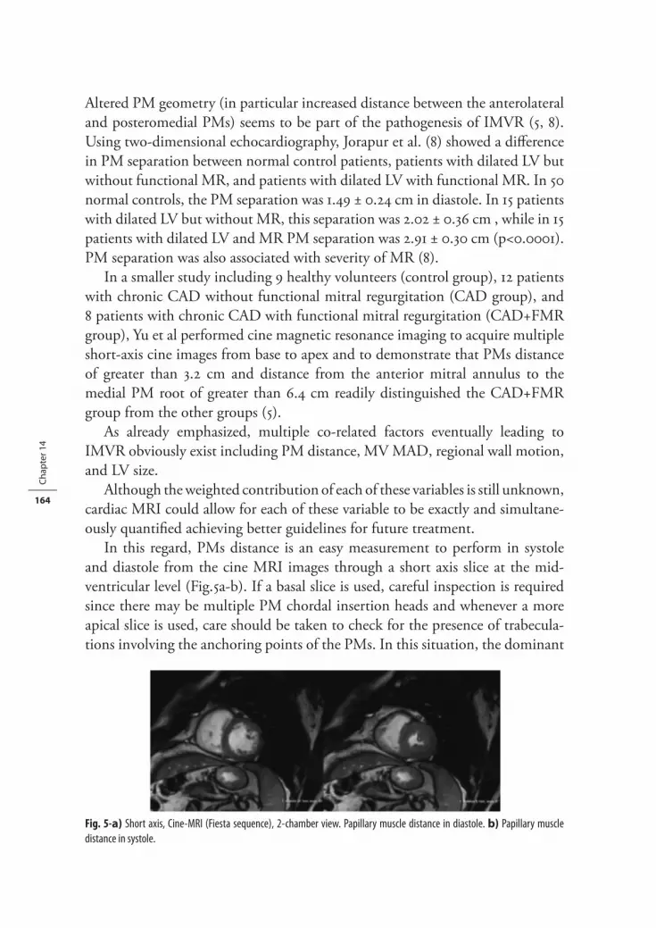
Chap
ter 1
4
164
Altered PM geometry (in particular increased distance between the anterolateral and posteromedial PMs) seems to be part of the pathogenesis of IMVR (5, 8). Using two-dimensional echocardiography, Jorapur et al. (8) showed a diff erence in PM separation between normal control patients, patients with dilated LV but without functional MR, and patients with dilated LV with functional MR. In 50 normal controls, the PM separation was 1.49 ± 0.24 cm in diastole. In 15 patients with dilated LV but without MR, this separation was 2.02 ± 0.36 cm , while in 15 patients with dilated LV and MR PM separation was 2.91 ± 0.30 cm (p<0.0001). PM separation was also associated with severity of MR (8).
In a smaller study including 9 healthy volunteers (control group), 12 patients with chronic CAD without functional mitral regurgitation (CAD group), and 8 patients with chronic CAD with functional mitral regurgitation (CAD+FMR group), Yu et al performed cine magnetic resonance imaging to acquire multiple short-axis cine images from base to apex and to demonstrate that PMs distance of greater than 3.2 cm and distance from the anterior mitral annulus to the medial PM root of greater than 6.4 cm readily distinguished the CAD+FMR group from the other groups (5).
As already emphasized, multiple co-related factors eventually leading to IMVR obviously exist including PM distance, MV MAD, regional wall motion, and LV size.
Although the weighted contribution of each of these variables is still unknown, cardiac MRI could allow for each of these variable to be exactly and simultane-ously quantifi ed achieving better guidelines for future treatment.
In this regard, PMs distance is an easy measurement to perform in systole and diastole from the cine MRI images through a short axis slice at the mid-ventricular level (Fig.5a-b). If a basal slice is used, careful inspection is required since there may be multiple PM chordal insertion heads and whenever a more apical slice is used, care should be taken to check for the presence of trabecula-tions involving the anchoring points of the PMs. In this situation, the dominant
Fig. 5-a) Short axis, Cine-MRI (Fiesta sequence), 2-chamber view. Papillary muscle distance in diastole. b) Papillary muscle distance in systole.
Giuseppe Text alt.indd 164Giuseppe Text alt.indd 164 08-Sep-09 14:32:12 PM08-Sep-09 14:32:12 PM

Ischemic mitral valve regurgitation: the new challenge for magnetic resonance imaging 165
body of the PM should be used to fi nd the center of the PM. To avoid these pitfalls, measurements are more reliable when done at the midventricular level (16).
Myocardial Scar Assessment
MRI has advantages over the more commonly used methods of viability assess-ment and viability by MRI includes detection of both regional function and morphology.
Th e main clinical tool for MRI viability assessment is gadolinium contrast agent. Nonviable scar tends to have a signifi cantly higher concentration of contrast 10–20 min following infusion than the concentration in normal, viable myocardium (delayed enhancement). With the inversion-recovery imaging techniques, easy visualization of nonviable, scarred regions, is possible as these territories will appear very bright and normal myocardium will be dark on the myocardium nulled inversion-recovery images (fi g.6).
Th e extent of myocardial segmental scarring (MRI hyperenhancement) can be graded using a previously described method (17). After dividing the ventricle in 16 segments as above suggested to investigate the LV regional contractility (9), the amount of hyper-enhancement from scar (infracted myocardium) to nulled signal (viable myocardium) can be semi-quantitatively evaluated in each segment using a 6-grade system as follows: 0= 0, 1=1 to 24, 2= 25 to 49,
Fig. 6 Short axis, 2-chamber view. Delayed enhancement sequence shows large area of sub-endocardial necrosis (fi brosis) of the mid-ventricular inferior wall.
Giuseppe Text alt.indd 165Giuseppe Text alt.indd 165 08-Sep-09 14:32:12 PM08-Sep-09 14:32:12 PM

Chap
ter 1
4
166
3= 50 to 74, 4= 75 to 99, and 5= 100 segmental scar (17). Total scarring can be calculated as the average of the scar grade over the entire LV.
Th e importance of regional myocardial perfusion and regional scarring in patients with IMVR has been poorly investigated. Srichai et al. evaluated MRI left ventricular geometric, functional, and scar measurements in addition to mitral valve geometric variables in a series of 60 patients with varying degrees of MR (none, mild, moderate, and severe) determined by echocardiography (18). At multivariate analysis, mitral systolic tenting area (p < 0.0001) in a statistical model with scarring of the anterior-lateral region (p < 0.05) proved to be the most powerful independent predictor of MR severity (18). Th ese results diff er somewhat from other studies that have shown higher incidences of IMVR in patients with inferior compared with anterior myocardial infarction (19, 20). However, diagnosis of regional scarring in these studies was not performed with MRI and was based on regional contractility dysfunction which may represent only myocardial hibernation as opposed to transmural scarring. Interestingly in Srichai et al. MRI evaluation, impairment of ventricular contractile function in the inferior-posterior, but not in the anterior-lateral region, was associated, at univariate analyses, with increasing severity of MR (18). However, as said, degree of regional scarring (particularly antero-lateral) as opposed to regional function proved a stronger determinant of IMVR severity in multivariate analysis.
Quantitative MRI with delayed-enhancement imaging for assessment of LV myocardial scarring could add to our understanding of the diverse mechanisms involved in the development of IMVR and eventually lead a more appropriate and tailored approach to its treatment. In this context, there is possibly a plethora of patients with more complex IMVR patterns including primary involvement of the anterolateral as opposed to the posteromedial PM and adjacent ventricular segments, and/or associated severe left ventricular geometrical distortion requir-ing a more complicated surgical approach rather than a simple annuloplasty to address the mitral regurgitation.
CONCLUSION
Although cardiac MRI off ers certain intrinsic advantages over other diagnostic tools in the study of MV function, there is at the present lack of a standardized scheme for data collection and analysis that could eventually direct the decision making process for cardiac surgeons and cardiologists and lead future under-standing for accurate diagnosis, operative planning, and follow-up of IMVR.
Giuseppe Text alt.indd 166Giuseppe Text alt.indd 166 08-Sep-09 14:32:13 PM08-Sep-09 14:32:13 PM

Ischemic mitral valve regurgitation: the new challenge for magnetic resonance imaging 167
Th e present manuscript summarizes those that are, in our opinion, the main parameters that should be routinely investigated while adopting MRI technology to assess patients with IMVR. Our considerations are the result of a multidisciplinary approach to this complex ethiopathogenetic entity and involve expertise spanning from radiology, cardiology, and cardiac surgery all aiming at defi ning those that are the crucial parameters to guide adequate understanding in the complexity of IMVR pathogenesis and, as a result, to allow for correct therapeutic planning.
Giuseppe Text alt.indd 167Giuseppe Text alt.indd 167 08-Sep-09 14:32:13 PM08-Sep-09 14:32:13 PM

Chap
ter 1
4
168
REFERENCES
1. Rankin JS, Hickey MSJ, Smith LR, Muhlbaier L, Reves JG, Pryor DB, Wechsler AS. Ischemic mitral regurgitation. Circulation 1989; 79 (Suppl.I): I-116–21
2. McGee EC, Gillinov MA, Blackstone EH, Rajeswaran J, Cohen G, Najam F, Shiota T, Sabik JF, Lytle BW, McCarthy PM, Cosgrove DM. Recurrent mitral regurgitation after annuloplasty forfunctional ischemic mitral regurgitation. J Th orac Cardiovasc Surg 2004; 128: 916-24
3. White HD, Norris RM, Brown MA, Brandt PW, Whitlock RM, Wild CJ. Left ventricular end-systolic volume as the major determinant of survival after recovery from myocardial infarction. Circulation 1987; 76: 44–51.
4. Hamer AW, Takayama M, Abraham KA, Roche AH, Kerr AR, Williams BF, Ramage MC, White HD. End-systolic volume and long-term survival after coronary artery bypass graft surgery in patients with impaired left ventricular function. Circulation 1994; 90: 2899–904.
5. Yu HY, Su MY, Liao TY, Peng HH, Lin FY, Tseng WYI. Functional mitral regurgitation in chronic ischemic coronary artery disease: Analysis of geometric alterations of mitral apparatus with magnetic resonance imaging. J Th orac Cardiovasc Surg 2004; 128: 543-51
6. Kaji S, Nasu M, Yamamuro A, Tanabe K, Nagai K, Tani T, Tamita K, Shiratori K, Kinoshita M, Senda M, Okada Y, Morioka S. Annular Geometry in Patients With Chronic Ischemic Mitral Regurgitation: Th ree-Dimensional Magnetic Resonance Imaging Study. Circulation 2005; 112; 409-414
7. Kongsaerepong V, Shiota M, Gillinov AM, Song JM, Fukuda S, McCarthy PM, Williams T, Savage R, Daimon M, Th omas JD, Shiota T. Echocardiographic predictors of successful versus unsuccessful mitral valve repair in ischemic mitral regurgitation. Am J Cardiol. 2006 Aug 15; 98(4): 504-8.
8. Jorapur V, Voudouris A, Lucariello RJ. Quantifi cation of annular dilatation and papillary muscle separation in functional mitral regurgitation: role of anterior mitral leafl et length as reference. Echocardiography. 2005 Jul; 22(6): 465-72.
9. Schiller NB, Shah PM, Crawford M, DeMaria A, Devereux R, Feigenbaum H, Gutgesell H, Reichek N, Sahn D, Schnittger I. Recommendations for quantitation of the left ventricle by two-dimensional echocardiography. American Society of Echocardiography Committee on Standards, Subcommittee on Quantitation of Two-Dimensional Echocardiograms. J Am Soc Echocardiogr. 1989 Sep-Oct; 2(5): 358-67.
10. Grigioni F, Enriquez-Sarano M, Zehr KJ, Bailey KR, Tajik AJ. Ischemic mitral regurgitation: long-term outcome and prognostic implications with quantitative Doppler assessment. Circula-tion. 2001 Apr 3; 103(13): 1759-64.
11. Simpson IA, Shiota T, Gharib M, Sahn DJ. Current status of fl ow convergence for clinical applications: is it a leaning tower of “PISA”? J Am Coll Cardiol 1996; 27: 504–9.
12. Zoghbi WA, Enriquez-Sarano M, Foster E, Grayburn PA, Kraft CD, Levine RA, Nihoyan-nopoulos P, Otto CM, Quinones MA, Rakowski H, Stewart WJ, Waggoner A, Weissman NJ; American Society of Echocardiography. Recommendations for evaluation of the severity of native valvular regurgitation with two-dimensional and Doppler echocardiography. J Am Soc Echocardiogr 2003; 16: 777–802.
Giuseppe Text alt.indd 168Giuseppe Text alt.indd 168 08-Sep-09 14:32:13 PM08-Sep-09 14:32:13 PM

Ischemic mitral valve regurgitation: the new challenge for magnetic resonance imaging 169
13. Kon MW, Myerson SG, Moat NE, Pennell DJ. Quantifi cation of regurgitant fraction in mitral regurgitation by cardiovascular magnetic resonance: comparison of techniques. J Heart Valve Dis 2004; 13: 600–7.
14. Gelfand EV, Hughes S, Hauser TH, Yeon SB, Goepfert L, Kissinger KV, Rofsky NM, Manning WJ. Severity of mitral and aortic regurgitation as assessed by cardiovascular magnetic resonance: optimizing correlation with Doppler echocardiography. J Cardiovasc Magn Reson. 2006; 8(3): 503-7.
15. Calafi ore AM, Gallina S, Di Mauro M, Gaeta F, Iaco AL, D’Alessandro S, Mazzei V, Di Giam-marco G. Mitral valve procedure in dilated cardiomyopathy: repair or replacement? Ann Th orac Surg 2001; 71: 1146 –1153.
16. Lloyd SG, Buckberg GD; RESTORE Group.Use of cardiac magnetic resonance imaging in surgical ventricular restoration. Eur J Cardiothorac Surg. 2006 Apr; 29 Suppl 1: S216-24.
17. Schvartzman PR, Srichai MB, Grimm RA, Obuchowski NA, Hammer DF, McCarthy PM, Kasper JM, White RD. Nonstress delayed-enhancement magnetic resonance imaging of the myocardium predicts improvement of function after revascularization for chronic ischemic heart disease with left ventricular dysfunction. Am Heart J. 2003 Sep; 146(3): 535-41.
18. Srichai MB, Grimm RA, Stillman AE, Gillinov AM, Rodriguez LL, Lieber ML, Lara A, Weaver JA, McCarthy PM, White RD. Ischemic mitral regurgitation: impact of the left ventricle and mitral valve in patients with left ventricular systolic dysfunction. Ann Th orac Surg. 2005 Jul; 80(1): 170-8.
19. Kumanohoso T, Otsuji Y, Yoshifuku S, Matsukida K, Koriyama C, Kisanuki A, Minagoe S, Levine RA, Tei C. Mechanism of higher incidence of ischemic mitral regurgitation in patients with inferior myocardial infarction: quantitative analysis of left ventricular and mitral valve geometry in 103 patients with prior myocardial infarction. J Th orac Cardiovasc Surg. 2003 Jan; 125(1): 135-43.
20. Izumi S, Miyatake K, Beppu S, Park YD, Nagata S, Kinoshita N, Sakakibara H, Nimura Y. Mechanism of mitral regurgitation in patients with myocardial infarction: a study using real-time two-dimensional Doppler fl ow imaging and echocardiography. Circulation. 1987 Oct; 76 (4): 777-85
Giuseppe Text alt.indd 169Giuseppe Text alt.indd 169 08-Sep-09 14:32:13 PM08-Sep-09 14:32:13 PM

Giuseppe Text alt.indd 170Giuseppe Text alt.indd 170 08-Sep-09 14:32:13 PM08-Sep-09 14:32:13 PM

Chapter 15
I schemic mitral valve regurgitation in patients
with depressed ventricular function: cardiac
geometrical and myocardial perfusion evaluation
with magnetic resonance imaging
Giuseppe D’Ancona, MD 1
Domenico Biondo, MS 2
Giuseppe Mamone, MD 3
Gianluca Marrone, MD 3
Francesco Pirone, MD 1
Gianluca Santise, MD 1
Sergio Sciacca, MD 1
Michele Pilato, MD 1
1 Department of CT Surgery, ISMETT@ University of Pittsburgh Medical Center, Palermo, Italy2 Offi ce of Research and Biomedical Sciences ISMETT@ University of Pittsburgh Medical Center,
Palermo, Italy3 Department of Radiology, ISMETT@ University of Pittsburgh Medical Center, Palermo, Italy
Eur J Cardiothor Surg 34(2008)964-948
Giuseppe Text alt.indd 171Giuseppe Text alt.indd 171 08-Sep-09 14:32:13 PM08-Sep-09 14:32:13 PM

Chap
ter 1
5
172
ABSTRACT
Objective
To investigate geometrical and functional changes involving the left ventricle (LV) and mitral valve (MV) apparatus in patients with depressed LV ejection fraction (LVEF) and ischemic MV regurgitation (IMVR).
Methods
A series of patients with 3 vessels CAD and depressed LVEF underwent cardiac MRI to investigate MV/LV geometry and function, and myocardial perfusion/vitality. Geometrical data were indexed by anterior MV leafl et length. Two groups were identifi ed: CAD without IMVR (Group CAD), and with IMVR (Group IMV).
Results
Eleven patients were enrolled in the CAD group and 13 in the IMV group.IMVR volume was signifi cantly higher in the IMV group (24.0± 12.0 vs.
4.5±5.2; p<0.0001). LVEF was comparable (IMV 34.6±13.0 vs. CAD 31.5±13.0; p=ns). Indexed MV/LV geometrical variables were comparable in the two groups. Perfusion/vitality study showed inferior myocardial necrosis occurred more often in the IMV group (p=0.01). At Pearson test, MV regurgitation occurrence correlated with inferior myocardial necrosis (r=0.5; p=0.006), non-indexed systolic/diastolic annular inter-commissural diameters (r=0.4; p=0.04) and MV annular areas (r=0.4; p=0.04). Papillary muscles distance (PMD) and LV vol-umes inversely correlated with LVEF (r=-0.6; p<0.05 and r=-0.8; p<0.001). At multivariable analysis, no independent determinants for IMVR were identifi ed and LV volumes were the sole determinants for LVEF (p<0.05).
Conclusion
In patients with depressed LVEF, IMV cannot be explained by LV geometrical modifi cations alone. Although PMD, LV volumes, and LVEF are correlated, they have no direct impact in the development of IMVR. In contrast, inferior myocardial necrosis and increased inter-commissural MV diameters may lead to deformity of MV complex and subsequent IMV.
Giuseppe Text alt.indd 172Giuseppe Text alt.indd 172 08-Sep-09 14:32:13 PM08-Sep-09 14:32:13 PM

Ischemic mitral valve regurgitation in patients with depressed ventricular function 173
INTRODUCTION
Ischemic mitral valve regurgitation (IMVR) has a controversial patho-physiology that assumes an even more complex aspect in patients with depressed left ventricu-lar ejection fraction (LVEF). LV remodeling, dilatation, and dysfunction result in geometrical changes in the MV apparatus, including papillary muscles (PMs) displacement and MV annular dilatation that, consequently, can lead to IMVR.
Although a global change in the geometrical relationships among the vari-ous components of the unit ventricle-mitral valve apparatus may take place in patients with impaired LV function, these modifi cations may not always justify the development of IMVR.
In this regard, in our clinical practice we frequently observe a plethora of patients with severely compromised cardiac contractility and dilated ventricular chambers that present with absolutely preserved MV function.
To understand why patients with equally impaired LVEF behave diff erently in relation to their MV continence, we should extend our analyses beyond the simple geometrical evaluation of the heart and consider, simultaneously, the impact of regional myocardial vitality and perfusion. For this purpose, a com-prehensive imaging modality such as cardiac magnetic resonance imaging (MRI) could allow us to depict a clearer image of IMVR in patients with depressed LVEF thus clarifying the purely geometrical and functional modifi cations that lead to this deleterious occurrence.
MATERIALS AND METHODS
In our Institution, patients with depressed LVEF of ischemic origin are rou-tinely submitted to cardiac MRI.
We have summarized our standardized protocol for cardiac MRI in patients with IMVR, and specifi ed the variables of interest to specifi cally evaluate MV function/morphology, LV function/morphology, and myocardial function/vital-ity (global and segmental) (1).
In this manuscript, we report our cardiac MRI fi ndings in a series of non-consecutive patients with 3 vessels coronary artery disease (CAD), depressed LVEF (<40), and with or without IMVR.
After admission, patients underwent trans-thoracic echocardiography to confi rm cardiac function and evaluate MV function/morphology.
Patients who presented MV or MV apparatus structural abnormalities were excluded from this analysis as well as those that had already undergone any sort
Giuseppe Text alt.indd 173Giuseppe Text alt.indd 173 08-Sep-09 14:32:13 PM08-Sep-09 14:32:13 PM
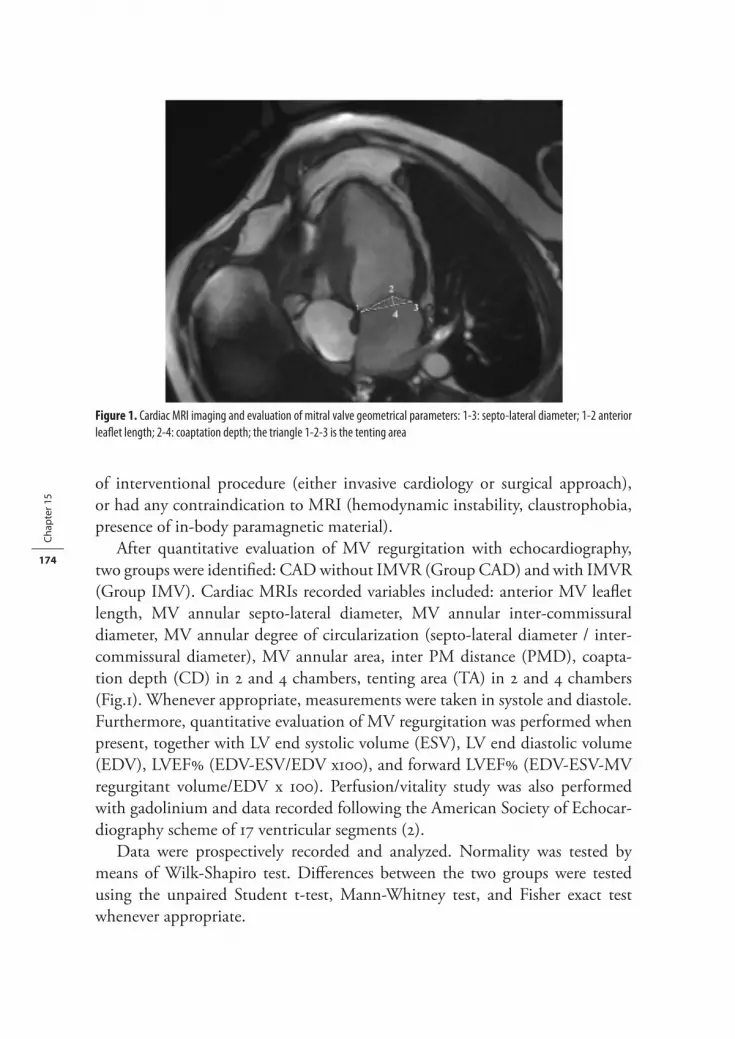
Chap
ter 1
5
174
of interventional procedure (either invasive cardiology or surgical approach), or had any contraindication to MRI (hemodynamic instability, claustrophobia, presence of in-body paramagnetic material).
After quantitative evaluation of MV regurgitation with echocardiography, two groups were identifi ed: CAD without IMVR (Group CAD) and with IMVR (Group IMV). Cardiac MRIs recorded variables included: anterior MV leafl et length, MV annular septo-lateral diameter, MV annular inter-commissural diameter, MV annular degree of circularization (septo-lateral diameter / inter-commissural diameter), MV annular area, inter PM distance (PMD), coapta-tion depth (CD) in 2 and 4 chambers, tenting area (TA) in 2 and 4 chambers (Fig.1). Whenever appropriate, measurements were taken in systole and diastole. Furthermore, quantitative evaluation of MV regurgitation was performed when present, together with LV end systolic volume (ESV), LV end diastolic volume (EDV), LVEF (EDV-ESV/EDV x100), and forward LVEF (EDV-ESV-MV regurgitant volume/EDV x 100). Perfusion/vitality study was also performed with gadolinium and data recorded following the American Society of Echocar-diography scheme of 17 ventricular segments (2).
Data were prospectively recorded and analyzed. Normality was tested by means of Wilk-Shapiro test. Diff erences between the two groups were tested using the unpaired Student t-test, Mann-Whitney test, and Fisher exact test whenever appropriate.
Figure 1. Cardiac MRI imaging and evaluation of mitral valve geometrical parameters: 1-3: septo-lateral diameter; 1-2 anterior leafl et length; 2-4: coaptation depth; the triangle 1-2-3 is the tenting area
Giuseppe Text alt.indd 174Giuseppe Text alt.indd 174 08-Sep-09 14:32:13 PM08-Sep-09 14:32:13 PM

Ischemic mitral valve regurgitation in patients with depressed ventricular function 175
To investigate correlation between the diff erent MRI geometrical variables, the presence of MVR, and LVEF, Pearson correlation test was performed in the overall group.
As LVEF did not have a normal distribution, logarithmic transformation of LVEF was used.
Multivariable analysis was performed to identify independent determinants for occurrence of IMVR (logistic regression, stepwise forward procedure) and for Log LVEF (linear regression, stepwise procedure).
Th e two multivariable analysis models were built including all those vari-ables that at Pearson correlation test had a correlation with IMVR and LVEF (p<0.05). All statistical analyses were performed using SPSS (SPSS Inc., Chicago, Ill, United States).
RESULTS
Eleven patients were enrolled in the CAD group and 13 in the IMV group.Geometrical measurements were considered before and after indexing for
anterior MV leafl et length, as previously indicated (3). Univariate analysis fi ndings are summarized in table 1. As expected, MV regurgitation volume was signifi cantly higher in the IMV group (24.0± 12.0 vs. 4.5±5.2; p<0.001) (Table 1). Conventional LVEF was comparable in the two groups (IMV 34.6±13.0 vs. CAD 31.5±13.0; p=ns) as well as forward LVEF (IMV 24.4±12.1 vs. CAD 33.6±12.4; p=ns) (table 1). Although annular MV area, and annular inter-com-missural diameters in systole and diastole were signifi cantly higher in the IMV group, after indexing for anterior MV leafl et length no statistically signifi cant diff erence was noticed between the two groups (table 1).
Gadolinium perfusion/vitality study was performed to evaluate the presence of necrotic segments (myocardium that after contrast infusion presents with a pattern of delayed enhancement). Th e total number of necrotic segments was comparable in the two groups (IMV 6.3±4.1 vs. CAD 6.7±4.9; p=0.8) (Table 2). A specifi c analysis of the diff erent segmental areas involved (inferior, lateral, anterior, septal, apical), showed a higher occurrence of inferior myocardial necrosis in the IMV group (IMV 61 vs. CAD 9; p=0.01) and a trend towards a higher rate of septal necrosis in the CAD group (IMV 54 vs. CAD 91; p=0.07) (Table 2).
At Pearson test, inferior myocardial necrosis had the strongest correlation with occurrence of IMVR (correlation ratio R=0.54; p=0.006) (Table 3). Other variables signifi cantly correlated with occurrence of IMVR were: non-indexed
Giuseppe Text alt.indd 175Giuseppe Text alt.indd 175 08-Sep-09 14:32:13 PM08-Sep-09 14:32:13 PM

Chap
ter 1
5
176
Table 1. Univariate comparison between patients with depressed LVEF%, 3 vessels CAD, and w/o IMVR. Data are expressed in systole (s) and diastole (d) and before and after indexing for anterior leafl et length when appropriate.
CAD IMV p-valueAge (years) 61.4±10.8 61.9±5.7 0.80
Gender M/F 9_2 9_4 0.20
LVEF% 31.5±13 34.6±13 0.77
Forward LVEF% 33.6±12.4 24.4±12.1 0.12
MVR Volume cc 4.5±5.2 24.0± 12.0 < 0.001
Anterior MV leafl et length mm 25.5±4.0 28.6±2.2 0.07
LV End Systolic Volume cc 142.2±94.6 148.4±63.7 0.80
LV End Diastolic Volume cc 208.1±32.5 225.3±64.0 0.60
Papillary muscles distance (s) mm 18.6±9.8 18.8±5.0 0.90
Papillary muscles distance (d) mm 24.8±8.4 26.7±5.2 0.50
Indexed Papillary muscles distance (s) 0.6±0.1 0.7±0.2 0.60
Indexed Papillary muscles distance (d) 0.9±0.1 0.9±0.2 0.70
Septo-lateral diameter (s) mm 32.0±3.6 34.6±2.7 0.08
Septo-lateral diameter (d) mm 33.6±3.4 36.4±2.7 0.06
Indexed Septo-lateral diameter (s) 1.2±0.1 1.2± 0.1 0.30
Indexed Septo-lateral diameter (d) 1.3±0.07 1.3± 0.1 0.20
Inter-commissural diameter (s) mm 31.2±2.5 35.0±4.5 0.04
Inter-commissural diameter (d) mm 33.0±3.6 37.0±4.3 0.04
Indexed Inter-commissural distance (s) 1.2±0.1 1.2±0.1 0.80
Indexed Inter-commissural distance (d) 1.3±0.1 1.3±0.2 0.80
MV Area (s) mm2 788.0±128.1 960.7±185.0 0.03
MV Area (d) mm2 876.9±168.4 1068.7±204. 3 0.04
Indexed MV Area (s) 31.1±3.8 33.6±5.9 0.30
Indexed MV Area (d) 34.6±5.8 37.2±5.7 0.30
MV Circularization degree (s) 1.0±0.1 0.9±0.08 0.50
MV Circularization degree (d) 1.0±0.08 0.9±0.05 0.30
Coaptation Depth 2chambers mm 7.7± 1.6 8.2± 1.7 0.50
Coaptation Depth 4 chambers mm 7.7± 2.2 7.8± 3.0 0.80
Tenting Area 2 chambers cm2 1.4±0.9 1.5± 0.4 0.70
Tenting Area 4 chambers cm2 1.5±0.9 1.4± 0.3 0.70
Table 2. Cardiac MRI vitality study: Mean total number of necrotic segments (within a 17 segments schema) and rate of necrosis within the diff erent segmental areas in the CAD and IMV groups.
CAD IMV p-valueDelayed Enhancement Total Segments 6.7±4.9 6.3±4.1 0.80
Inferior 9% 61% 0.01
Lateral 18% 46% 0.21
Anterior 45% 54% 1.00
Septal 91% 54% 0.07
Apical 64% 70% 1.00
Giuseppe Text alt.indd 176Giuseppe Text alt.indd 176 08-Sep-09 14:32:13 PM08-Sep-09 14:32:13 PM

Ischemic mitral valve regurgitation in patients with depressed ventricular function 177
inter-commissural diameter and MV annular area (both in systole and diastole), and septal myocardial necrosis (Table 3).
Left ventricular volumes (end-systolic and diastolic), and inter-papillary muscles distance (in systole and diastole) had the strongest negative correlation with the logarithmic transformed of LVEF (Table 4).
Th ose variables that had a signifi cant correlation with occurrence of IMVR and the logarithmic transformed of LVEF were tested with multivariable analysis.
No independent determinant for occurrence of IMVR was identifi ed (table 5). Left ventricular volumes (end-systolic and diastolic) were the sole indepen-dent determinants for Log10 LVEF (table 6).
Table 3. Pearson Correlation between the presence of ischemic MV regurgitation and cardiac MRI geometrical and vitality parameters. R= correlation coeffi cient, (s): systole, (d): diastole
R p-value
Inter-commissural diameter (s) 0.46 0.04
Inter-commissural diameter (d) 0.46 0.04
MV Annular Area (s) 0.48 0.03
MV Annular Area (d) 0.46 0.04
Inferior Necrosis 0.54 0.006
Septal Necrosis -0.46 0.04
Table 4. Pearson Correlation between Log LVEF% and cardiac MRI fi ndings. R= correlation coeffi cient, LV: Left Ventricular, (s): systole, (d): diastole
R p-value
Papillary Muscles Distance (s) -0.67 0.004
Papillary Muscles Distance (d) -0.56 0.02
LV End-systolic Volume -0.83 <0.001
LV End-diastolic Volume -0.73 0.001
Table 5. Logistic regression (stepwise forward procedure) analysis. Dependent Variable: occurrence of ischemic MV regurgitation, (s): systole
p-value
Inferior Myocardial Necrosis 0.16
MV Annular Area(s) 0.99
Table 6. Linear regression (stepwise procedure) analysis.p-value
LV End Systolic Volume 0.022*
LV End Diastolic Volume 0.036*
Dependent Variable: Log10 LVEF %.LV: Left Ventricular
Giuseppe Text alt.indd 177Giuseppe Text alt.indd 177 08-Sep-09 14:32:13 PM08-Sep-09 14:32:13 PM

Chap
ter 1
5
178
DISCUSSION
MV insuffi ciency occurs in 15 of patients after myocardial infarction (MI) and in the presence of structurally normal MV leafl ets and subvalvular apparatus (4). IMVR is associated with a controversial diagnostic-therapeutic pattern as its patho-physiologic bases have yet to be fully elucidated.
As this condition is associated with excess cardiac morbidity and mortality even in the presence of mild to moderate regurgitation (5), many authors have focused their research on identifying the mechanisms that would lead to the development of IMVR in an attempt to better stratify patients at risk and guide treatment.
Correct understanding of mechanisms leading to IMVR is even more crucial in patients with depressed LVEF.
We have evaluated with cardiac MRI the impact of LV global and segmental myocardial scarring, LVEF, as well as various geometric and functional vari-ables of the MV apparatus, on the development of IMVR in patients with LV dysfunction.
Use of cardiac MRI in a similar context has been previously suggested by Sirchai et al (6).
Our main fi nding is a higher rate of necrotic areas in the inferior myocardial segments in patients with IMVR. Furthermore, myocardial segmental delayed enhancement had the strongest univariate correlation with occurrence of MV regurgitation.
In our experience, LV dimensions and LVEF seem to have no direct impact on the development of IMVR and, consequently, equally enlarged ventricles may develop unequal degrees of MV regurgitation.
We believe that, per se, an enlarged ventricle will not automatically lead to an ischemic MV and that the location of the myocardial necrosis will play the main role in the ethio-pathogenesis of MV regurgitation. Similar results were reported by Kumanohoso et al. (7) in an evaluation involving patients with prior myocardial infarction (MI) (anterior and inferior) and normal control subjects. Th e authors evaluated the grade of IMVR by means of quantitative echocardiog-raphy and reported that, although global LV dilatation and dysfunction were signifi cantly less pronounced in patients with inferior MI, the percentage of mitral regurgitation jet area and the incidence of signifi cant regurgitation were greater in these patients (7).
Th is would confi rm that IMVR may occur in normally sized ventricles and, in the event of extreme LV dilatation, will occur mainly if the inferior myocar-dium is necrotic.
Giuseppe Text alt.indd 178Giuseppe Text alt.indd 178 08-Sep-09 14:32:13 PM08-Sep-09 14:32:13 PM

Ischemic mitral valve regurgitation in patients with depressed ventricular function 179
We should also emphasize the fact that, in our present analysis, most of the patients had only mild MV. In fact, in patients with more advanced mitral regur-gitation, the eff ect of blood volume loading on the left ventricle would become more substantial and, at this advanced stage, it would become more diffi cult to recognize cause from eff ect.
Th e role of MV annular dilatation in the pathogenesis of ischemic MV regurgitation remains controversial. In this regard, many authors have reported that isolated annular dilation does not usually cause important functional mitral regurgitation as leafl et tissue redundancy protects against leafl et abnormal coap-tation in isolated annular dilatation (8,9).
However, many others have reported, in diff erent cohorts of patients with IMVR and in animal experimentation, signifi cant increases in either MV septo-lateral or inter-commissural diameters (10-14).
Diff erently from the majority of the available literature, and as suggested by Jorapur et al (3), we have used anterior mitral leafl et length to index geometric measurements of the MV apparatus. Anterior leafl et and intertrigonal distance (the distance between the trigones, which corresponds to the fi brous portion of the mitral annulus) are not signifi cantly aff ected by left ventricular remodelling and have been used as reference measurements for sizing mitral annuloplasty rings.
Th e importance of using an indexing parameter is refl ected in our univariate analysis where MV inter-commissural distance and MV annular area are signifi -cantly correlated to occurrence of MV regurgitation only before being indexed by anterior MV leafl et length.
Th e current leading surgical technique for the treatment of ischemic MV regurgitation is based on the assumption that MV annulus is fi rmly anchored along the circumference of the anterior leafl et by the tough fi brous skeleton of the heart and, therefore, dilatation of the MV annulus primarily aff ects the posterior leafl et. As a consequence, all current operative mitral repair strategies contemplate reduction of the MV inlet area by posterior leafl et annuloplasty.
We believe that ischemic MV regurgitation occurs within a more complex scenario that is actually refl ected by the high rate of MV regurgitation recur-rence after simple posterior annuloplasty (15). In this context, Gelsomino et al. have recently shown that independent predictors of recurrent MV regurgitation after restrictive annuloplasty include mainly ventricular variables such as end systolic volume, systolic sphericity index, myocardial performance index, and wall motion score index (16).
Giuseppe Text alt.indd 179Giuseppe Text alt.indd 179 08-Sep-09 14:32:13 PM08-Sep-09 14:32:13 PM

Chap
ter 1
5
180
Altered PMs geometry has been hypothesized as part of the pathogenesis of ischemic MV regurgitation (3,7).
In several MRI studies, Kaji et al. confi rmed the importance of the posterior PM displacement (with consequent increase in posterior papillary muscle-septal annulus distance) (11). Yu et al. reported similar fi ndings concluding that the inter- PMs distance of more than 32 mm and the distance from the anterior mitral annulus to the posterior PM root of more than 64 mm readily distin-guished ischemic MV patients from healthy controls and isolated CAD patients (14).
Finally, inter- PMs distance was proposed as an independent predictor for mid-term failure after MV repair for IMVR (15).
In our analysis, inter-PMs distance (absolute and indexed) was homoge-neously distributed in patients with and without IMVR and, furthermore, there was not signifi cant correlation between inter-PMs distance and occurrence of MV regurgitation in the overall group. Our fi ndings may be biased by the fact we did not calculate the distance between PMs and MV annulus although we did focus on inter- papillary muscles distance. On the other hand, we found no correlation between the amount of IMVR and the coaptation depth (which would be a surrogate of the distance between PMs and MV annular plane).
In our univariate analysis, non-indexed inter- PMs distance correlated with LVEF. At multivariable analysis, LV volumes were the sole independent deter-minants for LVEF.
Th is confi rms the fact that PMs distance may be a good indicator of LV function and dilatation albeit not necessarily correlated to the degree of IMVR.
LIMITATIONS
We should emphasize that our multivariable analysis showed none of the inves-tigated parameters have an independent impact towards occurrence of IMVR. Th is could be explained by the small size of the investigated sample. Also, in our analysis we considered 10 cc as cut-off to divide patients with and without IMVR and, within the patients with diff erent degrees of IMVR, we made no distinction between moderate and severe MV regurgitation. Although this deci-sion was dictated by sample size limitations, fi ndings could have been diff erent especially when comparing the severe IMVR patients with the sole CAD ones.
Finally, as already underlined in the discussion, evaluation of inter-PMs distance was limited to the inter-PMs measurements without taking into consid-eration the distance from the PMs to the MV annulus. We deliberately decided
Giuseppe Text alt.indd 180Giuseppe Text alt.indd 180 08-Sep-09 14:32:13 PM08-Sep-09 14:32:13 PM

Ischemic mitral valve regurgitation in patients with depressed ventricular function 181
to concentrate on this parameter as we accepted MV coaptation depth and MV tenting area as good indirect indicators of the distance between PMs and MV.
CONCLUSION
Although various factors play a role in the ethio-pathogenesis of IMVR, we believe that a comprehensive imaging modality such as MRI could allow us to depict a clearer image of this elusive condition.
We are aware that the relatively small number of patients studied in the pres-ent analysis may not be suffi cient to clearly diff erentiate the causes in the genesis of IMVR.
At the same time we can suggest that in patients with depressed LVEF, isch-emic MV cannot be explained by global LV and MV geometrical modifi cations alone. Although PM distance, LV volumes, and LVEF are correlated, they have no direct independent impact in the development of IMVR. In contrast, inferior myocardial necrosis and increased inter-commissural MV diameters may lead to deformity of MV complex and subsequent IMVR.
Giuseppe Text alt.indd 181Giuseppe Text alt.indd 181 08-Sep-09 14:32:13 PM08-Sep-09 14:32:13 PM

Chap
ter 1
5
182
REFERENCES
1. D’Ancona G, Mamone G, Marrone G, Pirone F, Santise G, Sciacca S, Pilato M. schemic mitral valve regurgitation: the new challenge for magnetic resonance imaging. Eur J Cardiothorac Surg. 2007; 32: 475-80.
2. Schiller NB, Shah PM, Crawford M, DeMaria A, Devereux R, Feigenbaum H, Gutgesell H, Reichek N, Sahn D, Schnittger I. Recommendations for quantitation of the left ventricle by two-dimensional echocardiography. American Society of Echocardiography Committee on Standards-Subcommittee on Quantitation of Two-Dimensional Echocardiograms. J Am Soc Echocardiogr 1989; 2: 358–67.
3. Jorapur V, Voudouris A, Lucariello RJ. Quantifi cation of annular dilatation and papillary muscle separation in functional mitral regurgitation: role of anterior mitral leafl et length as reference. Echocardiography 2005; 22: 465–72.
4. Rankin JS, Hickey MSJ, Smith LR, Muhlbaier L, Reves JG, Pryor DB, Wechsler AS. Ischemic mitral regurgitation. Circulation 1989; 79(Suppl. I). I-116-21.
5. Grigioni F, Enriquez-Sarano M, Zehr KJ, Bailey KR, Tajik AJ. Ischemic mitral regurgitation: long-termoutcome and prognostic implicationswith quantitative Doppler assessment. Circula-tion 2001; 103: 1759–64.
6. Srichai MB, Grimm RA, Stillman AE, Gillinov AM, Rodriguez LL, Lieber ML, Lara A, Weaver JA, McCarthy PM, White RD. Ischemic mitral regurgitation: impact of the left ventricle and mitral valve in patients with left ventricular systolic dysfunction. Ann Th orac Surg 2005; 80: 170–8.
7. Kumanohoso T, Otsuji Y, Yoshifuku S, Matsukida K, Koriyama C, Kisanuki A, Minagoe S, Levine RA, Tei C. Mechanism of higher incidence of ischemic mitral regurgitation in patients with inferior myocardial infarction: quantitative analysis of left ventricular and mitral valve geometry in 103 patients with prior myocardial infarction. J Th orac Cardiovasc Surg 2003; 125: 135–43.
8. Otsuji Y, Kumanohoso T, Yoshifuku S, Matsukida K, Koriyama C, Kisanuki A, Minagoe S, Levine RA, Tei C. Isolated annular dilation does not usually cause important functional mitral regurgitation: comparison between patients with lone atrial fi brillation and those with idiopathic or ischemic cardiomyopathy. J Am Coll Cardiol. 2002; 39: 1651–1656.
9. Green GR, Dagum P, Glasson JR, Daughters GT, Bolger AF, Foppiano LE, Berry GJ, Ingels NB, Jr., Miller DC. Mitral annular dilatation and papillary muscle dislocation without mitral regurgitation in sheep. Circulation. 1999; 100: II95–II102.
10. Lai DT, Tibayan FA, Myrmel T, Timek TA, Dagum P, Daughters GT, Liang D, Ingels NB, Jr, Miller DC. Mechanistic insights into posterior mitral leafl et inter-scallop malcoaptation during acute ischemic mitral regurgitation. Circulation. 2002; 106: I40 –I45.
11. Kaji S, Nasu M, Yamamuro A, Tanabe K, Nagai K, Tani T, Tamita K, Shiratori K, Kinoshita M, Senda M, Okada Y, Morioka S. Annular geometry in patients with chronic ischemic mitral regurgitation: three-dimensional magnetic resonance imaging study. Circulation 2005; 112: 409–14
12. Kwan J, Shiota T, Agler DA, Popovic ZB, Qin JX, Gillinov MA, Stewart WJ, Cosgrove DM, McCarthy PM, Th omas JD. Geometric diff erences of the mitral apparatus between ischemic
Giuseppe Text alt.indd 182Giuseppe Text alt.indd 182 08-Sep-09 14:32:13 PM08-Sep-09 14:32:13 PM
Ischemic mitral valve regurgitation in patients with depressed ventricular function 183
and dilated cardiomyopathy with signifi cant mitral regurgitation: real-time three-dimensional echocardiography study. Circulation. 2003; 107: 1135–1140.
13. Kongsaerepong V, Shiota M, Gillinov AM, Song JM, Fukuda S, McCarthy PM, Williams T, Savage R, Daimon M, Th omas JD, Shiota T. Echocardiographic predictors of successful versus unsuccessful mitral valve repair in ischemic mitral regurgitation. Am J Cardiol 2006; 98: 504–8.
14. Yu HY, Su MY, Liao TY, Peng HH, Lin FY, Tseng WYI. Functional mitral regurgitation in chronic ischemic coronary artery disease: analysis of geometric alterations of mitral apparatus with magnetic resonance imaging. J Th orac Cardiovasc Surg 2004; 128: 543–51.
15. Kolh P. Surgical correction of ischaemic mitral regurgitation still a long way to go. Eur Heart J. 2008; 29: 147-9.
16. Gelsomino S, Lorusso R, De Cicco G, Capecchi I, Rostagno C, Caciolli S, Romagnoli S, Da Broi U, Stefàno P, Gensini GF. Five-year echocardiographic results of combined undersized mitral ring annuloplasty and coronary artery bypass grafting for chronic ischaemic mitral regurgitation. Eur Heart J. 2008 Jan; 29: 231-40.
17. Roshanali F, Mandegar MH, Yousefnia MA, Rayatzadeh H, Alaeddini F.A prospective study of predicting factors in ischemic mitral regurgitation recurrence after ring annuloplasty.Ann Th orac Surg. 2007; 84: 745-9
Giuseppe Text alt.indd 183Giuseppe Text alt.indd 183 08-Sep-09 14:32:13 PM08-Sep-09 14:32:13 PM

Giuseppe Text alt.indd 184Giuseppe Text alt.indd 184 08-Sep-09 14:32:13 PM08-Sep-09 14:32:13 PM

Chapter 16
D iscussion
Giuseppe D’Ancona, MD
Giuseppe Text alt.indd 185Giuseppe Text alt.indd 185 08-Sep-09 14:32:13 PM08-Sep-09 14:32:13 PM

Giuseppe Text alt.indd 186Giuseppe Text alt.indd 186 08-Sep-09 14:32:13 PM08-Sep-09 14:32:13 PM

Discussion 187
Every medical procedure requires a quality control method, and this is particu-larly true for complex procedures such as heart operations.
Medical practice is urging us towards quality control for scientifi c, clinical, ethical, as well as legal purposes. For these reasons, all the available armamentaria should be applied to provide evidence of good clinical practice.
Intraoperative graft patency verifi cation after coronary surgery should be routine part of the operation.
Aim of coronary artery bypass grafting is to increase blood fl ow to ischemic myocardium and it is important to document objectively the procedure success. Th is statement acquires further impact if we realize that 5-20 of performed grafts fail before discharge from the hospital and up to 30 before 1 year.
Many of these early failures are probably secondary to technical issues that could be promptly solved if adequately diagnosed intraoperative.
As reported in chapter 11, cardiac surgeons are overtly sceptic towards intra-operative graft patency verifi cation and most of them still trust their fi ngertips’ tactile feedback as evidence of coronary graft patency.
In being complementary with PCI, cardiac surgery has lost its supremacy in the treatment of coronary artery disease, and cardiac surgeons should concentrate on how to optimize the quality of care they are providing.
In perspective, and as summarized in the various chapters of this thesis, there is enough scientifi c evidence justifying the use of intraoperative coronary graft patency verifi cation tools.
Th e intraoperative tools that surgeons presently have to test coronary graft patency include imaging techniques and fl ow assessment devices.
Although most surgeons use neither of these technologies, in the future we should consider to adopt simultaneously the two diff erent technologies.
In this context, future research should be supported by manufacturing compa-nies and focus on two main goals:1) the development of a device that will allow surgeons to identify simultane-
ously and intraoperative the anatomical and functional parameters of the coronary targets and newly constructed coronary anastomoses;
2) the realization of a system of automated interpretation of coronary graft fl ow curves and anastomosis status.
Imaging modalities most often do not clearly demonstrate the functional capac-ity and reserve of the anastomoses and, on the other side, fl ow assessment devices do not clearly address the degree of patency of the anastomoses.
Giuseppe Text alt.indd 187Giuseppe Text alt.indd 187 08-Sep-09 14:32:13 PM08-Sep-09 14:32:13 PM
Chap
ter 1
6
188
A unique diagnostic tool could achieve a simultaneous iconographic and functional evaluation by integrating diff erent technologies (fl uorescence imag-ing and ultrasound?).
Alternatively, ultrasound technology may become the gold standard for intra-operative coronary graft patency verifi cation due to its dual ability to provide both morphological and functional information.
Although perivascular transit time fl ow measurement (an ultrasound based technology) has been widely demonstrated as a sensitive tool for intraoperative quality assessment of newly constructed coronary grafts, its application cannot be extended to the evaluation of coronary targets due to limitations intrinsic to transit time technology.
Diff erently from transit time fl ow measurement probes, Doppler epi-coronary probes are formed by a single crystal and do not require dissecting and encircling the target artery under study and, therefore, are more easily applicable to test the status of the native coronaries and their blood fl ow before and after the revascularization has been performed.
In this regard, Doppler technology has recently resurged as a valuable intraop-erative armamentarium to help cardiac surgeons select adequate coronary targets for revascularization and depicting both anatomical and functional features of newly constructed anastomosis.
As documented in our animal experiments (chapter 8), epicardial coronary artery Doppler represents a valuable tool to detect coronary arterial fl ow velocity in basal condition, after coronary occlusion, and during reperfusion. Moreover, potential applications of a custom-made epicardial ultrasound probe were previ-ously investigated demonstrating its ability to successfully visualize and assess coronary arteries and anastomoses on all sides of the heart.
We auspicate that, in the very next future, multimodal ultrasound probes will be designed and will include, in the same product, the possibility to visualize coronary targets and coronary anastomoses, to evaluate coronary fl ows and fl ow velocities (with Doppler) before and after surgical revascularization, and to record grafts fl ow values and fl ow patterns (with transit time) at the end of the operation.
Correct interpretation of intraoperative fi ndings is not always immediate and routine use and further technological amelioration of the existing devices will enhance the coronary graft quality control process.
Although transit time technology can clearly show diff erences between hemodynamically normal and abnormal grafts, correct interpretation is limited by our inability to analyze fl ow curves in their diff erent components. In this
Giuseppe Text alt.indd 188Giuseppe Text alt.indd 188 08-Sep-09 14:32:13 PM08-Sep-09 14:32:13 PM

Discussion 189
context, a system of automated interpretation of intraoperative fi ndings should be designed keeping into consideration the fl ow curve patterns.
Giuseppe Text alt.indd 189Giuseppe Text alt.indd 189 08-Sep-09 14:32:13 PM08-Sep-09 14:32:13 PM

Giuseppe Text alt.indd 190Giuseppe Text alt.indd 190 08-Sep-09 14:32:13 PM08-Sep-09 14:32:13 PM

Chapter 17
Summary English
Summary Dutch
Curriculum vitae
List of publications
Acknowledgement
Portfolio
Giuseppe Text alt.indd 191Giuseppe Text alt.indd 191 08-Sep-09 14:32:13 PM08-Sep-09 14:32:13 PM

Giuseppe Text alt.indd 192Giuseppe Text alt.indd 192 08-Sep-09 14:32:14 PM08-Sep-09 14:32:14 PM

S ummary English
CHAPTER 1 & CHAPTER 2
Introductory Chapters: Intraoperative Graft Patency Verifi cation in Coro-nary SurgeryTh ese two introductory chapters (Chapter 1 and 2) summarize the basis of vascular fl ow measurement focusing on coronary rheology and discussing the technological aspects of fl owmetry.
A comprehensive insight into ultrasound based transit time fl ow measure-ment is given.
Guidelines for correct interpretation of coronary grafts fl ow patterns are given.
Comment Phase Chapters: Clinical Experience with Transit Time Flow Measurement and Validation of Epicoronary Doppler in the animal modelTh is session discusses clinical use of transit time fl ow measurement to intraop-eratively test graft patency during coronary surgery.
CHAPTER 3
Chapter 3 is a short communication commenting upon a previous publication and emphasizing how strict protocols for fl ow measurement have to be followed in order to achieve the maximal clinical benefi t of this technology.
CHAPTER 4
Chapter 4 summarizes the clinical experience with transit time fl ow measure-ment in beating heart coronary surgery. Th e fi ndings in over 1000 tested grafts are reported and indications for correct interpretation of fl ow patterns are given on the basis of the fi ndings on revised grafts.
Giuseppe Text alt.indd 193Giuseppe Text alt.indd 193 08-Sep-09 14:32:14 PM08-Sep-09 14:32:14 PM

194
Sum
mar
y En
glis
h
CHAPTER 5
Chapter 5 and 6 suggest some clinical applications for transit time fl ow measure-ment. Chapter 5 shows how fl owmetry provides an extra tool to direct supportive measures such as cardiac pacing after CABG.
CHAPTER 6
Chapter 6 shows that in reoperative coronary artery surgery the physiologic signifi cance of suspect lesions by angiography can be confi rmed by direct intra-operative measurement of blood fl ow in the old grafts; such interpretation can prevent unnecessary graft revisions.
CHAPTER 7
In this chapter the infl uence of heparin dosing in patients undergoing off -pump coronary revascularization is evaluated in terms of coronary graft patency as detected by intraoperative transit time fl ow measurement.
CHAPTER 8
Chapter 8 validates, in an animal model, a newly-designed epicardial coronary artery Doppler probe. Th e experiments have been conducted in the laboratories of the Erasmus University hospital and have shown that epicardial coronary arte-rial Doppler represents a valuable tool to detect coronary arterial fl ow velocity in basal condition. Although changes in fl ow velocity are easily recorded after coronary occlusion and reperfusion, modifi cations after partial coronary stenosis are not clearly defi ned.
CHAPTER 9
Chapter 9 reports the use of intraoperative transit time technology as gold-standard to directly measure right ventricular output (intraoperative probe placement around the main pulmonary artery) and validate a new system for invasive cardiac output monitoring (True-Continuous Cardiac Output).
Giuseppe Text alt.indd 194Giuseppe Text alt.indd 194 08-Sep-09 14:32:14 PM08-Sep-09 14:32:14 PM

Summary English 195
CHAPTER 10
In this chapter transit time fl ow measurement is used to test diff erent techniques of coronary anastomoses construction.
CHAPTER 11
Th is chapter summarizes the fi nding of a survey about the clinical use of intraop-erative graft patency verifi cation by cardiac surgeons. It interestingly shows that the majority of surgeons use their fi nger tips to detect coronary grafts quality.
CHAPTER 12
Chapter 12 reviews the modern means available to intraoperative test and document the status of newly constructed coronary anastomosis. A distinction should be made between imaging techniques and fl ow assessment devices. Both methods have clear limitations, and we should consider, as a future perspective, using the two diff erent technologies simultaneously.
CHAPTER 13
Th is chapter discusses the very controversial topic of coronary revascularization: should cardiologists and cardiac surgeons approach this pathology together and aim at hybrid coronary revascularization?
CHAPTER 14-15
Th e last two chapters report a very recent experience on a very elusive issue: i.e., ischemic mitral valve regurgitation.
In these chapters the author attempts to give an explanation of the patho-physiologic bases of ischemic mitral valve regurgitation by using a modern and complete diagnostic and investigational tool such as cardiac MRI.
Giuseppe Text alt.indd 195Giuseppe Text alt.indd 195 08-Sep-09 14:32:14 PM08-Sep-09 14:32:14 PM

Giuseppe Text alt.indd 196Giuseppe Text alt.indd 196 08-Sep-09 14:32:14 PM08-Sep-09 14:32:14 PM

S ummary Dutch
Nederlandse samenvatting
HOOFDSTUK 1
In dit hoofdstuk worden de fysiologische aspecten van de bloedstroom in de coronairvaten besproken. Met name de rheologie in coronair vaten en de diverse aspecten van bloedstroom metingen worden belicht. Inzicht wordt gegeven in de op ultrageluid gebaseerde stroommetingen. Tevens worden richtlijnen besproken die dienen voor de juiste interpretatie van bloedstroom patronen in coronaire bypasses. De klinische toepasbaarheid ervan wordt toegelicht.
HOOFDSTUK 2
De toepassing van bloedstroom metingen tijdens coronair chirurgie wordt beschreven. Bij 161 patiënten die off -pump coronair chirurgie ondergingen werden in totaal 323 anastomosen gemaakt. Alle bypasses werden met behulp van TTFM (Transit Time Flow Measurement) gecontroleerd. In totaal warden 32 omleidingen gereviseerd (9.9) op basis van afwijkende stroom patronen. Bij alle gereviseerde bypasses werd een technische onvolkomenheid gevonden.
HOOFDSTUK 3
Hoofdstuk 3 is een commentaar op een wetenschappelijke publicatie van Hiro-tani et al. (Hirotani T, Kameda T, Shirota S, Nakao Y. An evaluation of the intraoperative transit time measurements of coronary bypass fl ow. Eur J Cardio-thorac Surg 2001; 19: 848–852). Benadrukt wordt hoe strikt protocollen voor bloedstroom metingen moeten worden gevolgd om tot de juiste beslissingen te komen of een bypass doorgankelijk is of niet.
HOOFDSTUK 4
Dit hoofdstuk vormt een beschrijving van de klinische ervaring van bloed-stroommeting tijdens ‘kloppend hart’ coronair chirurgie. De bevindingen van
Giuseppe Text alt.indd 197Giuseppe Text alt.indd 197 08-Sep-09 14:32:14 PM08-Sep-09 14:32:14 PM

198
Ned
erla
ndse
sam
enva
ttin
g
meer dan 1000 coronair bypass grafts worden gerapporteerd en aanwijzingen worden gegeven om tot een juiste interpretatie van de stromingspatronen te komen. Belangrijk hier is de discussie welke bypasses moeten worden herzien op basis van deze stroommetingen.
HOOFDSTUK 5
Hoofdstuk 5 beschrijft de toepassing van perioperatieve bloedstroom meting om myocardperfusie te optimaliseren door het elektrisch stimuleren(‘pacen’) van het hart.
HOOFDSTUK 6
De afwijkingen in bypass grafts die met bloedstroom metingen kunnen wor-den vastgesteld, kunnen gerelateerd worden aan preoperatieve angiografi sche bevindingen. Tijdens een coronaire reoperatie kan zo vastgesteld worden welke afwijkingen daadwerkelijk fysiologische consequenties hebben en welke bypass grafts gereviseerd dienen te worden. Dergelijke metingen kunnen onnodige graft herzieningen voorkomen.
HOOFDSTUK 7
In dit onderzoek is onderzocht welke invloed de heparine dosering heeft op de doorgankelijkheid van coronair bypass grafts bij patiënten die een off -pump coronair revascularisatie ondergaan. Geconcludeerd wordt dat onvoldoende antistolling tijdens de operatie leidt tot trombose in de bypass graft.
HOOFDSTUK 8
Hoofdstuk 8 valideert, in een diermodel, een nieuw ontworpen epicardiale coronaire Doppler sonde. De experimenten tonen aan dat een epicardiale coronair arteriële Doppler meting een waardevol instrument is voor het bepalen van coronaire bloedstroom snelheid onder basale omstandigheden. Hoewel de veranderingen in de stroomsnelheid eenvoudig kunnen worden vastgesteld
Giuseppe Text alt.indd 198Giuseppe Text alt.indd 198 08-Sep-09 14:32:14 PM08-Sep-09 14:32:14 PM

Nederlandse samenvatting 199
na coronaire occlusie en reperfusie, zijn de veranderingen die optreden tijdens gedeeltelijke coronaire occlusie minder duidelijk.
HOOFDSTUK 9
In hoofdstuk 9 wordt het gebruik van ‘intraoperative transittime’ metingen vergeleken met een nieuwe continue cardiac output meting die berust op een thermisch principe.
HOOFDSTUK 10
In dit hoofdstuk worden stroom karakteristieken besproken voor de verschil-lende anastomose technieken tussen de linker arterie thoracica interna en de linker coronair arterie (LAD).
HOOFDSTUK 11
Dit hoofdstuk geeft de bevindingen weer van een enquête die gehouden is betreff ende de vraag hoe chirurgen intraoperatief de doorgankelijkheid van grafts vaststellen. Het blijkt dat de meerderheid van de chirurgen hun vingertoppen gebruiken om de bloedstroom in een bypass te verifi ëren.
HOOFDSTUK 12
Hoofdstuk 12 geeft een overzicht van de nieuwe methoden die beschikbaar zijn om intraoperatief de doorgankelijkheid van bypasses vast te stellen. Er wordt onderscheid gemaakt tussen beeldvormende technieken en technieken die de bloedstroom beoordelen. Beide methoden hebben duidelijke beperkingen, en overwogen moet worden of in de toekomst de twee verschillende technologieën beiden moeten worden gebruikt.
Giuseppe Text alt.indd 199Giuseppe Text alt.indd 199 08-Sep-09 14:32:14 PM08-Sep-09 14:32:14 PM
200
Ned
erla
ndse
sam
enva
ttin
g
HOOFDSTUK 13
Dit hoofdstuk behandelt het controversiële onderwerp van coronaire revasculari-satie: moet cardiologen en chirurgen deze coronair pathologie samen aanpakken en zich richten op behandeling door zowel CABG als PCI?
HOOFDSTUK 14-15
De laatste twee hoofdstukken beschrijven de pathosfysiologische aspecten van ischemische mitralisklep insuffi ciëntie. Deze insuffi ciëntie kan onderzocht worden met behulp van een cardiale MRI. De meerwaarde van deze methode zou ook liggen bij patiënten die een ischemisch mitralisklep lijden hebben in combinatie met een slechte linker ventrikelfunctie.
Giuseppe Text alt.indd 200Giuseppe Text alt.indd 200 08-Sep-09 14:32:14 PM08-Sep-09 14:32:14 PM

C urriculum vitae
Giuseppe D’Ancona is a Cardiac Surgeon working at the Mediterranean Institute for Advanced Th erapies and Transplantation in Palermo Italy.
He graduated in Medicine at the University of Milan where he specialized in Cardiac Surgery after completing fellowships at the State University of New York (Buff alo) and Quebec Heart Institute (Laval University). During his training and afterwards he has developed a strong Academic approach to medicine and surgery contributing in over 100 publications in peer reviewed journals, editing 3 technical textbooks in cardiac surgery related topics, and presenting his clinical research fi ndings at national and international meetings.
Dr. D’Ancona has developed many research interests including intraoperative graft patency verifi cation in coronary surgery, quality control in cardiac surgery, ischemic mitral valve regurgitation physio-pathology, use of magnetic resonance imaging in cardiology, cell therapy for advanced heart failure, and percutaneous treatment of cardiovascular disease.
Dr. D’Ancona has enrolled in the COEUR PhD program at the Rotterdam University in 2004 and has focused his doctoral activity in the fi eld of intraop-erative coronary graft patency verifi cation.
Giuseppe Text alt.indd 201Giuseppe Text alt.indd 201 08-Sep-09 14:32:14 PM08-Sep-09 14:32:14 PM

Giuseppe Text alt.indd 202Giuseppe Text alt.indd 202 08-Sep-09 14:32:14 PM08-Sep-09 14:32:14 PM

L ist of Publications
PAPERS
Santise G, D’Ancona G, Falletta C, Pirone F, Sciacca S, Turrisi M, Biondo D, Pilato M.Donor pharmacological hemodynamic support is associated with primary graft failure in human heart transplantation.Interact Cardiovasc Th orac Surg. 2009 Jun 29.
Santise G, D’Ancona G, Mamone G, Caruso S, Frenda A, Sciacca S, Pirone F, Pilato M.Echocardiography in Acute Native Aortic Valve Endocarditis.J Cardiothorac Vasc Anesth. 2009 Jun 22. [Epub ahead of print]
Marrone G, Sciacca S, D’Ancona G, Pilato M, Luca A, Gridelli B. A Rare Case of Left Ventricular Intramural Hemangioma Diagnosed Using 1.5-T Cardiac MRI with Histopathological Correlation and Successfully Treated by Surgery.Cardiovasc Intervent Radiol. 2009 Mar 19. [Epub ahead of print]
D’Ancona G, Parrinello M, Santise G, Pirone F, Arcadipane A, Sciacca S, Turrisi M, Pilato M.Intraoperative Validation of a New System for Invasive Continuous Cardiac Output Measurement.Intensive Care Med. 2009 May; 35(5): 943-7.
D’Ancona G, Bartolozzi F, AP Kappetein, M Pilato, A Bogers.Intraoperative graft patency verifi cation in coronary artery surgery: modern diagnostic tools.J Cardiothorac Vasc Anesth. 2009 Apr; 23(2): 232-8.
D’Ancona G, Biondo D, Mamone G, Marrone G, Pirone F, Santise G, Sciacca S, Pilato M.Ischemic mitral valve regurgitation in patients with depressed ventricular func-tion: cardiac geometrical and myocardial perfusion evaluation with magnetic resonance imaging.Eur J Cardiothorac Surg. 2008 Nov; 34(5): 964-8.
Giuseppe Text alt.indd 203Giuseppe Text alt.indd 203 08-Sep-09 14:32:14 PM08-Sep-09 14:32:14 PM

204
List
of P
ublic
atio
ns
D’Ancona G, Hartman JM, Bartolozzi F, van Deel E, Duncker DJ, Bogers AJ, Pilato M, Kappetein AP.Epicardial coronary artery Doppler: validation in the animal model.Interact Cardiovasc Th orac Surg. 2008 Aug; 7(4): 634-7.
Santise G, Sciacca S, D’Ancona G, Pilato M.Circulatory support system as a bridge to decision in patients with refractory acute cardiogenic shock: is there a space for extracorporeal membrane oxygen-ation?J Th orac Cardiovasc Surg. 2008 Mar; 135(3): 717
D’Ancona G, Mamone G, Marrone G, Pirone F, Santise G, Sciacca S, Pilato M.Ischemic mitral valve regurgitation: the new challenge for magnetic resonance imaging.Eur J Cardiothorac Surg. 2007 Sep; 32(3): 475-80
Santise G, D’Ancona G, Sciacca S, Pirone F, Gruttadauria S, Gridelli B, Pilato M.A suspicious abdominal computed tomography scan fi nding after aortic valve replacement.J Cardiothorac Vasc Anesth. 2007 Aug; 21(4): 612-4.
D’Ancona G, Salerno TA, Aoukar PS, Karamanoukian H. Use of nitinol-U clips and fl ow characteristics of LIMA-LAD anastomoses. Interact Cardiovasc Th orac Surg. 2003 Sep; 2(3): 237-40.PMID: 17670037 [PubMed]
D’Ancona G, Mamone G, Marrone G, Pirone F, Santise G, Sciacca S, Pilato M. Protruding left intercostal mass after left ventricular aneurysmectomy. Ann Th orac Surg. 2007 Aug; 84(2): 657-9.PMID: 17643659 [PubMed - in process]
D’Ancona G, Baglini R, Clemenza F, Pirone F, Santise G, Scardulla C, Sciacca S, Pilato M. Left anterior descending coronary artery bridge: contraindication to cardiac transplantation? J Heart Lung Transplant. 2007 Jun; 26(6): 637-8. Epub 2007 May 2.PMID: 17543789 [PubMed - in process]
Giuseppe Text alt.indd 204Giuseppe Text alt.indd 204 08-Sep-09 14:32:14 PM08-Sep-09 14:32:14 PM

List of Publications 205
D’Ancona G, Hargrove M, Hinchion J, Ramesh BC, Chughtai JZ, Anjum MN, O’Donnell A, Aherne T. Coronary grafts fl ow and cardiac pacing modalities: how to improve perioperative myocardial perfusion. Eur J Cardiothorac Surg. 2004 Jul; 26(1): 85-8.PMID: 15200984 [PubMed - indexed for MEDLINE]
Pande RU, Patel Y, Powers CJ, D’Ancona G, Karamanoukian HL. Th e tele-communication revolution in the medical fi eld: present applications and future perspective. Curr Surg. 2003 Nov-Dec; 60(6): 636-40.PMID: 14972207 [PubMed - indexed for MEDLINE]
D’Ancona G, Baillot R, Poirier B, Dagenais F, de Ibarra JI, Bauset R, Mathieu P, Doyle D. Determinants of gastrointestinal complications in cardiac surgery.Tex Heart Inst J. 2003; 30(4): 280-5.PMID: 14677737 [PubMed - indexed for MEDLINE]
D’Ancona G, Bauset R, Normand JP, Turcotte R, Dagenais F. Endovascular stent-graft repair of a complicated penetrating ulcer of the descending thoracic aorta: a word of caution. J Endovasc Th er. 2003 Oct; 10(5): 928-31.PMID: 14656180 [PubMed - indexed for MEDLINE]
D’Ancona G, Saez de Ibarra JI, Baillot R, Mathieu P, Doyle D, Metras J, Desaulniers D, Dagenais F. Determinants of stroke after coronary artery bypass grafting. Eur J Cardiothorac Surg. 2003 Oct; 24(4): 552-6.PMID: 14500074 [PubMed - indexed for MEDLINE]
Pande RU, Nader ND, Donias HW, D’Ancona G, Karamanoukian HL.Fast-Tracking Cardiac Surgery. Heart Surg Forum. 2003; 6(4): 244-8.PMID: 12928208 [PubMed - indexed for MEDLINE]
Patel YR, Donias HW, Boyd DW, Pande RU, Amodeo JL, Karamanoukian RL, D’Ancona G, Karamanoukian HL. Are you ready to become a robo-surgeon? Am Surg. 2003 Jul; 69(7): 599-603.PMID: 12889624 [PubMed - indexed for MEDLINE]
Donias HW, D’Ancona G, Pande RU, Schimpf D, Kawaguchi AT, Karama-noukian HL. Heparin dose, transfusion rates, and intraoperative graft patency in minimally invasive direct coronary artery bypass. Heart Surg Forum. 2003; 6(3): 176-80.
Giuseppe Text alt.indd 205Giuseppe Text alt.indd 205 08-Sep-09 14:32:14 PM08-Sep-09 14:32:14 PM

206
List
of P
ublic
atio
ns
PMID: 12821433 [PubMed - indexed for MEDLINE]
Semere W, Karamanoukian HL, Levitt M, Edwards T, Murero M, D’Ancona G, Donias HW, Glick PL. A pediatric surgery study: parent usage of the Internet for medical information. J Pediatr Surg. 2003 Apr; 38(4): 560-4.PMID: 12677566 [PubMed - indexed for MEDLINE]
Amodeo JL, D’Ancona G, Donias HW, Kawaguchi AT, Karamanoukian HL.Novel proximal and distal anastomotic devices for coronary artery bypass surgery.J Cardiothorac Vasc Anesth. 2003 Feb; 17(1): 101-12.PMID: 12635071 [PubMed - indexed for MEDLINE]
Donias HW, Karamanoukian HL, D’Ancona G, Hoover EL. Minimally invasive mitral valve surgery: from Port Access to fully robotic-assisted surgery. Angiology. 2003 Jan; 54(1): 93-101.PMID: 12593501 [PubMed - indexed for MEDLINE]
D’Ancona G, Vassiliades TA, Boyd WD, Donias HW, Stahl KD, Karama-noukian H. Is hybrid coronary revascularization favored by cardiologists or cardiac surgeons? Heart Surg Forum. 2002; 5(4): 393-5.PMID: 12538124 [PubMed - indexed for MEDLINE]
D’Ancona G, Dagenais F, Bauset R. Endoluminal stenting of the aorta as treat-ment of aortoesophageal fi stula due to primary aortic disease. Tex Heart Inst J. 2002; 29(3): 216-7.PMID: 12224728 [PubMed - indexed for MEDLINE]
Czibik G, D’Ancona G, Donias HW, Karamanoukian HL. Robotic cardiac surgery: present and future applications. J Cardiothorac Vasc Anesth. 2002 Aug; 16(4): 495-501.PMID: 12154434 [PubMed - indexed for MEDLINE]
Bhatt KA, Karamanoukian HL, Bergsland J, D’Ancona G, Stephan R.Intraoperative graft verifi cation in renal transplants.Vasc Endovascular Surg. 2002 Mar-Apr; 36(2): 93-6.PMID: 11951095 [PubMed - indexed for MEDLINE]
Giuseppe Text alt.indd 206Giuseppe Text alt.indd 206 08-Sep-09 14:32:14 PM08-Sep-09 14:32:14 PM

List of Publications 207
D’Ancona G, Donias HW, Karamanoukian RL, Bergsland J, Karamanoukian HL.OPCAB therapy survey: off -pump clopidogrel, aspirin or both therapy survey.Heart Surg Forum. 2001; 4(4): 354-8.PMID: 11803149 [PubMed - indexed for MEDLINE]
D’Ancona G, Karamanoukian H, Kawaguchi AT, Ricci M, Salerno TA, Berg-sland J.Myocardial revascularization of the beating heart in high-risk patients.J Card Surg. 2001 Mar-Apr; 16(2): 132-9.PMID: 11766831 [PubMed - indexed for MEDLINE]
Murero M, D’Ancona G, Karamanoukian H.Use of the Internet by patients before and after cardiac surgery: telephone survey.J Med Internet Res. 2001 Jul-Sep; 3(3): E27.PMID: 11720969 [PubMed - indexed for MEDLINE]
D’Ancona G, Karamanoukian H, Lima R, Ricci M, Bergsland J, Rossman J, Salerno T.Hemodynamic eff ects of elevation and stabilization of the heart during off -pump coronary surgery.J Card Surg. 2000 Nov-Dec; 15(6): 385-91.PMID: 11678461 [PubMed - indexed for MEDLINE]
Ricci M, Bergsland J, D’Ancona G, Salerno TA, Karamanoukian HL.Reoperative grafting of the LAD system using the LAST approach: technical details.J Card Surg. 2000 Nov-Dec; 15(6): 383-4.PMID: 11678460 [PubMed - indexed for MEDLINE]
D’Ancona G, Karamanoukian H, Kawaguchi A, Bergsland J.Coronary artery exposure in off -pump cabg: a word of caution.Heart Surg Forum. 2001; 4(3): 243-5; discusiion 245-6.PMID: 11673145 [PubMed - indexed for MEDLINE]
D’Ancona G, Karamanoukian H, Ricci M, Salerno T, Lajos T, Bergsland J.Reoperative coronary artery bypass grafting with and without cardiopulmonary bypass: determinants of perioperative morbidity and mortality.Heart Surg Forum. 2001; 4(2): 152-8; dicussion 158-9.PMID: 11550654 [PubMed - indexed for MEDLINE]
Giuseppe Text alt.indd 207Giuseppe Text alt.indd 207 08-Sep-09 14:32:14 PM08-Sep-09 14:32:14 PM

208
List
of P
ublic
atio
ns
Abraham R, Karamanoukian HL, Jajkowski MR, D’Ancona G, Salerno TA, Bergsland J.Low ejection fraction is not a contraindication to off -pump coronary artery surgery.Heart Surg Forum. 2001; 4(2): 141-4; discussion 144-6.PMID: 11544626 [PubMed - indexed for MEDLINE]
Abraham R, Karamanoukian HL, Jajkowski MR, von Fricken K, D’Ancona G, Bergsland J, Salerno TA. Does avoidance of cardiopulmonary bypass decrease the incidence of stroke in diabetics undergoing coronary surgery? Heart Surg Forum. 2001; 4(2): 135-40.PMID: 11544620 [PubMed - indexed for MEDLINE]
D’Ancona G, Karamanoukian H, Ricci M, Kawaguchi A, Bergsland J, Salerno T.Myocardial revascularization on the beating heart after recent onset of acute myocardial infarction. Heart Surg Forum. 2001; 4(1): 74-9.PMID: 11502503 [PubMed - indexed for MEDLINE]
D’Ancona G, Karamanoukian HL, Ricci M, Bergsland J, Salerno TA. Echocar-diographic diagnosis of left ventricular density: diagnostic pitfalls.J Cardiothorac Vasc Anesth. 2001 Jun; 15(3): 394-5.PMID: 11426378 [PubMed - indexed for MEDLINE]
D’Ancona G, Karamanoukian HL, Soltoski P, Salerno TA, Bergsland J.Changing referral pattern in off -pump coronary artery bypass surgery: a strategy for improving surgical results.Heart Surg Forum. 1999; 2(3): 246-9.PMID: 11276485 [PubMed - indexed for MEDLINE]
Karamanoukian HL, D’Ancona G, Salerno TA, Choi S, Schmid S, Bergsland J.Steal syndrome documented by transit time fl ow measurement technique in an “H” Graft. Heart Surg Forum. 1999; 2(3): 239-41.PMID: 11276483 [PubMed - indexed for MEDLINE]
D’Ancona G, Karamanoukian HL, Salerno TA, Schmid S, Bergsland J.Flow measurement in coronary surgery.Heart Surg Forum. 1999; 2(2): 121-4.PMID: 11276468 [PubMed - indexed for MEDLINE]
Giuseppe Text alt.indd 208Giuseppe Text alt.indd 208 08-Sep-09 14:32:14 PM08-Sep-09 14:32:14 PM

List of Publications 209
Tang LW, D’Ancona G, Bergsland J, Kawaguchi A, Karamanoukian HL.Robotically assisted video-enhanced-endoscopic coronary artery bypass graft surgery.Angiology. 2001 Feb; 52(2): 99-102.PMID: 11228093 [PubMed - indexed for MEDLINE]
Kerr PC, Ricci M, Abraham R, D’Ancona G, Salerno TA.Redo left anterior descending artery grafting via left anterior small thoracotomy: an alternative approach.Ann Th orac Surg. 2001 Jan; 71(1): 384-5.PMID: 11216796 [PubMed - indexed for MEDLINE]
Ricci M, Karamanoukian HL, D’Ancona G, Bergsland J, Salerno TA.Exposure and mechanical stabilization in off -pump coronary artery bypass graft-ing via sternotomy. Ann Th orac Surg. 2000 Nov; 70(5): 1736-40.PMID: 11093536 [PubMed - indexed for MEDLINE]
D’Ancona G, Karamanoukian HL, Ricci M, Schmid S, Spanu I, Apfel L, Berg-sland J, Salerno TA. Intraoperative graft patency verifi cation: should you trust your fi ngertips?Heart Surg Forum. 2000; 3(2): 99-102.PMID: 11074961 [PubMed - indexed for MEDLINE]
Bergsland J, D’Ancona G, Karamanoukian H, Ricci M, Schmid S, Salerno TA.Technical tips and pitfalls in OPCAB surgery: the buff alo experience.Heart Surg Forum. 2000; 3(3): 189-93.PMID: 11074971 [PubMed - indexed for MEDLINE]
D’Ancona G, Karamanoukian H, Lajos T, Ricci M, Bergsland J, Salerno T.Posterior thoracotomy for reoperative coronary artery bypass grafting without cardiopulmonary bypass: perioperative results.Heart Surg Forum. 2000; 3(1): 18-22; discussion 22-3.PMID: 11064541 [PubMed - indexed for MEDLINE]
Ricci M, Karamanoukian HL, D’Ancona G, Bergsland J, Salerno TA.Coronary artery bypass grafting in the presence of atheromatous or calcifi ed aorta: On-pump or off -pump? Heart Surg Forum. 2000; 3(1): 12-4. No abstract available.PMID: 11064539 [PubMed - indexed for MEDLINE]
Giuseppe Text alt.indd 209Giuseppe Text alt.indd 209 08-Sep-09 14:32:14 PM08-Sep-09 14:32:14 PM

210
List
of P
ublic
atio
ns
Ricci M, Karamanoukian HL, D’Ancona G, Bergsland J, Salerno TA.Use of the “single suture” technique to expose the anterior surface of the heart. J Card Surg. 1999 Nov-Dec; 14(6): 460-1.PMID: 11021372 [PubMed - indexed for MEDLINE]
D’Ancona G, Karamanoukian H, Ricci M, Bergsland J, Salerno TA.Preoperative angiography and intraoperative transit time fl ow measurement to detect coronary graft patency in reoperations: an integrated approach--a case report.Angiology. 2000 Sep; 51(9): 777-80.PMID: 10999619 [PubMed - indexed for MEDLINE]
D’Ancona G, Karamanoukian HL, Ricci M, Bergsland J, Salerno TA.Graft patency verifi cation in coronary artery bypass grafting: principles and clinical applications of transit time fl ow measurement.Angiology. 2000 Sep; 51(9): 725-31.PMID: 10999613 [PubMed - indexed for MEDLINE]
Ricci M, Karamanoukian HL, D’Ancona G, DeLaRosa J, Karamanoukian RL, Choi S, Bergsland J, Salerno TA. Survey of resident training in beating heart operations.Ann Th orac Surg. 2000 Aug; 70(2): 479-82.PMID: 10969666 [PubMed - indexed for MEDLINE]
Ricci M, Karamanoukian HL, D’Ancona G, Salerno TA, Bergsland J.Reoperative “off -pump” circumfl ex revascularization via left thoracotomy: how to prevent graft kinking.Ann Th orac Surg. 2000 Jul; 70(1): 309-10.PMID: 10921740 [PubMed - indexed for MEDLINE]
Ricci M, Karamanoukian HL, Jajkowski MR, D’Ancona G, Bergsland J, Salerno TA.Th e innominate artery as an infl ow site in coronary reoperations without cardio-pulmonary bypass. Ann Th orac Surg. 2000 May; 69(5): 1606-8.PMID: 10881863 [PubMed - indexed for MEDLINE]
Ricci M, Karamanoukian HL, Abraham R, Von Fricken K, D’Ancona G, Choi S, Bergsland J, Salerno TA. Stroke in octogenarians undergoing coronary artery surgery with and without cardiopulmonary bypass.
Giuseppe Text alt.indd 210Giuseppe Text alt.indd 210 08-Sep-09 14:32:14 PM08-Sep-09 14:32:14 PM

List of Publications 211
Ann Th orac Surg. 2000 May; 69(5): 1471-5.PMID: 10881825 [PubMed - indexed for MEDLINE]
Soltoski P, Bergsland J, Salerno TA, Karamanoukian HL, D’Ancona G, Ricci M, Panos AL. Techniques of exposure and stabilization in off -pump coronary artery bypass graft.J Card Surg. 1999 Sep-Oct; 14(5): 392-400.PMID: 10875598 [PubMed - indexed for MEDLINE]
D’Ancona G, Karamanoukian HL, Ricci M, Schmid S, Bergsland J, Salerno TA.Graft revision after transit time fl ow measurement in off -pump coronary artery bypass grafting. Eur J Cardiothorac Surg. 2000 Mar; 17(3): 287-93.PMID: 10758390 [PubMed - indexed for MEDLINE]
Donatelli F, Triggiani M, Benussi S, D’Ancona G.Inferior epigastric artery as a conduit for myocardial revascularization: a two-year clinical and angiographic follow-up. Cardiovasc Surg. 1998 Oct; 6(5): 520-4.PMID: 9794274 [PubMed - indexed for MEDLINE]
Donatelli F, Pocar M, Triggiani M, Moneta A, Lazzarini I, D’Ancona G, Pelenghi S, Grossi A. Surgery of cavo-atrial renal carcinoma employing circulatory arrest: immediate and mid-term results. Cardiovasc Surg. 1998 Apr; 6(2): 166-70.PMID: 9610830 [PubMed - indexed for MEDLINE]
Triggiani M, D’Ancona G, Nascimbene S, Benussi S, Villa E, Donatelli F, Grossi A.Timing for surgical treatment in native infective endocarditis. A seven-year experience. Minerva Cardioangiol. 1997 Oct; 45(10): 467-70.PMID: 9489314 [PubMed - indexed for MEDLINE]
Donatelli F, Triggiani M, D’Ancona G, Blasio A, Santoro F, Marchetto G, Benussi S, Grossi A. Transmyocardial laser revascularization in patients with peripheral coronary atherosclerosis. Indications and preliminary results]G Ital Cardiol. 1997 May; 27(5): 430-5.PMID: 9244748 [PubMed - indexed for MEDLINE]
Triggiani M, Donatelli F, D’Ancona G, Grossi A.Reinforced closure of precarious sternum after median sternotomy.
Giuseppe Text alt.indd 211Giuseppe Text alt.indd 211 08-Sep-09 14:32:14 PM08-Sep-09 14:32:14 PM

212
List
of P
ublic
atio
ns
Ann Th orac Surg. 1997 Apr; 63(4): 1221-2. No abstract available.PMID: 9124962 [PubMed - indexed for MEDLINE]
Triggiani M, D’Ancona G, Nascimbene S, Benussi S, Donatelli F, Paolini G, Galli L, Grossi A. Six-and-half years’ experience with the St. Jude BioImplant porcine prosthesis.J Heart Valve Dis. 1997 Mar; 6(2): 138-44.PMID: 9130121 [PubMed - indexed for MEDLINE]
Triggiani M, Donatelli F, Benussi S, Marchetto G, Guarracino F, Oppizzi M, D’Ancona G, Grossi A. Emergency surgical revascularization in acute myocar-dial infarct. Th e preliminary results of a prospective studyCardiologia. 1996 Nov; 41(11): 1089-95.PMID: 9064206 [PubMed - indexed for MEDLINE]
Donatelli F, Triggiani M, Benussi S, D’Ancona G, Grossi A.Runoff dependence of inferior epigastric artery grafts in coronary artery operations.J Th orac Cardiovasc Surg. 1995 Nov; 110(5): 1567-8. PMID: 7475211 [PubMed - indexed for MEDLINE]
D’Ancona G, Karamanoukian HL, Bergsland J, Salerno TA.Th oracotomy approach in reoperations.Ann Th orac Surg. 2000 Mar; 69(3): 972.PMID: 10750809 [PubMed - indexed for MEDLINE]
D’Ancona G, Karamanoukian H, Bergsland J, Salerno T.Hemodynamic changes during off -pump coronary artery bypass graft surgery.J Cardiothorac Vasc Anesth. 2000 Apr; 14(2): 235-6; author reply 237. PMID: 10794352 [PubMed - indexed for MEDLINE]
D’Ancona G, Karamanoukian HL, Salerno TA, Ricci M, Bergsland J.Flow measurement in coronary artery surgery.Ann Th orac Surg. 2000 Apr; 69(4): 1300-1.PMID: 10800860 [PubMed - indexed for MEDLINE]
D’Ancona G, Karamanoukian H, Ricci M, Bergsland J, Salerno T.Is an axial fl ow pump necessary during beating heart surgery?J Cardiothorac Vasc Anesth. 2000 Jun; 14(3): 361-2.PMID: 10890500 [PubMed - indexed for MEDLINE]
Giuseppe Text alt.indd 212Giuseppe Text alt.indd 212 08-Sep-09 14:32:14 PM08-Sep-09 14:32:14 PM

List of Publications 213
Ricci M, Karamanoukian HL, D’Ancona G, Bergsland J, Salerno TA.Preventing neurologic complications in coronary artery surgery: the “off -pump, no-touch” technique. Am Heart J. 2000 Sep; 140(3): 345-7.PMID: 10966526 [PubMed - indexed for MEDLINE]
Ricci M, Salerno TA, D’Ancona G, Bergsland J, Karamanoukian HL.A peril of minimally invasive cardiac surgery.J Card Surg. 1999 Nov-Dec; 14(6): 482-3.PMID: 11021376 [PubMed - indexed for MEDLINE]
Ricci M, Karamanoukian HL, D’Ancona G, Jajkowski MR, Bergsland J, Salerno TA.Avulsion of an H graft during closed-chest cardiopulmonary resuscitation after minimally invasive coronary artery bypass graft surgery.J Cardiothorac Vasc Anesth. 2000 Oct; 14(5): 586-7. PMID: 11052446 [PubMed - indexed for MEDLINE]
D’Ancona G, Ricci M, Karamanoukian H.Surfactant rescue or liquid ventilation for adult respiratory distress syndrome.J Cardiothorac Vasc Anesth. 2000 Dec; 14(6): 760-2..PMID: 11139126 [PubMed - indexed for MEDLINE]
Ricci M, D’Ancona G, Bergsland J, Salerno TA, Karamanoukian HL.Selective graft and coronary sinus perfusion in off -pump CABG: is it necessary?Ann Th orac Surg. 2001 Mar; 71(3): 1069-72.PMID: 11269446 [PubMed - indexed for MEDLINE]
D’Ancona G, Karamanoukian HL, Bergsland J.Is intraoperative measurement of coronary blood fl ow a good predictor of graft patency?Eur J Cardiothorac Surg. 2001 Nov; 20(5): 1075-7.PMID: 11675218 [PubMed - indexed for MEDLINE]
D’Ancona G, Donias HW, Bergsland J, Karamanoukian HL.Myocardial stunning after off -pump coronary artery bypass grafting: safeguards and pitfalls.Ann Th orac Surg. 2001 Dec; 72(6): 2182-3.PMID: 11789835 [PubMed - indexed for MEDLINE]
Giuseppe Text alt.indd 213Giuseppe Text alt.indd 213 08-Sep-09 14:32:14 PM08-Sep-09 14:32:14 PM

214
List
of P
ublic
atio
ns
Donias HW, Karamanoukian HL, D’Ancona G, Hoover EL.Applying robotic assistance to laparoscopic cholecystectomy.Ann Surg. 2002 Mar; 235(3): 446; author reply 446-7..PMID: 11917168 [PubMed - indexed for MEDLINE]
D’Ancona G, Karamanoukian H.Minimally invasive bypass surgery.N Engl J Med. 2002 Dec 26; 347(26): 2165-8; author reply 2165-8.PMID: 12501848 [PubMed - indexed for MEDLINE]
D’Ancona G, Saez de Ibarra J, Baillot R, Dagenais F.Risk factors for stroke after cardiac operations.Ann Th orac Surg. 2004 Aug; 78(2): 755-6; author reply 756.PMID: 15276576 [PubMed - indexed for MEDLINE]
D’Ancona G, Santise G, Sciacca S, Pirone F, Pilato M.Simplifi ed left atrial volume reduction and modifi ed maze procedure as treat-ment for permanent atrial fi brillation during concomitant mitral surgery.Ann Th orac Surg. 2007 Jul; 84(1): 357; author reply 357-8.PMID: 17588459 [PubMed - indexed for MEDLINE]
PRESENTATIONS AND ABSTRACTS:
D’Ancona G et al: Surgical Management of Extensive Acute Myocardial Infarc-tion and Postinfarction Cardiogenic Shock. Results of a Prospective Protocol. Presented at the 45th congress of the European Society of Cardiovascular Sur-gery, Venice, Italy, 9/1996
D’Ancona G et al: Surgery of cavo-atrial renal carcinoma employing circulatory arrest: Immediate and Midterm Results. Presented at the 45.th congress of the European Society of Cardiovascular Surgery, Venice, Italy, 9/1996
D’Ancona G et al: Changing referral pattern in off -pump coronary artery bypass grafting: A strategy for improving surgical results. Presented at the ISMICS meeting, Paris, France, 6/1999
Giuseppe Text alt.indd 214Giuseppe Text alt.indd 214 08-Sep-09 14:32:14 PM08-Sep-09 14:32:14 PM

List of Publications 215
D’Ancona G et al: Hemodynamic changes during off -pump coronary artery bypass surgery. Invited speaker to the II Minimally Invasive Coronary Surgery Meeting: “Back to the future”, Buff alo, NY, USA, 8/1999
D’Ancona G et al: Graft revision after TTFM measurement in OPCAB surgery. Presented at the EACTS meeting, Glasgow, Scotland, 9/1999
D’Ancona G et al: Coronary graft fl ow measurement in OPCAB surgery. Invited Speaker to the IInd “Beating Heart CABG” meeting in Belo Horizonte, Brasil, 12/1999
D’Ancona G et al: Myocardial revascularization on the beating heart after recent onset of AMI. Presented at the 80th AATS meeting, Toronto, Canada, 5/2000
D’Ancona G et al: OPCAB in high risk patients. Invited Speaker to the “Japan off -pump” meeting, Tokyo, Japan, 7/2000
D’Ancona G et al: Intraoperative graft patency verifi cation. Invited speaker to the satellite symposium “TTFM in Coronary Surgery” EACTS meeting, Frank-furt, Germany, 9/2000
D’Ancona G et al: Th e Buff alo Experience with Transit Time Flow Measure-ment. Invited speaker to the IInd “Bosnia and Herzegovina” cardiac surgery meeting, Mostar, Bosnia and Herzegovina, 10/2000
D’Ancona G et al: OPCAB in Patients with Depressed LVEF. Invited Speaker to the meeting “Innovative Surgical Th erapies of CHF”, Buff alo, NY, USA, 3/2001
D’Ancona G et al: GI complications in cardiac surgery. Presented at the EACTS meeting, Monte Carlo, Monaco, 9/2002
D’Ancona G et al: Off -pump vs. on-pump CABG: A surgeon matched study. Presented at the CCS meeting, Edmonton, Alberta, Canada, 10/2002
D’Ancona G et al: Use of MRI in Ischemic Mitral Valve Regurgitation. Presented at the Italian Association of Medical Cardiology, Florence, Italy, 6/2007
Giuseppe Text alt.indd 215Giuseppe Text alt.indd 215 08-Sep-09 14:32:14 PM08-Sep-09 14:32:14 PM

216
List
of P
ublic
atio
ns
D’Ancona G et al: Surgery for Ischemic Mitral Valve Regurgitation: Single Center Experience. Presented at the Italian Association of Medical Cardiology, Florence, Italy, 6/2007
D’Ancona G et al: Aortic Valve Repair: Single Center Experience. Presented at the Italian Association of Medical Cardiology, Florence, Italy, 6/2007
Sciacca S, D’Ancona G, Santise G, et al. Entirely heparin coated extracorporeal membrane oxygenator: initial single center experience. Presented at European Society Organ Transplantation, Prague, Czech Republic, 10/2007
Santise G, D’Ancona G, Sciacca S , et al. Perioperative routine use of nitric oxide for heart transplantation: a single center initial experience. Presented at European Society Organ Transplantation, Prague, Czech Republic, 10/2007
D’Ancona G et al. Use of MRI in ischemic mitral valve regurgitation. Presented at the Denton Cooley Society, Houston, TX, USA, 10/2007
D’Ancona G et al: Ischemic mitral valve regurgitation: Morphofunctional and Myocardial Vitality Evaluation With Cardiac MRI. Presented at the 18th World Congress of the World Society of Cardio-Th oracic Surgeons, to be held at Kos Island, Greece, from April 30th to May 3rd 2008.
D’Ancona G et al: Extracorporeal Life Support : Time Related Performance of a Fully Heparinized Circuit and Polymethilpentene Oxygenator. To be presented at the International Society for Heart and Lung Transplantation, Paris, France, April 2009
TECHNICAL TEXTBOOKS (AS MAIN EDITOR OR CO-EDITOR)
Salerno, Ricci, Karamanoukian, D’Ancona, Bergsland. Beating Heart Coronary Artery Surgery. Published by Futura, January 2001
D’Ancona, Karamanoukian, Ricci, Salerno, Bergsland, Intraoperative Graft Patency Verifi cation in Cardiac and Vascular Surgery. Published by Futura., March 2001
Giuseppe Text alt.indd 216Giuseppe Text alt.indd 216 08-Sep-09 14:32:14 PM08-Sep-09 14:32:14 PM

List of Publications 217
Sie, D’Ancona, Bartolozzi, Beukema, Doty. Manual of surgical treatment of Atrial Fibrillation. Published by Blackwell, January 2008
TECHNICAL TEXTBOOKS: (AUTHOR IN BOOK CHAPTERS)
G D’Ancona. Myocardial Protection- in “Cardiac Surgery Secrets”, edited by P Soltoski, H Karamanoukian; published by Hanley & Belfus · February 2004
G. D’Ancona. Th e Mini-cardioplegia- in “Myocardial Protection”, edited by T Salerno, M Ricci; published by Blackwell- December 2003
Giuseppe Text alt.indd 217Giuseppe Text alt.indd 217 08-Sep-09 14:32:14 PM08-Sep-09 14:32:14 PM

Giuseppe Text alt.indd 218Giuseppe Text alt.indd 218 08-Sep-09 14:32:14 PM08-Sep-09 14:32:14 PM

A cknowledgements
I would like to spend the fi rst words of this acknowledgement session to thank Prof. Bogers and Dr. Kappetein for giving me the possibility to achieve the academic goal I have been chasing now for the last 5 years.
I want to thank you both for your trust and your open understanding of my scientifi c interests. Frankly, I consider myself very fortunate in associating with your “free academic spirits” and I hope to continue a prosperous collaboration in the next years.
A special thank to Prof. Duncker that has allowed me to work in his lab and has provided precious suggestions (being the “fl ow-guru” that he is!!!).
Many thanks to Elza van Deel, for being so good at what she does, and always so happy about it. A friendly thank to Michael Hartman, recalling our nice cooperation and our shared interests. Michael I wish you well in freezing Groningen (do not worry I will be warm in Palermo!!!). For my dear colleague Fabio Bartolozzi: part of this achievement is also yours, as we started this project together.
For Elisabeth Duininck: thank you for your precious help and patience....and for sounding very upset on the phone!!! We made it.
Apart from the friends and colleagues that have helped me in the last phases of this trip, my academic path spans back to my time at the State University of new York in Buff alo, where I started my clinical research career. In this moment, I want to remark my gratitude towards Drs. Salerno and Bergsland that, since 1998, have triggered my scientifi c curiosity and allowed me to develop my aca-demic interests. Th ank you always for your trust and support and long lasting friendship. I would also like to thank Dr. Karamanoukian for his constant “push to produce papers”. You were a great motivator. Also, I want to thank all the Authors and friends that have contributed in 2001 to the printing of my fi rst book on Intraoperative Graft Patency Verifi cation.
My greetings and thanks to the Vikings Arne and Truls from Medistim for their nice support.
I want also to thank my colleague and present chief at ISMETT, Michele Pilato, for supporting my research interests and for his constant “direct and indircet” feedback.
Many thanks to Dr Levine and Dr Gridelli from UPMC and ISMETT for their direct support in involving Dr London in my “defense” project (and of course many thanks to Dr London for being so kind to accept my invitation).
Giuseppe Text alt.indd 219Giuseppe Text alt.indd 219 08-Sep-09 14:32:14 PM08-Sep-09 14:32:14 PM

220
Ackn
owle
dgem
ents
And fi nally, allow me to acknowledge my happy gratitude towards my life com-panion, “light” wizard, team-mate, and best friend Monica, that always supports me inspite of my restless entropic soul.
Giuseppe Text alt.indd 220Giuseppe Text alt.indd 220 08-Sep-09 14:32:14 PM08-Sep-09 14:32:14 PM

P hD Portfolio
Research School: COEURPhD period: 2004-2009
PhD Training year Workload (ECTS)
General Academic Skills
Laboratory animal science (small animals)Institutional Animal Care and Use Committee (IACUC) Licence, University of Pittsburgh Medical Center, Pittsburgh, PA, USA
2007 1.5
Laboratory animal science (large animals)Institutional Animal Care and Use Committee (IACUC) Licence, University of Pittsburgh Medical Center, Pittsburgh, PA, USA
2007 1.5
In Depth Courses
Endovascular Treatment of Th oracic Aorta pathology,Arizona Heart Center, Phoenix, AZ, USA
2009 1.8
Italian Society of Cardiac Surgery: Post-graduate courses 2008 0.6Mitral Valve Repair for Degenaritive Mitral Valve Disease. Palma De Mallorca, Spain
2005 0.6
Presentations
D’Ancona G et al: Myocardial revascularization on the beating heart after recent onset of AMI. Presented at the 80th AATS meeting, Toronto, Canada,
5/2000 0.6
D’Ancona G et al: OPCAB in high risk patients. Invited Speaker to the “Japan off -pump” meeting, Tokyo, Japan,
7/2000 0.6
Giuseppe Text alt.indd 221Giuseppe Text alt.indd 221 08-Sep-09 14:32:14 PM08-Sep-09 14:32:14 PM

222
PhD
Por
tfol
io
D’Ancona G et al: Intraoperative graft patency verifi ca-tion. Invited speaker to the satellite symposium “TTFM in Coronary Surgery” EACTS meeting, Frankfurt, Germany,
9/2000 0.6
D’Ancona G et al: Th e Buff alo Experience with Transit Time Flow Measurement. Invited speaker to the IInd “Bosnia and Herzegovina” cardiac surgery meeting, Mostar, Bosnia and Herzegovina,
10/ 2000 0.6
D’Ancona G et al: OPCAB in Patients with Depressed LVEF. Invited Speaker to the meeting “Innovative Surgical Th erapies of CHF”, Buff alo, NY, USA,
3/2001 0.6
D’Ancona G et al: GI complications in cardiac surgery. Presented at the EACTS meeting, Monte Carlo, Monaco,
9/2002 0.6
D’Ancona G et al: Off -pump vs. on-pump CABG: A surgeon matched study. Presented at the CCS meeting, Edmonton, Alberta, Canada,
10/2002 0.6
D’Ancona G et al: Use of MRI in Ischemic Mitral Valve Regurgitation. Presented at the Italian Association of Medical Cardiology, Florence, Italy,
6/2007 0.6
D’Ancona G et al: Surgery for Ischemic Mitral Valve Regurgitation: Single Center Experience. Presented at the Italian Association of Medical Cardiology, Florence, Italy,
6/2007 0.6
D’Ancona G et al: Aortic Valve Repair: Single Center Experience. Presented at the Italian Association of Medical Cardiology, Florence, Italy,
6/2007 0.6
Sciacca S, D’Ancona G, Santise G, et al. Entirely heparin coated extracorporeal membrane oxygenator: initial single center experience. Presented at European Society Organ Transplantation, Prague, Czech Republic,
10/2007 0.6
Santise G, D’Ancona G, Sciacca S , et al. Perioperative routine use of nitric oxide for heart transplantation: a single center initial experience. Presented at European Society Organ Transplantation, Prague, Czech Republic,
10/2007 0.6
D’Ancona G et al. Use of MRI in ischemic mitral valve regurgitation. Presented at the Denton Cooley Society, Houston, TX, USA,
10/2007 0.6
Giuseppe Text alt.indd 222Giuseppe Text alt.indd 222 08-Sep-09 14:32:14 PM08-Sep-09 14:32:14 PM

PhD Portfolio 223
D’Ancona G et al: Ischemic mitral valve regurgitation: Morphofunctional and Myocardial Vitality Evaluation With Cardiac MRI. Presented at the 18th World Con-gress of the World Society of Cardio-Th oracic Surgeons, to be held at Kos Island, Greece.
4/2008 0.6
D’Ancona G et al: Extracorporeal Life Support : Time Related Performance of a Fully Heparinized Circuit and Polymethilpentene Oxygenator. Presented at the International Society for Heart and Lung Transplanta-tion, Paris, France
4/2009 0.6
Ischemic Mitral Valve Regurgitation And Cardiac Magnetic Resonance Imaging: Independent Impact Of Segmental Myocardial Vitality. Presented at the Society for Heart Valve Disease, 5th Biennial Meeting, Berlin, Germany
6/2009 0.6
Conferences and SeminarsItalian Association of Medical Cardiology, Florence, Italy,
2007 0.9
European Society Organ Transplantation, Prague, Czech Republic
2007 1.2
Denton Cooley Society, Houston, TX, USA, 2008 0.918th World Congress of the World Society of Cardio-Th oracic Surgeons, to be held at Kos Island, Greece, from April 30th to May 3rd 2008.
2008 0.9
International Society for Heart and Lung Transplanta-tion, Paris, France
2009 1.2
XXII Endovascular Symposium, Phoenix, AZ, USA 2009 1.8
Didactic SkillsFlow measurement in coronary surgery 2000-2009 0.9
Intraoperative graft patency verifi cation 2000-2009 0.9
Teaching ActivitiesTeaching and assisting trainees in intraoperative and periopeartive management of cardiac surgery patients
2002-2009 9
Supervising trainees in research protocols designing, data collection, analysis and academic publications
2002-2009 9
Giuseppe Text alt.indd 223Giuseppe Text alt.indd 223 08-Sep-09 14:32:14 PM08-Sep-09 14:32:14 PM
224
PhD
Por
tfol
io
Mentoring residents in cardiac surgery specialization thesis development
2007-2009 9
Responsible for Academic Research at the Department of Cardiac Surgery @ ISMETT
2006-2009 9
Abstract Reviewer for the EACTS Meeting 2009 1.5Invited Discussant on Intraoperative Graft Patency Verifi cation, EACTS Meeting
2009 1.5
OthersReviewer Annals of Th oracic Surgery 2004- present 1.5Member of the ISMETT ethical Committe 2008- present 1.5Chief of the ISMETT Skin Care Committe 2008- present 1.5
Giuseppe Text alt.indd 224Giuseppe Text alt.indd 224 08-Sep-09 14:32:14 PM08-Sep-09 14:32:14 PM


Intraoperative Flow Measurement in Coronary Artery Surgery
Giuseppe D’Ancona
Intraoperative Flow M
easurement in C
oronary Artery Surgery
Giuseppe D
’Ancona


![[Health-related quality of life, anxiety and depression before and after coronary artery bypass grafting]](https://img.pdfslide.net/doc/110x75/63465ddf6cfb3d40640a1baa/health-related-quality-of-life-anxiety-and-depression-before-and-after-coronary.jpg)


