Embed Size (px)
Citation preview

GSH or Palmitate Preserves Mitochondrial Energetic/Redox Balance, Preventing Mechanical Dysfunction inMetabolically Challenged Myocytes/Hearts FromType 2 Diabetic MiceCarlo G. Tocchetti,
1Viviane Caceres,
1Brian A. Stanley,
1Chaoqin Xie,
2Sa Shi,
1Walter H. Watson,
3
Brian O’Rourke,1Regina C. Spadari-Bratfisch,
1Sonia Cortassa,
1Fadi G. Akar,
2Nazareno Paolocci,
1,4
and Miguel A. Aon1
In type 2 diabetes, hyperglycemia and increased sympathetic drivemay alter mitochondria energetic/redox properties, decreasing theorganelle’s functionality. These perturbations may prompt or sus-tain basal low-cardiac performance and limited exercise capacity.Yet the precise steps involved in this mitochondrial failure remainelusive. Here, we have identified dysfunctional mitochondrial res-piration with substrates of complex I, II, and IV and loweredthioredoxin-2/glutathione (GSH) pools as the main processes ac-counting for impaired state 4→3 energetic transition shown bymitochondria from hearts of type 2 diabetic db/db mice upon chal-lenge with high glucose (HG) and the b-agonist isoproterenol(ISO). By mimicking clinically relevant conditions in type 2 dia-betic patients, this regimen triggers a major overflow of reactiveoxygen species (ROS) from mitochondria that directly perturbscardiac electro-contraction coupling, ultimately leading to heartdysfunction. Exogenous GSH or, even more so, the fatty acid pal-mitate rescues basal and b-stimulated function in db/db myocyte/heart preparations exposed to HG/ISO. This occurs because bothinterventions provide the reducing equivalents necessary tocounter mitochondrial ROS outburst and energetic failure. Thus,in the presence of poor glycemic control, the diabetic patient’sinability to cope with increased cardiac work demand largelystems from mitochondrial redox/energetic disarrangements thatmutually influence each other, leading to myocyte or whole-heartmechanical dysfunction. Diabetes 61:3094–3105, 2012
Diabetic cardiomyopathy is a life-threatening com-plication of type 2 diabetes (1,2), encompassingsysto-diastolic and autonomic dysfunction (3) in-dependent of coronary artery disease or hyper-
tension (3,4). Inefficient glycemic control and sympatheticderegulation contribute to limited exercise capacity and pos-tural hypotension in these patients (5–7). Along with insulinresistance, the pathogenesis of diabetes includes unregulatedlipid metabolism, altered adipokine secretion/signaling, andoxidative stress (8). High glucose (HG) and elevated levels ofcirculating fatty acids (FAs) are prominent traits of type 2diabetes (9,10) where substrate utilization switches from glu-cose to FA. The increased uptake/utilization by the heart viab-oxidation (11) provides energy and likely compensates fordecreased insulin signaling/glucose metabolism (3).
In patients with poor glycemic control, hyperadrenergicactivity may also intervene to compensate for postural hypo-tension (5,12). This may compound defective glycemic control(13), increasing mitochondrial reactive oxygen species (ROS)levels (14). Oxidative stress associated with obesity, HG, andhyperlipidemia stems from elevated ROS production and/ordecreased antioxidant defense. In turn, significant oxidativedamage happens in plasma and adipose tissue, as reflected byincreased lipid peroxidation and protein carbonylation (15).When unrestricted, this ROS emission may cause insulin re-sistance (16), and mitochondrial dysfunction is central tothese phenomena (14,17). HG and adrenergic stimulationmay be additive factors in elevating cardiac oxidative stress intype 2 diabetic subjects (18,19) through increased tri-carboxylic acid cycle activity (17,20,21). ROS-mediated car-diac complications in diabetes occur via amplification ofHG-induced activation of signaling pathways, leading to dif-ferent manifestations of the metabolic syndrome (3). Exces-sive ROS burden augments the mitochondrial propensity tooscillations and mechanical dysfunction in hearts duringischemia-reperfusion injury (22–24). High mitochondrial H2O2outflow and augmented levels of manganese superoxide dis-mutase are evident in nonstimulated perfused db/db hearts(25). Moreover, both acute glucose ingestion and chronic high-fat diet render the rodent or human skeletal muscles moreoxidized, with altered tissue glutathione (GSH)/large-capacityGSH (GSSG) ratio and absolute GSH levels (26).
Changes in plasmatic glucose levels, increased FA avail-ability, and alterations of the autonomic nervous balancemay unleash simultaneously in type 2 diabetic patients. Mi-tochondria sense all these changes. Yet, despite evidence
From the 1Division of Cardiology, Johns Hopkins University School of Medi-cine, Baltimore, Maryland; the 2Cardiovascular Research Center, Divisionof Cardiology, Mount Sinai School of Medicine, New York, New York; the3Department of Medicine, Division of Gastroenterology, Hepatology,and Nutrition, University of Louisville, Louisville, Kentucky; and the4Dipartimento di Medicina Clinica e Sperimentale, Universita di Perugia,Perugia, Italy.
Corresponding author: Miguel A. Aon, [email protected] 19 January 2012 and accepted 31 May 2012.DOI: 10.2337/db12-0072This article contains Supplementary Data online at http://diabetes
.diabetesjournals.org/lookup/suppl/doi:10.2337/db12-0072/-/DC1.C.G.T., V.C., N.P., and M.A.A. contributed equally to this work.C.G.T. is currently affiliated with the Division of Cardiology, National Cancer
Institute, Pascale Foundation, Naples, Italy.V.C. is currently affiliated with the Departamento de Farmacologia, Universidade
Federal de Sao Paulo (UNIFESP/EPM), Sao Paulo, Brazil.S.S. is currently affiliated with the Department of Pathophysiology, Harbin
Medical University, Harbin, China.R.C.S.-B. is currently affiliated with the Departamento de Biociencias, Campus
Baixada Santista-UNIFESP, Sao Paulo, Brazil.� 2012 by the American Diabetes Association. Readers may use this article as
long as the work is properly cited, the use is educational and not for profit,and the work is not altered. See http://creativecommons.org/licenses/by-nc-nd/3.0/ for details.
3094 DIABETES, VOL. 61, DECEMBER 2012 diabetes.diabetesjournals.org
ORIGINAL ARTICLE

proving the presence of highly oxidizing conditions in car-diomyopathy (14,27), sites of mitochondrial-driven excessiveROS emission remain ill-defined in type 2 diabetes (3,11). Noris it clear whether impaired energetics and shift in substrateschiefly contribute to alter mitochondrial redox balance,particularly in the presence of increased cardiac workload,due for instance to exercise or postural changes. Finally, itis still undetermined whether alterations in mitochondrialredox/energetic assets are necessary and sufficient to per-turb cardiac electro-mechanical coupling in db/db myocytes/hearts subjected to combined metabolic/energetic stress.
By use of type 2 diabetic db/dbmyocytes/hearts subjectedto combined energetic and redox stress (HG + b-adrenergicstimulation), in this study, we aimed at 1) unveiling mito-chondrial steps liable for energetic impairment and redoximbalance (Fig. 1), 2) correlating these changes with myocyte/heart mechanical dysfunction due to metabolic/redox chal-lenge, and 3) testing whether a shift in redox balance mayhelp rescue mitochondrial redox assets, and thus function,in metabolically challenged myocytes/hearts.
RESEARCH DESIGN AND METHODS
Male C57BLKS/J-leprdb/leprdb diabetic (db/db; stock number 000642) andnondiabetic C57BLKS/J-lepr+/lepr+ (+/+; stock number 000662) mice, 8–10weeks of age, were obtained from The Jackson Laboratory (Bar Harbor, ME)
and housed ad libitum with food and water. For all experiments, animals 10–12weeks of age were used when db/db mice were severely hyperglycemic (28). Atthe time the mice were killed, wild-type (WT) versus db/db body mass (g), heartweight (mg), and blood glucose levels were on average 25 6 0.6 versus 44 6 1.4;183 6 10 versus 148 6 6; and 220 6 9 versus 523 6 12 mg/dL, respectively. Allprocedures involving the handling of animals were approved by the Animal Careand Use Committee of the Johns Hopkins University and the Mount Sinai Hospitaland adhered to the National Institutes of Health Public Health Service guidelines.Mitochondrial isolation from murine hearts. Isolation and handling ofmitochondria were performed as described previously (29). In mouse heartmitochondria, respiratory control ratios (RCRs; state 3/state 4) of 5 or higherwere obtained.Mitochondrial physiological studies. Respiration was assayed in freshly iso-lated mitochondria with a high-throughput automated 96-well extracellular fluxanalyzer (Seahorse XF96; Seahorse Bioscience, Billerica, MA) as described in detailelsewhere (30). In brief, a medium containing (in mmol/L) 137 KCl, 2 KH2PO4, 0.5EGTA, 2.5 MgCl2, 20 HEPES at pH 7.2 and 37°C, with 0.2% FA-free BSA was used.Mitochondria were assayed in polyethyleneimine-coated XF96 plates. After re-moving the solution of polyethyleneimine, the equivalent of 5–15 mg mitochondrialprotein was transferred to each well. The status of the different complexes of therespiratory chain was evaluated with substrates of complex I, II, and IV. Maximalrespiratory capacity was analyzed in the presence of 1 mmol/L ADP and 50 mmol/Ldinitrophenol. Mitochondrial protein was determined using the bicinchoninic acidmethod, BCA protein assay kit (Thermo Scientific, Barrington, IL). NADPH, DCm,and H2O2 were determined as described elsewhere (29), using the same medium asabove for measuring respiration (excluding BSA).Cardiomyocyte functional and redox studies. The same batch of freshlyisolated cardiomyocytes was assayed in parallel for contractility and two-photon imaging.
FIG. 1. Major energetic and redox pathways in mitochondria. The scheme shows the respiratory complexes (I–IV), the ATP synthase (ATPsynth),and the ANT in the inner mitochondrial membrane. Also displayed are the major sites of superoxide (O2
.2) production and its conversion to H2O2
by manganese superoxide dismutase (MnSOD), the tricarboxylic acid cycle, and the H2O2 scavenging pathways in the matrix. O2.2
can be producedby complexes I and III (CI and CIII) of the electron transport chain through FET or RET, which depends on NADH- or FADH-linked substrates suchas G/M or Succ, donating electrons to complex I or II, respectively. FET and RET, the two main modes of electron transport known, producemarkedly different ROS overflow (RET→FET) (29). In RET, electrons flow from complex II to I instead of complex III, which occurs in mito-chondria oxidizing high amounts of Succ, but FET is considered the physiological mode. The NADPH/NADP
+coupled with the highest (negative)
redox potential (approximately 2360 to 2400 mV) is the main electron donor of the large-capacity GSH (GSSG) and the Trx (Trx[SH]2, TrxSS)systems responsible for scavenging H2O2 via GSH peroxidase (GPx) and peroxiredoxin (Prx) enzymes, respectively. Highlighted in red are theincreased (↑: O2
.2, H2O2, and TrxSS) or decreased (↓: ATP, GSH, and respiratory complex I, II, and IV) levels or activity found after two-photon
microscopy imaging of intact cardiomyocytes, contractile measurements, and bioenergetic, redox, and ROS measurements in isolated mitochondriafrom db/db mice. Oligomycin (Oligo) or carboxyatractyloside (CATL) inhibits the ATP synthase or the ANT (indicated by blunted red bars),blocking the state 4→3 transition and mimicking the dysfunctional energetic-redox phenotype shown by db/db mice hearts (Fig. 3). AcCoA, acetyl-coenzyme A; CAT, catalase; CIV, respiratory complex IV; CoQ, coenzyme Q; Cytc, cytochrome c; FMN, flavin mononucleotide; Fum, fumarate; GR,glutathione reductase; THD, transhydrogenase.
C.G. TOCCHETTI AND ASSOCIATES
diabetes.diabetesjournals.org DIABETES, VOL. 61, DECEMBER 2012 3095

Two-photon microscopy. Experiments with intact cardiomyocytes wereperformed at 37°C in a thermostatically controlled flow chamber mountedon the stage of an upright microscope (Nikon E600FN) attached to a mul-tiphoton laser scanning system, as described previously (31). Cells wereloaded with fluorescent probes and imaged as previously described (29,31)(Fig. 2, legend).Sarcomere shortening and calcium transients.Myocytes were isolated aspreviously described (32). After isolation, cells were placed in a perfusionchamber with a flow-through rate of 2 mL/min. Sarcomere shortening wasassessed by real-time imaging, and whole-cell Ca2+ transients by Fura 2-AMfluorescence using an inverted fluorescence microscope (Nikon TE2000) andIonOptix (Myocam) software (32).Assay of the functional activity of the thioredoxin reductase/
thioredoxin system in intact mitochondria. The functional activity of thethioredoxin reductase (TrxR2)/thioredoxin (Trx2) system was monitored inintact mitochondria through the redox status of Trx2 (33). To determine theproportion of reduced and oxidized Trx2, a redox Western blot was performedas described previously (33,34). The redox potential of the Trx2 redox couplewas quantified through the Nernst potential as previously described (33).Assessment of left ventricular function and coronary tone in
Langendorff-perfused WT and db/db hearts. After anesthesia, heartswere harvested and the aorta cannulated and retrogradely perfused with Krebs-Henseleit (KH) buffer warmed and gassed with 95% O2 and 5% CO2. Heartswere paced at 600 bpm (10 Hz, 4-ms duration, 4 V), and left ventricular (LV)function was monitored with a water-filled balloon connected to a pressuretransducer coupled to a continuous data recording system. After stabilization,in addition to LV function parameters, baseline values of coronary perfusionpressure (CPP) were obtained through a side arm of the aortic cannula con-nected to a separate pressure transducer.High-resolution optical action potential mapping in ex vivo perfused
WT and db/db hearts. db/db and WT mice, 8–10 weeks of age (n = 13), ofeither sex were killed, and their hearts were rapidly excised, cannulated, per-fused, and placed in a custom-designed imaging chamber. Perfusion pressurewas maintained at ;60 mmHg and temperature at 36.5 6 1°C throughout theentire protocol. Unipolar electrocardiograms were recorded for rhythm analysisusing silver electrodes (Grass Instruments). Hearts were stained with di-4-ANEPPS (20 mmol/L) for 5 min, allowing the high-resolution mapping of opticalaction potentials (APs), and therefore, the measurement of epicardial AP du-ration (APD), normalized upstroke velocity, and conduction velocity (CV)during steady-state pacing, as previously described (35,36).Statistical analysis. Data were analyzed with the software GraphPad Prism(version 3; San Diego, CA) or MicroCal Origin. The statistical significance of thedifferences between groups and treatments was evaluated with two-way ANOVAusing Tukey multiple comparison test or with a Student t test (small samples,paired t test with two-tailed P values), and the results were presented asmean 6 SEM (95% CI).
RESULTS
db/db myocytes exhibit more oxidized redox statusunder metabolic and energetic stress. Are changes inredox status occurring in WT and db/db myocytes atbaseline and after metabolic and redox stress? To answerthis question, WT and db/db myocytes were loaded withROS or GSH sensors and imaged with two-photon laserscanning fluorescence microscopy (Fig. 2A). We comparedNADH, GSH, and normalized ROS levels between groupsat baseline and during exposure to stress produced by HG(30 mmol/L glucose) and HG + b-agonist isoproterenol(ISO, 10 nmol/L), with or without the FA palmitate (Palm).At baseline, NADH and GSH levels were comparable be-tween WT and db/db myocytes (Gluc 5) (Fig. 2B and C),and exposing myocytes to HG or HG + ISO determineda progressive GSH oxidation (Fig. 2C) in both groups,without affecting NADH (Fig. 2B). However, stressingdb/db myocytes either with HG or HG + ISO led to amore prominent decay of GSH levels compared with WT(Fig. 2C), with higher MitoSOX fluorescence (Fig. 2D). Infact, during exposure to HG + ISO, O2
.2 production indb/db myocytes was approximately twofold greater thanthat in WT cells (Fig. 2D), with more elevated, althoughnonsignificant, CM-DCF fluorescence in both WT anddb/db myocytes (Fig. 2E).
To establish whether changes in substrate availability/utilization by the heart can affect cellular/mitochondrialredox balance, we used Palm in the range of serumconcentrations found in type 2 diabetes (37). This FArestored redox balance in db/db cardiomyocytes (Fig. 2)in a concentration-independent manner (SupplementaryFig. 1). Under any of the conditions tested, Palm efficientlycountered exaggerated ROS emission (15–60-fold, P ,0.001), increasing GSH levels (20–40%, P , 0.001) in bothWT and db/db. NADH remained nearly all reduced (;93%)(Fig. 2B). Together, these data indicate that stress con-ditions, such as the HG + ISO regimen, unleash the pro-pensity of the db/db heart to build up oxidative stress,whereas the use of Palm may overcome some of theseadverse events.Mitochondrial energetic transition is compromised inthe heart from db/db animals. What sites are liable foraltered redox state in type 2 diabetic hearts subjected tometabolic/energetic stress? To answer this question, wemeasured mitochondrial respiration, coupling of oxidativephosphorylation (OxPhos), NADH levels, DCm, and rate ofH2O2 emission as a function of respiration state and elec-tron transport mode in isolated mitochondria from WT anddb/db hearts. Mitochondrial respiration was first quantifiedin the presence of substrates of complex I, II, and IV andthe coupling of OxPhos under each condition (Fig. 3A–D).The dynamic response of mitochondrial NADH and DCmto 5 mmol/L glutamate/malate (G/M) or 5 mmol/L succinate(Succ) and the specific rate of H2O2 emission under for-ward (FET) and reverse electron transport (RET) werealso investigated (Fig. 3 and Supplementary Fig. 2).
First, we found that the basic bioenergetic behavior ofmitochondria from WT and db/db hearts was noticeablydifferent. The RCR (state 3/4), a measure of the degree ofcoupling of OxPhos, was markedly decreased at the levelof the three main respiratory complexes in db/db mito-chondria (Fig. 3A). In db/db mitochondria, a trend tohigher values of state 4 respiration was evident in bothcomplex I and II (Fig. 3B and C). Thus, it is plausible thatthe overall mechanism of energy transduction is impairedin db/db mitochondria, including electron flow and ATPsynthesis.
Mitochondria from db/db hearts exhibited significantlygreater H2O2 emission levels, both under FET (Fig. 3E andF) and RET (Fig. 3G and H) modes of electron transport.ROS emission during state 4→3 transition decreased lessin db/db mitochondria, especially during FET, the physio-logical mode of electron transport (Fig. 3E), and in theface of similar mitochondrial NADH and DCm dynamicresponse to complex I and II substrates (SupplementaryFig. 2). In RET, the decrease in ROS emission observedduring state 4→3 transition, both in WT and db/db mito-chondria (Fig. 3G), is likely due to the pronounced DCmdepolarization elicited by ADP (Supplementary Fig. 2), towhich ROS by RET is extremely sensitive (38).
The state 4→3 transition is a fundamental one in mito-chondrial physiology, representing the shift from a highlyenergized (i.e., high DCm) and reduced redox status of lowrespiration and ATP synthesis to high respiration and ATPsynthesis in response to elevated ATP demand (Fig. 3B–D,E, and F) (29,39). Hence, we further investigated whetherhigh ROS outflow during 4→3 transition could be localizedat a site other than the respiratory complexes. To mimicthe db/db phenotype, we inhibited the adenine nucleotidetranslocator (ANT) (with carboxyatractyloside) or the ATPsynthase (with oligomycin) in WT mitochondria, measuring
REDOX DYSFUNCTION IN HEARTS FROM DIABETIC MICE
3096 DIABETES, VOL. 61, DECEMBER 2012 diabetes.diabetesjournals.org

FIG. 2. Imaging of WT and db/db cardiomyocyte redox status under normal glucose or HG without or with b-adrenergic stimulation and in theabsence or presence of Palm. ROS levels were monitored with the probes 5-(6)-chloromethyl-2, 7-dichlorohydrofluorescein diacetate (CM-H2DCFDA), MitoSOX (MSOX), and reduced GSH with GSH S-bimane (GSB) (31) (all from Invitrogen, Eugene, OR). A: Freshly isolated murinecardiomyocytes were loaded with MitoSOX (2 mmol/L, left panel) and CM-H2DCFDA (2 mmol/L, middle panel) and imaged with two-photon laserscanning fluorescence microscopy. B: GSH was imaged in myocytes loaded with the membrane-permeant indicator monochlorobimane (50 mmol/L,bottom panel), reporting the level of GSH as the fluorescent product GSB (31). C: NADPH autofluorescence was imaged simultaneously with theROS probes and used for normalizing MitoSOX (D) and CM-H2DCFDA (E) signals. The NADPH signal was calibrated with 1 mmol/L potassiumcyanide (KCN) (100% reduction) and 5 mmol/L carbonyl cyanide 4-(trifluoromethoxy)phenylhydrazone (FCCP) (0% reduction or maximal oxi-dation). Baseline imaging of cells was performed with Tyrode, pH 7.5, containing 1 mmol/L Ca
2+and normal (5 mmol/L) glucose, followed by the
same solution with 30 mmol/L glucose (HG), in the absence or the presence of 10 nmol/L ISO (Gluc 30 + Iso). The same protocol was repeated in thepresence of two Palm concentrations (0.4 or 0.8 mmol/L) for WT or db/db (Supplementary Fig. 1). Cardiomyocytes were incubated for 30 or 3 minunder HG conditions without or with ISO, respectively. Depicted are the results obtained from paired determinations in two independentexperiments with n = 30 for each treatment and fluorescent probe. For all fluorescent signals (GSB, MitoSOX, and CM-DCF), WT and db/dbcardiomyocytes were compared by two-way ANOVA within treatment (e.g., 5 mmol/L glucose) for the absence (control) or presence of Palm. Inaddition, the normalized ROS signals over NADH (for which fluorescence did not change by treatments) were compared across treatments by two-way ANOVA when Palm was absent or present. *P < 0.05; **P < 0.01; ***P < 0.001. a.u., arbitrary units. (A high-quality digital representation ofthis figure is available in the online issue.)
C.G. TOCCHETTI AND ASSOCIATES
diabetes.diabetesjournals.org DIABETES, VOL. 61, DECEMBER 2012 3097
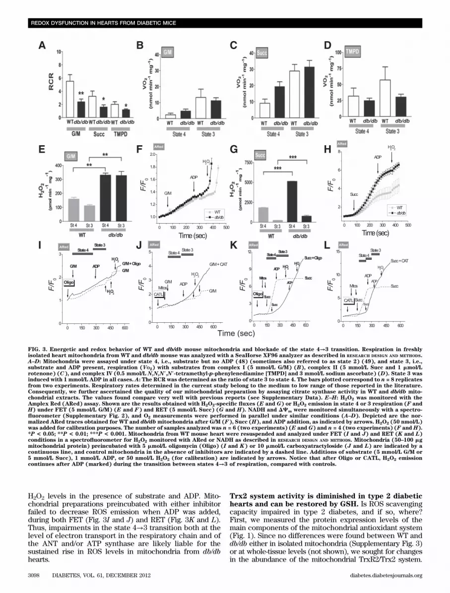
H2O2 levels in the presence of substrate and ADP. Mito-chondrial preparations preincubated with either inhibitorfailed to decrease ROS emission when ADP was added,during both FET (Fig. 3I and J) and RET (Fig. 3K and L).Thus, impairments in the state 4→3 transition both at thelevel of electron transport in the respiratory chain and ofthe ANT and/or ATP synthase are likely liable for thesustained rise in ROS levels in mitochondria from db/dbhearts.
Trx2 system activity is diminished in type 2 diabetichearts and can be restored by GSH. Is ROS scavengingcapacity impaired in type 2 diabetes, and if so, where?First, we measured the protein expression levels of themain components of the mitochondrial antioxidant system(Fig. 1). Since no differences were found between WT anddb/db either in isolated mitochondria (Supplementary Fig. 3)or at whole-tissue levels (not shown), we sought for changesin the abundance of the mitochondrial TrxR2/Trx2 system.
FIG. 3. Energetic and redox behavior of WT and db/db mouse mitochondria and blockade of the state 4→3 transition. Respiration in freshlyisolated heart mitochondria from WT and db/db mouse was analyzed with a SeaHorse XF96 analyzer as described in RESEARCH DESIGN AND METHODS.A–D: Mitochondria were assayed under state 4, i.e., substrate but no ADP (48) (sometimes also referred to as state 2) (49), and state 3, i.e.,substrate and ADP present, respiration (VO2) with substrates from complex I (5 mmol/L G/M) (B), complex II (5 mmol/L Succ and 1 mmol/Lrotenone) (C), and complex IV (0.5 mmol/L N,N,N9,N9-tetramethyl-p-phenylenediamine [TMPD] and 3 mmol/L sodium ascorbate) (D). State 3 wasinduced with 1 mmol/L ADP in all cases. A: The RCR was determined as the ratio of state 3 to state 4. The bars plotted correspond to n = 8 replicatesfrom two experiments. Respiratory rates determined in the current study belong to the medium to low range of those reported in the literature.Consequently, we further ascertained the quality of our mitochondrial preparation by assaying citrate synthase activity in WT and db/db mito-chondrial extracts. The values found compare very well with previous reports (see Supplementary Data). E–H: H2O2 was monitored with theAmplex Red (ARed) assay. Shown are the results obtained with H2O2-specific fluxes (E and G) or H2O2 emission in state 4 or 3 respiration (F andH) under FET (5 mmol/L G/M) (E and F) and RET (5 mmol/L Succ) (G and H). NADH and DCm were monitored simultaneously with a spectro-fluorometer (Supplementary Fig. 2), and O2 measurements were performed in parallel under similar conditions (A–D). Depicted are the nor-malized ARed traces obtained for WT and db/dbmitochondria after G/M (F), Succ (H), and ADP addition, as indicated by arrows. H2O2 (50 nmol/L)was added for calibration purposes. The number of samples analyzed was n = 6 (two experiments) (E and G) and n = 4 (two experiments) (F and H).*P < 0.05; **P < 0.01; ***P < 0.001. Mitochondria from WT mouse heart were resuspended and analyzed under FET (I and J) and RET (K and L)conditions in a spectrofluorometer for H2O2 monitored with ARed or NADH as described in RESEARCH DESIGN AND METHODS. Mitochondria (50–100 mgmitochondrial protein) preincubated with 5 mmol/L oligomycin (Oligo) (I and K) or 10 mmol/L carboxyatractyloside (J and L) are indicated by acontinuous line, and control mitochondria in the absence of inhibitors are indicated by a dashed line. Additions of substrate (5 mmol/L G/M or5 mmol/L Succ), 1 mmol/L ADP, or 50 nmol/L H2O2 (for calibration) are indicated by arrows. Notice that after Oligo or CATL, H2O2 emissioncontinues after ADP (marked) during the transition between states 4→3 of respiration, compared with controls.
REDOX DYSFUNCTION IN HEARTS FROM DIABETIC MICE
3098 DIABETES, VOL. 61, DECEMBER 2012 diabetes.diabetesjournals.org

Again, no differences were observed (Fig. 4A). Therefore,we asked whether changes in Trx2 redox status, i.e.,oxidized/reduced (TrxSS/Trx[SH]2), occurred under non-energized and energized conditions (Supplementary Fig. 2),using G/M as a substrate (FET) (33). This approach revealedmarked differences between WT and db/db (Fig. 4B). Atbaseline, only 34% of Trx2 was in its reduced status, both inWT and db/db (Fig. 4B, top right). However, substrate ad-dition to energize mitochondria to state 4 led to significantlygreater Trx(SH)2 in WT versus db/dbmitochondria. Trx(SH)2increased in parallel with NADPH and DCm when G/M wasadded (Supplementary Fig. 2). In state 3, Trx2(SH)2 levelsremained as high as in state 4. Since the reducing power ofTrx2 is determined by the ratio TrxSS/Trx(SH)2, we calcu-lated the latter and expressed it as redox potential. Uponsubstrate addition to WT mitochondria, the redox potentialbecame more negative, thus more reducing, from 2294 6 5to 2325 6 2 mV. This increment was less pronounced inmitochondria from db/db mice, from 2294 6 5 to 2313 62 mV (Fig. 4B, bottom right). The 12-mV difference inTrx2 redox potential accounts for a significant 2.3-foldrise in the TrxSS/Trx(SH)2 ratio.
Finally, we investigated whether altered Trx2 redoxcould be reversed by treatment with exogenous GSH ethylester (GSHee). Preincubation of db/db mitochondria withGSHee caused a significant increase in Trx(SH)2 (from64 6 3 to 78 6 1%), equivalent to a twofold rise in the
TrxSS/Trx(SH)2 ratio (Fig. 4B, right). Noteworthy, thesevalues are comparable to those found in WT Trx(SH)2levels and similar to those found in state 3. Thus, in ad-dition to their energetic impairment, db/db mitochondriaexhibit diminished ROS scavenging capacity via the Trx2system.Function is impaired in db/db myocytes undercombined HG + ISO regimen. Next, we tested whetherthe HG + ISO regimen, which unveiled major redox/energetic deficits in cardiac db/db mitochondria, wouldreverberate on mechanical function and Ca2+ transient ofmyocytes from WT and db/db hearts. Changes in the am-plitude and kinetics of contractility and Ca2+ transients inWT and db/db myocytes were measured under normal(5 mmol/L) and HG conditions, in the presence or ab-sence of ISO, with and without Palm at two differentconcentrations.Minus Palm. Fractional shortening (FS) and Ca2+ tran-sient amplitude/kinetics were similar in WT versus db/dbmyocytes at 5 and 30 mmol/L glucose (SupplementaryTables 1 and 2). Next, we examined the effects of b-stimulationvia ISO on db/db and WT myocytes. At 5 mmol/L glucose,ISO response was similar in both groups (Table 1). Con-versely, HG + ISO unveiled marked differences betweendb/db and WT myocytes: in db/db, ISO-induced enhance-ment of contractility/relaxation was lower, coupled to al-tered Ca2+ kinetics (Table 2 and Fig. 5). These findings
FIG. 4. Abundance, redox status, and response of Trx system fromWT and db/dbmitochondria to preincubation with GSHee. Freshly isolated heartmitochondria from WT and db/dbmice were handled and analyzed for TrxR2 and Trx2 protein abundance by Western blot (WB) and for Trx-SS andTrx(SH)2 status by redox WB. A: Shown are three representative examples (left) and the statistical comparison between WT and db/db (n = 6experiments) (right). As in Supplementary Fig. 3, protein was normalized to total protein abundance based on Direct Blue 71 staining. Trx(SH)2and Trx-SS were determined in fresh, active mitochondria (150–200 mg mitochondrial protein), under baseline (nonenergized) (B), state 4 (St 4)(5 mmol/L G/M), and state 3 (St 3) (+1 mmol/L ADP) of respiration in paired samples as described elsewhere (33). B: Representative redox WB ofTrx2 from WT and db/db mitochondria (left), and the amount of Trx(SH)2 (right) (top, in %) and redox potential (bottom, in mV) comparativelybetween WT and db/db mitochondria, in the absence or presence of GSHee preincubation (3 mmol/L GSHee for 30 min; n = 6 experiments). *P <0.05; ***P < 0.001.
C.G. TOCCHETTI AND ASSOCIATES
diabetes.diabetesjournals.org DIABETES, VOL. 61, DECEMBER 2012 3099

suggest a deficit in cAMP-dependent protein kinase–mediatedenhancement in contractility and Ca2+ handling of db/dbmyocytes under HG.Plus Palm. Under HG, Palm had a significant negativeinfluence on the amplitude of Ca2+ transient and sarco-mere shortening in db/db myocytes at both concentrationstested (Supplementary Tables 1 and 2). Yet, its adminis-tration in db/db myocytes challenged with HG + ISO im-proved contractility and Ca2+ handling to a higher extentthan that seen in WT (Table 2). Palm preserved the car-diomyocyte response to the b-stimulant under HG inmyocytes from db/db hearts (Fig. 5 and Table 2) as well asin WT and db/db cardiomyocytes at 5 mmol/L glucose(Table 1). Thus, Palm is beneficial for db/db car-diomyocytes exposed to a concomitant energetic and re-dox challenge.GSH or Palm offsets mechanical dysfunction in db/dbcardiomyocytes challenged with HG and b-agonists.To strengthen the causative link between impaired con-tractility and the more oxidized redox status of db/dbcardiomyocytes, and to assess whether a shift in sub-strates may affect their function under challenging con-ditions, we tested the impact of cell-permeable GSHee orPalm. Exogenous GSHee offset ISO-mediated changes inROS and GSH levels of db/db cardiomyocytes exposed toHG (Supplementary Fig. 4); all functional indexes re-covered to WT levels after GSH (Fig. 5A–D). Moreover,GSHee replenished the thiol intracellular pool by ;20%,without significantly changing NADH levels (Supple-mentary Fig. 4, insets), countering, in turn, the rise inROS levels (Supplementary Fig. 4). To do so, GSHeeacted through the reconstitution of the Trx2 pool (Fig. 4),ultimately limiting ROS emission in mitochondria fromHG/ISO-challenged db/db mice and preventing myocytemechanical deficiency.
Even more so, Palm significantly improved both con-tractility and Ca2+ handling, beyond the levels required tofully offset HG + ISO adverse effects (Fig. 5A–D and Table2). This beneficial impact was coincident with a more re-duced redox status (Fig. 2), both under normal conditionsand HG. Thus, in diabetes, FAs may have a role that goesbeyond that of energetic providers (11,40); this contention
is reinforced by the fact that the impact of GSHee andPalm on myocyte function is not additive (Fig. 5).WT and db/db intact heart LV function andelectrophysiological performance. The combined HG +ISO regimen in the absence or presence of Palm uncoveredmajor differences in db/db and WT myocyte redox con-ditions (Fig. 2) and electro-contraction (E-C) couplingimpairment (Fig. 5) that were not apparent at baseline (Fig.2, Table 1, and Supplementary Table 1). These alterationswere underlined by mitochondrial energetic/redox alterations(Fig. 3). To evaluate the functional implications of thesechanges at the whole-heart level, we used Langendorff-perfused mouse hearts and high-resolution optical AP im-aging. With the former approach, we evaluated possiblechanges in LV function and coronary vascular response(CPP) under HG (Fig. 6 and Supplementary Fig. 6) andeuglycemia (EG) (Supplementary Figs. 5 and 7) in theabsence or presence of ISO and Palm (Fig. 6A–D and Sup-plementary Figs. 5–7A–D). As in cardiomyocytes (Fig. 5 andTable 2), whole WT hearts exhibited better LV function versusdb/db hearts when exposed to HG + ISO (Fig. 6A–D). Opticalmapping revealed that although HG alone did not alter APD inpaced WT and db/db hearts, the HG + ISO regimen uncoveredimportant functional differences between groups. Under HG+ ISO challenge, there was a significant decrease in bothAPD50 (P = 0.019) and 75 (P = 0.030) in db/db comparedwith WT hearts (Fig. 6E–H and Supplementary Fig. 8).
Notably, Palm (same concentration as in cardiomy-ocytes) significantly improved the functional performanceof db/db hearts exposed to HG + ISO by decreasing CPPand increasing LV-developed pressure (LVDP) concomi-tantly with the rates of db/db heart contraction (dP/dtmax)and relaxation (dP/dtmin) (Fig. 6C and D), whereas this FAdid not affect WT heart performance (Fig. 6A–D). Similarresults were obtained at both Palm concentrations testedunder HG (Fig. 6 and Supplementary Fig. 6). However, theLVDP, dP/dtmax, and dP/dtmin, but not CPP, of WT heartswere significantly decreased under EG + ISO at the twoPalm concentrations tested (Supplementary Figs. 5 and 7).In essence, Palm improved LV functional performance ofhearts from diabetic animals subjected to metabolic/redoxchallenge.
TABLE 1Contractility and Ca2+ transient kinetics in ventricular myocytes isolated from WT and db/db mice, under a normal (5 mmol/L) glucoseregimen in the presence of ISO (10 nmol/L), without or with Palm at two different concentrations
Glucose 5 mmol/L + ISO
WT db/db
Control Palm 0.4 mmol/L Palm 0.8 mmol/L Control Palm 0.4 mmol/L Palm 0.8 mmol/L
FS (%) 9.2 6 0.4 10.7 6 1.1 12.4 6 0.3(P , 0.05 vs.
control)
9.4 6 0.6 10.6 6 0.6 11.9 6 1.3
Ca2+ transient (%) 43.9 6 2.4 47.2 6 6.8 38.1 6 7.4 52 6 5 47.7 6 3.9 58 6 10.5T50% peak shortening (ms) 75 6 1 70 6 3
(P , 0.05 vs.control)
75 6 2 79 6 1 83 6 2 78 6 2
T50 Ca2+ transient (ms) 161 6 3 152 6 5 164 6 5 183 6 5(P , 0.001 vs.WT control)
179 6 9 164 6 8(P , 0.03 vs.
control)
Data represent mean 6 SEM from at least 20 cells for each group isolated from at least four different hearts. T50% peak shortening, time frombaseline to 50% peak shortening; T50 Ca2+ transient, time to 50% Ca2+ transient decay.
REDOX DYSFUNCTION IN HEARTS FROM DIABETIC MICE
3100 DIABETES, VOL. 61, DECEMBER 2012 diabetes.diabetesjournals.org

DISCUSSION
Here we show that energetically/redox-stressed cardio-myocytes from type 2 diabetic db/db mice display impairedmitochondrial state 4→3 energetic transition, accountingfor uninhibited ROS emission, under both FET and RETmodes. Deficits in local pools of mitochondrial GSH andTrx2 account for this amplified ROS emission. The resultingmore-oxidizing intracellular environment correlates withaltered E-C coupling in cardiomyocytes/intact hearts. Thesealterations are causative and not incidental because exog-enous GSH rescues both mitochondrial/cellular redox bal-ance and E-C coupling. Also, shifting substrates, i.e., the useof the FA Palm, reestablishes proper myocyte redox milieuwhile improving function in metabolically/redox-challengedwhole hearts.
The balance between ROS production in the respiratorychain and the efficiency of the ROS scavenging systemsdetermines mitochondrial H2O2 emission as a function ofthe redox environment (29). Here we assign to mitochon-drial impairment in the key state 4→3 energetic transition,and defective mitochondrial GSH and Trx2 systems, aleading role in the excess ROS emission occurring in db/dbmyocytes, ultimately resulting in myocyte/whole-heartfunctional impairment. Uncoupling of OxPhos, along withthe inability of mitochondria to decrease ROS levels duringthe state 4→3 transition under FET, plays a major role inthe energetic/redox dysfunction. Recent data show thatunder states 4 and 3, mouse heart mitochondria diverta significant portion of total respiratory flow to ROS gen-eration, which is continuously offset by GSH/Trx scav-enging systems (30,33).
Indeed, HG + ISO provoked a concomitant rise in ROSlevels and GSH depletion that was associated with con-tractile and Ca2+ handling failure. Mechanistically, the re-setting of mitochondrial and cellular redox balanceoperated by exogenous GSH was mediated by the TrxR2/Trx2/peroxiredoxin 3 (Prx3) system, whose redox poten-tial became more reduced in the presence of additionalcytoplasmic GSH. The augmented scavenging capacitybestowed by GSHee could also indirectly increase SODactivity by lowering H2O2, which exerts a negative feed-back on this enzyme (41) (Supplementary Fig. 4). Herein,
intact ex vivo perfused db/db hearts exhibited altered vas-cular tone, impaired LV function, and significant shorteningof APD compared with WT hearts. These deficits are allcrucial for maintaining proper E-C coupling, and thuscontraction, under both basal and increased-workloadconditions. A possible mechanism responsible for APDshortening is the nonischemic activation of sarcolemmalATP-sensitive (K-ATP) channels, reflecting energeticshortage when energy demand is high in db/db hearts.Impaired myocardial Ca2+ handling is known to be in-volved in diabetic cardiomyopathy (42). Our study showsthat these alterations become particularly prominent whendiabetic cells are subjected to redox/energetic stress, thusexpanding previous observations showing that b-adrenergicresponse may be blunted in db/db myocytes (42,43).
The status of diabetic cardiomyopathy is maintained bythe compounding effects of HG, mitochondrial energydeficit, and oxidative stress. However, no major contractiledeficits are evident under HG alone, even in db/db hearts.This prompted us to use conditions of combined energetic/redox challenge (44). Since myocardial glucose metabo-lism (e.g., transport and glycolysis) is decreased and FAoxidation increased in diabetes (11), we analyzed the ef-fect of the saturated FA Palm on intracellular redox,contractility, and Ca2+ handling in myocytes/hearts. Palmdetermined a transition from oxidized-to-reduced cellularredox status in db/db cardiomyocytes, abating ROS levelsdrastically. This effect was coupled to a marked GSH riseboth in WT and db/db myocytes (Fig. 2B). Consequently,Palm significantly improved ISO-induced contractile re-serve in db/db cardiomyocytes. Conversely, its absencewas accompanied by marked deficits in Ca2+ handling andmyocyte mechanical properties (Fig. 5A–D). Myocardialsubstrate use in patients with diabetes involves increaseddelivery of FAs (3,11,45). At the whole-heart level, Palmproduced a substantial improvement in LV function indb/db hearts exposed to metabolic/redox stress, anda relative decrease in the WT. Previous reports have el-egantly shown that, in the presence of 11 mmol/L glucose,Palm has a negative impact on LV function of db/db mice(25). Present evidence is consistent with these findings,and may help to mechanistically explain the decline inLV function and efficiency seen in db/db hearts with
TABLE 2Contractility and Ca2+ transient kinetics in ventricular myocytes isolated from WT and db/db mice, under HG in the presence of ISO(10 nmol/L), without and with Palm at two different concentrations
Glucose 30 mmol/L + ISO
WT db/db
Control Palm 0.4 mmol/L Palm 0.8 mmol/L Control Palm 0.4 mmol/L Palm 0.8 mmol/L
FS (%) 8.99 6 0.6 14.8 6 0.7(P , 0.001 vs.
control)
10.8 6 1.0 7.4 6 0.7(P , 0.02 vs.WT control)
14.1 6 1.1(P , 0.001 vs.
control)
12.1 6 1.3(P , 0.002 vs.
control)Ca2+ transient (%) 44.7 6 2.9 64.6 6 5.3
(P , 0.05 vs.control)
50.9 6 5.8 49 6 3.6 43.8 6 4.1 55 6 9.7
T50% peak shortening (ms) 78 6 1 69 6 1(P , 0.001 vs.
control)
74 6 1 80 6 1 78 6 1 74 6 1(P , 0.01 vs.
control)T50 Ca2+ transient (ms) 159 6 2 150 6 2 147 6 6 181 6 5
(P , 0.001 vs.WT control)
175 6 11 164 6 7(P , 0.04 vs.
control)
Data represent mean 6 SEM from at least 20 cells for each group isolated from at least four different hearts. T50% peak shortening, time frombaseline to 50% peak shortening; T50 Ca2+ transient, time to 50% Ca2+ transient decay.
C.G. TOCCHETTI AND ASSOCIATES
diabetes.diabetesjournals.org DIABETES, VOL. 61, DECEMBER 2012 3101

Palm + glucose (25). However, here we aimed to testa different question, i.e., how isolated db/db myocytes/hearts respond to increased metabolic/redox stress usingthe HG + ISO regimen. Although Palm partially worsenedLV function in WT hearts, it definitely improved the per-formance of db/db hearts subjected to HG + ISO. Thus, ourstudy suggests that in animals with type 2 diabetes, FAs areimportant for handling increased workload that demandshigher energy supply while exposing cardiac tissue to thelikelihood of ROS imbalance (46).
FAs are a major source of acetyl-coenzyme A for thetricarboxylic acid cycle, contributing 60–70% of myocar-dial ATP at physiological levels (40). However, the higherenergetic budget provided by Palm (three times higherthan from glucose) is produced up to two-thirds in theform of reducing equivalents (24 NADH and 8 FADH2)from b-oxidation in mitochondria. This reducing powercontributes electrons to antioxidant systems and the mi-tochondrial respiratory/energetic machinery (Fig. 1). The
significantly lower levels of GSH found in db/db car-diomyocytes argue in favor of a more oxidized and con-stitutively compromised supply of reducing equivalentsto the GSH/Trx systems through NADPH (Fig. 1). Therescue of Trx(SH)2 to WT levels via GSHee supports thiscontention, suggesting that the achievement of a morebalanced redox status could be mediated by the TrxR2/Trx2/Prx3 pathway, a major controller of H2O2 emissionfrom mitochondria (30,33). The recovery of contractileactivity observed in db/db cardiomyocytes preincubatedwith GSHee, along with a more-reduced redox status incells, and of Trx2 in mitochondria, demonstrates for thefirst time that changes in mitochondrial pools of GSH andTrx2 can have a negative impact that propagates from mi-tochondria to the function of myocytes in challenged db/dbhearts.
Concerning the role of FAs, one intrinsic limitation ofour study is that we tested only the acute effects of Palm.In a more chronic stand, the upregulation of FA uptake is
FIG. 5. Contractile behavior of db/db cardiomyocytes under EG and hyperglycemia without or with b-adrenergic stimulation and GSHee or Palmpreincubation. Cardiomyocytes from db/db mouse hearts were isolated, handled, and analyzed for cell shortening (A and C) and Ca
2+transients
(B and D), in parallel with the imaging studies (Fig. 2). Left: Representative traces of the main experimental situations. EG and HG were used inthe absence or presence of b-adrenergic stimulation with 10 nmol/L ISO and without or with preincubation in the presence of 4 mmol/L GSHee for 3 hor Palm (0.4 or 0.8 mmol/L for WT and db/db, respectively), at room temperature (Tables 1 and 2). The number of samples analyzed was n = 20–30(from four to six hearts). T50% peak shortening, from baseline to 50% peak shortening; TR50, time to 50% relengthening; T50 Ca
2+, time to 50%
Ca2+
transient decay. *P < 0.05; **P < 0.01.
REDOX DYSFUNCTION IN HEARTS FROM DIABETIC MICE
3102 DIABETES, VOL. 61, DECEMBER 2012 diabetes.diabetesjournals.org

Gluc30
FIG. 6. LV function in Langendorff-perfused hearts and high-resolution optical AP imaging. Hearts were harvested, handled, and perfused asdescribed in RESEARCH DESIGN AND METHODS. Hearts were paced with a Radnoti pacing electrode (Monrovia, CA) at 600 bpm (10 Hz, 4-ms duration,4 V) using a Grass stimulator (Grass Instruments Co., Quincy, MA). LV function was monitored with a water-filled, customized latex balloonconnected to a P23XL pressure transducer with interface cable (Harvard Apparatus Instruments, Holliston, MA) and coupled to a BIOPACSystem (DA100, Santa Barbara, CA) for continuous data recording and offline analysis. LV end-diastolic pressure was set at 5–10 mmHg byadjusting the balloon volume with a Gilmont micrometer syringe (Cole-Parmer, Vernon Hills, IL). After stabilization in KH buffer (11 mmol/Lglucose; EG), the heart was exposed to ISO (10 nmol/L), followed by a drug-free perfusion period to recover baseline parameters. The sameheart was perfused for 1 h with KH buffer containing 30 mmol/L glucose, followed by exposure to ISO again. Shown are (in mmHg) the CPP,LVDP, and (in mmHg/s) maximal rates of contraction (dP/dtmax) and relaxation (dP/dtmin) observed under HG (30 mmol/L glucose), in theabsence or presence of ISO (10 nmol/L) and Palm (WT, 0.4 mmol/L; db/db, 0.8 mmol/L). The data presented correspond to n = 4 hearts in eachgroup of WT or db/db. The same protocol was applied at Palm 0.2 mmol/L WT and 0.4 mmol/L db/db under EG and HG (n = 6 hearts from eachgroup; see Supplementary Figs. 5–7). APD was measured in paced (140-ms pacing cycle length [PCL]) WT and db/db hearts during perfusionunder baseline, HG, and HG + ISO. The CCD-based, high-resolution optical mapping system is capable of measuring APs from 6,400 pixels of the4 3 4-mm
2epicardial surface with high temporal (1 ms) and spatial (50 mm) resolutions in intact murine hearts. Also measured were epicardial
conduction velocity (CV) and normalized AP upstroke velocity under the same conditions (Supplementary Fig. 8). E and F: Representativeepicardial APs measured from WT (red) and db/db (blue) mice during pacing at 140 ms PCL. Also shown is a CCD-based image of the mappedepicardial region with the pacing electrode in the lower left corner. G and H: The HG + ISO regimen uncovered a significant shortening of APD50and APD75 in db/db but not WT hearts. *P < 0.05; **P < 0.01.
C.G. TOCCHETTI AND ASSOCIATES
diabetes.diabetesjournals.org DIABETES, VOL. 61, DECEMBER 2012 3103

known to promote the accumulation of intracellular lipid,resulting in lipotoxic effects (47), likely due to deficits inlipase function, ultimately leading to cardiac disease(11,25). Notwithstanding, our findings suggest that, whenproperly used, FAs are important suppliers of reducingequivalents, particularly when db/db myocytes/hearts areexposed to HG and more workload.
ACKNOWLEDGMENTS
This work was supported by National Institutes of HealthGrant R01-HL-091923-01. C.G.T. was supported by Inter-national Society for Heart Research–European Section/Servier; V.C. by Coordenação de Aperfeiçoamento dePessoal de Nível Superior (CAPES), Brazil; and S.S. bythe National Natural Science Foundation of China Grant81100155.
No potential conflicts of interest relevant to this articlewere reported.
C.G.T., V.C., B.A.S., C.X., and S.S. researched data andcontributed to discussion. W.H.W., B.O., and R.C.S.-B.contributed to discussion and reviewed and edited themanuscript. S.C. and F.G.A. researched data, contributedto discussion, and reviewed and edited the manuscript.N.P. and M.A.A. researched data, contributed to discus-sion, and wrote the manuscript. M.A.A. is the guarantor ofthis work and, as such, had full access to all the data in thestudy and takes responsibility for the integrity of the dataand the accuracy of the data analysis.
Parts of this study were presented as a poster at the 54thAnnual Meeting of the Biophysical Society, San Francisco,California, 20–24 February 2010; 55th Annual Meeting ofthe Biophysical Society, Baltimore, Maryland, 5–9 March2011; and 56th Annual Meeting of the Biophysical Society,San Diego, California, 25–29 February 2012.
REFERENCES
1. Bell DSH. Heart failure: the frequent, forgotten, and often fatal complica-tion of diabetes. Diabetes Care 2003;26:2433–2441
2. Laakso M. Hyperglycemia and cardiovascular disease in type 2 diabetes.Diabetes 1999;48:937–942
3. Boudina S, Abel ED. Diabetic cardiomyopathy, causes and effects. RevEndocr Metab Disord 2010;11:31–39
4. Cohen-Solal A, Beauvais F, Logeart D. Heart failure and diabetes mellitus:epidemiology and management of an alarming association. J Card Fail2008;14:615–625
5. Bruce DG, Chisholm DJ, Storlien LH, Kraegen EW, Smythe GA. The effectsof sympathetic nervous system activation and psychological stress onglucose metabolism and blood pressure in subjects with type 2 (non-insulin-dependent) diabetes mellitus. Diabetologia 1992;35:835–843
6. Colberg SR, Albright AL, Blissmer BJ, et al. Exercise and type 2 diabetes:American College of Sports Medicine and the American Diabetes Associ-ation: joint position statement. Exercise and type 2 diabetes. Med SciSports Exerc 2010;42:2282–2303
7. Fang ZY, Sharman J, Prins JB, Marwick TH. Determinants of exercisecapacity in patients with type 2 diabetes. Diabetes Care 2005;28:1643–1648
8. Dobrin JS, Lebeche D. Diabetic cardiomyopathy: signaling defects andtherapeutic approaches. Expert Rev Cardiovasc Ther 2010;8:373–391
9. McGarry JD. What if Minkowski had been ageusic? An alternative angle ondiabetes. Science 1992;258:766–770
10. Wisneski JA, Gertz EW, Neese RA, Mayr M. Myocardial metabolism of freefatty acids. Studies with 14C-labeled substrates in humans. J Clin Invest1987;79:359–366
11. Lopaschuk GD, Ussher JR, Folmes CD, Jaswal JS, Stanley WC. Myocardialfatty acid metabolism in health and disease. Physiol Rev 2010;90:207–258
12. Cryer PE. Disorders of sympathetic neural function in human diabetesmellitus: hypoadrenergic and hyperadrenergic postural hypotension. Me-tabolism 1980;29(Suppl. 1):1186–1189
13. Huggett RJ, Scott EM, Gilbey SG, Stoker JB, Mackintosh AF, Mary DA.Impact of type 2 diabetes mellitus on sympathetic neural mechanisms inhypertension. Circulation 2003;108:3097–3101
14. Sivitz WI, Yorek MA. Mitochondrial dysfunction in diabetes: from molec-ular mechanisms to functional significance and therapeutic opportunities.Antioxid Redox Signal 2010;12:537–577
15. Styskal J, Van Remmen H, Richardson A, Salmon AB. Oxidative stress anddiabetes: what can we learn about insulin resistance from antioxidantmutant mouse models? Free Radic Biol Med 2012;52:46–58
16. Maechler P, Jornot L, Wollheim CB. Hydrogen peroxide alters mitochon-drial activation and insulin secretion in pancreatic beta cells. J Biol Chem1999;274:27905–27913
17. Friederich M, Hansell P, Palm F. Diabetes, oxidative stress, nitric oxideand mitochondria function. Curr Diabetes Rev 2009;5:120–144
18. Choi SW, Benzie IF, Ma SW, Strain JJ, Hannigan BM. Acute hyperglycemiaand oxidative stress: direct cause and effect? Free Radic Biol Med 2008;44:1217–1231
19. Evans JL, Goldfine ID, Maddux BA, Grodsky GM. Oxidative stress andstress-activated signaling pathways: a unifying hypothesis of type 2 di-abetes. Endocr Rev 2002;23:599–622
20. Brownlee M. Advanced protein glycosylation in diabetes and aging. AnnuRev Med 1995;46:223–234
21. Lowell BB, Shulman GI. Mitochondrial dysfunction and type 2 diabetes.Science 2005;307:384–387
22. Akar FG, Aon MA, Tomaselli GF, O’Rourke B. The mitochondrial origin ofpostischemic arrhythmias. J Clin Invest 2005;115:3527–3535
23. Aon MA, Cortassa S, Akar FG, Brown DA, Zhou L, O’Rourke B. From mito-chondrial dynamics to arrhythmias. Int J Biochem Cell Biol 2009;41:1940–1948
24. Slodzinski MK, Aon MA, O’Rourke B. Glutathione oxidation as a trigger ofmitochondrial depolarization and oscillation in intact hearts. J Mol CellCardiol 2008;45:650–660
25. Boudina S, Sena S, Theobald H, et al. Mitochondrial energetics in the heartin obesity-related diabetes: direct evidence for increased uncoupled res-piration and activation of uncoupling proteins. Diabetes 2007;56:2457–2466
26. Anderson EJ, Lustig ME, Boyle KE, et al. Mitochondrial H2O2 emissionand cellular redox state link excess fat intake to insulin resistance in bothrodents and humans. J Clin Invest 2009a;119:573–581
27. Ren J, Pulakat L, Whaley-Connell A, Sowers JR. Mitochondrial biogenesisin the metabolic syndrome and cardiovascular disease. J Mol Med (Berl)2010;88:993–1001
28. Buchanan J, Mazumder PK, Hu P, et al. Reduced cardiac efficiency and al-tered substrate metabolism precedes the onset of hyperglycemia and con-tractile dysfunction in two mouse models of insulin resistance and obesity.Endocrinology 2005;146:5341–5349
29. Aon MA, Cortassa S, O’Rourke B. Redox-optimized ROS balance: a unify-ing hypothesis. Biochim Biophys Acta 2010b;1797:865–877
30. Aon MA, Stanley BA, Sivakumaran V, et al. Glutathione/thioredoxin systemsmodulate mitochondrial H2O2 emission: an experimental-computationalstudy. J Gen Physiol 2012;139:479–491
31. Aon MA, Cortassa S, Maack C, O’Rourke B. Sequential opening of mito-chondrial ion channels as a function of glutathione redox thiol status.J Biol Chem 2007;282:21889–21900
32. Tocchetti CG, Wang W, Froehlich JP, et al. Nitroxyl improves cellular heartfunction by directly enhancing cardiac sarcoplasmic reticulum Ca2+ cy-cling. Circ Res 2007;100:96–104
33. Stanley BA, Sivakumaran V, Shi S, et al. Thioredoxin reductase-2 is es-sential for keeping low levels of H(2)O(2) emission from isolated heartmitochondria. J Biol Chem 2011;286:33669–33677
34. Go Y-M, Jones DP. Thioredoxin redox Western analysis. Current Protocolsin Toxicology 2009;17.12(Suppl. 41):1–12
35. Roepke TK, Kontogeorgis A, Ovanez C, et al. Targeted deletion of kcne2impairs ventricular repolarization via disruption of I(K,slow1) and I(to,f).FASEB J 2008;22:3648–3660
36. Akar FG, Tomaselli GF. Conduction abnormalities in nonischemic dilatedcardiomyopathy: basic mechanisms and arrhythmic consequences. TrendsCardiovasc Med 2005;15:259–264
37. Boardman N, Hafstad AD, Larsen TS, Severson DL, Aasum E. Increased O2cost of basal metabolism and excitation-contraction coupling in heartsfrom type 2 diabetic mice. Am J Physiol Heart Circ Physiol 2009;296:H1373–H1379
38. Schönfeld P, Wojtczak L. Fatty acids as modulators of the cellular pro-duction of reactive oxygen species. Free Radic Biol Med 2008;45:231–241
39. Wei AC, Aon MA, O’Rourke B, Winslow RL, Cortassa S. Mitochondrialenergetics, pH regulation, and ion dynamics: a computational-experimentalapproach. Biophys J 2011;100:2894–2903
40. Lopaschuk GD. Metabolic abnormalities in the diabetic heart. Heart FailRev 2002;7:149–159
REDOX DYSFUNCTION IN HEARTS FROM DIABETIC MICE
3104 DIABETES, VOL. 61, DECEMBER 2012 diabetes.diabetesjournals.org

41. Cortassa S, Aon MA, Winslow RL, O’Rourke B. A mitochondrial oscil-lator dependent on reactive oxygen species. Biophys J 2004;87:2060–2073
42. Belke DD, Dillmann WH. Altered cardiac calcium handling in diabetes.Curr Hypertens Rep 2004;6:424–429
43. Pereira L, Matthes J, Schuster I, et al. Mechanisms of [Ca2+]i transientdecrease in cardiomyopathy of db/db type 2 diabetic mice. Diabetes 2006;55:608–615
44. Bahmani F, Bathaie SZ, Aldavood SJ, Ghahghaei A. Glycine therapy in-hibits the progression of cataract in streptozotocin-induced diabetic rats.Mol Vis 2012;18:439–448
45. Leone TC, Kelly DP. Transcriptional control of cardiac fuel metabolism andmitochondrial function. Cold Spring Harb Symp Quant Biol 2011 76:175–182
46. Kohlhaas M, Liu T, Knopp A, et al. Elevated cytosolic Na+ increases mi-tochondrial formation of reactive oxygen species in failing cardiac my-ocytes. Circulation 2010;121:1606–1613
47. Brookheart RT, Michel CI, Schaffer JE. As a matter of fat. Cell Metab 2009;10:9–12
48. Chance B, Williams GR. Respiratory enzymes in oxidative phosphoryla-tion. III. The steady state. J Biol Chem 1955;217:409–427
49. Nicholls DG, Ferguson SJ. Bioenergetics 3. London, San Diego, AcademicPress, 2002
C.G. TOCCHETTI AND ASSOCIATES
diabetes.diabetesjournals.org DIABETES, VOL. 61, DECEMBER 2012 3105


