Embed Size (px)
Citation preview

67
H+-Gated Cation Channelsa
RAINER WALDMANN, GUY CHAMPIGNY, ERIC LINGUEGLIA, JAN R. DE WEILLE, C. HEURTEAUX, AND MICHEL LAZDUNSKIb
IPMC-CNRS, 660 route des Lucioles, Sophia Antipolis, 06560 Valbonne, France
ABSTRACT: H+-gated cation channels are members of a new family of ionic chan-nels, which includes the epithelial Na+ channel and the FMRFamide-activated Na+
channel. ASIC, the first member of the H+-gated Na+ channel subfamily, is expressedin brain and dorsal root ganglion cells (DRGs). It is activated by pHe variationsbelow pH 7. The presence of this channel throughout the brain suggests that the H+
might play an essential role as a neurotransmitter or neuromodulator. The ASICchannel is also present in dorsal root ganglion cells, as is its homolog DRASIC, whichis specifically present in DRGs and absent in the brain. Since external acidification isa major factor in pain associated with inflammation, hematomas, cardiac or muscleischemia, or cancer, these two channel proteins are potentially central players in painperception. ASIC activates and inactivates rapidly, while DRASIC has both a fastand sustained component. Other members of this family such as MDEG1 andMDEG2 are either H+-gated Na+ channels by themselves (MDEG1) or modulators ofH+-gated channels formed by ASIC and DRASIC. MDEG1 is of particular interestbecause the same mutations that produce selective neurodegeneration in C. elegansmechanosensitive neurons, when introduced in MDEG1, also produce neurodegener-ation. MDEG2 is selectively expressed in DRGs, where it assembles with DRASIC toradically change its biophysical properties, making it similar to the native H+-gatedchannel, which is presently the best candidate for pain perception.
+-gated cation channels are ligand-gated ion channels activated by the simplest ligandpossible. H+-gated cation channels with different pH sensitivities and kinetics were
reported in sensory neurons,1–7 in neurons of the central nervous system (CNS),7–9 and inoligodentrocytes.10 The extracellular pH in a tissue can decrease by more than two pHunits during tissue acidosis11 that accompanies inflammation and many ischemic condi-tions, and there is very convincing evidence that the sensation of pain parallels thedecrease in pH.12 H+-gated cation channels in sensory nerve endings were therefore pro-posed to be involved in the perception of pain that accompanies tissue acidosis.1,6,11
The recent cloning of four H+-gated cation channel subunits showed that they are mem-bers of the NaC/DEG superfamily. This family includes (i) epithelial Na+ channel (ENaC)subunits;13–17 (ii) a family of proteins designated as degenerins (DEG); after mutationsleading to a gain of function, these produce degeneration in mechanoreceptor neurons,which are required for touch sensation in the nematode18–22 and which could be mechano-sensitive channels; and (iii) a peptide-gated Na+ channel, FaNaC, which is directly gatedby the cardioexcitatory peptide FMRFamide in the snail Helix aspersa.23
The first H+-gated channel subunit was cloned only recently and designated asASIC124 (for acid sensing ionic channel 1). Like other members of the NaC/DEG fam-
aThis paper is dedicated to the memory of our friend and colleague Dr. Guy Champigny, who hascontributed in a major way to our current understanding of the properties of this ion channel family.
bCorresponding author. Institut de Pharmacologie Moléculaire et Cellulaire, CNRS, 660 route desLucioles, Sophia Antipolis, 06560 Valbonne, France. Phone: 33 (0)4 93 95 77 02 or 03; fax: 33 (0)493 95 77 04; e-mail: [email protected]
H
68 ANNALS NEW YORK ACADEMY OF SCIENCES
ily,22,25 it has two transmembrane domains with a large extracellular protein component.24
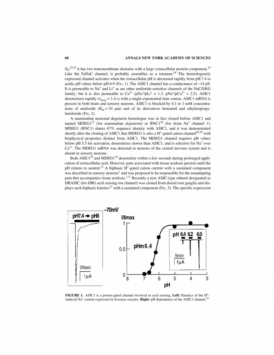
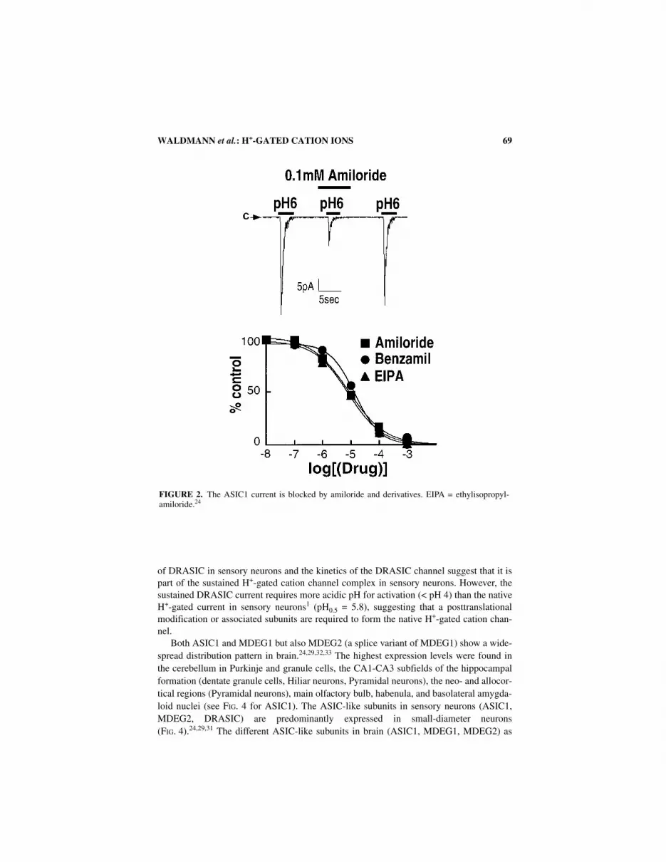
Like the FaNaC channel, it probably assembles as a tetramer.26 The heterologouslyexpressed channel activates when the extracellular pH is decreased rapidly from pH 7.4 toacidic pH values below pH 6.9 (FIG. 1). The ASIC1 channel has a conductance of ~14 pS.It is permeable to Na+ and Li+ as are other amiloride-sensitive channels of the NaC/DEGfamily; but it is also permeable to Ca2+ (pNa+/pLi+ = 1.3, pNa+/pCa2+ = 2.5). ASIC1desensitizes rapidly (τinact = 1.4 s) with a single exponential time course. ASIC1 mRNA ispresent in both brain and sensory neurons. ASIC1 is blocked by 0.1 to 1-mM concentra-tions of amiloride (KD = 10 µm) and of its derivatives benzamil and ethylisopropy-lamiloride (FIG. 2).
A mammalian neuronal degenerin homologue was in fact cloned before ASIC1 andnamed MDEG127 (for mammalian degenerin) or BNC128 (for brain Na+ channel 1).MDEG1 (BNC1) shares 67% sequence identity with ASIC1, and it was demonstratedshortly after the cloning of ASIC1 that MDEG1 is also a H+-gated cation channel29,30 withbiophysical properties distinct from ASIC1. The MDEG1 channel requires pH valuesbelow pH 5.5 for activation, desensitizes slower than ASIC1, and is selective for Na+ overCa2+. The MDEG1 mRNA was detected in neurons of the central nervous system and isabsent in sensory neurons.
Both ASIC124 and MDEG130 desensitize within a few seconds during prolonged appli-cation of extracellular acid. However, pain associated with tissue acidosis persists until thepH returns to neutral.12 A biphasic H+-gated cation current with a sustained componentwas described in sensory neurons1 and was proposed to be responsible for the nonadaptingpain that accompanies tissue acidosis.1,11 Recently a new ASIC-type subunit designated asDRASIC (for DRG acid sensing ion channel) was cloned from dorsal root ganglia and dis-plays such biphasic kinetics31 with a sustained component (FIG. 3). The specific expression
FIGURE 1. ASIC1 is a proton-gated channel involved in acid sensing. Left: Kinetics of the H+-induced Na+ current expressed in Xenopus oocytes. Right: pH dependence of the ASIC1 channel.24
WALDMANN et al.: H+-GATED CATION IONS 69
of DRASIC in sensory neurons and the kinetics of the DRASIC channel suggest that it ispart of the sustained H+-gated cation channel complex in sensory neurons. However, thesustained DRASIC current requires more acidic pH for activation (< pH 4) than the nativeH+-gated current in sensory neurons1 (pH0.5 = 5.8), suggesting that a posttranslationalmodification or associated subunits are required to form the native H+-gated cation chan-nel.
Both ASIC1 and MDEG1 but also MDEG2 (a splice variant of MDEG1) show a wide-spread distribution pattern in brain.24,29,32,33 The highest expression levels were found inthe cerebellum in Purkinje and granule cells, the CA1-CA3 subfields of the hippocampalformation (dentate granule cells, Hiliar neurons, Pyramidal neurons), the neo- and allocor-tical regions (Pyramidal neurons), main olfactory bulb, habenula, and basolateral amygda-loid nuclei (see FIG. 4 for ASIC1). The ASIC-like subunits in sensory neurons (ASIC1,MDEG2, DRASIC) are predominantly expressed in small-diameter neurons(FIG. 4).24,29,31 The different ASIC-like subunits in brain (ASIC1, MDEG1, MDEG2) as
FIGURE 2. The ASIC1 current is blocked by amiloride and derivatives. EIPA = ethylisopropyl-amiloride.24

70 ANNALS NEW YORK ACADEMY OF SCIENCES
well as the ASIC-like subunits in sensory neurons (ASIC1, MDEG2, DRASIC) are appar-ently coexpressed in the same neurons.29,33
Heteromultimeric assembly of subunits is commonly found in the DEG/NaC family ofion channels. The ENaC channel is a heteromultimer of three different types of subunit α,β, and γ,15 and genetic evidence indicates that the association of the nematode degenerinMEC4 and MEC10 is essential for the mechanosensitive function.20 Therefore, the appar-ent colocalization that we observed immediately suggested that ASIC/DRASIC/MDEG1subunits also form heteromultimeric channels. This was indeed recently demonstrated forseveral subunit combinations in a variety of ways29,33 (FIG. 5). For example, MDEG2, asplice variant of MDEG1 in which the first 236 amino acids of MDEG1 are replaced by anovel sequence turned out to be inactive when expressed alone. However coexpression ofthis splice variant MDEG2 modified the properties of both the DRASIC and MDEG1channel subunits.29 The homomultimeric MDEG1 channel has single exponential inactiva-tion kinetics and is highly selective for Na+. When coexpressed with MDEG2, the inacti-vation kinetics becomes biphasic, with a very slowly inactivating component thatdiscriminates only poorly between Na+ and K+. When MDEG2 is coexpressed with DRA-SIC, similar selectivity changes were observed, and the sustained Na+-selective DRASICcurrent (FIG. 3) become nonselective (pNa+ = pK+) (FIG. 5). Interestingly, the sustainedcomponent of the native biphasic H+-gated cation channel recorded from sensory neurons
FIGURE 3. The expression of the DRASIC channel in COS cells. A. Biphasic kinetics of the chan-nel activity after a pH jump from pH 7.3 to pH 4.31 B. Unitary currents recorded from an outside-outpatch held at −50mV.31 C. Current-voltage relations indicating the Na+ selectivity of both the peakand the sustained components of the DRASIC channel.31

WALDMANN et al.: H+-GATED CATION IONS 71
FIGURE 4. In situ hybridization indicating the distribution of ASIC1 in the brain (A) and in dorsal rootganglia (B). (a) low-power image; (b) high-power image.

72 ANNALS NEW YORK ACADEMY OF SCIENCES
also does not discriminate between Na+ and K+,1 suggesting that heteromultimeric DRA-SIC/MDEG2 channels are formed in vivo. Heteromultimeric assembly of ASIC subunitswas directly demonstrated for ASIC1 and MDEG1 by coimmunoprecipitation afterexpression in SF9 cells.33
Mutations in the C. elegans degenerins cause late-onset neurodegeneration in the nem-atode.18–20 Most of those mutations concern a substitution of an alanine situated justbefore the second transmembrane region for a bulkier amino acid, such as valine or pheny-lanaline. They were proposed to cause gain-of-function of the putative degenerin chan-nel.20 Interestingly, identical mutations introduced into ASIC and MDEG1 subunits canalso cause constitutive channel activity (shown for ASIC133 and MDEG127) as well as celldeath.27 Gain-of-function of the C. elegans degenerins causes neurodegeneration. Consti-tutively active ASIC/MDEG1 subunits have the potential to kill neurons, and it is possiblethat ASIC/MDEG1 mutations are involved in human forms of neurodegeneration just asmutations of their C. elegans homologues causes neuronal death in the nematode.
Beside creating constitutive Na+ channel activity, mutations in MDEG1 correspondingto those in the nematode degenerins sequences (FIG. 6) also drastically change the gatingproperties of the MDEG1 channel. While the MDEG1 channel is activated only at rela-tively acidic pH (pH0.5 = 4.1),30 mutations replacing the key Gly residue situated just
FIGURE 5. Assembly of MDEG2 with the DRASIC subunit changes the ionic selectivity of thesustained component of the current.
FIGURE 6. Mutations that lead to neurodegeneration of mechanosensitive neurons in C. eleganslead to drastic changes of both the kinetics and pH dependence of the channel when introduced inMDEG1. A. Higher part, comparative sequences of the C. elegans degenerin MEC4 and MDEG1just before the second transmembrane domain MII and identification of the Ala residue, which ismutated in nematode degenerins, and of the corresponding Gly430 residue in MDEG1. Lower part,kinetic changes observed when replacing Gly430 by other amino acids with bulky side chains. WT:wild type. B. Changes in the pH dependence of the MDEG1 channel after replacement of Gly430 bySer, Cys, Phe, Thr, and Val residues.

WALDMANN et al.: H+-GATED CATION IONS 73

74 ANNALS NEW YORK ACADEMY OF SCIENCES
before the second transmembrane domain (FIG. 6) by a Val, a Phe, or any residue bulkierthan Gly drastically alters the inactivation of the channel. It can even completely abolish it,and the channel then becomes permanently open after an acidification (FIG. 6). Thesemutations also drastically shift the pH dependence of the MDEG1 channel to much higherpH values (FIG. 6). The replacement of Gly430 by a Phe shifts the pH0.5 value from pH 4.1to pH 6.9. Clearly the protein domain situated just before the second transmembrane seg-ment is essential for the gating of H+-activated Na+ channels.
H+-gated cation channels are acid sensors, and this family of ion channels probably ful-fills this role throughout the body. Tissue acidosis causes pain,12 and an involvement ofH+-gated cation channels in nociception seems most likely. However, nociception cannotbe the only role of H+-gated cation channels, since most of the ASIC subunits cloned sofar are also abundant in the CNS, and some of the subunits such as MDEG1 are onlyexpressed there. The ASIC subunits in the CNS are highly conserved between species.ASIC2 protein sequences from rat and human are 99% identical, and this extreme conser-vation suggests a crucial role in neuronal function. ASIC channels are ligand-gated cationchannels with biophysical properties (kinetics, ion selectivities) similar to those of otherneuronal ligand-gated cation channels such as the ionotropic, purinergic, or NMDA recep-tors. The H+-gated channels in the CNS require acidic pH fluctuations for activation; andsome of them, such as the CNS-specific MDEG1 subunit (pH0.5 = 4.1)29,30 or the nativeH+-gated Na+ channel described in hypothalamic neurons8 (pH0.5 = 4.9), need a quiteacidic pH for activity. This imposes the question whether the proton is the physiologicalactivator in the CNS or whether there are other, yet-unidentified ligands. The proton is theonly known activator so far, and it will indeed be a physiological activator if pH fluctua-tions that occur in the CNS are sufficiently rapid and acidic to activate the ASIC channels.ASIC1 starts to open below pH ~7,24 a value that is very close to physiological pH. On theother hand, neuronal activity is known to be associated with pH fluctuations34,35 that couldprobably activate ASIC1. It is not clear however whether pH fluctuations acidic enough toactivate MDEG1 and the heteromultimeric ASIC1/MDEG1 channel are really takingplace. Neurons dispose of mechanisms that could produce localized and rapid acidic pHfluctuations. Synaptic vesicles are acidic inside,36,37 and a massive release of the contentof those acidic vesicles associated with repetitive stimulation could probably provide themicroenvironment for the activation of ASIC1 channels in the CNS. On the other hand,the synaptic degradative hydrolysis of neurotransmitters such as acetylcholine or ATP willalso produce protons. Recordings with extracellular electrodes have clearly shown extra-cellular pH shifts (0.1–0.3pH units) during sustained electrical activity in different regionsof the nervous system.34 However, the extent of the synaptic pH changes caused by synap-tic activity is not yet known, but it is expected to be potentially larger. A closer examina-tion of very local pH fluctuations in the CNS will be necessary to understand if there isenough acidity in specific regions of the CNS to activate the different ASIC-like channels.
Large changes in extracellular acidity are produced in the brain in the course ofischemia and epileptic seizures. Therefore, this class of ASIC-type channels will certainlybe activated in these pathophysiological conditions. This activation would be expected toproduce deleterious effects, including both cellular depolarization and a significant contri-bution to the well-known massive Na+ entry, especially in ischemia, when the (Na+, K+)ATPase will be less active in pumping Na+ out because of intracellular ATP depletion.
Blockers of the H+-gated cation channels that are more specific than amiloride wouldbe important in studying the role of those channels both in pain perception and in physio-

WALDMANN et al.: H+-GATED CATION IONS 75
logical and pathophysiological brain functions. Such specific inhibitors are not yet avail-able, but the search for such blockers will be greatly facilitated with the availability ofcDNA clones. Compared with the “classical” ligand-gated cation channels, the investiga-tion of H+-gated cation channels is still at an early stage. The idea that neurons might usethe simplest ligand possible for cell-to-cell communication is attractive, and it is possiblethat the proton will have to be promoted into the role of an important neurotransmitter orneuromodulator.
ACKNOWLEDGMENTS
This work was supported by the Centre National de la Recherche Scientifique (CNRS),the Association Française contre les Myopathies (AFM) and the Association pour laRecherche sur le Cancer (ARC).
REFERENCES
1. BEVAN, S. & J. YEATS. 1991. Protons activate a cation conductance in a sub-population of ratdorsal root ganglion neurons. J. Physiol. 433: 145–161.
2. KRISHTAL, O.A. & V.I. PIDOPLICHKO. 1981. Receptor for protons in the membrane of sensory neu-rons. Brain Res. 214: 150–154.
3. AKAIKE, N., O.A. KRISHTAL & T. MARUYAMA. 1990. Proton-induced sodium current in frog iso-lated dorsal root ganglion cells. J. Neurophysiol. 63: 805–813.
4. KOVALCHUK, YU.N., O.A. KRISHTAL & M.C. NOWYCKY. 1990. The proton-activated inward cur-rent of rat sensory neurons includes a calcium component. Neurosci. Lett. 115: 237–242.
5. DAVIES, N.W., H.D. LUX & M. MORAD. 1988. Site and mechanism of activation of proton-induced sodium current in chick dorsal root ganglion neurons. J. Physiol. 400: 159–187.
6. KRISHTAL, O.A. & V.I. PIDOPLICHKO. 1981. A receptor for protons in the membrane of sensoryneurons may participate in nociception. Neuroscience 6: 2599–2601.
7. AKAIKE, N. & S. UENO. 1994. Proton induced current in neuronal cells. Prog. Neurobiol. 43: 73-83.
8. UENO, S., T. NAKAYE & N. AKAIKE. 1992. Proton-induced sodium current in freshly dissociatedhypothalamic neurons of the rat. J. Physiol. 447: 309–327.
9. GRANTYN, R. & H.D. LUX. 1988. Similarity and mutual exclusion of NMDA- and proton-activated transient Na+-currents in rat tectal neurons. Neurosci. Lett. 89: 198–203.
10. SONTHEIMER, H., M. PEROUANSKY, D. HOPPE, H.D. LUX, R. GRANTYN & H. KETTENMANN. 1989.Glial cells of the oligodentrocyte lineage express proton-activated Na+ channels. J. Neurosci.Res. 24: 496–500.
11. REEH, P.W. & K.H. STEEN. 1996. Tissue acidosis in nociception and pain. Prog. Brain Res. 113:143–151.
12. STEEN, K.H., U. ISSBERNER & P.W. REEH. 1995. Pain due to experimental acidosis in human skin:Evidence for non-adapting nociceptor excitation. Neurosci. Lett. 199: 29–32.
13. CANESSA, C.M., J.D. HORISBERGER & B.C. ROSSIER. 1993. Epithelial sodium channel related toproteins involved in neurodegeneration. Nature 361: 467–470.
14. LINGUEGLIA, E., N. VOILLEY, R. WALDMANN, M. LAZDUNSKI & P. BARBRY. 1993. Expression clon-ing of an epithelial amiloride-sensitive Na+ channel. A new channel type with homologies toCaenorhabditis elegans degenerins. Febs Lett. 318: 95–99.
15. CANESSA, C.M., L. SCHILD, G. BUELL, B. THORENS, I. GAUTSCHI, J.D. HORISBERGER & B.C. ROSS-IER. 1994. Amiloride-sensitive epithelial Na+ channel is made of three homologous subunits.Nature 367: 463–467.

76 ANNALS NEW YORK ACADEMY OF SCIENCES
16. LINGUEGLIA, E., S. RENARD, R. WALDMANN, N. VOILLEY, G. CHAMPIGNY, H. PLASS, M. LAZDUNSKI &P. BARBRY. 1994. Different homologous subunits of the amiloride-sensitive Na+ channel aredifferently regulated by aldosterone. J. Biol. Chem. 269: 13736–13739.
17. WALDMANN, R., G. CHAMPIGNY, F. BASSILANA, N. VOILLEY & M. LAZDUNSKI. 1995. Molecularcloning and functional expression of a novel amiloride-sensitive Na+ channel. J. Biol. Chem.270: 27411–27414.
18. CHALFIE, M. & E. WOLINSKY. 1990. The identification and suppression of inherited neurodegen-eration in Caenorhabditis elegans. Nature 345: 410–416.
19. DRISCOLL, M. & M. CHALFIE. 1991. The Mec-4 gene is a member of a family of Caenorhabditiselegans genes that can mutate to induce neuronal degeneration. Nature 349: 588–593.
20. HUANG, M. & M. CHALFIE. 1994. Gene interactions affecting mechanosensory transduction inCaernorhabditis elegans. Nature 367: 467–470.
21. TAVERNARAKIS, N., W. SHREFFLER, S. WANG & M. DRISCOLL. 1997. unc-8, a Deg/EnaC familymember, encodes a subunit of a candidate mechanically gated channel that modulates C. ele-gans locomotion. Neuron 18: 107–119.
22. LAI, C.C., K. HONG, M. KINNELL, M. CHALFIE & M. DRISCOLL. 1996. Sequence and transmem-brane topology of MEC-4, an ion channel subunit required for mechanotransduction in Cae-norhabditis elegans. J. Cell. Biol. 133: 1071–1081.
23. LINGUEGLIA, E., G. CHAMPIGNY, M. LAZDUNSKI & P. BARBRY. 1995. Cloning of the amiloride-sen-sitive FMRFamide peptide-gated sodium channel. Nature 378: 730–733.
24. WALDMANN, R., G. CHAMPIGNY, F. BASSILANA, C. HEURTEAUX & M. LAZDUNSKI. 1997. A protongated cation channel involved in acid sensing. Nature 386: 173–177.
25. RENARD, S., E. LINGUEGLIA, N. VOILLEY, M. LAZDUNSKI & P. BARBRY. 1994. Biochemical analysisof the membrane topology of the amiloride-sensitive Na+ channel. J. Biol. Chem. 269: 12981–12986.
26. COSCOY, S., E. LINGUEGLIA, M. LAZDUNSKI & P. BARBRY. 1998. The Phe-Met-Arg-Phe-amide acti-vated sodium channel is a tetramer. J. Biol. Chem. 273: 8317–8322.
27. WALDMANN, R., G. CHAMPIGNY, N. VOILLEY, I. LAURITZEN & M. LAZDUNSKI. 1996. The Mamma-lian degenerin MDEG, an amiloride-sensitive cation channel activated by mutations causingneurodegeneration in C. elegans. J. Biol. Chem. 271: 10433–10436.
28. PRICE, M.P., P.M. SNYDER & M.J. WELSH. 1996. Cloning and expression of a novel human brainNa+ channel. J. Biol. Chem. 271: 7879–7882.
29. LINGUEGLIA, E., J.R. DE WEILLE, F. BASSILANA, C. HEURTEAUX, H. SAKAI, R. WALDMANN & M.LAZDUNSKI. 1997. A modulatory subunit of acid sensing ion channels in brain and dorsal rootganglion cells. J. Biol. Chem. 272: 29778–29783.
30. CHAMPIGNY, G., R. VOILLEY, R. WALDMANN & M. LAZDUNSKI. 1998. Mutations causing neurode-generation in Caenorhabditis elegans drastically alter the pH sensitivity and inactivation ofthe mammalian H+-gated Na+ channel MDEG1. J. Biol. Chem. 273: 15418–15422.
31. WALDMANN, R., F. BASSILANA, J. DE WEILLE, G. CHAMPIGNY, C. HEURTEAUX & M. LAZDUNSKI.1997. Molecular cloning of a non-inactivating proton-gated Na+ channel specific for sensoryneurons. J. Biol. Chem. 272: 20975–20978.
32. GARCIA-ANOVEROS, J., B. DERFLER, J. NEVILLE-GOLDEN, B.T. HYMAN & D.P. COREY. 1997. BNaC1and BNaC2 constitute a new family of human neuronal sodium channels related to degenerinsand epithelial sodium channels. Proc. Natl. Acad. Sci. USA 94: 1459–1464.
33. BASSILANA, F., G. CHAMPIGNY, R. WALDMANN, J.R. DE WEILLE, C. HEURTEAUX & M. LAZDUNSKI.1997. The acid-sensitive ionic channel subunit ASIC and the mammalian degenerin MDEGform a heteromultimeric H+-gated Na+ channel with novel properties. J. Biol. Chem. 272:28819–28822.
34. CHESLER, M. & K. KAILA. 1992. Modulation of pH by neuronal activity. Trends Neurosci. 15:396–402.
35. KRISHTAL, O.A., Y.V. OSIPCHUK, T.N. SHELEST & S.V. SMIRNOFF. 1987. Rapid extracellular pHtransients related to synaptic transmission in rat hippocampal slices. Brain Res. 436: 352–356.
36. NGUYEN, M.L. & S.M. PARSONS. 1995. Effects of internal pH on the acetylcholine transporter ofsynaptic vesicles. J. Neurochem. 64: 1137–1142.
37. WOLOSKER, H., D.O. DE SOUZA & L. DE MEIS. 1996. Regulation of glutamate transport into syn-aptic vesicles by chloride and proton gradient. J. Biol. Chem. 271: 11726–11731.


![Effects of [ 3 H]-BIDN, a novel bicyclic dinitrile radioligand for GABA-gated chloride channels of insects and vertebrates](https://img.pdfslide.net/doc/110x75/631cf78a93f371de1901b35a/effects-of-3-h-bidn-a-novel-bicyclic-dinitrile-radioligand-for-gaba-gated-chloride.jpg)


