Embed Size (px)
Citation preview
By
SALWA MOHAMMED EL-BASHIR KHOGALI
(B.V.Sc, 1979,M.Sc, 1988, University of Khartoum)
Faculty of Pharmacy
Department of Pharmacology
Supervision of:
Prof: Abdalla Mohammed El- Hassan
Co- Supervision:
Dr. Osama Yousif Mohammed
Prof : Ali Mohammed A / Majid
October 2004
: قال سبحانه وتعاىل
صدق اهللا العظيم
)31(سورة البقرة اآلية

Dedication
To my Family and The Soul of my Father
in recognition to their care
To the Soul of my husband
To my daughters
for their encouragement and support
To those whom I learned from
In appreciation of their kindness
To those who made it easier by their
Valuable assistance
To all
I dedicate this work which is a
Fruit of a seed they have sown

ACKNOWLEDGEMENT
My thanks and prayers, toAlla, who blessed upon this work and enabled
me to make it possible.
I am indebted to Professor Abdulla Mohm. ElHassan for his
council guidance and kind supervision. Thanks are extended to Dr.
Osama Yousif Mohmed and Professor Ali M. A./ Majid for their
criticism and sustained co supervision throughout the preparation of this
study.
Thanks also go to Dr. Hassan ElSubki, Dr. Salah Mukhtar; and
Dr. Kamal Salih and Dr. Faiza Ahmed Omer for their valuable advice. I
deeply appreciate Dr. Shiayuob for his critical and sustained
contributions.
I am indebted to my brother Hafez Khogali, Al-ElNaiem and Dr.
Ali M. Shamat for their outstanding help.
I also wish to thank Mr. Abdulrahman Adam, Madam Rowda
Hassan and Mr. Nasir Ibrahim for their technical help.
My daughters, mother, sisters, and brothers have cheerfully allow
this thesis to become a member of the family, I am deeply grateful to them
for their patience and bovyonancy , they have truly shared in its
prevalence.
Thanks are due everyone who offered me help durring my studies.
Special thanks are to the Director of the Cent. Vet. Res.Lab,,Soba and
Animal Resource Research Corporation for the financial support and
facilitions.

Thanks for all.
LIST OF CONTENT
No. Content Page
I اآليــــة
Dedication II
Acknowledgement III
List of Content IV
List of Tables XIII
List of Fiugers XIV
Abstract XVII
Arabic Abstract XIX
Chapter one Introduction and literature
1.1 Camel encyclopedia 1
1.1.1. General introduction 1
1.1.2 Origin and geographical distribution 2
1.1.3. Physical characteristic 2
1.1.4. Naming 4
1.1.5. Locomotion 4
1.1.6. Life span 4
1.1.7 Camel in holly books 4
1.1.8. Urine as medicine in Sunna 5
1.2 Liver 5
1.2.1. Liver as a tool for nutritional healing 5
1.2.2. Liver diseses 7
1.2.3. Hepatitis 7
1.2.3.1. Toxic hepatitis 7
1.2.3.2. toxemia perfusion hepatitis 8
1.2.3.3. Infectious hepatitis 8
1.2.3.4. Paracitic hepatitis 8
1.2.3.5. Nutritional hepatitis 9
1.2.3.6. Bacterial hepatitis 9
1.2.3.7. Chemical hepatitis 9
1.3 Liver cirrhosis 9
1.3.1. Epidemiology 10
1.3.2. Clinical findings and diagnosis 11
1.3.3. Treatment of cirrhosis 11
1.3.3.1 Drugs used in cirrhtic patients 13
1.3.4. Other associated disorder 15
1.3.4.1. Ascitis 15
1.3.4.2. Hepatic renal syndrome 16
1.4 Parasitic cirrhosis 16
1.4.1. Hepatic fasciolosis (kiver fluke disease) 16
1.4.1.1. Etiology 17
1.4..1.2. Prevalence 18
1.4.1.3. Life cycle 19
1.4.1.4. Treatment of Fasciolosis 20
1.4.1.5. Pharmacology of Fasciolocidal drug(Rafoxanide) 20
1.4.1.6. Actions and uses 20
1.5 Health and prevention of illness 20
1.5.1. Health 20
1.5.2. Urine 21
1.5.2.1. Camel urine 24
1.6 Urine therapy 24
1.6.1. Urine therapy in eastern countries 25
1.6.2. Urine therapy in western countries 26
1.6.3. Medicinal use of human urine 26
1.6.4. Medicinal use of animal urine 27
1.6.4.1. Medicinal use of camel urine 28
1.7 Aims of the present study 30
2 Chapter two: Materials and Methods 31
2.1 Materials 31
2.1.1. Experimental Animals 31
2.1.2. Naturally inffected calves 31
2.1.3. Sources of urine 31
2.1.4. Extraction of camel urine components for screening
programs.
32
2.1.4.1. Chloroformic extract 32
2.1.4.2. Protein precipitation 32
2.1.4.3. Uttra centrifugation of camel urine 32
2.1.4.4. Chemicals 33
2.2. Methods 35
2.2.1 Qualitative chemical tests: 35
2.2.1.1. Detection of chloride 35
2.2.1.2. Detection of bicarbonate 35
2.2.1.3. Detection of phosphate 36
2.2.1.4. Detection of sulphate 36
2.2.2. Quantitative chemical tests 36
2.2.2.1. Determination of sodium and potassium 36
2.2.2.2. Determination of magnesium, copper and zinc 36
2.2.2.3. Determination of calcium 36
2.2.2.4. Determination of total protein in urine 37
2.2.2.5. Determination of non protein nitrogen 37
2.2.2.6. Determination of amino acids in urine 37
2.2.2.7. Determination of uric acid in urine 38
2.2.2.8. Determination of urine albumin 38
2.2.2.9. Determination of serum total billirubin 38
2.3 Biochemical methods 39
2.3.1. Aspartate amino transferease 39
2.3.2. Alanine amino transferase 39
2.3.3. Alkaline phosphatase 40
2.3.4. Gamma glutamyl transferase 40
2.4. Heamatological methods 41
2.4.1. Haemoglobin concentration 41
2.4.2. Red blood cell count 41
2.4.3. White blood cell count 41
2.4.4. Packed cell volume 42
2.4.5. Mean corpuscular volume 42
2.4.6. Mean corpuscular heamoglobin concentration 42
2.4.7. Thrompoplastin test (TT) 42
2.4.8. Activated partial thronpoplastin time (APTT) 42
2.5 Pathological methods 42
2.5.1. Macroscopic examination 42
2.5.2. Microscopic examination 43
2.6 Pharmacological methods 43
2.6.1. ED50 determination 43
2.6.2. Assessment of activity of camel urine(EMU) and its
chlorofomic extract against CCL4 induced hepatic
toxcitiy in rats
44
2.6.3. Assessment of camel urine and its chloroformic
extract against paraxtamol toxicity.
44
2.6.4.
Evaluation of camel urine and its chloroformic
extract on isolated strips prepration
44
2.6.4.1 Isolated tissue responses to camel urine and it
extracts compared with animal urine.
45
2.6.4.1.1. Rat fundous strip preparation 45
2.6.4.1.2 Rat duodenum strip preparation 45
2.6.4.1.3 Rabbit jejunum strip preparation 45
2.6.4.1.4 Chick rectum strip preparation 46
2.6.5 Prepration of Tissue culture 46
2.6.5.1 Primary bovine kidney cells 46
2.6.5.2. Secoundary cell cultures 47
2.7. Chromatographic Methods 47
2.7.1 Extraction 48
2.7.2 Thin layer chromatography TLC 48
2.7.3 Preparative thin layer chromatography (PTC) 49
2.7.4 Gell filteration on sephadex A 25-150 49
2.7.5 Column Chromotography 50
2.7.6 Ion exchange chromatography 50
2.7.7 Electrophoresis 50
2.7.8 Gas liquid chromatography GLC 51
2.7.8.1 Protein hydrolysate of Biological Matierials 51
2.7.8.2. Derivatization methods 51
2.8. Confermatory Tests 52
2.8.1. Biuret Test 52
2.8.2. Acid Hydrolysis of Camel Urine and its
chloroformic extract
52
2.9. Analytical Screening of Camel Urine 52
2.9.1. Tests of Alkaloids and/or Nitrogenous Bases 53
2.10. Experimental Design and Treatment Protocol 53
2.11. Statistical Analysis 56
3. Chapter Three : Results 57
3.1. Urine analysis 57
3.1.1. Physical properties of camel urine 57
3.1.2. Qualitative analysis of sulphate, bicarbonate,
phosphate, and chloride.
58
3.1.3 Biochemical constituents of camel urine compared
to that of other species.
58
3.1.3.1. Determination of organic compounds of camel
urine.
58
3.1.3.2. Determination of electrolytes and trace element
constituents of camel urine.
59
3.2. Pharmacology of camel urine. 61
3.2.1. Assessment of Hepatoprotective effect of 24 hours
collected camel urine (24hcu) against CCL4
toxicity.
61
3.2.2 Assessment of Hepatoprotective effect of early
morning camel urine (EMU).
62
3.2.3 Assessment of Hepaterprotective effect of
chloroformic extract (CE) of camel urine.
66
3.2.4. Time Course of Hepatotoxic effect of CCL4 on Rat
Liver
67
3.3. Effect of camel urine (EMU) on liver AST, ALT,
and LDH against paracitamol.
68
3.4 Pharmacological screening. 68
3.4.1. Effect of camel urine and (CUPP) on isolated rat
fundus.
68
3.4.2. Compareson Effect of camel, Cattle and human
urine Protein precipitant on rat duodenum.
70
3.4.3. Effect of (CUPP) and camel urine on Isolated rabbit
jejunum.
71
3.4.4. Effect of camel urine and (CIPP) on isolated rabbit
rectum.
73
3.4.5. Effect of camel urine chloroformic extract (CE) on
the pre mentioned isolated stripts.
74
3.5 Camel urine in vitro tests on different cell cultures 74
3.5.1. Effect of concentrated urine (crude urine) and
diluted urine (1:1;1:3) v/v on liver cell culture.
74
3.5.2. Effect of (CUPP) on bovine kidney cell culture. 76
3.6. Clinical effect of camel urine treatment. 77
3.6.1. Pretreatment clinical observation. 77
3.6.1.1. Paracitological examination 78
3.6.1.2. Biochemical Examination 79
3.6.1.3. Haematological examination. 80
3.6.1.4 Ultrasongrophy 82
3.6.2. Observations Post treatment examination 88
3.6.2.1. Toxicity of camel (EMU) 88
3.6.2.2. General appearance of treated animals. 88
3.6.2.3. Faeal examination 94
3.6.2.4. Biochemical Investigations. 94
2.6.2.5. Haematological Findings 106
3.7. Post mortum clinical examination. 112
3.7.1. Macroscopic lesions 112
3.7.1.1. . Macroscopic lesions in calves naturally infected
with F. gigantica and unteated calves.
112
3.7.1.2. Macroscopic lesions in calves naturally infected
with F. gigantica and treated with camel urine
112
(EMU) 4ml/kg B.W.
3.7.1.3. Macroscopic lesions in calves naturally infected
with F. gigantica and treated with rafoxanide
4ml/10Kg.B.W.
112
3.7.2. Histopathological findings 116
3.7.2.1. Histopathological findings in calves naturally
infected with F. gigantica and untreated .
116
3.7.2.2. Histopathological findings in calves naturally
infected with F. gigantica and treated with camel
urine (EMU) 4ml/kg B.W.
120
3.7.2.3. Histopathological findings in calves naturally
infected with F. gigantica and treated with
Rafoxonide 4ml/10 kg.B.W.
123
3.8 Chromatography of camel urine 126
3.8.1 Thin layer chromatogram of camel urine,its
chloroformic extract,cattle urine(CE) and human
urine (CE).
126
3.8.2 Chromatogram of camel and cattle urine
chloroformic extract in methanol.
126
3.8.3. Thin layer chromatogram of camel urine butanolic
fraction.
127
3.8.4. Thin layer chromatogram of camel urine ethanolic
fraction.
127
3.8.5 Ultraviolet visible spectraphotometry 128
3.8.6 Infra Red (IR) spectrophotometry 129
4 CHAPTER FOUR: DISCUSSION 145
Physiochemical Investigations 145 4.1.
Physical Properties. 145
Chemical and Biochemical Analysis 146
Pharmacological Screening of Camel Urine 147
Hepatoprotective Effects of Camel Urine and its
Protein Precipitant and Chloroformic Extract.
148
CCL4 Toxicity
148
Time course of CCL4 Hepatoprotective effect on
Rat Liver.
149
Paracetamol Toxicity 149
Hepatoprotective Effect of Camel Urine against
CCL and Paracetamol Toxicity.
149
Response of Isolated Tissue Strips to Camel Urine
and its Extracts.
151
Isolated Rat Duodenum. 151
Isolated Rabbit Jejunem. 151
Isolated Rat Fundus. 152
4.2.
Cytotoxic Effects (invitro tests) of Camel Urine and
its Extracts on Bovine Cell Culture (Kidney and
Liver)
152
Camel Urine Therapy 152
Biological Role of Macro and Micro elements 152
4.3.
Effects of Camel Urine(EMU) on the Selected
Parameters
153
Camel Urine Chromatography 155
CONCLUSOIN 157
4.4
CHAPTER FIVE : REFERNCES 160
LIST OF TABLES Tables Page
1 Physical properties of camel urine 57
2 Organic constituents of camel urine compared to other
species
58
3 Electrolytes concentration in camel urine compared to other
species.
59
4 Mineral concentration of some important plant which is
browsed by camel.
60
5 CCL4 toxicity and percentage recovery achieved by camel
urine.
61
6(a) Hepatoprotective effect of(a) twenty four hour collected
camel urine (24huc).
62
6(b&c) (b) early morning urine (EMU) and (c) camel urine
cholroformic extract (CE) against CCL4 induced
hepatotoxicity in rats.
63
7 Time course of hepatotoxic effect of CCL4 on rat liver 67
8 The activity of hepatic enzymes against paracetamol
toxicity..
68
9 Examination of feacal samples for Parasitological infection. 69
10 Haematological and Biochemical analysis of naturally
infected calves.
81
11 Prothrombin time. 82
12 Post treatment faecal examination. 88
13 Average body weight 94
14 UV absorbance of methanolic,methanol in hydrochloric acid
and hydrolysate of camel urine.
128
15 Infra red spectrophotometery data. 129
LIST OF FIGURES Figures Page
1a The effect of Early Morning camel urine on
ALT(GPT) andAST(GOT) activities one hour
before Carbon tetrachloride IP injection.
64
1b Effect of Twenty Four Hour collected camel
urine onALT and AST activities one hour
before Carbon tetrachloride IP injection.
65
1c Effect of chlolorformic extract of young
female camel urine on ALT and ASTactivities
one hour before Carbon tetrachloride IP
injection..
66
2 Time course of hepatotoxic effect of CCL4 on
rat liver
67
3 Effect of of camel urine protein precipitate
(CUPP) on rat fundus.
69
4 Response of rat dudinum to a) camel urine
protein precipitate (CUPP) b) cattle urine
protein precipitate. c) Human urine protein
precipitate.
70
5a Effect of camel urine protein precipitate
(CUPP) on (a) isolated rabbit jejunum
72
5b (b) isolated rabbit rectum. 73
6 The effect of diluted urine and crude camel
urine on liver cell culture.( a, b and c )
75
7 Effect of camel urine protein precipitates
(CUPP) on kidney culture. (a, b and c)
76
8 Body condition of the infected untreated
calves.
77
9-13 Ultra sonographic findings in the livers of the
naturally infected calves.
83-87
14 – 18 Body score condition of Calvs before and after
treatment.
89-93
19 – 28 Time course of the effect of camel
urine(2,4ml/Kg) and Rafoxanide (4ml/10Kg)
on serum enzymes activities.
95-105
29-33 Haematological cncentrations following
adminstration of camel urine compared to
Rafoxanide.
107-111
35- a Macroscopic photography of infected
untreated- liver compared to treated one.
113
35-b Macroscopic photography of infected
untreated kidney compared to treated one.
114
35-c Macroscopic photography of infected
untreated- heart compared to treated one.
115
36 Histopathologial findings of different organs
from infected untreated calves (control) a-
liver b-kidney c-intestine d-lung e-heart.
117-119
37 Histopathologial findings of different organs
from infected calves treated with camel urine
(4ml/Kg.) a-liver b-kidney c-intestine d-lung e-
heart.
121-122
38 Histopathologial findings of different organs
from infected calves treated with Rafoxanide
(4ml/10Kg.) a-liver b-kidney c-intestine d-lung
e-heart.
124-125
39 Chromatographic photographs of camel urine
and it’s chloroformic extract treated with
0.6(N)HCL.
131
40 Chromatographic photographs of a)-camel
urine and it’s chloroformic extract .b)-
chloroformic extract of camel , cattle , and
human urine.
132
41 Dragndoff detection of Alkaloid in camel and
cattle chloroformic extract.
133
42a Chromatographic photographs of camel urine
butanolic fraction 4-1-1.
134
42b Chromatogric photographs of camel urine
ethanolic fraction.
135
43 Electrophoreies of 40% ammonium sulphate
precipitate of camel urine & other spp.
Compared to human serum.
136
44 UV Spectrophtometery of methanolic in
HCL(99:1) extract of camel urine (a&b).
138-139
45a Infrared spectra of chloroformic extract of
camel urine .
140
45b-e Infrared spectra of lophilized camel urine
(a,b,c,d) .
141-144
ABSTRACT
Arabian camel urine is reputed in our honourable sunna and flake
medicine for the treatment of a wide variety of health problems in
particular liver ailments. Pharmacological and therapeutic efflects of
camel urine were performed through in vivo and in vitro experiments,
with a comparative physical, biochemical and analytical analysis to know
the constituents of livestock urine (Camelus dromedarius).
The physical and biochemical investigation depicted that camel
urine has an alkaline pH 9.5± 0.5, with a pronounced amount of
potassium 7.25 ± 0.05 mmol/L, urea 14.2 ±3.3 mg/L, and total protein
8.87± 3.3 g/L, sodium, uric acid and creatinine were slightly low when
compared to other species. Pharmacological preliminary screening effect
of camel urine on isolated tissue strips of differed species (rats/rabbits
and chick) revealed in an stimulant effect on rat fundus which was
blocked by cyprohyptadine. Rabbit intestine showed a dose dependant
stimulant response to diluted urine , crude urine has a marked (relaxant)
effect followed by transient contraction on first washing, it was blocked
by atropine. Rat duodenum showed relaxant response. Camel urine and
Camel urine chlorofomic extract has a significant antihepatotoxic effect
at P<0.05, it was proven experimentally by using hepatotoxic agaents
(CCL4 and paracetamol) and compared to Silymarin (natural
hepatoprotective drug). Camel urine protein precipitate and camle urine
(diluted) has a significant cellular changes on Bovine kidney and liver
cell culture .
Clinical trails were also conducted for the treatment of naturally
infected calves with (F. gigantic) early moring female camel urine and
compared to the liver fluke commercial drug (Rafoxanide).
Livestock urine particularly that of Camelus dromedarius were
subjected to chemical and chromatographic investigations. Extraction,
separation and fractionation of camel urine revealed the presence of
alkaloid substances, purine bases and essential amino acids. IR
techniques revealed the presence of identical compounds but not similar
in 4 different aged female camel urine.
بسم اهللا الرحمن الرحيم
ملخص األطروحة
االستقصاء الطبي ألبوال األنعام
)أبوال اإلبل وحيدة السنام(
ه األطروحة لدراسة األثر الدوائي ألبوال اإلبـل وكمـا درسـت تهدف هذ
بعض الخواص الفيزيائية والفسيولوجية والكيميائية ألبوال اإلبل مقارنـة بـأبوال
.اإلنسان والحيوانات االخري
ألبوال اإلبل) pH( وجد أن درجة الحمضيةةمن خالل التحاليل الفسيولوجي
جية أن أبوال اإلبل تحتوي علي نسبة أعلي لوحظ من التحاليل البيولو 9.5±0.5
بينمـا ) 5mg/L. 3 ±14.3(واليوريـا ) mmol/L 0.5 ±7.25(من البوتاسيوم
نسبيا أقل عند مقارنتهـا بـأبوال )(uric acid, total protein(نسبة الصوديوم
.اإلنسان والبقر والماعز
والكلـي تمت دراسة أثر البول الخام، والبول مخففا علـي خاليـا الكبـد
وجد أنها تعمل علي تنشيط تكـاثر الخاليـا ) Bovine cell culture(المتكاثرة
.)dystruction(أما البول الخام فله اثر مثبط لنمو الخاليا . الكبدية
تحتوي هذه الدراسة أيضا علي سلسلة من التجارب التي توضح تأثير أبوال
اإلبل وأبوال اإلنسان والبقر ومستخلصاتها علي األعضاء المعزولة من الجـرذان
)Wistar Abino rats (واألرنب المحلي والكتكوت
علي ) relaxation(أظهر المستخلص البروتيني أن لكل األبوال أثر مثبط
الجرذان، أما البول الخام والمستخلص البـروتين فلهمـا أثـر منبـه اثني عشر
)stimulant ( علي األجزاء المعزولة من معدة الجرذ وأمعاء األرنب والتـي تـم
).Cyproheptadine and Atropine respectively . (حصرها بواسطة
أظهرت التجارب أن أبوال اإلبل لها المقدرة علي حماية كبد الجرذ المخربة
.Silymarinكيميائياً بواسطة رابع كلوريد الكربون والبرستمول مقارنة بدواء
عنيت هذه الدراسة في جزء منها علي التجارب السريرية باستخدام البـول
Fasciola(لمعالجة الديـدان الورقيـة ) Fresh urine(الصباحي للنوق البكرة
gigantica ( َلنتائج قوة وفعالية العـالج وقد أثبتت ا . في العجول المصابة طبيعيا
) Rafoxanide(بابوال االبل مقارنة بالمجموعة التي تمت معالجتها بدواء
أظهرت التحاليل البيانية اللونية أن أبوال اإلبل تحتوي علـي الكالويـدات
Alkaloids وفي التحليل الكيفي وجد أن أبوال اإلبل بها نـسبة . وتربينات ثالثية
,IR وعند استخدام . Sulphates والسلفات Bicarbonatesعالية من البكرونات
U.Vوجد أنها تحتوي علي مجموعات حيوية كثيرة وأحماض أمينية .
CHAPTER ONE
INTRODUCTION AND LITERATURE REVIEW
1.1.Camel encyclopedia
Order: Artiodactyla
The even toed hoofed mammals in which two
or four of the toes touch the ground forming
the “cloven hoof” many domestic animals
belong to this order.
Sub-order: Tylopodia
Family : Camelidae
Genus: Camelus
Species: Dromedary and Bactrian.
1.1.1.General introduction
Camels and llamas are quite distinct from other ruminants and
sometimes placed in a separate sub-order Tylopodia. They have no
horns, stomach with three (not four) compartments and peculiar feet.
These end in two toes, but the animals weight rests on two joints of the
toes instead of only on the end joint, so that there is no true hoof present
in the two species of camel.
The Bactrian and the Arabian camel or Dromedary, only the
former now exist in the wild states. The llama, alpaca, and the wild
guanaco are domesticated forms. They live in mountainous regions of
South America.
1.1.2. Origin and Geographical distribution.
Although camels, are found in Africa, Asia and the Arabian
peninsula, the family camelidae probably originated in north America
during the Eocene period (about 50 million years ago), before spreading
towards either south America, where the family evolved as llamas,
alpacas, quanacos and vicunas, or across the bearing strait into Asia, the
near east Arabia and Africa via north Africa (Higgins, 1984). The one-
humped camel was probably first domesticated about 3000BC in southern
Arabia. From there it spreads throughout its present range in the desert
and semi- desert of Africa and other near east, most notably the Sahara
desert.
The species has also been introduced into dry and arid region of
central Australia (Nowak, 1991).
1.1.3. Physical characteristics:
The dromedary camel is characterized by a long curved neck, deep
– narrow chest and a single hump. The hump is composed of fat bound
together by fibrous tissue (acting as food storage in times of need). The
size of the hump varies with nutritional status of the camel, becoming
smaller to non-existent during times of starvation. The lips of dromedary
camel are thickened and the upper lip is split to allow consumption of
thorny plants. Dromedary are typically caramel brown or sandy brown in
colour, however, shades can range from almost black to nearly white, hair
length is longer on the throat, shoulder and hump area. The feet of
dromedary are pad-shaped and adapted for traveling on sand (Ency.Bull.
1974). Male dromedary in comparison to female, are about 10% heavier,
weighing 400-600kg, and are about 10cm taller at shoulder height,
measuring 1.8-2.0m, additionally male dromedary have an inflatable soft
palate which is used to attract females durring the rutting season. The
one-humped camel have a total of 34 teeth, with a dental formula 1/3;
1/1; 3/2’ 3/3/ (Kohlen-Rolefson, 1991). The camel eyes are protected
from blowing sand and dust by a double row of eyelashes. Additionally,
on the onset of sandstorm the camel has the ability to close its nostrils to
prevent sand from entering (Phoenix Zoo, 1995). Water is conserved by
the camel’s ability to fluctuate its body temperature throughout the day
from 34oC to 41oC. This fluctuation in body temperature allows the camel
to conserve water by not sweating as the external temperature rises
(Schwarts and Dioli, 1992). Groups of camels also avoid the excess heat
from the environment by pressing against each other. The dromedary
camel may drink every 8 to 10 days and can tolerate greater than 30%
water loss of its body mass (Yagil, 1982, 1985). This condition is lethal
for most other mammals, since water is trnsfered from interstitial and
intracellular body fluids. When camels come across water, they are
capable of consuming enormous quantities, 100 litre in 10 minutes.
(Schmidt-Nielsen, 1979;Wilson, 1984) camels are unique among
mammals in their oval shape of the Red Blood Cell (Ibrahim and Mona
1989). Behaviouraly, the females have characteristic patterns when they
are in oestrus . They exhibit frequent urination (Ibrahim, 1989). Camels
become static and dance at the Arab songs (Sa'adi, 1928).
1.1.4. Naming:
There are many terms that describe the camel, this is clearly seen in
the early Arabic poetry and the “Seven odes” in particular the word “Ibil”
indicating the dromedary and Bactrain one and two humped respectively.
“Dhamel” is sometimes used for male, and “Naga” is the female “Ho
war” is a sucking young camel, once weaned, it is called “Faseel”,
“Bakar” and “Bakra” are two years old male and female respectively.
The male is ready to mount at this age. Also named “Galoud”. A 5 years
old is named “Nageib”.
1.1.5. Locomotion:
When the camel runs it moves both legs on one side in a parallel
manner (Phoenix Zoo, 1995).
1.1.6. Life-span:
The normal life span of a camel is around 30-45 years but the
working camels are generally retired at 20-25 years (Yagil, 1992).
The camel is returning as an animal of leisure and hobby. In large
areas of arid tracts of Africa, the camel is still a condition for human
survival.
1.1.7. Camel in Holly Books:
Camel was mentioned approximately 1800 B.C. before the time of
the prophet Abraham . In sura VI (AI Anaam) is used for Gud chewing
animals and such further details are given in verses (143, 144, 146). Sura
VII (AL ARAF) verse 73 & 77. Sura XXI “ALHAJ” verse 36, verse 17
sura “ALGashia” LXXXIII reads don’t they look at the camel how are
they created). Verse 160 (Al Dusougi, 1988).
1.1.8. Urine as medicine in Suna:
Narrated Anas: the climate of Medina did not suit some people, so
the prophet ordered them to follow his shepherd, i.e his camels, and drink
their milk and urine (as a medicine). So they followed the shepherd and
drank the camel milk and urine till their bodies became healthy. Then
they killed the shepherd and drove away the camels. When the news
reached the prophet, he sent-off some people in their pursuit, when they
were brought, he cut their hands and feet and their eyes were branded
with heated pieces of iron [Sahih Al-Bukhari Vol (7 and 8)] Sahih
Muslim, 1987). Therefore camel urine and milk were used for enteric
disorder remedy. Al-Tabari (Volume 8); Sirat Rasul Allah (Ibn Ishaq),
AlHawiAlka Bir, (Ibn Sa’D Volume 2).
1.2. The liver: It is a large organ with many regulatory and storage functions. The
liver is situated in the upper abdomen, and weighs about 2kg( 4.5lb) in
humans. It is divided into four lobes. The liver receives the products of
digestion, converts glucose to glycogen (a long chain carbohydrate used
for storage) and break down fats. It removes excess amino acids from the
blood, converting them to urea, which is excreted by the kidneys. The
liver also synthesizes vitamins, produces bile and blood-clotting factors,
and eliminates damaged red cells and toxins such as alcohol from the
blood.
1.2.1. The liver as a tool for nutritional healing:
The liver is the largest gland of the body and will regenerate itself
when part of it is damaged (Gabriel, 1986). Up to 25 percent of the liver
can be removed, and within a short period of time, it will grow back to its
original shape and size (Robbin, 1967). The liver has many functions,
perhaps the more important is its secretion of bile, this fluid is stored in
the gallbladder for release when needed for digestion. (unlike
mammalians, camels and horses have no gallbladder). Bile is necessary
for the digestion of fats; it breaks fat down into small globules, bile also
assists in the absorption of fat- soluble vitamins (A, D, E and K), and
helps to assimilate calcium. In addition, bile converts beta – Carotene to
vitamin A. It promotes intestinal peristalsis as well, which helps and
prevents constipation.
The absorbed food into the blood stream from the intestinal wall is
transported via the hepatic portal system to the liver. In the liver nutrients
such as iron and vitamins, A, B12 and D are removed from blood stream
and stored for further use. These stored substances are utilized for every
day activities and in time of physical stress. In addition, the liver plays an
important role in fat metabolism, in the synthesis of fatty acids from
amino acids and sugars, in production of lipoproteins, cholesterol, and
phospholipids, and in the oxidation of fats to produce energy. Finally
excess food is converted to fat in the liver, which is then transported to
fatty tissues of the body for storage. The liver also acts as a detoxifier.
Protein metabolism and bacterial fermentation of food in the intestine
produces ammonia as a by-product, which is detoxified by the liver
(Wayne, 1996). In addition to detoxifing ammonia, the liver also
combines toxic substances including metabolic waste, insecticide
residues, drugs, alcohol, and chemical with other substance that are less
toxic. The substances are then excreted though the kidneys. Thus in
order to have proper liver function there must be proper kidney function.
In addition to its many functions, the liver is responsible for
regulating blood sugar levels by converting thyroxine, a thyroid hormone,
into its more blood active form. Inadequate conversion by the liver may
lead to hypothryodism. The liver synthesized glucose tolerance factor
(GTF) from chromium and glutathione. GTF is required for insulin to
regulate blood sugar levels properly. The liver also breaks down
hormones like adrenaline, aldosterone, estrogen, and insulin after they
have performed their needed functions (John Maclead, 1987).
1.2.2. Liver Diseases:
Inflammation of the liver cell (hepatitis or hepatosis)
The cause of liver disease may be:
- Toxical
- Viral
- Bacterial
- Protozoal
- Fungal
- Parasitic
- Nutritional
- Chemical
1.2.3. (Hepatitis)
1.2.3.1. Toxic hepatitis may be caused by
a) Inorganic and organic poisons
b) Poisonous plants
1.2.3.2. Toxemia perfusion hepatitis.
This occurs in many bacterial infections regardless of their location.
By bacterial toxins or by shock, anoxia or vascular insufficiency. The
same position in case of burns, injury and in fraction.
1.2.3.3. Infectious hepatitis:
It is a diffuse hepatic lesion, in animals rarely caused by infectious
agents, but most significant are :
a) The virus of Rift Valley Fever causing local liver necrosis.
b) Systemic mycosis. Histoplasmosis may be accompanied by
multiple granulomatous lesions of the liver.
c) In case of salmonellosis, listeriosis, liptospirosis,
hepatotuberclosis and in case of infectious necrotic hepatitis
due to Clostridium novyi.
1.2.3.4. Parasitichepatitis
The main parasitic infestation of the liver include
- Acute and chronic liver fluke infestation
- Migrating larvae of ascaries species
- Malarial parasite
- Liver cirrhosis is associated with Schistomiasis and/or non-
alcoholic cirrhosis. Hunt et al, (1992).
- Lishmanial parasite
- Fibrosing granuloma of liver in case of schistosomaisis
1.2.3.5. Nutritional hepatitis:
Selenium and Vit. E deficiency in a diet, Multiple dietary
deficiency has been suggested to cause massive hepatic necrosis in lambs.
Also other mineral deficiency leads to fatty change and liver necrosis.
Metabolic changes involving protein, energy, nutrition and
malnutrition.
1.2.3.6. Bacterial hepatitis:
Bacterial diseases of the liver in sheep and cattle. Hepatic abscess
in cattle caused by Corynebacterium pyogenes, Streptococcus and
Staphylococcus.
Microscopic lesion in the liver fine coagulation, necrosis with zone
of leucocytes dominate with neutrophils, e.g. infectious necrotic hepatitis
caused by Clostridium novyi (Fanconi syndrome).
1.2.3.7.Chemical hepatitis:
Chemical inflamation of hepatocytes due to toxic and/or irritant
substances such as CCL4, pracetamol, chloroform etc. Carageenan inhibit
the regression of carbon tetrachloride induced collagen accumulation in
the liver of rats ( Szend et al. 1992).
1.3. Liver Cirrhosis:- Liver disorder may be classified as acute or chronic hepatitis,
inflammatory liver disease and hepatitis (non-inflammatory disease of the
liver). An actual curative therapeutic agent has not yet been found. In
fact most of the available remedies, rather support and promote the
process of healing or regeneration of the liver cells.
Liver Cirrhosis is a morophologic alteration of the liver that has
received a great amount of alterations. Histologically cirrhosis is
characterized by presence of sepate of collagen distributed throughout the
liver cells (Schinella and Becken 1975; Gabriel, 1986). The
development of fibrous sepate within the liver lobules together with
disorganized regeneration of liver cells following death (Brown et al.,
1989). Cirrhosis is associated with alcohol abuse, (Cotran, et al 1989;
Hunt and Mccosker 1992). Dietary derangement can induce fatty change
in the liver. Billiary cirrhosis following chronic obstruction of bile flow
(Cholestasis).
A greater exposure to hepatotoxic drugs and chemicals cause diffuse
liver toxicity leads to cirrhosis (Wayne, 1986). In the later stages of
cirrhosis many complications may develop, such as ascitis,
gastrointestinal bleeding, and mental deterioration encephalopathy
(Fracer and Ariell, 1985; Sax and Fischer, 1986). Hepatocellular
carcinoma develops in as many as 10% of human with long standing
cirrhosis.
1.3.1. Epidemiology
It is difficult to site an incidence of cirrhosis since patients do not
exhibit any signs of symptoms. A frequency ranging from 3 to 15% have
been shown from various hospitals. Cirrhosis is a leading cause of death
in the United States
(Anon, 1983). World wide the annual death rate from cirrhosis of all
causes is as high as 15 to 40 persons per 100,000 populations, (World
health statistics annual, 1985). In the third world countries, children are
frequently affected following maternally acquired hepatitis (Cotran, et al
1989; Anon, 1983). In 1155 patients with cirrhosis from a variety of
causes, the over all 5 year survival was about 40% (D”Amico, et al 1986)
the cause of death was liver failure in 49%, hepatorcellular carcinoma in
22%, bleeding in 14%, hepatic renal syndrome in 8%, and other causes in
the remainder. Cirrhosis has become one of the 5 most frequent causes of
death in persons over the age of 40 years (Anon, 1983).
1.3.2. Clinical findings and diagnosis:
Cirrhosis is insidious in its development and often produces no
clinical manifestations. Up to 50% of all cases discovered only at the time
of post mortem examination. Many patients seek medical help
complaining of vague, non-specific symptoms such as weight loss, loss of
appetite, nausea, vomiting, and ill-defined digestive disturbances. Others
were acutely ill with the full syndrome of acute alcoholic hepatitis
(Precursor to cirrhosis). They have jaundice, mildly elevated serum
aminotrnsferases (ALT and AST), and alkaline phosphates levels, a low
serum albumin level, evidence of impaired coagulation (Prolonged
prothrombin time) and might be equadual pain. Despite extensive
investigation of liver function and pathologies, there is no effective
therapy for many liver diseases. At base only symptomatic management
(rather support or promote the process of healing or regeneration of the
liver). Jaundice and ascetic are signs of advanced liver damage and are
late signs of cirrhosis (Chrestopher, et al 1995).
1.3.3. Treatment of cirrhosis
The drugs available in the modern system of medicine are the
corticosteroids and/or immunosuppressive agents, which bring about only
symptomatic relief (Handa, et al. 1986). Sudanese traditional camel owners used camel urine with or without milk for the treatment of jaundice,
hepatomeagerly, spleenomeagerly, ascites, and many internal disorders. Management of cirrhosis is largely symptomatic.
- Fluid and electrolytes balance should be maintained
- Anti-ascitics.
- Analgesics may be administrated to relive gastric pain.
- Dietary supplements rich in branched chain amino acids and low in
aromatic amino acids e.g. (Hepatic Aid).
- Vitamin replacement.
- Thiamin (B1).
- Vitamin K.
- Diuresis is the cornerstone of drug therapy of ascetic, but the diuresis
may be slow (Pockros, 1986).
Some of the interesting drugs that used for liver cirrhosis are: -
1- Thiamine.
2- Vitamin K.
3- Spironolactone.
4- Vasopressin.
5- Sodium tetradecyl sulfate or ethanolamine oleate
6- Dopamine.
7- BCAA: AAA ratio
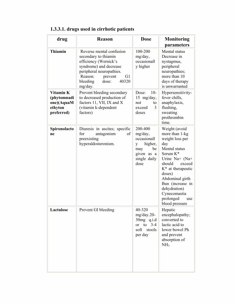
1.3.3.1. drugs used in cirrhotic patients
Monitoring parameters
Dose Reason drug
Mental status Decrease in nystagmus, peripheral neuropathies; more than 10 days of therapy is unwarranted
100-200 mg/day, occasionally higher
Reverse mental confusion secondary to thiamin efficiency (Wernick’s syndrome) and decrease peripheral neuropathies. Reason: prevent G1 bleeding dose: 40320 mg/day.
Thiamin
Hypersensitivity-fever chills, anaphylaxis, flushing, sweating prothrombin time.
Dose: 10-15 mg/day, not to exceed 3 doses
Prevent bleeding secondary to decreased production of factors 11, VII, IX and X (vitamin k-dependent factors)
Vitamin K (phytomnadione)(AquaMethyton preferred)
Weight (avoid more than 1-kg weight loss per day Mental status Serum K* Urine Na+ (Na+ should exceed K* at therapeutic doses) Abdominal girth Bun (increase in dehydration) Cynecomastia prolonged use blood pressure
200-400 mg/day, occasionally higher, may be given as a single daily dose
Diuresis in ascites; specific for antagonism of preexisting hyperaldosteronism.
Spironolactone
Hepatic encephalopathy; converted to lactic acid to lower bowel Ph and prevent absorption of NH3
40-320 mg/day.20-30mg q.i.d or to 3-4 soft stools per day
Prevent GI bleeding Lactulose
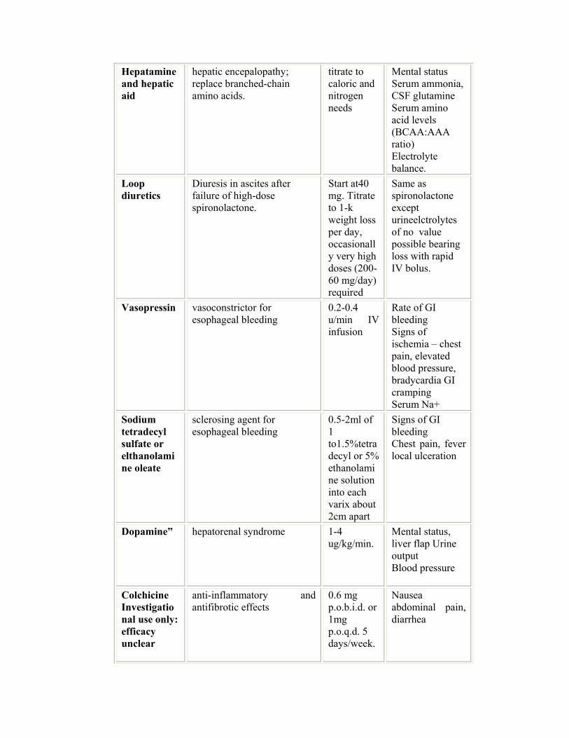
Mental status Serum ammonia, CSF glutamine Serum amino acid levels (BCAA:AAA ratio) Electrolyte balance.
titrate to caloric and nitrogen needs
hepatic encepalopathy; replace branched-chain amino acids.
Hepatamine and hepatic aid
Same as spironolactone except urineelctrolytes of no value possible bearing loss with rapid IV bolus.
Start at40 mg. Titrate to 1-k weight loss per day, occasionally very high doses (200-60 mg/day) required
Diuresis in ascites after failure of high-dose spironolactone.
Loop diuretics
Rate of GI bleeding Signs of ischemia – chest pain, elevated blood pressure, bradycardia GI cramping Serum Na+
0.2-0.4 u/min IV infusion
vasoconstrictor for esophageal bleeding
Vasopressin
Signs of GI bleeding Chest pain, fever local ulceration
0.5-2ml of 1 to1.5%tetradecyl or 5% ethanolamine solution into each varix about 2cm apart
sclerosing agent for esophageal bleeding
Sodium tetradecyl sulfate or elthanolamine oleate
Mental status, liver flap Urine output Blood pressure
1-4 ug/kg/min.
hepatorenal syndrome Dopamine”
Nausea abdominal pain, diarrhea
0.6 mg p.o.b.i.d. or 1mg p.o.q.d. 5 days/week.
anti-inflammatory and antifibrotic effects
Colchicine Investigational use only: efficacy unclear
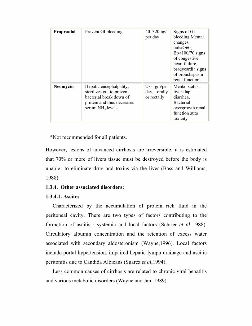
Signs of GI bleeding Mental changes, pulse>60; Bp>100/70 signs of congestive heart failure, bradycardia signs of bronchspasm renal function.
40–320mg/ per day
Prevent GI bleeding Propranlol
Mental status, liver flap diarrhea, Bacterial overgrowth renal function auto toxicity
2-6 gm/per day, orally or rectally
Hepatic encephalpahty; sterilizes gut to prevent bacterial break down of protein and thus decreases serum NH3 levels.
Neomycin
*Not recommended for all patients.
However, lesions of advanced cirrhosis are irreversible, it is estimated
that 70% or more of livers tissue must be destroyed before the body is
unable to eliminate drug and toxins via the liver (Bass and Williams,
1988).
1.3.4. Other associated disorders:
1.3.4.1. Ascites
Characterized by the accumulation of protein rich fluid in the
peritoneal cavity. There are two types of factors contributing to the
formation of ascitis : systemic and local factors (Schrier et al 1988).
Circulatory albumin concentration and the retention of excess water
associated with secondary aldosteronism (Wayne,1996). Local factors
include portal hypertension, impaired hepatic lymph drainage and ascitic
peritonitis due to Candida Albicans (Suarez et al,1994).
Less common causes of cirrhosis are related to chronic viral hepatitis
and various metabolic disorders (Wayne and Jan, 1989).
Pneumonia and other haematologic disorder due to chronic alcohol abuse
affect folic acid absorption, as well as iron. Fanconi syndrome causes
severe liver cirrhosis (Deeinhofer, 1996).
Endocrine disorders are seen in advanced cirrhosis because of the
inability of the liver to metabolize the steriod hormone of the adrenals
and gonads.
1.3.4.2. Hepatic renal syndrome
The concurrent impairment of renal function with hepatic failure is
termed the hepatic renal syndrome. There is functional change in the
kidney, caused by fluid and electrolyte disturbances, diuretic - induced
volume depletion, shock, or accumulation of unmetabolized toxic
substances.
1.4. Parasitic cirrhosis
Due to infestation with schistosoma and/or fluke, parasitic cirrhosis
could occur. The liver fluke may produce billary type of cirrhosis due to
lodgement in biliary channel. Also there is thickening and dilatations of
bile ducts, fibrous and cellular infilteration in the portal spaces, the flukes
on their remaindants may be found (Radostis, et al 2000).
1.4.1 Hepatic fascioliosis (Liver fluke disease)
It is a disease caused by Fasciola hepatica, mainly in sheep but
also in cattle and other ruminants, Fasciola gigantica, mainly infects
cattle and it also affects other ruminants. Both of these are large flukes
inhabiting the bile duct or intestines, causing damage to the liver resulting
in emaciation, jaundice and oedema (Solusby, 1982).
1.4.1.1. Etiology:
Fasciola hepatica is the most common and important liver fluke
and has a cosmopolitan distribution. Lymnial snails are intermediate
host, and release the infective forms, the metacericaria, onto the herbage.
Hepatic fasciolosis is an economically important disease of animals and
man, (Radostits, et al 2000). Mainly sheep and cattle, but other species may
provide a reservoir of infection .Fasciola hepatica may infest all domestic
animal including equidae and many wild life species(Owen, 1977).
Chronically infested sheep are the most important source of pasture
contamination (Boray 1985). Human cases are usually associated with the
ingestion of a harch plants such as water cress or uncooked vegetables
which are contaminated with encysted larvae. A similar but large fluke F.
gigantic larvae is restricted to warmer regions including parts of Africa and
Asia (Solusby, 1982; Leather, 1982). Fascioliosis is endemic in 61
countries and has become a food borne infection of public health importance
in parts of the world such as the Andean- highlands of Bolivia, Ecuador,
Peru, the Nile delta of Egypt and Northern Iran.
It is estimated that 2.4 million people are infected world –wide and
more than 180 million are at risk of infection. More than 60% of the
population is infected in high land of Bolivia. An outbreak along the
shores of the Caspain sea in Northern Iran between 1989-1991 infected
more than 10,000 people. Also outbreaks occurred in Algeria, Cuba and
France. The infection may be wide spread in humans than is appreciated, as
it is present in domestic animals in almost all countries where cattle and
sheep are reared (L. Savoili, 1998).
1.4.1.2. Prevalence:
One or two species of the genus fasciola occur in almost every
tropical country. In some areas it is enzootic and is a serious hazard in
regions where the conditions exist for the survival and multiplication of
the snail intermediate host.

The incidence and economic loss from fascioliasis is generally very
high. Fasciola gigantic occurs most commonly in Africa and Indian
subcontinent, Hawaii and the Philippines. Elsewhere F. hepatica is most
often found.
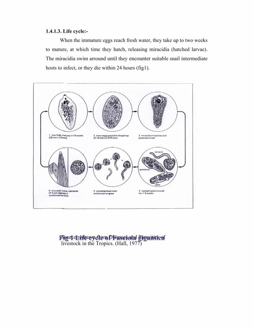
1.4.1.3. Life cycle:-
When the immature eggs reach fresh water, they take up to two weeks
to mature, at which time they hatch, releasing miracidia (hatched larvae).
The miracidia swim arround until they encounter suitable snail intermediate
hosts to infect, or they die within 24 hours (fig1).
Source redrawn from Diseases and Parasitics of livestock in the Tropics. (Hall, 1977)
1.4.1.4. Treatment of Fasciolosis
Rafoxanide is salicylanide compound
3`5 dintro-3 chloro-4(P-chlorophenoxy) –salyclanide
1.4.1.5 Pharmacology of Fasciolocidal Drug (Rafoxanide)
Rafoxanide is well absorbed by cattle and sheep with peak plasma
levels occurring between 24-48 hrs. after dosing. Rafoxanide is not
metabolized by cattle and sheep to any detectable degree, the half-life
varies from 5 to 10 days in sheep.
1.4.1.6. Action and uses
Rafoxanide is active against 99% of adult and immature fasciola
hepatica, more than 99% adult and up to 91% immature fasciola
gigantica.The mechanism of action is due to paralysis of the parasite.
1.5. Health and prevention of illness
1.5.1. Health:
It is an equilibrium between the mind, the body, and the external
world (environment), where disease is a disruption of this harmony.
Prevention and treatment involve creating the condition in which
the body could maintain and cure itself through its internal healing
mechanism. When disease did manifest itself -specific intervention would
be applied, but natural cures such as dietary changes were preferred over
drugs so people generally treated their illness with prayer and such
common sense approaches as good food, rest and whatever substances
they found in nature that were traditionally known to have medical
qualities such as simple herbs, plants, minerals, urine etc.
1.5.2. Urine:
Urine is not a waste product, but a purified, sterile watery solution,
it is an extra ordinary valuable physiological substance. It has been shown
through out the history of medical science right-up, until to day to have
profound medical uses. It is composed of many non toxic substances, the
toxic one’s are being removed from the body through the liver, intestine,
through the skin, and through out breath. The main function of the
kidney is to keep the composition of the blood in optimal balance. When
there is too much water, the kidney will remove it. Camel kidneys unlike
other mammalian species, play an important role in water release. It has
been clear for centuries that the camel has a degree of independence of
water greater than other domestic animals (Yagil, 1985 and 1993).
The composition of urine depends onthe life style of humans, and
on the type of grazing pasture of animals. Natural urine contains a
measurable amount of substances which has been used medically, even in
extremely large quantities without causing side effects.
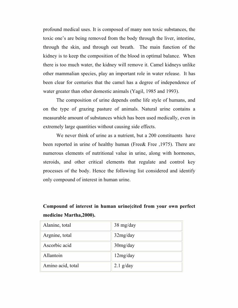
We never think of urine as a nutrient, but a 200 constituents have
been reported in urine of healthy human (Free& Free ,1975). There are
numerous elements of nutritional value in urine, along with hormones,
steroids, and other critical elements that regulate and control key
processes of the body. Hence the following list considered and identify
only compound of interest in human urine.
Compound of interest in human urine(cited from your own perfect
medicine Martha,2000).
Alanine, total 38 mg/day
Argnine, total 32mg/day
Ascorbic acid 30mg/day
Allantoin 12mg/day
Amino acid, total 2.1 g/day
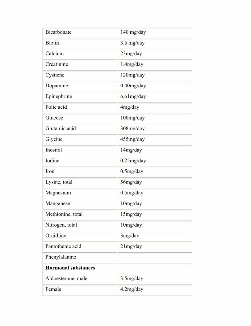
Bicarbonate 140 mg/day
Biotin 3.5 mg/day
Calcium 23mg/day
Creatinine 1.4mg/day
Cystiene 120mg/day
Dopamine 0.40mg/day
Epinephrine o.o1mg/day
Folic acid 4mg/day
Glucose 100mg/day
Glutamic acid 308mg/day
Glycine 455mg/day
Inositol 14mg/day
Iodine 0.25mg/day
Iron 0.5mg/day
Lysine, total 56mg/day
Magnesium 0.5mg/day
Manganese 10mg/day
Methionine, total 15mg/day
Nitrogen, total 10mg/day
Ornithine 3mg/day
Pantothenic acid 21mg/day
Phenylalanine
Hormonal substances
Aldoesterone, male 3.5mg/day
Female 4.2mg/day
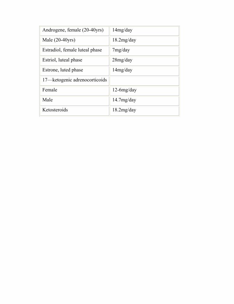
Androgene, female (20-40yrs) 14mg/day
Male (20-40yrs) 18.2mg/day
Estradiol, female luteal phase 7mg/day
Estriol, luteal phase 28mg/day
Estrone, luted phase 14mg/day
17—ketogenic adrenocorticoids
Female 12-6mg/day
Male 14.7mg/day
Ketosteroids 18.2mg/day
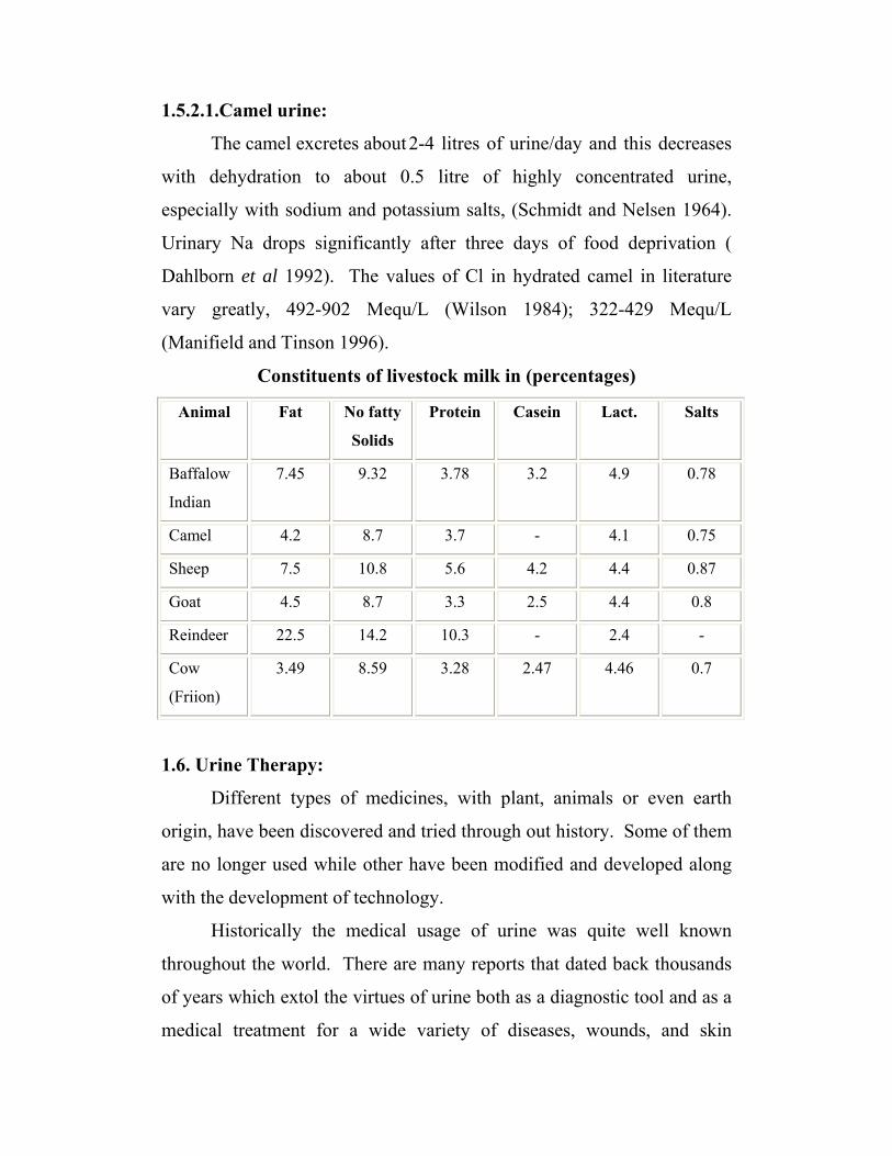
1.5.2.1.Camel urine:
The camel excretes about 2-4 litres of urine/day and this decreases
with dehydration to about 0.5 litre of highly concentrated urine,
especially with sodium and potassium salts, (Schmidt and Nelsen 1964).
Urinary Na drops significantly after three days of food deprivation (
Dahlborn et al 1992). The values of Cl in hydrated camel in literature
vary greatly, 492-902 Mequ/L (Wilson 1984); 322-429 Mequ/L
(Manifield and Tinson 1996).
Constituents of livestock milk in (percentages)
Animal Fat No fatty
Solids
Protein Casein Lact. Salts
Baffalow
Indian
7.45 9.32 3.78 3.2 4.9 0.78
Camel 4.2 8.7 3.7 - 4.1 0.75
Sheep 7.5 10.8 5.6 4.2 4.4 0.87
Goat 4.5 8.7 3.3 2.5 4.4 0.8
Reindeer 22.5 14.2 10.3 - 2.4 -
Cow
(Friion)
3.49 8.59 3.28 2.47 4.46 0.7
1.6. Urine Therapy:
Different types of medicines, with plant, animals or even earth
origin, have been discovered and tried through out history. Some of them
are no longer used while other have been modified and developed along
with the development of technology.
Historically the medical usage of urine was quite well known
throughout the world. There are many reports that dated back thousands
of years which extol the virtues of urine both as a diagnostic tool and as a
medical treatment for a wide variety of diseases, wounds, and skin
disorders. Urine as a medicine is used externally or internally to promote
or to maintain mammalian health. This is based on the principle of
“Natural cycles”, it has been known through out the centuries both in the
West and in the East. Urine therapy has proven helpful in a great number
of various diseases ranging from simple cold and throatache, to
tuberculosis and asthma (Kroon, 1996). Also in minor skin problems
such as itching to more serious skin disease such as eczema, psoriasis,
and even skin cancer. Urine therapy can be combined with any other
natural medicine to yield good results. Nowadays cancer is treated in
different parts of the world by urine therapy (Rostan, 1992).
1.6.1. Urine therapy in Eastern countries:
Urine therapy in Eastern countries has been practiced for thousands
of years, especially within Yoga and Tantra tradition where the use of
urine has been kept alive. It served particularly as a real “therapy”, a
method to clean the physical body of impurities, but also as a way to
further spiritual growth. The way of the Yoga is the practice of ingesting
ones own urine (Martha 2000; Shankandevanda, 1978) in Rome, India,
and Egypt ( Raojibhia, 1973 and Burzynsky, 1986).
Since 5000 years ago, old document has been found that describes
the practice of urine in different respects. This document consists of 107
verses (Slokas) it is called shivambu kalpavidhi.
1.6.2. Urine therapy in Western Countries:-
The urine therapy practice has been known on many cultures.
German encyclopedia stated that the Greek and Romans were aquanted
with the use of urine as a medicine (Plesch, 1947). The English man who
was the urine therapy pioneer, cured himself of tuberculosis, which had
been declared “incurable”(Armstrong, 1944). Also an Australian scientist
found a hormone in morning urine called melanin.
1.6.3.Medicinal use of human urine:-
- Urea has an anti septic and inhibitory effect on the growth of
microorganisms. (Wilson, 1906; Duncon 1918).
- Auto therapeutic agents (John, et al 1935 and Krebs 1934).
- In treatment of cancer. (Millar, 1933 and Burzynski et al 1977).
- Auto-urine vaccine therapy for nephritis. (Tiberi, 1934).
- Wounds and burns were treated by urine. (Leon, 1938)
- Urine extract was used for peptic ulcer, (Sandweiss, et al, 1941).
- A natural urine injection was used in medical practice extensively and
with excellent success on large a variety of disease conditions (Plesch,
1947).
- Anti-tubercle effect of human urine (Armstrong 1971; Bjornesjo,
1951; Tsuji, 1965).
- Urea is one of the most useful non-metabolized non-electrolyte
diuretics. In comparing the effect of urea with Diamox on intraocular
pressure, urea is found to be more effective.( The physicion’s Desk
Reference 1992).
- Natural anti bodies were found in the urine. (Martin, et al 1962).
- Recently anti-cancer. (Phalon, 1993). Anti hepatotoxic agents is found
in human urine.(Lai, et al 1999 ;Wen Chuan Lin,2003).
- It is used as miracle drug for AIDs, obesity, cancer, ageing (Kent,
1982) AIDs treatment ( Forber and Lederer 1989). In cancer, AIDS
and Autoimmune diagnosis (Burzynski, 1993).
- Anti-allergic effect (Dunne, 1981; William, 1982)
- Bactericidal effect repeated (Robert, et al 1987).
- The urokinase, a urine constituent that used is in dissolving blood
clots in veins, arteries heart and lung.( Mannucci and Angelo 1982).
- Urea has been used during the last two decades in the treatment of dry
skin, both clinically and in cosmetic products. (Gunnar, 1992; Serup,
1992).
There are a few more examples of commercial medical applications of
urine and urea in use today.
- Urea Phil: diuretic made from urea
- Urofollitropin: urine extract fertility drug.
- Puneaskin: urea cream for skin problems
- Amino-cerv: urea cream used for cervical treatments.
- Premarmin: urine extract estrogen supplement
- Panafil: urea papain ointment for skin ulcers, burns and infected
wounds.
1.6.3. Medicinal use of animals urine:-
A sick person will try the prescribed remedy regardless of the cost,
taste or odour. Therapeutic uses of animal’s urine have along history as
well as that of human. (Ibn-Albitar and Alrazi, 1925). Cow’s urine is
well known in India ( Raojibhai 1973; News week 1977). Such a therapy
of cow’s urine is known in different parts of southern Sudan (Ohaj,
1993). Goat urine with some medicinal plants used for treatment of
jaundice and ascites, (Ibn Sina, 1037). (Al Nasimi 1992) described the
general properties of all animal urine and mentioned the effect of camel
urine in treatment of ascetics. (Beaton, 1971), isolated anorexiginic
mobilizing substances from animal urine. While (Veerangevank 1992)
investigated trypsin inhibition substance from the urine of pregnant mare,
but few researches had been done on is the camel urine. Recently camel
urine is used in Arab Desert for the treatment of Leukemia and digestive
cancer(Kabariti et al., 1988; Muddathir, 1995 personal communication).
Also it was used as hair detergent in Morocco, such therapy is known in
some parts of Sudan, Saudi Arabia, and in Somalia (Ibrahim and Mona
1989 ; Ohaj, 1993), also used in healing of bad burns and injuries. It is
more extensively used for spleenomegaly, hepatormegali, liver disorder
and sometimes for fever (Ohaj 1998). The initial concern of using
camel’s urine lies in its more concentrated salt contents (almost twice
that of sea water .Yagil, 1994).
1.6.3.1.1. Medicinal use of Camel Urine Camel urine is used by the camel owners, and Beduines as medicine in different
ways. The Beduine in the Arab desert used to mix camel urine with milk and give it to
patients who were suffering from many enteric disorders and illnesses. Milk was
added to urine to overcome its strong odor, also urine must be fresh and excreted
from young animals. The duration of the treatment lasts for 2-3months for ascitic
patients (Ohaj, 1998). Kabarity (1988) used camel urine for a few weeks to treat
carcinogenic patients after which they were declared to be healthy. Ibrahim, (1989)
used camel urine as a hair detergent.
Alhawi AlKabeer used two ounces plus one pound of milk for the treatment
of ascites (Al-Razi, 1937). Also Alyahodi used goat urine plus fox apple. Dried and
burned –stool of camel was topically administered for skin dermatitis (Al-Yahodi
1958). They treated some patients with camel urine after boiling (Ali and Erwa,
1993). People in Yemen would dry camel urine under the sun and compress it in the
form of tablets to be used in case of burns and wet body injuries (Al-Zhrawi, Ibn
Elbitar, 1009). The percentages of use of camel urine among 5 nomadic tribes in
eastern Sudan as follows : 72% use camel urine for internal problems in general,
while 52%, 32%, 20%, and 32% used it for malaria, ascitis, dental problems and hair
shampoo respectively. Regarding the type of the animal which urine is used, 88%
used that of female and 12% of male. 72% drink it pure, whereas the remainder 28%
mix it with the milk (Ohaj 1998).
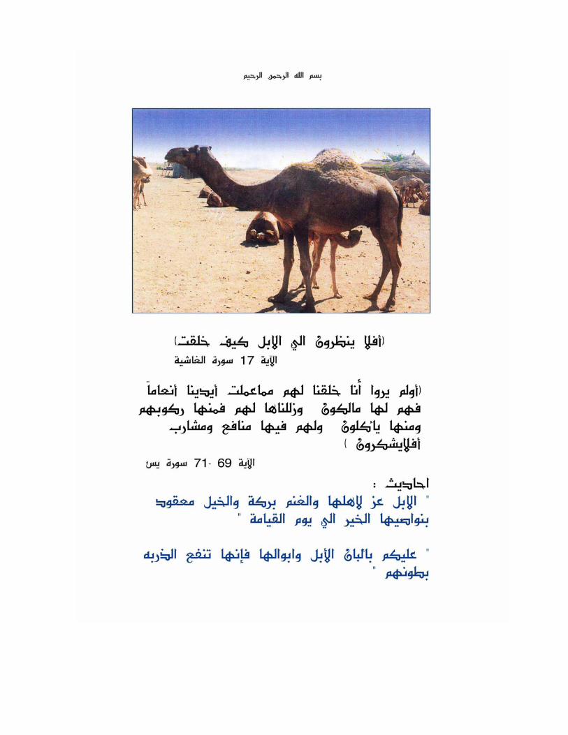
1.7. Aims of the present study Aims of the present work are to undertake greater in depth research
on Camel urine. 1. Physical and Chemical analysis of the normal constituents of camel urine
and compare it with that of other animals.
2. To assess camel urine as a remedy clammed in traditional medicine
from therapeutic point of view. ( Clinical studies on naturally infected
calves (Faschiolosis)).
3. Pharmacological investigation by invivo and invitro methods.
a) Camel urine hepatoprotectove effect against CCl4 & paracetamol.
b) Bioscreening response to camel urine and its extracts.
c) Camel urine effect on cell culture.
4. Chromatography of camel urine.
CHAPTER TWO
Materials and Methods
2.1. Materials
2.1.1. Experimental Animals 150 male and female Wistar albino rats weighing (90-250)gram were used. The animals were housed in groups and provided with a balanced diet and water ad
libitum.
2.1.2. Naturally infected calves
Twenty five calves naturally parasitized with Fasciola gigantica, aged 1-2 years, weighing 105-160kg, were used in this study. Animals were kept in pens at
the premises of the Central Vetarinary Research Laboratory (C.V.R.L.) at Soba. They are identified by plastic ear tags. Then the animals were alloted into five
groups and provided with a balanced diet and water ad libitum.
2.1.3. Sources of urine
Two adult female camel (5-6 years) urine was collected during
twenty four hours, from Soba animal house, at the C.V.R.L.,Soba,
Khartoum,Sudan.
Young female camel urine (6 month up to two years old) urine was
brought from different areas, Butana, Gazira (Wadballal).
The camel urine samples were collected by Tashweel technique which
was done by touching the abdominal side of the female camel near the
hide of the back leg. (Ohaj, 1998). By this technique urine sample could
be available at any time. Urine extracts were prepared at the department
of Biochemistry, Nutrition and Toxicology at the Central Veterinary
Research Laboratory Unit-Soba, Khartoum, Sudan.
2.1.4. Extraction of camel urine components for screening programs:
2.1.4.1. Chloroformic extract:-
Equal volumes of urine and chloroform were placed in a
volumetric flask V/V, and allowed to shake for 3 hours, at room
temperature using circular horizontal shaker. The mixture was poured in
a separating funnel, till two layers were clearly separate. The lower
chloroformic layer was displaced in a weighed beaker, the emulsified
layer was centrifuged and the clear chloroform layer was aspirated and
added to the former one, then left to complete dryness at room
temperature.
2.1.4.2. Protein precipitation:-
Native protein precipitate was obtained by the method of salt
saturation (Ammonium Sulphate 40% W/V – salting out). The
temperature was kept at 0oC. The mixture was allowed to shake for one
hour using (KARL Kolb) water bath shaker. Then the mixture was
centrifuged for 5 min at 4000 rpm, the supernatant was discarded and the
precipitate was weighed and dissolved in 1ml distilled water, some were
dissolved in Alkaline phosphate buffer pH 7.2 and kept at –20oC for
further analysis. 2.1.4.3. Ultra centrifugation of camel urine
Ultra centrifuge (Class R) was adapted to 10000 round per minute for 20 minutes at (0-10)ºC, then the precipitant was collected and dissolved in 0.5ml
phosphate buffer and frozen at –20oC for further analysis.
2.1.4.4. Chemicals
Chemical Sources
Acetic acid BDH
Alanine aminotransferase Linear-chemical kits
Alkaline phosphates Linear-chemical kits
Aspartate aminotransferase Linear-chemical kits
Atropine Sigama
Ammonium sulphate BDH
Albumin Linear chemical kits
Billirubin Linear chemical kits
Barium chloride BDH
Carbon tetra chloride (BDH)
Calcium chloride (BDH)
Chloroform BDH
Cilica Gell SD fine-Chemical India
Diethyl acetate Merck
Diethylchloro methane BDH
Ethylene diamine tetrachloride BDH
Ethanol BDH
Glacial acetic acid BDH
Gamma glularyle transferase Linear chemical kits
Glucose BDH
Hydrochloric acid SD fine-Chemical India
5-Hydroxytryptamine Sigama
Lactate dehydrogenase Linear chemical kits
n- Butanol Merek
Phentolamine Sigama
Picric acid Riedel DE haenag
Potassium dihydrogen phosphate Hopkin / Will
Propanol Mereic
Potassium chloride (BDH)
Sephadex sigma
Sodium carboxymethyl cellulose Merck
Sodium hydroxide Met lab.U.K

Trichloroacetic acid Perkin & William
2.2. Methods
2.2.1. Qualitative chemical tests
The pH of each sample was tested with a yellow litmus paper
(Advantic Tpyo Roshi Kashi Ltd. Japan). The litmus paper after being
dipped in the sample was compared to a colour- scaled paper with whole
range of 0 to 14.
Total urinary protein was detected by the method of Varley (1984).
This is simply done by placing 5ml of urine sample into a test tube and
adding few drops of glacial acetic acid. A cloudiness or turbidity
indicates the presence of urinary proteins.
2.2.1.1. Detection of chloride:
Inorganic compounds were detected qualitativly according to Vogel
method (1982).
1- Chloride one gram of the sample (freeze dried camel urine) was
placed in 100ml volumetric flask, the volume was completed to
100ml, and stirred for 10 minutes then filtered.
2- 10ml of the solution were diluted to 100ml with distilled water.
3- Then 0.5ml + 10ml of buffer (Contain 105g / L CH3COOH+ 9
gm/L HNO3) solution were mixed and then read on chloride meter.
2.2.1.2. Detection of Bicarbonate:
10mg of dried camel urine were dissolved in 5ml of distilled water.
The solution was then treated with a solution of magnesium sulphate; no
precipitate was produced (that means no carbonate). The solution was
then boiled; a white precipitate is produced (indicate that the bicarbonate
group is existing).
2.2.1.3. Detection of phosphate:
5ml of camel urine was treated with 5ml of silver nitrate solution.
A yellow precipitant was produced, the colour of the precipitant was not
changed on boiling and which is soluble in ammonia.
2.2.1.4. Detection of Sulphate:
About 50mg of freeze dried camel urine was dissolved in 5ml of
distilled water. The solution was then treated with 1ml of 2M
Hydrochloric acid and 1ml of barium chloride solution.
2.2.2. Quantitative chemical tests
2.2.2.1. Determination of sodium and potassium.
The sodium and potassium were estimated by flame photometer
(Jenway, PEP7). Two separate standard curves were prepared ranging
from 100 to 150 m Equ /L for sodium and potassium respectively. 0.2ml
of urine sample was completed to 20ml (1 to 100) with distilled water and
directly read on the (Flame photometer).
2.2.2.2. Determination of Magnesium, Copper and Zinc:
Mg, Cu and Zn were determined by the use of atomic absorption
spectrophotometer (AAS). Stock, working standard, and sample
specimens were prepared for each of the above elements as described by
Beaty (1978). The instrument was calibrated according to the instruction
manual and the absorbance was read.
2.2.2.3. Determination of calcium:
Urinary calcium was determined according to the method of
Sulkowitch (1937). Sulkwitch reagent was prepared by mixing 2.5g
oxalic acid, 2.5g ammonium sulphate and 5 ml glacial acetic acid
completed to 100 ml with distilled water. Equal volumes of the above
reagent and urine sample were mixed.
2.2.2.4. Determination of total protein in urine:
Urinary protein was determined by the method of Biuret reaction
as discribed by Weichselbaum (1946). The protein was concentrated by
precipitation using trichloroacetic acid. It was redissolved in an alkali
and measured spectrophotometrically at 420nm.
2.2.2.5. Determination of non-protein nitrogen:
(Urea, uric acid creatinine, and creatine)
The urea was determined by the manual method described by
(Evans, 1968; March et al., 1965). The urine sample was diluted 1 to 10
with distilled water. The possible proteins were precipitated with
tricholoroacetic acid and centrifuged, the sample absorbance was read
agai urea standard (10mmol/L) at 520nm.
The creatinine was determined by the method of alkaline picrate of
(Bonsnes and Taussky 1945). One ml of urine sample was completed to
100 with distilled water, 1ml picric was added to 3ml diluted urine,
followed by 1ml of NaOH. 3ml creatinine standard (0.3 micromol and
3ml distilled water) used as blank were treated the same way. The
absorbance was measured after 15 minutes at 500nm.
The creatine in urine was transferred to creatinine by heating urine
sample with picric acid before adding NaOH. Then total creatinine was
determined according to the above method.
2.2.2.6. Determination of amino acids in urine:
Amino acids in urine samples were determined according to the
method of (Goodwin 1968b, 1970) using dinitroflouro-benzene. 1ml of
urine sample was diluted to 10 ml with distilled water. Three drops of
phenophthaline and 200mmol/L NaOH was added until the mixture turn
pink. The mixture was boiled for 15 minutes at 70oC, cooled and 5ml
acidified dioxane were added. Stock and working standard of glutamic
acid glycine were prepared and treated as directed. The absorbance of the
sample was measured against that of standard.
2.2.2.7. Determination of Uric acid in urine:
Uric acid was estimated according to Cavawy (1954). Diluted
urine (1:100), 5ml of urine, 1ml of 10% sodium tungestate and 1ml of 2N
H2SO4 were mixed and centrifuged. 5ml of the supernatant was put in
one of two test tubes, (standard and blank). o each tube, 1ml of 10%
sodium carbonate and 1ml of phosphatungestic acid were added and left
to stand at room temp. for 30 minutes, then was read at 680 nm.
2.2.2.8. Determination of urine Albumin:
Urine albumin was determined according to (Kentman et al. 1971). 2.2.29. Determination of serum total Billirubin:-
Serum total Billinubin was determined according to( Kind and King 1954).
Principle:
Billirubin in the sample reacts with diazotized sulfanilic acid in the presence of DMSO. The formed coloured azobillirubin is measured photometerically: there is
two Billirubin fractions in serum, billirubin-glucuronide and free billimute which is bound to albumin.
2.3. Biochemical methods:
2.3.1. Aspartate amino transferase (AST:EC. 2.6.11.1)
Aspartate amino transferase, formerly known as glutamic
oxaloacetic acid transaminase (GOT) catalysis the reversible transfer of
the amino group from an amino acid to a keto acid. This enzyme is
mainly present in the liver and many other tissues including kidneys,
heart, and skeletal muscle. The enzyme activity was measured as
described by Reitman and Frankel (1957). The principle of the assay
depends on the intermolecular transfer of an amino group from aspartic
acid to α -keto glutaric acid without the intermediate formation of
ammonia, and measuring the amount of the reaction product
(Oxaloacetate).
Glutamic acid + oxaloacetic acid
AST
α -keto glutaric acid + aspartic acid.
Aspartate amino transferase catalysis the transfer of amino group
from aspartate to 2-oxogluterate forming glutamate and oxaloacetate.
The oxaloacetate is reduced to malate by dehydrogenase and NADH.
The rate of decrease in concentration of NADH is proportional to
concentration of AST present in the sample.
Aspartate + 2-oxogluterate AST Glutamate + oxaloacetate
NADH+ H+ oxaloacetate MDH Malate + NAD+
2.3.2. Alanine amino transferase (ALT:EC1.2.6.1.2 formally known
as GPT):
Alanine amino transferase, catalyses the transfer of an amino group
from alanine to 2-oxoglutarate forming glutamate and pyruvate. The
pyruvate produced is reduced to lactate-by-lactate dehydrogenase (LDH).
Lactate dehydrogenase catalyses the reduction of pyruvate by NADH is
proportional to concentration of ALT present in the sample
Alanine + H+ + Pyruvate ALT glutamate + pyrvate
NADH + H+ +pyruvate LDH Lactate + NAD+
2.3.3. Alkaline phosphates (Linear-Chemical)
(ALP; E.C.3.1.3.1):
Alkaline phosphates was determined according to Chemie,(1972)
Principle:
Alkaline phosphates catalyze the hydrolysis of p-
nitrophenylphosphate, in the presence of magnesium ions, liberating
inorganic phosphate and p-nitrophenol. The rate of p-nitrophenol
formation is proportional to the concentration of ALP present in the
sample.
4-nitrophenyl phosphate + H2O ALP 4-nitophenyl +P1
2.3.4. γ –Glutamyl transferase(Linear-Chemical)
(GGT:E.C.2.3.2.2.):
Priniciple:
γ –Glutamyl trasferase catalysis the transfer of a γ-glutamyl group
from γ-glutamyl-carboxy-4-nitroanalide, glutamate is proportional to the
concentration of GGT present in the sample.
γ-glutamyl-3-carboxy-4-nitroanalide + glycyl glycine
GGT
γ-glutamyl glycyl glycine + 3-carboxy-4-Nitroanaline
2.4 Haematological methods:
Blood samples for whole blood examinations were withdrawn from
the jugular vein at intervals in vacutainer tubes (5ml) containing ethylene
diamine tetrachloroaceticacid (EDTA) as anticoagulent
(Becton,Dickinson,France) as described by (Schalm 1965).
2.4.1. Determination of Haemoglobin (Hb)
The concentration of haemoglobin was measured by the
cyanomethaemoglobin technique using colorimeter (CIBA CORNING
colorimeter model 252). 0.2ml of blood was added to 4ml of Drabkins
solution (0.2gm of potassium ferricyanide and 1gm bicarbonate per litre
of distilled water). The haemoglobin concentration was measured in g/dl
of blood. This method is based on the conversion of haemoglobin by
Drabkins solution to cyanomethaemoglobin as described by ( Schalm
1965).
2.4.2. Red Blood Cells Count (RBC):
Erythrocytes were counted using Neubauer haemocytometer
(Hawksley and sons Ltd., England) using Hayems solution as a diluent
consisting of 0.5gm of mercuric chloride and 1 gm of sodium chloride
made up to 200ml with distilled water as estimated by (Schalm, 1965). 2.4.3.White Blood cells count (WBC):
Leukocytes were counted according to Schalm (1965) using
Neubauer haemocytometer. Turk’s solution (1% glacial acetic acid
coloured with gention violet made up to 200ml with distilled water) was
used as a diluent.
2.4.4. Packed Cell Volume (PCV):
Blood samples were drawn into microhaematocrit capillary tubes
and sealed at one end with cristaseal (Hawksley). The capillary tubes
were centrifuged at 8000 rpm for 5 minutes using a microhaematocrit
centrifuge (Hawsley and sons Ltd., England). The PCV percentage was
read with Hawskley microhaematocrit reader.(Scalm, 1965).
2.4.5. Mean Corpuscular Volume (MCV):
The MCV was calculated from the PCV and RBC values as
follows
MCV (fl) = PCV x 10 RBC
2.4.6. Mean corpuscular haemoglobin concentration (MCHC):
MCHC was calculated from the PCV and Hb values as follows
MCHC(%) = Hb (g/dl) x 100
PCV 2.4.7. Thrompoplastin test with calcium (TT):
Prothrombin time studies the total extrinsic clotting system. It
measures the clotting time of a plasma at 37oC in the presence of excess
tissue thromboplastin and calcium. The test is dependent on factors II, V,
VII and X.
2.4.8. Activated partial thromboplastin time (APTT):
Activated partial thromboplastin time (APTT) measures the
clotting time of plasma or serum at 37oC in the presence of a platelet
substitute and an activator.
2.5. Pathological methods
2.5.1. Macroscopic examination
Macroscopic examination was carried out on each necropsied animal, all
organs were examined for the presence of any gross changes. The liver
was examined carefully for the presence of fasciola worms. 2.5.2. Microscopic techniques:
Representative samples from liver, intestine, kidney, lung, heart, spleen
were taken and preserved in normal saline (10%). Tissues were trimmed and
dehydrated in serial dilution of alcohol, 70, 85, 95 and 100%, using automatic
tissue processor and cleared twice by xylene . Then embedded in paraffin wax
and 5µ sections were cut using a microtome. Sections were stained with
Haematoxylin and Eosin and special stain may also be use. Processing and
staining procedures were those described by Clayden (1971).
2.6.Pharmacological methods
2.6.1. ED50 Determination:
Preliminary experiments were carried out for each type of urine
collection and from chloromoforic extract of camel urine to determine the
approximate dose, which produces 0-100% lethality. Doses chosen based
on the honorable Haddith and on the preliminary data, which were
administered randomly. The tested agents were given camel urine orally.
While CE was injected intraperitoneally to each group of rats. Death on
each group was observed over the next 24 hours. Percent moralities in all
groups were then calculated.
2.6.2.Assessment of hepatoprotective activity of camel urine and its
chloroformic extract against CCL4 induced hepatic toxicity in rats:
CCL4 was dissolved in 2% Na carboxy methyl cellulose CMC (15% v/v)
and administered orally at a dose of 10ml/kg and IP at a dose of 1ml/kg to
Wistar Ablino rats (CCL4 control group). The rise in ALT (GPT) was
taken as evidence for impaired liver function. The peak of increase in
serum ALT activity was reached 24 hours after CCL4 administration
(Mansour, 2000; Handa et al. 1986). 2.6.3.Assessment of Camel urine hepato protective activity against paracetamol
induced hepato toxicity:
Paracetamol was dissolved in distilled water (1g in 100 ml). A
dose of 1 g/kg body weight was orally administrated to male and female
Wistar albino rats (Control group) to damage liver cells . (Tee et al, 1987)
paracetamol was given 1 hour after chloroformic extract of camel urine,
24 hour later samples were collected from treated and control groups, and
frozen at –20oC till analyzed for ALT activities.
2.6.4. Evaluation of camel urine and its chloroformic extract (CE) on
isolated tissue strips preparations.
Preparation of physiologic salt solution: Kreb's solution is prepared
as follows
NaCl, 34.6; KCl, 1.75; CaCl2, 1.4; MgSO4, 1.45; NaHCO3, 10.5; KH2PO4
).8; in grams/5 liter (Kitchen, 1984).
The physiologic salt solution should be clear, not milky or cloudy.
Tyrod's solution is prepared of the following concentrations NaCl,
40; glucose, 5; CaCl2, 1; MgCl2, 1; MgCl2 0.5; NaH2PO4, 0.025;
NaHCO3, 5; in grams per liters.
2.6.4.1. Rat fundus strip preparation:
A Wistar Albino rat is killed by a blow on the head according to
the method of Vane (1957), and the throat was cut, the abdomen was
opened, the stomach was removed and placed on a petri dish containing
aerated Kreb's solution, cuts were made along the junction of the fundus
and pylorus. The fundus was opened and cuts were made alternatively
from each side to make a strip about 4 cm long. A thread was attached at
each end of the preparation, and it was mounted to the organ bath with
aerated Kreb's solution, temp. was maintained at 37oC.
2.6.4.2. Rat dudenum-strip preparation:
The rat (Wistar albino) duodenal strip was prepared according to
the method of Kitchen (1984). The duodenal strip (Proximate part of
intestine) 2-3 cm long was mounted in an organ bath filled with an
aerated Tyrod's solution, temp. was maintained to 37oC.
2.6.4.3. Rabbit jejunum preparation:
Rabbit of local strains weighing 1.5-2kg. were used. The animal
was killed by dislocating the neck and exsanguinated. The abdomen was
exposed. The first 2-3 cm of the jejunum was taken out and placed in a
petri-dish containing tyrod's solution at room temperature. The fat and
connective tissues were removed. Then the tissue was tied by thread at
the two ends transferred to an organ bath (25ml), containing an aerated
Tyrode's solution temp. was maintained at 37oC (Trendelenburg, 1917).
The urine and its extraction were left in contact with tissue for 30
seconds, in a dose cycle of 3 min.
2.6.4.4. Chick rectum strip preparation:
A young chick (7-15 day old) was killed with chloroform. The
abdomen was exposed and the last terminal part of the intestine (rectum)
attached to anal orifice was cut and placed on a petri dish containing
aerated Tyrod's solution. A thread was attached to the strip and then
mounted to the organ bath.
2.6.5. Preparation of tissue culture:
2.6.5.1. Primary bovine kidney cells:
Calf kidneys obtained from calves 1-4 weeks old were prepared
according to reported method of FAO, (1984). One or both kidneys were
separated from the animal under aseptic conditions and carried to the
laboratory in sterile beaker. Fats and capsule removed from the organ
and the cortex was cut in fragments from the medulla, placed in sterile
petri dish and minced with a pair of scissors. The tissue was washed in a
balanced salt solution free from calcium and magnesium, as phosphate
diluent (PD) containing 200 IU penicillin and 100µg streptomycin.
Tissue fragments were transferred to a grooved trypsinization flask
agitated by magnetic stirrer bar and washed in Hank's solution until clear.
After discarding the last wash pre-warmed 0.25% trypsin in Hanks
balanced salt solution (1: 9 dilution) replaced to cover the tissue and
repeated the washing. After the pieces of tissue settled to the bottom of
the flask, the supernatant was discarded since it contains dead cells and
blood. Fresh warmed trypsin was added to the tissue and stirred
overnight at 4oC, the digested tissue was allowed to settle and the
supernatant filtered through three layers of sterile gauze in a flask
containing bovine serum to stop trypsin action. The suspension was
centrifuged and the supernatant was discarded, the pellet cells were
suspended as 1ml packed cell was added to 250-300ml (GMEM) plus5-
10% bovine serum. Cell suspension was dispensed into tissue culture
flasks and incubated at 37oC to form monolayers within 4-5 days post-
seeding.
2.6.5.2. Secondary cell cultures:
Secondary cell culture was propagated from the primary culture as
follows:
After removing the growth medium from the fully-grown culture,
the sheet of cells was washed twice with phosphate diluent (PD). Trypsin
solution warmed to 37oC added to cover
cell sheet and left for 5 minutes. Cells removed by pipetting gently with
some (PD) and bovine serum was added to stop trypsin action. Cells
were centrifuged at 1000 rpm for 5 minutes and the supernatant was
discarded. Then suspended in twice the original volume of growth
medium containing antibiotics with 5-10% calf serum, distributed into
tissue culture glasses and incubated at 37oC.
2.7.Chromatographic methods:.
Chromatography is a method used for purification and
identification of chemical substances that consist of many compounds on
the basis of physical methods of separation, such as; thin layer
chromatography (TLC); gel and ion-exchange method, in addition to gas
liquid chromatography (GLC), and high performance liquid
chromatography (HPLC). Separations are made on the basis of several
differential molecular properties, especially charge, solubility, affinity
and adsorption.
2.7.1. Extraction:
As a primary requirement, the substance to be extracted must be
soluble, not only in aqueous biological media, but also in a phase which
is immiscible (or only slightly miscible) with water e.g. ether,
chloroform, carbon tetrachloride etc. If the aqueous phase shaken with
the organic solvent, the compound distribute itself between the two
phases in accordance with its distribution coefficient. If the aqueous
phase is repeatedly extracted with a fresh organic phase, a group of
substances can almost be completely separated.
2.7.2. Thin layer chromatography:
The stationary phase is a thin layer [0.1-0.5mm] of the grained
silica gel ( Kessel Gell G ) deposited on a carrier plate [20x20 cm] made
of glass, plastic, or aluminum foil. 1-5µl of sample is applied as on a spot
of the starting line about [10-15mm] from the bottom edge of the plate.
Then placed in a tightly closed tank containing a suitable eluting solvents.
The mobile phase diffusing up by capillary action.
Thin-layer chromatography is based on adsorption or partition.
A measurement of the rate of migration of substance is provided by
RF value.
Rf = distance traveled by the compound distance traveled by solvents
2.7.3.Preparative thin layer chromatography [PTLC]
Silica gel Kessel Gell G 0.5 mm thickness, were used. Fraction A
(chloroformic extract); fraction B (Butanolic extract) fraction C
(ethanolic extract), were applied in a form of streeks and the
chromatogram was developed using n-butanol-acetic acid and water
(BAW) as a solvent system in a ratio of 4:1:1.
On the bases of Ultrviolt light (U.V.) absorption, each streek was
composed of more than one band.
Different developing systems were used. Propanol and water in a
ratio of 70:30; n-Butanol, acetic acid and water in a ratio 4:5:1.
2.7.4. Gel filtration on sephadex A25-150:
Sephadex R (A25-120 SDEAE) was allowed to swell in (M)
methanol, for 24 hours. The gel was then made in a form of thick slurry,
degassed and then packed into the column. A 2.5x100 cm glass column
was fixed at a vertical stand. The bottom (outlet) was clamped and the
thick slurry was poured gently into the glass column till it was 3/4 filled
with methanol and the methanol was allowed to flow over until the gel
packed and degassed 5ml of camel urine were added. Then methanol was
added to form 20ml eluted layer over the gel. Introduction of air bubble
was avoided during the packing and elution process. Samples were
collected as 20ml/tube. 60 fractions were obtained. Followed by 20
fractions elution by methanol in 2N HCL in a ratio of (99:1).
2.7.5. Column chromatography
Gel filtration on sephadex (Fluka biochemical DEAE (A25-120)
and an ionexchange, were the principle technique employed for the
purification of camel urine protein and amino acid classes.
2.7.6. Ion exchange chromatography
60 Mesh or ion-exchange Resin column used for the isolation of
camel urine protein, amino acid, amide and polypeptides by a method
similar to the procedure employed by Fey et al. (1976).
Microgranules mesh 60 was first soaked with n-butanol solution
(BDH), which it self-constituting the starting mobile phase solution. The
resin was then packed in 1.5x30 cm glass column, fixed in a vertical
position. The outlet was closed and one third of he column was filed with
starting solution, 10 ml of camel urine were applied to the 60-mesh resin
column. The elution was performed from top downwards at speed of
1ml/min. Samples were collected as 3ml/tube. The elution profile of each
preparation was chromatographed by TIC and PTLC. Protein fractions
were more identified by Ultra violet spectrophotometer (UV), infra red
spectra and gas chromatography (GC).
2.7.7. Electrophoresis:
This technique was performed according to Ouchterlony and
Nilson (1979). It was employed for purity testing as well as identification
of isolated camel urine fractions.
The technique was carried out in commercial sheets of agrose gel
in 0.1 M barbitone buffer, pH 8.6. were noticed.
Five hundred ml of 0.1 M barbitone buffer (pH 8.6) were poured
into electrophoresis chamber (Ciba).
0.5µl of camel urine protein, milk protein, and that of other species
were applied onto the agrose gel membrane. The membrane was then
stretched across the two chambers, with its edges submerged in the
barbital buffer. The anode was specified, and the electrophoresis was run
at 70 volt, 12 m A for half an hour.
It was then removed and placed in a fixative dye solution, for 5
min. the strips were then destained and cleaned with ethyl alcohol.
The resulting peaks were designated, according to their speed of
migration towards the anode as albumin, A1, A2, B1, B2 and γ globulins.
2.7.8. Gas liquid chromatography (GLC):
2.7.8.1. Protein hydrolysate of biological materials
One gram of sample was refluxed with 10ml constant. boiling HCI
for 20 hours. The hydrolysate was filtered through glass fiber (Reve
Angel 934A4 filter paper), and the sample was taken to dryness at room
temperature on a rotary evaporator, the dry residue was redissolved in 1
litre of 0.1 N HCI in a 25ml flat bottom flask, lyophilized, then dried and
derivatized for gas chromatography.
2.7.8.2. Derivatization method:
1- Removal of water to give dry amino acids
2- Esterification of the amino acids to form methyl ester hydrochlorides.
3- Interestrification of the methyl ester to form n-butyl ester
hydrochlorides.
4- Acylation of n-butanol ester hydrochlorides to form n-trifluoroacetyl-
buytl esters.
2.8.Confirmatory chemical test of chloroformic extract:
2.8.1.Biuret test:
The biuret stock reagent was prepared following the method described
by (Henry,et al 1974). Forty five grams of sodium potassium tarterate
were transferred to a 1 litre flask containing 400ml 0.2 N NaOH. Two
hundred ml of this stock solution were diluted to 1 litre with alkaline
iodide solution to a bout 4 ml of the Biuret working solution were added
to a soluble amount of chloroformic extract crude urine and mixed
thoroughly. The mixture was allowed to stand for 10 minutes and the
change in colour was observed.
2.8.2. Acid hydrolosis of camel urine and chloroformic extract:
Two to three drops 0.6 N HCl were added to a few mls of camel
urine, chloroformic extract each, and heated at 120oC for 3 hours. The
hydrolysate obtained, the references solution and authentic samples of six
amino acids (glycine, DL- serine, L-Tyrosine, L-Alanine histadine
monochloride, 4-Aminobuteric acid and L- Tryptorphan) were
chromatographer on silica gel plates using propanol-water, Butanol-acetic
acid and water as developing systems. The developed plates were dried,
sprayed with ninhydrin reagent and the Rf values of the detected spots
were calculated. 2.9. Analytical screening of camel urine
Analytical screening for the constituents present in the camel urine extracts were carried out using the methods of (Ibrahim 1991). These studies were carried out on the liquid sample of urine and on the successive extracts of camel and cattle urine.
2.9.1.Tests of alkaloids and/or nitrogenous bases:-
a) The residues obtained from 20ml of Alcoholic extract, (ethanol and water;
35:15) was dissolved in 2ml dilute hydrochloric acid, and tested with Mayer’s
and Wagner reagents (1.3g of mercuric chloride were dissolved in 60ml of
water , and 5g of potassium sulphite (KS) in 10ml of water. The two solutions
are mixed and diluted to 100ml with distilled water).
b) Urine samples of camel (from adult and young female camel) and cattle were
extracted with chloroform (v/v). Chloroformic layer was then evaporated,
using steam to evaporate chloroform, the residue was dissolved in few mls of
methanol. Then each sample was spotted on TLC plate and developed with
methanol and ammonium water (100:1.5) to a suitable distance. The plates
were then air dried, visualized under U.V. (Bright-Blue spots were seen),
sprayed with Dragndoff reagent. The Rf of the detected compounds were
calculated.
2.10.Experimental design and treatment protocol
Experiments:
1.Physical and biochemical analysis of camel, cattle, goat and human
urine.
2.Time course of hepatotoxic effect of (CCL4) on rat liver.
3.Hepatoprotective effect against carbon tetrachloride (CCL4) pre and
post treatment with camel urine, and urine extracts. 4.Hepatoprotective
effect against paracetamol compared with Silymarin natural
hepatoprotective drug.
5.Clinical trials on naturally infected calves (fascioliasis).
6.In vitro effects of camel urine on cell culure and isolated strips.
7.Chromatography and electrophoresis.
8.phytochemical analysis of camel urine.
Experiment (1)
Group Treatment Dosage Route
1 2% carboxyl methyl cellulose (CMC)
10ml/kg B.W. Orally
2 2% (CMC) +CCL4 10ml 2% (CMC/kg 150µl+kg B.W. Orally
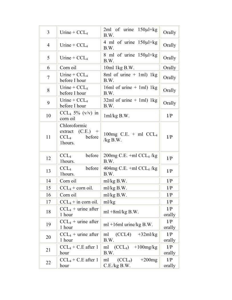
3 Urine + CCL4 2ml of urine 150µl+kg B.W. Orally
4 Urine + CCL4 4 ml of urine 150µl+kg B.W. Orally
5 Urine + CCL4 8 ml of urine 150µl+kg B.W. Orally
6 Corn oil 10ml 1kg B.W. Orally
7 Urine + CCL4 before I hour
8ml of urine + 1ml) 1kg B.W. Orally
8 Urine + CCL4 before I hour
16ml of urine + 1ml) 1kg B.W. Orally
9 Urine + CCL4 before I hour
32ml of urine + 1ml) 1kg B.W. Orally
10 CCL4 5% (v/v) in corn oil 1ml/kg B.W. I/P
11
Chloroformic extract (C.E.) + CCL4 before 1hours.
100mg C.E. + ml CCL4 /kg B.W. I/P
12 CCL4 before 1hours.
200mg C.E. +ml CCL4 /kg B.W. I/P
13 CCL4 before 1hours.
404mg C.E. +ml CCL4 /kg B.W. I/P
14 Corn oil ml/kg B.W. I/P 15 CCL4 + corn oil. ml/kg B.W. I/P 16 Corn oil ml/kg B.W. I/P 17 CCL4 + in corn oil. ml/kg I/P
18 CCL4 + urine after 1 hour ml +8ml/kg B.W. I/P
orally
19 CCL4 + urine after 1 hour ml +16ml urine/kg B.W. I/P
orally
20 CCL4 + urine after 1 hour
ml (CCL4) +32ml/kg B.W.
I/P orally
21 CCL4 + C.E after 1 hour
ml (CCL4) +100mg/kg B.W.
I/P orally
22 CCL4 + C.E after 1 hour
ml (CCL4) +200mg C.E./kg B.W.
I/P orally
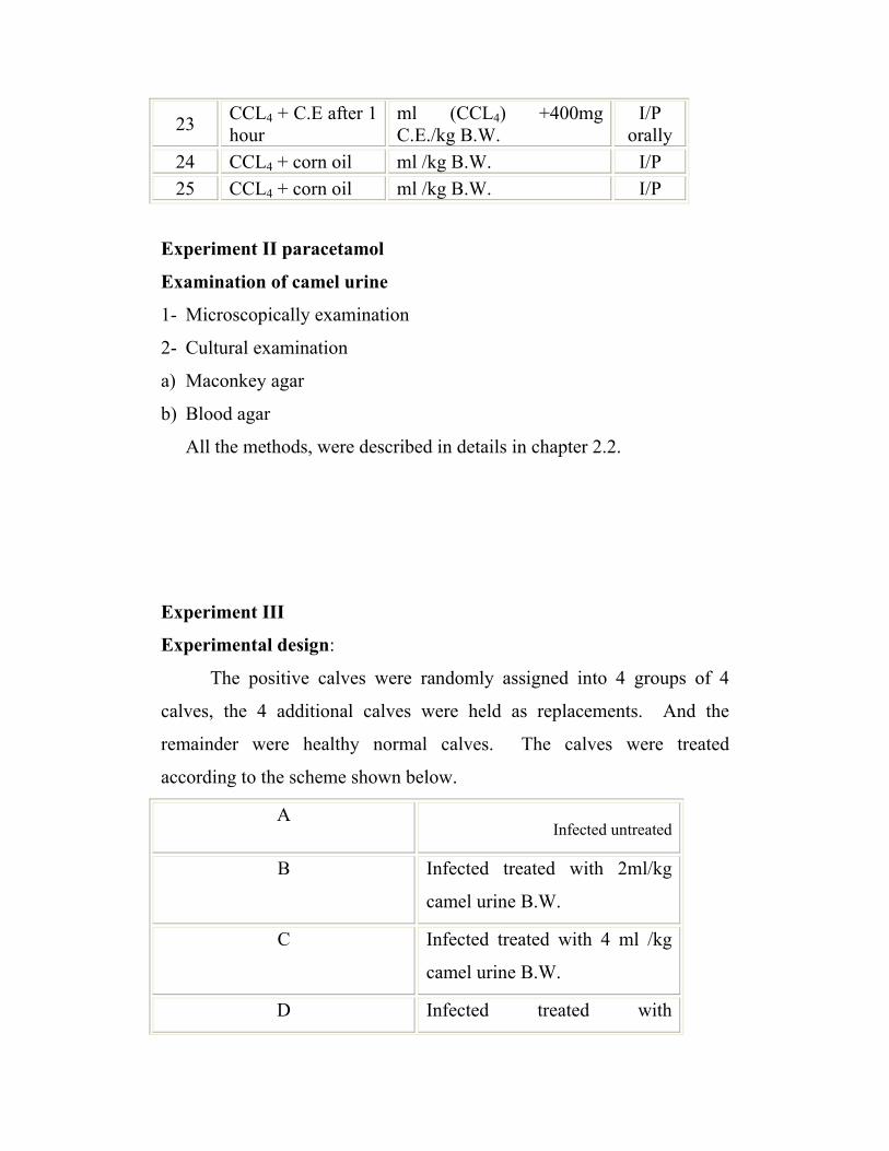
23 CCL4 + C.E after 1 hour
ml (CCL4) +400mg C.E./kg B.W.
I/P orally
24 CCL4 + corn oil ml /kg B.W. I/P 25 CCL4 + corn oil ml /kg B.W. I/P
Experiment II paracetamol
Examination of camel urine
1- Microscopically examination
2- Cultural examination
a) Maconkey agar
b) Blood agar
All the methods, were described in details in chapter 2.2.
Experiment III
Experimental design:
The positive calves were randomly assigned into 4 groups of 4
calves, the 4 additional calves were held as replacements. And the
remainder were healthy normal calves. The calves were treated
according to the scheme shown below.
A Infected untreated
B Infected treated with 2ml/kg
camel urine B.W.
C Infected treated with 4 ml /kg
camel urine B.W.
D Infected treated with
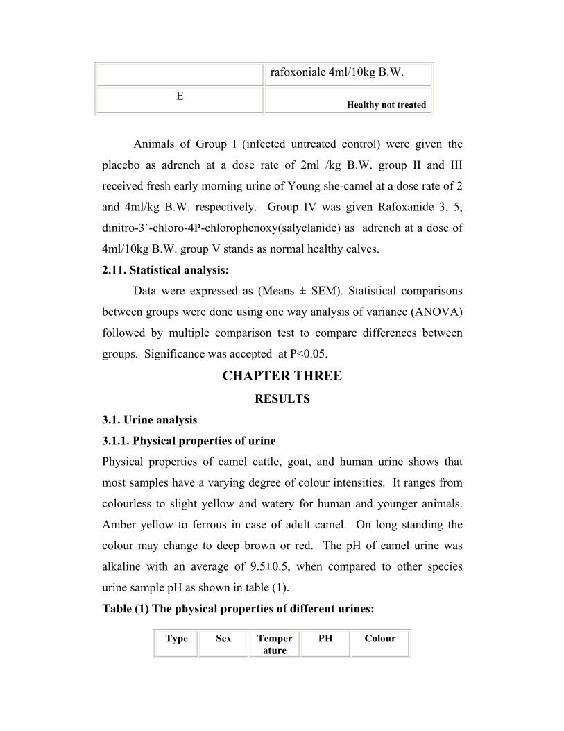
rafoxoniale 4ml/10kg B.W.
E Healthy not treated
Animals of Group I (infected untreated control) were given the
placebo as adrench at a dose rate of 2ml /kg B.W. group II and III
received fresh early morning urine of Young she-camel at a dose rate of 2
and 4ml/kg B.W. respectively. Group IV was given Rafoxanide 3, 5,
dinitro-3`-chloro-4P-chlorophenoxy(salyclanide) as adrench at a dose of
4ml/10kg B.W. group V stands as normal healthy calves.
2.11. Statistical analysis:
Data were expressed as (Means ± SEM). Statistical comparisons
between groups were done using one way analysis of variance (ANOVA)
followed by multiple comparison test to compare differences between
groups. Significance was accepted at P<0.05.
CHAPTER THREE RESULTS
3.1. Urine analysis
3.1.1. Physical properties of urine
Physical properties of camel cattle, goat, and human urine shows that
most samples have a varying degree of colour intensities. It ranges from
colourless to slight yellow and watery for human and younger animals.
Amber yellow to ferrous in case of adult camel. On long standing the
colour may change to deep brown or red. The pH of camel urine was
alkaline with an average of 9.5±0.5, when compared to other species
urine sample pH as shown in table (1).
Table (1) The physical properties of different urines:
Type Sex Temperature
PH Colour
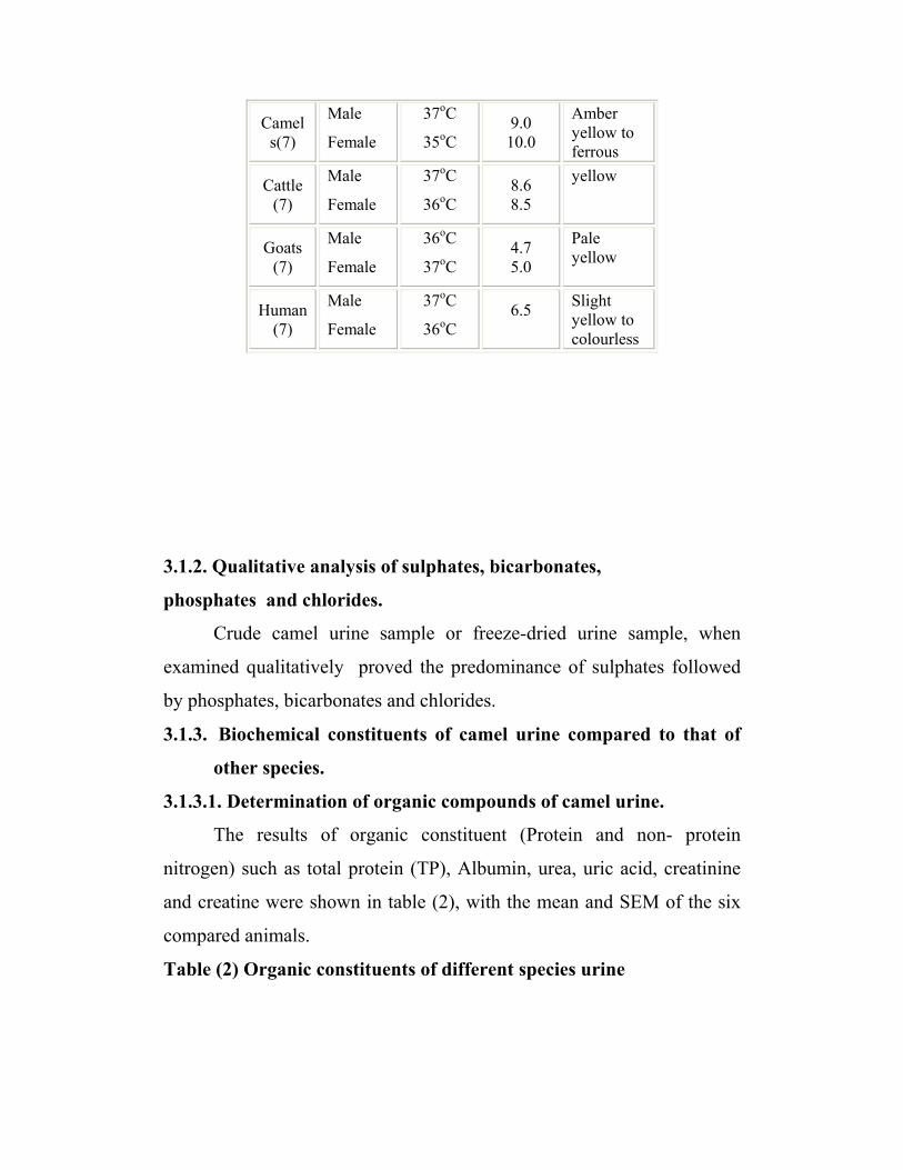
Camels(7)
Male
Female
37oC
35oC 9.0 10.0
Amber yellow to ferrous
Cattle (7)
Male
Female
37oC
36oC 8.6 8.5
yellow
Goats (7)
Male
Female
36oC
37oC 4.7 5.0
Pale yellow
Human (7)
Male
Female
37oC
36oC 6.5
Slight yellow to colourless
3.1.2. Qualitative analysis of sulphates, bicarbonates,
phosphates and chlorides.
Crude camel urine sample or freeze-dried urine sample, when
examined qualitatively proved the predominance of sulphates followed
by phosphates, bicarbonates and chlorides.
3.1.3. Biochemical constituents of camel urine compared to that of
other species.
3.1.3.1. Determination of organic compounds of camel urine.
The results of organic constituent (Protein and non- protein
nitrogen) such as total protein (TP), Albumin, urea, uric acid, creatinine
and creatine were shown in table (2), with the mean and SEM of the six
compared animals.
Table (2) Organic constituents of different species urine
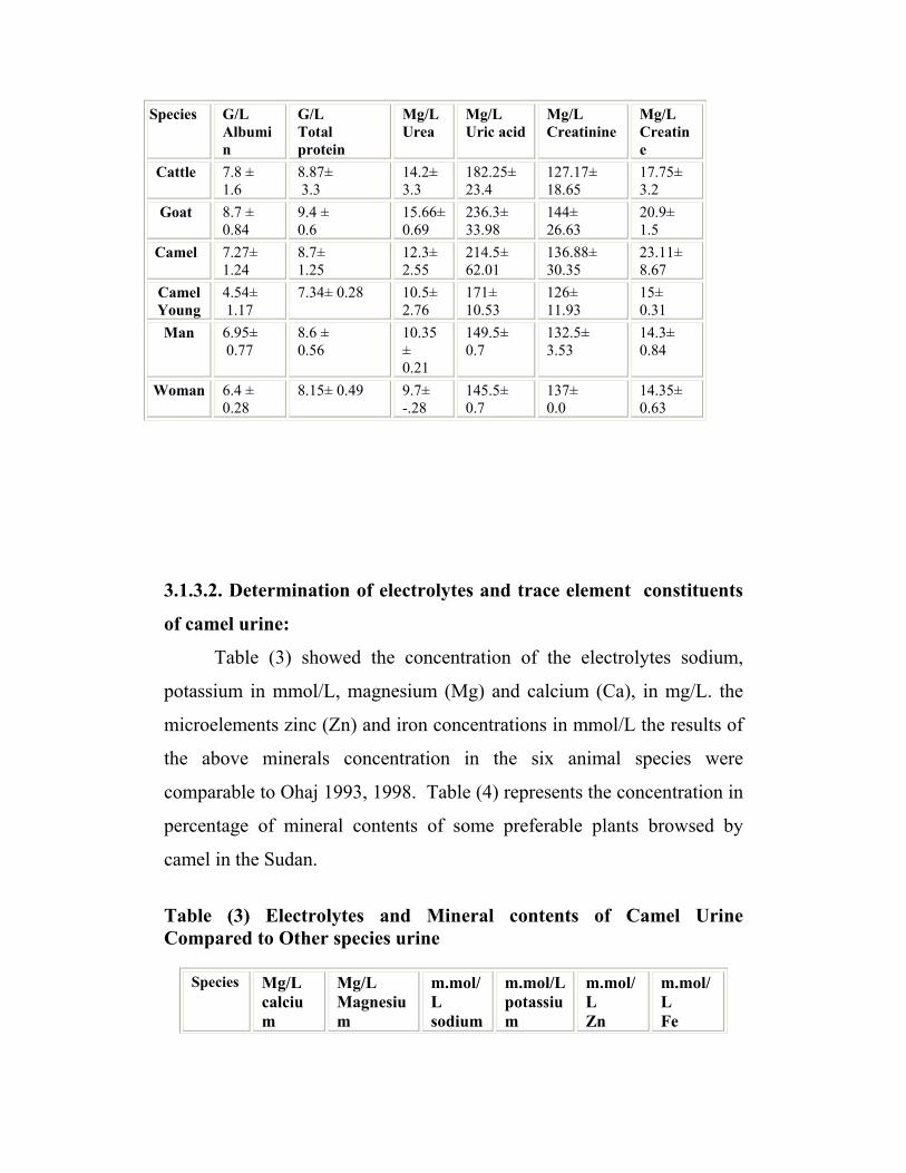
Species G/L Albumin
G/L Total protein
Mg/L Urea
Mg/L Uric acid
Mg/L Creatinine
Mg/L Creatine
Cattle 7.8 ± 1.6
8.87± 3.3
14.2± 3.3
182.25± 23.4
127.17± 18.65
17.75± 3.2
Goat 8.7 ± 0.84
9.4 ± 0.6
15.66± 0.69
236.3± 33.98
144± 26.63
20.9± 1.5
Camel 7.27± 1.24
8.7± 1.25
12.3± 2.55
214.5± 62.01
136.88± 30.35
23.11± 8.67
Camel Young
4.54± 1.17
7.34± 0.28 10.5± 2.76
171± 10.53
126± 11.93
15± 0.31
Man 6.95± 0.77
8.6 ± 0.56
10.35 ± 0.21
149.5± 0.7
132.5± 3.53
14.3± 0.84
Woman 6.4 ± 0.28
8.15± 0.49 9.7± -.28
145.5± 0.7
137± 0.0
14.35± 0.63
3.1.3.2. Determination of electrolytes and trace element constituents
of camel urine:
Table (3) showed the concentration of the electrolytes sodium,
potassium in mmol/L, magnesium (Mg) and calcium (Ca), in mg/L. the
microelements zinc (Zn) and iron concentrations in mmol/L the results of
the above minerals concentration in the six animal species were
comparable to Ohaj 1993, 1998. Table (4) represents the concentration in
percentage of mineral contents of some preferable plants browsed by
camel in the Sudan.
Table (3) Electrolytes and Mineral contents of Camel Urine Compared to Other species urine
Species Mg/L
calcium
Mg/L Magnesium
m.mol/L sodium
m.mol/L potassium
m.mol/L Zn
m.mol/L Fe
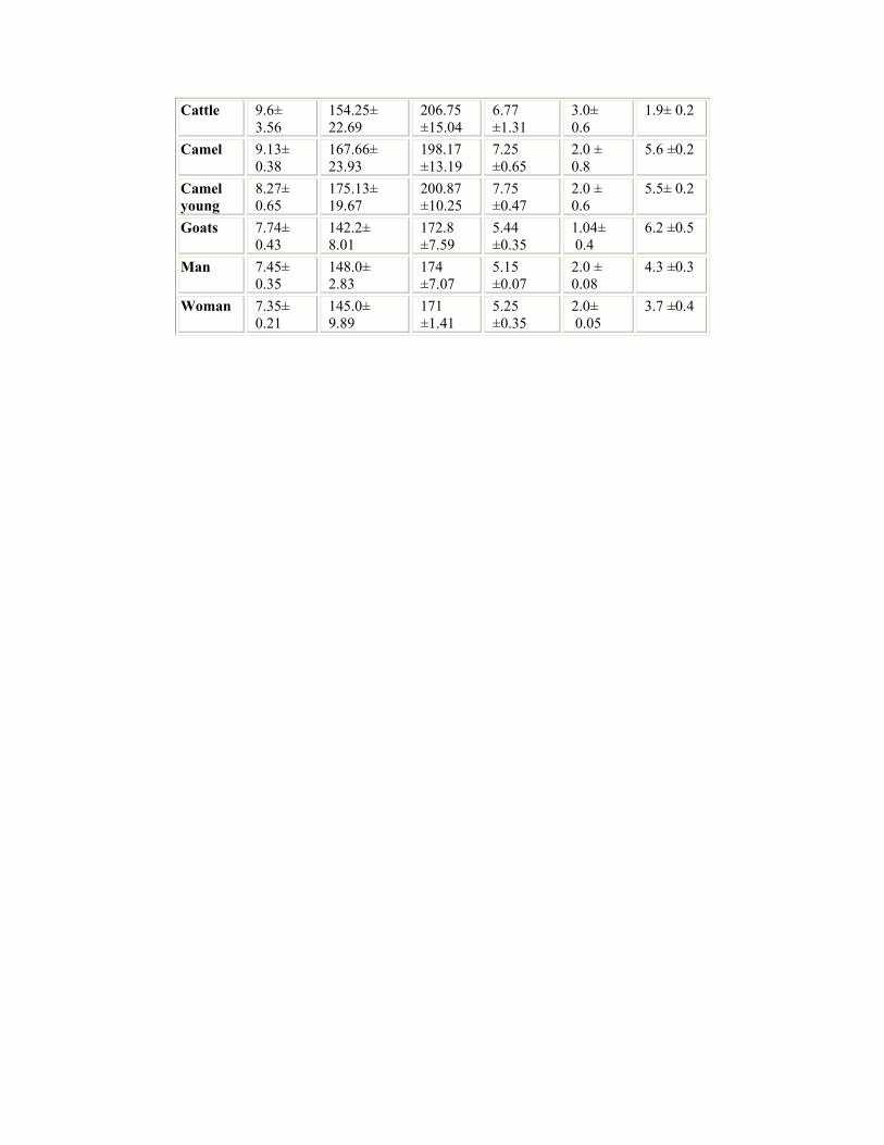
Cattle 9.6± 3.56
154.25± 22.69
206.75 ±15.04
6.77 ±1.31
3.0± 0.6
1.9± 0.2
Camel 9.13± 0.38
167.66± 23.93
198.17 ±13.19
7.25 ±0.65
2.0 ± 0.8
5.6 ±0.2
Camel young
8.27± 0.65
175.13± 19.67
200.87 ±10.25
7.75 ±0.47
2.0 ± 0.6
5.5± 0.2
Goats 7.74± 0.43
142.2± 8.01
172.8 ±7.59
5.44 ±0.35
1.04± 0.4
6.2 ±0.5
Man 7.45± 0.35
148.0± 2.83
174 ±7.07
5.15 ±0.07
2.0 ± 0.08
4.3 ±0.3
Woman 7.35± 0.21
145.0± 9.89
171 ±1.41
5.25 ±0.35
2.0± 0.05
3.7 ±0.4
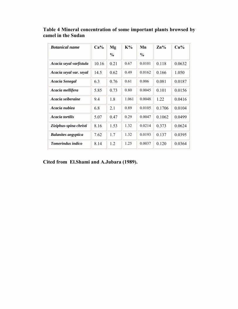
Table 4 Mineral concentration of some important plants browsed by camel in the Sudan
Botanical name Ca% Mg
%
K% Mn
%
Zn% Cu%
Acacia seyal varfistula 10.16 0.21 0.67 0.0101 0.118 0.0632
Acacia seyal var. seyal 14.5 0.62 0.49 0.0162 0.166 1.050
Acacia Senegal 6.3 0.76 0.61 0.006 0.081 0.0187
Acacia mellifera 5.85 0.73 0.80 0.0045 0.101 0.0156
Acacia seiberaine 9.4 1.8 1.061 0.0048 1.22 0.0416
Acacia nubiea 6.8 2.1 0.89 0.0105 0.1706 0.0104
Acacia tortilis 5.07 0.47 0.29 0.0047 0.1062 0.0499
Ziziphus-spina christi 8.16 1.53 1.32 0.0214 0.373 0.0624
Balanites aegyptica 7.62 1.7 1.32 0.0193 0.137 0.0395
Tomerindus indico 8.14 1.2 1.25 0.0037 0.120 0.0364
Cited from El.Shami and A.Jubara (1989).
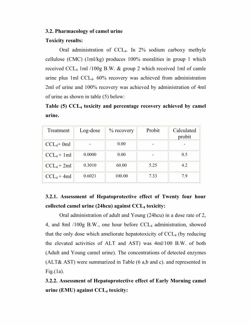
3.2. Pharmacology of camel urine
Toxicity results:
Oral administration of CCL4. In 2% sodium carboxy methyle
cellulose (CMC) (1ml/kg) produces 100% moralities in group 1 which
received CCL4 1ml /100g B.W. & group 2 which received 1ml of camle
urine plus 1ml CCL4. 60% recovery was achieved from administration
2ml of urine and 100% recovery was achieved by administration of 4ml
of urine as shown in table (5) below:
Table (5) CCL4 toxicity and percentage recovery achieved by camel
urine.
Treatment Log-dose % recovery Probit Calculated probit
CCL4+ 0ml - 0.00 - -
CCL4 + 1ml 0.0000 0.00 - 0.5
CCL4 + 2ml 0.3010 60.00 5.25 4.2
CCL4 + 4ml 0.6021 100.00 7.33 7.9
3.2.1. Assessment of Hepatoprotective effect of Twenty four hour
collected camel urine (24hcu) against CCL4 toxicity:
Oral administration of adult and Young (24hcu) in a dose rate of 2,
4, and 8ml /100g B.W., one hour before CCL4 administration, showed
that the only dose which ameliorate hepatotoxicity of CCL4 (by reducing
the elevated activities of ALT and AST) was 4ml/100 B.W. of both
(Adult and Young camel urine). The concentrations of detected enzymes
(ALT& AST) were summarized in Table (6 a,b and c). and represented in
Fig.(1a).
3.2.2. Assessment of Hepatoprotective effect of Early Morning camel
urine (EMU) against CCL4 toxicity:
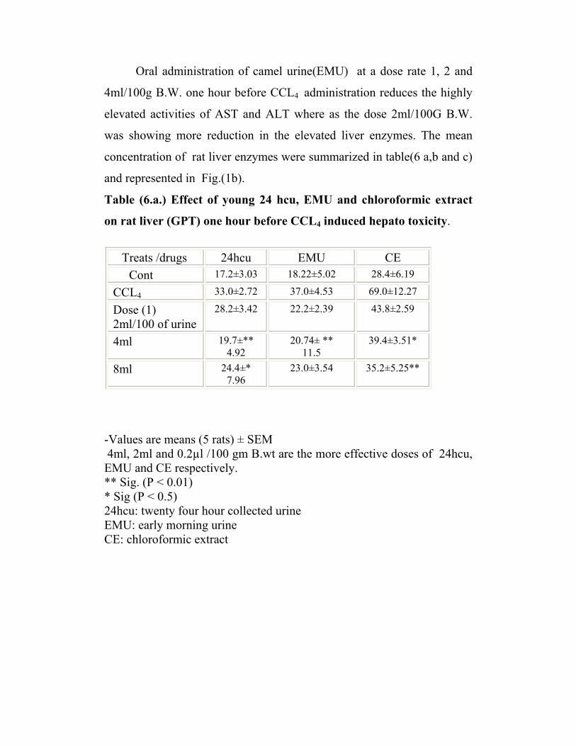
Oral administration of camel urine(EMU) at a dose rate 1, 2 and
4ml/100g B.W. one hour before CCL4 administration reduces the highly
elevated activities of AST and ALT where as the dose 2ml/100G B.W.
was showing more reduction in the elevated liver enzymes. The mean
concentration of rat liver enzymes were summarized in table(6 a,b and c)
and represented in Fig.(1b).
Table (6.a.) Effect of young 24 hcu, EMU and chloroformic extract
on rat liver (GPT) one hour before CCL4 induced hepato toxicity.
Treats /drugs 24hcu EMU CE Cont 17.2±3.03 18.22±5.02 28.4±6.19
CCL4 33.0±2.72 37.0±4.53 69.0±12.27
Dose (1) 2ml/100 of urine
28.2±3.42 22.2±2.39 43.8±2.59
4ml 19.7±** 4.92
20.74± ** 11.5
39.4±3.51*
8ml 24.4±* 7.96
23.0±3.54 35.2±5.25**
-Values are means (5 rats) ± SEM 4ml, 2ml and 0.2µl /100 gm B.wt are the more effective doses of 24hcu, EMU and CE respectively. ** Sig. (P < 0.01) * Sig (P < 0.5) 24hcu: twenty four hour collected urine EMU: early morning urine CE: chloroformic extract
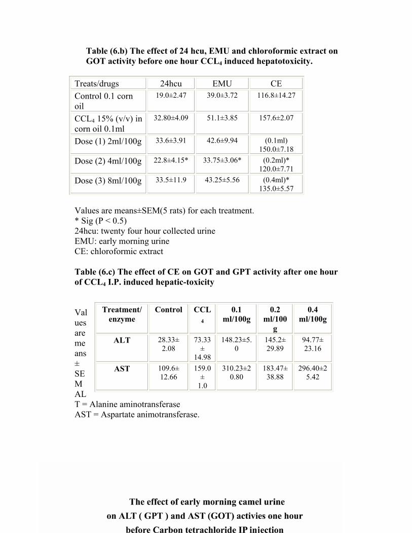
Table (6.b) The effect of 24 hcu, EMU and chloroformic extract on GOT activity before one hour CCL4 induced hepatotoxicity.
Treats/drugs 24hcu EMU CE Control 0.1 corn oil
19.0±2.47 39.0±3.72 116.8±14.27
CCL4 15% (v/v) in corn oil 0.1ml
32.80±4.09 51.1±3.85 157.6±2.07
Dose (1) 2ml/100g 33.6±3.91 42.6±9.94 (0.1ml) 150.0±7.18
Dose (2) 4ml/100g 22.8±4.15* 33.75±3.06* (0.2ml)* 120.0±7.71
Dose (3) 8ml/100g 33.5±11.9 43.25±5.56 (0.4ml)* 135.0±5.57
Values are means±SEM(5 rats) for each treatment. * Sig (P < 0.5) 24hcu: twenty four hour collected urine EMU: early morning urine CE: chloroformic extract Table (6.c) The effect of CE on GOT and GPT activity after one hour of CCL4 I.P. induced hepatic-toxicity Values are means ± SEM ALT = Alanine aminotransferase AST = Aspartate animotransferase.
Treatment/ enzyme
Control CCL4
0.1 ml/100g
0.2 ml/100
g
0.4 ml/100g
ALT 28.33± 2.08
73.33±
14.98
148.23±5.0
145.2± 29.89
94.77± 23.16
AST 109.6± 12.66
159.0±
1.0
310.23±20.80
183.47± 38.88
296.40±25.42

Fig (2)
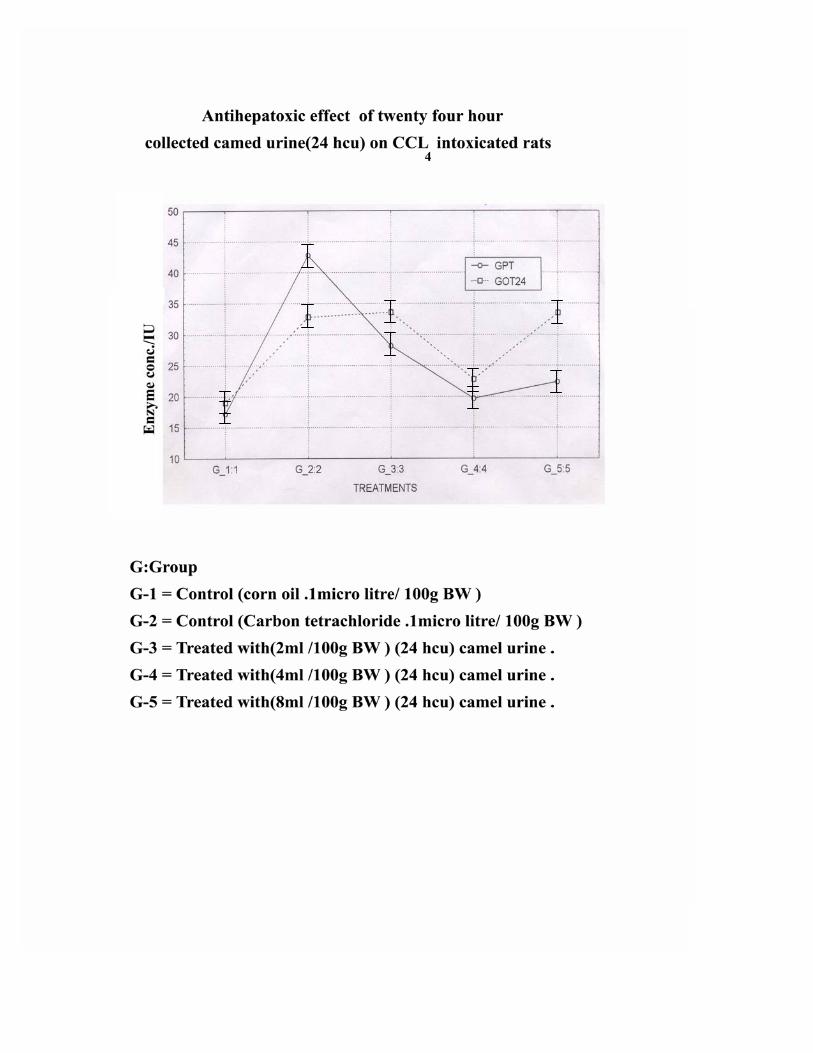
3.2.2 Assessment of Hepatoprotective effect of early morning
camel urine (EMU):
Oral administration at a dose rate of 1, 2 and 4 ml /100 g B.W. one
hour before CCL4 administration reduces the highly elevated activities of
AST and ALT whereas the dose 2ml/100g B.W. was showing more
reduction in the elevated enzymes. The mean concentration of the
reduced enzymes were summarized in Table (6) and represented in Fig.
(6b).
Fig (3)
/ L
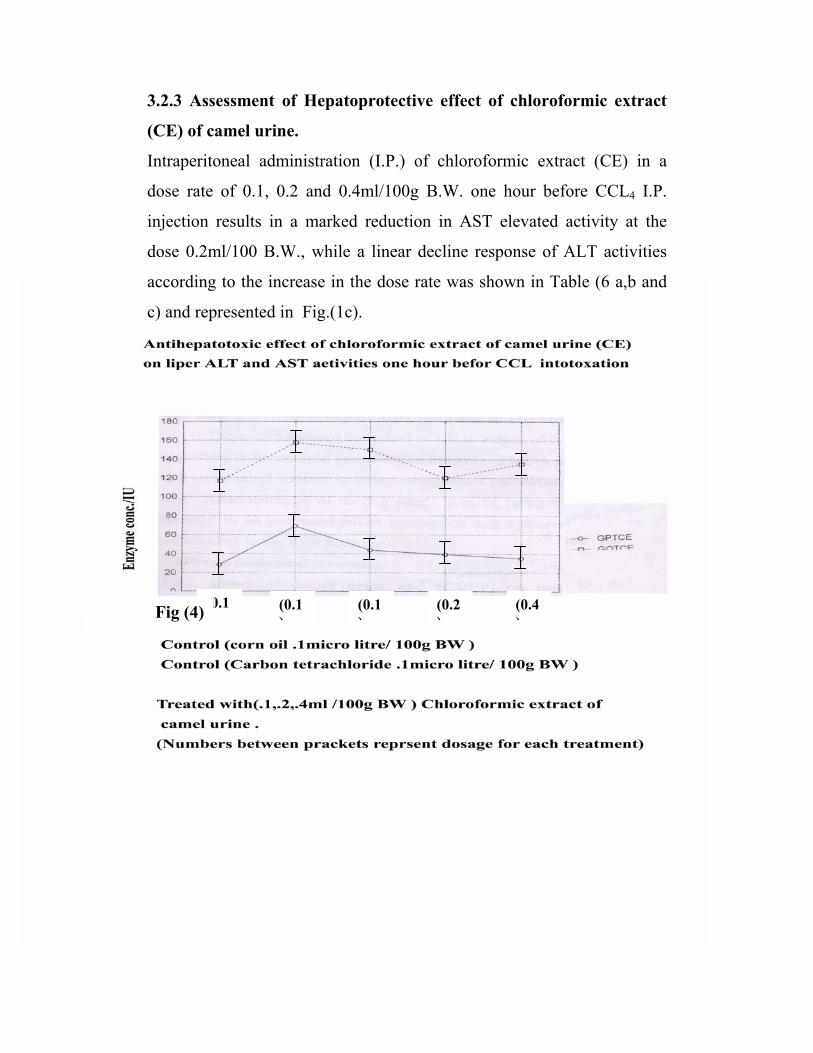
3.2.3 Assessment of Hepatoprotective effect of chloroformic extract
(CE) of camel urine.
Intraperitoneal administration (I.P.) of chloroformic extract (CE) in a
dose rate of 0.1, 0.2 and 0.4ml/100g B.W. one hour before CCL4 I.P.
injection results in a marked reduction in AST elevated activity at the
dose 0.2ml/100 B.W., while a linear decline response of ALT activities
according to the increase in the dose rate was shown in Table (6 a,b and
c) and represented in Fig.(1c).
(0.1)
(0.1)
(0.1)
(0.2)
(0.4)Fig (4)
3.2.4. Time course of hepatotoxic effect of CCL4 on rat liver: Enzyme levels in 5 rats serum were returned to normal levels after
96 hours, as shown in table (7) Fig.(2).
Table (7) Time course of hepatotoxic effect of CCL4 on rat liver:
Time/
enzyme
0 hour
24 hours
48 hour
72 hours
96
hours
GPT
(ALT)
28.33±
2.08 88.0±18.36 65.33±5.68 48.27±2.41
28.67±
6.5
GOT
(AST) 109.6±
12.66 208.13±8.95 181.27±8.73
161.93±
10.55
146.67±
6.11
- Values are means ± SEM
3.3. Effect of Camel urine (EMU) on serum AST, ALT and LDH
against paracetamol hepatotoxicity:
Fig (2) : Time course of hepatotoxic effect of CCL4 on rat liver
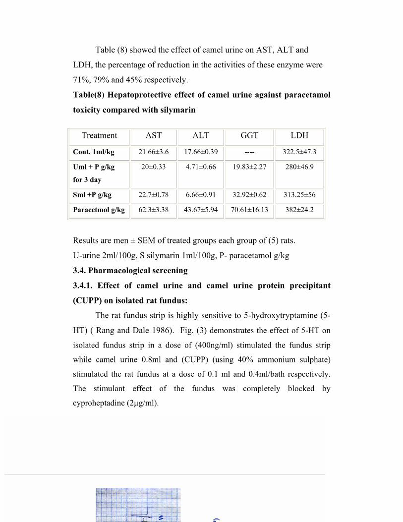
Table (8) showed the effect of camel urine on AST, ALT and
LDH, the percentage of reduction in the activities of these enzyme were
71%, 79% and 45% respectively.
Table(8) Hepatoprotective effect of camel urine against paracetamol
toxicity compared with silymarin
Treatment AST ALT GGT LDH
Cont. 1ml/kg 21.66±3.6 17.66±0.39 ---- 322.5±47.3
Uml + P g/kg
for 3 day
20±0.33 4.71±0.66 19.83±2.27 280±46.9
Sml +P g/kg 22.7±0.78 6.66±0.91 32.92±0.62 313.25±56
Paracetmol g/kg 62.3±3.38 43.67±5.94 70.61±16.13 382±24.2
Results are men ± SEM of treated groups each group of (5) rats.
U-urine 2ml/100g, S silymarin 1ml/100g, P- paracetamol g/kg
3.4. Pharmacological screening
3.4.1. Effect of camel urine and camel urine protein precipitant
(CUPP) on isolated rat fundus:
The rat fundus strip is highly sensitive to 5-hydroxytryptamine (5-
HT) ( Rang and Dale 1986). Fig. (3) demonstrates the effect of 5-HT on
isolated fundus strip in a dose of (400ng/ml) stimulated the fundus strip
while camel urine 0.8ml and (CUPP) (using 40% ammonium sulphate)
stimulated the rat fundus at a dose of 0.1 ml and 0.4ml/bath respectively.
The stimulant effect of the fundus was completely blocked by
cyproheptadine (2µg/ml).
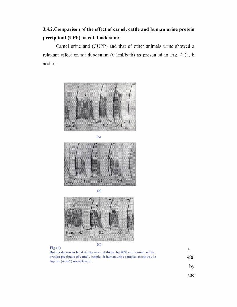
3.4.2.Comparison of the effect of camel, cattle and human urine protein
precipitant (UPP) on rat duodenum:
Camel urine and (CUPP) and that of other animals urine showed a
relaxant effect on rat duodenum (0.1ml/bath) as presented in Fig. 4 (a, b
and c).
3.4.3. Effect of (CUPP) and camel urine on Isolated rabbit jejunum.
Rabbit jejunum has a myogenic contraction (Rang and Dale,1986
and 2003). Relaxation of these contractions is produced by
sympathomimetics or direct muscle relaxants. Fig. (5.a.) shows the
influence of (CUPP) on contracting rabbit jejunum. This effect was
completely blocked by atropine (2.5µg/ml). Camel urine induced
transient relaxation followed by increased contraction.
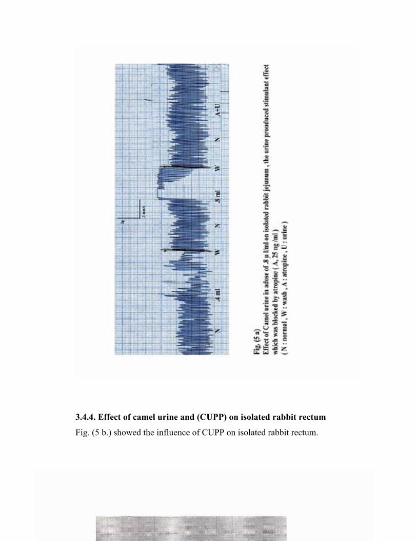
3.4.4. Effect of camel urine and (CUPP) on isolated rabbit rectum
Fig. (5 b.) showed the influence of CUPP on isolated rabbit rectum.

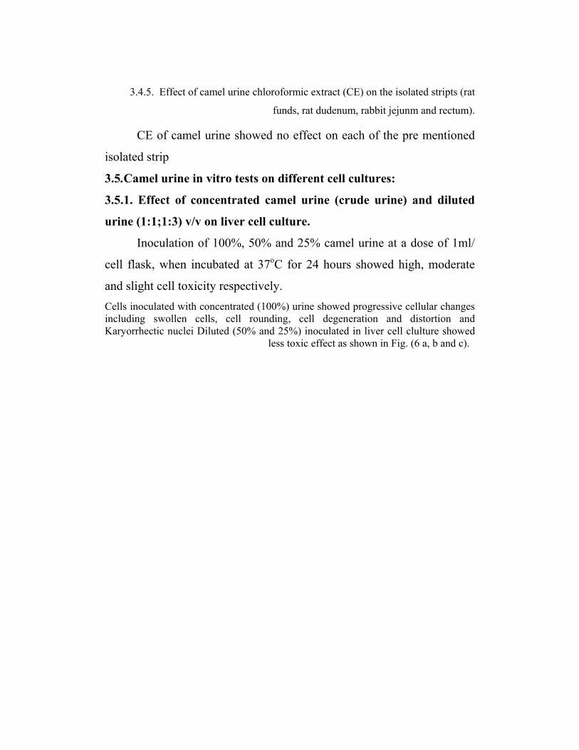
3.4.5. Effect of camel urine chloroformic extract (CE) on the isolated stripts (rat
funds, rat dudenum, rabbit jejunm and rectum).
CE of camel urine showed no effect on each of the pre mentioned
isolated strip
3.5.Camel urine in vitro tests on different cell cultures:
3.5.1. Effect of concentrated camel urine (crude urine) and diluted
urine (1:1;1:3) v/v on liver cell culture.
Inoculation of 100%, 50% and 25% camel urine at a dose of 1ml/
cell flask, when incubated at 37oC for 24 hours showed high, moderate
and slight cell toxicity respectively. Cells inoculated with concentrated (100%) urine showed progressive cellular changes including swollen cells, cell rounding, cell degeneration and distortion and Karyorrhectic nuclei Diluted (50% and 25%) inoculated in liver cell clulture showed
less toxic effect as shown in Fig. (6 a, b and c).
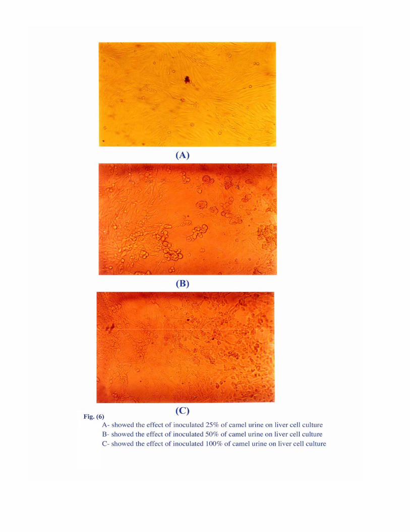
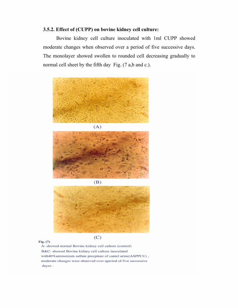
3.5.2. Effect of (CUPP) on bovine kidney cell culture:
Bovine kidney cell culture inoculated with 1ml CUPP showed
moderate changes when observed over a period of five successive days.
The monolayer showed swollen to rounded cell decreasing gradually to
normal cell sheet by the fifth day Fig. (7 a,b and c.).
3.6. Clinical effect of camel urine treatment:
3.6.1. Pretreatment clinical observation for selecting naturally
infected animals.
clinical signs are more or less the same in all naturally infected animals.
Although the intensity of chronicity is varied between the infected calves.
The pre-dominant observations were loss condition, emaciation, and
rough coats in some cases. As shown in Fig.(8).
3.6.1.1. Parasitological examination:-
sedimentation and floatation methods of faecal samples resulted in 15
positive samples with Faschiola gigantica and 2 cases with mixed
infection faschiola and Schistosoma bovis, 8 calves were infected with
Strongylus and 3 of them have faschiola. One animal showed Tricuris
and two were infected with Theileria after blood film examination (Table
9).
Table (9) examination of fecal sample for parasitological infection
Animal
No. Fasciola
eggs Schistosoma Others in blood faecal
S Theileria T 35 - - + 36 - - + 37 + - + + 38 - - + 39 + - 40 + 41 + +++ 42 + - 43 - - + 44 ++ - - - - 45 - - +
46 + + 47 + + 48 + - 49 - - + 50 51 + 52 + - 53 - - + 54 + + 55 + - 56 - - Strongylus 57 + - 58 + - 59 - -
3.6.1.2. Biochemical assessment of liver function test:
Biochemical assay of serum enzymes activity, such as aspartate
amino transferase (AST), alanine amino transferase (ALT), Alkaline
phosphates (ALP), lactate dehydrogenase (LDH) and Gamma glutamate
transferease (GGT) revealed an increase in the enzyme activities and
slight increase in the concentrations of serum electrolytes and minerals.
3.6.1.3. Haematological examination:
Anaemia with a reduced haemoglubin (Hb) concentration and red blood
cell (RBC) count was evident, and a parallel fall in PCV was detected.
Prothrombin time result in slight change in clothing time.
Haematological and biochemical results were represented in table ( 10
and 11).
Table (10) Haematological and biochemical analysis of naturally infected calves
(from white Nile province- Sudan)
Measurement Mean values for
the diagnosed
animal
Reference values
PCV % 18 28-38
leukocyte TWBC (µl) 3450±150.1 4000-10000
Total billirubin (g/L) 0.2 ± 0.01 0.8-8.6
AST (µ/l) 89 ± 11.4 40-80
GGT (u/L) 3.6 ± 0.2 6-17
Urea (mmol/L) 1.8 ± 0.05 2.5-7.5
Sodium (mmol/L) 155 ± 5.1 140-150
Potassium (mmol/L) 6.7 ± 1 4-5
Calcium (mmol/L) 8.7 ± 2.2 2-2.6
Phosphates(mmol/L) 4.9 ± 1.02 1.3-1.25
Magnesium (mmol/L) 2.2 ± 0.92 0.7-1.1
Units ALP 97 ± 3.9 0.0-500
Units LDH 464.42 ± 36.5 692-1445
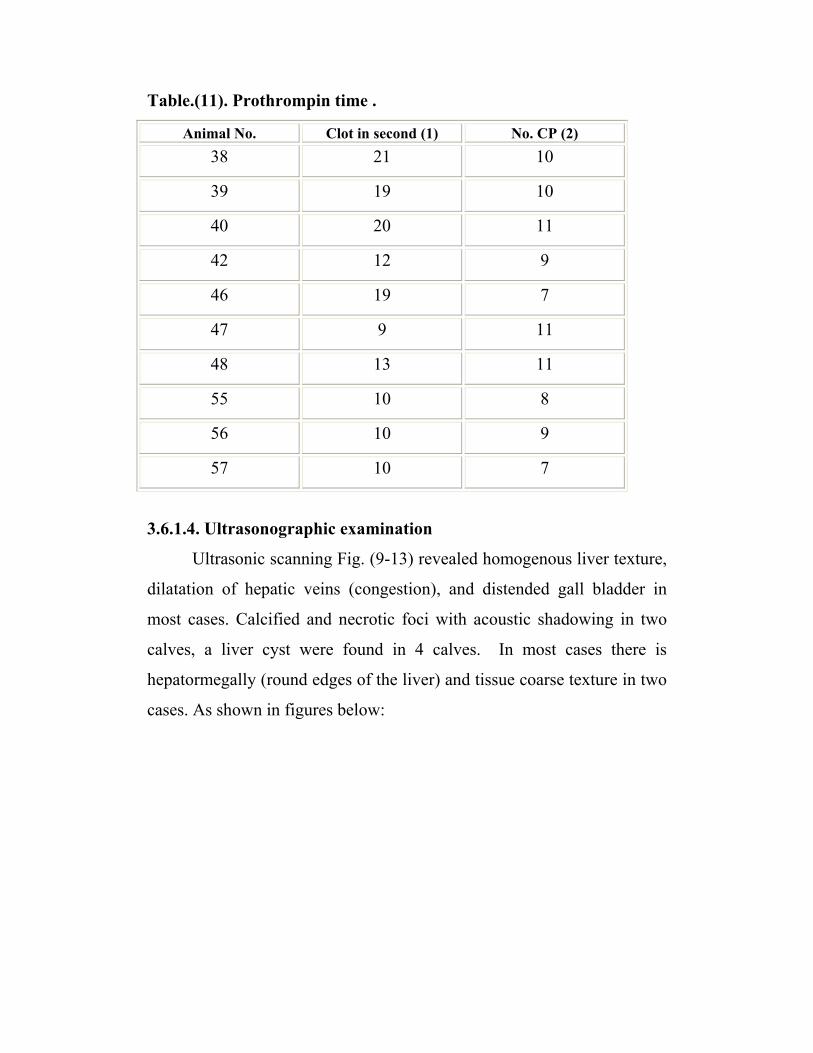
Table.(11). Prothrompin time .
Animal No. Clot in second (1) No. CP (2) 38 21 10
39 19 10
40 20 11
42 12 9
46 19 7
47 9 11
48 13 11
55 10 8
56 10 9
57 10 7
3.6.1.4. Ultrasonographic examination
Ultrasonic scanning Fig. (9-13) revealed homogenous liver texture,
dilatation of hepatic veins (congestion), and distended gall bladder in
most cases. Calcified and necrotic foci with acoustic shadowing in two
calves, a liver cyst were found in 4 calves. In most cases there is
hepatormegally (round edges of the liver) and tissue coarse texture in two
cases. As shown in figures below:
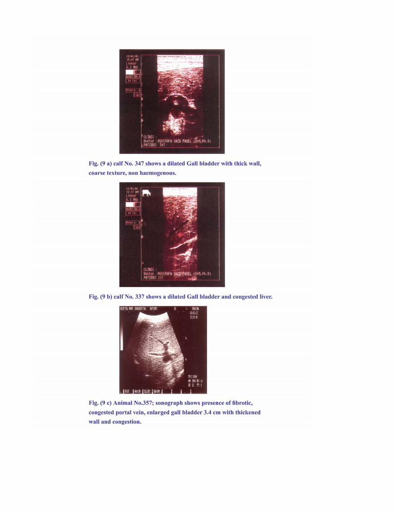
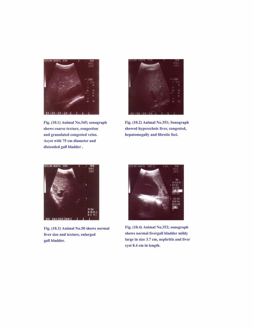
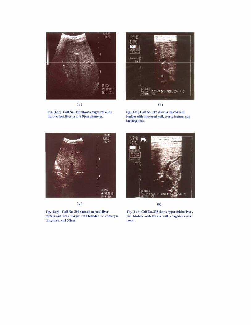
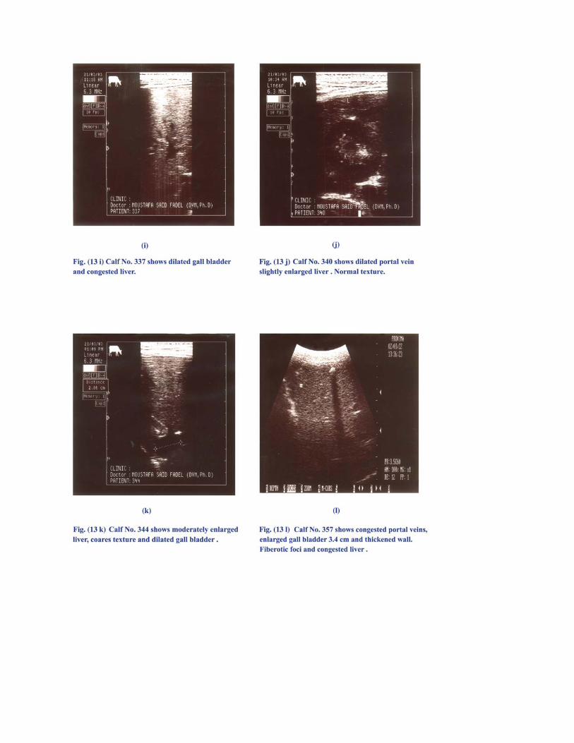
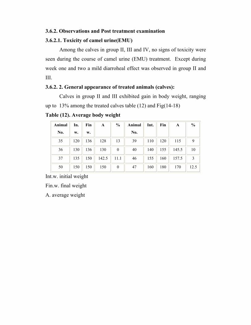
3.6.2. Observations and Post treatment examination
3.6.2.1. Toxicity of camel urine(EMU)
Among the calves in group II, III and IV, no signs of toxicity were
seen during the course of camel urine (EMU) treatment. Except during
week one and two a mild diarroheal effect was observed in group II and
III.
3.6.2. 2. General appearance of treated animals (calves):
Calves in group II and III exhibited gain in body weight, ranging
up to 13% among the treated calves table (12) and Fig(14-18)
Table (12). Average body weight
Animal
No.
In.
w.
Fin
w.
A
% Animal
No.
Int. Fin A %
35 120 136 128 13 39 110 120 115 9
36 130 136 130 0 40 140 155 145.5 10
37 135 150 142.5 11.1 46 155 160 157.5 3
50 150 150 150 0 47 160 180 170 12.5
Int.w. initial weight
Fin.w. final weight
A. average weight
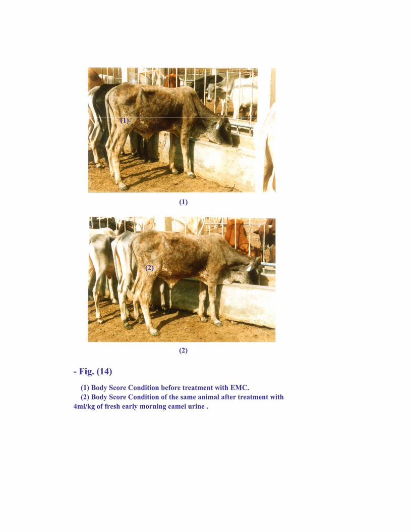
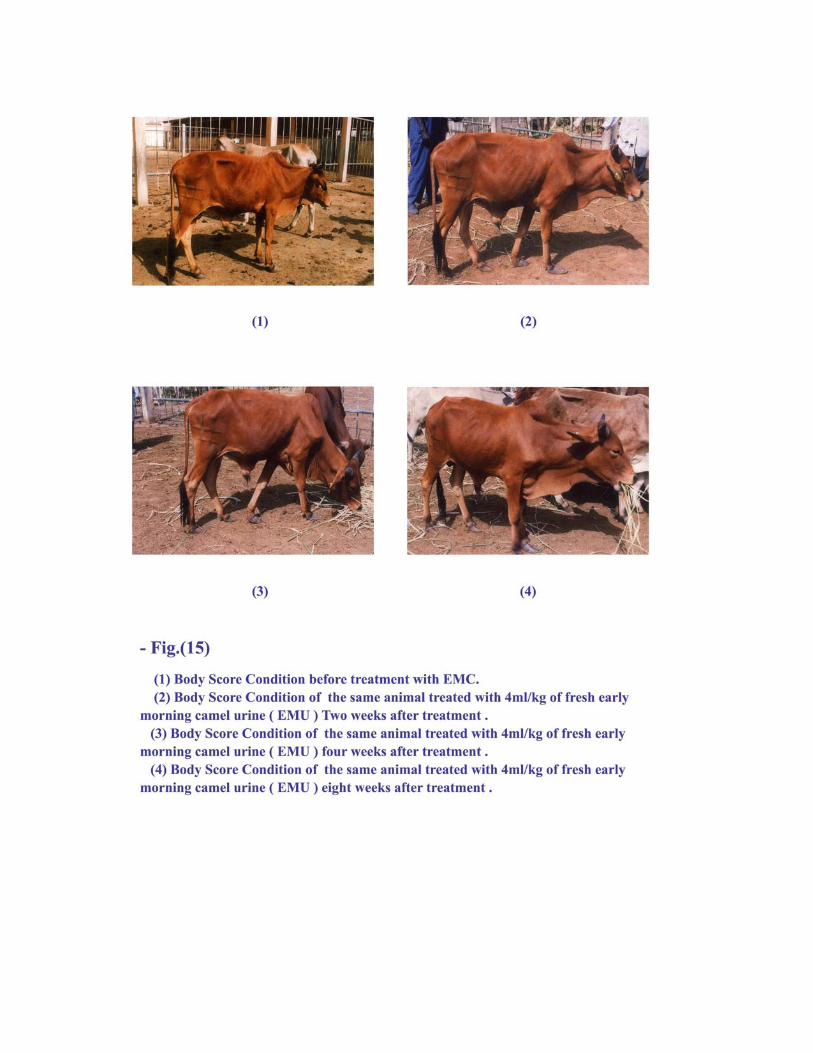
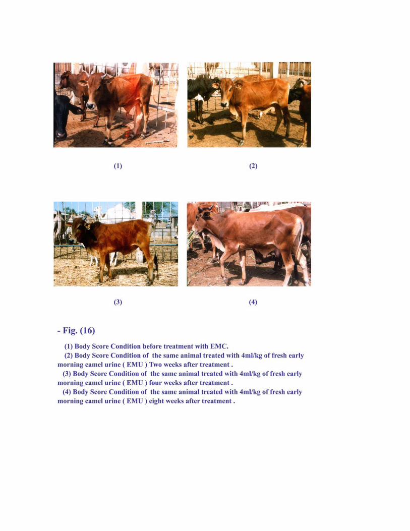
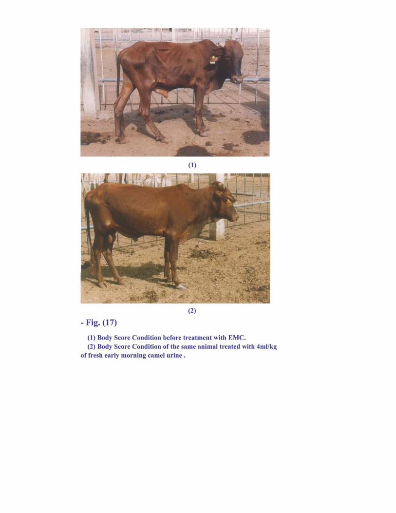
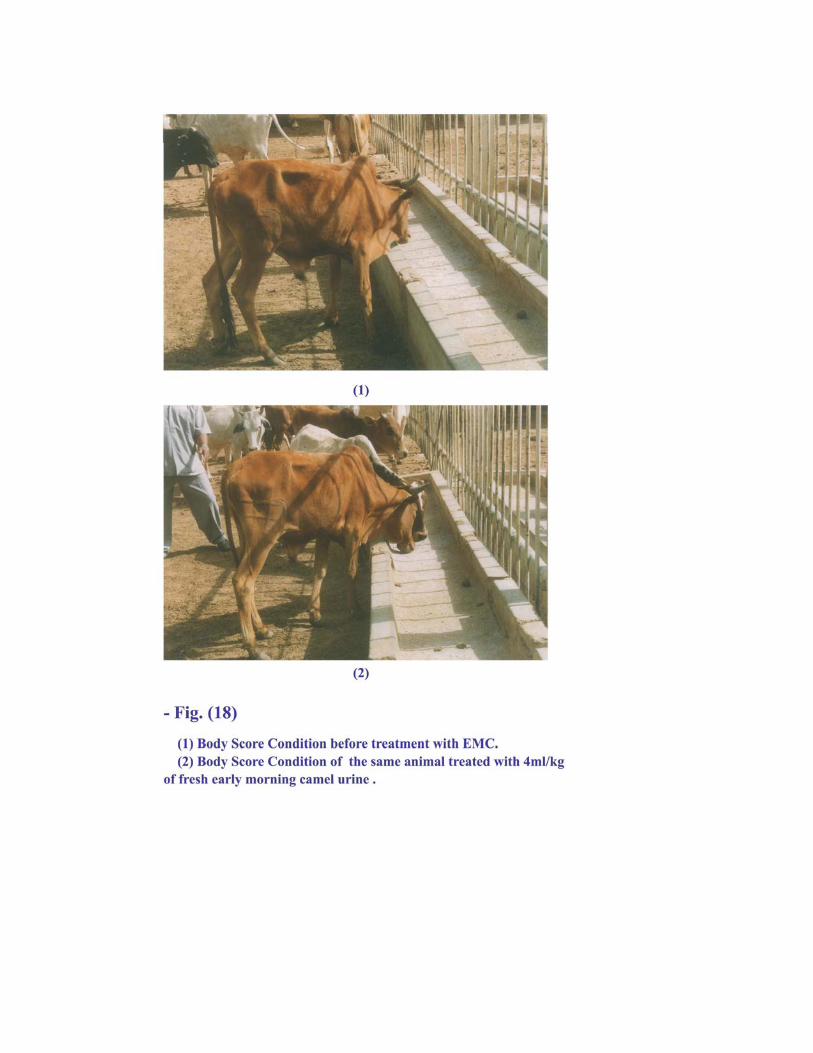
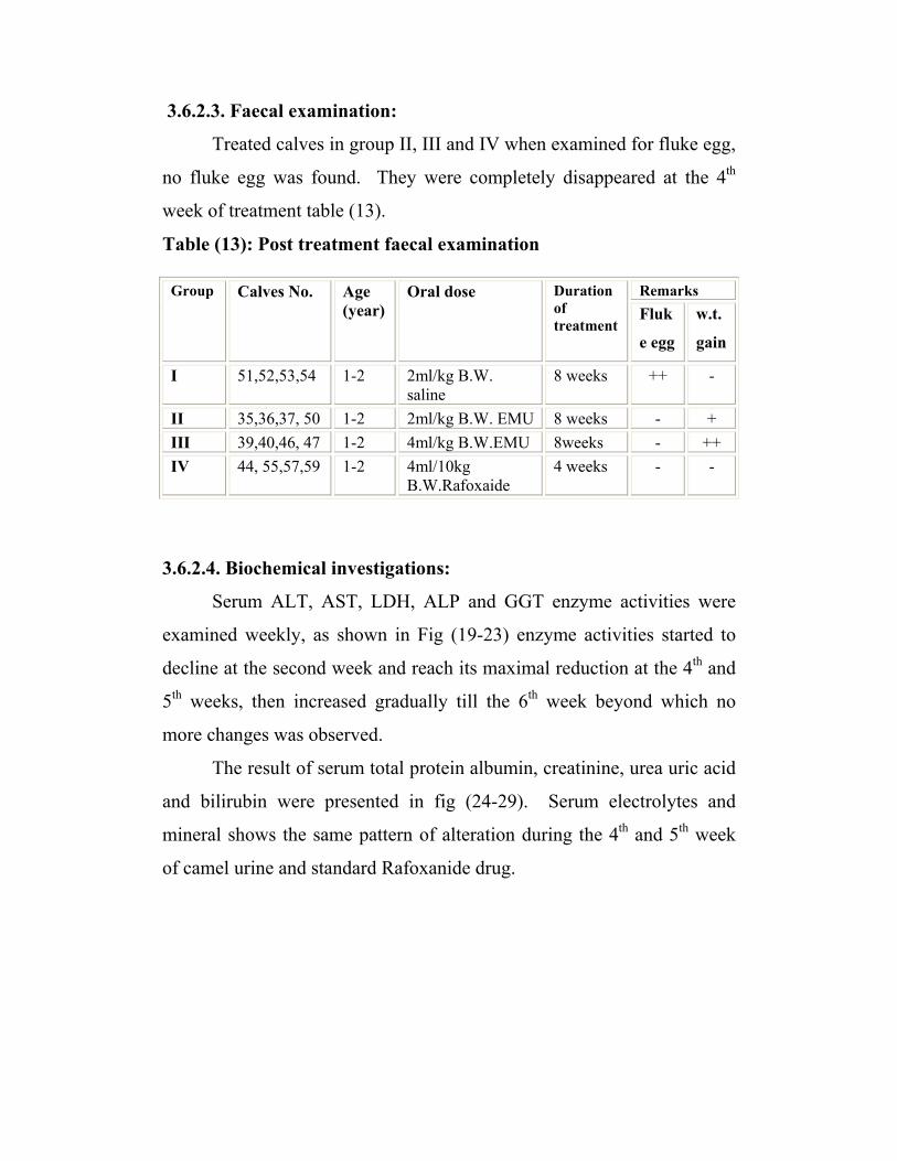
3.6.2.3. Faecal examination:
Treated calves in group II, III and IV when examined for fluke egg,
no fluke egg was found. They were completely disappeared at the 4th
week of treatment table (13).
Table (13): Post treatment faecal examination
Remarks Group Calves No. Age (year)
Oral dose Duration of treatment
Fluk
e egg
w.t.
gain
I 51,52,53,54 1-2 2ml/kg B.W. saline
8 weeks ++ -
II 35,36,37, 50 1-2 2ml/kg B.W. EMU 8 weeks - + III 39,40,46, 47 1-2 4ml/kg B.W.EMU 8weeks - ++ IV 44, 55,57,59 1-2 4ml/10kg
B.W.Rafoxaide 4 weeks - -
3.6.2.4. Biochemical investigations:
Serum ALT, AST, LDH, ALP and GGT enzyme activities were
examined weekly, as shown in Fig (19-23) enzyme activities started to
decline at the second week and reach its maximal reduction at the 4th and
5th weeks, then increased gradually till the 6th week beyond which no
more changes was observed.
The result of serum total protein albumin, creatinine, urea uric acid
and bilirubin were presented in fig (24-29). Serum electrolytes and
mineral shows the same pattern of alteration during the 4th and 5th week
of camel urine and standard Rafoxanide drug.
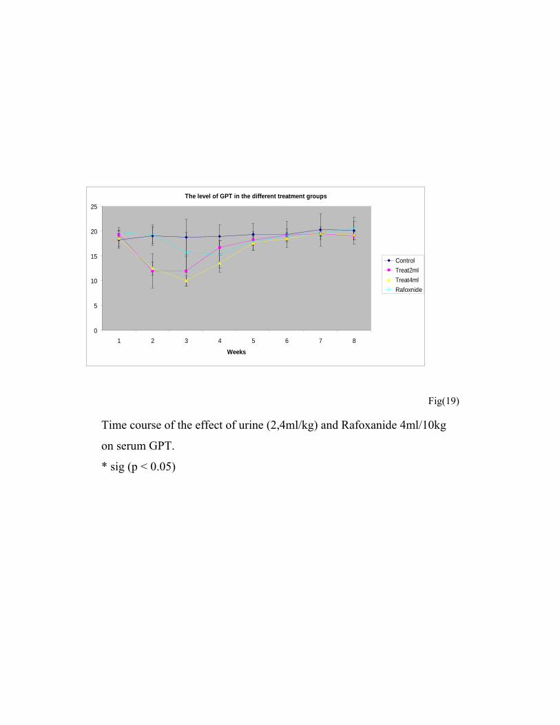
The level of GPT in the different treatment groups
0
5
10
15
20
25
1 2 3 4 5 6 7 8
Weeks
ControlTreat2mlTreat4mlRafoxnide
Fig(19)
Time course of the effect of urine (2,4ml/kg) and Rafoxanide 4ml/10kg
on serum GPT.
* sig (p < 0.05)
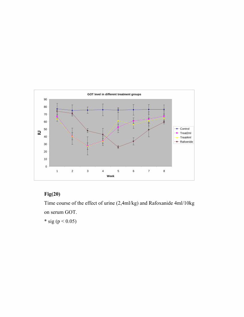
GOT level in different treatment groups
0
10
20
30
40
50
60
70
80
90
1 2 3 4 5 6 7 8
Week
IU
ControlTreat2mlTreat4mlRafoxnide
Fig(20)
Time course of the effect of urine (2,4ml/kg) and Rafoxanide 4ml/10kg
on serum GOT.
* sig (p < 0.05)
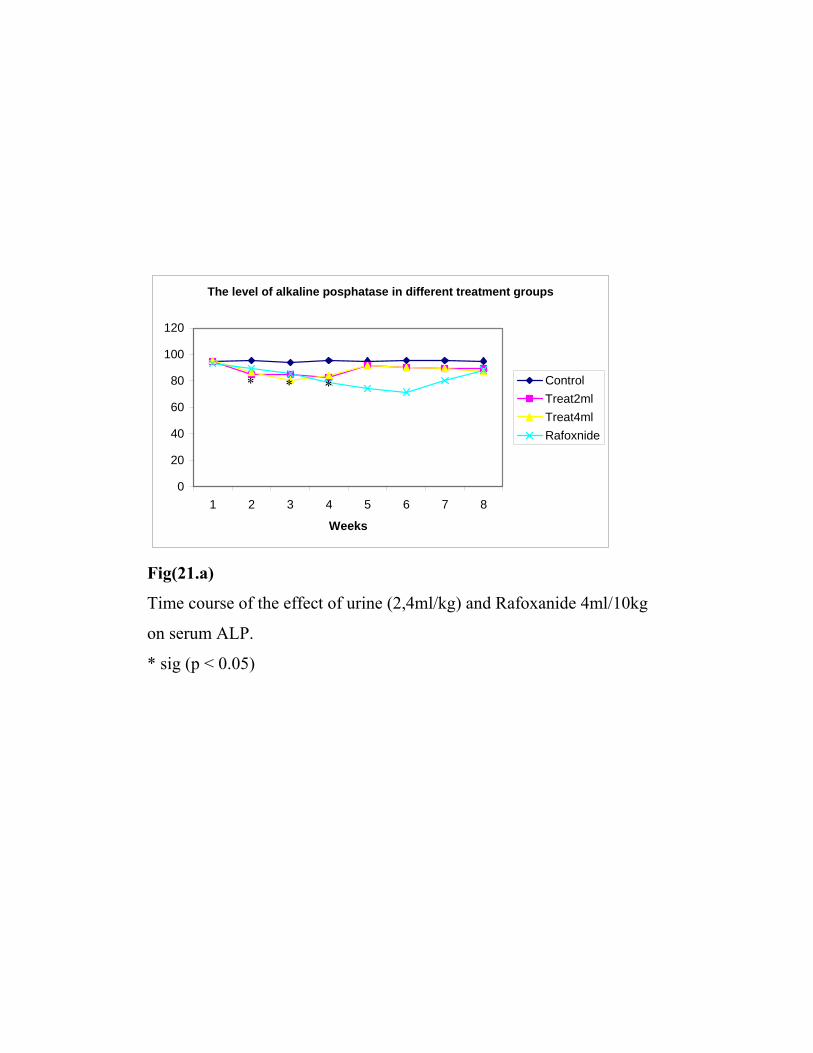
The level of alkaline posphatase in different treatment groups
0
20
40
60
80
100
120
1 2 3 4 5 6 7 8
Weeks
ControlTreat2mlTreat4mlRafoxnide
Fig(21.a)
Time course of the effect of urine (2,4ml/kg) and Rafoxanide 4ml/10kg
on serum ALP.
* sig (p < 0.05)
* * *
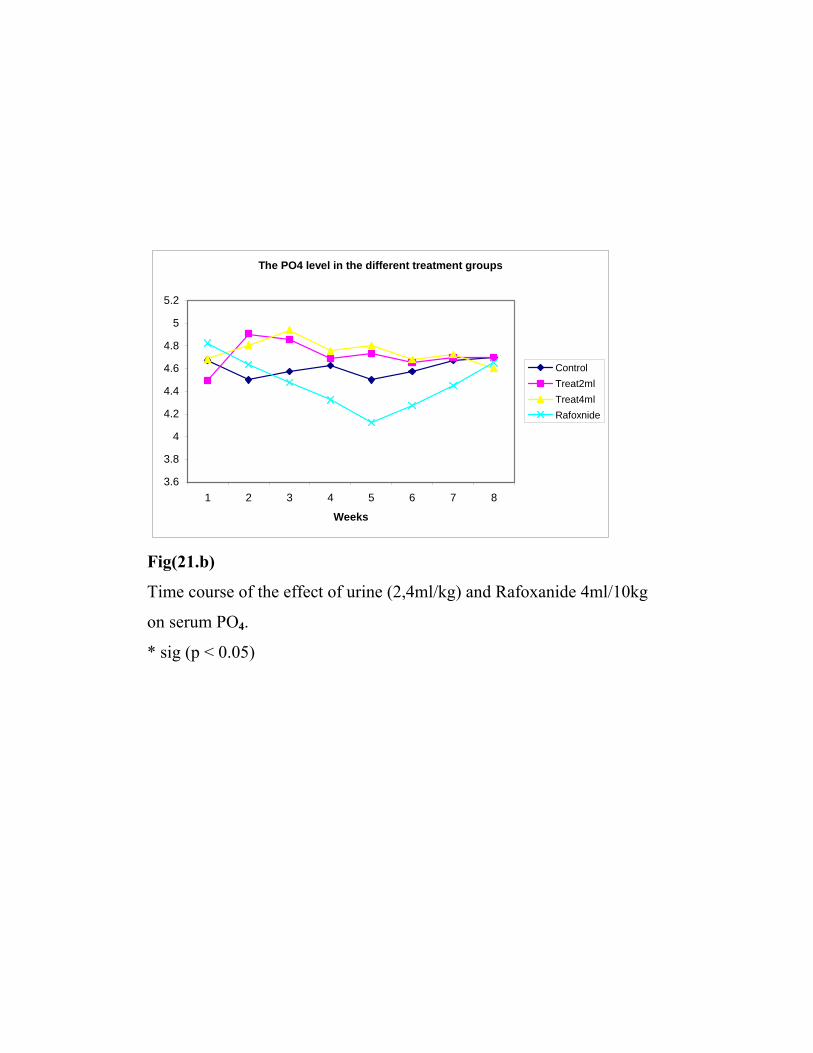
The PO4 level in the different treatment groups
3.6
3.8
4
4.2
4.4
4.6
4.8
5
5.2
1 2 3 4 5 6 7 8
Weeks
ControlTreat2mlTreat4mlRafoxnide
Fig(21.b)
Time course of the effect of urine (2,4ml/kg) and Rafoxanide 4ml/10kg
on serum PO4.
* sig (p < 0.05)
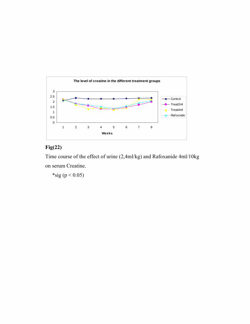
The level of creatine in the different treatment groups
00.5
11.5
22.5
3
1 2 3 4 5 6 7 8
Weeks
Control
Treat2ml
Treat4ml
Rafoxnide
Fig(22)
Time course of the effect of urine (2,4ml/kg) and Rafoxanide 4ml/10kg
on serum Creatine.
*sig (p < 0.05)
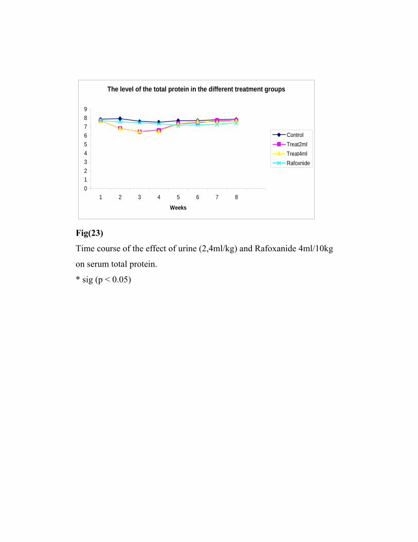
The level of the total protein in the different treatment groups
0123456789
1 2 3 4 5 6 7 8
Weeks
ControlTreat2mlTreat4mlRafoxnide
Fig(23)
Time course of the effect of urine (2,4ml/kg) and Rafoxanide 4ml/10kg
on serum total protein.
* sig (p < 0.05)
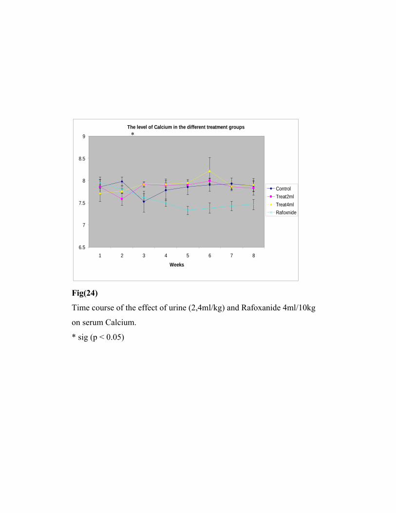
The level of Calcium in the different treatment groups
6.5
7
7.5
8
8.5
9
1 2 3 4 5 6 7 8
Weeks
ControlTreat2mlTreat4mlRafoxnide
Fig(24)
Time course of the effect of urine (2,4ml/kg) and Rafoxanide 4ml/10kg
on serum Calcium.
* sig (p < 0.05)
*
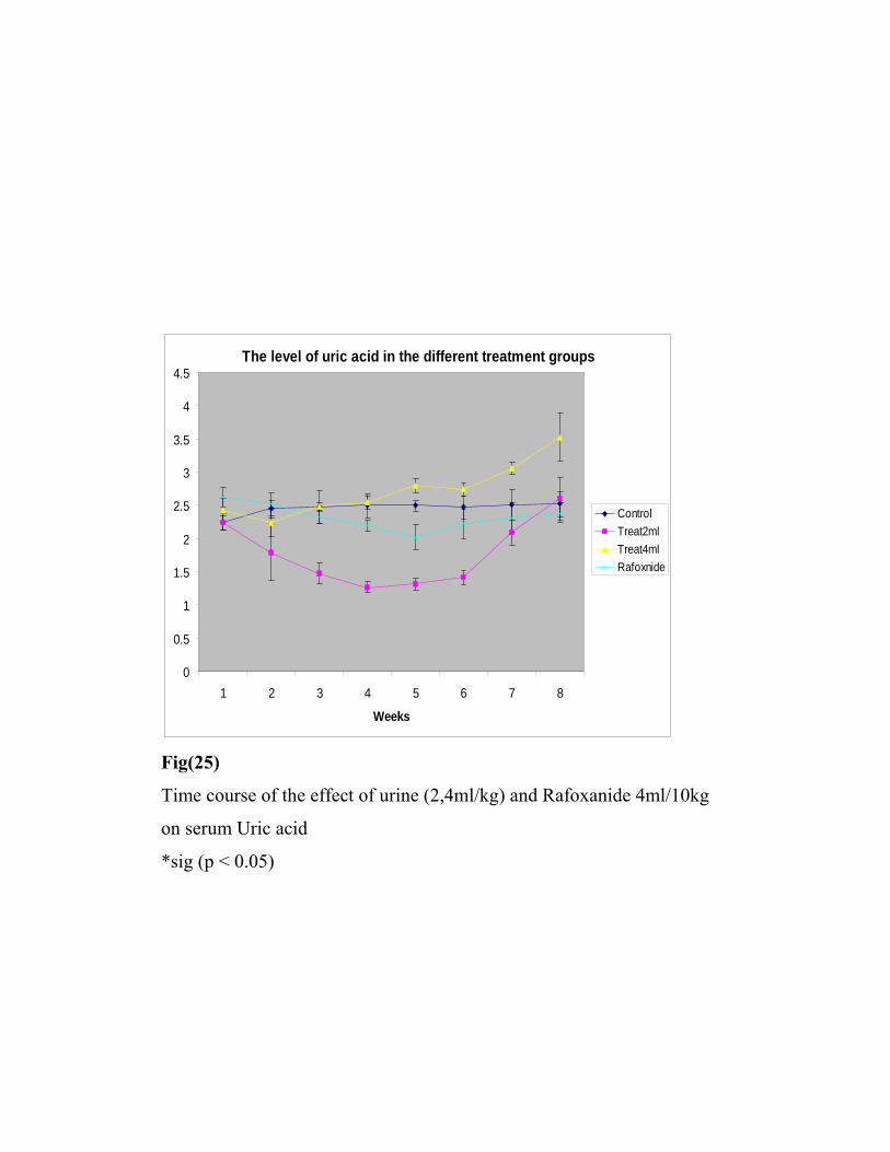
The level of uric acid in the different treatment groups
0
0.5
1
1.5
2
2.5
3
3.5
4
4.5
1 2 3 4 5 6 7 8
Weeks
ControlTreat2mlTreat4mlRafoxnide
Fig(25)
Time course of the effect of urine (2,4ml/kg) and Rafoxanide 4ml/10kg
on serum Uric acid
*sig (p < 0.05)
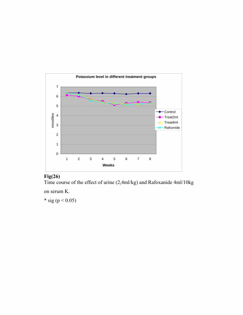
Potassium level in different treatment groups
0
1
2
3
4
5
6
7
1 2 3 4 5 6 7 8
Weeks
mm
ol/li
tre
ControlTreat2mlTreat4mlRafoxnide
Fig(26) Time course of the effect of urine (2,4ml/kg) and Rafoxanide 4ml/10kg
on serum K.
* sig (p < 0.05)
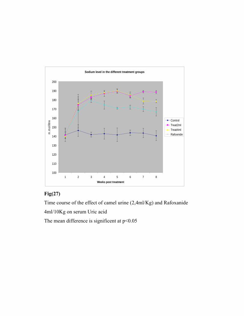
Sodium level in the different treatment groups
100
110
120
130
140
150
160
170
180
190
200
1 2 3 4 5 6 7 8
Weeks post treatment
m.m
ol/li
tre
ControlTreat2mlTreat4mlRafoxnide
Fig(27)
Time course of the effect of camel urine (2,4ml/Kg) and Rafoxanide
4ml/10Kg on serum Uric acid
The mean difference is significent at p<0.05
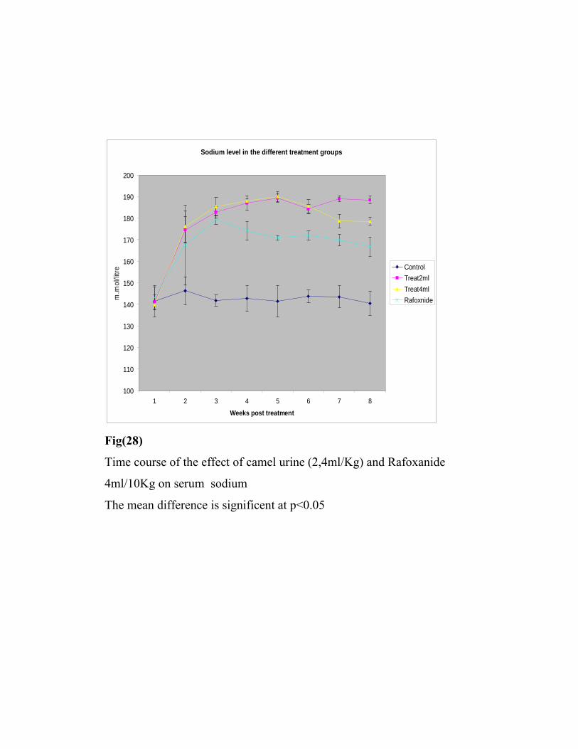
Sodium level in the different treatment groups
100
110
120
130
140
150
160
170
180
190
200
1 2 3 4 5 6 7 8
Weeks post treatment
m.m
ol/li
tre
ControlTreat2mlTreat4mlRafoxnide
Fig(28)
Time course of the effect of camel urine (2,4ml/Kg) and Rafoxanide
4ml/10Kg on serum sodium
The mean difference is significent at p<0.05

3.6.2.5. Haematological findings:
Haematological parameter resulted in increased Hb and PCV. Also
some animals showed an increase in RBCs and WBCs count as shown in
Fig.(30-34).


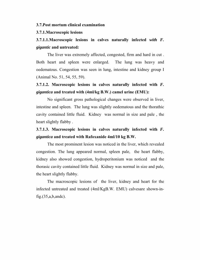
3.7.Post mortum clinical examination
3.7.1.Macroscopic lesions
3.7.1.1.Macroscopic lesions in calves naturally infected with F.
gigantic and untreated:
The liver was extremely affected, congested, firm and hard in cut .
Both heart and spleen were enlarged. The lung was heavy and
oedematous. Congestion was seen in lung, intestine and kidney group I
(Animal No. 51, 54, 55, 59).
3.7.1.2. Macroscopic lesions in calves naturally infected with F.
gigantica and treated with (4ml/kg B.W.) camel urine (EMU):
No significant gross pathological changes were observed in liver,
intestine and spleen. The lung was slightly oedematous and the thorathic
cavity contained little fluid. Kidney was normal in size and pale , the
heart slightly flabby .
3.7.1.3. Macroscopic lesions in calves naturally infected with F.
gigantica and treated with Rafoxanide 4ml/10 kg B.W.
The most prominent lesion was noticed in the liver, which revealed
congestion. The lung appeared normal, spleen pale, the heart flabby,
kidney also showed congestion, hydroperitonium was noticed and the
thorasic cavity contained little fluid. Kidney was normal in size and pale,
the heart slightly flabby.
The macroscopic lesions of the liver, kidney and heart for the
infected untreated and treated (4ml/KgB.W. EMU) calvesare shown-in-
fig.(35,a,b,andc).
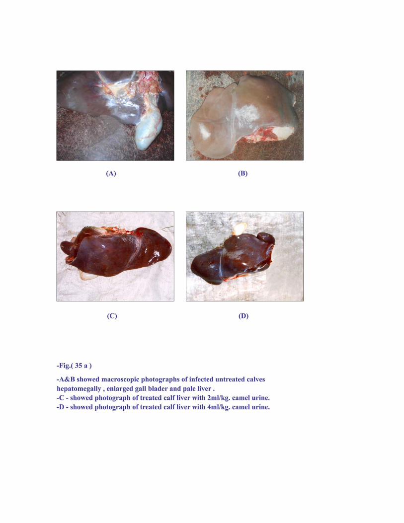
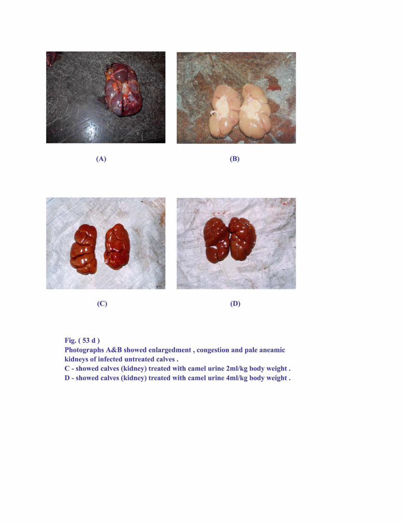
Fig.(21 b)
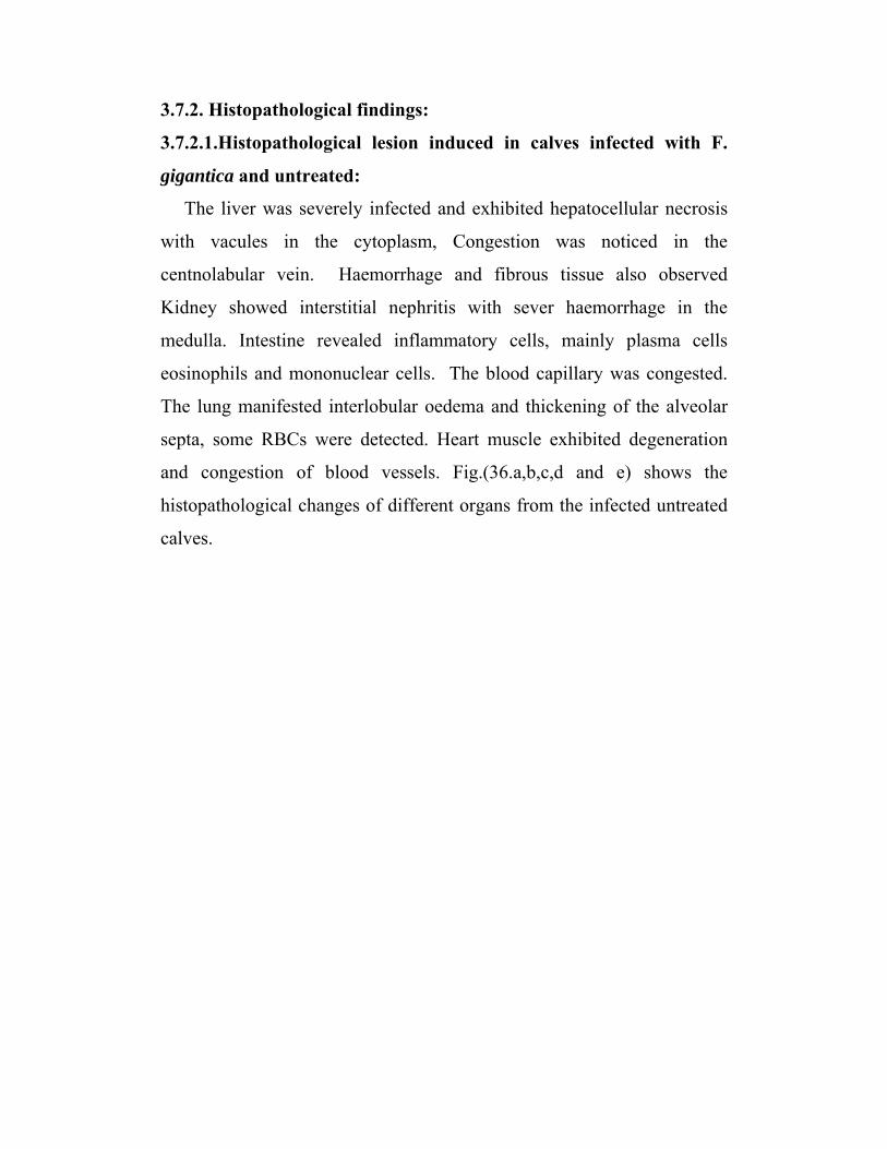
3.7.2. Histopathological findings:
3.7.2.1.Histopathological lesion induced in calves infected with F.
gigantica and untreated:
The liver was severely infected and exhibited hepatocellular necrosis
with vacules in the cytoplasm, Congestion was noticed in the
centnolabular vein. Haemorrhage and fibrous tissue also observed
Kidney showed interstitial nephritis with sever haemorrhage in the
medulla. Intestine revealed inflammatory cells, mainly plasma cells
eosinophils and mononuclear cells. The blood capillary was congested.
The lung manifested interlobular oedema and thickening of the alveolar
septa, some RBCs were detected. Heart muscle exhibited degeneration
and congestion of blood vessels. Fig.(36.a,b,c,d and e) shows the
histopathological changes of different organs from the infected untreated
calves.
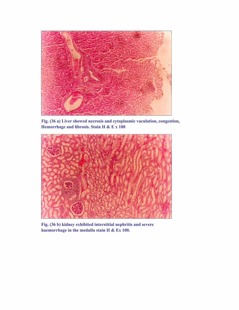
Fig
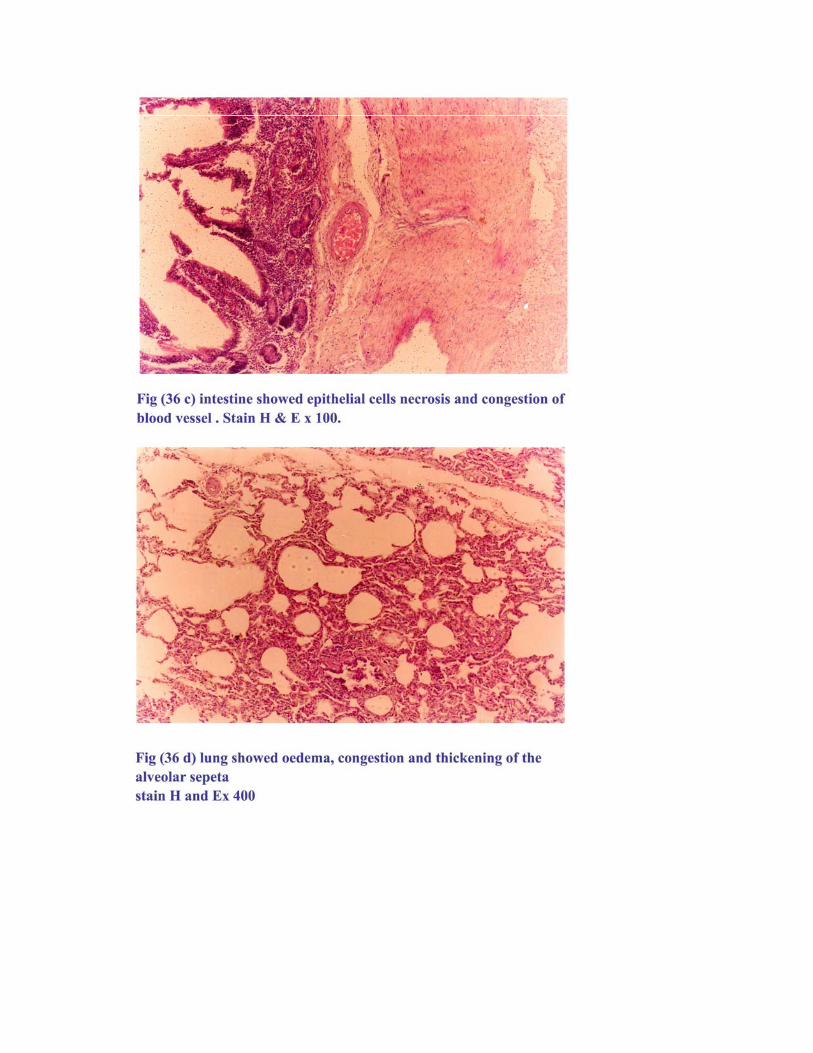
Lung showed oedema, congestion and thickening of alveolar spta stain H&E X400
Fig
Fig (25)
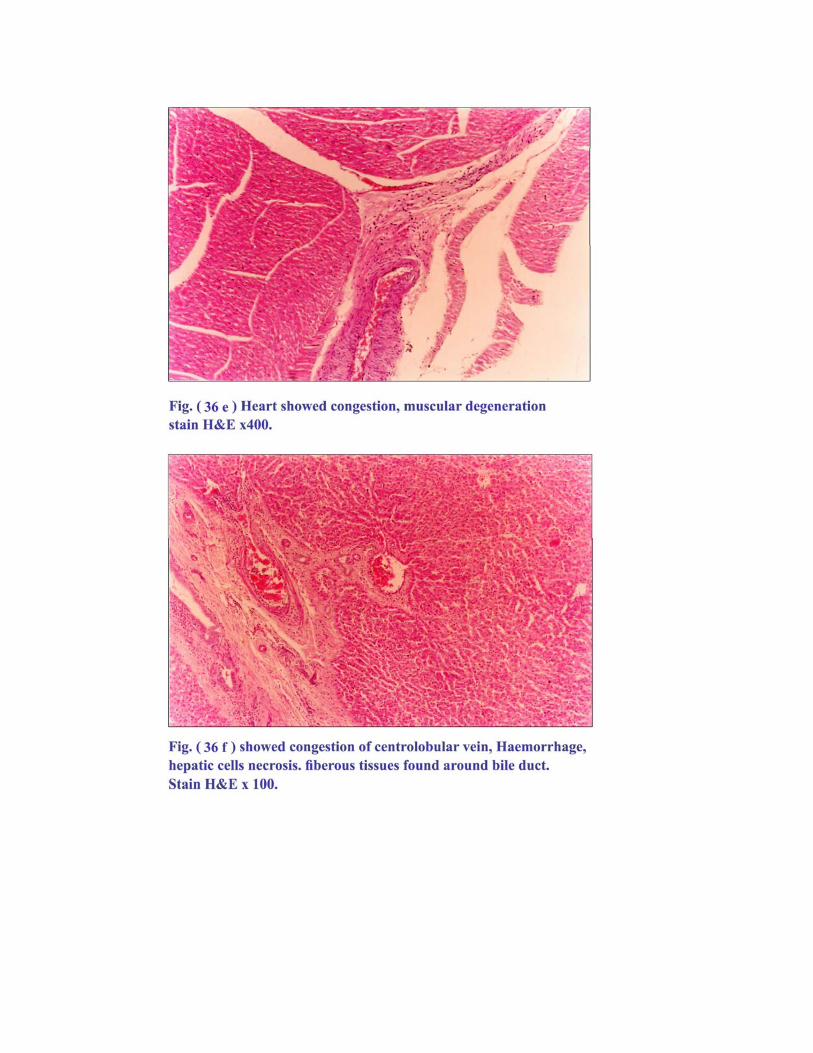
Fig
Fig
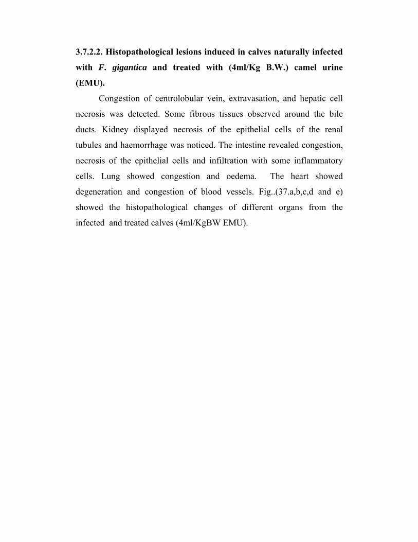
3.7.2.2. Histopathological lesions induced in calves naturally infected
with F. gigantica and treated with (4ml/Kg B.W.) camel urine
(EMU).
Congestion of centrolobular vein, extravasation, and hepatic cell
necrosis was detected. Some fibrous tissues observed around the bile
ducts. Kidney displayed necrosis of the epithelial cells of the renal
tubules and haemorrhage was noticed. The intestine revealed congestion,
necrosis of the epithelial cells and infiltration with some inflammatory
cells. Lung showed congestion and oedema. The heart showed
degeneration and congestion of blood vessels. Fig..(37.a,b,c,d and e)
showed the histopathological changes of different organs from the
infected and treated calves (4ml/KgBW EMU).
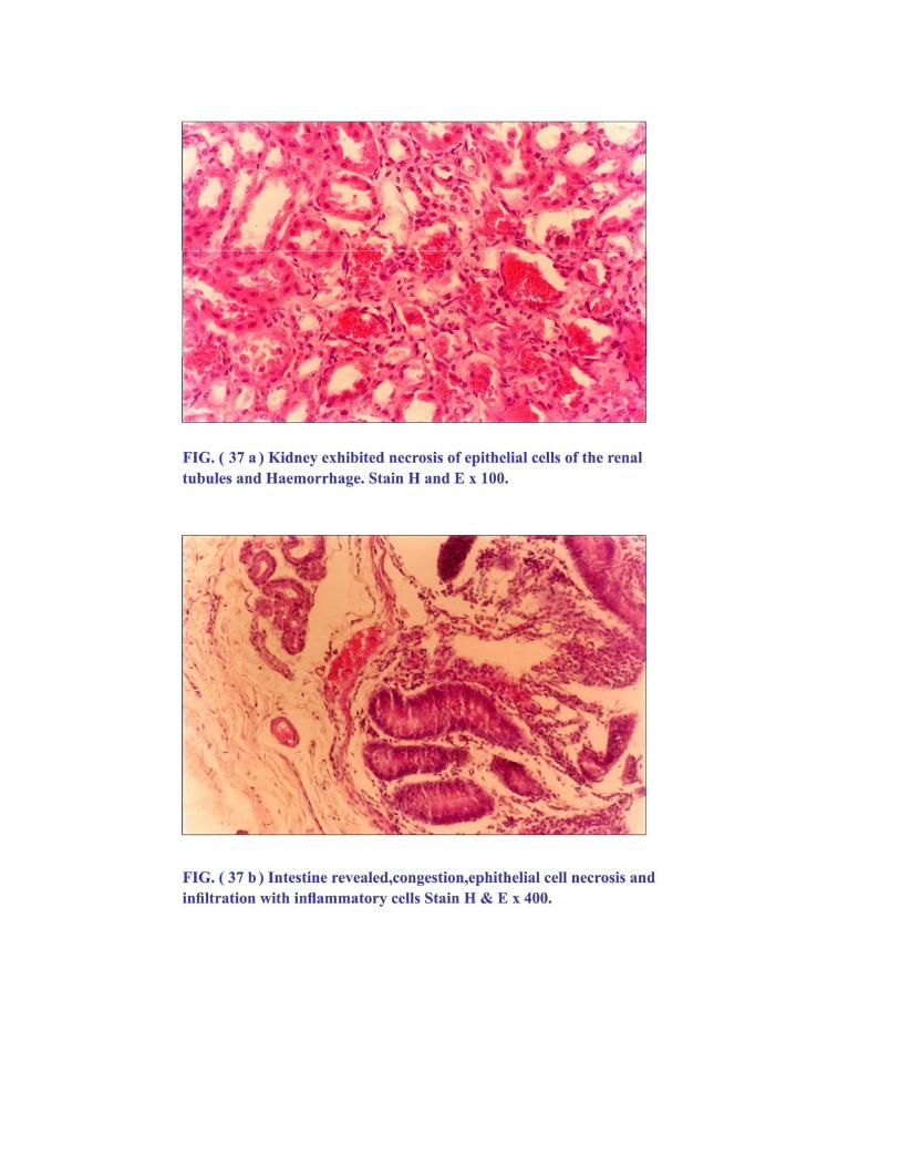
Fig
Fig
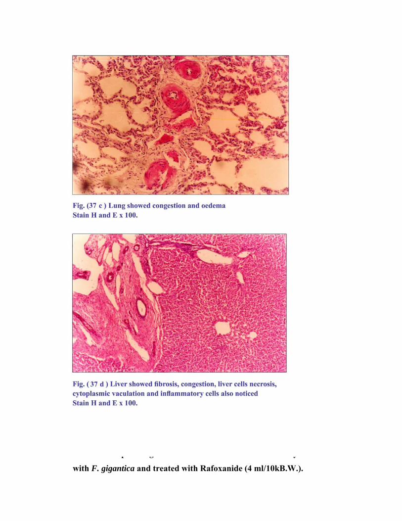
3.7.2.3. Histopathological lesions induced in calves naturally infected
with F. gigantica and treated with Rafoxanide (4 ml/10kB.W.).
Liver showed congestion, fibrosis and inflammatory cells.
Hepatorcellular necrosis was observed And some vacules were noticed on
the sytoplasm. Intestine revealed congestion of blood vessel and some
inflammatory cells. Kidney exhibited severe haemorrhage in the medulla
and necrosis of the epithelial cells and renal tubule.
The lung showed oedema and congestion. Fig(38.a,b,c,d and e) show the
histopathological changes of different organs from the inffected and
treated calves.(4ml/10Kg.B.W.Rafoxanide).
Fig
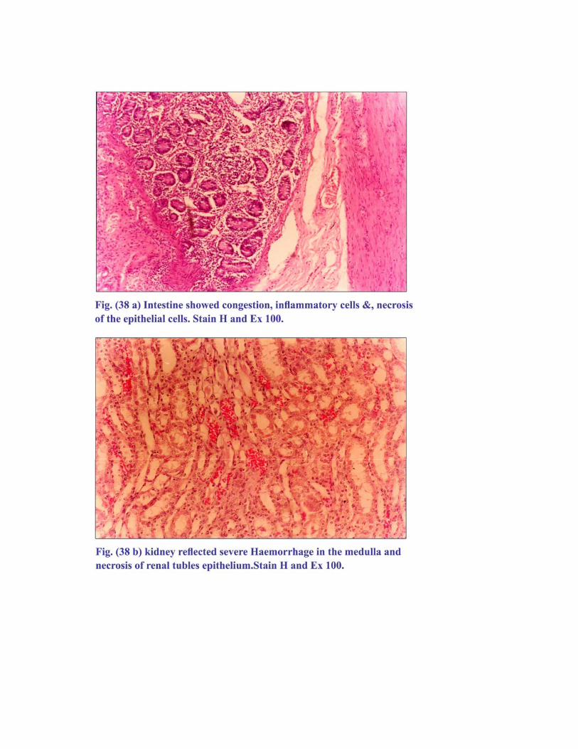
Fig
Fig
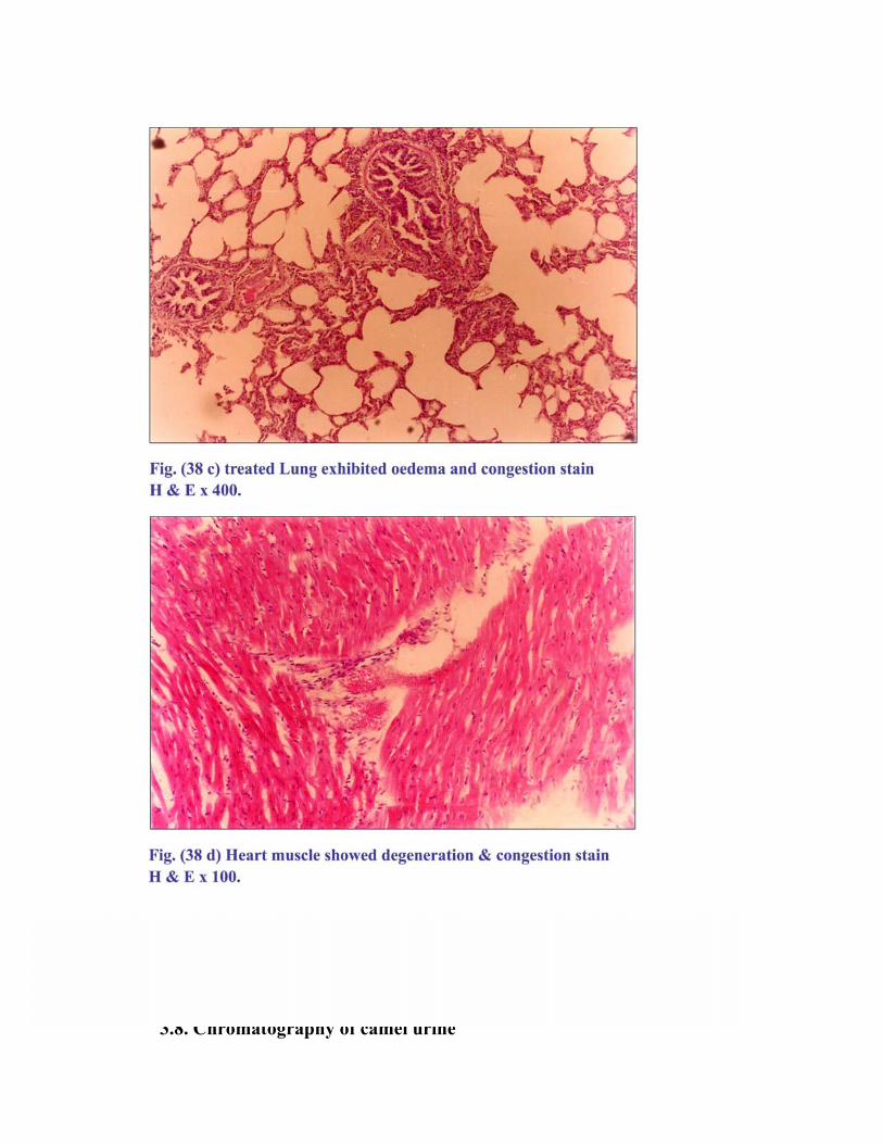
3.8. Chromatography of camel urine
3.8.1. Thin layer chromatogram of Camel urine, its chloroformic
extract, cattle urine (CE) and human urine (CE).
Camel urine, its chloroformic extract and that of cattle and
human urine when chromatographed on TLC it revealed a positive
reaction with Ninhydrin reagent, with one oval yellow spot for each
sample, whereas human urine showed no reaction. The RF values were
0.81, 0.79 and 0.6 for camel. As presented in Fig.(39.a and b).
Also chromatogram of (CE) hydrolysate by using 0.6 N HCL revealed
one yellow spot after Ninhydrin spraying with R = 0.65 Fig (39.c.).
3.8.2. Thin layer chromatogram of camel and cattle urine (CE) in
methanol:
Thin layer chromatogram of camel urine (CE), cattle urine (CE)
were dissolved in drops of methanol, they revealed a light blue-small
rounded-spots under short UV light [254mm]. When sprayed with
Ninhydrin a yellow 5 spots with RF values of 0.6, 0.7 0.75, 0.8, and 0.85,
for she-camel 1 & 3 years old and cattle (young and adult) Fig. (40).
3.8.3. Thin layer chromatogram of camel urine butanolic fraction:
Butanolic fraction (1,2,3) (4,5) (6,7,8) (9) (10,11,12) revealed one
violet spot for each, with RF equal 0.24. Fraction 13, 14, 15 resulted in
three components of violet, pink and yellow colouration after , Ninhydrin
spraying their Rf vales were 0.24 0.82, 0.88 respectively. Crude urine
showed the same zone of separation with addition of one more yellow
spot with 0.92 Rf value.
With Dragindoff reagent one yellowish to orange spot was
appeared against each stain spot with Rf of 0.6, 0.7, 0.75, 0.8 and 0.85
Fig. (41.a.). Another plate was exposed to potassium iodide vapor a
positive orange spots were seen (spot IA and 3B) Fig.(41.b.).
3.8.4.Thin layer chromatogram of camel urine ethanolic fraction:
Preparative thin layer chromatography (PTLC) of ethanolic
fractions and that of chloroformic extract spotted on silica gel (0.5mm)
and soaked in Butanol: acetic acid: water solvent system BAW (
4:1:5;4:1:1) for 45 minutes showed bright blue colour under short UV
light (254nm). When sprayed with ninhydrin and activated at 110 C,
Butanolic fraction (1-5) showed one violet spot of 0.24 Rf value. Fraction
13,14,15 gives 3 bands of violet, pink and yellow coluration Rf values
0.24, 0.82 and 0.88. crude urine spot of young she-camel revealed eight
bands of 0.08, 0.17, 0.25, 0.67, 0.79, 0.88 and 0.92 Rf values. Most of
which were yellow and pink in colour. Ethanolic fraction resulted in 3
bands of 0.21, 0.82 and 0.88 Rf values with the same colouration, where
chloroformic extract revealed a large round pink spot and slight yellow
band above it with Rf values of 0.8 and 0.88 respectively Fig.(42).
3.8.5. Ultraviolet visible sepectrophotometry (UV. V/S)
UV spectrophotometer [ 200-400] nm revealed components
ranged between [288-237] nm. Methanolic and methanol in HCl (99;1)
fraction resulted in the presence of amino acids as compared to 13
standard amino acids ranged between 200-400 nm as shown in Fig. (44.a
and b.) table (14).
Most of which are similar to the reference amino acids.
Table (14) Represent UV absorbance of methanolic, methanol in
hydrochloric acid and hydrolysate of camel urine
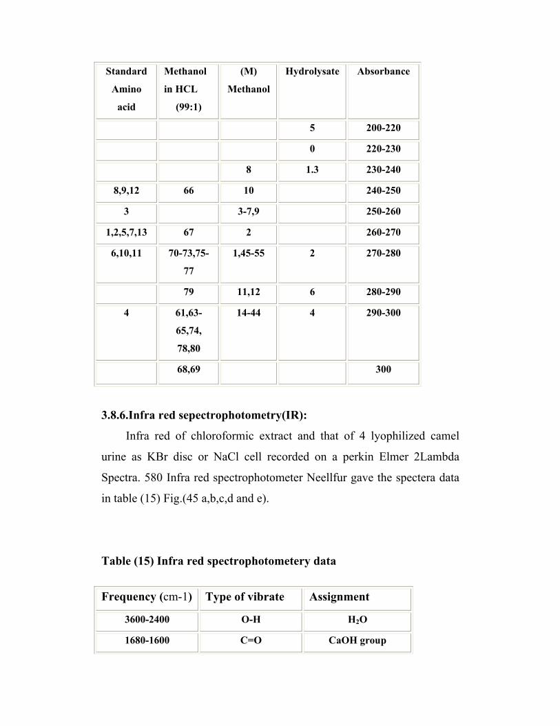
Standard
Amino
acid
Methanol
in HCL
(99:1)
(M)
Methanol
Hydrolysate Absorbance
5 200-220
0 220-230
8 1.3 230-240
8,9,12 66 10 240-250
3 3-7,9 250-260
1,2,5,7,13 67 2 260-270
6,10,11 70-73,75-
77
1,45-55 2 270-280
79 11,12 6 280-290
4 61,63-
65,74,
78,80
14-44 4 290-300
68,69 300
3.8.6.Infra red sepectrophotometry(IR):
Infra red of chloroformic extract and that of 4 lyophilized camel
urine as KBr disc or NaCl cell recorded on a perkin Elmer 2Lambda
Spectra. 580 Infra red spectrophotometer Neellfur gave the spectera data
in table (15) Fig.(45 a,b,c,d and e).
Table (15) Infra red spectrophotometery data
Frequency (cm-1) Type of vibrate Assignment
3600-2400 O-H H2O
1680-1600 C=O CaOH group
1600-1500 C-C Aromatic
1448-1097 NH NH2 group
1322 -S=O So2group
582 CI CI
Remarks of IR analysis:
All the spectra showed the following characteristic absorption
bands:
1- 3600-2400 trade band characteristic of O-H st. vib. H banded
mainly of acids if H2 O is present.
2- 1680 cm -1 and neighbours, are of C= O St. vib.
* more than one C = O St. vib.
III – 1124 cm –1 charac. Of C- O St. vib.
** I, II, III strongly pointed to – COOH carboxylic acid.
IV 1600 m 1500 1484 stands mainly for C- C of aromatic system.
773, 691 bonding and pattern of subs.
V- To large degree sample zone 4 identical 1, 2 similar to 4 but not
identical.

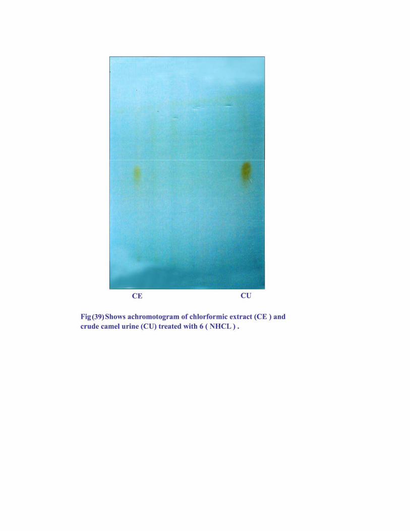
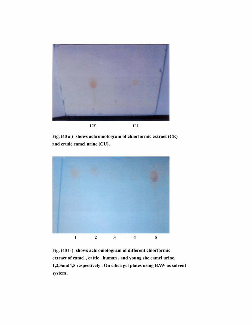
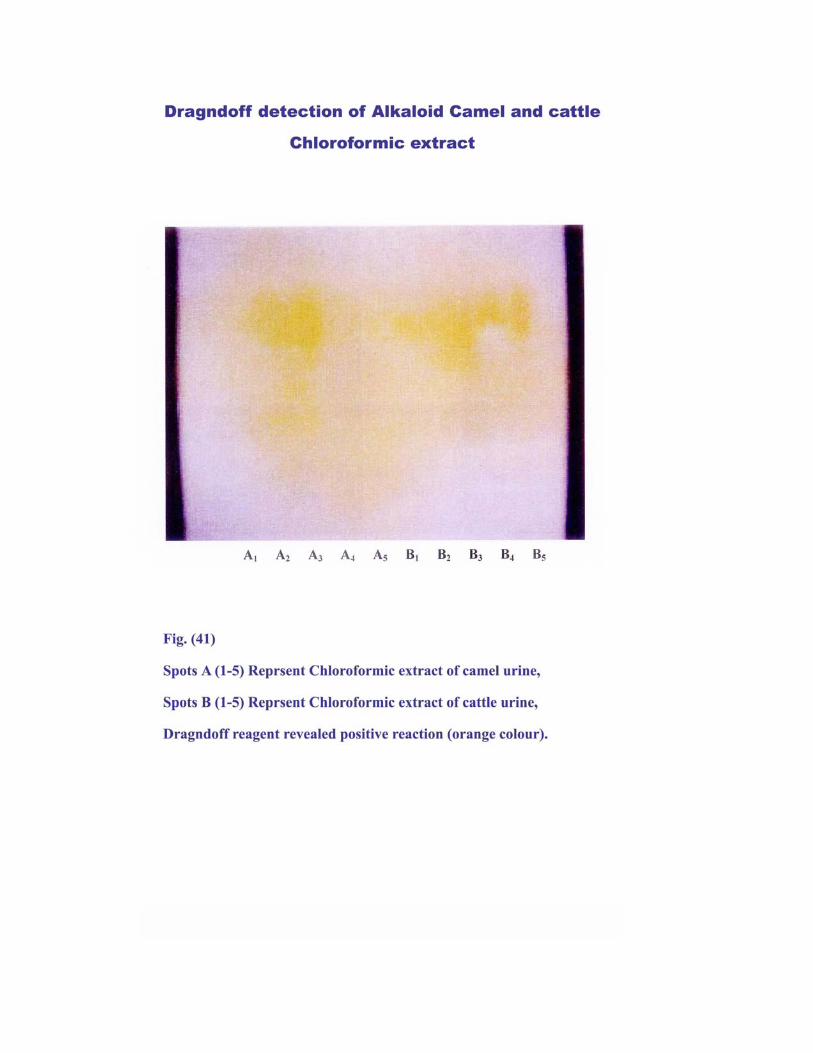
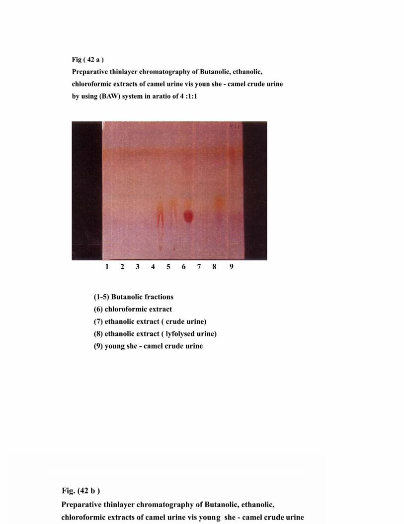
Spots A (1-5) reprsent Chloroformic of extract camel urine, Spots B (1-5) reprsent Chloroformic of extract camel urine. Dragnoff reagent revealed positive reaction (orange colour).

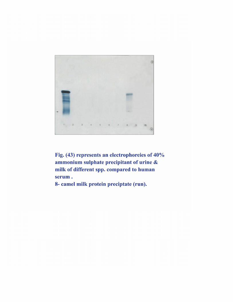
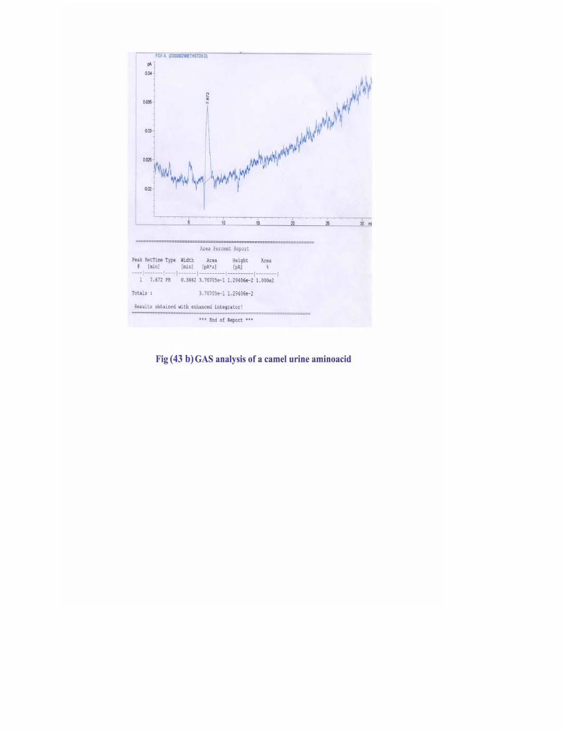
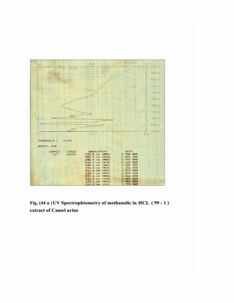
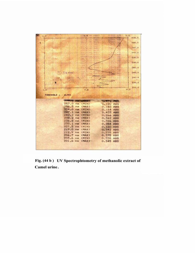
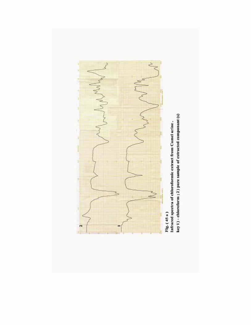
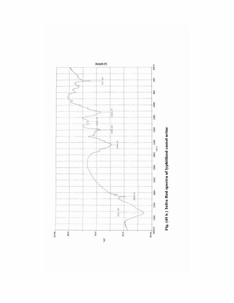
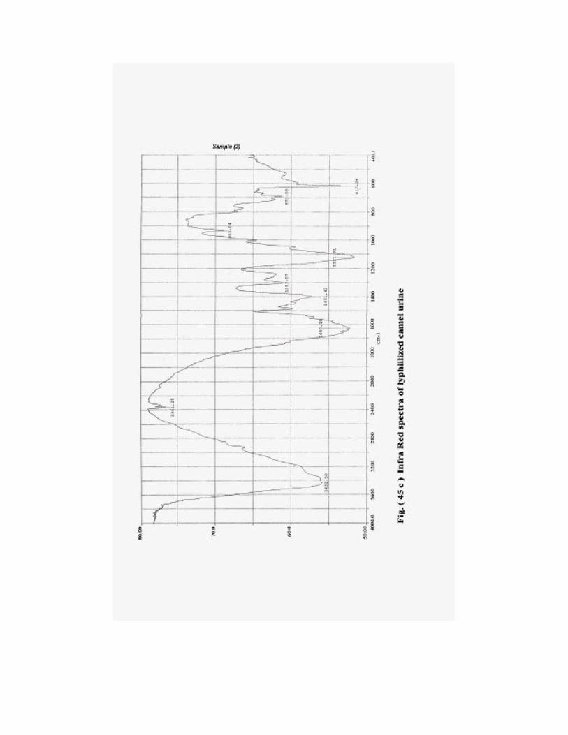
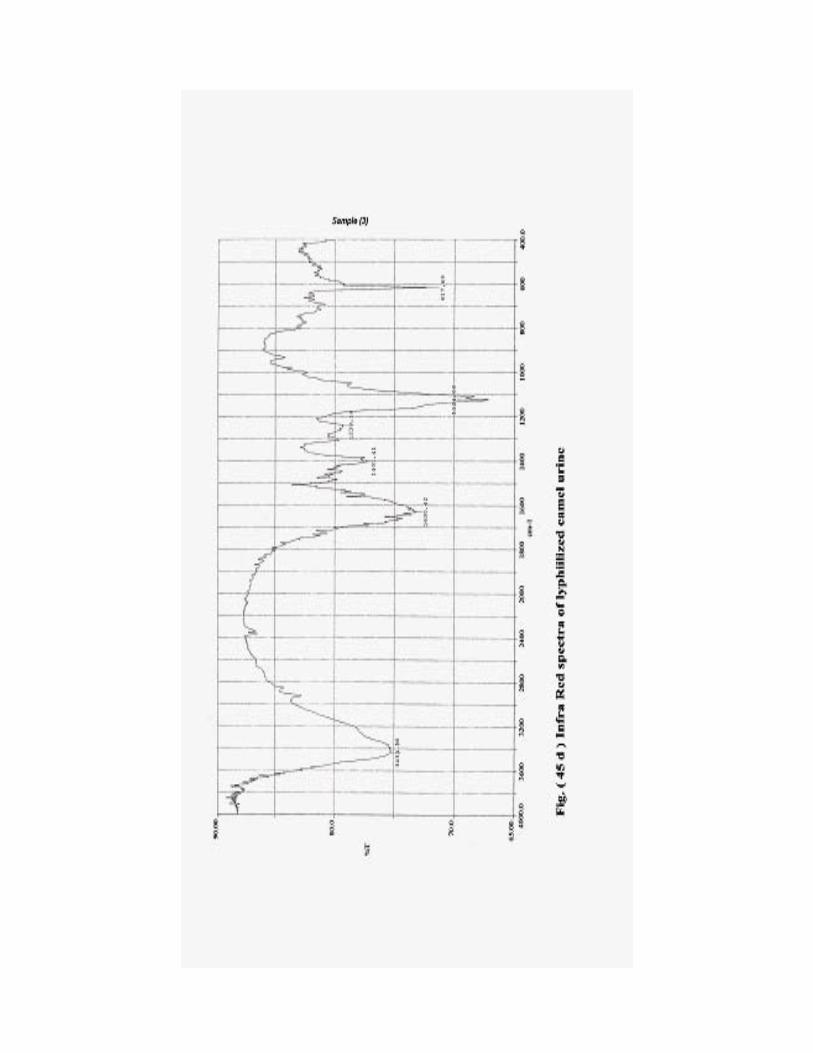
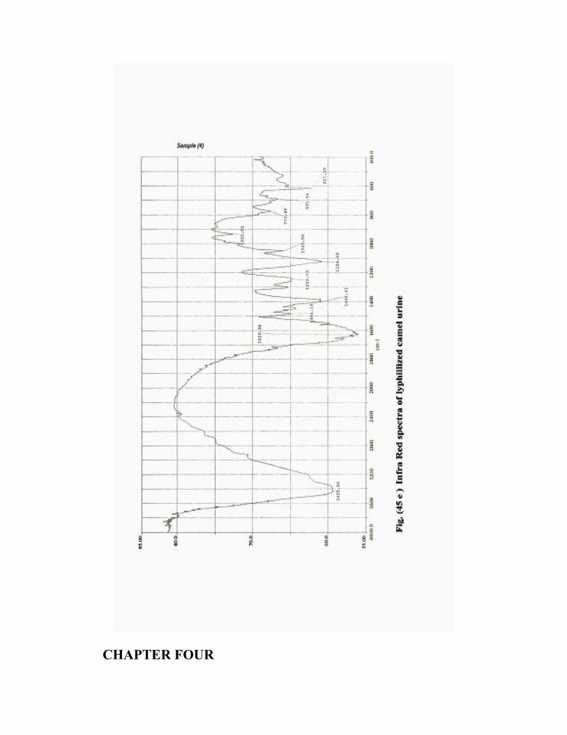
CHAPTER FOUR
DISCUSSION
Medicinal usage of plant and/or animal products, represents one of
the most essential sources of drugs. In Sudan and many other countries
camel owners use camel urine to treat a wide range of health problems.
Camel urine has antibacterial, antifungal, antiviral, antineoplastic and
therapeutic applications on ascitic patients as reported by Ohaj (1998),
Amer and Alhendi (1996) and Wisal (2002).
No pharmacological investigation has been published on livestock
urine, so the aims of the present study are to make more considerable
physical, biochemical, pharmacological and clinical investigations on
camel urine.
4.1. Physiochemical Investigations
4.1.1. Physiological properties
Most animal's urine is clear watery solutions with exception of
Equine urine which is viscous due to the presence of mucoproteins, while
birds urine is creamy and thick due to formation of uric acid, which is
excreted with faeces, (Ibrahim 1989). Camel urine is amber yellow to
deep brown in colour, with offensive odour. This is in agreement with
observation reported by Ohaj (1989)and Wisal (2002).
In this study camel urine gave an alkaline pH with a mean value of
9.5±0.5 this likely due to the presence of greater reservoirs of bicarbonate
in the intestine of camel (Maloiy and Clemens -1980), which is slightly
higher than the pH (7.42) reported by Wermery and Wensvoort (1992) in
Blood and Urine of camel Amer and Alhendi (1996) reported acid pH of
(6.5) in 14 male urine samples while 6 samples showed an alkaline pH. In
this study, the pH of young female camel. (6 month up to 1 year old)
urine sample was (6.7), this is in agreement with pH 6.5 that was reported
by Manefield and Tinson (1996). This variation in pH may be due to:
(1) feed intake (High energy diets), such diets can cause metabolic
acidosis and mineral disturbances.
(2) environmental condition.
(3) different areas of sample collection.
4.1.2. Chemical and biochemical analysis
Human urine is the most analyzed urine, whereas camel and goat
urine were studied as a tools of dehydration effects on desert animals,
Musa (1978) and Yagil (1984 and 1994).
Potassium was the most predominant electrolyte followed by
sodium and calcium. Camel in their normal habitat are exposed to
dehydration salty bushes or salty water Dahl and Hjout (1980) and Yagil
(1985). The well adapted and functioning camel kidney which has a
relatively thick medulla (Medulla cortex 4.1cm) with long loop of Henle
for water and sodium reabsorption and hence the ability to concentrate
urine Wilson (1989).
Urea is the major end product of nitrogen metabolism, as
summarized in table (2) this appears reasonable because of the counter
current metabolism for urine concentration, which permits to excrete
highly soluble urea with very little water. The average urea level 2-3g/dl
and 10.5g/dl for male and female respectively which was in agreement
with Ohaj (1998) reports but disagrees with Amer and Alhendi (1996)
who reported the mean value to be 1.95g/dl. Bolyne (1977) and Etzon and
Yagil (1986) reported a mean value of 1.8g.dl in control and1.4g/dl in
dehydrated ones.
In this study, the results showed, that goat urine has higher values
of urea followed by cattle, camel and human. Wilson (1989) reported
higher levels of camel urine sample urea of dehydrated and fully watered
camels 5.7g/dl and 3.7g/dl respectively. Our result is in contrast with the
result of Read (1925) who found no urea in camel urine.
Uric acid was found to be higher in goat and camel, than in other
animal’s urine. These results were similar to that reported by Ohaj
(1998), and higher than that reported by Amer (1996), Uric acid mean
values were 236.3, 214.5, 182.25, 145.5 mg/L in goat, camel, cattle and
human urine respectively.
Murphy et al. (1969) and Cracer et al. (1988) found low level
concentration of uric acid in cow, sheep, goat, pigs and horse
respectively, this may be attributed to the action of liver uricase enzyme
which converts uric acid into allanatoin (Margon, 1959). It is assumed
that camel livers do not release uricase enzyme (Murphy et al. 1969 and
Cracer et al. 1988). The mean value of creatinine and creatine were
higher in males, because the male has more muscles. This result was in
accordance with Ohaj (1988) and Wisal (2002) findings.
4.2. Pharmacological screening of camel urine:
The pharmacological screening was carried out to evaluate if the
twenty four hour (24hcu) collected urine early morning urine (EMU),
Ammonium Sulphate Protein Precipitate of camel urine (ASPPOCU), and
chloroformic extract (CE) of camel urine used have other activity that
might be considered of interest. and to establish explain the general
effects experimentally on blood and liver enzymes of normal and
intoxicated rats.
4.2.1. Hepatoprotective effect of camel urine and its extract:
In this study camel urine, (ASPPOCU), and chloroformic extract
(CE) were screened for their hepatoprotective activity against CCL4 as
standard hepatotoxic agent Handa, (1986) and Mansour, (2000) and
paracetamol as a necrotic agent at high doses (1g/kg) induces
hepatocellular damage Tee, et al (1983), compared to silymarin (1g/kg)
standard anti-hepatotoxic agents. Such a screening process represents an
essential stage for selecting the most potent hepatoprotective form of
camel urine for further detailed pharmacological and chromatographical
investigation.
4.2.1.1. CCL4 toxicity:
CCL4 is a colourless volatile liquid with characteristic sweet odor,
it is miscible with most aliphatics and stable in the presence of air and
light. Decomposition may produce phosgene, CO and hydrochloric acid
Phol et al. (1983)and Watanable et al. (1982). The final step in the
biotransformaton of CCL4 is catalysed by cytochrome p450 enzyme
leading to formation of reactive trichloromethyl radical.
In the oxidative biotransformation, the most important pathway of
the trichloromethy redical is the formation of the more reactive
trichloromethy peroxide, which covalently links to macromolecules, and
lipid peroxidation occurs via metabolic intermediates of CCL4, (Dezwat
et al. 1977; Nagi et al. 1999; Mansour, 2000). Liver and kidney are the
target organs for CCL4 toxicity.
4.2.1.2. Time course of CCL4 hepatotoxic effect on rat liver
A daily decrease of CCL4 elevated levels of ALT and AST were
showed in Fig. (4) after ninety six hours ALT and AST were reduced to
normal levels. Similar findings were reported by Teschke, et al (1933)
and Murphy, et al (1969)
4.2.1.3. Paracetamol toxicity
Decomposition of paraminophenol compounds may produce its
analgesic and antipyratic agent, in this group of compounds the analgesic
effect is due to N-acetyl paraminophenol (paracetamol) that found in the
liver, also it has slight toxic effect. The sulphate ester and glucuronide of
paracetamol account for about 75-80% of ingested drug Jewis,(1964).
4.2.1.4. Hepatoprotective effect of camel urine against CCL4 and
paracetamol toxicity:
Different doses of (24HCU), (EMU) and chloroformic extract of
camel urine were tested against liver damage induced by CCl4 in two
ways, one hour prior and one hour after CCl4 administration. The doses
that protect liver against CCl4 toxicity (in rats) were 4ml/100g BW of all
(24hcu) tested urine 2ml and 0.2ml of (EMU) and chloroformic extract
respectively.
Camel urine and its chloroformic extract significantly (P<0.05)
reduced the highly elevated levels of ALT and AST induced by CCl4.
The result of the present study indicated that I.P. administration of
CCL4 at a dose rate of 0.1ul/100g B.W. induced damage of liver cells of
experimental rats, which was manifested biochemically by significant
elevation of AST, ALT enzyme activities (P<0.05).
These forms of camel urine, significantly reversed the elevated
level of serum ALT and AST activity induced by CCL4 at 4ml and
2ml/100g B.W. fig. (1, 2 and 3). Other doses did not protect liver cells,
whereas 2ml dose of (24hcu) may be below the therapeutic value while
the 8ml of the (24hcu) was found to be too high or may be due the long
storage of urine lead to some changes in 24hcu urine constituents, since
the rats died in less than 55 minutes. On the bases of these result we could
clearly suggested that camel urine has an active component (s) which
may have an important role as an endogenous anti-oxidant and/or could
act as cytoprotective agent against tissue damage mediated by toxic
substances. The percentage of hepatic protection with camel urine is
82%, and with CE is 95% against CCL4 induced toxicity.
There is no published data concerning camel urine hepatoprotective
activity, whereas similar hepatoprotective effects were reported by Ali et
al (2001). Wen (1999) and Shuang (2003) reported similar findings by
using human urine extracts. Findings were reported by many authors
dealing with some plants having hepatoprotective activity, such as
Ballanitis agypticaca, Mellen et al. (1985), Robbin (1967) and Ali et al.
(2001) and Raja,(2002). Solanum nigieum plant has a protective effect as
reported by Prichard et al.(1987) and Rana and Avadhood, (1992).
Calotropis procera root (CE) do posses a hepatoprotective effect Basu et
al. (1992). Rhazya stricta, Balanitis aegyptiaca and Haplophy Lum
tuberculatum hepatoprotective effects were reported by Ali et al (2001)
4.2.2. Response of isolated tissue strips to camel urine and its extracts:
Serotomin causes increased gastrointestinal motility and
contraction of isolated strips of intestine, this being partly due to a direct
effect on smooth muscle cells and partially due to indirect effect on
enteric neurones Rang and Dale, (1987).
Cyproheptadien the standard amide at concentration of (4.4x10⁻⁵
M) significantly inhibits the contractile response induced by 5-HT (5x10⁻⁵
M) Rang and Dale (1987).
4.2.2.1. Isolated rat duodenum
Produces spontaneous contractile action. Addition of 0.1ml/bath of
camel urine protein precipitate (CUPP), abolished the contractile
responses of this tissue this suggest that (CUPP) may possess adrenaline
like substances.
4.2.2.2. Isolated rabbit jejunum
It was directly stimulated by (CUPP) and diluted urine (DU, 1:3), at
dose rates of 0.1 ml/bath and 0.8ml/bath respectively, camel urine exerts
a daul effect on rabbit jejunum. The stimulant effect appeared to be
mediated via muscarinic receptor stimulation as the effect was blocked by
atropine 0.2ml/bath (5x10⁻⁵M), which implies an acetylchlorine like
action. This was similar to result reported by Fischer and his Co.
worker.(1983). Crude urine 0.1ml/bath (CU) cause marked relaxant effect
on isolated rabbit jejunum followed by a transient contraction after wash
suggesting that concentrated urine has an irritant effect.
Addition of small, medium and high dose of (CE). 0.1, 0.2,
0.4µl/bath respectively producing slight effect on the isolated rectum of
rabbit and chick, this suggest that camel urine may contain polar and non
polar substances that affect the isolated tissue than do the (CE).
4.2.2.3. Isolated rat Fundus
Camel urine precipitant 0.8ml/bath significantly stimulated the
isolated rat fundus (P<0.05). More over the diluted camel urine (1:3)
significantly stimulated the isolated rat fundus strip at doses of 0.2, 0.4.
and 0.8ml/bath. No effect was produced when CE was administrated,
similar findings were reported by Guthum, (1958). We might suggest that
camel urine may contain 5-HT like substances and / or mediated amines.
4.2.3. Cytotoxic effect in vitro test of camel urine (ASPPCU) on
bovine kidney and liver cell culture:
Concentrated camel urine when inoculated in tissue culture
(BLCS) caused 100% cytopathic effect after 24 hours, while 1:10 caused
damage to the cell culture at 50% where diluted urine (DU, 1:3) shows
growth action of the liver cell culture which was in agreement with
Umberto et al.(2000). These observations suggest that camel urine and
(CUPP) may contain some components which enhance or initiate cell
growth or alter cell membrane permeability.
4.3. Camel urine therapy:
4.3.1. Biological Role of Macroand Micro elements
Macro elements are commonly described; essential and
nonessential. The first one so defined because their lack leads to
functional disorders, and complementation with a physiological dose can
prevent or cure these disorder (Mertz, 1981). These elements are
generally included in enzyme molecules, for example, copper in
cytochrome oxidase, Alkaline phosphates, DNA and RNA polymerases,
and dehydrogenases, Manganese in pyruvate carboxylase and selenium in
glutathione peroxidase. Some are included also in hormones (iodine in
thyroid hormone), vitamins (cobalt in VB12) or metaloproteins (iron in
haemoglobin and myoglobin).
4.3.2. Effect of (EMU) camel urine on the selected parameters
Results of the parameter selected in this study, may be used as a
surveillance tools for detecting the incidence of curelly and clinical status
of the animal health. Camel urine (EMU) at a dose rate of 2 and 4 ml/kg
B.W. changed serum enzymes, AST, ALT, ALP, GGT, and LDH;
electrolytes (Na and K); minerals, Ca, P, and Mg. Among the mentioned
parameters, AST and ALT showed a significant (P<0.01) reduction at 4th
week along with Rafoxanide treatment. Change in serum Ca, P, Na, and
K concentrations also decreased at week 3 and 4; whereas gradual
increase in Na concentration was observed. Although according to these
results, camel urine in general observations, started to exert its effect at
the first week this may be attributed to its high concentrations of K, Na,
Ca and Mg; and their salts e.g. NaCl, MgSO4, NaH2O, which have a
pronounced physiological and pharmacological effect on the potentiality
of cell membrane permeability and ion exchange system. Moreover, the
increased influence of camel urine may be referred to alteration in its
absorption and local metabolic degradation, taking into account the
changes in Bovine ruminal microflora environment. Rumen pH generally
varies from 6.3 to 7.0, because the composition of fibrous feeds usually
leads to considerable recycling of buffer in saliva resulting from the
greater time of mastication and regurgitation. Also mineral acids reduces
the ruminal pH Dunand and Kawashima, (1980). A significant lowering
of enzyme levels (Table 1) (P<0.05) compared to infected untreated
control animals. Changes in serum constituents were significantly
correlated at week 3 and 4 rather than in week eight. AST, and ALT
showed a highly significant correlation (P<0.01), r = 0.778 and 0.639 in
week 3 and 4. Also there is a highly interaction between GGT and urea
(P<0.01) at weak eight r = 0.731, while total protein and albumin were
significantly correlated (P<0.01),with time r = 0.656 at the first and 8th
week followed by no significant interaction at weak 3 and 4. Total
protein and creatinine were significantly correlated (P<0.01) and
(P<0.05) in the 3rd and 4th week respectively. Ca and P showed no
significant correlation in first and 8th week, whereas a highly significant
correlation (P<0.01) was obtained in the 3rd and 4th weak, r= 0.690 and
0.635. Urea is formed solely in the liver from the catabolism of amino acids, and it is
the main excretion product of protein metabolism. Urea diffuses freely at the same concentration throughout the body water.
Blood urea concentration is influenced by the extent to which
amino acids are oxidized and on the absorption of NH3 from the rumen,
Wallace (1979). Uric acid is a principal end- product of nucleic acid and
purine metabolism in man, via final common path way of conversion of
xanthine by means of xanthine oxidase to uric acid.
The rise in RBCs and WBCs concentration in group II and III may
indicate that camel urine has some factor(s) (e.g. Interlukine) which
initiate or enhance haemopiotic stem cells to promote synthesis of new
RBC, and WBC cells; also it may indicate that microflora were
generating antibodies against the given (EMU) of camel and by the 4th
week, the animal internal organs acid base balance adapted and
equilibrated the change in buffer system. Microflora too become totally
adapted to the changed environmental condition.
Results of histopathology revealed slight recovery in the
hepatocyte texture. Camel urine as observed from the results of the
treated calves showed a parallel correlations with that of Rafoxanide.
This suggests that camel urine may contain active substances present in
Rafoxanide i.e. substituted phenols, salicylanide or nitrnoxil.
4.3.3. Camel urine chromatography
Extraction of active substances from camel urine, using the method
described by Ibrahim (1989), showed that Chloroformic extract gave poor
yield. Precipitation with ether appears to be an inefficient way to recover
the active material from camel urine. Much better recovery has been
obtained by ethanolic, methanolic and chloroform in methanol fraction,
which provides the presence of alkaloids and which was similar to the
observation found by Umberto et al. (1987) using HPLC analysis of
camel and zebo cattle.
Elusion of camel urine from sephadex DAEA [25-150] was found
more satisfactory than the butanolic elution, recovery was good and the
active material (s) were concentrated in a few fractions. As shown in this
study columns of IRC 60 mesh and sephadex have the advantage that
active ingredient can be adsorbed from small volumes of urine. If elution
is carried out with acid little activity is lost.
Conclusion
The results of experimentally examined camel urine, camel urine
chloroformic extract and camel urine protein precipitant when compared
to standard drugs on hepatoprotective effect, responses to various isolated
strips, chromatographic and curative effect show certain similarities to
many investigators using human and rat urine Gaddum (1953) and Horton
(1958).
In this investigation diluted camel urine appeared to be more active
than protein precipitant, crude urine and chloroformic extract on in vitro
experiments. Early morning urine of young female camel is the most
effective on in vivo one’s the CUPP and camel urine seems to have a
similar role of action to that of 5-HT, parallel assay provided more
evidence of response relationship of all these mediated peptides and
amino acids.
From all these biological and biochemical comparisons it is
concluded that the three forms of camel urine do have the same active
principle(s) that are responsible for the activity of camel urine studied,
more over camel urine of four different ages female camel emphasizing
their similarity without implying identity by using IR technique.
Indeed, these experiments will suffice to illustrate the hypothesis
that camel urine may cure and prevent liver and internal organ disorder.
The knowledge gained from this study could improve the application of
camel urine for animal health and nutrition, as well as to assist in the
alleviation of diseases in significant measures.These goals have been
realized in this research of camel urine physiology, biochemistry,
pharmacology and chromatography which could be summerised in:
1)Camel urine has an alkline pH.
2)Camel urine has a pronounced level of potasiom and urea.
3) Camel urine consists of bicarbonates,sulphates,phosphates. and
chlorides.
4) Antioxidant agents.
5) Purgative and little laxative effects
6) Alkaloids – nitrogenous basis.
7) Promoting or supporting agent(s).
Recommendations for future work:
More considerable investigation must be done to know:
1) The structure of the macromolecules of camel urine
2) How the potential energy available from oxidation of camel urine can
derive the main-fold energy requiring processes of the living cells. (its
action on liver metabolizing enzyme).
3) The primary strands that the genetic apparatus encodes instruction for
the precise ordering of the amino acids in the primary strands of
protein.
4)Quantitative biological activity-directed fractionation of (CE) of camel
urine.
5)Determination of the most potant fraction of GC-MS.
6)Synthesis and optimisation of the active conistituant(s)
7)Formulation of the active ingredients in the pharmaceutical dosage.
Abbas,B.(1998) Ethno-veterinary practices among camel pastoralist in
Butana. Proc. of 8th . Arab Vet. Conf. Khartoum. March, 1998.
Al Tabari “The Victory of islam” volume 8, published by SUNY,
translated by Michael Fisher . page 97,98.
Ali,B.A and Erwa, H.H .(1993). Effect of Nigella sativa on ingestion
ability in mice peritoneal macrophases. Saudi pharm. J. 1:18.22.
Amer, H.A. and Alhendi, A.B. (1996). physical, Biochemical and
microscopic analysis of camel urine.J. of camel practice and Research,
June :17-21
Anon (1983). Trends in mortality from cirrhosis and alcoholism. United
States. MMWR 35: 703-705.
AOAC (1990) Official method of analysis. Association of official Analytical Chemist. Washington. DC.
Archibald, R.G. (1933). The use of the fruits of the tree Balanites aegyptiaca in control of schistosomiasis in the Sudan Trans. Roy. Soc. Trop. Med. Hyg.
27, 207-210.
Armstrong, J.W. (1971).The water of life. Health science .press,
Rustingtong. Surssex. England.
Bailey, C.B. and Palch, C.C. (1961b). Saliva secretion and its relation
to feeding in cattle. The composition and rate of secretion of mixed
saliva in the cow during rest Br. J. Nut, 15: 383-402.
Bass, N. and Williams, R.(1988). Guide to drug dosage in hepatic
disease. Clin pharmacokinet 15: 396-420.
Basu, A., Sen, T., Ray, R. N. and Chaudhuri. A.K.N. (1992).
Hepatoprotective effects of Calotropis procerera root.
Bjornesjo,K.B. (1951). “On the effect of human urine on tubercle Bacilli
II: The tuberculostatic Effect on Various Urine Constituents” Acta
ScandinavicaVol.25,No.5,447-455.
Boray,J.C.(1985). In: World animal science B2: Practs, Pest sand
Predictors. Eds Gaafar,S.M. Amisterdam:Elsevier,p179.
Bosnes ,R.W.and toussky ,H.H.(1945) Determination of creatinine in
serum and urine J. of Biol .chem .158.
Burzyniski,S.R. (1986). Anit-neoplaston History of the Research, Drugs
Exptl. Clin. Res. Supply 1,XII, 1-9.
Burzynski Stanislaw R. (1977). “Anti-neoplaston A in cancer therapy:
physiology”. Chemistry & physics, vol.9,485.
Carbon Tetrachloride (1999) Environmental Health Criteria(EHC)
(208).
Chang-Ho, Lee and colleagues (1999). Carbon tetrachloride Pathway
Map(Anaerobic).
Chemie, D.G.K. (1972). Journal of clinical chemistry and clinical Biochemistry 10, 182-192.
Chrestopher, S. W.(1995) Daived son's, Principles and Practice of
Medicines.
Christy, M. (2000). Clinically tested medicinal proved book, Your own
perfect medicine.
Clayden, E.C.(1971). Practical section, cutting and staining technique 5th
ed. Churchill Livingston, Edinburgh.
Cotran, R. , Kumar, V. and Robbins, S.(1989), Pathological basis of
disease. Ed. 4. Philadelphia: W.B. Saunders company, 941-957.
D’Amico, G., Morabito,A.,Pagliaro,L. , Marubini,E. (1986)Survival
and prognostic indicators in compensated and de compensated cirrhosis.
Dig. Dis. Sci.31:468-475.
Dahl,G. , Hjort, A. (1979). Dromedary pastoralism in Africa and Arabia.
in IFS (Scandinavian Institute of African Studies) Ist symp camels
Sudan. 447- 458.
Dahlborn, K., Benlamlih, S., Zine-Filali, R., Gueroulali, A., Hossaini-
Hilali,J., Qukessou, M.(1992). Food deprivation and refeeding in the
camel (Camelus Dromedaries) Amer. J. Phys. 262:1000-1005.
Davis, R. H., Donta, J.J.,Hantman, G.M., and Haas, R.C., (1994).
Anti inflammatory and wound healing of growth substance in Aloe Vera.
Journal of American Pediatric Association 84: 77-81.
Deshande, U. R., Gadre, S. G., Raste, A.S., Pillia, D., Bhide, S. V.,
Samuel A. M., (1988). Protective effect of turmeric (Curcuma Longal.)
extract on carbon tetrachloride induced liver damage in rats. Indian J.
Biol. 36 : (6), 573-577.
Duncan, C. H. (1918) Auto therapy, New York.
Dunne,M.P. (1981). The use of injected and sublingual urine in the
treatment of allergies. A preliminary report, held at the Oxford Medical
Symposium.
El. Gazali, G.E.B., El-Tohami, M. S, Khalid H. E, Abdalah, W.
E.,Yagi S.M. (1998) Medicinal plants in Sudan. Medicinal plants in
Khartoum province. National center for research, Khartoum, Sudan. PP.
61, 62, 85, 86, 129-130, 153-154.
El. Shami,M,E.and A.Jubara (1989). Comparative study of utilization
of browse plants by camel and goats. In: camel news letter No. (60).
Elamin, H.M. (1990). Balanitaceae. Trees and shrubs of the Sudan
Illka Press. 426.
Eldisougi, M. (1988). An appeal for modified camel productivity.
Uppsala conference, March 1988, 15-18.
Etzion, Z. and Yagil R., (1986). Renal function in camels (Camelus
dromedaries) following rapid dehydration. Physiol zool. 59 (5): 558-5
Evans,R.T.(1968).Manual and automated method for measuring urea
based on modification of its reaction with diacetylmonoxime and
thiosemicarbazide J. clin. path.2:527-529.
FAO (1984). Report of FAO expert consultation on Rinderpest
Diagnosis, vaccine production and quality control. Held in Rome 15-19
October.
Forber, C., and Lederer, B.,(1989) “AIDS, words from the front” Spin
magazine June, 5: (3), 90-107.
Ford, A.L and Milligan, L.P (1970). Tracer studies of urea recycling in
sleep. Canadian journal of animal science, 50: 129-135.
Fracer, C. Ariell, A. (1985). Hepatic encephalopathy. Med 313: 869-873.
Frankel, S. and Reitman, S. (1963). Clinical laboratory methods and
diagnosis Vol 2, 6th ed.. The C.V. Mosny Company.
Free, A. H. and Free, H.M.(1975). Urinalysis in clinical and laboratory
practice. lnc .U.S.A:13-17.
Gabriel L. Plaa (1986). Toxicology 3rd Edition. PP. 287.
Goodwin, J.F.(1970). Thin layer chromatography of urinary amino acid
1, standard method, of clinical chemistry, 51: 377.
Gunner, S. (1992). Urea in the treatment of dry skin. Your own perfect
medicine, report 40: 149.
Handa ,S.S., Anupam, S. and Chakrabort, K.K. (1986). Natural
products and plants as liver protecting drugs. Fitoterapia (57): (5), 307-
351.
Hall, (1977). Diseases and Parasitic of livestock in the Tropics. Encyclopedia. Bulletin. 1974.
Henry, R. J. cannon, D. C. and Winkelman, J.W.(1974). Protein
determination by Biuret, clinical chemistry, 2nd Edition, Harper and Row
publisher, New York. London. 411-416.
Herman, J. R. (1980). Autouro-therapy. New Yourk state: J. of
med.vol.80 No.7, 1149-1154.
Higgins, A.J., Ed. (1984). The camel in health and disease Br. Vet. J., 140(5): 482-504.
Horton, E.W. (1958). Human Urinary Kinin excretion. Amer. J. of
physiology 14: 125-132.
Houpt, T.R. (1965). Utilization of blood urea in ruminants Amer. J.
Physiol. 197: 115-120.
Houpt, T.R. and Houpt, K.A. (1968). Transfer of urea nitrogen across
the rumen wall. Amer. J. Physiol. 214: 1296-1303.
Hunt, S. and Mc Cosker, P. J.(1992). Observation on serum adenosine
activity in experimentally produced liver disease of cattle and sheep.
Yellow wood, Lantana, Carbon tetrachloride and chronic copper
poisoning, Br. Vet. J., 126:74-81.
Ibn alrzaq, Tas-hil al-Mana fil al-Tibbi wal Hikma (“The Facilitation
of Benefits in Medicine and Wisdom”) 1206 Khayriya Cairo ed.
P.60.
Ibn Ishag’s “Sirat Rasul Allah” (The Life Of Prophet Of God)
Ibn Saad. ”Kitab al-Tabaqat al-Kabir”. published by the Pakistan
Historical Society pages 114 - 115.
In. “ Remington’s Pharmaceutical sciences”, (1980). OSOL A (Ed),
Mack publishing company, Easton, Pennsylvania. P. 119.
Jamis, O.F. (2002). Protein component test. Gale Encyclopedia of medicine Gale Research.
John Armstrong (1944) the water of life ,C. W. Daniel publishers.
John, H. ,Foulger, M.D. and Lee, F. M. D.(1933).Antiseptic and
Bactericidal Action of Urea. J. Lab. and Clin Med.
Kabariti, A. Mazruai, S. and Elgendi ,A.(1988).camel urine a
possible anti carcinogenic agent. Arab Gulf : J. Sci. Res .Agric. Biol. sci.
Kalsen CD, Plaa GL, (1969). Comparison of the biochemical alteration
elected in livers from rats treated with carbon tetrachloride, chlorofor and
1,1,1 trichloroethane. Biochem. Pharmacol. 18: 2019-27.
Kaneko (1997). Clinical biochemistry of domestic animals, 5th Edition,
New York academic press.
Kent, S. (1982). “DHEA: Miracle Drug?”,Geriatic,Vol.37,No. 9,157-161.
Khan, M.M.: Sahih Al-Bukhari (1974) . The translation of the
meanings of the Koran (Al-Medina: Islamic university ).
Kind, P. R.N. and King, E.J. (1954). Clin. Pathol. 7, 322.
Kitchen Lan (1984). Isolated small intestine peruse archges physio. 102-
103.
Krebs, M. (1934). “Auto-Urine therapy” Society of pediatricians,
Leipziq: 442-444.
Krone. (1991). Carbon tetra chloride pathway map-(Anaerobic)
(Compounds and Reactions) (BBD Main Menu)
Kroon, C.V. (1996). The Golden Fountain, Auto urine therapy. Gate way Books ISBNO (73) (2) 244-256.
Lai, T. Y. ,Wu, Y. W. Lin, W.C.(1999).Effect of a urinary preparation
on liver injury by short-term carbon tetrachloride treatment in rats. Amer.
J. Clini Med. 27(2),241-250.
Lea and Febiger (1981) Pharmacology,8th.edt.
Leather, C. W. Foreyt, W. J. Fetcher, A. and Foreyt, K.M.(1982)
JAVAN, Etiology ofFasciola,180:1451-1454.
Lee, S. P. Kauffman, R. G. Kim, B.C.(2000). Low-Voltage electrical
stimulation effects on proteolysis and lamb tenderness. J. Food Sci,
65:786-790.
Leon, M. (1938). Treatment of infected wounds with urea. England
medical journal, the London lancet.
Lewis, PA. Woodward, AJ. and Maddock, J.(1984).High liquid chromatographic assay for N- acetylcystrine in plasma and urine. J Pharm Sci,73:996-998.
Macfarlane, W. V. B. J. Morris and B. Howards (1963).turnover and
distribution of water in desert camels, sheep, cltle and kangores.
Maclead, J. (1987). David sons, principles and practice of medicine, 5th
Edition.
Mahmud al-Nusaymi, al-Tibb al-Nabawi wal-Iim al-Hadith(3),242.
Malendowiez, L.K. and Colby, H. D. (1982). Effect of carbon tetra
chloride on adreno-cortical Function in rats. Toxical. Pharm acol. 62, 32-
37.
Malioy, G.M.O. and Clemens, E.T.(1980). Colonic absorption and
secretion of electrolytes as seen in five species of east African
herbivorous mammals. Comp. Biochem & Physi 67A: 21-25.
Malioy, G.M.O.(1972). Renal salt and water exertion in th camel
(camelus dromedaries).Symp Zool Soc Lond, 31:243-259.
Manifield, G. W. and Tinson, A.H.(1996). Detection of mineral status
of grazing ruminants. In nutrition of grazing ruminants in warm climates
(Edited by Mc dowell, L. R.) New York, USA; Academy press, Inc. pp
339-357.
Mannucci, P.M, and D’Angelo, A. (1982). Urokinase, Basic And
Clinical Aspects, Serrano Symposia. Academic Press.
Mansour (2000). Protective effect of thymoquinone and des fermium
amine against carbon tetra chloride induced hepatoxicity in normal mice.
Life since, 66, 2583-2591.
March, W.H.B. Finger hunt, and Miller, H. (1965). Determination of
urea in urine; Journal of clinical chemistry, 11: 624.
Melen, K., Hultberg, B., Haegerstrand, I., Isaksson, A., Joelsson, B.,
and Bengmark, S. (1985). Lysosomal enzyme in plasma, liver and
spleen from rats with carbon tetrachloride induced liver cirrhosis.
Enzyme, 33, 84-88.
Mertz, W. (1981) Since, 213: 1332-1338
Millar, W. M (1933) “Urea crystals in cancer” Journal. A. M. A., May
page 1684.
Mona,S.(1989).camel urine as hair detergent (B.sc. dissertation) Ahfad
University, Khartoum, Sudan.
Morgan H. C., (1959). A comparison of uric acid determination and
sulfo bromo Phahalein sodium retention tests as an index to canine liver
dysfunction. Amer. J. vet. Res. 20: 372.
Mousa, H.M.(1978).Some bioloscal aspecli of dehydration in desert
goots and desert sheep (M.sc. thesis) U of K ,Sudan .
Murphy, S.D., and Malleys (1969). Effect of CCL4 or induction of liver
Enzymes by acute corticosterone. Toxical Apple. Pharmacology.
Granner D. K., Murray R. K., Mayeres R. A. Radwel V. W. (1988).
Harprn’s Biochemistry, 21st Edition Appleton and Lange, Norwalk,
Connecticut/San Mateo, California.
Nagi, M., Alam, K. Badary, O.AL-Shabanah, O. Al-Swaf, H., and Al-
bekairi, A.(1999). Thymoquinone protection against carbon tetra
chloride hepatortoxcity in mice via an anti-oxidant mechanism.
Biochem. Mol. Biol. Int., 47, 153-159.
Nature(lond) 197:270-271
Ohaj, H.M. (1993). Camels urine as a medicament in Sudan (B.Sc.
Dissertation) University of Gezira, Sudan.
Ohaj, H.M. (1998). Clinical trails for treatment of ascites with camel
urine M.Sc. University of Gezira. Sudan.
Oregon State University (1999). Blood chemistry reference ranges collage of Vet. Medicine, Oregon State University, Corvallis, Oregon.
Pages 677 - 678.
Perry, S.G. (1969). Peak Identification in gas chromatography.
Chromate rev., 9, 1-22.
Phalon, R.(1993).New support for old therapies, Forbes Magazine,
12/20:254.
Plesch, (1947). "Urine therapy" medical press. London. Vol.218: August
6, 128-133.
Pockros, P. and Reynolds, T.(1986). Rapid diuresis in Patients with
ascites from chronic liver diseases: The important of peripheral edema.
Gastro. Enterology 90: 1827-1833.
Poll, M., Weben, L.W., Becken, E. and Stampft, A. (2001).
Pathogeneses of carbon tetra chloride. Induced hepatocyte injury bio
activation of CCL4 by cyto chrome p450 and effects on lipid homeostasis
znatuntrosch, 100 (1-2), 111-121.
Radostits, O. M. Gay, C.C., Blood, D.C. and Hinchcliff, K.W.(2000).
Raja, A. M. (2002). Hepatoprotective effect of two Sudanese plants on
experimental liver damage In laboratory animals. M. V. Sc. U of K.
Rana, A. C. and Avadhoot, Y. (1992). Experimental evaluation of
hepatoprotective activity of Gymnema sylvestre and Curcuma zedoaria.
Fitoterapia, 63: (1), 60-62.
Rang, H.P. and Dale, M.M .(1987). Pharmacology 5th Edition. Churchill
Livingstone, London.
Raojibhai, P.(1973). Manav Mootra (Auto Urine Therapy) Bharat Sevak
Samj Publication, Pankornaka, India.
Read, B.E.(1925). Urea excretion in camels. J. of Biological chemistry
64: 615.
Reid, T.F.S. (1973). Fasciolosis: in ”Helminthes diseases of cattle and
sheep and houses in Europe” Proceedings of workshop held at veterinary
school of Glasgow, 81-92. Edited by G.M. Urquhart and J. Armoun.
Reitman, S. and Frankel, S. (1957). A calorimetric method for the
determination of serum glutamic oxaloacetic acid and glutamic pyretic
transaminase. American journal of. Clin. Path., 28:56.
Robbins, S. L. (1967). The liver and billary tract Pathology 3rd ed. W. B.
Saunders Company, Philadelphia, U.S.A.
Rocco, V. and Ware, A.(1986).: Cirrhotic as cities, Path physiology,
diagnosis and management. Ann Intern Med 105: 573-6) 585 Fraser C,
Arieff A: Hepatic encephalopathy. (N Engl) Med 313: 869-873
Rohdenburg, G.L.; Nagy, S.M. (1937). growth stimulating and
inhibiting substances in human urine. Amer. J. of cancer, 29: 66.
Sahih Al-Bukhari, vol 7. Bk. 71, 590.
Sahih Muslim book 16 number 4131 – 4132.
Sandweiss, D. j., Sugarman, M. H., Friendman, M. H. F, and
Saltzstein, H. C., (1941). “The effect of urine extracts on peptic ulcer”.
Journ. D. D: 371-382.
Sarker, S. D., Bartholomew, B., and Nosh, R. J. (2000). Alkhaloids
from Balanites aegyptiace. Fitoterpia, 71, 328-330.
Sax, H. , Talamini, M. Fischer, J. (1980). Clinical use of branched-
chain animo acids in liver disease, sepsis, trauma and burns, Arch Surg
121: 358-366.
Schalm, O. W. (1965). Veterinary Hematology, 4th ed. Lea & Febiger.
Philadelphia, U.S.A.
Schmidt –Nielsen ,B.K.(1964) .Desert animals: physiological problems
of heat and water. pp 277 ( Clarendon press, Oxford).
Schmidt-Nielson, B.K. Schmidt-Nielson, T. R. Hjoupt, T.R. and Jarnum, S.A. (1957). Urea excretion in the camel Amer J. Physiol: 188: 477-484.
Schrier, R. Arroyo, V. and Bernardi, M. (1988). Peripheral arterial
vasodilatation hypothesis: a proposal for the initiation of renal sodium
and water retention in cirrhosis. Hepatolology 8: 1151-1157.
Schwartz, H.J. and Dioli, M. C. (1992). The one-humped camel in
eastern Africa, a pictorial guide to disease, health care and management.
ISBN 3-8236-1218-2.
Serup, J., (1992) “A double –blind comparison of two creams containing
urea as the active ingredient”, Acta Derm venereal (Stockholm), Suppl.
177: 34-38.
Serup, J.M.D.(1992). A three hours test for rapid comparison of effect of
moisturizers and active constituents urea. Published in Acta
Dermvenereol. In Your own perfect medicine (book), report 41, page
151.
Shan kardevan and Dr. S.(1978). Amaroli, Bihar School,8-9.
Siebert, B.D. and MacFarlane , W.V.(1974).Desert cattle and
camels :J. of physiology and zoology 48 (1) :36-48.
Singh, H. and Singha, R.P.(1982). Milch cattle –critical to Anti Poverty
Rural Development. Indian J.Agri.Econ.35:160.
Soulsby, E.G.(1982) Helminthes, Arthropods and Protozoa of
Domesticated Animals. London: Bailliere.
Tee LBG, Davies DS, Seddon CE, and Boobis AR(1978).Species differences in the hepatotoxicity of paracetamol are due to differences in the rate of conversion to its
cytotoxic metabolite. Biochem Pharmacol, 36:1041-1052.
Teschke, R. vienken W., Golden man L., (1933). Carbon tetra chloride
levels and serum activities of liver enzymes following acute CCL4
intoxication, Toxical letter, June, 17(1-2): 175-180.
The Physician’s Desk Reference(1992). for Non-prescription Drugs
page 1200.
Thompson, J.B.(1990). Drug evaluation techniques in pharmacology.
VCH publisher INC- New York, 251-259.
Tiberi, R.(1934) “Value of auto-uro-Vaccine therapy in acute
hemorrhagic nephritis.” La Diagnosis, 14: 183-196.
Trend elenburg, U. (1917). As stated in text book in vitro practical 1984
by Kitchen Lan. Archives of experimental pathology and pharmacology.
30-55.
Tsuji, S. (1965). “Isolation of polypeptide from human urine having
marked tuberculostatic activity” from the fifth division of the tuber
culosis research institute, Kyoto Tokyo University, Japan.
Umberto, M. Ahmed, M. O . Abdullahi, S. M. Dario, D. M. and Pier,
L.I.(1987) Purina Salvage as Metabolite and Energy Saving Mechanism
in Camelus Dromedarius: The Recovery of Guanine. Comp.
Biochem.Physiol.Vol.87B, No.1, pp. 157-160.
Vane, J.R.(1957). A sensitive method for the assay of 5.HT, Brit. J. of
pharmacology 12: 344.
Varley, H. Gowen Lock, A.H. and Bell, M. (1984). Practical clinical biochemistry 5th edition vol. 1 William Heinemann medical books, Ltd.,
London.
Veerangerant, K. Singh, K and Waschter, E. (1992). Characterization
of Trypsin inhibitor substance in Equine urine, Biochemical Int. 0158. 26
(3): 403-413. Veterinary Medicine.9th ed.:347-360.
Veterinary ultra songraphyan atlas of diagnostic ultrasound in dog and cat.
Vetibraun Dr. Med. Vet. Katherina wild, dr. Med. Vet.
Vischer. P. and Kasals - Stenzel J,.(1983).J Biol Chem.Pharmacol.
Vogel(1982) Text book of qualitative inorganic analysis.3rd ed.
Wagner H., Geyer B. Kiso Y. Hikno H., Roa G. H., (1986) Planta
media., 370.
Wallace, R. J. (1979). APPL. Bacte 47: 443-455.
Wayne, A. , Radjan, K. and Pharm, D.(2000). Clinical Pharmacology
and Therapeutic, 5th ed
Wayne, A.(1996) Clinical Pharmacology and therapeutics,5th ed ,462. .
Weichselbaum, T.E. (1946). An accurate and rigid method for the
determination of proteins in small amounts of blood serum and plasma.
American Journal of Clinical Pathology. 16(tech. Sec. 10) 40-49.
Wensvoort, J. (1992).Proc. 1st Int. Camel Conf. Dubai. U.A.E.,319-322.
Wermery, U. and Wensvoort, J.(1992) Experimentally induced
rumen acidosis in a year old camel bull (Camelus Dramedarius). A
preliminary report. Br. Vet. Journal 148: 167-170.
William, D. L. (1982).Specific Immunologic unresponsiveness. Basic
and clinical immunology PHD. Your own perfect medicine p.138
Willson, R. T. (1989). Ecophysiology of the camelidae and desert
ruminants. Springedverlag Berlin Heidelberg, New York, London, Paris,
Tokyo, Hong Kong.
Willson, W.J.(1906) Polymorphism as exhibited by bacterial growth on
media containing urea. J. Path. Bact. London 11: 394.
Wilson, R. T (1984). The camel. (Longman, London / New York).
Wilson, C.W.M.(1984). The Protective Effect of “Auto-Immue Buccal
Urine Therapy (AIBUT) Against the Raynaud Phenomenon” Medical
Hypothesis,Vol.13:99-10
Wilson,C.W.M., and Lewis,A..(1983). “Auto-Immune Therapy Against
Human Allergic Disease: A Physiological Self Defense Factor” Medical
Hypothesis,Vol.12:143.
Wisal. G. A (2002). Anti Bacterial and anti frugal effect of camel urine
(Camelus Dromedarius) M. V. Sc. U of K.
World Health Statistics Annual .Geneva: World Health Organization.
(1985).
Yagil R. (1985). The desert camel. Comparative physiological adaptation Com. Anim. Nutri. Vol. 5 based. Switzerland. Karger.
Yagil, R. (1982). Camels and camel milk animal production and health paper No. 26 FAO, Rome: 26.
Yagil, R. (1992). Renal function and water metabolism in the dromedary,
in Bourke, Mallick and Pollalt Moving points in nephrology. Pp. 161-
171, Kargen, Based (1993).
Yagil, R. (1994). Science and camel milk production comm.. coll.
Dromedaries et chameaux: animaux laiteirs. Nouakchott, Mauritania.


