Embed Size (px)
Citation preview

HB-EGF synthesis and release induced by cholesterol
depletion of human epidermal keratinocytes is controlled by
extracellular ATP and involves both p38 and ERK1/2
signaling pathways.
Séverine Giltaire1, Sylviane Lambert1 and Yves Poumay1*
1Cell and Tissue Laboratory, URPHYM, Narilis, University of Namur (FUNDP), B-5000
Namur, Belgium
*Corresponding author:
Professor Yves Poumay
Cell and Tissue Laboratory, URPHYM, University of Namur (FUNDP), 61, rue de Bruxelles,
B-5000 Namur, Belgium
Tel: +32 81 724257
Fax: +32 81 724261
Email: [email protected]
Keywords: keratinocytes, HB-EGF, ATP, cholesterol depletion
Abbreviations used: HB-EGF: heparin-binding epidermal growth factor-like growth factor,
MCD: methyl-beta-cyclodextrin, ATP: Adenosine 5’-triphosphate, EGFR: epidermal growth
factor receptor.
Contract grant sponsor: Fonds de la Recherche Fondamentale Collective
Contract grant number: 2.4.522.10F

ABSTRACT
The Heparin-Binding EGF-like Growth Factor (HB-EGF) is an autocrine/paracrine
keratinocyte growth factor which binds to the EGF (Epidermal Growth Factor) receptor
family and plays a critical role during the re-epithelialization of cutaneous wound by
stimulating the keratinocytes proliferation and migration. In this study, cellular stressing
condition in autocrine cultures of human keratinocytes was induced by cholesterol depletion
methyl-beta-cyclodextrin (MCD). MCD treatment induces the expression and the release of
HB-EGF. By analysis of the culture media, large amounts of cellular ATP were measured
particularly after 1h of MCD treatment. To investigate whether ATP contributes to the
expression of HB-EGF, the nonhydrolyzable ATP analogue, ATP-S, was used to mimic the
extracellular ATP released. We report that keratinocytes stimulated with ATP--S induce HB-
EGF expression and activate EGFR and ERK1/2. Using an antagonist of P2 purinergic
receptors, we demonstrate that HB-EGF synthesis induced by lipid rafts disruption is
dependent on ATP interaction with P2 purinergic receptors. Moreover, our data suggest that
both MAPKs p38 and ERK1/2 are involved together or independently in the regulation of
HB-EGF gene expression. These findings provide new insight into the signalling pathway by
which HB-EGF is expressed after lipid rafts disruption. In summary, after lipid raft
disruption, keratinocytes release large amount of extracellular ATP. ATP induces HB-EGF
synthesis and release by interacting with the P2 purinergic receptor and through p38 and
ERK1/2 signaling in response to a challenging environment. A release of ATP acts as an early
stress response in keratinocytes.

INTRODUCTION
The skin is the most outer organ of the mammalian body and so its major function is to
provide at the epidermis level, a protective barrier against dehydration and insults from the
environment. The epidermis is mainly composed of keratinocytes which undergo a complex
differentiation program leading to keratinization. The maintenance of the keratinized barrier is
a process regulated by keratinocyte proliferation and differentiation, thus when injuries affect
the epidermal barrier it is crucial to rapidly restore the original tissue. The epidermis is able to
hyperproliferate in response to several stimuli, including growth factors secreted by the
keratinocyte itself or by other cells (macrophages, platelets …). More particularly,
keratinocytes respond to members of the epidermal growth factor (EGF) family. Here, we
focus on the Heparin-Binding EGF-like Growth Factor (HB-EGF) which is an autocrine
keratinocyte growth factor (Hashimoto et al., 1994; Raab and Klagsbrun, 1997). Similarly to
all other members of the EGF family of growth factors, HB-EGF is synthesized as a
transmembrane protein, proHB-EGF, which can be shed enzymatically by metalloproteases in
order to release the soluble growth factor (Nishi and Klagsbrun, 2004). At cellular level, it has
been shown that HB-EGF binds and activates the EGF receptor (EGFR), stimulating the
keratinocyte proliferation and migration (Nishi and Klagsbrun, 2004). In vivo, Marikovsky et
al, (1993) have first detected HB-EGF in wound fluid of pigs and HB-EGF also appeared
after analysis of burn wound fluid from human, who had sustained partial thickness burns
(Marikovsky et al., 1993; McCarthy et al., 1996). These findings support an important role of
HB-EGF in skin wound healing and studies reveal that during the re-epithelialization of
cutaneous wound, HB-EGF stimulates the migration and the proliferation of keratinocytes
(Hashimoto et al., 1994; Tokumaru et al., 2000).
Cholesterol, an essential component of the plasma membrane involved in membrane structure
and function, is especially present in specific microdomains called lipid rafts. Previous studies
from our laboratory have shown that cellular stress with methyl--cyclodextrin, a molecule
that extracts cholesterol from the plasma membrane and thereby disrupts of lipids rafts,
strongly induces the expression of HB-EGF in human keratinocytes through the activation of
p38 mitogen-activated protein kinase (Jans et al., 2004; Mathay et al., 2008). However, the
mechanisms leading to the expression of HB-EGF have not been fully established.

ATP is known to be the principal intracellular energy source in cells but it has been
demonstrated that a number of cell types release ATP in the extracellular environment in
response to mechanical stress or biological activation (Hansen et al., 1993; Pastore et al.,
2007). The first evidence that ATP has extracellular effects was noted in 1929 by Drury &
Szent-Gyorgyi in their study of the effects of adenine compounds on the heart (Drury and
Szent-Gyorgyi, 1929). Since this discovery of an involvement in cardiac function, many
studies have revealed that extracellular ATP contributes to regulate of a variety of biological
processes, including neurotransmission, smooth muscle contraction, vasodilatation, bone
metabolism, cell proliferation and differentiation, platelet activation and inflammation
(Agteresch et al., 1999; Birk et al., 2002; Burnstock and Knight, 2004; Hoebertz et al., 2003;
Schwiebert and Zsembery, 2003). Despite this knowledge, the mechanisms and physiological
roles of cellular ATP release are incompletely understood. Once released, the extracellular
ATP activates P2 purinergic receptors, a family of transmembrane receptors which has been
divided into two classes: the P2X (ligand-gated ion channels) receptors and the P2Y (G-
protein-coupled) receptors (Boarder and Hourani, 1998). During cutaneous wound healing,
skin is exposed to extracellular ATP released from platelets and damaged cells (Burrell et al.,
2005; Huang et al., 1989; Weinger et al., 2005) and Dixon et al., (1999) have demonstrated
that ATP released promotes keratinocyte proliferation. Moreover, during wound healing,
extracellular ATP acts synergistically with growth factors such as PDGF, TGF- or EGF to
enhance DNA synthesis and to promote cell proliferation (Wang et al., 1990). Recently, Yin
et al. demonstrated that ATP released from epithelial cells upon scratch wound was able to
induce HB-EGF shedding (Yin et al., 2007).
In the present work, we focused on the keratinocyte stress response induced by cholesterol
depletion using MCD. We demonstrate for the first time that stress conditions like lipid rafts
disruption by MCD in human keratinocyte, induce a strong release of ATP which can be
responsible for the expression and shedding of HB-EGF. These results suggest that a release
of ATP, consecutive to membrane microdomains alterations in keratinocytes, induces HB-
EGF expression and release which can be crucial for epidermal healing and homeostasis.

MATERIALS AND METHODS
Antibodies and chemicals
MBCD, Suramin, CRM197 and Apyrase were obtained from Sigma-Aldrich (Munich,
Germany). PD 98059 was from Calbiochem VWR (Leuven, Belgium). Goat anti-human HB-
EGF antibody was obtained from R&D Systems (Abingdon, UK). Mouse anti-phospho-
ERK1/2, rabbit anti-ERK1/2, rabbit anti-p38, anti-phospho-p38 and rabbit anti-human EGFR
antibody were purchased from Cell Signaling (Leiden, The Netherlands). Rabbit anti-human
phospho-EGFR Tyr1173 antibody was from Biosource (Nivelles, Belgium). Serum-free
keratinocyte growth medium was from Lonza (Verviers, Belgium). Serum-free keratinocyte
complete culture medium (Epilife and HKGS) and keratinocyte autocrine culture medium
(Epilife without HKGS) were from Cascade Biologics (Mansfield, UK).
Culture of human keratinocytes
Normal human adult abdominal skin samples were obtained from plastic surgery (Dr Bienfait,
Clinique St Luc, Namur-Bouge, Belgium). Superficial skin samples were cut with a
dermatome and keratinocytes were isolated following the trypsin float technique (Wille et al.,
1984). Primary cultures were initiated in KGM2. Proliferating primary cultures were
trypsinized and keratinocytes were plated into secondary cultures at 6 X 10³ cells/cm2 in
complete culture medium (EpiLife containing HKGS). When the cells covered approximately
50% of the culture substratum, keratinocytes were switched to an autocrine medium (EpiLife
alone) which does not contain any growth factor (Minner et al., 2010). In such conditions,
keratinocytes proliferate autonomously until the confluence of the culture is reached
concomitantly with cell growth arrest (Poumay and Pittelkow, 1995). All experiments were
performed at confluence in autocrine culture conditions.
Keratinocytes treatments
For cholesterol depletion, confluent keratinocyte cultures were incubated in the presence of
7.5mM MCD for 1 hour followed by incubation in autocrine culture medium for different
periods (recovery times). This current working MCD concentration has been previously used
for treatment of keratinocytes (Jans et al., 2004; Lambert et al., 2008) as Jans et al. (2004)
showed that 1h of MCD is efficient to decrease significantly the concentration of cholesterol
in keratinocytes. MCD loaded with cholesterol was used as control to avoid any effect not
resulting from cholesterol extraction (Mathay et al., 2008; Mathay and Poumay, 2010).
For scratch wound assay, multiple linear scratch wounds were made with a 200 µl pipette tip
in confluent keratinocytes cultures. Then keratinocytes were washed with PBS to remove
cellular debris and were incubated in fresh autocrine culture medium for different indicated
periods (recovery times).
Protein extraction and Western blotting
Before lysis, cells were washed with phosphate-buffered saline (PBS) and then scraped into
twice concentrated Laemmli sample buffer (62.5mM Tris–HCl, 2% SDS, 8.7% glycerol,
0.05% bromophenol blue, 0.2% dithiothreitol). The proteins of the cell lysates were analyzed
by SDS-polyacrylamide gel electrophoresis and transferred onto polyvinylidene difluoride
membranes (GE Healthcare Bio-Sciences, Uppsala, Sweden). Blocking of the membrane in
PBS/1% Tween 20/5% skimmed milk (blocking buffer) was followed by incubation of the
membrane with primary antibody diluted in blocking buffer. After three washing steps, the
membrane was incubated with a HRP-conjugated secondary antibody in blocking buffer.
Finally, a POD Chemoluminescence Substrate (Roche Diagnostics, Mannheim, Germany)
was used in order to visualize the recognized protein bands. For the visualisation, an
ImageQuant 350 (GE Healthcare Bio-Sciences, Uppsala, Sweden) was used.
Measurement of the release of HB-EGF
Release of HB-EGF in the culture medium was quantified by quantitative sandwich
enzyme-linked immunosorbent assay (ELISA), using the DuoSet ELISA human HB-EGF kit
from R&D systems (Abingdon, U.K.). Using a microplate reader (Molecular Devices,
Sunnyvale, CA, U.S.A.), the optical density was determined at 540 nm (wavelength
correction at 450 nm). The amount of HB-EGF present in the culture supernatants was
calculated on the basis of a standard curve with the Softmax Pro 5.2 program.
Lactate dehydrogenase (LDH) release
Lactate dehydrogenase (LDH) release was measured with the CyTox-ONETM Homogeneous
Membrane Integrity Assay from Promega (Leiden, Netherlands) according to the
manufacturer’s protocol. Fluorescence was measured using a fluorescence plate reader
(Thermo Scientific, Zellick, Belgium) and LDH release was determined using background
control and positive control cell lysis in Triton X-100 (maximum LDH release).

Measurement of extracellular ATP
Release of extracellular ATP was quantified using the Molecular Probes’ ATP determination
kit based on the requirement of luciferase for ATP to produce light. After treatment (scratch
wound or cholesterol depletion), the media were harvested and luciferase activity in the
presence of these media was measured using a luminometer. The amount of extracellular ATP
was calculated in reference to a standard curve.
Measurement of cell survival: MTT assay
MTT solution (Sigma-Aldrich, Bornem, Belgium) dissolved in culture medium was used at a
concentration of 0.5 mg/ml during 1h. After this incubation, the MTT solution was discarded
and isopropanol was added to each well for 1h at room temperature. The optical density of
MTT extraction solution was determined using a microplate reader (550 nm) (Molecular
Devices, Sunnyvale, CA, U.S.A.).
Cholesterol extraction and quantification
Cells were scraped into demineralised water before being sonicated (1 minute). Lipids and
proteins were then separated in chloroform/methanol (2:1). NaCl (0.05 M) was added to the
organic phase, which was then washed twice with 0.36 M CaCl2/methanol (1:1). 1% Triton X-
100/acetone was added and samples were evaporated in air flow with SpeedVac SC100
(Thermo Electron Corporation). The extracts were then solubilised with demineralised water
before measurement of cholesterol.
Cholesterol was quantified using the Amplex Red Cholesterol Assay Kit (Invitrogen)
according to the instructions of the manufacturer.
Statistical analysis
Data were analysed by analysis of variance (ANOVA 1) after testing the homogeneity of
variance (Bartlett). Post hoc comparisons were performed by pairwise Scheffe’s test
(*p<0.05, **p<0.01, ***p<0.001). All data represent a mean of three independent
experiments.

RESULTS
Lipid rafts disruption in keratinocytes induces the expression and the shedding of HB-
EGF which subsequently binds to the EGFR.
Previous studies of human keratinocytes have shown that cholesterol depletion by MCD
induces the activation of p38 and ERK1/2 MAPKs, followed by the expression of proHB-
EGF which has been shown to depend on p38 activity (Mathay et al., 2008). Although
keratinocytes can produce several growth factors of the EGF family including amphiregulin,
betacellulin, epiregulin, transforming growth factor- and HB-EGF, it has been shown that
after treatment for 1h with MCD or 1h of MCD treatment followed by 1h of recovery
period, HB-EGF is the only member of the family that is rapidly and strongly induced in these
conditions (Mathay et al., 2010).
In this work, time-course experiments have been performed after lipid rafts disruption (1h
MCD) in order to study the recovery period varying from 1 hour to 18 hours in normal
autocrine culture medium. Figure 1a confirms that protein expression of proHB-EGF is
indeed expressed following stress conditions in keratinocytes where cholesterol has been
extracted from the plasma membrane. Immediately after lipid rafts disruption, EGFR and the
MAPK ERK1/2 and p38 were phosphorylated. However, whereas the phosphorylations of
EGFR and p38 MAPK were rapidly down-regulated, the ERK1/2 MAPK remained activated
during the full period of recovery.
HB-EGF is synthesized as a precursor transmembrane protein that has to be cleaved
enzymatically to release a soluble growth factor (mature HB-EGF) which then can bind and
activate the EGFR. Thus, to investigate whether the mature form of HB-EGF was shedded
and secreted in the cellular environment following cholesterol depletion by MCD, we
analysed HB-EGF concentration in the culture medium using the ELISA technique. Figure 1b
illustrates that HB-EGF is mainly secreted after long recovery periods (4h, 8h and 18h) in
accordance with the maximum of expression of its precursor (1h, 2h, 4h, 8h) as seen in figure
1a. When the GM6001 metalloprotease inhibitor was added, we observed a decrease in HB-
EGF concentration in the culture medium particularly after 4h, 8h and 18h of recovery
periods, confirming the implication of metalloprotease in the shedding of HB-EGF (data not
shown).
To explore if the mature form of HB-EGF secreted was able to activate the EGFR, cells were
incubated after cholesterol depletion in the presence of CRM197, a non-toxic mutant of

diphtheria toxin. CRM197 binds to the proHB-EGF as well as to the mature form of HB-EGF,
impairing the binding between the EGFR and HB-EGF and subsequently inhibiting the
mitogenic action of HB-EGF. CRM197 does not bind to other growth factors of the EGF
family (Mitamura et al., 1995). We focused on the timing where HB-EGF is principally
secreted and found that after 4h and 8h of recovery times, the phosphorylation of EGFR
decreases in the presence of CRM197 (Figure 1c), demonstrating that part of the EGFR
phosphorylation following cholesterol depletion is partly due to receptor binding by HB-EGF
ligand. Comparing data in Figure 1a, there was a surprising slight increase in the
phosphorylation of EGFR after 4h of recovery. However generally, the phosphorylation of
EGFR decreases during the recovery periods.
These results suggest that perturbation of lipid rafts in keratinocytes plasma membrane of
induces a stress response which results in the synthesis and shedding of HB-EGF by
epidermal cells as found during skin wound repair at the margin of the healing epidermis
(Mathay et al., 2008). A similar pattern of precursor expression and mature form release is
observed when keratinocytes are wounded in a scratch wound assay (Figure 2a and b),
indicating that HB-EGF, due to its mitogenic and chemotactic properties, is likely released by
this cell type in order to participate to healing of the epidermal tissue. To investigate the
importance of HB-EGF synthesis and release in scratch wound assays, cells were incubated in
the presence or absence of CRM197. We report that keratinocytes migration was reduced in
the presence of CRM197, suggesting involvement of HB-EGF in cell migration and
proliferation (Figure 2c).
These data suggest that HB-EGF must be considered as a key factor in epidermal stress
response, as it was already suggested by studies revealing that HB-EGF neutralization with
CRM197 or with specific neutralizing antibody produces an impaired wound closure (Block
et al., 2004; Xu et al., 2004).
ATP--S treatment induces HB-EGF synthesis.
Membrane cholesterol depletion, like other cellular stress, induces an HB-EGF synthesis
(Mathay et al., 2008). However, the mechanisms leading to the expression of HB-EGF have
not been fully established although a role for p38 MAPK was identified. It is known that ATP
signalling in epithelial cells can play a critical role in tissue wound repair since ATP is a
known extracellular signalling molecule inducing cell proliferation and migration and thus
promoting wound closure (Klepeis et al., 2004; Weinger et al., 2005). Because the shedding
of HB-EGF has already been linked to extracellular ATP in other epithelial cells (Yin et al.,
2007), we wondered whether ATP could play a role in the early stages of keratinocyte stress
response. To investigate whether ATP can contribute directly to cellular protein expression of
HB-EGF, ATP-S, the nonhydrolyzable ATP analogue, was added to culture medium in
order to mimic an extracellular ATP release. Figure 3 illustrates that EGFR and ERK1/2 were
activated 30 minutes after ATP-S stimulation in keratinocytes, followed by expression of
HB-EGF as detected after 1h and up to 4h. The HB-EGF concentration released in the
medium were also measured simultaneously, revealing a weak shedding of HB-EGF after 4h
and 8h only (data not shown), probably due to very low concentrations of released HB-EGF.
In addition, increasing concentrations of ATP-S stimulate EGFR phosphorylation and HB-
EGF protein expression (Figure 3b).
This result demonstrates that some extracellular ATP is able to induce the synthesis of HB-
EGF in keratinocytes. Moreover, in good accordance with response to the cholesterol
depletion, we also observe phosphorylation of EGFR and ERK1/2 after ATP-S treatment,
suggesting that early cell response after cholesterol depletion could be due to some release of
ATP by keratinocytes.
Cholesterol depletion induces ATP release from keratinocytes.
Because extracellular ATP was demonstrated as being able to induce HB-EGF synthesis, the
question was then to know whether cholesterol depletion could induce some ATP release. In
order to investigate whether keratinocytes do release ATP after cholesterol depletion, the
concentrations of ATP were measured in the culture media. Briefly, confluent keratinocyte
cultures were treated for 1h with MCD followed by incubation in autocrine culture medium
for different periods of recovery. The culture media were harvested and analysed using the
luciferase-luciferin ATP bioluminescent assay. Figure 4 illustrates that during incubation 1h
with MCD, keratinocytes release a high concentration of ATP (355 nM) which corresponds
to an approximately 21-fold increase over the control condition (16 nM). During the first hour
of recovery time, we observe a reduced release of extracellular ATP secreted in the culture
medium compared with the sole incubation with MCD (1h). This release is also seen up to 4
hours of recovery, and then returns to normal values after 8h of recovery. These results
strongly suggest that the extracellular ATP released by keratinocytes during cholesterol
depletion could explain the induced HB-EGF expression after this treatment.

The question was then to determine whether some cell lysis could be responsible for ATP
release. To quantify eventual cell lysis, we measured the cytosolic lactate dehydrogenase
(LDH) activity in the culture media after MCD treatment and during the recovery period.
The cell membrane is impermeable to LDH, but when the integrity of the plasma membrane is
altered or disrupted, LDH is rapidly released into the culture medium and therefore can be
considered as a marker for cell damage. Keratinocyte cultures were grown to confluence and
treated for 1h with MCD, followed by recovery periods in autocrine culture medium. The
culture medium was collected and then analysed to measure the amount of LDH activity
released during the recovery period. Figure 5a shows that MCD did not cause any immediate
damage to the cell membrane. Some LDH release progressively happened during recovery,
but its kinetics could not explain the cellular release of ATP, discarding damages to plasma
membranes as a potential explanation. MTT assays were also performed and showed that
cholesterol depletion does not affect cellular viability (data not shown). Finally,
measurements of cholesterol have shown that during recovery periods, cholesterol is
progressively re-synthesized, reaching a cholesterol concentration in keratinocytes after 17h
of recovery period that is similar to the initial concentration measured in control conditions
(Figure 5b).
HB-EGF synthesis induced by lipid rafts disruption is dependent on ATP interaction
with purinergic receptors.
Released ATP stimulates cells via the activation of the P2 purinergic receptors in plasma
membranes (Klepeis et al., 2004). To elucidate whether the expression of HB-EGF observed
in MCD treatment could be due to the interaction between the extracellular ATP with the P2
purinergic receptors, we added suramin, a known antagonist of the P2 purinergic receptor.
Suramin is a non-selective antagonist P2 receptor inhibitor (Dunn and Blakeley, 1988).
Keratinocyte cultures were incubated 1h with suramin, followed by 1h of MCD treatment
before washing and incubation for cell recovery in autocrine culture medium containing 200
µM of suramin (Figure 6a). Results illustrate downregulations of the phosphorylations of
EGFR and ERK1/2, suggesting that the lipid rafts-induced activation of EGFR and ERK1/2 is
potentially mediated by the P2 purinergic receptor signalling pathway. On the other hand, the
p38 MAPK was still phosphorylated in the presence of suramin whilst HB-EGF synthesis was
completely impaired. This was surprising because previous data have shown that HB-EGF
synthesis, particularly during short recovery periods after disruption of lipid rafts, is

dependent on p38 MAPK activity (Mathay et al., 2008). Thus, to control the efficiency of p38
activity after suramin treatment, the phosphorylation of Hsp27, a down-stream target of p38
activity in keratinocytes (Garmyn et al., 2001), was checked by Western blot analysis (Figure
6a). The state of phosphorylation of Hsp27 is in concordance with the activity of p38,
indicating that the MAPK p38 activity is not affected by suramin. To confirm the implication
of extracellular ATP in the synthesis of HB-EGF after cholesterol depletion, keratinocytes
were treated with apyrase. Figure 6b shows that in the presence of apyrase, HB-EGF protein
expression is also downregulated.
Because these data indicate that suramin is responsible for some downregulation of ERK1/2
phosphorylation, followed by a strong decrease in HB-EGF protein expression (Figure 6a),
we decided to assess whether the MAPK ERK1/2 could participate in the synthesis of HB-
EGF. For this purpose, the MEK inhibitor PD98069 was employed. Interestingly, this
experiment revealed that inhibition of ERK1/2 phosphorylation by PD98069 decreases
detection of HB-EGF protein after 1h, 2h and 4h of recovery (Figure 7a). However, since the
expression of HB-EGF is not completely abolished by this treatment, one can deduce that the
induction of the synthesis of HB-EGF is probably dependent on an interaction between p38
and ERK1/2. Indeed, Sharma et al. (2003) observed that a two-way cross-talk between p38
and ERK1/2 MAPKs in corneal epithelium exists and that, when ERK1/2 pathway is
interrupted by specific inhibitor, the MAPK p38 is concomitantly up-regulated (Sharma et al.,
2003). Moreover, our previous results showed that the inhibition of p38 did not completely
abolish the protein expression of HB-EGF after long time (8h and 18h) recovery periods
(Mathay et al. 2008). For this reason, we analysed the effect of PD98069 combined with the
p38 MAPK inhibitor PD169316 during the cell response to lipid rafts disruption. Figure 7b
shows a complete impairing of HB-EGF protein expression in the presence of both inhibitors,
supporting a two-way cross-talk between p38 and ERK1/2 in controlling the induction of HB-
EGF.
Altogether our results demonstrate that extracellular ATP acts as an early keratinocytes stress
response able to induce ERK1/2 signalling through suramin-antagonized receptor (likely the
P2-purinergic receptor), leading to strong cooperation with p38 in order to induce the
transient expression of HB-EGF by this cell type.

DISCUSSION
Previous data from our laboratory have shown that cellular stresses, such as cholesterol
depletion or oxidative stress, strongly induced the expression of HB-EGF in human
keratinocytes (Jans et al., 2004; Mathay et al., 2008). However the mechanisms leading to the
synthesis of HB-EGF have not been fully understood.
It has been shown that cholesterol depletion using MCD disorganizes the structures of lipid
rafts (Kabouridis et al., 2000) and initiates keratinocytes signalling pathways including the
activation of EGFR (Jans et al., 2004; Lambert et al., 2008). The present work precisely
focuses on the keratinocyte stress response consecutive to cholesterol depletion and which has
been shown previously to include a strong induction of HB-EGF expression (Mathay et al.,
2008). Here, cells and culture media following cholesterol depletion, during a recovery period
(Lambert et al., 2008; Mathay et al., 2008) were analysed by Western blotting and ELISA and
revealed that HB-EGF is mainly released 4h, 8h and 18h following lipid rafts disruption.
Using the non-toxic mutant of diphtheria toxin, CRM197, we provide evidence that HB-EGF
secreted in the culture media is likely binding the EGFR, partly inducing its phosphorylation
after 4h and 8h of recovery. Since HB-EGF expression has been also demonstrated as
dependent on p38 phosphorylation in stress conditions (Mathay et al., 2008), further
characterization of the early stress response in keratinocyte was initiated.
An important role for ATP in keratinocyte proliferation, differentiation and cell-to-cell
communication has previously been underlined (Burrell et al., 2005; Dixon et al., 1999). In
vivo, during skin wound healing, keratinocytes are exposed to high levels of ATP released
from platelets, damaged cells including keratinocytes, and such exposure promotes epidermal
healing. In addition, because a recent study has shown that extracellular ATP is responsible
for HB-EGF shedding from epithelial cells after scratch wound (Yin et al., 2007), we
investigated whether the nonhydrolyzable ATP analogue, ATP--S, was able to induce HB-
EGF expression in autocrine keratinocyte culture model. Interestingly, HB-EGF was indeed
detected after ATP--S treatment, together with phosphorylation of EGFR and ERK1/2.
Because related signaling pathways were also activated after MCD treatment, we
investigated whether keratinocytes do release ATP in their environment after cholesterol
depletion. Using an ATP bioluminescent assay, we showed that keratinocytes release large
amount of extracellular ATP upon cholesterol depletion. However, other stresses, like

oxidative stress or treatment with phorbol ester, which are known to also increase HB-EGF
expression in keratinocytes, do not induce release of ATP in the culture media (data not
shown).
Mechanisms responsible for the release of ATP are multiple and probably differ from cell
type to another. There are several potential mechanisms, including cytolysis, ATP-binding
cassette (ABC) protein, neighboring ion channels or exocytosis of vesicles (Bodin and
Burnstock, 2001; Schwiebert and Zsembery, 2003). Because MCD extracts cholesterol from
plasma membrane and thereby disrupts lipid rafts, we evaluated the damage to cell membrane
by measurement of the release of lactate dehydrogenase (LDH) after MCD treatment of
keratinocytes. No significant release of LDH was observed, suggesting good integrity of the
cell membrane. Many mitochondrial proteins are found in plasma membrane such as ATP
synthase complex which are able to generate extracellular ATP in cell environment (Kim et
al., 2004; Zhao et al., 2004). Kim et al. (2004) have also demonstrated that plasma membrane
lipid rafts do contain ATP synthase complex. One possible explanation could be that the
disruption of lipid rafts by MCD causes the activation of ATP synthase. On the other hand,
Feranchak et al. (2010) have reported the vesicular localisation of ATP in a model of liver cell
line, suggesting that exocytosis could be involved in ATP release.
Once released, ATP can be rapidly degraded or interacts and activates in an autocrine or
paracrine manner the P2 purinergic receptors (Schwiebert and Zsembery, 2003). Among P2
purinergic receptors, P2Y1 and P2Y2 receptors were identified in the basal layer of the
epidermis and implicated in keratinocytes proliferation (Dixon et al., 1999; Greig et al., 2003).
After cholesterol depletion, the released ATP acts as an extracellular mediator by interacting
with P2 purinergic receptors, leading to HB-EGF synthesis. Indeed, when keratinocytes are
incubated with suramin, an antagonist of P2 purinergic receptors, followed by MCD
treatment, the HB-EGF synthesis was abolished as well as the phosphorylation of EGFR and
ERK1/2.
The release of extracellular ATP is therefore an early signal after cholesterol depletion,
identified to be at least partially upstream of the EGFR activation. Recently, Liu et al. have
suggested a mechanism for the transactivation of the EGFR by the P2Y2 receptor. They have
identified two SH3-binding domains in the P2Y2 receptor that are necessary for the G-
protein-coupled receptor (GPCR) to bind directly and activate the Src nonreceptor tyrosine
kinase, leading to Src-dependent transactivation of several receptor tyrosine kinases,
including the EGFR (Liu et al., 2004). Similarly, Buzzi et al. have suggested that in human

colon cancer Caco-2 cells stimulated by ATP, GPCR may transactivate the EGFR via
activation of tyrosine kinases such as Src, or via activation of metalloproteinases to generate
EGFR ligands (Buzzi et al., 2009).
ERK1/2 is known to be implicated in cell survival and proliferation (Seger and Krebs, 1995).
Recently, Satoh et al. demonstrated that ERK2 can contribute to wound healing after a partial-
thickness burn (Satoh et al., 2009). Thus we investigated whether the activation of ERK1/2
could be involved in HB-EGF expression. Although previous data had indicated that HB-EGF
synthesis was dependent on p38 activation, ERK inhibition appears to partially impair the
early HB-EGF expression in keratinocytes. These findings provide new insight into the
signalling pathway by which HB-EGF is expressed after lipid rafts disruption. Our data
suggest that after p38 and ERK1/2 activation, both MAPKs are involved together or
independently in the regulation of HB-EGF gene expression. This concurs with studies
showing that inhibition of one of these two MAPKs impairs wound healing (Satoh et al.,
2009; Sharma et al., 2003).
In conclusion, after lipid raft disruption by cholesterol extraction, keratinocytes respond to
this cellular stress by release large amounts of extracellular ATP. This release, by interacting
with the P2 purinergic receptor, induces HB-EGF synthesis and shedding (Figure 8). This
growth factor is considered as a good marker of the keratinocyte’s response to a challenging
environment and could even be involved in atopic dermatitis (Mathay et al., 2008). Thus our
data suggest that the release of extracellular ATP acts as an early stress response in
cholesterol-depleted keratinocytes.
ACKNOWLEDGEMENTS
We thank Françoise Herphelin and Valérie De Glas for their excellent technical help and
Emily Ruban for proofreading the language. SG is research fellow of the FRIA. This work
was financially supported by FNRS grant 1.5.033.06F and FRFC grant 2.4.522.10F to YP.

REFERENCES ARTICLE
Agteresch HJ, Dagnelie PC, van den Berg JW, Wilson JH. 1999. Adenosine triphosphate:
established and potential clinical applications. Drugs 58(2):211-232.
Birk AV, Broekman MJ, Gladek EM, Robertson HD, Drosopoulos JH, Marcus AJ, Szeto HH.
2002. Role of extracellular ATP metabolism in regulation of platelet reactivity. J Lab
Clin Med 140(3):166-175.
Block ER, Matela AR, SundarRaj N, Iszkula ER, Klarlund JK. 2004. Wounding induces
motility in sheets of corneal epithelial cells through loss of spatial constraints: role of
heparin-binding epidermal growth factor-like growth factor signaling. J Biol Chem
279(23):24307-24312.
Boarder MR, Hourani SM. 1998. The regulation of vascular function by P2 receptors:
multiple sites and multiple receptors. Trends Pharmacol Sci 19(3):99-107.
Bodin P, Burnstock G. 2001. Purinergic signalling: ATP release. Neurochem Res 26(8-
9):959-969.
Burnstock G, Knight GE. 2004. Cellular distribution and functions of P2 receptor subtypes in
different systems. Int Rev Cytol 240:31-304.
Burrell HE, Wlodarski B, Foster BJ, Buckley KA, Sharpe GR, Quayle JM, Simpson AW,
Gallagher JA. 2005. Human keratinocytes release ATP and utilize three mechanisms
for nucleotide interconversion at the cell surface. J Biol Chem 280(33):29667-29676.
Buzzi N, Bilbao PS, Boland R, de Boland AR. 2009. Extracellular ATP activates MAP kinase
cascades through a P2Y purinergic receptor in the human intestinal Caco-2 cell line.
Biochim Biophys Acta 1790(12):1651-1659.
Dixon CJ, Bowler WB, Littlewood-Evans A, Dillon JP, Bilbe G, Sharpe GR, Gallagher JA.
1999. Regulation of epidermal homeostasis through P2Y2 receptors. Br J Pharmacol
127(7):1680-1686.
Drury AN, Szent-Gyorgyi A. 1929. The physiological activity of adenine compounds with
especial reference to their action upon the mammalian heart. J Physiol 68(3):213-237.
Dunn PM, Blakeley AG. 1988. Suramin: a reversible P2-purinoceptor antagonist in the mouse
vas deferens. Br J Pharmacol 93(2):243-245.
Garmyn M, Mammone T, Pupe A, Gan D, Declercq L, Maes D. 2001. Human keratinocytes
respond to osmotic stress by p38 map kinase regulated induction of HSP70 and
HSP27. J Invest Dermatol 117(5):1290-1295.
Greig AV, Linge C, Terenghi G, McGrouther DA, Burnstock G. 2003. Purinergic receptors
are part of a functional signaling system for proliferation and differentiation of human
epidermal keratinocytes. J Invest Dermatol 120(6):1007-1015.
Hansen EH, Gundstrup M, Mikkelsen HS. 1993. Determination of minute amounts of ATP by
flow injection analysis using enzyme amplification reactions and fluorescence
detection. J Biotechnol 31(3):369-380.
Hashimoto K, Higashiyama S, Asada H, Hashimura E, Kobayashi T, Sudo K, Nakagawa T,
Damm D, Yoshikawa K, Taniguchi N. 1994. Heparin-binding epidermal growth
factor-like growth factor is an autocrine growth factor for human keratinocytes. J Biol
Chem 269(31):20060-20066.
Hoebertz A, Arnett TR, Burnstock G. 2003. Regulation of bone resorption and formation by
purines and pyrimidines. Trends Pharmacol Sci 24(6):290-297.
Huang N, Wang DJ, Heppel LA. 1989. Extracellular ATP is a mitogen for 3T3, 3T6, and
A431 cells and acts synergistically with other growth factors. Proc Natl Acad Sci U S
A 86(20):7904-7908.

Jans R, Atanasova G, Jadot M, Poumay Y. 2004. Cholesterol depletion upregulates involucrin
expression in epidermal keratinocytes through activation of p38. J Invest Dermatol
123(3):564-573.
Kabouridis PS, Janzen J, Magee AL, Ley SC. 2000. Cholesterol depletion disrupts lipid rafts
and modulates the activity of multiple signaling pathways in T lymphocytes. Eur J
Immunol 30(3):954-963.
Kim BW, Choo HJ, Lee JW, Kim JH, Ko YG. 2004. Extracellular ATP is generated by ATP
synthase complex in adipocyte lipid rafts. Exp Mol Med 36(5):476-485.
Klepeis VE, Weinger I, Kaczmarek E, Trinkaus-Randall V. 2004. P2Y receptors play a
critical role in epithelial cell communication and migration. J Cell Biochem
93(6):1115-1133.
Lambert S, Ameels H, Gniadecki R, Herin M, Poumay Y. 2008. Internalization of EGF
receptor following lipid rafts disruption in keratinocytes is delayed and dependent on
p38 MAPK activation. J Cell Physiol 217(3):834-845.
Liu J, Liao Z, Camden J, Griffin KD, Garrad RC, Santiago-Perez LI, Gonzalez FA, Seye CI,
Weisman GA, Erb L. 2004. Src homology 3 binding sites in the P2Y2 nucleotide
receptor interact with Src and regulate activities of Src, proline-rich tyrosine kinase 2,
and growth factor receptors. J Biol Chem 279(9):8212-8218.
Marikovsky M, Breuing K, Liu PY, Eriksson E, Higashiyama S, Farber P, Abraham J,
Klagsbrun M. 1993. Appearance of heparin-binding EGF-like growth factor in wound
fluid as a response to injury. Proc Natl Acad Sci U S A 90(9):3889-3893.
Mathay C, Giltaire S, Minner F, Bera E, Herin M, Poumay Y. 2008. Heparin-binding EGF-
like growth factor is induced by disruption of lipid rafts and oxidative stress in
keratinocytes and participates in the epidermal response to cutaneous wounds. J Invest
Dermatol 128(3):717-727.
Mathay C, Pierre M, Pittelkow MR, Depiereux E, Nikkels A, Colige A, Poumay Y. 2010.
Transcriptional profiling after lipid raft disruption in keratinocytes identifies critical
mediators of atopic dermatitis pathways. JInvest Dermatol doi:10.1038/jid.2010.272:in
press.
Mathay C, Poumay Y. 2010. Lipid rafts and the oxidative stress hypothesis. J Invest Dermatol
130(5):1457-1459.
McCarthy DW, Downing MT, Brigstock DR, Luquette MH, Brown KD, Abad MS, Besner
GE. 1996. Production of heparin-binding epidermal growth factor-like growth factor
(HB-EGF) at sites of thermal injury in pediatric patients. J Invest Dermatol 106(1):49-
56.
Minner F, Herphelin F, Poumay Y. 2010. Study of epidermal differentiation in human
keratinocytes cultured in autocrine conditions. Methods Mol Biol 585:71-82.
Mitamura T, Higashiyama S, Taniguchi N, Klagsbrun M, Mekada E. 1995. Diphtheria toxin
binds to the epidermal growth factor (EGF)-like domain of human heparin-binding
EGF-like growth factor/diphtheria toxin receptor and inhibits specifically its mitogenic
activity. J Biol Chem 270(3):1015-1019.
Nishi E, Klagsbrun M. 2004. Heparin-binding epidermal growth factor-like growth factor
(HB-EGF) is a mediator of multiple physiological and pathological pathways. Growth
Factors 22(4):253-260.
Pastore S, Mascia F, Gulinelli S, Forchap S, Dattilo C, Adinolfi E, Girolomoni G, Di Virgilio
F, Ferrari D. 2007. Stimulation of purinergic receptors modulates chemokine
expression in human keratinocytes. J Invest Dermatol 127(3):660-667.
Poumay Y, Pittelkow MR. 1995. Cell density and culture factors regulate keratinocyte
commitment to differentiation and expression of suprabasal K1/K10 keratins. J Invest
Dermatol 104(2):271-276.

Raab G, Klagsbrun M. 1997. Heparin-binding EGF-like growth factor. Biochim Biophys Acta
1333(3):F179-199.
Satoh Y, Saitoh D, Takeuchi A, Ojima K, Kouzu K, Kawakami S, Ito M, Ishihara M, Sato S,
Takishima K. 2009. ERK2 dependent signaling contributes to wound healing after a
partial-thickness burn. Biochem Biophys Res Commun 381(1):118-122.
Schwiebert EM, Zsembery A. 2003. Extracellular ATP as a signaling molecule for epithelial
cells. Biochim Biophys Acta 1615(1-2):7-32.
Seger R, Krebs EG. 1995. The MAPK signaling cascade. FASEB J 9(9):726-735.
Sharma GD, He J, Bazan HE. 2003. p38 and ERK1/2 coordinate cellular migration and
proliferation in epithelial wound healing: evidence of cross-talk activation between
MAP kinase cascades. J Biol Chem 278(24):21989-21997.
Tokumaru S, Higashiyama S, Endo T, Nakagawa T, Miyagawa JI, Yamamori K, Hanakawa
Y, Ohmoto H, Yoshino K, Shirakata Y, Matsuzawa Y, Hashimoto K, Taniguchi N.
2000. Ectodomain shedding of epidermal growth factor receptor ligands is required for
keratinocyte migration in cutaneous wound healing. J Cell Biol 151(2):209-220.
Wang DJ, Huang NN, Heppel LA. 1990. Extracellular ATP shows synergistic enhancement
of DNA synthesis when combined with agents that are active in wound healing or as
neurotransmitters. Biochem Biophys Res Commun 166(1):251-258.
Weinger I, Klepeis VE, Trinkaus-Randall V. 2005. Tri-nucleotide receptors play a critical role
in epithelial cell wound repair. Purinergic Signal 1(3):281-292.
Wille JJ, Jr., Pittelkow MR, Shipley GD, Scott RE. 1984. Integrated control of growth and
differentiation of normal human prokeratinocytes cultured in serum-free medium:
clonal analyses, growth kinetics, and cell cycle studies. J Cell Physiol 121(1):31-44.
Xu KP, Ding Y, Ling J, Dong Z, Yu FS. 2004. Wound-induced HB-EGF ectodomain
shedding and EGFR activation in corneal epithelial cells. Invest Ophthalmol Vis Sci
45(3):813-820.
Yin J, Xu K, Zhang J, Kumar A, Yu FS. 2007. Wound-induced ATP release and EGF
receptor activation in epithelial cells. J Cell Sci 120(Pt 5):815-825.
Zhao Y, Zhang W, Kho Y. 2004. Proteomic analysis of integral plasma membrane proteins.
Anal Chem 76(7):1817-1823.
FIGURE LEGENDS
Figure 1: HB-EGF is synthesized and released after cholesterol depletion in human
keratinocytes cultures.
a. Confluent keratinocytes were left untreated (Ctl 1h and Ctl 18h) or were treated 1h
with MCD (7.5 mM) then followed by several recovery periods in culture medium
(0, 1h, 2h, 4h, 8h and 18h). After each treatment, proteins were extracted and analysed
by Western blotting using specific antibodies (EGFR, p38, ERK, their phosphorylated
forms and HB-EGF). b. The culture media were harvested and analysed by enzyme-
linked immunosorbent assay (ELISA) to measure the concentration of HB-EGF
released after cholesterol depletion. The results shown are representative data obtained

in three experiments and were analysed by ANOVA 1 after testing the homogeneity of
variance (Bartlett). Post hoc comparisons were performed by pairwise Scheffe’s test
(**p<0.01). c. Confluent keratinocytes were left untreated (Ctl 1h and Ctl 18h) or
treated 1h with MCD (7.5 mM) following by several recovering periods in culture
medium (2h, 4h, 8h and 18h) in the presence of CRM197 (10 g/ml). After each
treatment, proteins were extracted and analysed by Western blotting using specific
antibodies against EGFR and its phosphorylated form.

Figure 2: HB-EGF is synthesized and released after scratch wound in human keratinocytes
cultures.
a. Confluent keratinocytes were left untreated (Ctl 1h and Ctl 18h) or cultures were wounded
followed by several recovery periods in culture medium (0, 1h, 2h, 4h, 8h, 18h and 24h).
Proteins were then extracted and analysed by Western blotting using specific antibodies
(EGFR, p38, ERK, their phosphorylated forms and HB-EGF). b. The culture media were
harvested and analysed by enzyme-linked immunosorbent assay (ELISA) to measure the
concentration of HB-EGF released after cholesterol depletion. The results shown are
representative data obtained in three experiments and were analysed by ANOVA 1 after
testing the homogeneity of variance (Bartlett) (*p<0.05, **p<0.01, ***p<0.001). Post hoc
comparisons were performed by pairwise Scheffe’s test. c. Scratch-wounded keratinocytes
were incubated in the presence or in absence (Ctl) of CRM197 (10ng/ml). Cells were
photographed by phase contrast microscopy immediately after the scratch-wound (0h) or after
18h of recovery period.

Figure 3: ATP--S treatment induces HB-EGF expression.
a. Confluent keratinocytes were left untreated (Ctl 30 min and Ctl 8h) or were treated with
ATP--S (50 µM) during 30 min, 1h, 2h, 4h and 8h and cells lysates were analysed by
Western blotting using specific antibodies against EGFR, ERK, their phosphorylated forms
and HB-EGF. b. Confluent keratinocytes were left untreated (Ctl 1h) or treated with several
concentrations of ATP--S (12.5, 25, 50 or 100 µM) during 1h. Cells lysates were then
analysed by Western blotting using specific antibodies against EGFR, pEGFR and HB-EGF.

Figure 4: Cholesterol depletion induces the released of ATP in the culture media.
Confluent keratinocyte were treated 1 h with MCD (7.5 mM) followed by recovery time (0h,
1h, 2h, 4h, 8h or 18h). The culture media were harvested for each condition and the
concentrations of extracellular ATP were analysed using an ATP Bioluminescent Assay Kit.
The results shown are representative data obtained in four experiments and were analysed by
ANOVA I after testing the homogeneity of variance (Bartlett). Post Hoc comparisons were
performed by pairwise Scheffe’s test (*p<0.05, **p<0.01, ***p<0.001).
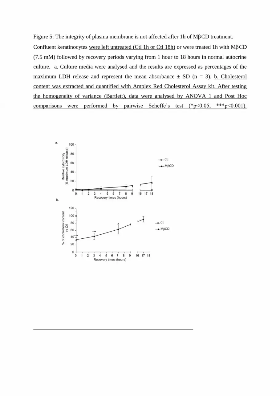
Figure 5: The integrity of plasma membrane is not affected after 1h of MCD treatment.
Confluent keratinocytes were left untreated (Ctl 1h or Ctl 18h) or were treated 1h with MCD
(7.5 mM) followed by recovery periods varying from 1 hour to 18 hours in normal autocrine
culture. a. Culture media were analysed and the results are expressed as percentages of the
maximum LDH release and represent the mean absorbance ± SD (n = 3). b. Cholesterol
content was extracted and quantified with Amplex Red Cholesterol Assay kit. After testing
the homogeneity of variance (Bartlett), data were analysed by ANOVA 1 and Post Hoc
comparisons were performed by pairwise Scheffe’s test (*p<0.05, ***p<0.001).

Figure 6: HB-EGF synthesis after lipid raft disruption is dependent on extracellular ATP
released in keratinocytes.
a. Confluent keratinocyte cultures were pretreated 1h with suramin (200 µM) followed by 1h
of MCD treatment (7.5mM). Then cells were incubated in autocrine culture medium during
several recovering periods (0, 1h, 2h, 4h, 8h and 18h). After each treatment, proteins were
extracted and analysed by Western blotting using specific antibodies against EGFR, p38,
ERK, HSP27 and their phosphorylated forms and HB-EGF.
b. Confluent keratinocyte cultures were treated 1h with 1h MCD (7.5mM) in the presence of
apyrase (10U/ml) or the vehicle. Then cells were incubated in autocrine culture medium
during several recovering periods (0, 1h, 2h, 4h, 8h and 18h) in the presence of apyrase
(10U/ml) or the vehicle. After each treatment, proteins were extracted and analysed by
Western blotting using specific antibodies against EGFR, p38, ERK, and their phosphorylated
forms and HB-EGF.

Figure 7: Early HB-EGF protein expression induced by disruption of lipid rafts is dependent
on ERK and p38 MAPK activity.
a. Confluent keratinocyte cultures were left untreated or were pretreated 1h with the ERK
inhibitor, the PD98069 (2 µM) then left untreated or treated with MCD (7.5mM) for 1h in
the presence of PD98069 (2 µM) or the vehicle. Then cells were incubated in autocrine
culture medium during several recovering periods (0, 1h, 2h, 4h, 8h and 18h) in the presence
of PD98069 (2 µM) or the vehicle. Cells lysates were then analysed by Western blotting using
specific antibodies against EGFR, ERK, their phosphorylated forms and HB-EGF.
b. Confluent keratinocyte cultures were pretreated 1h with PD98069 (2 µM) and PD169316
(15 µM). Then cultures were treated 1h with MCD (7.5mM) in the presence of both
inhibitors which were also part of the recovery medium (0, 1h, 2h, 4h, 8h and 18h). Cells
lysates were then analysed by Western blotting.

Figure 8: HB-EGF synthesis and release induced by MCD treatment is controlled by
extracellular ATP and involves both p38 and ERK1/2 signaling pathways in keratinocytes.
After cholesterol depletion, keratinocytes release large amounts of extracellular ATP. By
interaction with P2 purinergic receptors, ATP induces EGFR phosphorylation, HB-EGF
synthesis and release through signaling pathways involving p38 and ERK1/2. Once released,
HB-EGF further activates EGFR.


