Embed Size (px)
Citation preview

This article was downloaded by: [ ]On: 22 September 2011, At: 04:34Publisher: Taylor & FrancisInforma Ltd Registered in England and Wales Registered Number: 1072954Registered office: Mortimer House, 37-41 Mortimer Street, London W1T 3JH, UK
Journal of Natural HistoryPublication details, including instructions for authors andsubscription information:http://www.tandfonline.com/loi/tnah20
Toward a taxonomic revision of themedicinal leech Hirudo medicinalisLinnaeus, 1758 (Hirudinea:Hirudinidae): re-description ofHirudo troctina Johnson, 1816 fromNorth AfricaFred O. P. Hechtel & Roy T. Sawyer
Available online: 06 Dec 2010
To cite this article: Fred O. P. Hechtel & Roy T. Sawyer (2002): Toward a taxonomic revisionof the medicinal leech Hirudo medicinalis Linnaeus, 1758 (Hirudinea: Hirudinidae): re-description of Hirudo troctina Johnson, 1816 from North Africa, Journal of Natural History,36:11, 1269-1289
To link to this article: http://dx.doi.org/10.1080/00222930110048945
PLEASE SCROLL DOWN FOR ARTICLE
Full terms and conditions of use: http://www.tandfonline.com/page/terms-and-conditions
This article may be used for research, teaching and private study purposes. Anysubstantial or systematic reproduction, re-distribution, re-selling, loan, sub-licensing, systematic supply or distribution in any form to anyone is expresslyforbidden.
The publisher does not give any warranty express or implied or make anyrepresentation that the contents will be complete or accurate or up to date. Theaccuracy of any instructions, formulae and drug doses should be independentlyverified with primary sources. The publisher shall not be liable for any loss, actions,claims, proceedings, demand or costs or damages whatsoever or howsoever caused

arising directly or indirectly in connection with or arising out of the use of thismaterial.
Dow
nloa
ded
by [
] a
t 04:
34 2
2 Se
ptem
ber
2011
Journal of Natural History, 2002, 36, 1269–1289
Toward a taxonomic revision of the medicinal leech Hirudo medicinalisLinnaeus, 1758 (Hirudinea: Hirudinidae): re-description of Hirudotroctina Johnson, 1816 from North Africa
FRED O. P. HECHTEL† and ROY T. SAWYER*‡
†School of Biological Sciences, University of Wales, Swansea SA2 8PP,UK‡Biopharm (UK) Ltd., Bryngelen House, 2 Bryngwili Road, Hendy,Carms. SA4 1XB, UK; e-mail: [email protected]
(Accepted 2 February 2001)
The medicinal leech Hirudo medicinalis Linnaeus, 1758 is genotype of the genusHirudo and constitutes the taxonomic basis for the family Hirudinidae and eventhe class Hirudinea. In spite of its unique taxonomic signi� cance, H. medicinalishas never been characterized in terms of criteria currently accepted for theHirudinea. As part of a broader revision of this species, Hirudo troctina Johnson,1816 from North Africa is re-described based on the foregut and internal dia-gnostic characters. The authors con� rm that H. troctina is a separate speciesfrom H. medicinalis as currently understood. In addition to external diŒerences(four or six metameric dorsal black dots in H. troctina), the male and femalereproductive anatomy consistently distinguish H. troctina (vagina bulbous,upright and not folded; epididymis massive in relation to ejaculatory bulb) fromH. medicinalis (vagina a folded, elongated tube in non-juveniles; epididymis notmuch larger than ejaculatory bulb). Furthermore, several possible errors in thedescription of taxonomic features of the foregut of H. medicinalis (e.g. claimedabsence of a sulcus and salivary papillae) appear to have been perpetuated in theliterature, requiring a re-evaluation of the genus Hirudo.
Keywords: Hirudo troctina, Hirudo medicinalis, North Africa, Morocco, Tunisia,Algeria, Hirudinea, medicinal leech, leech trade, medical history.
IntroductionThe medicinal leech Hirudo medicinalis Linnaeus, 1758 is genotype of the genus
Hirudo and constitutes the taxonomic basis for the family Hirudinidae and even theclass Hirudinea. In spite of its unique taxonomic signi� cance, H. medicinalis hasnever been characterized in terms of criteria currently accepted for the Hirudinea.
In the 19th century the many recognized variations of this species were based
*To whom correspondence should be addressed.
Journal of Natural HistoryISSN 0022-2933 print/ISSN 1464-526 2 online © 2002 Taylor & Francis Ltd
http://www.tandf.co.uk/journalsDOI: 10.1080/00222930110048945
Dow
nloa
ded
by [
] a
t 04:
34 2
2 Se
ptem
ber
2011

F. O. P. Hechtel and R. T. Sawyer1270
almost entirely on external pigmentation. Moquin-Tandon (1846: 248) listed anumber of named varieties of medicinal leech sold in Europe and concluded thatthey all represented the same species, Hirudo medicinalis, with the single exceptionof the so-called ‘Dragon’ or Hirudo troctina Johnson, 1816, a closely allied formoriginating from North Africa. The most experienced hirudinologist of his day,Moquin-Tandon clearly considered H. troctina to be a distinctive species separatefrom H. medicinalis. However, his conclusion was based on external criteria alone.
In this paper we re-describe the little known H. troctina based on a detailedanatomical study of internal characters, mainly of the foregut and reproductivesystems. We critically discuss its taxonomic relationship with H. medicinalis ascurrently de� ned (Sawyer, 1986: 684, 716). One outcome of this study is the revela-tion that several critical errors in the description of taxonomic features of the foregutof H. medicinalis appear to have been perpetuated in the literature. This calls intoquestion the taxonomic de� nition of H. medicinalis itself. Accordingly, this paper ispart of a planned broader revision of H. medicinalis. Until such a revision iscompleted and a type locality designated, no conclusions can be made concerningthe precise taxonomic status of the H. medicinalis used in this study.
The leeches of North Africa remain incompletely studied. Of the jawed leeches(Hirudinidae) only three species have been reported: Limnatis nilotica (Savigny,1822 ), Haemopis sanguisuga (Linnaeus, 1758) and Hirudo troctina. The so-callednasal leech Limnatis nilotica occurs throughout North Africa from Morocco, Algeria,Tunisia, Libya, Egypt and Asia Minor. Since Biblical and Roman times ‘nasalleeches’ have caused serious disability by invading for prolonged periods the nasalpassages of cattle, horses, camels and occasionally humans. Limnatis nilotica hasonly rudimentary teeth and is unable to penetrate the skin of mammals, except thedelicate membranes of the nares. While common in most of Europe, Haemopissanguisuga has only been reported once in North Africa, from a single specimenfrom the Atlas Mountains of Morocco (Moore, 1939). A single colony of H.sanguisuga has been found in the nearby Maltese island of Gozo where it is considereda locally endangered species (Schembri, 1986). In spite of its name, Haemopissanguisuga is strictly predaceous and incapable of sucking blood.
The third species H. troctina was originally described, based on external featuresalone, by Rawlins Johnson (1816) from specimens found in local pharmacies inBristol, England. The author, who was unaware of the geographic origin of thisimported species, gave this leech the speci� c name troctina (‘trout’) due to thesimilarity of its spotted pigmentation to that of the sides of a trout. Similarly, theFrench hirudinologist Alfred Moquin-Tandon (1827) described a species Sanguisugainterrupta which he found in pharmacies around Montpelier. Again, the author wasunaware of the origin of this species. By 1846 Moquin-Tandon, in consultation withJohnson, acknowledged the synonomy of the two nominal species, with H. troctinataking precedence. The problem of the geographical origin of this medically usefulspecies was resolved � nally by Gervais (1835) who demonstrated that the speciesoriginated from North Africa where it was known in the leech trade as the Algerian‘Dragon’. The name ‘Dragon’ refers to the similarity of the leech’s pigmentation tothat of the uniforms of the French military dragoons.
In the 19th century H. troctina was exported in large quantities into Englandand France for medicinal purposes as a replacement for H. medicinalis. This is theonly leech known from North Africa with well-developed teeth and jaws capable ofsucking blood through human skin, hence its medical desirability. The commercial
Dow
nloa
ded
by [
] a
t 04:
34 2
2 Se
ptem
ber
2011

North African medicinal leech 1271
trade in H. troctina declined before this species was properly characterized taxonom-ically so that questions remain as to the true phylogenetic relationship of this species,especially to H. medicinalis. Apart from a very brief mention by Moore (1939) theinternal anatomy of H. troctina has never been described.
Materials and methodsWe examined preserved specimens of H. troctina from various localities broadly
representative of its known geographical range in northwestern Africa, namelyTunisia, Algeria and Morocco. Specimens were obtained from The Natural HistoryMuseum (London) [BMNH] and the Museum National d’Histoire Naturelle (Paris)[MNHN]. All dissections were carried out by F. O. P. Hechtel and numbered21Z–36Z in his dissection notes. Preserved specimens were pinned under alcoholand dissected along the dorsal midline from X a2/b5 to XV b2/a2 to reveal diagnosticfeatures of the male and female reproductive systems. The specimens were thenturned over and repinned. Cutting the foregut along the ventral midline to X b2/a2revealed features of the jaws and pharynx. The drawings were made freehand withthe aid of an ocular micrometer. Where possible at least two mature specimens weredissected from each country. To ensure nomenclatural continuity we examinedspecimens identi� ed by Moore (1939) as H. troctina from Morocco. Being aware ofthe uncertainties of labelling and constraints on dissecting old museum material, theauthors would have preferred to base conclusions on live material. However, allattempts to obtain live H. troctina from North Africa failed. The study of thisspecies was con� ned to North Africa in that specimens labelled ‘troctina’ fromoutside this region were not examined. It remains open whether H. troctina occursoutside North Africa.
In order to assess the correct taxonomic status of H. troctina we also examinedspecimens of H. medicinalis, the genotype of the genus Hirudo. With one exception(Dissection 23Z), each live specimen of H. medicinalis was narcotized gradually inethanol prior to being � xed in 70% ethanol in a straight, � at state. Care was takennot to stretch the specimen overly beyond the relaxed state. Unfortunately, as withH. troctina, no type material for H. medicinalis exists. Accordingly we chose forcomparative purposes specimens of H. medicinalis from western Turkey. This areawas chosen because of the similarity of arid habitats of these two circum-Mediterranean regions. The eight specimens of H. medicinalis examined in this study,along with accompanying dissection notes, have been deposited in The NaturalHistory Museum (London) [BMNH 2001.283–290].
In order to maximize clarity for subsequent workers, descriptions of the taxonom-ically pertinent characters stand alone for each dissection. In our experience withleech taxonomy lumping descriptions has been a major cause of confusion in thepast. Care was taken to examine specimens of diŒering sizes and states of reproduct-ive maturity to eliminate transient diagnostic features as much as possible. It wasfound that annulation, position of gonopores and eyes, and some other externalfeatures for H. troctina were indistinguishable from H. medicinalis. This being thecase these characters are only described in detail for one specimen of each species,unless otherwise noted.
The systematics and specialized terminology follow that of Sawyer (1986). Inthe context of this paper the male atrium is understood to comprise the prostateand penis sheath. A sulcus is a groove or � ssure in the ventral surface of the dorsallip of the cephalic sucker, often varying in depth depending on the method of
Dow
nloa
ded
by [
] a
t 04:
34 2
2 Se
ptem
ber
2011

F. O. P. Hechtel and R. T. Sawyer1272
narcotization and preservation, leading from the crypt of the dorso-median jaw tothe anterior rim of the sucker.
Caution. In this study we must emphasize the di� culty of comparing museumspecimens, which are often preserved under harsh � eld conditions, with freshlynarcotized material. All the museum material, with the exception of one largespecimen (Dissection 21Z), had been preserved in a highly contracted and ventrallycurved state. When such specimens are pinned for dissection there can be somedistortion of the precise segmental boundaries of certain structures. Similarly, thelength measurements of contracted specimens can be reduced by as much as half, ifnot more, than had they been relaxed prior to � xation. In greatly contractedspecimens it is not always possible to con� rm annulation, but we are con� dent thatthere is no signi� cant diŒerence between the two species. The friability of preservedmaterial made it especially di� cult to de� ne with precision the anatomy of the cropof H. troctina. Apart from black markings the pigmentation was sometimes totallyfaded in H. troctina. The black pigmentation, however, was so conserved that wecan con� rm that four (sometimes six) metameric black spots were nearly alwaysdiscernible on the dorsum of H. troctina, even in otherwise faded specimens; andconversely they were not found in any specimen of H. medicinalis examined inthis study.
Systematic account
Family HIRUDINIDAE Whitman, 1886Subfamily HIRUDININAE Richardson, 1969
Genus Hirudo Linnaeus, 1758Hirudo troctina Johnson, 1816
Hirudo Troctina Johnson, 1816: 31–32 (no type locality speci� ed); Blanchard, 1894: 41–42;Blanchard, 1908: 310; Johansson, 1914: 837–840; Moore, 1939: 82–83.
Sanguisuga interrupta Moquin-Tandon, 1827: 118–119, pl. 6, � gure 2; Letheby, 1844: 256,� gure 3.
Sanguisuga troctina Moquin-Tandon, 1846: 335–337, pl. 11, � gures 19–22 .
Material examined. Morocco (note: this material was previously examined byMoore, 1939). Lake Aguelman, Sidi Ali, Moyen Atlas Mts, eight specimens, twodissected 40.3 mm (BMNH 1939.8.18.5-6) and 43.0 mm (BMNH 1939.8.18.7-8).Oumer Rbia River, south-west of Azrou, one specimen dissected 29.0 mm (BMNH1939.8.18.13). The following material was examined externally, Lake C, Ouiouane,80 km south west of Azrou, ca 1500 m, three specimens 26.0, 28.5 and 31.0 mm(BMNH 1939.8.18.10–12). No. 558 Timhadit, Guigou River, 40 km south of Azrou,ca 2000 m, one specimen 27.0 mm (BMNH1939.8.18.9). Algeria. ‘Algeria’, fourspecimens, two dissected 50.5 and 59.0 mm, 1876 (1870?) (MNHN A796). Tunisia.Sousse, one large specimen, dissected 114 mm, 1899 (MNHN A806). ‘Tunisia’, 11specimens, two dissected 57.0 and 67.5 mm, 1888 (MNHN A802).
Known distribution. Hirudo troctina has been reported previously from thefollowing localities in northwestern Africa. MOROCCO: Lake Aguelman, Sidi Ali(130 km south Meknes, ca 2100 m); Timhadit, Guigou River (40 km south Azrou,ca 2000 m); Ouiouane (80 km south-west Azrou, ca 1400 m); Oum er Rbia River(south-west Azrou); Aguelman Aziza Lake (20 km west Oum er Rbia Springs)(Moore, 1939). ALGERIA: El Kreider (Johansson, 1914), Teniet-el-Haad (3 dayssouth of Milianah) (Moquin-Tandon, 1846: 336), Lake Holloula (west of Mitidja)
Dow
nloa
ded
by [
] a
t 04:
34 2
2 Se
ptem
ber
2011

North African medicinal leech 1273
(Ebrard 1857: 188), ‘Constantine’ (Ebrard, 1857: 38), Tiaret (Ghelma) (Bertherand,1855). TUNISIA: Oued el Amor (Tabarca region); Camp de la Sante (A õ n-Drahamregion) (Blanchard, 1908), ‘Tunis’ (Ebrard, 1857: 37).
Reports in the early literature of this species from Spain (near Corogne)(Blanchard, 1894: 42) and from Sardinia and Libya (‘Tripoli’) (Ebrard, 1857: 37)remain unsubstantiated .
Designation of neotype. No type material exists for H. troctina, and the originaldescription did not specify a type locality. In order to stabilize the nomenclature ofthis species we designate the following specimen as the neotype for this species:BMNH 1939.8.18.5-6 from Stn B, Lake Aguelman, Sidi Ali, Moyen AtlasMountains, Morocco. We choose this specimen because its identi� cation was con-� rmed by Moore (1939), as well as in this study (our Dissection 30Z). (Due tofriability of the neotype, its dissection was limited to establishing identi� cation.Figure 2 ( left) was taken from Dissection 27Z where additional tissue was removedto allow for a complete drawing.)
Hirudo medicinalis Linnaeus, 1758Hirudo medicinalis Ray, 1710: 3; Linnaeus, 1758: 649; Moquin-Tandon, 1846: 327, pls vii–xi.Sanguisuga medicinalis Savigny, 1822: 114; Moquin-Tandon, 1827: 114, pl. v, � gure 2.Sanguisuga o� cinalis Savigny, 1822: 330.Hirudo o� cinalis Derheims, 1825: 9, 11.
Material examined. Specimens of H. medicinalis were collected as part ofanother study from a pond south of Edirne, Turkey, at diŒerent times of the yearduring 1997–2000. Most dissections were made on specimens collected fromFebruary to April, inclusively. Eight specimens were dissected: 43.3, 54.0, 61.0, 74.3,90.0, 117.0, 122.0 and 167.0 mm. Some specimens were bred in the laboratory fromthe same stock for more controlled evaluation of developmental changes of taxo-nomic characters.
Distribution. The original range of H. medicinalis was from western and south-ern Europe to the Ural Mountains (Sawyer, 1986). Today its distribution is muchcontracted to the point that it is recognized as a threatened or endangered speciesin most countries of western Europe (Sawyer, 1981).
Results
Taxonomic characterization of Hirudo troctina
Morocco: specimen one (Dissection 30Z)BMNH: ‘Hirudo troctina 1939.8.18. 5-6 Lake Aguelman (Stn.B) Sidi Ali, Moyen
Atlas Mts., Morocco’. Specimen contracted and tightly curled.Externals. Measurements (mm): length 40.3, maximum body width 8.94, width
at buccal ring 3.38 (very contracted), width at male gonopore 7.12, length anteriorto male gonopore 9.35, maximum width of caudal sucker 4.15. Gonopores separatedby � ve annuli, male at XI b5/b6, female at XII b5/b6. Annulation: 15 complete(� ve-annulate) somites, IX–XXIII; (incomplete cephalic and caudal somites toocontracted to distinguish clearly). Eyes: � ve pairs, one pair each on the a2 annulusof V and VI (due to contraction it was not possible to determine the annuli of the� rst three pairs with certainty). Sulcus: present as a narrow groove running fromthe crypt of the dorso-median jaw to the dorsal rim of the cephalic sucker.
Dow
nloa
ded
by [
] a
t 04:
34 2
2 Se
ptem
ber
2011

F. O. P. Hechtel and R. T. Sawyer1274
Pigmentation: background faded to pale beige, markings faded to greyish dusky.Metameric intermediate and supra-marginal dots on b2 annulus of complete somitesand equivalent of incomplete somites, supra-marginal streak apparent as very slightlydarker than background, dusky sub-marginal stripe, venter marked heavily withirregularly sized and shaped blotches.
Foregut. Foregut based on ventral dissection to X b2/a2 (� gure 1). Trignathous,monostichodont, jaws papillate. Pharynx: fusiform chamber with muscular ridgesdisintegrated beyond description, but no ridges terminate between the jaws.
Internals. Dorsal dissection from X a2/b5 to XV b2/a2. Male system fullydeveloped, female possibly not totally developed, male terminal system 3–3.5 timessize of complete female system.
Male reproductive system. Epididymes: massive, globular masses of tightlypacked ducting standing upright on either side of the prostate. Ejaculatory bulbs:developed, small in relation to epididymes, the dorso-cephalic rim of which theycircle. Male terminal organs lie within XI and XII (more precise location notpossible). Vasa deferentia: small remaining sections reveal ducts broad with heavyglandular cover. Testisacs: very little left of � rst pair; though damaged, su� cient ofsecond pair right testisac to estimate size as approximately three times that of theovisacs, ovoid (to retain integrity of the specimen the second pair left testisac notrevealed); unable to state precise location, but � rst pair in region of XIII and XIV.Atrium: prostate, large, bulbous with glandular cover; penis sheath, a long, broadduct re� exed anteriorly.
Female reproductive system. Vagina: bulbous in form (a long swollen uprightsac, leaning slightly caudad and tapering gently towards junction with ventral bodywall, precise location not possible to ascertain, but in the region of XII–XIII ),caecate, no vaginal duct (vagina enters directly into ventral body wall ). Commonoviduct enters vagina just below its dorsal tip to form a small, distinct vaginalcaecum. Ovisacs: small, delicate and damaged in dissection. Common oviduct (albu-men gland): a broad, long, convoluted duct lying adnate on the cephalic face of thevagina, with some glandular covering, not extensive.
Fig. 1. Comparison of the foreguts of Hirudo troctina ( left) [Dissection 30Z, neotype] andHirudo medicinalis (right) [Dissection 35Z]. Ventral view. j, jaw; p, salivary papillae; s,sulcus; t, teeth. Scale bar: 1 mm.
Dow
nloa
ded
by [
] a
t 04:
34 2
2 Se
ptem
ber
2011

North African medicinal leech 1275
Morocco: specimen two (Dissection 34Z)BMNH: ‘Hirudo troctina 1939.8.18. 7-8 Lake Aguelman (Stn.9) Sidi Ali, Moyen
Atlas Mts. 130 Km South of Meknes 6,700 ft’. Specimen contracted.Externals. Measurements (mm): length 43.0, maximum body width 8.75, width
at buccal ring 3.79, width at male gonopore 6.4, length anterior to male gonopore9.4, maximum width of caudal sucker 4.67. Sulcus: present as a narrow grooverunning from the crypt of the median-dorsal jaw to the dorsal rim of the cephalicsucker. Pigmentation: background faded to pale beige, markings faded to greyishdusky. Metameric intermediate dots on b2 annulus of complete somites and equiva-lent of incomplete somites, sub-marginal dusky stripe, venter sparsely maculated.
Foregut. No foregut dissection undertaken; it was considered unnecessary todisturb the integrity of a further specimen as all H. troctina specimens examined sofar show clearly, without exception, trignathous, monostichodont, papillate charac-ters and a fusiform pharynx with no pharyngeal ridges terminating between jaws.
Internals. Dorsal dissection from X a2/b5 to XV b2/a2. Male and femalesystems fully developed, male terminal system 3.5–4 times size of complete femalesystem. Both systems lie in the region of XI, XII, XIII; precise locations obscuredby contraction.
Male reproductive system. Epididymes: massive, tightly packed, globular massesof ducting. Ejaculatory bulbs: fusiform, well developed, but small in relation toepididymes the dorso-cephalic faces of which they circle. Vasa deferentia: a smallsection dissected revealed a duct broad with glandular cover. Testisacs: due tofragility of the material not dissected. Atrium: prostate, large, bulbous with a heavyglandular cover; penis sheath, a long, broad duct re� exed anteriorly.
Female reproductive system. Vagina: bulbous in form (a broad, elongate, uprightsac), caecate, no vaginal duct (vagina enters directly into ventral body wall ).Common oviduct enters vagina just below its dorsal tip to form a small, distinctvaginal caecum. Ovisacs: left large, right very small, possibly rudimentary (stunted)or partially removed in dissection. Common oviduct (albumen gland): broad, long,convoluted duct lying adnate on cephalic face of vagina with heavy glandular cover.
Morocco: specimen three (Dissection 31Z)BMNH: ‘Hirudo troctina 1939.8.18.13 Sta. D, No. 403, Oumer Rbia River, South
west of Azrou’. A small specimen, contracted.Externals. Measurements (mm): length 29.0, maximum body width 7.5, width
at buccal ring 2.95, width at male gonopore 5.2, length anterior to male gonopore7.4, maximum width of caudal sucker 3.9. Sulcus: present as a narrow grooverunning from the crypt of the dorso-median jaw to the dorsal rim of the cephalicsucker. Pigmentation: background faded to pale beige, markings greyish dusky;metameric intermediate spots and smaller supra-marginal spots on b2 annulus of allcomplete somites, dusky sub-marginal stripe, venter virtually immaculate.
Foregut. Foregut based on ventral dissection to X b2/a2. Trignathous, monos-tichodont, jaws papillate. Pharynx, a fusiform chamber, pharyngeal ridges muchdisintegrated, however, no ridges terminate between the jaws.
Internals. Dorsal dissection from X a2/b5 to XV b2/a2. Male system nearlyfully developed, female not developed, male terminal system approximately six timeslarger than complete female system (atrium alone being more than twice the size ofentire female system).
Dow
nloa
ded
by [
] a
t 04:
34 2
2 Se
ptem
ber
2011

F. O. P. Hechtel and R. T. Sawyer1276
Male reproductive system. Epididymes: developed, but not massive, globularmasses of tightly packed ducting standing on either side of the prostate. Ejaculatorybulbs: well developed, approximately the same size as the epididymes the dorso-cephalic rim of which they circle. Paired terminal organs lie within XI and XII(more precise location not possible). Testisacs: � rst pair much disintegrated, su� cientto estimate size to be approximately 2.5 times size of ovisacs, located in region ofXIII, XIV. Atrium: prostate, large, bulbous with some glandular cover; penis sheath,a long, broad duct re� exed anteriorly.
Female reproductive system. Vagina: a simple tube standing upright ( leaningslightly caudad, in region of XII/XIII ), caecate, no vaginal duct (vagina entersdirectly into ventral body wall ), common oviduct enters vagina just below the dorsaltip to form a vaginal caecum. Ovisacs: small, though relatively large in comparisonwith undeveloped rest of female system, located somewhere in XII. Common oviduct(albumen gland)–vaginal complex: very small indeed, almost vestigial (breadthhardly more than the ventral nerve cord which partly conceals it); the commonoviduct a long, broad duct convoluted and lying adnate on the dorso-cephalic faceof the vagina with no sign of glandular cover.
Algeria: specimen one (Dissection 26Z)MNHN A 796: ‘Hirudo troctina, Algerie. M. Mores, No. 23, 1876 [or 1870?] ’.
Specimen contracted and tightly curled.Externals. Measurements (mm): length 59.0, maximum body width 14.82, width
at buccal ring 3.58, width at male gonopore 11.25, length anterior to male gonopore12.26, caudal sucker deformed through curling. Sulcus: present as a narrow grooverunning from the crypt of the median-dorsal jaw to the dorsal rim of the cephalicsucker. Pigmentation: faded, pattern obliterated.
Foregut. Foregut based on ventral dissection to X b2/a2. Trignathous, monos-tichodont, jaws papillate. Pharynx: a short fusiform chamber, midline six muscularpharyngeal ridges merging in pairs to form three ridges each terminating at the baseof a jaw; pharynx stretches caudad into cephalic half of IX, short of GIX.
Internals. Dorsal dissection from X a2/b5 to XV b2/a2. Male and femalesystems fully developed, male terminal system 1.5–2 times larger than completefemale system.
Male reproductive system. Epididymes: massive, globular masses of coiled duct-ing. Ejaculatory bulbs: fusiform, well developed but small in relation to epididymes,the dorso-cephalic rim of which they circle. Male terminal organs lie within caudalhalf of XI and cephalic half of XII. Vasa deferentia: ducts with heavy glandularcover. Testisacs: from the little which survives of the � rst pair they appear to belarger than the ovisacs. (Due to friability of the material in general and the fragilityof the testisacs in particular no attempt was made to reveal the second pair.) Atrium:prostate, large bulbous, heavily invested with glandular tissue, penis sheath a broadlong duct re� exed anteriorly.
Female reproductive system. Vagina: bulbous in form (a large, broad, ovoid sac,dorso-ventrally compressed, located on the margin of XII/XIII ), caecate, no vaginalduct (vagina enters directly into ventral body wall ). Common oviduct enters thevagina just below its dorsal tip to form a small, distinct vaginal caecum. Ovisacs:ovoid, located in caudal half of XII. Common oviduct (albumen gland): broad,long convoluted duct tightly packed to a compact mass adnate on dorso-cephalicface of vagina and heavily invested with glandular tissue.
Dow
nloa
ded
by [
] a
t 04:
34 2
2 Se
ptem
ber
2011
North African medicinal leech 1277
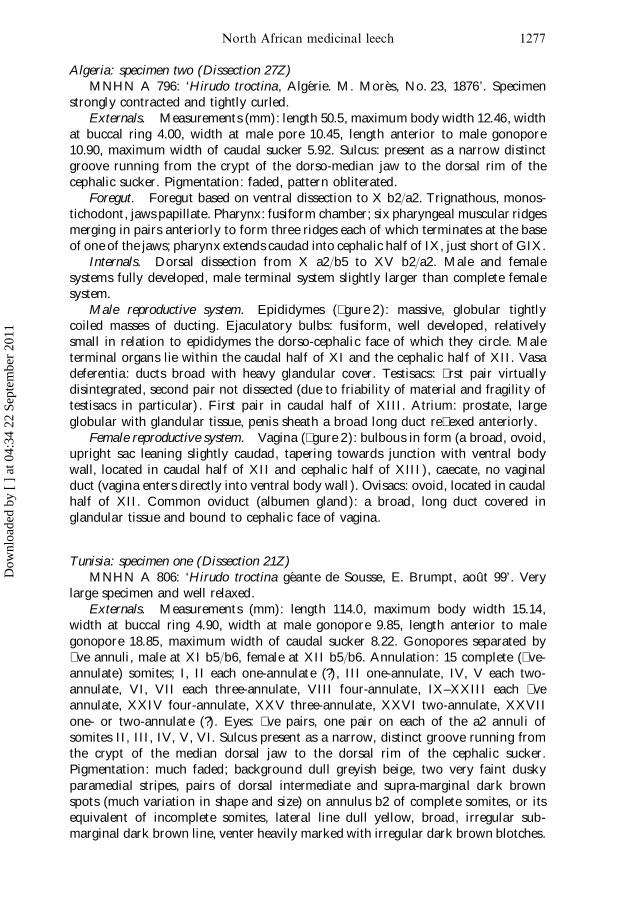
Algeria: specimen two (Dissection 27Z)MNHN A 796: ‘Hirudo troctina, Algerie. M. Mores, No. 23, 1876’. Specimen
strongly contracted and tightly curled.Externals. Measurements (mm): length 50.5, maximum body width 12.46, width
at buccal ring 4.00, width at male pore 10.45, length anterior to male gonopore10.90, maximum width of caudal sucker 5.92. Sulcus: present as a narrow distinctgroove running from the crypt of the dorso-median jaw to the dorsal rim of thecephalic sucker. Pigmentation: faded, pattern obliterated.
Foregut. Foregut based on ventral dissection to X b2/a2. Trignathous, monos-tichodont, jaws papillate. Pharynx: fusiform chamber; six pharyngeal muscular ridgesmerging in pairs anteriorly to form three ridges each of which terminates at the baseof one of the jaws; pharynx extends caudad into cephalic half of IX, just short of GIX.
Internals. Dorsal dissection from X a2/b5 to XV b2/a2. Male and femalesystems fully developed, male terminal system slightly larger than complete femalesystem.
Male reproductive system. Epididymes (� gure 2): massive, globular tightlycoiled masses of ducting. Ejaculatory bulbs: fusiform, well developed, relativelysmall in relation to epididymes the dorso-cephalic face of which they circle. Maleterminal organs lie within the caudal half of XI and the cephalic half of XII. Vasadeferentia: ducts broad with heavy glandular cover. Testisacs: � rst pair virtuallydisintegrated, second pair not dissected (due to friability of material and fragility oftestisacs in particular) . First pair in caudal half of XIII. Atrium: prostate, largeglobular with glandular tissue, penis sheath a broad long duct re� exed anteriorly.
Female reproductive system. Vagina (� gure 2): bulbous in form (a broad, ovoid,upright sac leaning slightly caudad, tapering towards junction with ventral bodywall, located in caudal half of XII and cephalic half of XIII ), caecate, no vaginalduct (vagina enters directly into ventral body wall ). Ovisacs: ovoid, located in caudalhalf of XII. Common oviduct (albumen gland): a broad, long duct covered inglandular tissue and bound to cephalic face of vagina.
Tunisia: specimen one (Dissection 21Z)MNHN A 806: ‘Hirudo troctina geante de Sousse, E. Brumpt, aout 99’. Very
large specimen and well relaxed.Externals. Measurements (mm): length 114.0, maximum body width 15.14,
width at buccal ring 4.90, width at male gonopore 9.85, length anterior to malegonopore 18.85, maximum width of caudal sucker 8.22. Gonopores separated by� ve annuli, male at XI b5/b6, female at XII b5/b6. Annulation: 15 complete (� ve-annulate) somites; I, II each one-annulate (?), III one-annulate, IV, V each two-annulate, VI, VII each three-annulate, VIII four-annulate, IX–XXIII each � veannulate, XXIV four-annulate , XXV three-annulate, XXVI two-annulate, XXVIIone- or two-annulate (?). Eyes: � ve pairs, one pair on each of the a2 annuli ofsomites II, III, IV, V, VI. Sulcus present as a narrow, distinct groove running fromthe crypt of the median dorsal jaw to the dorsal rim of the cephalic sucker.Pigmentation: much faded; background dull greyish beige, two very faint duskyparamedial stripes, pairs of dorsal intermediate and supra-marginal dark brownspots (much variation in shape and size) on annulus b2 of complete somites, or itsequivalent of incomplete somites, lateral line dull yellow, broad, irregular sub-marginal dark brown line, venter heavily marked with irregular dark brown blotches.
Dow
nloa
ded
by [
] a
t 04:
34 2
2 Se
ptem
ber
2011

F. O. P. Hechtel and R. T. Sawyer1278
Fig. 2. Comparison of male and female reproductive systems of Hirudo troctina from NorthAfrica ( left) [Dissection 27Z] and Hirudo medicinalis from western Turkey (right)[Dissection 28Z]. Dorsal view. Inset ( left): left lateral view of vagina [Dissection 27Z].Inset (right): left lateral view of vagina of Dissection 29Z (a more typical vaginalmorphology for this species than Dissection 27Z). co, common oviduct; e, epididymis;eb, ejaculatory bulb; ed, ejaculatory duct; o, oviduct; os, ovisac; ps, penis sheath; pt,prostate; ts, testisac; v, vagina; vc, vaginal caecum; vd, vas deferens. Roman numeralsindicate the location of respective somatic ganglia to assist in establishing the locationof the reproductive organs. Scale bar: 1 mm.
Foregut. Foregut based on ventral dissection to X b2/a2. Trignathous, monos-tichodont, jaws papillate. Right lateral jaw extracted and stained with Dela� eld’sHaematoxylin; 71 1 teeth, large conical pegs reducing in size distally and eventuallydisappearing in contracted tissue; approximately 28 papillae on the dorsal and 37on the ventral face, diameters of largest papillae range from 53 to 113 mm. Pharynx,short fusiform chamber, six longitudinal muscular ridges which fuse in pairsanteriorly so that none terminate between the jaws.
Internals. Dorsal dissection from X a2/b5 to XV b2/a2. Male and femalesystems fully developed, male terminal system 2.5–3 times larger than completefemale system.
Male reproductive system. Epididymes: large, broad discoid masses of tightlycoiled ducting standing upright on either side of the prostate. Ejaculatory bulbs:fusiform, developed, relatively small in relation to the large epididymes, the dorso-cephalic rims of which they circle. Vasa deferentia: small sections remaining show abroad glandular covered duct. Testisacs: ovoid, approximately twice the size of theovisacs, � rst pair over the border between XIII and XIV. Atrium: a large gobular
Dow
nloa
ded
by [
] a
t 04:
34 2
2 Se
ptem
ber
2011

North African medicinal leech 1279
prostate covered extensively with glandular tissue concealing what appears to be ashort penis sheath (the latter not revealed due to friability of the material ).
Female reproductive system. Vagina: bulbous in form (an upright elongated sac,leaning caudally, located in XIII ); caecate, no vaginal duct (vagina enters directlyinto the ventral body wall ); common oviduct joins the vagina just short of thedorsal tip creating a distinct vaginal caecum; Ovisacs: ovoid, located in caudal halfof XII. Common oviduct (albumen gland): a broad duct adnate on the cephalicface of the vagina, covered with glandular tissue.
Tunisia: specimen two (Dissection 22Z)MNHM A 802: ‘Hirudo troctina, Tunisie—prepare par liquid de Kleinenberg,
1888’. Specimen contracted.Externals. Measurements (mm): length 57.0, maximum body width 10.25, width
at buccal ring 3.65, width at male gonopore 6.65, length anterior to male gonopore13.95, maximum width of caudal sucker 4.37. Sulcus: present as a narrow, distinctgroove running from the crypt of the dorso-median jaw to the dorsal rim of thecephalic sucker. Pigmentation: colour and pattern almost totally faded; backgroundpigmentation lost (pale beige), two faint dusky para-medial stripes, pairs of dorsalintermediate and supra-marginal dusky spots on annulus b2 of complete somites orits equivalent on incomplete somites, broad dusky irregular submarginal stripe,venter extensively marked with irregular dusky blotches.
Foregut. Foregut based on ventral dissection to X b2/a2. Trignathous, monos-tichodent, jaws papillate. Pharynx much disintegrated, no pseudognaths between thejaws suggesting that muscular ridges terminate at base of jaws, where extant theridges indicate there were approximately six at the midline of the pharynx.
Internals. Dorsal dissection from X a2/b5 to XV b2/a2. Male and femalesystems fully developed. Male terminal system more than 2.5 times size of completefemale system.
Male reproductive system. Epididymes: massive, discoid (� attening from globu-lar due to pressure of blood meal ), tightly packed masses of ducting. Ejaculatorybulbs: fusiform, well developed but small in relation to epididymes the dorso-cephalicface of which they circle. Male terminal system lies within caudal half of XI andcephalic half of XII. Vasa deferentia: small remaining sections reveal ducts withheavy glandular cover. Testisacs: � rst pair in poor condition, second pair more orless intact, large ovoid, approximately 2.5 times the size of the ovisacs. Atrium:prostate, large, bulbous, heavily invested with glandular tissue, penis sheath a broad,long duct re� exed anteriorly.
Female reproductive system. Vagina: bulbous in form (long and swollen,standing upright, located on the margin of XII/XIII ); caecate, no vaginal duct(vagina enters directly into ventral body wall ). Common oviduct joins the vaginajust below its dorsal tip to form a small distinct vaginal caecum. Ovisacs: ovoid,located in caudal half of XII. Common oviduct (albumen gland): a broad, longduct bound to the cephalic face of the vagina invested with glandular tissue.
Tunisia: specimen three (Dissection 25Z)MNHM A 802: ‘Hirudo troctina, Tunisie, prepare par liquid de Kleinenberg,
1888’. Specimen contracted.Externals. Measurements (mm): length 67.5, maximum body width 10.27, width
at buccal ring 4.25, width at male gonopore 7.38, length anterior to male gonopore
Dow
nloa
ded
by [
] a
t 04:
34 2
2 Se
ptem
ber
2011

F. O. P. Hechtel and R. T. Sawyer1280
15.47, maximum width of caudal sucker 5.56. Sulcus: present as a narrow, distinctgroove running from the crypt of the dorso-median jaw to the dorsal rim of thecephalic sucker. Pigmentation: colour and pattern almost totally faded; backgroundpigmentation lost (pale beige), two faint dusky para-medial stripes, pairs of dorsalintermediate and supra-marginal dusky spots on annulus b2 of complete somites orits equivalent of incomplete somites, broad dusky sub-marginal stripe, venter markedextensively with irregular dusky blotches.
Foregut. Foregut based on ventral dissection to X b2/a2. Trignathous, monos-tichodont, jaws papillate. Pharynx: short fusiform chamber reaching into cephalichalf of IX, just short of GIX, no pseudognaths between jaws; six muscular pharyngealridges, the two lateral pairs merging anteriorly before terminating at the base of therespective jaws, two partially obliterated ridges suggest the same arrangement forthe dorso-median jaw.
Internals. Dorsal dissection from X a2/b5 to XV b2/a2. Male and femalesystems fully developed, male terminal system approximately 2.5–3 times size ofcomplete female system.
Male reproductive system. Epididymes: massive, thick discoid tightly packedmasses of ducting standing upright on either side of the prostate. Ejaculatory bulbs:fusiform, developed, relatively small in relation to large epididymes the dorso-cephalic rims of which they circle. Male terminal organs lie within caudal half ofXI and cephalic half of XII. Vasa deferentia: ducts broad with a heavy glandularcover from caudad of � rst testisacs to junction with epididymes. Testisacs: ovoid,approximately twice the size of the ovisacs, � rst pair spanning margin of XIII andXIV. Atrium: prostate, large, globular, heavily invested with glandular tissue; penissheath, broad, long duct re� exed anteriorly.
Female reproductive system. Vagina: bulbous in form (an upright elongated sacleaning slightly caudad, located on the margin of XII/XIII); caecate, no vaginalduct (vagina enters directly into ventral body wall ). Common oviduct enters justbelow the dorsal tip of the vagina to form a small, distinct caecum. Ovisacs: ovoid,in caudal half of XII (right ovisac spanning border with XIII ). Common oviduct(albumen gland): a broad duct covered with glandular tissue and lying adnate oncephalic face of vagina.
Taxonomic characterization of Hirudo medicinalis
Turkey: specimen one (Dissection 28Z)BMNH 2001.283: Edirne. Except where stated all H. medicinalis were live speci-
mens narcotized gradually in ethanol and � xed in 70% ethanol, in a straight, � atand relaxed state.
Externals. Measurements (mm): length 90, maximum body width 7.72, widthat buccal ring 5.25, width at male gonopore 6.36, length anterior to male gonopore18.18, maximum width of caudal sucker 6.38. Gonopores separated by � ve annuli,male at caudal margin of XI b5, female at caudal margin of XII b5. Annulation:15 complete (� ve-annulate) somites; I, II one-annulate (?), III one-annulate, IV, Vtwo-annulate, VI, VII three-annulate, VIII four-annulate , IX–XXIII � ve-annulate,XXIV four-annulate, XXV three-annulate, XXVI, XXVII two-annulate. Sulcus: anarrow, distinct groove running from the crypt of the dorso-median jaw to thedorsal rim of the cephalic sucker. Pigmentation: background of dorsum very deepolive green (in most natural light almost blackish) with two para-median and two
Dow
nloa
ded
by [
] a
t 04:
34 2
2 Se
ptem
ber
2011

North African medicinal leech 1281
intermediate irregularly shaped black-lined dark orange stripes; the para-medianstripes being oŒset outwards on a2 and b5 of each annulus of complete somites;four very irregular dark orange dorso-lateral stripes randomly interspersed withirregular black maculation from small spots to large blotches; bright yellow stripesalong the lateral line sandwiched between supra- and sub-marginal solid blackstripes; venter medium olive green, immaculate.
Foregut. Foregut based on ventral dissection to X b2/a2. Trignathous, monos-tichodont, jaws papillate. Pharynx: a fusiform chamber reaching caudad into IXjust short of GIX, six muscular pharyngeal ridges merge in pairs each of whichterminates at the base of a jaw (no ridges terminate between jaws).
Internals. Dorsal dissection from X a2/b5 to XV b2/a2. Male and femalesystems fully developed, male terminal system approximately twice the size ofcomplete female system.
Male reproductive system (� gure 2). Epididymes: medium size, discoid massesof tightly packed ducting standing upright on either side of the atrium. Ejaculatorybulbs: fusiform, developed, slightly smaller than the epididymes the dorso-cephalicrim of which they circle. Male terminal organs lie within XI and extend into cephalichalf of XII. Vasa deferentia: broad, gland-covered ducts. Testisacs: ovoid, large,approximately three times the size of the ovisacs. First pair in caudal half of XIII.Atrium: prostate, large, globular with glandular tissue; penis sheath, a long, broadduct re� exed anteriorly.
Female reproductive system (� gure 2). Vagina: tubular in form (a long, broad,curved tube with a central swelling, from the caudal margin of XII into the cephalichalf of XIII ), caecate, no vaginal duct (vagina enters directly into ventral bodywall ). Common oviduct enters the vagina sub-terminally to form a small, distinctcaecum. Ovisacs: left ovoid, right roughly globular, in caudal half of XII. Commonoviduct (albumen gland): a broad duct heavily invested in glandular tissue lyingadnate in the cephalic curve of the vagina.
Fig. 3. Illustration of Hirudo troctina (from Moquin-Tandon, 1846, pl. XI ).
Dow
nloa
ded
by [
] a
t 04:
34 2
2 Se
ptem
ber
2011

F. O. P. Hechtel and R. T. Sawyer1282
Turkey: specimen two (Dissection 29Z)BMNH 2001.284: Edirne.Externals. Measurements (mm): length 117. Sulcus: a narrow groove running
from the crypt of the dorso-median jaw to the dorsal rim of the cephalic sucker.Foregut. Foregut based on ventral dissection to X b2/a2. Trignathous, monos-
tichodont, jaws papillate (papillae in right jaw barely discernible in situ). Six pharyn-geal ridges merge in pairs to form three each of which terminates in the base of a jaw.
Internals. Dorsal dissection from X a2/b5 to XV b2/a2. Male and femalesystems developed, probably not fully mature and not sexually active.
Male reproductive system. Epididymes: small, loosely packed masses of � accidducting standing upright on either side of the atrium. Ejaculatory bulbs: fusiform,well developed, only slightly smaller than the epididymes, the dorso-cephalic rimsof which they circle.
Female reproductive system. Vagina (� gure 2): tubular in form (a long, broadtube of even width), caecate, no vaginal duct (vagina enters directly to the ventralbody wall ). Common oviduct enters vagina sub-terminally to form a small vaginalcaecum. Ovisacs: ovoid. Common oviduct (albumen gland): a long convoluted tubefolded up against the cephalic face of the vagina, a small amount of glandular tissuerestricted to the right side of the common oviduct between it and the vagina.
Turkey: specimen three (Dissection 23Z)BMNH 2001.285: Edirne. This specimen was chosen as it had lain preserved for
some time in formalin, i.e. it was not recently narcotized in ethanol as in the otherdissections of H. medicinalis described herein. In addition, in having a fully developedclitellum, the specimen presumably had a sexually mature female system. A relativelylarge specimen.
Externals. Measurements (mm): length 122.0, maximum body width (not atclitellum) 12.23, maximum body width at clitellum 13.44, width at buccal ring 6.34,width at male gonopore 12.4, length anterior to male gonopore 22.2, maximumwidth of caudal sucker 10.0. Sulcus: present as a narrow groove running from thecrypt of the median-dorsal jaw to the dorsal rim of the cephalic sucker.
Foregut. Foregut based on ventral dissection to X b2/a2. Trignathous, monos-tichodont, jaws papillate. Pharynx: somewhat disintegrated; su� cient to determinethe absence of ridges terminating between the jaws.
Internals. Dorsal dissection from X a2/b5 to XV b2/a2. Male and femalesystems fully developed, male terminal system approximately 1.5 times size of com-plete female system. Female system mature (sexually active). A thick layer of clitellarglandular tissue beneath longitudinal muscles of body wall.
Male reproductive system. Epididymes: medium size, broad discoid masses ofducting standing upright on either side of atrium. Ejaculatory bulbs: fusiform,developed, not small, but just over half the size of the epididymes, the dorso-cephalicrim of which they circle. Male terminal organs lie within the caudal half of XI andcephalic half of XII. Vasa deferentia: gland-covered ducts, proximally not broad,broadening with gland cover increase distally at GXIII. Testisacs: ovoid, large,slightly larger than ovisacs, � rst pair in caudal half of XIII, second pair in caudalhalf of XIV. Atrium: prostate, large globular, covered with glandular tissue; penissheath, a broad, medium length duct re� exed anteriorly.
Female reproductive system. Vagina: tubular in form (an upright, curved, elong-ate swollen fusiform tube located over caudal half of XII and cephalic half of XIII ),
Dow
nloa
ded
by [
] a
t 04:
34 2
2 Se
ptem
ber
2011

North African medicinal leech 1283
caecate, no vaginal duct (vagina enters directly into ventral body wall ). Commonoviduct enters just below the dorsal tip of the vagina to form a small vaginal caecum.Ovisacs: large, pyriform due to swelling (sexual activity) where oviduct emerges, incaudal half of XII. Common oviduct (albumen gland): a broad duct covered witha thick layer of glandular tissue lying adnate on cephalic face of vagina.
Turkey: specimen four (Dissection 24Z)BMNH 2001.286: Edirne. A very large specimen with a developed clitellum,
chosen because of its size and ripe state of sexual maturity.Externals. Measurements (mm): length 167.0, maximum body width (not at
clitellum) 15.95, maximum body width at clitellum 17.78, width at buccal ring 6.18,width at male gonopore 14.9, length anterior to male gonopore 29.2, caudal suckerdeformed. Sulcus: present as a narrow groove running from the crypt of the median-dorsal jaw to the dorsal rim of the cephalic sucker.
Foregut. Foregut based on ventral dissection to X b2/a2. Trignathous, monos-tichodont, jaws papillate. Pharynx: a fusiform chamber reaching to margin of VIIIand IX, six muscular ridges fusing in pairs to form three, each of which terminatesin the base of a jaw.
Internals. Dorsal dissection from X a2/b5 to XV b2/a2. Male and femalesystems fully developed, male terminal system approximately twice the size ofcomplete female system.
Male reproductive system. Epididymes: large, more or less triangular-shapedbundles of ducting standing upright either side of the atrium. Ejaculatory bulbs:fusiform, developed, not small, just over half the size of the epididymes, the dorso-cephalic edges of which they line. Male terminal organs lie in the caudal half of XIand cephalic half of XII. Vasa deferentia: medium-broad, gland-covered ducts.Testisacs: ovoid, approximately the size of the ovisacs, � rst pair in caudal half ofXIII, second pair in caudal half of XIV. Atrium: prostate, large globular, coveredwith glandular tissue; penis sheath, a broad medium length duct re� exed anteriorly.
Female reproductive system. Vagina: tubular in form (an upright, curved, elong-ate swollen banana-shaped tube broadening slightly towards entry to ventral bodywall; entire system lies within the caudal half of XII), caecate, no vaginal duct(vagina enters directly into ventral body wall ). Common oviduct enters just belowthe dorsal tip of the vagina to form a small vaginal caecum. Ovisacs: large, globularwith swelling at point of entry of oviduct, in caudal half of XII. Common oviduct(albumen gland): a broad duct covered with a thick layer of glandular tissue lyingadnate on cephalic face of vagina.
Turkey: specimen � ve (Dissection 32Z)BMNH 2001.287: Edirne. A small specimen chosen to match approximately the
size of the BMNH (London) and most MNHM (Paris) specimens of H. troctina.Externals. Measurements (mm): length 54, maximum body width 8.45, width
at buccal ring 4.9, width at male gonopore 5.9, length anterior to male gonopore12.79, maximum width of caudal sucker 5.94. Sulcus: present as a narrow grooverunning from the crypt of the dorso-median jaw to the dorsal rim of the cephalicsucker.
Foregut. Foregut based on ventral dissection to X b2/a2. Trignathous, monos-tichodont, jaws papillate. Pharynx: a fusiform chamber, rather short, reaching onlyto GVIII; with nine (individual character) muscular ridges at the midline all of
Dow
nloa
ded
by [
] a
t 04:
34 2
2 Se
ptem
ber
2011

F. O. P. Hechtel and R. T. Sawyer1284
which merge cephalad into three ridges each of which then terminates in the baseof one of the jaws.
Internals. Dorsal dissection from X a2/b5 to XV b2/a2. Male and femalesystems undeveloped. Male terminal system 2.5–3 times size of complete femalesystem.
Male reproductive system. Epididymes: very undeveloped, consisting of small,loose bunches of � accid ducting lying on either side of the atrium. Ejaculatory bulbs:well developed, larger than the epididymes which they surmount. Male terminalorgans lie in the caudal half of XI and the cephalic half of XII. Vasa deferentia:thin ducts with no gland cover. Atrium: prostate, large globular with beginnings ofgland cover around junctions of ejaculatory ducts with prostate; penis sheath, a longbroad duct re� exed anteriorly.
Female reproductive system. Vagina: tubular in form (a long broad tube, thedistal half slightly narrower, located in caudal half of XII and cephalic margin ofXIII ), caecate, no vaginal duct (vagina enters directly to the ventral body wall ).Common oviduct enters vagina sub-terminally to form a small, distinct vaginalcaecum. Ovisacs: globular, small. Common oviduct (albumen gland): a long broadduct lying against cephalic face of vagina, no sign of glandular tissue.
Turkey: specimen six (Dissection 33Z)BMNH 2001.288: Edirne. A small specimen chosen to match approximately the
size of the BMNH (London) and most MNHN (Paris) specimens of H. troctina.Externals. Measurements (mm): Length 61.0, maximum body width 9.16, width
at buccal ring 5.1, width at male gonopore 7.2, length anterior to male gonopore15.28, maximum width of caudal sucker 6.62. Sulcus: present as a narrow grooverunning from the crypt of the dorso-median jaw to the dorsal rim of the cephalicsucker.
Foregut. Foregut based on ventral dissection to X b2/a2. Trignathous (theepidermis had come away removing teeth and leaving only faint traces of somepapillae). Pharynx: a fusiform chamber reaching caudad into IX, six muscular ridgesmerge in pairs into three each of which terminates at the base of a jaw.
Internals. Dorsal dissection from X a2/b5 to XV b2/a2. Male and femalesystems undeveloped.
Male reproductive system. Epididymes: not fully developed, small, loose bundlesof � accid ducting lying on either side of the atrium. Ejaculatory bulbs: well developed,approximately the same size or just slightly smaller than the epididymes, the dorso-cephalic rims of which they circle. Male terminal organs lie in the caudal half of XIand into the cephalic margin of XII. Vasa deferentia: narrow ducts with thin glandcover throughout, slightly more from caudal half of XII. Atrium: prostate, largeglobular with beginnings of gland cover around junctions of ejaculatory ducts withprostate; penis sheath, a long broad duct re� exed anteriorly.
Female reproductive system. Vagina: tubular in form (a long broad, curved tube,both ends slightly tapering, in the caudal half of XII and into the cephalic marginof XIII ), caecate, no vaginal duct (vagina enters directly to the ventral body wall ).Common oviduct enters vagina sub-terminally to form a small, distinct vaginalcaecum. Ovisacs: globular, small. Common oviduct (albumen gland): a long broadduct lying against the cephalic face of the vagina, without gland cover.
Dow
nloa
ded
by [
] a
t 04:
34 2
2 Se
ptem
ber
2011

North African medicinal leech 1285
Turkey: specimen seven (Dissection 35Z)BMNH 2001.289: Edirne. A medium-sized, laboratory-bred specimen.Externals. Measurements (mm): length 74.3, maximum body width 7.87, width
at buccal ring 4.92, width at male gonopore (specimen previously dissected), lengthanterior to male gonopore 17.66, maximum width of caudal sucker 7.05. Sulcus:present as a narrow groove running from the crypt of the dorso-median jaw to thedorsal rim of the cephalic sucker.
Foregut. Foregut based on ventral dissection to X b2/a2 (� gure 1). Trignathous,monostichodont, jaws papillate. Pharynx: a fusiform chamber reaching caudad intocephalic margin of IX, six muscular ridges fusing in pairs to form three ridges, eachof which terminates in the base of a jaw.
Internals. Dorsal dissection from X a2/b5 to XV b2/a2. Male and femalesystems developed, but not fully mature. Male terminal system very slightly largerthan complete female system.
Male reproductive system. Epididymes: small, undeveloped, consisting of smallbundles of ducting on either side of the atrium, approximately the same size as theejaculatory bulbs which encircle their dorso-cephalic edges. Ejaculatory bulbs: welldeveloped. Male terminal organs lie in the caudal half of XI and the cephalic marginof XII. Vasa deferentia: narrow ducts with gland cover. Atrium: prostate, large,globular with beginnings of gland cover; penis sheath, a long broad duct re� exedanteriorly.
Female reproductive system. Vagina: tubular in form (a long, broad, curvedtube located in caudal half of XII and cephalic margin of XIII ), caecate, no vaginalduct (vagina enters directly to the ventral body wall ). Common oviduct entersvagina sub-terminally to form a small distinct vaginal caecum. Ovisacs: globular,small. Common oviduct (albumen gland) a long, broad duct lying on the cephalicface of the vagina, no gland cover.
Turkey: specimen eight (Dissection 36Z)BMNH 2001.290: Edirne. A small specimen chosen to match approximately the
size of the BMNH (London) and most MNHN (Paris) specimens of H. troctina.Externals. Measurements (mm): Length 43.3, maximum body width 6.18, width
at buccal ring 4.21, width at male gonopore (specimen previously dissected), lengthanterior to male gonopore 11.02, maximum width of caudal sucker 4.72. Sulcus: anarrow groove running from the crypt of the dorso-median jaw to the dorsal rimof the cephalic sucker.
Foregut. Foregut based on ventral dissection to X b2/a2. Trignathous, monos-tichodont, jaws papillate. Pharynx: a fusiform chamber reaching to margin of VIIIand IX, six muscular ridges fusing in pairs to form three, each of which terminatesat the base of a jaw.
Internals. Dorsal dissection from X a2/b5 to XV b2/a2. Specimen demonstrateshirudinid protandrous character; the male system, though not fully mature, is farmore advanced than the female; female system incipient. Male terminal system fourto � ve times size of complete female system.
Male reproductive system. Epididymes: small, not developed, consisting of smallbundles of ducting on either side of the atrium, approximately the same size as theejaculatory bulbs which circle their dorso-cephalic edges. Ejaculatory bulbs: welldeveloped. Male terminal system lies in the caudal half of XI and the cephalicmargin of XII. Vasa deferentia: narrow ducts with beginnings of gland cover. Atrium:
Dow
nloa
ded
by [
] a
t 04:
34 2
2 Se
ptem
ber
2011

F. O. P. Hechtel and R. T. Sawyer1286
prostate, large globular with beginnings of gland cover; penis sheath, a long broadduct re� exed anteriorly.
Female reproductive system. Virtually incipient. Vagina: tubular in form (anarrow, straight tube located in the caudal half of XII reaching to the cephalicmargin of XIII ), caecate, no vaginal duct (vagina enters directly to the ventral bodywall ). Common oviduct enters vagina sub-terminally to form a small, distinct vaginalcaecum. Ovisacs: globular, very small. Common oviduct (albumen gland): a longnarrow duct lying against cephalic face of the vagina, no glandular tissue.
DiscussionBased on the many specimens examined externally and dissected in this study
we con� rm Johnson (1816) that H. troctina is a separate species from H. medicinalisas currently understood. They are clearly closely allied forms, but are consistentlydistinguishable using either external or internal characters. Our con� dence is basedon a careful analysis of the internal anatomy, particularly the male and femalereproductive systems.
Vagina. In all cases, except one, the vagina of H. troctina is a broad, elongatesac swollen in the middle and tapering at both ends (i.e. not tubular in form). Itstands upright from the ventral body wall, leaning somewhat caudad. The exceptionis the smallest specimen dissected (Dissection 31Z, 29.0 mm). In this immaturespecimen the vagina is a simple tube standing upright and leaning slightly caudad.A virtually identical condition is found in immature H. medicinalis (e.g.Dissection 36Z, 43.3 mm). The vagina of developed, sexually mature H. medicinalisis tubular in form, i.e. a long broad tube of more or less uniform diameter throughoutits length.
Epididymis. In all cases except one, the epididymis of H. troctina is very large,even massive, and essentially dwarfs the ejaculatory bulb which is otherwise welldeveloped. The exception is the immature specimen mentioned above(Dissection 31Z). In this case, the epididymis is large but is only about the samesize as the ejaculatory bulb. In mature H. medicinalis the epididymis is relativelysmaller in relation to the ejaculatory bulb. We emphasize it is the relative size of theepididymis in relation to the ejaculatory bulb which distinguishes the adults of thesetwo species. In sexually immature specimens we are unable to demonstrate a signi� c-ant diŒerence.
Foregut. In having a sulcus and salivary papillae H. troctina diŒers from H.medicinalis as currently de� ned (see Sawyer, 1986: 684, 716). However, in this studywe consistently observed a sulcus and salivary papillae on fresh specimens of H.medicinalis from western Turkey. In fact, in this study we could not distinguish theforegut of H. troctina from Morocco, Algeria and Tunisia from that of H. medicinalisfrom Turkey.
Sulcus. The sulcus is a ventral groove in the dorsal lip of the oral sucker whichruns from the expanding crypt of the dorso-median jaw to the sucker rim. In thisstudy we always found such a groove on preserved H. troctina (con� rming Johansson,1914, and Moore, 1939). However, we also found a sulcus on carefully relaxedH. medicinalis. While in the case of the latter its attenuation varied somewhat withcondition of the specimen, it was always present. Furthermore, we could not discernany signi� cant diŒerence in this character between the H. troctina and H. medicinalisfrom Turkey.
Dow
nloa
ded
by [
] a
t 04:
34 2
2 Se
ptem
ber
2011
North African medicinal leech 1287
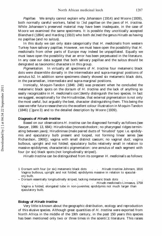
Papillae. We simply cannot explain why Johansson (1914) and Moore (1939),both normally careful workers, failed to � nd papillae on the jaws of H. troctina.While Johansson’s preserved material may have been inadequate, in the case ofMoore we examined the same specimens. It is possible they uncritically acceptedBlanchard (1894) and Harding (1910) who both de� ned the genus Hirudo as havingno papillae (and no sulcus).
In this study we can only state categorically that H. medicinalis from westernTurkey have salivary papillae. However, we must leave open the possibility that H.medicinalis from other parts of Europe may indeed be unpapillated. Equally wemust leave open the possibility that an error has been perpetuated in the literature.In any case our data suggest that both salivary papillae and the sulcus should bedenigrated as taxonomic characters in this group.
Pigmentation. In virtually all specimens of H. troctina four metameric blackdots were discernible dorsally in the intermediate and supra-marginal positions ofannulus b2. In addition some specimens clearly showed six metameric black dots,in the paramedian, intermediate and supra-marginal positions.
Ironically, Moquin-Tandon (1846: 248) was prescient when he concluded themetameric black spots on the dorsum of H. troctina and the lack of anything soeasily recognizable in H. medicinalis con� dently distinguish the two species. In factwe suggest, exceptionally for the Hirudinidae, that external pigmentation is not onlythe most useful, but arguably the best, character distinguishing them. This being thecase we refer future researchers to the excellent colour illustration in Moquin-Tandon(1846) (� gure 3) and to the detailed description by Moore (1939).
Diagnosis of Hirudo troctinaBased on our observations H. troctina can be diagnosed formally as follows (see
Sawyer, 1986: 13, 684): Hirudinidae (monostichodont ; no pharyngeal ridges termin-ating between jaws); Hirudininae (male paired ducts of ‘hirudoid’ type: i.e. epididy-mis and ejaculatory bulb present and looped, not forming linear series [seeRichardson, 1969]); vagina with small distinct caecum; no vaginal duct; vaginabulbous, upright and not folded; ejaculatory bulbs relatively small in relation tomassive epididymes; characteristic pigmentation: one annulus of each segment withfour (or six) black spots (not longitudinally striped).
Hirudo troctina can be distinguished from its congener H. medicinalis as follows:
1 Dorsum with four (or six) metameric black dots . . Hirudo troctina Johnson, 1816Vagina bulbous, upright and not folded; epididymis massive in relation to ejaculat-ory bulb.
– Dorsum essentially longitudinally striped, lacking metameric black dots . . . . .. . . . . . . . . . . . . . . Hirudo medicinalis Linnaeus, 1758Vagina a folded, elongated tube in non-juveniles; epididymis not much larger thanejaculatory bulb.
Biology of Hirudo troctinaVery little is known about the geographic distribution, ecology and reproduction
of this elusive species. Although great quantities of H. troctina were exported fromNorth Africa in the middle of the 19th century, in the past 150 years this specieshas been mentioned only two or three times in the scienti� c literature. This raises
Dow
nloa
ded
by [
] a
t 04:
34 2
2 Se
ptem
ber
2011

F. O. P. Hechtel and R. T. Sawyer1288
the question whether there has been a serious decline in the natural populations ofthis elusive leech, as has happened with H. medicinalis now recognized as endangered(Sawyer, 1981).
In order to stimulate research into the biology and taxonomy of H. troctina andrelated leeches of North Africa, the following practical key to known jawed leechesof the region should prove useful:
1 Segmentally repeating pigment pattern on dorsum . . Hirudo troctina Johnson, 1816Anus small; caudal sucker about two-thirds to three-quarters maximum body width;each jaw with about 65 (60–100) teeth.
– No segmentally repeating pigment pattern on dorsum . . . . . . . . . 2
2 Caudal sucker relatively small, about half maximum body width . . . . . . .. . . . . . . . . . . . . . Haemopis sanguisuga (Linnaeus, 1758)Anus prominent; each jaw with two rows of fewer than 20 teeth. . . . . . .
– Caudal sucker large, its diameter about equal to or larger than maximum body width. . . . . . . . . . . . . . . Limnatis nilotica (Savigny, 1822)Anus small; each jaw with about 40–50 teeth.
AcknowledgementsWe thank J.-L. Justine, Laboratoire de Biologie Parasitaire, Protistologie,
Helminthologie, Museum national d’Histoire naturelle, Paris, for furnishing pre-served specimens from Tunisia and Algeria; S. Halsey, Department of Zoology, TheNatural History Museum, London, for furnishing material from Morocco; P.J. Schembri, Department of Biology, University of Malta, Malta, and C. Savona-Ventura, Institute of Health Care, University of Malta, Gwardamangia, Malta, forinformation on the leeches of Malta and its leech trade with North Africa.
ReferencesBertherand, E. L., 1855, Medecine et hygiene des Arabes (Paris: G. Bailliere).Blanchard, R., 1894, Hirudinees de l’Italie continentale et insulaire, Bollettino dei Musei di
Zoologia ed Anatomia Comparata della R. Universita di Torino, No. 192, 9, 1–84.Blanchard, R., 1908, Hirudinees, in H. Gadeau de Kerville (ed.) Voyage Zoologique en
Khroumirie (Tunisie), V–VI 1906 (Paris), pp. 307–310.Derheims, J. L., 1825, Histoire Naturelle et Medicale des Sangsues (Paris).Ebrard, E., 1857, Nouvelle Monographie des Sangsues Medicinales (Paris: J. B. Bailliere et � ls).Gervais, P., 1835, Sur la patrie de la sangsue interrompue, Bulletin de la Societe d’Historie
Naturelle de France, No. 1 (original unobtainable, cited in Moquin-Tandon, 1846 andBlanchard, 1894).
Harding, W. A., 1910, A revision of the British leeches, Parasitology, 3, 130–201.Johansson, L., 1914, Ergebnisse einer von Prof. Franz Werner in Sommer 1910 mit
Unterstutzung aus dem Legate Wedl ausgefuhrten zoologischen Forschungsreise nachAlgerien, Sitzungsberichte der Akademie der Wissenschaften, Wien, 123, 837–852.
Johnson, J. R., 1816, A Treatise on the Medicinal Leech: Including its Medical and NaturalHistory, with a Description of its Anatomical Structure; also Remarks upon the Diseases,Preservation and Management of Leeches (London).
Letheby, H., 1844, Classi� cation and structure of the leech, Pharmaceutical Journal, London,4(6), 252–257.
Linnaeus, C., 1758, Systema Naturae, Lipsiae, 10th edn, Hirudinea, pp. 648–651.Moore, J. P., 1939, Leeches (Hirudinea) from the Atlas Mountains of Morocco, Annals and
Magazine of Natural History, London, Ser. 11, 3, 80–87.Moquin-Tandon, A., 1827, Monographie de la Famille des Hirudinees (Montpellier: Maison
de Commerce).
Dow
nloa
ded
by [
] a
t 04:
34 2
2 Se
ptem
ber
2011

North African medicinal leech 1289
Moquin-Tandon, A., 1846, Monographie de la Famille des Hirudinees, new edn (Paris: J.-B.Bailliere).
Ray, J., 1710, Historia Insectorium (London).Richardson, L. R., 1969, A contribution to the systematics of the hirudinid leeches, with
description of new families, genera and species, Acta Zoologica Academiae ScientiarumHungaricae, Budapest, 15 (1/2), 97–149.
Savigny, J. C., 1822, Systeme des Annelides (Paris).Sawyer, R. T., 1981, Why we need to save the medicinal leech, Oryx, 16, 165–168.Sawyer, R. T., 1986, Leech Biology and Behaviour (Oxford: Oxford University Press).Schembri, P. J., 1986, A note on non-marine leeches (Annelida: Hirudinea) from the Maltese
islands, The Central Mediterranean Naturalist, I (4), 81–83.
Dow
nloa
ded
by [
] a
t 04:
34 2
2 Se
ptem
ber
2011


