Embed Size (px)
Citation preview

Hen Egg White Lysozyme Permeabilizes Escherichia coli Outer andInner MembranesMelanie Derde,*,†,‡ Valerie Lechevalier,†,‡ Catherine Guerin-Dubiard,†,‡ Marie-Francoise Cochet,†,‡
Sophie Jan,†,‡ Florence Baron,†,‡ Michel Gautier,†,‡ Veronique Vie,# and Francoise Nau†,‡
†Agrocampus Ouest, UMR1253 Science et technologie du lait et de l’œuf, 65 rue de St-Brieuc, F-35042 Rennes, France‡INRA, UMR1253 Science et technologie du lait et de l’œuf, 65 rue de St-Brieuc, F-35042 Rennes, France#Universite de Rennes 1, Institut de physique de Rennes, UMR6251, CNRS, 263 Av. General Leclerc, F-35042 Rennes, France
ABSTRACT: Natural preservatives answer the consumer demand for long shelf life foods, synthetic molecules being perceivedas a health risk. Lysozyme is already used because of its muramidase activity against Gram-positive bacteria. It is also described asactive against some Gram-negative bacteria; membrane disruption would be involved, but the mechanism remains unknown. Inthis study, a spectrophotometric method using the mutant Escherichia coli ML-35p has been adapted to investigate membranedisruption by lysozyme for long durations. Lysozyme rapidly increases the permeability of the outer membrane of E. coli due tolarge size pore formation. A direct delayed activity of lysozyme against the inner membrane is also demonstrated, but withoutevidence of perforations.
KEYWORDS: lysozyme, antimicrobial activity, membrane permeability, Gram-negative bacteria
■ INTRODUCTION
Food additives, including preservatives, worry 66% of theEuropean consumers, and synthetic additives are perceived asmore dangerous than natural additives.1,2 On the other hand,consumers demand safer food products, with a long shelf life.Research for novel, natural food preservatives is thus stimulated,and biological resources are therefore widely screened. Specialattention is given to those molecules that have a wideantimicrobial spectrum. Peptides and proteins are consideredas potential candidates. One of the natural antimicrobial proteinswidely studied is hen egg white lysozyme (HEWL). HEWL iswell-known for its muramidase activity against Gram-positivebacteria. It is therefore used as a food additive (E1105) to controlGram-positive spoilers in winemaking and cheese refining.3−5
Several studies suggest that HEWL also acts against some Gram-negative bacteria; mechanisms such as perturbation of DNA orRNA synthesis and membrane permeabilization would beresponsible for lysozyme activity against these micro-organ-isms.3,6−8 The efficacy of lysozyme against Gram-negativebacteria can be increased by modifying the protein by proteolysisto obtain small active peptides,9−11 by fusion of chemicalmoieties,12−16 or by heat-denaturation.17−20
Membrane disruption is a major mechanism by whichantibacterial peptides and proteins act on both Gram-negativeand Gram-positive bacteria. The cationic and amphipathiccharacter of most of these peptides and proteins suggestselectrostatic and hydrophobic interactions with the bacterial cellwall. Such interactions could disturb the bacterial membrane,leading to bacterial cell death, or translocation of the peptide orprotein into the cytoplasm, where it interacts with intracellulartargets.21,22
Membrane permeabilization is an attractive hypothesis toexplain lysozyme activity against Gram-negative bacteria,considering the recent discovery of lysozyme inhibitors in the
periplasm of species such as E. coli.23,24 Gram-negative bacteriaare naturally protected against lysozyme by the outer membrane,a physical barrier preventing entrance into the cell of moleculesbigger than 650 Da.25 However, the presence of periplasmiclysozyme inhibitors in some Gram-negative bacteria suggeststhat lysozyme is able to cross their outer membrane. Yet, verylittle is known about the membrane activity of lysozyme.Especially, there is no experimental evidence of the capability oflysozyme to directly act, or not, on the inner membrane of Gram-negative bacteria.Different techniques have been used to detect bacterial
membrane permeabilization, such as detection of potassiumleakage, LPS monitoring, NPN uptake, DiSC3 uptake, or atomicforce microscopy (AFM).26−33 One of the most popular andsimple assays is a spectrophotometric method using Escherichiacoli ML-35p. This method was first described by Lehrer in 1988and was later optimized for smaller sample volumes by Epand in2010.32,33 It has been used to detect membrane activity ofantibacterial peptides such as cecropin A, melittin, ceragenins,Cg-BPI, and indolicidin and of antibacterial proteins such ashuman defensins and HEWL.32−35 E. coliML-35p is constitutivefor β-galactosidase expressed in the cytoplasm and produces a β-lactamase in the periplasm, which is encoded on a plasmid(pBR322). This bacterial strain also lacks lactose permease.32,33
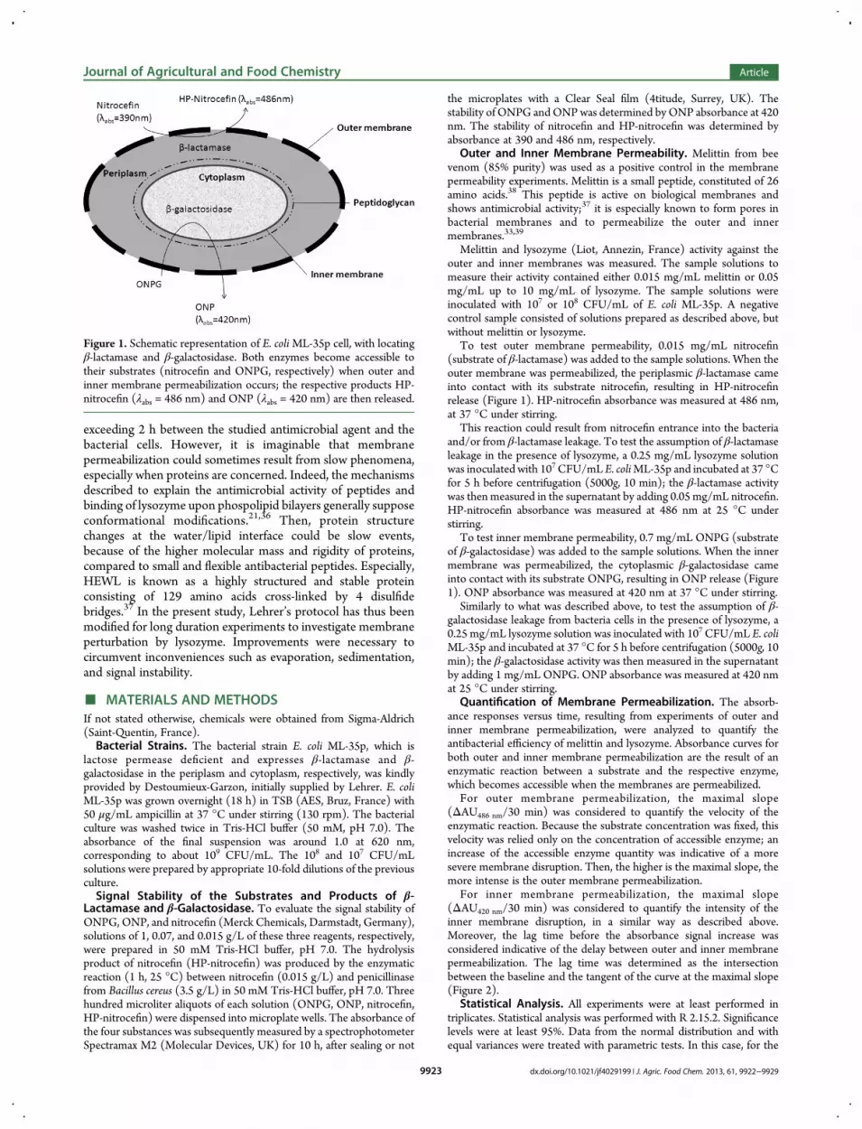
The measurement of β-lactamase and β-galactosidase activitiesthus enables the detection of outer and inner membranepermeabilization, respectively (Figure 1).In the literature, membrane permeability has been measured
using the E. coli ML-35p mutant with a contact time never
Received: July 3, 2013Revised: September 13, 2013Accepted: September 18, 2013Published: September 18, 2013
Article
pubs.acs.org/JAFC
© 2013 American Chemical Society 9922 dx.doi.org/10.1021/jf4029199 | J. Agric. Food Chem. 2013, 61, 9922−9929
exceeding 2 h between the studied antimicrobial agent and thebacterial cells. However, it is imaginable that membranepermeabilization could sometimes result from slow phenomena,especially when proteins are concerned. Indeed, the mechanismsdescribed to explain the antimicrobial activity of peptides andbinding of lysozyme upon phospolipid bilayers generally supposeconformational modifications.21,36 Then, protein structurechanges at the water/lipid interface could be slow events,because of the higher molecular mass and rigidity of proteins,compared to small and flexible antibacterial peptides. Especially,HEWL is known as a highly structured and stable proteinconsisting of 129 amino acids cross-linked by 4 disulfidebridges.37 In the present study, Lehrer’s protocol has thus beenmodified for long duration experiments to investigate membraneperturbation by lysozyme. Improvements were necessary tocircumvent inconveniences such as evaporation, sedimentation,and signal instability.
■ MATERIALS AND METHODSIf not stated otherwise, chemicals were obtained from Sigma-Aldrich(Saint-Quentin, France).Bacterial Strains. The bacterial strain E. coli ML-35p, which is
lactose permease deficient and expresses β-lactamase and β-galactosidase in the periplasm and cytoplasm, respectively, was kindlyprovided by Destoumieux-Garzon, initially supplied by Lehrer. E. coliML-35p was grown overnight (18 h) in TSB (AES, Bruz, France) with50 μg/mL ampicillin at 37 °C under stirring (130 rpm). The bacterialculture was washed twice in Tris-HCl buffer (50 mM, pH 7.0). Theabsorbance of the final suspension was around 1.0 at 620 nm,corresponding to about 109 CFU/mL. The 108 and 107 CFU/mLsolutions were prepared by appropriate 10-fold dilutions of the previousculture.Signal Stability of the Substrates and Products of β-
Lactamase and β-Galactosidase. To evaluate the signal stability ofONPG, ONP, and nitrocefin (Merck Chemicals, Darmstadt, Germany),solutions of 1, 0.07, and 0.015 g/L of these three reagents, respectively,were prepared in 50 mM Tris-HCl buffer, pH 7.0. The hydrolysisproduct of nitrocefin (HP-nitrocefin) was produced by the enzymaticreaction (1 h, 25 °C) between nitrocefin (0.015 g/L) and penicillinasefrom Bacillus cereus (3.5 g/L) in 50 mM Tris-HCl buffer, pH 7.0. Threehundred microliter aliquots of each solution (ONPG, ONP, nitrocefin,HP-nitrocefin) were dispensed into microplate wells. The absorbance ofthe four substances was subsequently measured by a spectrophotometerSpectramax M2 (Molecular Devices, UK) for 10 h, after sealing or not
the microplates with a Clear Seal film (4titude, Surrey, UK). Thestability of ONPG andONPwas determined byONP absorbance at 420nm. The stability of nitrocefin and HP-nitrocefin was determined byabsorbance at 390 and 486 nm, respectively.
Outer and Inner Membrane Permeability. Melittin from beevenom (85% purity) was used as a positive control in the membranepermeability experiments. Melittin is a small peptide, constituted of 26amino acids.38 This peptide is active on biological membranes andshows antimicrobial activity;37 it is especially known to form pores inbacterial membranes and to permeabilize the outer and innermembranes.33,39
Melittin and lysozyme (Liot, Annezin, France) activity against theouter and inner membranes was measured. The sample solutions tomeasure their activity contained either 0.015 mg/mL melittin or 0.05mg/mL up to 10 mg/mL of lysozyme. The sample solutions wereinoculated with 107 or 108 CFU/mL of E. coli ML-35p. A negativecontrol sample consisted of solutions prepared as described above, butwithout melittin or lysozyme.
To test outer membrane permeability, 0.015 mg/mL nitrocefin(substrate of β-lactamase) was added to the sample solutions. When theouter membrane was permeabilized, the periplasmic β-lactamase cameinto contact with its substrate nitrocefin, resulting in HP-nitrocefinrelease (Figure 1). HP-nitrocefin absorbance was measured at 486 nm,at 37 °C under stirring.
This reaction could result from nitrocefin entrance into the bacteriaand/or from β-lactamase leakage. To test the assumption of β-lactamaseleakage in the presence of lysozyme, a 0.25 mg/mL lysozyme solutionwas inoculated with 107 CFU/mL E. coliML-35p and incubated at 37 °Cfor 5 h before centrifugation (5000g, 10 min); the β-lactamase activitywas then measured in the supernatant by adding 0.05 mg/mL nitrocefin.HP-nitrocefin absorbance was measured at 486 nm at 25 °C understirring.
To test inner membrane permeability, 0.7 mg/mL ONPG (substrateof β-galactosidase) was added to the sample solutions. When the innermembrane was permeabilized, the cytoplasmic β-galactosidase cameinto contact with its substrate ONPG, resulting in ONP release (Figure1). ONP absorbance was measured at 420 nm at 37 °C under stirring.
Similarly to what was described above, to test the assumption of β-galactosidase leakage from bacteria cells in the presence of lysozyme, a0.25 mg/mL lysozyme solution was inoculated with 107 CFU/mL E. coliML-35p and incubated at 37 °C for 5 h before centrifugation (5000g, 10min); the β-galactosidase activity was then measured in the supernatantby adding 1 mg/mL ONPG. ONP absorbance was measured at 420 nmat 25 °C under stirring.
Quantification of Membrane Permeabilization. The absorb-ance responses versus time, resulting from experiments of outer andinner membrane permeabilization, were analyzed to quantify theantibacterial efficiency of melittin and lysozyme. Absorbance curves forboth outer and inner membrane permeabilization are the result of anenzymatic reaction between a substrate and the respective enzyme,which becomes accessible when the membranes are permeabilized.
For outer membrane permeabilization, the maximal slope(ΔAU486 nm/30 min) was considered to quantify the velocity of theenzymatic reaction. Because the substrate concentration was fixed, thisvelocity was relied only on the concentration of accessible enzyme; anincrease of the accessible enzyme quantity was indicative of a moresevere membrane disruption. Then, the higher is the maximal slope, themore intense is the outer membrane permeabilization.
For inner membrane permeabilization, the maximal slope(ΔAU420 nm/30 min) was considered to quantify the intensity of theinner membrane disruption, in a similar way as described above.Moreover, the lag time before the absorbance signal increase wasconsidered indicative of the delay between outer and inner membranepermeabilization. The lag time was determined as the intersectionbetween the baseline and the tangent of the curve at the maximal slope(Figure 2).
Statistical Analysis. All experiments were at least performed intriplicates. Statistical analysis was performed with R 2.15.2. Significancelevels were at least 95%. Data from the normal distribution and withequal variances were treated with parametric tests. In this case, for the
Figure 1. Schematic representation of E. coliML-35p cell, with locatingβ-lactamase and β-galactosidase. Both enzymes become accessible totheir substrates (nitrocefin and ONPG, respectively) when outer andinner membrane permeabilization occurs; the respective products HP-nitrocefin (λabs = 486 nm) and ONP (λabs = 420 nm) are then released.
Journal of Agricultural and Food Chemistry Article
dx.doi.org/10.1021/jf4029199 | J. Agric. Food Chem. 2013, 61, 9922−99299923

comparison of means the Student t test was used. Data from otherdistributions or with unequal variances were treated with nonparametrictests. In this case, for the comparison of means the Wilcoxon rank sumtest was used.
■ RESULTSPreliminary Protocol Improvements for Reliable
Measurements of Outer and Inner Membrane Disruptionfor Long Durations. Extension of Lehrer’s method to longdurations implies that substrates and products of β-lactamase andβ-galactosidase are time-stable. Nitrocefin and ONPG absor-bances were both stable at 37 °C for durations as long as 10 h(data not shown). On the contrary, the absorbances of HP-nitrocefin (Figure 3A) and ONP (Figure 3B) were not stable.
Sealing the microplate improved ONP stability from an 80%decrease to only 26% decrease of absorbance. Similarly, HP-nitrocefin absorbance decreased only 12% under sealedconditions compared to 28% under nonsealed conditions.To prove the relevance of sealing microplates, melittin has
been used as a reference antibacterial agent. When the outermembrane permeabilization was measured by melittin, themaximum absorbance was higher under sealed conditions,compared to nonsealed ones (Figure 4A). Moreover, a highermaximal slope was observed with a sealed microplate (Figure4C).When the inner membrane permeabilization was measured
(Figure 4B), the absorbance signal corresponding to ONP
release was dramatically different, depending on whether themicroplate was sealed or not. Especially, during the first 3 h, theslope of the absorbance curve was much higher under sealedconditions. Because of this initial discrepancy, and despite anequivalent maximal slope between 3 and 5 h, the maximumabsorbance was lower under nonsealed conditions (Figure 4B).With regard to the latter results, experiments with lysozyme
will be performed only with sealed microplates.Lysozyme Activity against Outer and Inner Mem-
branes of E. coli. Lehrer’s method was applied to test HEWLactivity, including the modifications as described above. Theresults exhibited that 0.25 mg/mL HEWL disturbed the outermembrane of E. coli because β-lactamase activity was detectableafter around 0.5 h of incubation, whereas no absorbance wasmeasured in the negative control, that is, without lysozyme(Figure 5A). Despite HP-nitrocefin content subsequentlyincreasing in the negative control, it remained much lowerthan in the presence of 0.25 mg/mL lysozyme, throughout the10 h experiment. Moreover, the supernatant of the bacterialsuspension treated with lysozyme contained β-lactamase activity,unlike the supernatant of the negative control (Figure 5C).During the first 2 h, a slight β-galactosidase activity was also
measured, but in a similar way in samples with and withoutlysozyme (Figure 5B). On the contrary, when the experimentwas extended to durations as long as 2.7 h and longer, β-galactosidase activity was muchmore extensive in the presence of0.25 mg/mL HEWL, compared to the negative control (Figure5B). However, the supernatant of the bacterial suspensiontreated with lysozyme did not contain β-galactosidase activity(data not shown).
Membrane Permeabilization Depending on LysozymeConcentration and E. coli Inoculum.When 107 CFU/mL E.coli was inoculated, β-lactamase activity, that is, outer membranepermeabilization, remained unchanged whatever the lysozymeconcentration was, from 0.05 to 10 mg/mL (maximal slopearound 0.0025 ΔAU486 nm/min; Figure 6A). On the contrary,when the inoculum was 108 CFU/mL, the intensity of outermembrane permeabilization increased when lysozyme concen-tration increased from 0.05 to 0.5 mg/mL; above 0.5 mg/mLHEWL, the intensity of outer membrane permeabilizationdecreased when the lysozyme concentration increased (Figure6A).For both inocula levels, β-galactosidase activity, that is, inner
membrane permeabilization, increased when lysozyme concen-tration increased (higher maximal slope; Figure 6B). Moreover,the higher inoculum showed systematically higher maximalslopes. Considering the lag time, when 107 CFU/mL wasinoculated, lag time strongly decreased when lysozymeconcentration increased (Figure 6C). On the contrary, with108 CFU/mL inoculum, the lag time first remained stablebetween 0.05 and 0.5 mg/mLHEWL and then slightly decreasedwhen lysozyme concentration increased over 0.5 mg/mLHEWL(Figure 6C). Lag time was systematically shorter with 108 CFU/mL inoculum compared to 107 CFU/mL.
■ DISCUSSIONMembrane permeabilization is a major mechanism involved inthe activity of many antimicrobial molecules, especiallyantimicrobial peptides and proteins.9,10 Most of the studiesaiming to highlight such bacterial membrane disruptionare limited to short-time experiments (<2 h). However, it isconceivable that membrane permeabilization could sometimesneed more time, especially when proteins are concerned. Indeed,
Figure 2. Quantification of inner membrane permeabilization usingmaximal slope and lag time.
Figure 3. Absorbance stability of the reaction products of β-lactamaseand β-galactosidase in nonsealed (full line) and sealed (dashed line)microplates. (A)HP-nitrocefin is detected by absorbance at 486 nm. (B)ONP is detected by absorbance at 420 nm. Standard deviation wascalculated from triplicates (gray dotted line). Results were not correctedwith a reference measurement, meaning that the absorbance valuesinclude the absorbance of the microplate and the buffer solution.
Journal of Agricultural and Food Chemistry Article
dx.doi.org/10.1021/jf4029199 | J. Agric. Food Chem. 2013, 61, 9922−99299924

mechanisms described to explain the antimicrobial activity ofpeptides and the interaction between lysozyme and lipid bilayersgenerally suppose conformational modifications.21,36 Then,
protein structure changes at the water/cell membrane interfacemight be slower than with peptides, because proteins aregenerally much more rigid molecules compared to peptides.
Figure 4. Permeabilization of the outer membrane (A) and inner membrane (B) of E. coliML-35p (107 CFU/mL) bymelittin (15 μg/mL) in nonsealed(full line) and sealed (dashed line) microplates, as evidenced by HP-nitrocefin and ONP release, respectively. Standard deviations were calculated fromnine replicates (gray dotted line). Results were corrected with a reference absorbance including the absorbance of the microplate and the buffer solution.(C) Comparison of the maximal slopes for outer membrane permeabilization with and without sealing. Different letters (a, b) indicate significantdifference (p < 0.05, Student t test).
Figure 5. Permeabilization of outer membrane (A) and inner membrane (B) of E. coliML-35p (107 CFU/mL) for 10 h, in the presence of 0.25 mg/mLHEWL (full line), and without lysozyme (dashed line). Standard deviations were calculated from triplicates (gray dotted line). Results were correctedwith a reference absorbance including the absorbance of the microplate and buffer solution. (C) Externalization of β-lactamase in the absence andpresence of 0.25 mg/mL of lysozyme measured by supernatant enzyme activity. Results stem from six replicates. Different letters (a,b) indicatesignificant difference (p < 0.01, Wilcoxon rank sum test).
Journal of Agricultural and Food Chemistry Article
dx.doi.org/10.1021/jf4029199 | J. Agric. Food Chem. 2013, 61, 9922−99299925

Especially, HEWL is known as a particularly rigid protein cross-linked by four disulfide bridges.37 The extension to longdurations of the traditional methods to investigate bacterialmembrane permeabilization by proteins is then a relevantchallenge. The popular and simple spectrophotometric methoddeveloped by Lehrer has here been selected for such anadaptation.32
Sealing Microplates Is an Efficient Way To Improve theReliability of Lehrer’s Method for Long Experiments. Toextend Lehrer’s method to durations longer than 2 h, thesubstrates (ONPG and nitrocefin) and the products (ONP andHP-nitrocefin) of both enzymatic reactions need to be stable at37 °C. This condition was not fulfilled for ONP, even for short-time experiments (Figure 3B), and to a lesser extent for HP-nitrocefin (Figure 3A). ONP, which results from ONPGhydrolysis by β-galactosidase, turns out to be especially unstablewhen nonsealed plates are used (Figure 3B). This is likely theresult from the high volatility of this compound at 37 °C, becausethe ONP signal decrease is largely limited with sealedmicroplates. In these conditions, only a slight decrease isobserved at the very beginning of the experiment (Figure 3B),probably due to the partial evaporation of ONP in the gas phase,between the liquid phase and the film, until the gas/liquidequilibrium was reached for this chemical compound. Thisobservation suggests that a minimal headspace between theliquid phase and the film should be preferred; however, it cannotbe reduced to zero, because of practical considerations such assealing difficulty and risk of cross-contamination betweenadjacent wells. Although much less significant than for ONP,the HP-nitrocefin signal also decreases throughout the 10 hexperiment when performed without sealing; this decrease issmaller when microplates are previously sealed (Figure 3A).Sealing microplates as proposed in this study appears then to bean easy and efficient way to improve the reliability of Lehrer’s
method when time extension up to 10 h is needed. Moreover,sealing avoids cross-contamination between wells, which canhappen because of microplate stirring.When the modified method (sealing microplates) was
performed to measure the melittin antibacterial activity againstE. coli, the results were significantly improved compared to theoriginal method: higher initial rates were measured for bothouter and inner membrane permeabilization (Figure 4). Thisindicates that the technical adjustments proposed in this studysolve the underestimation induced by the original protocol. Thisunderestimation is quite moderate for outer membranepermeabilization (Figure 4A), but a huge difference exists forthe measurement of the inner membrane permeabilization(Figure 4B). In the latter case, the use of sealed microplatesappears absolutely necessary, even for short-time experiments.Indeed, even in the very first moments of the test, because anextensive and quick disappearance of ONP occurs simulta-neously with ONP enzymatic release, the initial rate ofpermeabilization is underestimated by 80% when nonsealedmicroplates are used.
HEWL Disrupts Outer and Inner Membranes of E. coli.The method adjustments proposed above enabled theinvestigation of lysozyme membrane activity for durations aslong as 10 h. With such long experiments, the ability of HEWL topermeabilize both outer and inner membranes of E. coli wasdemonstrated. Indeed, both β-lactamase and β-galactosidaseactivities were detected when HEWL (0.25 mg/mL) was addedto an E. coli culture, as indicated by HP-nitrocefin and ONPrelease, respectively (Figure 5). The weak absorbance signalsobtained with the negative control likely result from thespontaneous lysis of bacteria that occurs during the 10 hexperiments. However, both β-lactamase and β-galactosidaseactivities were higher when HEWL was added, undoubtedlyproving the membrane permeabilization induced by lysozyme.
Figure 6. Membrane permeabilization as a function of lysozyme concentration for 107 CFU/mL (●) and 108 CFU/mL (◇) inocula: (A) outermembrane permeabilization as evidenced by β-lactamase activity; inner membrane permeabilization as evidenced by (B) β-galactosidase activity and(C) lag time between outer and inner membrane permeabilization. Standard deviations were calculated from triplicates.
Journal of Agricultural and Food Chemistry Article
dx.doi.org/10.1021/jf4029199 | J. Agric. Food Chem. 2013, 61, 9922−99299926

Outer membrane permeabilization has already been describedby Wild et al. and Pelligrini et al. for a similar HEWLconcentration.7,40 These authors observed the outer membranepermeabilization using electron microscopy and Lehrer’smembrane permeabilization assay, respectively. The originalLehrer method enabled this because outer membrane perme-abilization occurs after around 30 min, for 0.25 mg/mLlysozyme. However, these authors conclude that no innermembrane permeabilization occurs due to the direct action ofHEWL.The present study highlights that, when Lehrer’s method is
extended to long durations, HEWL induces inner membranepermeabilization, too, but this is only detectable after 2.7 h ofincubation with 0.25 mg/mL HEWL and 107 CFU/mL E. coliinoculum. It is then a slow phenomenon, compared to what isusually observed with antibacterial peptides. The delay necessaryfor the detection of the inner membrane permeabilization couldbe explained by the succession of hurdles that HEWL has to getover: passing through the outer membrane, peptidoglycanhydrolysis or diffusion through the peptidoglycan network,41
and finally disturbance of the inner membrane.To ensure that the inner membrane permeabilization is not the
result of cell lysis caused by peptidoglycan disintegration, thepresence of β-galactosidase was investigated in the supernatant ofthe E. coli cells (107 CFU/mL) treated with 0.25 mg/mLlysozyme (as explained under Outer and Inner MembranePermeability). β-Galactosidase would be present in the super-natant when peptidoglycan disintegration and thus cell lysisoccur.42 However, no β-galactosidase activity could be measuredin the supernatant, demonstrating that this enzyme was notleaking out of the cytoplasm; then, there was no cell lysis, and theβ-galactosidase activity detected when E. coli cells are in thepresence of lysozyme resulted from the diffusion of ONPG intothe cell. This confirms that HEWL directly acts on the innermembrane of E. coli, modifying its permeability, and independentof the lysozyme enzymatic activity on peptidoglycan.It is noticeable that, in opposition to β-galactosidase, β-
lactamase activity was measured in the supernatant of E. coli cellstreated with 0.25 mg/mL lysozyme (Figure 5C). This proves thatHEWL disrupts the outer membrane in such a way that thisenzyme of 28.9 kDa can leak out of the periplasm. Large sizepores inside the outer membrane are thus induced by HEWL.HEWL Acts by a Two-Step Process: Saturation of the
Outer Membrane before Entrance into the Cell andPermeabilization of the InnerMembrane.The extent of theouter membrane permeabilization, quantified by the β-lactamaseactivity (Figure 6A), was unchanged between 0.05 and 10 mg/mL HEWL with inoculation of 107 CFU/mL. On the contrary,the outer membrane permeabilization increased when HEWLconcentration increased from 0.05 to 0.5 mg/mL withinoculation of 108 CFU/mL. This suggests that a critical ratiofor outer membrane saturation should be 0.05 mg/mLHEWL:107 CFU/mL E. coli. Indeed, β-lactamase activity didnot increase when >0.05 mg/mL lysozyme was added to 107
CFU/mL. Moreover, the maximum β-lactamase activity when108 CFU/mL was inoculated was reached at a ratio 0.5 mg/mLlysozyme:108 CFU/mL, that is, the same ratio as 0.05 mg/mLHEWL:107 CFU/mL.As far as the inner membrane is concerned, a dose−response
effect occurred for 0.05−10 mg/mL HEWL at both E. coliinocula (107 and 108 CFU/mL). The extent of the innermembrane permeabilization increased when HEWL concen-tration increased (Figure 6B). The maximal slopes obtained with
108 CFU/mL inoculum were systematically higher than thoseobtained with 107 CFU/mL. This is consistent with the higherquantity of β-galactosidase potentially accessible to ONPG whenthe bacterial inoculum was higher. Simultaneously, the lag timedecreased when the HEWL concentration increased, for both E.coli inocula (Figure 6C). Because the lag time is the delay neededfor the release of detectable quantities of ONP, a lag timedecrease indicates a faster increase of ONP concentration,related to a higher β-galactosidase activity. Therefore, the lagtime is consistently shorter with 108 CFU/mL E. coli comparedto 107 CFU/mL.When the inoculum was 107 CFU/mL E. coli, the lag time
regularly and strongly decreased when HEWL increased from0.05 to 10 mg/mL. This suggests that the higher the HEWLconcentration in the bulk, the higher the quantity of HEWLreaching the inner membrane. The lag time decrease is thenconsistent with the assumption of the outer membranesaturation with HEWL concentration of 0.05 mg/mL or higherand 107 CFU/mL E. coli. Indeed, because of such a saturation,each additional HEWLmolecule added in the bulk remains “free”(not entrapped into the outer membrane), able to cross over thedisrupted outer membrane and to reach the inner membrane.When 108 CFU/mL E. coli was inoculated, the lag time was
constant between 0.05 and 0.5 mg/mL HEWL. Because theouter membrane would not be saturated with HEWL moleculesin these conditions, as suggested above, each additional HEWLmolecule added in the bulk would then be essentially entrappedinside the outer membrane and then unavailable for deeperpenetration into the bacteria cell. On the contrary, with HEWLconcentrations >0.5 mg/mL, meaning when outer membranesaturation is achieved, the lag time decreased when HEWLconcentration increased, similarly to what was observed when107 CFU/mL E. coli was inoculated; in these conditions, eachadditional HEWL molecule remains “free” and able to enter intothe cell. However, even with the highest lysozyme concentration,that is, 10 mg/mL, the lag time remained >50 min; this could bethe minimal delay for lysozyme entrance into the cell andinteraction with the inner membrane.
Outer Membrane Permeabilization Is Reduced WhenE. coli Inoculum and HEWL Concentration Are Simulta-neously High. At high inoculum levels, quorum sensing canplay a major role in bacterial resistance against antimicrobialagents.43 Quorum sensing is a cell-to-cell communicationbetween bacteria by excretion of signal molecules, which canbe detected by other bacteria of the same or other species.44 In E.coli K12, AI-2 is one of those signal molecules that up-regulatesseveral genes related to the outer membrane such as rfaY; rfaYcontrols the LPS-core biosynthesis. Stress induction of AI-2 hasbeen demonstrated due to the addition of glucose, Fe3+, NaCl,and dithiothreitol.45,46 Thus, quorum sensing can be a stress-induced phenomenon.In the present study, it is noticeable that E. coli outer
membrane permeabilization decreased when HEWL concen-tration exceeds 1 mg/mL and when 108 CFU/mL wasinoculated, but this was not observed when inoculum was 107
CFU/mL (Figure 6A). The assumption of a lysozyme stress (≥1mg/mL) could be proposed. This stress could activate quorumsensing between bacterial cells, in a dose-dependent manner. Inthese conditions, that is, high inoculum and high HEWLconcentration, the outer membrane permeabilization of somebacterial cells may induce the expression of signal molecules,which could activate defense mechanisms by the sister cells.These defense mechanisms could include changes in the outer
Journal of Agricultural and Food Chemistry Article
dx.doi.org/10.1021/jf4029199 | J. Agric. Food Chem. 2013, 61, 9922−99299927

membrane composition, thus decreasing permeabilization bylysozyme. However, more investigations are needed to provesuch hypothetical mechanisms.This study then demonstrated that HEWL is able to
permeabilize the outer and inner membranes of E. coli. Asequential event is proposed (Figure 7): first, entrapping ofHEWL molecules inside the outer membrane, inducing itsdisruption with large size pore creation; then, transfer of “free”HEWL into the cell, to the inner membrane having increasedpermeability, but without massive cytoplasm leakage. Whereasthe first step is quite rapid, the second one is a much longerphenomenon, depending on the quantity of “free” HEWL andthen depending on the initial HEWL concentration in the bulk.Experiments are in progress to investigate the interactionsbetween HEWL and E. coli membranes.
■ AUTHOR INFORMATIONCorresponding Author*(M.D.) Mailing address: Agrocampus Ouest, UMR1253 STLO65, rue de St-Brieuc, 35042 Rennes cedex, France. Phone: +33/2.23.48.55.74. E-mail: [email protected].
FundingThis research was funded by Conseil Regional de Bretagne.
NotesThe authors declare no competing financial interest.
■ ACKNOWLEDGMENTSWe thank Delphine Destoumieux-Garzon for kindly providingthe E. coli ML-35p strain.
■ ABBREVIATIONS USEDHEWL, hen egg white lysozyme; LPS, lipopolysaccharide; NPN,1-N-phenylnaphthylamine; DiSC3, 3,3-dipropylthiadicarbocya-
nine iodide; ONPG, o-nitrophenylgalactoside; ONP, o-nitro-phenol; HP-nitrocefin, hydrolysis product of nitrocefin
■ REFERENCES(1) TNS Opinion and Social. Special Eurobarometer 354: Food-related risks, 2010.(2) Bruhn, C. M. Consumer attitudes toward food additives. In FoodAdditives, 2; Branen, A. L., Davidson, P. M., Salminen, S., Thorngate, J.H., Eds.; Dekker: New York, 2002; pp 111−119.(3) Masschalck, B.; Michiels, C. W. Antimicrobial properties oflysozyme in relation to foodborne vegetative bacteria. Crit. Rev.Microbiol. 2003, 29, 191−214.(4) Sonni, F.; Chinnici, F.; Natali, N.; Riponi, C. Pre-fermentativereplacement of sulphur dioxide by lysozyme and oenological tannins:effect on the formation and evolution of volatile compounds during thebottle storage of white wines. Food Chem. 2011, 129, 1193−1200.(5) Carini, S.; Mucchetti, G.; Neviani, E. Lysozyme: activity againstClostridia and use in cheese production − a review.Microbiol., Aliments,Nutr. 1985, 3, 299−320.(6) Pellegrini, A.; Thomas, U.; Vonfellenberg, R.; Wild, P. Bactericidalactivities of lysozyme and aprotinin against Gram-negative and Gram-positive bacteria related to their basic character. J. Appl. Bacteriol. 1992,72, 180−187.(7) Pellegrini, A.; Thomas, U.; Wild, P.; Schraner, E.; von Fellenberg,R. Effect of lysozyme or modified lysozyme fragments on DNA andRNA synthesis and membrane permeability of Escherichia coli.Microbiol.Res. 2000, 155, 69−77.(8) Laible, N. J.; Germaine, G. R. Bactericidal activity of humanlysozyme, muramidase-inactive lysozyme, and cationic polypeptidesagainst Streptococcus-sanguis and Streptococcus-faecalis inhibition bychitin oligosaccharides. Infect. Immun. 1985, 48, 720−728.(9) Mine, Y.; Ma, F.; Lauriau, S. Antimicrobial peptides released byenzymatic hydrolysis of hen egg white lysozyme. J. Agric. Food Chem.2004, 52, 1088−1094.(10) Ibrahim, H. R.; Thomas, U.; Pelligrini, A. A helix-loop-helixpeptide at the upper lip of the active site cleft of lysozyme confers potent
Figure 7.Hypothetical sequential events explaining the action of HEWL on outer and inner membranes of E. coli. Lys, lysozyme; β-Lact, β-lactamase; β-Gal, β-galactosidase; Nit, nitrocefin, and HP-Nit, HP-nitrocefin, substrate and product of β-lactamase, respectively; ONPG and ONP, substrate andproduct of β-galactosidase, respectively.
Journal of Agricultural and Food Chemistry Article
dx.doi.org/10.1021/jf4029199 | J. Agric. Food Chem. 2013, 61, 9922−99299928

antimicrobial activity with membrane permeabilization action. J. Biol.Chem. 2001, 47, 43767−43774.(11) Ibrahim, H. R.; Imazato, K.; Ono, H. Human lysozyme possessesnovel antimicrobial peptides within its N-terminal domain that targetbacterial respiration. J. Agri. Food Chem. 2011, 59, 10336−10345.(12) Ibrahim, H. R.; Kato, A.; Kobayashi, K. Antimicrobial effects oflysozyme against Gram-negative bacteria due to covalent binding ofpalmitic acid. J. Agri. Food Chem. 1991, 39, 2077−2082.(13) Ibrahim, H. R.; Hatta, H.; Fujiki, M.; Kim, M.; Yamamoto, T.Enhanced antimcirobial action of lysozyme against Gram-negative andGram-positive bacteria due to modification with perillaldehyde. J. Agric.Food Chem. 1994, 42, 1813−1817.(14) Ibrahim, H. R.; Kobayashi, K.; Kato, A. Length of hydrocarbonchain and antimicrobial action to Gram-negative bacteria of fattyacylated lysozyme. J. Agric. Food Chem. 1993, 41, 1164−1168.(15) Nakamura, S.; Kato, A.; Kobayashi, K. Bifunctional lysozymegalactomannan conjugate having excellent emulsifying properties andbactericidal effect. J. Agric. Food Chem. 1992, 40, 735−739.(16) Nakamura, S.; Kato, A.; Kobayashi, K. New antimicrobialcharacteristics of lysozyme dextran conjugate. J. Agric. Food Chem. 1991,39, 647−650.(17) Ibrahim, H. R. On the novel catalytically-independentantimicrobial function of hen egg-white lysozyme: a conformation-dependent activity. Nahrung 1998, 42, 187−193.(18) Ibrahim, H. R.; Higashiguchi, S.; Koketsu, M.; Juneja, L. R.; Kim,M.; Yamamoto, T.; Sugimoto, Y.; Aoki, T. Partially unfolded lysozyme atneutral pH agglutinates and kills Gram-negative and Gram-positivebacteria through membrane damage mechanism. J. Agric. Food Chem.1996, 44, 3799−3806.(19) Ibrahim, H. R.; Higashiguchi, S.; Juneja, L. R.; Kim, M.;Yamamoto, T. A structural phase of heat-denatured lysozyme with novelantimicrobial action. J. Agric. Food Chem. 1996, 44, 1416−1423.(20) Ibrahim, H. R.; Higashiguchi, S.; Sugimoto, Y.; Aoki, T. Role ofdivalent cations in the novel bactericidal activity of the partially unfoldedlysozyme. J. Agric. Food Chem. 1997, 45, 89−94.(21) Nguyen, L. T.; Haney, E. F.; Vogel, H. J. The expanding scope ofantimicrobial peptide structures and their modes of action. TrendsBiotechnol. 2011, 29, 464−472.(22) Jenssen, H.; Hamill, P.; Hancock, R. E. Peptide antimicrobialagents. Clin. Microbiol. Rev. 2006, 19, 491−511.(23) Deckers, D.; Masschalck, B.; Aertsen, A.; Callewaert, L.; VanTiggelen, C. G. M.; Atanassova, M.; Michiels, C. W. Periplasmiclysozyme inhibitor contributes to lysozyme resistance in Escherichia coli.Cell Mol. Life Sci. 2004, 61, 1229−1237.(24) Callewaert, L.; Aertsen, A.; Deckers, D.; Vanoirbeek, K. G.;Vanderkelen, L.; Van Herreweghe, J. M.; Masschalck, B.; Nakimbugwe,D.; Robben, J.; Michiels, C. W. A new family of lysozyme inhibitorscontributing to lysozyme tolerance in Gram-negative bacteria. PloSPathog. 2008, 4, e1000019.(25) Nikaido, H. Molecular basis of bacterial outer membranepermeability revisited. Microbiol. Mol. Biol. Rev. 2003, 67, 593−656.(26) Alakomi, H.; Paananen, A.; Suihko, M.; Helander, I.; Saarela, M.Weakening effect of cell permeabilizers on Gram-negative bacteriacausing biodeterioration.Appl. Environ. Microbiol. 2006, 72, 4695−4703.(27) Vaara, M. Increased outer-membrane resistance to ethyl-enediaminetetraacetate and cations in novel lipid A mutants. J. Bacteriol.1981, 148, 426−434.(28) Orlov, D. S.; Nguyen, T.; Lehrer, R. I. Potassium release, a usefultool for studying antimicrobial peptides. J. Microbiol. Methods 2002, 49,325−328.(29) Silvestro, L.; Weiser, J. N.; Axelsen, P. H. Antibacterial andantimembrane activities of cecropin A in Escherichia coli. Antimicrob.Agents Chemother. 2000, 44, 602−607.(30) Wu, M.; Maier, E.; Benz, R.; Hancock, R. E. Mechanism ofinteraction of different classes of cationic antimicrobial peptides withplanar bilayers and with the cytoplasmic membrane of Escherichia coli.Biochemistry 1999, 38, 7235−7242.(31) Meincken, A.; Holroyd, D. L.; Rautenbach, M. Atomic forcemicroscopy study of the effect of antimicrobial peptides on the cell
envelope of Escherichia coli. Antimicrob. Agents Chemother. 2005, 49,4085−4092.(32) Lehrer, R. I.; Barton, A.; Ganz, T. Concurrent assessment of innerand outer-membrane permeabilization and bacteriolysis in Escherichiacoli by multiple-wavelength spectrophotometry. J. Immunol. Methods1988, 108, 153−158.(33) Epand, R. F.; Pollard, J. E.; Wright, J. O.; Savage, P. B.; Epand, R.M. Depolarization, bacterial membrane composition, and theantimicrobial action of ceragenins. Antimicrob. Agents Chemother.2010, 54, 3708−3713.(34) Gonzalez, M.; Gueguen, Y.; Destoumieux-Garzon, D.;Romestand, B.; Fievet, J.; Pugniere, M.; Roquet, F.; Escoubas, J. M.;Vandenbulcke, F.; Levy, O.; Saune, L.; Bulet, P.; Bachere, E. Evidence ofa bactericidal permeability increasing protein in an invertebrate, theCrassostrea gigas Cg-BPI. Proc. Natl. Acad. Sci. U.S.A. 2007, 104, 17759−17764.(35) Turner, J.; Cho, Y.; Dinh, N. N.; Waring, A. J.; Lehrer, R. I.Activities of LL-37, a cathelin-associated antimicrobial peptide of humanneutrophils. Antimicrob. Agents Chemother. 1998, 42, 2206−2214.(36) Gorbenko, G. P.; Ioffe, V. M.; Kinnunen, P. K. Binding oflysozyme to phospholipid bilayers: evidence for protein aggregationupon membrane association. Biophys. J. 2007, 93, 140−153.(37) Canfield, R. E.; Liu, A. K. The disulfide bonds of egg whitelysozyme (muramidase). J. Biol. Chem. 1965, 240, 1997−2002.(38) Terwilliger, T. C.; Eisenberg, D. The structure of melittin. II.Interpretation of the structure. J. Biol. Chem. 1982, 257, 6016−6022.(39) Lee, M. T.; Sun, T. L.; Hung, W. C.; Huang, H. W. Process ofinducing pores in membranes by melittin. Proc. Natl. Acad. Sci. U.S.A.2013, DOI: 10.1073/pnas.1307010110.(40) Wild, P.; Gabrieli, A.; Schraner, E. M.; Pellegrini, A.; Thomas, U.;Frederik, P. M.; Stuart, M. C. A.; Vonfellenberg, R. Reevaluation of theeffect of lysoyzme on Escherichia coli employing ultrarapid freezingfollowed by cryoelectronmicroscopy or freeze substitution.Microsc. Res.Tech. 1997, 39 (3), 297−304.(41) Vollmer, W.; Bertsche, U. Murein (peptidoglycan) structure,architecture and biosynthesis in Escherichia coli. Biochim. Biophys. Acta2008, 1778, 1714−1734.(42) Silhavy, T. J; Kahne, D.; Walker, S. The bacterial cell envelope.Cold Spring Harbor Perspect. Biol. 2010, 2, a000414.(43) Mah, T. F.; O’Toole, G. A. Mechanisms of biofilm resistance toantimicrobial agents. Trends Microbiol. 2001, 9, 34−39.(44) Waters, C. M.; Bassler, B. L. Quorum sensing: cell-to-cellcommunication in bacteria. Annu. Rev. Cell Dev. Biol. 2005, 21, 319−346.(45) DeLisa, M. P.; Wu, C. F.; Wang, L.; Valdes, J. J.; Bentley, W. E.DNA microarray-based identification of genes controlled by auto-inducer 2-stimulated quorum sensing in Escherichia coli. J. Bacteriol.2001, 183, 5239−5247.(46) DeLisa, M. P.; Valdes, J. J.; Bentley, W. E. Mapping stress-inducedchanges in autoinducer AI-2 production in chemostat-cultivatedEscherichia coli K-12. J. Bacteriol. 2001, 183, 2918−2928.
Journal of Agricultural and Food Chemistry Article
dx.doi.org/10.1021/jf4029199 | J. Agric. Food Chem. 2013, 61, 9922−99299929


