Embed Size (px)
Citation preview

pharmaceuticals
Article
Herbal-Based Formulation Containing Eurycoma longifolia andLabisia pumila Aqueous Extracts: Safe for Consumption?
Bee Ping Teh 1,* , Norzahirah Ahmad 1 , Elda Nurafnie Ibnu Rasid 1, Nor Azlina Zolkifli 1,Umi Rubiah Sastu@Zakaria 1, Norliyana Mohamed Yusoff 1, Azlina Zulkapli 2, Norfarahana Japri 1,June Chelyn Lee 1 and Hussin Muhammad 1
�����������������
Citation: Teh, B.P.; Ahmad, N.; Ibnu
Rasid, E.N.; Zolkifli, N.A.;
Sastu@Zakaria, U.R.; Mohamed
Yusoff, N.; Zulkapli, A.; Japri, N.; Lee,
J.C.; Muhammad, H. Herbal-Based
Formulation Containing Eurycoma
longifolia and Labisia pumila Aqueous
Extracts: Safe for Consumption?.
Pharmaceuticals 2021, 14, 142. https://
doi.org/ 10.3390/ph14020142
Received: 28 November 2020
Accepted: 17 December 2020
Published: 10 February 2021
Publisher’s Note: MDPI stays neu-
tral with regard to jurisdictional clai-
ms in published maps and institutio-
nal affiliations.
Copyright: © 2021 by the authors. Li-
censee MDPI, Basel, Switzerland.
This article is an open access article
distributed under the terms and con-
ditions of the Creative Commons At-
tribution (CC BY) license (https://
creativecommons.org/licenses/by/
4.0/).
1 Herbal Medicine Research Centre, Institute for Medical Research, National Institutes of Health,Ministry of Health Malaysia, Shah Alam 40170, Selangor Darul Ehsan, Malaysia;[email protected] (N.A.); [email protected] (E.N.I.R.); [email protected] (N.A.Z.);[email protected] (U.R.S.); [email protected] (N.M.Y.); [email protected] (N.J.);[email protected] (J.C.L.); [email protected] (H.M.)
2 Medical Resource Research Centre, Institute for Medical Research, Jalan Pahang,Kuala Lumpur 50588, Wilayah Persekutuan Kuala Lumpur, Malaysia; [email protected]
* Correspondence: [email protected]; Tel.: +60-33362-7961
Abstract: A combined polyherbal formulation containing tongkat ali (Eurycoma longifolia) and kacipfatimah (Labisia pumila) aqueous extracts was evaluated for its safety aspect. A repeated dose 28-daytoxicity study using Wistar rats was conducted where the polyherbal formulation was administeredat doses 125, 500 and 2000 mg/kg body weight to male and female treatment groups daily via oralgavage, with rats receiving only water as the control group. In-life parameters measured includemonitoring of food and water consumption and clinical and functional observations. On day 29,blood was collected for haematological and biochemical analysis. The rats were necropsied andthe organs were collected for histopathological examination. This study showed that the combinedformulation did not induce any significant toxicity effect at any dose level in terms of morbidity,mortality, behaviour, functional observation, body weight, food and water consumption, wholeblood haematology and serum biochemistry. However, there were some microscopic changes in thehistopathological examinations of some organs given 2000 mg/kg body weight, which may suggestan early response to the polyherbal formulation. From this study, the no observed adverse effect levelis estimated to be more than 500 mg/kg body weight but not exceeding 2000 mg/kg body weight.The observed effects at the highest dose indicate the need for further study of longer dosing duration.
Keywords: Eurycoma longifolia; Labisia pumila; tongkat ali; kacip fatimah; toxicity
1. Introduction
Traditionally, medicinal plants have been used in disease prevention and treatment formany generations and their potential pharmaceutical values have easily drawn worldwideattention. At the global stage, the World Health Organization (WHO) has launched theWHO Traditional Medicine Strategy 2014–2023 to reinforce the use of medicinal plantsin healthcare services and management [1]. Consumers at present are conscious aboutgrowing price [2] and frequent adverse events often associated with conventional drugs [3].Consequently, the trend towards maintaining health and wellbeing using medicinal plantsas an alternative treatment is expanding. However, some medicinal plants have been re-ported to trigger allergic reactions [4,5], modify bioavailability of conventional drugs [6–8]and cause organ toxicity such as hepatotoxicity, nephrotoxicity and cardiotoxicity [9–11].Therefore, the consumption of medicinal plants merely based on traditional practices thatlack valid scientific evidence may overrule their benefits due to these possible undesirablehealth effects. Evidence-based safety studies investigating medicinal plants and the prod-ucts thereof are beneficial to both the scientific community for pharmaceutical development,as well as to the consumers in seeking alternative plant-based therapies.
Pharmaceuticals 2021, 14, 142. https://doi.org/10.3390/ph14020142 https://www.mdpi.com/journal/pharmaceuticals
Pharmaceuticals 2021, 14, 142 2 of 29
Root aqueous extracts of Eurycoma longifolia Jack (family Simaroubaceae, commonlyknown as tongkat ali) and roots or whole plant aqueous extracts of Labisia pumila (Blume)Fern-Vill (family Primulaceae, kacip fatimah), which are native plants of Southeast Asia,are two popular choices of herbs consumed in this region [12]. Tongkat ali roots have beentraditionally used to improve various health conditions such as fever, boils, wounds, ulcer,infertility, bleeding gums, aches, dysentery, glandular swelling, edema and as an afterbirthmedication and tonic [13,14]. The whole plant of kacip fatimah is used but mainly theroots are used traditionally to aid in the labour process of contraction and induction, helpregain body strength after giving birth and relieve other health problems such as flatulence,dysentery, dysmenorrhoea and gonorrhoea [15,16]. The scientific investigations into theseclaims looked into different extraction processes of these main plant parts and largelyexplored tongkat ali’s effect on male fertility [17–24] and its antimalarial action [20,25–29],while kacip fatimah has been tested for its potential in women’s reproductive health [30–35]and, to some extent, their cytotoxic [35,36], antimicrobial [37,38], anti-inflammatory [30,39],antioxidant [39,40] and anti-osteoporosis [41–44] capacities. Improvements in male sexualhealth is evidenced by improvements in seminal and fertility parameters, as well asincreased testosterone levels in in vivo models and clinical trials, largely attributed toits quassinoid content [45–47]. The phytoestrogenic potency of kacip fatimah shown bythe suppression of proinflammatory cytokines (responsible for osteoporosis) combinedwith its high scavenging properties (for reducing oxidative stress), regulating hormonalchanges and providing relief of post-menopausal symptoms [30,48]. Although there is noknowledge of both herbs used in combination traditionally, the use of these two herbs incombination is of scientific interest as they show potential health benefits in different organsystems.
The idea of using combinatorial herbal formulations is an age-old method in traditionalmedicines with the aim to boost the effects of the combined herbs in a synergistic man-ner [49,50]. However, along with the benefits of these polyherbal formulations, some harm-ful effects may also be exacerbated through a combined formulation [51]. Both the aqueousextracts of tongkat ali and kacip fatimah evaluated independently showed a high toler-ance value of up to 5000 mg/kg body weight in rats [52,53]. However, Singh et al. (2009)reported the no observed adverse effect level (NOAEL) for their kacip fatimah extract to be50 mg/kg [54].
The use of an individual NOAEL value as a step towards determining the safe startingdose in humans must be done with caution in a combination formulation [55,56]. TheNOAEL value accounting for the combined formula and the effect of repeated exposurevia safety testing using the specific formulation that accounts for all the compounds mustbe established. Some safety information of tongkat ali and kacip fatimah as individualherbal plants in their aqueous extracts for both plants, as well as methanolic extract andpowdered form for tongkat ali, which were tested in the rat model and clinically (fortongkat ali), are available [57–61], however, the safety data for the combination of thesetwo medicinal plants in any animal model have not been established. Previously, anacute oral toxicity study for the combined extracts was conducted in our facility, wherethe NOAEL value was determined to be more than 2000 mg/kg (unpublished work).Therefore, it is crucial to identify any health risks associated with the repeated use ofthis combined herbal formulation. In this study, the potential toxicity of a polyherbalformulation termed the P.SLP formulation, containing aqueous extracts from E. longifolia(roots) and L. pumila (whole plant), was evaluated in both genders of Wistar rats followingdaily oral administration for 28 days.
2. Results2.1. Chemical Identification of Test Item (P.SLP Formulation)
Chromatographic identification analysis is commonly used for the quality assessmentand species authentication of medicinal plants, including herbal products such as healthsupplements. In this present study, high-performance thin layer chromatography (HPTLC)

Pharmaceuticals 2021, 14, 142 3 of 29
and liquid chromatography–mass spectrometry (LC-MS) methods were used to authen-ticate the P.SLP formulation containing aqueous extracts from E. longifolia and L. pumila.Eurycomanone and gallic acid were used as chemical markers to identify E. longifolia and L.pumila-based formulations. A mixture of chloroform:methanol (9:1 v/v) (as mobile phase 1(MP1)) were used to detect eurycomanone, which is detected at a retention factor (Rf) of0.13 under ultraviolet (UV) 254 nm before derivatisation and under UV 366 nm (green zone)and under white light (violet zone) after derivatisation with 10% sulphuric acid solution.The MP 2, a mixture of ethyl acetate:formic acid (3:1 v/v) was used to detect both gallicacid at Rf of 0.81 and eurycomanone at Rf of 0.53 under UV 254 nm before derivatisation.The markers were also observed after derivatisation with 10% sulphuric acid under UV366 nm (gallic acid at Rf of 0.81 (blue zone) and eurycomanone at Rf of 0.53 (violet zone))and under white light (gallic acid at Rf of 0.81 (violet zone) and eurycomanone at Rf of 0.53(violet zone)) (Figure 1).
Pharmaceuticals 2021, 14, x FOR PEER REVIEW 3 of 28
2. Results 2.1. Chemical Identification of Test Item (P.SLP Formulation)
Chromatographic identification analysis is commonly used for the quality assess-ment and species authentication of medicinal plants, including herbal products such as health supplements. In this present study, high-performance thin layer chromatography (HPTLC) and liquid chromatography–mass spectrometry (LC-MS) methods were used to authenticate the P.SLP formulation containing aqueous extracts from E. longifolia and L. pumila. Eurycomanone and gallic acid were used as chemical markers to identify E. longi-folia and L. pumila-based formulations. A mixture of chloroform:methanol (9:1 v/v) (as mo-bile phase 1 (MP1)) were used to detect eurycomanone, which is detected at a retention factor (Rf) of 0.13 under ultraviolet (UV) 254 nm before derivatisation and under UV 366 nm (green zone) and under white light (violet zone) after derivatisation with 10% sul-phuric acid solution. The MP 2, a mixture of ethyl acetate:formic acid (3:1 v/v) was used to detect both gallic acid at Rf of 0.81 and eurycomanone at Rf of 0.53 under UV 254 nm before derivatisation. The markers were also observed after derivatisation with 10% sul-phuric acid under UV 366 nm (gallic acid at Rf of 0.81 (blue zone) and eurycomanone at Rf of 0.53 (violet zone)) and under white light (gallic acid at Rf of 0.81 (violet zone) and eurycomanone at Rf of 0.53 (violet zone)) (Figure 1).
Figure 1. High-performance thin layer chromatography (HPTLC) profiles of gallic acid (GA), eu-rycomanone (EU) and methanol solution of P.SLP formulation (TI) observed under 254 nm (before derivatisation), 366 nm and visible light after derivatisation with 10% sulphuric acid solution.
Figure 1. High-performance thin layer chromatography (HPTLC) profiles of gallic acid (GA), eu-rycomanone (EU) and methanol solution of P.SLP formulation (TI) observed under 254 nm (beforederivatisation), 366 nm and visible light after derivatisation with 10% sulphuric acid solution.
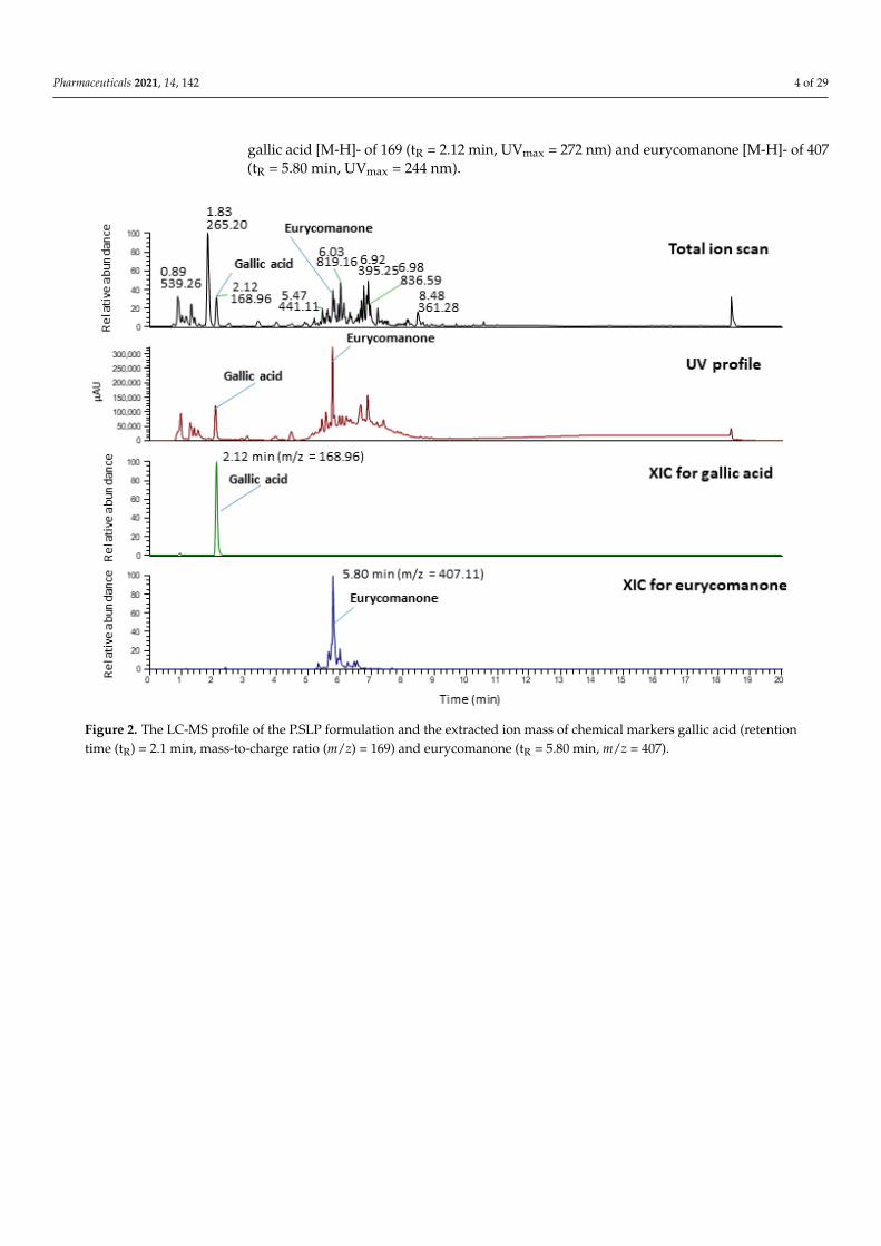
Meanwhile, identification of the formulation was also evaluated on an LC-MS instru-ment coupled to a UV detector. The markers, gallic acid and eurycomanone were detectedand identified based on mass-to-charge ratio (m/z), retention time (tR) and UV profile withreference to the chemical standards. The chromatogram of the formulation is shown inFigure 2 whilst its UV profile is shown in Figure 3. The LC-MS-UV results showed thatthe marker compounds gallic acid and eurycomanone were both detected in the sample:
Pharmaceuticals 2021, 14, 142 4 of 29
gallic acid [M-H]- of 169 (tR = 2.12 min, UVmax = 272 nm) and eurycomanone [M-H]- of 407(tR = 5.80 min, UVmax = 244 nm).
Pharmaceuticals 2021, 14, x FOR PEER REVIEW 4 of 28
Meanwhile, identification of the formulation was also evaluated on an LC-MS instru-ment coupled to a UV detector. The markers, gallic acid and eurycomanone were detected and identified based on mass-to-charge ratio (m/z), retention time (tR) and UV profile with reference to the chemical standards. The chromatogram of the formulation is shown in Figure 2 whilst its UV profile is shown in Figure 3. The LC-MS-UV results showed that the marker compounds gallic acid and eurycomanone were both detected in the sample: gallic acid [M-H]- of 169 (tR = 2.12 min, UVmax = 272 nm) and eurycomanone [M-H]- of 407 (tR = 5.80 min, UVmax = 244 nm). 2.2. Mortality, Clinical and Functional Observations
No morbidity or mortality were observed in any rats, except for a female rat in the high-dose satellite group that was found dead on day 28 of dosing. The post-mortem find-ings (data not shown) indicated the cause of death was due to technical error and not formulation related. No clinical sign of toxicity was observed in the any P.SLP formula-tion-treated or control groups (Table 1).
No significant difference was found in the general activity, locomotor and stereo-typed movements for all groups. However, the forelimb grip strength of female rats in the medium- and high-dose groups was shown to be significantly stronger compared to the vehicle control group (Table 2). 2.3. Body Weight
The body weights gradually increased each week for all groups except for a slight decrease (0.59%) in the sixth week in the male vehicle control satellite group. However, there was no significant difference in the percentage weekly body weight change between P.SLP formulation-treated groups and their respective vehicle control groups (Table 3). 2.4. Food and Water Consumption
All rats had normal food and water consumption (94.45–187.36 g/week; 197.2–694.8 mL/week) throughout the study. No significant difference in the percentage of weekly food and water consumption between any P.SLP formulation-treated groups and their respective control groups was found in either gender (Tables 4 and 5).
Figure 2. The LC-MS profile of the P.SLP formulation and the extracted ion mass of chemical markers gallic acid (retentiontime (tR) = 2.1 min, mass-to-charge ratio (m/z) = 169) and eurycomanone (tR = 5.80 min, m/z = 407).

Pharmaceuticals 2021, 14, 142 5 of 29
Pharmaceuticals 2021, 14, x FOR PEER REVIEW 5 of 28
Figure 2. The LC-MS profile of the P.SLP formulation and the extracted ion mass of chemical markers gallic acid (retention time (tR) = 2.1 min, mass-to-charge ratio (m/z) = 169) and eurycoma-none (tR = 5.80 min, m/z = 407).
Figure 3. UV spectrum of (A) gallic acid standard (1 mg/mL) and (B) eurycomanone standard (1 mg/mL) overlaid with methanol solution of the P.SLP formulation.
Figure 3. UV spectrum of (A) gallic acid standard (1 mg/mL) and (B) eurycomanone standard (1 mg/mL) overlaid withmethanol solution of the P.SLP formulation.
2.2. Mortality, Clinical and Functional Observations
No morbidity or mortality were observed in any rats, except for a female rat in thehigh-dose satellite group that was found dead on day 28 of dosing. The post-mortemfindings (data not shown) indicated the cause of death was due to technical error and notformulation related. No clinical sign of toxicity was observed in the any P.SLP formulation-treated or control groups (Table 1).

Pharmaceuticals 2021, 14, 142 6 of 29
Table 1. Summary of mortality and clinical signs of toxicity in female and male rats.
Female Rats Male Rats
Dose (mg/kg) (Group)
Parameters
Dose (mg/kg) (Group)
0(VehicleControl)
125(LowDose)
500(Medium
Dose)
2000(HighDose)
0(VehicleControl
Satellite)
2000(High-Dose
Satellite)
0(VehicleControl)
125(LowDose)
500(Medium
Dose)
2000(HighDose)
0(VehicleControl
Satellite)
2000(High-Dose
Satellite)
0/5 0/5 0/5 0/5 0/5 1/5 Mortality(died/dosed) 0/5 0/5 0/5 0/5 0/5 0/5
NAD NAD NAD NAD NAD NAD Clinical signs of toxicity NAD NAD NAD NAD NAD NAD
0/5 0/5 0/5 0/5 0/5 0/5 Incidence of clinical signsof toxicity 0/5 0/5 0/5 0/5 0/5 0/5
N/A N/A N/A N/A N/A N/A Onset of clinical signs oftoxicity (day) N/A N/A N/A N/A N/A N/A
N/A N/A N/A N/A N/A N/A Duration/severity ofclinical signs of toxicity N/A N/A N/A N/A N/A N/A
0/5 0/5 0/5 0/5 0/5 0/5 Incidence of lesions/dosed 0/5 0/5 0/5 0/5 0/5 0/5
N/A N/A N/A N/A N/A N/A Type of lesions N/A N/A N/A N/A N/A N/A
N/A N/A N/A N/A N/A N/A Severity of lesions N/A N/A N/A N/A N/A N/A
Satellite groups were kept for an additional 14 days without treatment to detect delayed occurrence or persistence of, or recovery from, toxic effects. A female rat in the high-dose satellite group died due totechnical error in dosing. NAD = no abnormality detected, N/A = not available.
Pharmaceuticals 2021, 14, 142 7 of 29
No significant difference was found in the general activity, locomotor and stereotypedmovements for all groups. However, the forelimb grip strength of female rats in themedium- and high-dose groups was shown to be significantly stronger compared to thevehicle control group (Table 2).
2.3. Body Weight
The body weights gradually increased each week for all groups except for a slightdecrease (0.59%) in the sixth week in the male vehicle control satellite group. However,there was no significant difference in the percentage weekly body weight change betweenP.SLP formulation-treated groups and their respective vehicle control groups (Table 3).
2.4. Food and Water Consumption
All rats had normal food and water consumption (94.45–187.36 g/week;197.2–694.8 mL/week) throughout the study. No significant difference in the percent-age of weekly food and water consumption between any P.SLP formulation-treated groupsand their respective control groups was found in either gender (Tables 4 and 5).
2.5. Haematology and Clinical Biochemistry
Administration of the P.SLP formulation showed no significant effect on haematocrit(HCT), haemoglobin concentration (HGB), erythrocyte count (RBC), total leukocyte count(WBC) and platelet count (PLT) when compared to the respective control groups (Table 6).No significant difference was found in potassium (K), glucose (GLUC), total cholesterol(CHOL), urea (BUN), creatinine (CREA), total protein (TP), albumin (ALB), triglyceride(TGL), uric acid (URCA), alanine aminotransferase (ALT), alkaline phosphatase (ALP) andaspartate aminotransferase (AST) levels when compared to the respective control groups(Tables 7 and 8).
2.6. Gross Pathology Examination
Gross lesions were found in the lung, intestinal tract, liver, kidneys, ovaries anduterine horn across all dose groups (Table 9). Other macroscopically examined organs suchas the heart, spleen, stomach, adrenals and testes did not show any abnormalities.
2.7. Relative Organ Weight
There was no significant difference in the organ weight relative to 100 g body weightin either gender for all organs other than the left ovary. A significantly lower relativeweight was observed for the left ovary in the low-dose (0.0188), medium-dose (0.0166) andhigh-dose satellite (0.0183) groups compared to their respective control groups (vehiclecontrol: 0.0256, vehicle control satellite: 0.0205) (Tables 10 and 11).
2.8. Histopathology
There were some microscopic findings on the histological appearance of the organtissues (liver, kidneys, adrenals, testes, ovaries, uterine horn, spleen, lungs, heart, stomachand intestinal tract) for low and medium groups, but it was not a P.SLP formulation-related toxicity effect. However, the heart, lung, liver, spleen, kidneys, stomach, intestinaltract, ovaries and adrenal from the high-dose groups (five females, four males) and thehigh-dose satellite groups (four females, five males) were found to have some histologicalobservations indicating an early response to exogenous toxicity (Table 12).

Pharmaceuticals 2021, 14, 142 8 of 29
Table 2. Summary of functional observations on motor activity and grip strength in female and male rats.
Female Rats Male Rats
Dose (mg/kg) (Group)
Parameters
Dose (mg/kg) (Group)
0(VehicleControl)
125(LowDose)
500(Medium
Dose)
2000(HighDose)
0(VehicleControl
Satellite)
2000(High-Dose
Satellite)
0(VehicleControl)
125(LowDose)
500(Medium
Dose)
2000(HighDose)
0(VehicleControl
Satellite)
2000(High-Dose
Satellite)
1335.40 ±251.54
1273.00 ±314.98
1278.00 ±257.02
1029.80 ±234.67
1399.80 ±197.64
1252.00 ±299.80
Generalactivity
1042.80 ±306.00
1072.00 ±183.94
1220.40 ±61.06
1121.20 ±159.34
927.60 ±352.26
790.40 ±282.33
91.40 ± 19.31 83.80 ±23.47
83.00 ±11.68
75.40 ±11.95
90.00 ±11.92 95.25 ± 19.60 Stereotyped
movement84.00 ±
19.9680.80 ±
14.6096.60 ±
16.8685.00 ±
12.3173.60 ±
42.96 55.60 ± 19.83
1244.00 ±249.15
1189.20 ±294.44
1195.00 ±252.36
954.40 ±226.39
1309.80 ±190.73
1156.75 ±283.99 Locomotion 958.80 ±
291.39991.20 ±
177.561123.80 ±
51.801036.20 ±
154.56854.00 ±
310.84734.80 ±
263.68
555.32 ±144.31
683.76 ±164.89
859.74 ±104.77 *
857.42 ±155.00 *
828.20 ±120.30
891.85 ±119.11
Forelimb gripstrength (g)
894.54 ±143.06
873.68 ±149.43
1008.78 ±160.67
943.12 ±98.40
920.84 ±140.86
997.30 ±66.07
1002.08 ±177.52
955.72 ±59.88
1013.48 ±156.98
1027.84 ±74.99
947.00 ±158.82
995.08 ±244.18
Forelimb andhind limb grip
strength (g)
1068.46 ±72.42
1146.04 ±227.37
1200.40 ±218.91
1154.40 ±85.66
1091.04 ±133.74
1127.62 ±178.69
Satellite groups were kept for an additional 14 days without treatment to detect delayed occurrence or persistence of, or recovery from, toxic effects. Values are expressed as mean ± standard deviation (n = 5).Data for one dead animal were excluded from data analysis, therefore n = 4 for female high-dose satellite group. A statistically significant difference was considered at p < 0.05 and is denoted with an asterisk(against vehicle control group) in the table. Statistical tests involved were normality, Levene’s, one-way ANOVA, Kruskal–Wallis and/or Tukey HSD (honestly significant difference) tests.

Pharmaceuticals 2021, 14, 142 9 of 29
Table 3. Percentages of weekly body weight change (%) in female and male rats.
Female Rats Male Rats
Dose (mg/kg) (Group) WeeklyBody
WeightChange
Dose (mg/kg) (Group)
0(VehicleControl)
125(Low Dose)
500(Medium
Dose)
2000(HighDose)
0(VehicleControl
Satellite)
2000(High-Dose
Satellite)
0(VehicleControl)
125(Low Dose)
500(Medium
Dose)
2000(HighDose)
0(VehicleControl
Satellite)
2000(High-Dose
Satellite)
10.41 ± 5.23 8.49 ± 1.65 9.78 ± 2.34 8.89 ± 5.26 6.32 ± 2.02 7.96 ± 2.89 Week 1 12.92 ± 3.67 12.20 ± 1.58 13.10 ± 1.57 13.74 ± 4.27 13.45 ± 2.56 12.33 ± 2.042.98 ± 3.22 5.78 ± 1.77 5.31 ± 1.40 5.39 ± 1.90 7.19 ± 3.61 8.97 ± 3.42 Week 2 6.78 ± 2.58 8.34 ± 1.84 9.08 ± 1.97 8.57 ± 0.74 7.61 ± 3.18 7.72 ± 0.696.17 ± 5.17 2.03 ± 2.31 4.14 ± 2.63 4.11 ± 3.60 2.95 ± 5.17 1.43 ± 3.23 Week 3 6.36 ± 1.16 6.41 ± 1.15 6.90 ± 1.18 6.23 ± 1.11 7.31 ± 2.14 5.09 ± 2.321.81 ± 1.99 3.51 ± 3.47 4.16 ± 3.55 4.39 ± 2.88 3.47 ± 4.22 4.88 ± 2.69 Week 4 2.96 ± 1.11 3.40 ± 1.19 4.39 ± 1.65 3.13 ± 1.07 5.81 ± 0.54 4.64 ± 1.47
N/A N/A N/A N/A 2.73 ± 3.49 1.79 ± 0.67 Week 5 N/A N/A N/A N/A 9.35 ± 9.89 4.81 ± 1.34N/A N/A N/A N/A 0.78 ± 2.42 3.45 ± 1.02 Week 6 N/A N/A N/A N/A -0.59 ± 8.95 3.02 ± 1.09
Satellite groups were kept for an additional 14 days without treatment to detect delayed occurrence or persistence of, or recovery from, toxic effects. Values are expressed as mean ± standard deviation (n =5).Data for one dead animal were excluded from data analysis, therefore n = 4 for female high-dose satellite group. The above values were found to be not statistically significantly different (p ≥ 0.05) against therespective control groups. Statistical tests involved were normality, Levene’s, one-way ANOVA and/or Kruskal–Wallis tests. NAD = no abnormality detected, N/A = not available.

Pharmaceuticals 2021, 14, 142 10 of 29
Table 4. Mean food consumption (g) in female and male rats.
Female Rats Male Rats
Dose (mg/kg) (Group)Weekly FoodConsumption
Dose (mg/kg) (Group)
0(VehicleControl)
125(LowDose)
500(Medium
Dose)
2000(HighDose)
0(VehicleControl
Satellite)
2000(High-Dose
Satellite)
0(VehicleControl)
125(LowDose)
500(Medium
Dose)
2000(HighDose)
0(VehicleControl
Satellite)
2000(High-Dose
Satellite)
118.81 ±18.27
115.09 ±10.68
118.75 ±4.14
118.42 ±13.48
112.01 ±6.82 120.69 ± 7.62 Week 1 170.50 ±
15.83163.38 ±
15.48171.88 ±
8.13162.10 ±
14.83174.30 ±
13.20170.49 ±
12.48
124.49 ±12.62
118.91 ±16.79
128.31 ±5.78
122.10 ±12.89
118.91 ±2.43
130.12 ±13.76 Week 2 168.53 ±
18.67166.04 ±
12.46173.39 ±
9.96163.51 ±
14.01176.59 ±
10.77166.94 ±
14.28
126.75 ±10.80
116.83 ±10.71
127.29 ±2.60
123.48 ±9.17
118.40 ±6.19 122.90 ± 9.07 Week 3 170.66 ±
21.76165.07 ±
16.91174.58 ±
9.19158.75 ±
14.62174.35 ±
12.36165.23 ±
16.63
110.24 ±13.49
100.46 ±8.75
112.10 ±11.26
108.76 ±7.43
112.00 ±4.62 117.33 ± 7.00 Week 4 147.02 ±
17.50143.65 ±
12.20151.89 ±
7.31136.08 ±
16.14172.85 ±
15.19156.11 ±
16.95
N/A N/A N/A N/A 116.34 ±5.88 99.49 ± 5.04 Week 5 N/A N/A N/A N/A 172.37 ±
17.74165.34 ±
14.94
N/A N/A N/A N/A 126.28 ±8.37 113.95 ± 6.51 Week 6 N/A N/A N/A N/A 150.87 ±
13.63153.05 ±
19.47
Satellite groups were kept for an additional 14 days without treatment to detect delayed occurrence or persistence of, or recovery from, toxic effects. Values are expressed as mean ± standard deviation (n = 5).Data for one dead animal were excluded from data analysis, therefore n = 4 for female high-dose satellite group. The above values were found to be not statistically significantly different (p ≥ 0.05) against therespective control groups. Statistical tests involved were normality, Levene’s, one-way ANOVA and/or Kruskal–Wallis tests. N/A = not available.

Pharmaceuticals 2021, 14, 142 11 of 29
Table 5. Mean water consumption (g) in female and male rats.
Female Rats Male Rats
Dose (mg/kg) (Group)Weekly WaterConsumption
Dose (mg/kg) (Group)
0(VehicleControl)
125(LowDose)
500(Medium
Dose)
2000(HighDose)
0(VehicleControl
Satellite)
2000(High-Dose
Satellite)
0(VehicleControl)
125(Low Dose)
500(Medium
Dose)
2000(HighDose)
0(VehicleControl
Satellite)
2000(High-Dose
Satellite)
347.6 ±99.0
292.2 ±36.3 340.0 ± 95.5 316.4 ± 85.0 321.8 ± 37.0 275.0 ± 67.4 Week 1 419.0 ± 89.4 371.0 ± 82.1 316.2 ± 34.0 406.4 ±
122.3 358.4 ± 88.9 345.6 ± 36.3
460.6 ±158.2
325.4 ±33.3 378.8 ± 88.3 335.2 ± 99.1 347.6 ± 60.4 326.8 ± 62.1 Week 2 409.8 ± 82.8 372.0 ± 80.4 334.8 ± 45.3 423.2 ±
121.3 391.2 ± 96.7 374.4 ± 50.9
497.0 ±197.8
332.0 ±42.9 409.4 ± 71.4 342.0 ±
114.7 354.2 ± 66.0 281.3 ± 49.6 Week 3 407.8 ±111.2 364.0 ± 97.0 298.0 ± 55.5 393.0 ±
131.6 368.0 ± 71.0 366.0 ± 46.2
368.2 ±129.4
288.2 ±41.5
400.6 ±100.9 302.2 ± 69.9 366.4 ±
105.2 292.3 ± 55.5 Week 4 362.8 ±104.4 330.4 ± 91.0 291.6 ± 34.7 346.0 ± 89.0 354.6 ± 54.6 351.8 ± 66.6
N/A N/A N/A N/A 357.4 ±117.6 266.8 ± 69.6 Week 5 N/A N/A N/A N/A 349.0 ± 78.2 334.2 ± 47.4
N/A N/A N/A N/A 310.8 ± 85.6 381.5 ±277.4 Week 6 N/A N/A N/A N/A 317.0 ± 64.8 323.0 ± 78.5
Satellite groups were kept for an additional 14 days without treatment to detect delayed occurrence or persistence of, or recovery from, toxic effects. Values are expressed as mean ± standard deviation (n = 5).Data for one dead animal were excluded from data analysis, therefore n = 4 for female high-dose satellite group. The above values were found to be not statistically significantly different (p ≥ 0.05) against therespective control groups. Statistical tests involved were normality, Levene’s, one-way ANOVA and/or Kruskal–Wallis tests. N/A = not available.

Pharmaceuticals 2021, 14, 142 12 of 29
Table 6. Haematology findings in female and male rats.
Female Rats Male Rats
Dose (mg/kg) (Group)
Parameters
Dose (mg/kg) (Group)
0(VehicleControl)
125(Low Dose)
500(Medium
Dose)
2000(HighDose)
0(VehicleControl
Satellite)
2000(High-Dose
Satellite)
0(VehicleControl)
125(LowDose)
500(Medium
Dose)
2000(HighDose)
0(VehicleControl
Satellite)
2000(High-Dose
Satellite)
33.0 ± 3.1 33.3 ± 1.6 33.3 ± 2.5 32.6 ± 2.5 36.3 ± 2.6 35.8 ± 1.1 HCT (%) 36.4 ± 2.2 36.5 ± 3.2 37.6 ± 1.8 35.9 ± 2.3 38.4 ± 2.2 38.4 ± 0.412.6 ± 0.5 12.8 ± 0.3 12.9 ± 0.9 12.7 ± 0.6 13.6 ± 0.8 13.6 ± 0.4 HGB (g/dL) 13.7 ± 0.6 14.0 ± 0.8 14.2 ± 0.7 13.9 ± 0.7 14.3 ± 0.7 14.5 ± 0.2
6.27 ± 0.48 6.64 ± 0.43 6.68 ± 0.64 6.70 ± 0.48 7.17 ± 0.50 7.22 ± 0.27 RBC (106
cells/mm)7.26 ± 0.51 7.21 ± 0.59 7.61 ± 0.31 7.22 ± 0.40 7.83 ± 0.47 7.90 ± 0.19
3.6 ± 1.3 3.6 ± 1.5 3.0 ± 2.0 4.4 ± 2.8 3.4 ± 1.6 3.3 ± 1.0 WBC (103
cells/mm)7.2 ± 1.0 8.1 ± 1.8 8.5 ± 3.4 7.2 ± 4.0 6.0 ± 1.3 7.4 ± 1.6
1002 ± 123 1132 ± 157 1023 ± 119 1206 ± 119 972 ± 182 1017 ± 133 PLT (103
cells/mm)1124 ± 168 1134 ± 234 1154 ± 188 1316 ± 147 1208 ± 57 1082 ± 134
Satellite groups were kept for an additional 14 days without treatment to detect delayed occurrence or persistence of, or recovery from, toxic effects. Values are expressed as mean ± standard deviation (n = 5).Data for one dead animal were excluded from data analysis, therefore n = 4 for female high-dose satellite group. The above values were found to be not statistically significantly different (p ≥ 0.05) against therespective control groups. Statistical tests involved were normality, Levene’s, one-way ANOVA, Kruskal–Wallis and/or Tukey HSD tests. HCT = haematocrit, HGB = haemoglobin, RBC = red blood cells, WBC =white blood cells, PLT = platelet.

Pharmaceuticals 2021, 14, 142 13 of 29
Table 7. Clinical biochemistry liver profile parameter in female and male rats.
Female Rats Male Rats
Dose (mg/kg) (Group)
Parameters
Dose (mg/kg) (Group)
0(VehicleControl)
125(LowDose)
500(Medium
Dose)
2000(HighDose)
0(VehicleControl
Satellite)
2000(High-Dose
Satellite)
0(VehicleControl)
125(Low Dose)
500(Medium
Dose)
2000(HighDose)
0(VehicleControl
Satellite)
2000(High-Dose
Satellite)
Liver Profile
62.3 ± 2.1 62.8 ± 1.8 60.4 ± 2.3 60.0 ± 2.9 62.0 ± 1.0 62.5 ± 3.3 Total protein(g/L) 60.2 ± 3.1 65.6 ± 3.9 61.8 ± 4.2 60.4 ± 2.1 61.0 ± 3.4 60.4 ± 2.6
14.7 ± 0.5 15.0 ± 0.0 13.8 ± 1.3 14.0 ± 1.2 14.4 ± 0.9 15.0 ± 0.8 Albumin(g/L) 13.2 ± 1.3 13.8 ± 0.8 13.2 ± 0.8 12.6 ± 0.5 12.8 ± 0.4 12.6 ± 0.5
32.1 ± 5.9 35.0 ± 8.5 34.2 ± 4.3 28.8 ± 5.9 34.4 ± 5.7 30.3 ± 3.7 ALT (U/L) 40.2 ± 10.1 34.4 ± 5.8 32.8 ± 6.1 31.8 ± 5.1 37.8 ± 6.2 34.4 ± 7.483.2 ± 14.2 79.0 ± 23.5 67.2 ± 22.3 51.4 ± 9.8 67.4 ± 22.5 54.8 ± 12.7 ALP (U/L) 127.4 ± 24.8 108.20 ± 14.8 119.0 ± 25.1 93.6 ± 36.8 91.0 ± 9.2 90.6 ± 7.3109.8 ± 6.0 96.8 ± 14.8 100.2 ± 9.7 104.6 ± 23.2 81.2 ± 11.7 78.8 ± 11.7 AST (U/L) 80.3 ± 13.0 74.6 ± 17.1 74.8 ± 7.0 76.6 ± 9.0 70.8 ± 10.6 62.0 ± 3.5
Satellite groups were kept for an additional 14 days without treatment to detect delayed occurrence or persistence of, or recovery from, toxic effects. Values are expressed as mean ± standard deviation (n = 5).Data for one dead animal were excluded from data analysis, therefore n = 4 for female high-dose satellite group. The above values were found to be not statistically significantly different (p ≥ 0.05) against therespective control groups. Statistical tests involved were normality, Levene’s, one-way ANOVA, Kruskal–Wallis, Tukey HSD and/or Mann–Whitney U tests. ALT = alanine amino-transferase, ALP = alkalinephosphatase, AST = aspartate amino-transferase.

Pharmaceuticals 2021, 14, 142 14 of 29
Table 8. Clinical biochemistry renal and lipid profile parameter on female and male rats.
Female Rats Male Rats
Dose (mg/kg) (Group)
Parameters
Dose (mg/kg) (Group)
0(VehicleControl)
125(Low Dose)
500(Medium
Dose)
2000(HighDose)
0(VehicleControl
Satellite)
2000(High-Dose
Satellite)
0(VehicleControl)
125(Low Dose)
500(Medium
Dose)
2000(HighDose)
0(VehicleControl
Satellite)
2000(High-Dose
Satellite)
RenalProfile
4.1 ± 0.3 3.8 ± 0.2 4.2 ± 0.5 4.0 ± 0.4 3.9 ± 0.6 3.7 ± 0.2 Potassium(mmol/L) 4.0 ± 0.3 4.4 ± 0.4 4.1 ± 0.3 4.5 ± 0.2 4.0 ± 0.4 4.0 ± 0.3
38.4 ± 7.6 33.8 ± 2.7 39.0 ± 2.5 35.2 ± 5.4 39.4 ± 7.4 41.5 ± 9.3 Creatinine(µmol/L) 30.4 ± 3.2 32.2 ± 3.6 34.0 ± 7.0 27.2 ± 6.3 37.0 ± 6.1 32.4 ± 5.7
7.1 ± 0.7 6.7 ± 0.7 7.5 ± 1.0 6.1 ± 1.4 6.6 ± 1.1 7.2 ± 0.7 Urea(mmol/L) 6.7 ± 0.4 6.9 ± 0.9 6.9 ± 0.7 6.6 ± 1.1 6.5 ± 1.1 7.1 ± 1.3
94.3 ± 9.1 78.6 ± 30.5 111.8 ± 22.0 95.2 ± 25.8 77.8 ± 27.5 98.8 ± 35.7 Uric acid(µmol/L) 77.2 ± 5.8 78.80 ± 18.1 71.0 ± 28.2 68.6 ± 14.3 69.6 ± 15.7 82.4 ± 48.6
LipidProfile
1.9 ± 0.2 2.1 ± 0.4 2.1 ± 0.2 1.7 ± 0.1 2.1 ± 0.3 2.0 ± 0.2Total
cholesterol(mmol/L)
1.7 ± 0.2 2.3 ± 0.4 1.8 ± 0.2 1.4 ± 0.3 2.3 ± 0.1 2.1 ± 0.3
0.4 ± 0.1 0.5 ± 0.1 0.4 ± 0.1 0.4 ± 0.2 0.7 ± 0.2 0.5 ± 0.2 Triglyceride(mmol/L) 0.7 ± 0.2 1.0 ± 0.4 1.0 ± 0.3 1.0 ± 0.3 0.9 ± 0.2 0.9 ± 0.3
6.71 ± 0.40 6.70 ± 0.81 6.87 ± 0.71 7.26 ± 0.66 8.42 ± 1.13 7.45 ± 0.78 Glucose(mmol/L) 8.70 ± 0.56 8.58 ± 0.79 8.96 ± 0.90 9.64 ± 1.72 8.57 ± 0.74 9.31 ± 2.05
Satellite groups were kept for an additional 14 days without treatment to detect delayed occurrence or persistence of, or recovery from, toxic effects. Values are expressed as mean ± standard deviation (n = 5).Data for one dead animal were excluded from data analysis, therefore n = 4 for female high-dose satellite group. The above values were found to be not statistically significantly different (p ≥ 0.05) against therespective control groups. Statistical tests involved were normality, Levene’s, one-way ANOVA, Kruskal–Wallis, Tukey HSD and/or Mann–Whitney U tests.

Pharmaceuticals 2021, 14, 142 15 of 29
Table 9. Summary of gross pathology findings in female and male rats.
Female Rats Male Rats
Dose (mg/kg) (Group)
Organs
Dose (mg/kg) (Group)
0(VehicleControl)
125(Low Dose)
500(Medium
Dose)
2000(HighDose)
0(VehicleControl
Satellite)
2000(High-Dose
Satellite)
0(VehicleControl)
125(Low Dose)
500(Medium
Dose)
2000(HighDose)
0(VehicleControl
Satellite)
2000(High-Dose
Satellite)
5/5 4/5 5/5 5/5 5/5 4/5 Lung 4/5 5/5 4/5 5/5 5/5 4/50/5 0/5 0/5 0/5 0/5 0/5 Heart 0/5 0/5 0/5 0/5 0/5 0/50/5 0/5 0/5 0/5 0/5 0/5 Spleen 0/5 0/5 0/5 0/5 0/5 0/50/5 0/5 0/5 0/5 0/5 0/5 Stomach 0/5 0/5 0/5 0/5 0/5 0/51/5 3/5 3/5 3/5 0/5 0/5 IT 0/5 2/5 1/5 1/5 0/5 5/53/5 2/5 3/5 3/5 1/5 2/5 Liver 1/5 2/5 2/5 1/5 1/5 1/50/5 0/5 0/5 0/5 0/5 0/5 Kidneys 0/5 0/5 0/5 0/5 0/5 5/50/5 0/5 0/5 0/5 0/5 0/5 Adrenals 0/5 0/5 0/5 0/5 0/5 0/50/5 0/5 0/5 1/5 0/5 0/5 Ovaries N/A N/A N/A N/A N/A N/A0/5 0/5 1/5 0/5 0/5 1/5 Uterine horn N/A N/A N/A N/A N/A N/A
N/A N/A N/A N/A N/A N/A Testes 0/5 0/5 0/5 0/5 0/5 0/5
Satellite groups were kept for an additional 14 days without treatment to detect delayed occurrence or persistence of, or recovery from, toxic effects. Values indicate the number of rats with organ(s) that showedmacroscopic lesions during gross examination but were not necessarily P.SLP formulation related. IT = intestinal tract. N/A = not available.

Pharmaceuticals 2021, 14, 142 16 of 29
Table 10. Relative organ weight in female and male rats.
Female Rats Male Rats
Dose (mg/kg) (Group)
Organs
Dose (mg/kg) (Group)
0(VehicleControl)
125(Low Dose)
500(Medium
Dose)
2000(HighDose)
0(VehicleControl
Satellite)
2000(High-Dose
Satellite)
0(VehicleControl)
125(Low Dose)
500(Medium
Dose)
2000(HighDose)
0(VehicleControl
Satellite)
2000(High-Dose
Satellite)
0.4444 ±0.0454
0.4358 ±0.0209
0.4191 ±0.0330
0.3871 ±0.0310
0.3905 ±0.0135
0.3893 ±0.0216 Lung 0.3399 ±
0.04560.3528 ±
0.02030.3135 ±
0.040.3486 ±
0.04320.3057 ±
0.03180.3459 ±
0.0890
0.2890 ±0.0307
0.2850 ±0.0252
0.2876 ±0.0132
0.2930 ±0.0219
0.2729 ±0.0158
0.2769 ±0.0104 Heart 0.2494 ±
0.03310.2639 ±
0.02310.2559 ±
0.01320.2539 ±
0.03570.2297 ±
0.01850.2390 ±
0.0229
0.2332 ±0.0281
0.2292 ±0.0246
0.2282 ±0.0304
0.2180 ±0.0196
0.2028 ±0.0126
0.2000 ±0.0139 Spleen 0.2063 ±
0.03310.2052 ±
0.02440.1816 ±
0.02270.1829 ±
0.01240.1731 ±
0.01830.1962 ±
0.0205
0.5471 ±0.0688
0.5849 ±0.0919
0.5867 ±0.0651
0.5605 ±0.0247
0.5328 ±0.0269
0.5273 ±0.0155 Stomach 0.4714 ±
0.04940.4856 ±
0.04900.4743 ±
0.03290.4823 ±
0.06180.4533 ±
0.03660.4418 ±
0.0263
2.6132 ±0.6347
2.6427 ±0.5049
2.6492 ±0.7822
2.5011 ±0.3024
2.3388 ±0.4279
2.1914 ±0.1792 IT 1.8803 ±
0.34182.1921 ±
0.25802.0550 ±
0.28912.0597 ±
0.28142.0162 ±
0.26761.7278 ±
0.0613
2.7278 ±0.1933
2.5790 ±0.1221
2.5379 ±0.0668
2.6613 ±0.0964
2.4010 ±0.1312
2.5351 ±0.0276 Liver 2.9989 ±
0.41243.1014 ±
0.32722.8786 ±
0.11103.0926 ±
0.15272.7864 ±
0.23142.9335 ±
0.1799
0.3155 ±0.0174
0.3255 ±0.0291
0.3051 ±0.0061
0.3325 ±0.0233
0.2904 ±0.0300
0.3216 ±0.0228
KidneyRight
0.3248 ±0.0252
0.3339 ±0.0269
0.3082 ±0.0085
0.3238 ±0.0135
0.3083 ±0.0192
0.3318 ±0.0355
0.3172 ±0.0171
0.3254 ±0.0198
0.2910 ±0.0119
0.3199 ±0.0316
0.2836 ±0.0300
0.3147 ±0.0230
KidneyLeft
0.3252 ±0.0204
0.3283 ±0.0264
0.3111 ±0.0129
0.3171 ±0.0251
0.3034 ±0.0265
0.3270 ±0.0371
Satellite groups were kept for an additional 14 days without treatment to detect delayed occurrence or persistence of, or recovery from, toxic effects. Values are expressed as mean ± standard deviation (n = 5).Data for one dead animal were excluded from data analysis, therefore n = 4 for female high-dose satellite group. The above values were found to be not statistically significantly different (p ≥ 0.05) against therespective control groups. Statistical tests involved were normality, Levene’s, one-way ANOVA, Kruskal–Wallis and/or Tukey HSD tests. IT = intestinal tract.

Pharmaceuticals 2021, 14, 142 17 of 29
Table 11. Relative organ weight (adrenals and reproductive organs) in female and male rats.
Female Rats Male Rats
Dose (mg/kg) (Group)
Organs
Dose (mg/kg) (Group)
0(VehicleControl)
125(Low Dose)
500(Medium
Dose)
2000(HighDose)
0(VehicleControl
Satellite)
2000(High-Dose
Satellite)
0(VehicleControl)
125(Low Dose)
500(Medium
Dose)
2000(HighDose)
0(VehicleControl
Satellite)
2000(High-Dose
Satellite)
0.0152 ±0.0036
0.0137 ±0.0012
0.0157 ±0.0024
0.0134 ±0.0044
0.0130 ±0.0013
0.0130 ±0.0010 Adrenal Right 0.0083 ±
0.00160.0086 ±
0.00120.0077 ±
0.00090.0077 ±
0.00150.0065 ±
0.00130.0071 ±
0.0012
0.0168 ±0.0043
0.0148 ±0.0012
0.0149 ±0.0023
0.0161 ±0.0023
0.0143 ±0.0018
0.0130 ±0.0027 Adrenal Left 0.0082 ±
0.00100.0091 ±
0.00140.0084 ±
0.00070.0086 ±
0.00120.0077 ±
0.00130.0081 ±
0.0007
0.0275 ±0.0035
0.0252 ±0.0083
0.0207 ±0.0042
0.0223 ±0.0065
0.0196 ±0.0024
0.0169 ±0.0028 Ovary Right N/A N/A N/A N/A N/A N/A
0.0256 ±0.0029
0.0188 ±0.0036 *
0.0166 ±0.0033 *
0.0202 ±0.0040
0.0205 ±0.0019
0.0183 ±0.0043 # Ovary Left N/A N/A N/A N/A N/A N/A
0.0624 ±0.0864
0.0718 ±0.1002
0.1533 ±0.0964
0.1619 ±0.0969
0.1732 ±0.0535
0.2122 ±0.0421 Uterine Horn N/A N/A N/A N/A N/A N/A
N/A N/A N/A N/A N/A N/A Testes Right 0.4504 ±0.0596
0.4121 ±0.0309
0.4068 ±0.0361
0.4320 ±0.0251
0.3924 ±0.0289
0.4110 ±0.0316
N/A N/A N/A N/A N/A N/A Testes Left 0.4595 ±0.0874
0.4096 ±0.0272
0.4099 ±0.0366
0.4290 ±0.0238
0.3936 ±0.0303
0.4156 ±0.0480
Satellite groups were kept for an additional 14 days without treatment to detect delayed occurrence or persistence of, or recovery from, toxic effects. Values are expressed as mean ± standard deviation (n = 5).Data for one dead animal were excluded from data analysis, therefore n = 4 for female high-dose satellite group. Statistically significant difference was considered at p < 0.05 and is denoted with an asterisk(significantly different against female vehicle control group) and a hash mark (significantly different against female vehicle control satellite group) in the table. Statistical tests involved were normality, Levene’s,one-way ANOVA, Kruskal–Wallis and/or Tukey HSD tests.

Pharmaceuticals 2021, 14, 142 18 of 29
Table 12. Summary of microscopic findings in female and male rats.
Female Rats Male Rats
Dose (mg/kg) (Group)
Organs
Dose (mg/kg) (Group)
0(VehicleControl)
125(LowDose)
500(Medium
Dose)
2000(HighDose)
0(VehicleControl
Satellite)
2000(High-Dose
Satellite)
0(VehicleControl)
125(Low Dose)
500(Medium
Dose)
2000(HighDose)
0(VehicleControl
Satellite)
2000(High-Dose
Satellite)
0/5 5/5 4/5 4/5 5/5 5/5 Lung 2/5 5/5 5/5 5/5 5/5 5/50/5 0/5 0/5 5/5 0/5 5/5 Heart 0/5 0/5 0/5 5/5 0/5 5/50/5 2/5 0/5 1/5 0/5 0/5 Spleen 0/5 0/5 0/5 1/5 0/5 0/50/5 0/5 0/5 4/5 0/5 5/5 Stomach 0/5 0/5 0/5 5/5 0/5 5/50/5 3/5 0/5 4/5 0/5 5/5 IT 0/5 2/5 1/5 5/5 0/5 5/50/5 5/5 3/5 4/5 0/5 5/5 Liver 0/5 2/5 1/5 5/5 0/5 5/50/5 5/5 3/5 4/5 0/5 5/5 Kidneys 0/5 0/5 0/5 5/5 0/5 5/50/5 0/5 0/5 1/5 0/5 0/5 Adrenals 0/5 0/5 0/5 0/5 0/5 0/50/5 0/5 0/5 1/5 0/5 0/5 Ovaries N/A N/A N/A N/A N/A N/A0/5 0/5 0/5 0/5 0/5 0/5 Uterine Horn N/A N/A N/A N/A N/A N/A
N/A N/A N/A N/A N/A N/A Testes 0/5 0/5 0/5 0/5 0/5 0/5
Satellite groups were kept for an additional 14 days without treatment to detect delayed occurrence or persistence of, or recovery from, toxic effects. Values indicate the number of rats with organ(s) that showedmicroscopic lesions but were not necessarily P.SLP formulation related. IT = intestinal tract.

Pharmaceuticals 2021, 14, 142 19 of 29
In the 2000 mg/kg dose-treated groups, some sections of the heart showed cell degen-eration, congestion and hyaline tissue degeneration in the blood vessels and the musclefibres were slightly pale and less dense. The lung tissues showed bronchial infiltration withepithelial and red blood cells. Hepatocytes were found swollen and the central veins werecongested with blood. Mild depletion of lymphoid cells was observed in sections of spleen.Sections of kidneys showed tubular hyalinisation and degeneration of tubule epithelialcells within the renal medulla region. Sections of stomach showed cell degeneration anddepletion of cytoplasmic granules and there was also vacuolated villous epithelium foundin the sections of intestinal tract. Swelling and degeneration of cytoplasmic cells werefound in the adrenal sections. Cytoplasmic vacuolation was observed in the sections of theovaries (Figure 4).
Pharmaceuticals 2021, 14, x FOR PEER REVIEW 18 of 28
(a) (b) (c)
(d) (e) (f)
(g) (h) (i)
(j)
Figure 4. Histological specimens of rats’ tissues were collected after 28 days of treatment. Tissue samples were stained with haematoxylin and eosin. Representative histological pictures from the 2000 mg/kg dose groups were taken at the following magnifications: (a) heart (10×), (b) lung (20×), (c) liver (20×), (d) spleen (40×), (e) left side of kidney (10×), (f) right side of kidney (20×), (g) stom-ach (20×), (h) intestinal tract (10×), (i) left side of adrenal (20×) and (j) right side of ovary (40×).
3. Discussion Prediction of the adverse effects of the polyherbal formulation in humans over a
range of doses, dosage regimens and exposure durations by means of the rat model is the main goal in this current study. Repeated daily oral administration of the P.SLP poly-herbal formulation for 28 days in rats is commensurate with its repeated exposure for 2.7 years in humans [62]. Other than being inexpensive and easy to maintain, the rat model was chosen as it is one of the recommended and preferred rodent species by regulatory authorities, as it is a sufficiently characterised species, with high sensitivity in expressing any toxic responses [63]. The Wistar rats (Rattus norvegicus) were chosen in order to main-tain a consistent animal model, and were similarly used in the previous 14-day single oral dose study (unpublished work). Clinical and functional observations, body weight changes, food and water consumption and blood parameters did not show any significant
Figure 4. Histological specimens of rats’ tissues were collected after 28 days of treatment. Tissue samples were stained withhaematoxylin and eosin. Representative histological pictures from the 2000 mg/kg dose groups were taken at the followingmagnifications: (a) heart (10×), (b) lung (20×), (c) liver (20×), (d) spleen (40×), (e) left side of kidney (10×), (f) right side ofkidney (20×), (g) stomach (20×), (h) intestinal tract (10×), (i) left side of adrenal (20×) and (j) right side of ovary (40×).

Pharmaceuticals 2021, 14, 142 20 of 29
3. Discussion
Prediction of the adverse effects of the polyherbal formulation in humans over a rangeof doses, dosage regimens and exposure durations by means of the rat model is the maingoal in this current study. Repeated daily oral administration of the P.SLP polyherbalformulation for 28 days in rats is commensurate with its repeated exposure for 2.7 years inhumans [62]. Other than being inexpensive and easy to maintain, the rat model was chosenas it is one of the recommended and preferred rodent species by regulatory authorities,as it is a sufficiently characterised species, with high sensitivity in expressing any toxicresponses [63]. The Wistar rats (Rattus norvegicus) were chosen in order to maintain aconsistent animal model, and were similarly used in the previous 14-day single oral dosestudy (unpublished work). Clinical and functional observations, body weight changes,food and water consumption and blood parameters did not show any significant toxicityeffects at any dose level. However, some findings, namely on one mortality (incidentaldeath) and the relative ovary weight parameter in some dose groups, as well as findingsin gross pathology examination and histology assessment, particularly in the high-dosegroup, are reported in this study.
There was one death in the female high-dose satellite group due to personnel technicalerror that may have led to gavage-facilitated reflux resulting in the spontaneous death [64].The statistically significant low weights of the ovaries showed no clear dose relationship andthere was a lack of correlation with histological findings, except in the high-dose satellitegroup. Hence, these two circumstances were not considered to be P.SLP formulationrelated.
Organs, including ovary in the high-dose and high-dose satellite groups, were foundto show microscopic histology findings associated with exogenous toxicity. Exogenoustoxicity is caused by a toxin that is externally presented to the body and may originatefrom the materials supplied to the rats, such as the food pellets, drinking water andbedding. Nevertheless, in this study, all materials supplied to the rats were certified tobe safe for their respective use. Our previous 14-day single oral dose study showed noacute toxicity findings at any dose levels tested (5, 50, 300 and 2000 mg/kg body weight)(unpublished work), therefore, the repeated exposure of the P.SLP formulation at a highdose (2000 mg/kg) could account for the microscopic changes of organs in the high-dosegroups.
Of the few publications on the repeated oral administration of tongkat ali extracts at1000 mg/kg (aqueous extract), 2000 mg/kg (powdered root) and 2400 mg (aqueous extract)in rats, none reported any treatment-related mortality or clinical signs of toxicity [52,58,59].A study on eurycomanone, which is a prominent chemical marker in E. longifolia, suggeststhat once it is absorbed in rodents, it is not easily metabolised and it can actively exert itspharmacological activities [65]. It is inferred that the low percentage of bioavailability, atapproximately 11%, is unlikely to cause severe toxicity effects [65,66]. However, no steadystate data are available to compare eurycomanone’s cumulative effect when administeredin a repeated dose study of either the extract or the compound form.
As for the kacip fatimah aqueous extract, there is a 100-fold difference betweenthe lowest reported NOAEL at 50 mg/kg and the highest reported tolerable dose at5000 mg/kg [53,54]. This huge difference may indicate that these reported doses may notbe the representative NOAEL doses for kacip fatimah aqueous extracts. A closer look atkacip fatimah’s active ingredient, gallic acid, showed that the repeated oral administrationof gallic acid at 900 mg/kg in mice (calculated rat dose of approximately 450 mg/kg) didnot elicit any toxicity [67]. Therefore, it is deduced that the presence of gallic acid in theP.SLP formulation at 0.07% is safe.
Ultimately, findings from this study are useful for determining the safe human dosefor this combined formulation for its subsequent clinical use [68]. When extrapolating tothe human equivalent dose (HED), the uncertainties associated with animal data are offsetby the routine use of a 10-fold interspecies uncertainty factor [69,70]. The calculated HEDfor the high dose level (2000 mg/kg) by dividing by the safety factor of 10 is 32.25 mg/kg

Pharmaceuticals 2021, 14, 142 21 of 29
(equivalent to 1.94 g of the formulation taken by a 60 kg human). The recommended dailyhuman dosage is two capsules (250 mg of P.SLP formulation per capsule) equivalent to8.3 mg/kg body weight or 0.5 g P.SLP formulation intake by a 60 kg human [71]. Com-parison between the calculated HED (1.94 g/60 kg human) and the recommended humandose (0.5 g/60 kg human) indicates that the highest tested concentration (2000 mg/kg)in this study is equivalent to 3.9 times more than the recommended intake in humans.In essence, the recommended dose level is four-fold lower than the highest tested doselevel and corresponds to the medium dose level (500 mg/kg) tested in this current study.The medium dose level seems unlikely to reveal any P.SLP formulation-related toxicityresponse.
By contrast, some of the gross lesions detected in some organs during the grossexamination had no P.SLP formulation-related toxicity signs when they were analysedmicroscopically. Mechanical injury, such as puncture holes due to the technical approach orphysical pressure during organ harvesting or grossing, could have possibly caused thesereported lesions. Consequently, pneumocytes within the interalveolar septa would com-press one another and the extra pressure on the alveolar wall could have possibly causedrupture and losses of alveolar spaces [72]. The need for improved handling techniquesand finer tools is critical to avoid any unwanted lesions on the organ tissues [73]. Anotherrecorded observation includes the reddish colour of the uterine horn, which may be dueto the respective female rats being “in heat” (oestrus cycle), resulting in the appearanceof prominent blood vessels [74]. However, vaginal smear to confirm such an observationwas not conducted in this study. The macroscopic lesions and microscopic findings on thelungs and heart were likely associated with the trauma from blood collection via cardiacpuncture, which allowed maximum blood volume collection [75]. Lesions on the organtissues were deemed accidental and unrelated to the formulation administration.
The use of isoflurane in this study is of minimal concern. Inhalation of isofluranewith oxygen as the carrier gas was used as the anaesthetic agent to produce the stateof unconsciousness, thus preventing the animal from suffering. The blood solubilitycoefficient of the inhalation mode is lower than in the injection mode, which results in theanimals taking a longer time to lose consciousness, compared to the injection mode [76].However, the use of oxygen as the carrier gas in the inhalation mode improves tissueoxygenation during anaesthesia that maintains homeostasis and cellular metabolism in thetissues [77]. In addition, the metabolism of volatile-based anaesthetics including isofluraneis negligible and would interfere minimally with liver function and the metabolism of thetest item in a study [78,79]. Hence, the effect of using isoflurane on the findings from thistoxicology study is not significant.
The effects of the daily administration of doses at 125, 500 and 2000 mg/kg/dayfor 28 days consecutively, along with all the investigated experimental parameters inthe rat model, were sufficiently assessed to obtain relevant toxicology data of the P.SLPformulation. The observed early exogenous toxicity effects at the highest dose indicate theneed for further study of a longer dosing duration. Recently, extensive studies have beencarried out on other combined herbs to further understand undesirable effects such as anypotential interaction (synergistic or antagonistic) with other substances, such as food andconventional drugs [80,81]. Therefore, a similar approach for this polyherbal formulationshould also be implemented where its efficacy or any tendency to cause specific organtoxicity is explored.
4. Materials and Methods4.1. Test Item
The test item (P.SLP formulation) was provided by Biotropics Malaysia Berhad(762243-P) (Lot 21, Jalan U1/19, Section U1, Hicom Glenmarie Industrial Park, 40150Shah Alam, Selangor Darul Ehsan, Malaysia). It contains, namely, Physta® Tongkat Ali(trade name of E. longifolia plant aqueous extract) with a bioactive composition of 0.12%

Pharmaceuticals 2021, 14, 142 22 of 29
eurycomanone and SLP+® Kacip Fatimah (trade name of L. pumila plant aqueous extract)with 0.07% gallic acid content (certificate of analysis number: BMB/P17/RE019).
4.2. Chemicals and Reagents
Chemical standard gallic acid (CAS no.: 84633-29-4) was purchased from Sigma-Aldrich (St. Louis, MO, USA) and eurycomanone (CAS no.: 149-91-7) from ChromaDexCorp. (Los Angeles, CA, USA). Solvents used such as methanol, chloroform, ethyl ac-etate, formic acid, sulphuric acid and acetonitrile were purchased from Merck & Co., Inc.(Darmstadt, Germany).
4.3. Chemical Identification of the Test Item
Chemical identification of the test item was obtained using the HPTLC and LC-MStechniques. The identity of E. longifolia and L. pumila in the formulation was verified usinggallic acid and eurycomanone as the chemical markers [82,83].
The chemical standards gallic acid and eurycomanone were prepared at a concentra-tion of 1 mg/mL in methanol. For the sample preparation, 1 g of the test item was dissolvedin methanol and sonicated for 15 min. After sonication for 15 min, the test item solutionwas filtered through a 0.2 µm filter and used for the chemical identification analysis.
The HPTLC chromatography was performed on 10 × 10 cm HPTLC silica gel 60 F254plates. Standard and test item solutions of 5 µL were separately applied to the plate as 8 mmwide bands with an automatic TLC applicator Linomat-V (CAMAG, Muttenz, Switzerland),8 mm from the bottom. Two different mobile phases consisting of chloroform:methanol(9:1 v/v) (MP1) and ethyl acetate:formic acid (3:1 v/v) (MP2) were used per chromatograph.The plate was developed in a 10 × 10 twin glass chamber saturated with mobile phaseat room temperature up to 7 cm. After development, the plates were air dried and thenderivatisation of the chromatogram was performed by dipping the plate in 10% sulphuricacid in water, heated at 105 ◦C for 5 min or until the colour of the zones became visible.The plates were observed under the CAMAG UV cabinet at 254 nm (before derivatisation)and under UV 366 nm and white light (after derivatisation).
The LC-MS analysis was performed using a Dionex Ultimate 3000 UHPLC system(Thermo, Sunnyvale, CA, USA) coupled to a diode array detector and ion-trap massspectrometer (Thermo, San Jose, CA, USA). Chromatography separation was carriedout using an HSS T3 column (2.1 mm × 100 mm, 1.8 µm, Waters, Milford, MA, USA).Gradient elution was performed with 0.1% formic acid in water (A) and 0.1% formic acid inacetonitrile (B) at a flow rate of 0.3 mL/min. The initial condition was 5% B, which was heldfor 3 min and then increased to 100% B over 9 min, and kept constant at 100% B for 5 min.Finally, the initial condition was restored and held for 3 min to re-equilibrate the system.The total run time was 20 min. The column oven was maintained at 40 ◦C. The injectionvolume was 1 µL. The mass spectrometer was operated in electrospray ionisation (ESI)negative mode. Nitrogen was used as nebuliser gas at a pressure of 100 psi, as carrier gasat 15 L/min and 150 ◦C and as sheath gas at 35 L/min at 320 ◦C. Identification of chemicalmarker peaks was performed by comparison against the gallic acid and eurycomanonestandard.
4.4. Preparation of Test Item for Animal Study
The P.SLP formulation was added to reverse osmosis water (vehicle) to achieve con-centrations of 12.5, 50.0 and 200.0 mg/mL. The formulation was then administered orallyonce daily for a period of 28 days. The volume given was adjusted to the body weight ofthe rats and the pre-determined dosing volume of 10 mL/kg body weight. The oral routewas used in this study as it is the recommended route for rats, as well as the intended routefor human use.

Pharmaceuticals 2021, 14, 142 23 of 29
4.5. Experimental Animals
Rat (Rattus norvegicus) model, i.e., Wistar rats was used in this study. Sixty Wistarrats (30 rats per gender, eight to nine weeks old upon exposure to the P.SLP formulation,weighing 210–220 g for females and 309–312 g for males) were obtained from Beijing VitalRiver Laboratory, Animal Technology Co., Ltd., No. 4 Yangshan Road, Chaoyang District,Beijing, 100107, P.R. China.
The rats were housed in individually ventilated cages with corn-cob bedding (CornCob Laboratory Bedding (Biocob corncob), Biosys Corporation Pte Ltd., 111 North BridgeRoad, #27-01 Peninsula Plaza, Singapore 179098) for a 14-day quarantine period to monitortheir health condition. Using the same housing system, the rats were then acclimatisedto the laboratory condition and human handling for five days prior to the start of P.SLPformulation administration. The experimental room was maintained at a temperatureof 19–26 ◦C and humidity at 35–65%, with a 12 h light–dark cycle. The rats were givenstandard rodent pellet diet (Specialty Feeds, 3150 Great Eastern Highway, Glen Forrest,Western Australia 6071) and an unlimited supply of reverse osmosis water. The handlingof rats was performed in accordance with the animal handling guideline by the Ministry ofHealth Malaysia [84].
4.6. Animal Experimental Procedures
This study was conducted in accordance with test guideline 407 under the Organisa-tion for Economic Cooperation and Development principles of good laboratory practice [68]at the Non-Clinical Research Facility, Good Laboratory Practice Section, Institute for Medi-cal Research, Malaysia. Care and use of study animals were handled in compliance with thetest facility’s standard operating procedures. From the findings of the single dose 14-dayoral toxicity study (unpublished work), the NOAEL was more than 2000 mg/kg, thereforethe dose 2000 mg/kg was selected as the highest dose in this study. The medium and lowdoses were calculated such that they were four-fold lower than the high and medium dose,respectively, as recommended by the test guideline. Therefore, the three dose levels usedin this study were 125, 500 and 2000 mg/kg body weight.
For each gender, the rats were equally divided into six groups (five rats per group)randomly. The P.SLP formulation-treated groups were low-dose, medium-dose, high-doseand high-dose satellite groups, while the control groups were vehicle control and vehiclecontrol satellite groups (Figure 5). Administration of the formulation was performed viaoral gavage using a ball-tipped intubation needle fitted on a syringe. All the rats wereadministered with the formulation for 28 days, except for the satellite groups that werekept for an additional 14 days without dosing to detect delayed occurrence or persistenceof, or recovery from, toxic effects.
On the final day of oral dosing, the feed pellets were removed to allow overnightfasting. The rats were necropsied on the next day. Each rat was anaesthetised usingisoflurane (0.25–2.0% isoflurane with 0.5–2.0 L/min of oxygen, Veterinary Companies ofAustralia Pty Ltd., New South Wales, Australia) for blood sampling and exsanguinationby abdominal aorta, as advised by the veterinarian. The euthanised rats were subjected togross pathology examinations by the veterinarian. Any abnormality was recorded. Theweighed organs were then subjected to gross and histopathological examination.
4.6.1. Mortality, Clinical and Functional Observation
The rats were observed for morbidity and mortality twice daily, once in the morningand once in the afternoon, at approximately the same time. General clinical observa-tions were conducted once daily two hours after dosing. Detailed clinical observations(changes in skin, fur, eyes and mucous membranes, occurrence of secretions and excretion,autonomic activity, changes in gait, posture and response to handling, clonic or tonicmovements, stereotypies, bizarre behaviour and abnormalities of the oral cavity) were con-ducted once before the first exposure to the P.SLP formulation and once weekly thereafter.Functional observation was conducted during the fourth week of exposure in order to

Pharmaceuticals 2021, 14, 142 24 of 29
assess the rats’ grip strength using a grip strength meter (BIOSEB, Vitrolles, France) andmotor activity (general activity, locomotor and stereotyped movements) using an actimeter(Actitrack, Panlab, S.L., Barcelona, Spain). The observation procedures were performed bythe veterinarian and laboratory personnel.
Pharmaceuticals 2021, 14, x FOR PEER REVIEW 22 of 28
ard rodent pellet diet (Specialty Feeds, 3150 Great Eastern Highway, Glen Forrest, West-ern Australia 6071) and an unlimited supply of reverse osmosis water. The handling of rats was performed in accordance with the animal handling guideline by the Ministry of Health Malaysia [84]. 4.6. Animal Experimental Procedures
This study was conducted in accordance with test guideline 407 under the Organisa-tion for Economic Cooperation and Development principles of good laboratory practice [68] at the Non-Clinical Research Facility, Good Laboratory Practice Section, Institute for Medical Research, Malaysia. Care and use of study animals were handled in compliance with the test facility’s standard operating procedures. From the findings of the single dose 14-day oral toxicity study (unpublished work), the NOAEL was more than 2000 mg/kg, therefore the dose 2000 mg/kg was selected as the highest dose in this study. The medium and low doses were calculated such that they were four-fold lower than the high and me-dium dose, respectively, as recommended by the test guideline. Therefore, the three dose levels used in this study were 125, 500 and 2000 mg/kg body weight.
For each gender, the rats were equally divided into six groups (five rats per group) randomly. The P.SLP formulation-treated groups were low-dose, medium-dose, high-dose and high-dose satellite groups, while the control groups were vehicle control and vehicle control satellite groups (Figure 5). Administration of the formulation was per-formed via oral gavage using a ball-tipped intubation needle fitted on a syringe. All the rats were administered with the formulation for 28 days, except for the satellite groups that were kept for an additional 14 days without dosing to detect delayed occurrence or persistence of, or recovery from, toxic effects.
On the final day of oral dosing, the feed pellets were removed to allow overnight fasting. The rats were necropsied on the next day. Each rat was anaesthetised using isoflu-rane (0.25–2.0% isoflurane with 0.5–2.0 L/min of oxygen, Veterinary Companies of Aus-tralia Pty Ltd., New South Wales, Australia) for blood sampling and exsanguination by abdominal aorta, as advised by the veterinarian. The euthanised rats were subjected to gross pathology examinations by the veterinarian. Any abnormality was recorded. The weighed organs were then subjected to gross and histopathological examination.
Figure 5. Study design for repeated dose 28-day oral toxicity study. Total number of rats involved was 30 females and 30 males.
4.6.1. Mortality, Clinical and Functional Observation
Figure 5. Study design for repeated dose 28-day oral toxicity study. Total number of rats involved was 30 females and30 males.
4.6.2. Body Weight
The body weight for each rat was measured prior to dosing, weekly thereafter andprior to necropsy. The weekly percentage body weight change was calculated.
4.6.3. Food and Water Consumption
Each feed pellet packet was weighed and the amount of water in the drinking bottlewas measured prior to placement in the cage. The leftover amounts of feed pellets anddrinking water in the cage of each rat was recorded weekly. The consumptions for bothwere then calculated as a percentage.
4.6.4. Haematology and Clinical Biochemistry
The collection of blood was carried out via cardiac puncture and abdominal aorta.Whole blood was transferred to ethylenediaminetetraacetic acid tubes (for the haematologyanalysis) as well as gel and clot activator tubes (for the clinical biochemistry analysis). Thefollowing parameters were analysed for each whole blood sample using a haematologyanalyser (CDS Medonic CA 620, Plantation, FL, USA): HCT, HGB, RBC, WBC and PLT.Serum samples were analysed using a clinical biochemistry analyser (Siemens Dimension®
Xpand Plus, Newark, DE, USA) for the selected parameters: K, GLUC, CHOL, BUN, CREA,TP, ALB, TGL, URCA, ALT, ALP and AST. The blood sampling procedures were conductedby the laboratory personnel and veterinarian.
4.6.5. Gross Pathology Examination
All rats were subjected to a gross pathology examination of the body surface andsubcutis on necropsy day. Initial inspection was also made on their organs (liver, kidneys,
Pharmaceuticals 2021, 14, 142 25 of 29
adrenals, testes, ovaries, uterine horn, spleen, lungs, heart, stomach and intestinal tract).All examination procedures were conducted by the veterinarian.
4.6.6. Relative Organ Weight
The collected organs were cleaned of any adherent tissues or fats and weighed. Allpaired organs were weighed separately. The relative organ weight of the organ (organweight relative to 100 g body weight of rat) was then calculated. The fasting body weightof the rat prior to necropsy was used for calculating the relative organ weight.
4.6.7. Histopathology
Organs harvested from all rats were preserved in 10% formalin for histopathology eval-uations. Full histopathological examinations were conducted on all collected organs. Thehistopathological reading, interpretation and reporting were performed by the veterinarypathologist.
4.7. Statistical Analysis
The mean value (x) and standard deviation (σ) were calculated for each variable mea-sured. The statistical analysis was performed using a normality test (Kolmogorov–Smirnovtest and Shapiro–Wilk test) and variance homogeneity test (Levene’s test). Normally dis-tributed data were analysed using a parametric test (one-way ANOVA) and data that weresignificantly different in the parametric test were further analysed using a post hoc test(Tukey HSD (honestly significant difference)). Data that were not normally distributedwere analysed using a non-parametric test (Kruskal–Wallis) and data that were signifi-cantly different in the non-parametric test were further analysed using another post hoc test(Mann–Whitney U). Analysis of data was carried out using a Microsoft Excel worksheet(Excel, version 2013, Jones, Chicago, IL, USA) and SPSS (SPSS, version 23.0, SPSS Inc.,Chicago, IL, USA). Male and female test systems were considered separately for analysis.All the groups were subjected to statistical comparison with the respective control group.Statistically significant differences were recognised at p < 0.05.
5. Conclusions
The NOAEL for the polyherbal P.SLP formulation is suggested to be more than500 mg/kg body weight but not exceeding 2000 mg/kg body weight. The early responseto exogenous toxicity suggests a study of a longer dosing duration to further investigatethe toxicity effects of the formulation.
Author Contributions: Conceptualisation, B.P.T., N.A., A.Z. and H.M.; Methodology, B.P.T., N.A.,N.A.Z., A.Z., J.C.L. and H.M.; Software, B.P.T. and N.A.; Validation, B.P.T. and N.A.; Formal analysis,B.P.T. and N.A.; Investigation, B.P.T., N.A., E.N.I.R., N.A.Z., U.R.S., N.M.Y., A.Z., J.C.L. and H.M.;Resources, B.P.T., N.A., A.Z. and H.M.; Data curation, B.P.T., N.A. and J.C.L.; Writing—originaldraft preparation, B.P.T.; Writing—review and editing, B.P.T., N.A., E.N.I.R., N.A.Z., U.R.S., N.M.Y.,N.J., A.Z., J.C.L. and H.M.; Visualisation, B.P.T. and N.A.; Supervision, A.Z. and H.M.; Projectadministration, B.P.T., N.A., E.N.I.R. and N.A.Z. All authors have read and agreed to the publishedversion of the manuscript.
Funding: This study is funded by the Ministry of Agriculture, Malaysia and with approval from theNational Institutes of Health, Malaysia (NMRR-15-2436-24582).
Institutional Review Board Statement: Ethical approval for the animal use in this study was grantedby the Institutional Animal Care and Use Committee of the Ministry of Health Malaysia (approvalnumber ACUC/KKM/02(6/2015)).
Data Availability Statement: Please refer to suggested Data Availability Statements in section “MDPIResearch Data Policies” at https://www.mdpi.com/ethics.
Acknowledgments: We would like to thank the Director General of Health Malaysia for his permis-sion to publish this manuscript. The tested P.SLP formulation was supplied by Biotropics MalaysiaBerhad (762243-P). The authors acknowledge Syed Muhammad Asyraf Syed Taha, Murizal Zainol,Norizah Awang, Mohd Ridzuan Mohd Abd Razak, Siti Khuzaimah Maarof, Mohd Fairulnizal Md

Pharmaceuticals 2021, 14, 142 26 of 29
Noh, Ami Fazlin Syed Mohamed, Mohd Isa Wasiman, Mohd Shafarin Shamsuddin, Sia Juo Yiing,Tiffiny Ho Chau Dee, Nabilah Md Razak, Amirrudin Muhammad, Bazilah Jusoh, Wan Abdul HakimWan Lokman, Wan Mohamad Adham Wan Zainuzzaman, Nurulfariza Ramli, Izwah Hambiah, Qur-ratul Ain Suami Abdullah, Vivian James Lumanib, Jumriani Abd Ajis, Siau Thien Chen, NoorulafifaOmar, Muhammad Saiful Anuar Md Bajuri, Aravithamala Sockalinggam and Syazwani Sirdar Alifor their contribution in this study. The authors would also like to thank Siti Hajar Muhd Rosli forthe English language editing of this manuscript.
Conflicts of Interest: The Non-Clinical Research Facility, Good Laboratory Practice Section, Institutefor Medical Research is appointed by the sponsor, as an independent research team and scientists, toconduct the study (including data analysis and interpretation) according to the approved procedures.This facility strictly adheres to the Organisation for Economic Cooperation and Development princi-ples of good laboratory practice. Therefore, the authors would like to declare no conflict of interestwith regards to the study, authorship and publication of this manuscript.
References1. World Health Organization. WHO Traditional Medicine Strategy: 2014–2023. Available online: https://www.who.int/traditional-
complementary-integrative-medicine/publications/trm_strategy14_23/en/ (accessed on 1 October 2020).2. Hill, S.; Bero, L.; McColl, G.J.; Roughead, E. Expensive medicines: Ensuring objective appraisal and equitable access. Bull. World
Health Organ. 2015, 93, 4. [CrossRef]3. Zhang, P.; Wang, F.; Hu, J.; Sorrentino, R. Exploring the Relationship between Drug Side-Effects and Therapeutic Indications.
AMIA Ann. Symp. 2013, 2013, 1568–1577.4. Jamal, J.A.; Houghton, P.J.; Ridzwan, R. Contact dermatitis caused by kacip Fatimah. In Towards Bridging Science and Herbal
Industry; Chang, Y.S., Mastura, M., Vimala, S., Zainon, A.S., Eds.; Forest Research Institute of Malaysia: Kuala Lumpur, Malaysia,2001.
5. Faizal, B.; Noormalin, A.; Zailatul, H.M.Y.; Mastuty, S.; Ali, M.; Izzah, A.R.; Shonali, S.; Shahnaz, M. Allergic reaction to Eurycomalongifolia Jack—A case report. Med. J. M. 2010, 65 (Suppl. A), 42.
6. Salman, S.A.B.; Sulaiman, S.A.; Wahab, M.S.A.; Ismail, Z.; Ismail, R.; Yuen, K.H.; Gan, S.H.; Msc, S.A.B.S.; Mpharm, R.I.Modification of propranolol’s bioavailability by Eurycoma longifolia water-based extract. J. Clin. Pharm. Ther. 2010, 35, 691–696.[CrossRef] [PubMed]
7. Showande, S.J.; Adegbolagun, O.M.; Igbinoba, S.I.; Fakeye, T.O. In vivo pharmacodynamic and pharmacokinetic interactions ofHibiscus sabdariffa calyces extracts with simvastatin. J. Clin. Pharm. Ther. 2017, 42, 695–703. [CrossRef]
8. Guo, M.; Wang, T.-Y.; Yang, J.; Chang, H.; Ji, S.; Gao, F. Interaction of clopidogrel and fufang danshen dripping pills assay incoronary heart disease based on non-target metabolomics. J. Ethnopharmacol. 2019, 234, 189–196. [CrossRef] [PubMed]
9. Brown, A.C. Liver toxicity related to herbs and dietary supplements: Online table of case reports. Part 2 of 5 series. Food Chem.Toxicol. 2017, 107, 472–501. [CrossRef] [PubMed]
10. Brown, A.C. Kidney toxicity related to herbs and dietary supplements: Online table of case reports. Part 3 of 5 series. Food Chem.Toxicol. 2017, 107, 502–519. [CrossRef]
11. Brown, A.C. Heart Toxicity Related to Herbs and Dietary Supplements: Online Table of Case Reports. Part 4 of 5. J. Diet. Suppl.2017, 15, 516–555. [CrossRef]
12. Malaysian Herbal Monograph Committee. Malaysian Herbal Monograph 2015; Institute for Medical Research: Kuala Lumpur,Malaysia, 2015.
13. Gimlette, J.D.; Burkhill, I.H. The Medical Book of Malayan Medicine; The Gardens’ Bulletin Straits Settlements; Botanic Gardens:Singapore, 1930; Volume 6, p. 329.
14. Burkill, I.H.; Haniff, M. Malay Village Medicine; The Gardens’ Bulletin Straits Settlement 2; Botanic Gardens: Singapore, 1930;p. 182.
15. Isa, W.A.R.W.M.; Amin, I.M.; Ishak, N. Designing Mobile Information Architecture (IA) M-Health Learning Application forTraditional Malay Medicinal Plants with Medicinal Properties Using User Persona. Adv. Sci. Lett. 2018, 24, 603–607. [CrossRef]
16. Burkill, I.H. A Dictionary of the Economic Products of the Malay Peninsula; Ministry of Agriculture: Putrajaya, Malaysia, 1966; Volume2, p. 1311.
17. Chen, Y.; Phang, W.-M.; Mu, A.K.-W.; Chan, C.-K.; Low, B.-S.; Sasidharan, S.; Chan, K.-L. Decreased expression of alpha-2-HSglycoprotein in the sera of rats treated with Eurycoma longifolia extract. Front. Pharmacol. 2015, 6. [CrossRef] [PubMed]
18. Solomon, M.C.; Henkel, R.; Erasmus, N. In vivo effects of Eurycoma longifolia Jack (Tongkat Ali) extract on reproductive functionsin the rat. Andrologia 2013, 46, 339–348. [CrossRef] [PubMed]
19. Low, B.-S.; Choi, S.-B.; Wahab, H.A.; Das, P.K.; Chan, K.-L. Eurycomanone, the major quassinoid in Eurycoma longifoliaroot extract increases spermatogenesis by inhibiting the activity of phosphodiesterase and aromatase in steroidogenesis.J. Ethnopharmacol. 2013, 149, 201–207. [CrossRef] [PubMed]
20. Low, B.-S.; Das, P.K.; Chan, K.-L. Standardized quassinoid-rich Eurycoma longifolia extract improved spermatogenesis andfertility in male rats via the hypothalamic–pituitary–gonadal axis. J. Ethnopharmacol. 2013, 145, 706–714. [CrossRef] [PubMed]

Pharmaceuticals 2021, 14, 142 27 of 29
21. Teh, C.-H.; Abdulghani, M.A.; Morita, H.; Shiro, M.; Hussin, A.H.; Chan, K.-L. Comparative X-ray and conformational analysis ofa new crystal of 13α,21-dihydroeurycomanone with eurycomanone from Eurycoma longifolia and their anti-estrogenic activityusing the uterotrophic assay. Planta Med. 2010, 77, 128–132. [CrossRef] [PubMed]
22. Wahab, N.A.; Mokhtar, N.M.; Halim, W.N.H.; Das, S. The effect of eurycoma longifolia jack on spermatogenesis in estrogen-treatedrats. Clinics 2010, 65, 93–98. [CrossRef]
23. Zanoli, P.; Zavatti, M.; Montanari, C.; Baraldi, M. Influence of Eurycoma longifolia on the copulatory activity of sexually sluggishand impotent male rats. J. Ethnopharmacol. 2009, 126, 308–313. [CrossRef]
24. Ang, H.; Lee, K.L.; Kiyoshi, M. Sexual Arousal in Sexually Sluggish Old Male Rats after Oral Administration of Eurycomalongifolia Jack. J. Basic Clin. Physiol. Pharmacol. 2004, 15, 303–309. [CrossRef]
25. Ridzuan, M.A.R.M.; Sow, A.; Rain, A.N.; Ilham, A.M.; Zakiah, I. Eurycoma longifolia extract-artemisinin combination: Parasitemiasuppression of Plasmodium yoelii-infected mice. Trop. Biomed. 2007, 24, 111–118.
26. Wernsdorfer, W.H.; Ismail, S.; Chan, K.L.; Congpuong, K.; Wernsdorfer, G. Activity of Eurycoma longifolia root extract againstPlasmodium falciparum in vitro. Wien. Klin. Wochenschr. 2009, 121, 23–26. [CrossRef]
27. Ridzuan, M.A.R.M.; Rain, A.N.; Zhari, I.; Zakiah, I. Effect of Eurycoma longifolia extract on the Glutathione level in Plasmodiumfalciparum infected erythrocytes in vitro. Trop. Biomed. 2005, 22, 155–163.
28. Ang, H.H.; Chan, K.L.; Mak, J.W. Effect of 7-day daily replacement of culture medium containing Eurycoma longifolia Jackconstituents on the Malaysian Plasmodium falciparum isolates. J. Ethnopharmacol. 1995, 49, 171–175. [CrossRef]
29. Lam, C.K.; Choo, C.; Abdullah, N.R.; Ismail, Z. Antiplasmodial studies of Eurycoma longifolia Jack using the lactate dehydroge-nase assay of Plasmodium falciparum. J. Ethnopharmacol. 2004, 92, 223–227. [CrossRef]
30. Nadia, M.E.; Nazrun, A.S.; Norazlina, M.; Isa, N.M.; Norliza, M.; Nirwana, S.I. The Anti-Inflammatory, Phytoestrogenic, andAntioxidative Role ofLabisia pumilain Prevention of Postmenopausal Osteoporosis. Adv. Pharmacol. Sci. 2012, 2012, 1–7.[CrossRef]
31. Kadir, A.A.; Hussain, N.H.N.; Bebakar, W.M.W.; Mohd, D.M.; Mohammad, Z.W.; Hassan, I.I.; Shukor, N.; Kamaruddin, N.A.;Mohamud, W.N.W. The Effect ofLabisia pumilavar.alataon Postmenopausal Women: A Pilot Study. Evid. Based Complement.Altern. Med. 2012, 2012, 1–6. [CrossRef] [PubMed]
32. Manneras, L.; Fazliana, M.; Nazaimoon, W.W.; Lönn, M.; Gu, H.; Östenson, C.; Stener-Victorin, E. Beneficial metabolic effects ofthe Malaysian herb Labisia pumila var. alata in a rat model of polycystic ovary syndrome. J. Ethnopharmacol. 2010, 127, 346–351.[CrossRef]
33. Fazliana, M.; Nazaimoon, W.W.; Gu, H.F.; Östenson, C.-G. Labisia pumila extract regulates body weight and adipokines inovariectomized rats. Maturitas 2009, 62, 91–97. [CrossRef]
34. Jamal, J.A.; Houghton, P.J.; Milligan, S.R. Testing of Labisia pumila for oestrogenic activity using a recombinant yeast screen.J. Pharm. Pharmacol. 1998, 50, 79. [CrossRef]
35. Jamal, J.A.; Houghton, P.J.; Milligan, S.R.; Jantan, I. The oestrogenic and cytotoxic effects of the extracts of Labisia pumila var.alata and Labisia pumila var. pumila in vitro. Malays. J. Med. Sci. 2003, 1, 53–60.
36. Zakaria, Y.; Rahmat, A.; Hawariah, L.P.A.; Abdullah, N.R.; Houghton, P.J. Eurycomanone induce apoptosis in HepG2 cells viaup-regulation of p53. Cancer Cell Int. 2009, 9, 16. [CrossRef]
37. Kong, C.; Khalil, I.; Rahman, N.A.; Tan, M.-W.; Nathan, S. Discovery of potential anti-infectives against Staphylococcus aureususing a Caenorhabditis elegans infection model. BMC Complement. Altern. Med. 2014, 14, 4. [CrossRef] [PubMed]
38. Karimi, E.; Jaafar, H.Z.E.; Ahmad, S. Phytochemical Analysis and Antimicrobial Activities of Methanolic Extracts of Leaf, Stemand Root from Different Varieties of Labisa pumila Benth. Molecules 2011, 16, 4438–4450. [CrossRef] [PubMed]
39. Dang, N.H.; Choo, Y.-Y.; Dat, N.T.; Nam, N.H.; Chau, V.M.; Lee, J.-H.; Dang, N.H. 7-Methoxy-(9H-β-Carbolin-1-il)-(E)-1-PropenoicAcid, a β-Carboline Alkaloid FromEurycoma longifolia, Exhibits Anti-Inflammatory Effects by Activating the Nrf2/HemeOxygenase-1 Pathway. J. Cell. Biochem. 2016, 117, 659–670. [CrossRef] [PubMed]
40. Ibrahim, M.H.; Jaafar, H.Z.E. The Relationship of Nitrogen and C/N Ratio with Secondary Metabolites Levels and AntioxidantActivities in Three Varieties of Malaysian Kacip Fatimah (Labisia pumila Blume). Molecules 2011, 16, 5514–5526. [CrossRef][PubMed]
41. Shuid, A.N.; Abu Bakar, M.F.; Shukor, T.A.A.; Muhammad, N.; Mohamed, N.; Soelaiman, I.N. The anti-osteoporotic effect ofEurycoma Longifolia in aged orchidectomised rat model. Aging Male 2010, 14, 150–154. [CrossRef]
42. Fathilah, S.N.; Shuid, A.N.; Mohamed, N.; Muhammad, N.; Soelaiman, I.N. Labisia pumila protects the bone of estrogen-deficientrat model: A histomorphometric study. J. Ethnopharmacol. 2012, 142, 294–299. [CrossRef]
43. Effendy, N.M.; Mohamed, N.; Muhammad, N.; Mohamad, I.N.; Shuid, A.N. Eurycoma longifolia: Medicinal Plant in thePrevention and Treatment of Male Osteoporosis due to Androgen Deficiency. Evid. Based Complement. Altern. Med. 2012, 2012,1–9. [CrossRef]
44. Effendy, N.M.; Khamis, M.F.; Shuid, A.N. The effects of Labisia pumila extracts on bone microarchitecture of ovariectomized-induced osteoporosis rats: A micro-CT analysis. J. X-Ray Sci. Technol. 2017, 25, 101–112. [CrossRef]
45. Kotirum, S.; Ismail, S.B.; Chaiyakunapruk, N. Efficacy of Tongkat Ali (Eurycoma longifolia) on erectile function improvement:Systematic review and meta-analysis of randomized controlled trials. Complement. Ther. Med. 2015, 23, 693–698. [CrossRef]
46. Thu, H.E.; Mohamed, I.N.; Hussain, Z.; Jayusman, P.A.; Shuid, A.N. Eurycoma Longifolia as a potential adoptogen of male sexualhealth: A systematic review on clinical studies. Chin. J. Nat. Med. 2017, 15, 71–80. [CrossRef]

Pharmaceuticals 2021, 14, 142 28 of 29
47. Rehman, S.U.; Choe, K.; Yoo, H.H. Review on a Traditional Herbal Medicine, Eurycoma longifolia Jack (Tongkat Ali): ItsTraditional Uses, Chemistry, Evidence-Based Pharmacology and Toxicology. Molecules 2016, 21, 331. [CrossRef] [PubMed]
48. Mohammad, N.A.; Razaly, N.I.; Rani, M.D.M.; Aris, M.S.M.; Effendy, N. An Evidence-Based Review: The Effects of MalaysianTraditional Herbs on Osteoporotic Rat Models. Malays. J. Med. Sci. 2018, 25, 6–30. [CrossRef] [PubMed]
49. Barik, C.S.; Kanungo, S.K.; Tripathy, N.K.; Panda, J.R.; Padhi, M. A review on therapeutic potential of polyherbal formulation. Int.J. Pharm. Sci. Drug Res. 2015, 7, 211–228.
50. Che, C.-T.; Wang, Z.J.; Chow, M.S.S.; Lam, C.W.K. Herb-Herb Combination for Therapeutic Enhancement and Advancement:Theory, Practice and Future Perspectives. Molecules 2013, 18, 5125–5141. [CrossRef] [PubMed]
51. Ekor, M. The growing use of herbal medicines: Issues relating to adverse reactions and challenges in monitoring safety. Front.Pharmacol. 2014, 4, 177. [CrossRef]
52. Shuid, A.; Siang, L.; Chin, T.; Muhammad, N.; Mohamed, N.; Soelaiman, I. Acute and Subacute Toxicity Studies of Eurycomalongifolia in Male Rats. Int. J. Pharmacol. 2011, 7, 641–646. [CrossRef]
53. Wan Ezumi, M.F.; Siti Amrah, S.; Suhaimi, A.W.M.; Mohsin, S.S.J. Evaluation of the female reproductive toxicity of aqueousextract of Labisia pumila var. alata in rats. Indian J. Pharmacol. 2007, 39, 30. [CrossRef]
54. Singh, G.; Ganjoo, M.; Youssouf, M.; Koul, A.; Sharma, R.; Singh, S.; Sangwan, P.; Koul, S.; Ahamad, D.; Johri, R. Sub-acute toxicityevaluation of an aqueous extract of Labisia pumila, a Malaysian herb. Food Chem. Toxicol. 2009, 47, 2661–2665. [CrossRef]
55. Docea, A.O.; Gofita, E.; Goumenou, M.; Calina, D.; Rogoveanu, O.; Varut, M.; Olaru, C.; Kerasioti, E.; Fountoucidou, P.;Taitzoglou, I.; et al. Six months exposure to a real life mixture of 13 chemicals’ below individual NOAELs induced non monotonicsex-dependent biochemical and redox status changes in rats. Food Chem. Toxicol. 2018, 115, 470–481. [CrossRef]
56. Tsatsakis, A.; Docea, A.O.; Constantin, C.; Calina, D.; Zlatian, O.; Nikolouzakis, T.K.; Stivaktakis, P.; Kalogeraki, A.; Liesivuori, J.;Tzanakakis, G.; et al. Genotoxic, cytotoxic, and cytopathological effects in rats exposed for 18 months to a mixture of 13 chemicalsin doses below NOAEL levels. Toxicol. Lett. 2019, 316, 154–170. [CrossRef]
57. Fuad, W.E.M.; Sulaiman, S.A.; Islam, M.N.; Wahab, M.S.A.; Jamalullail, S.M.S. Evaluation of the Teratogenicity of AqueousExtract of Labisia pumila var. alata in rats. Malays. J. Med. Sci. 2005, 12, 13–21.
58. Choudhary, Y.K.; Bommu, P.; Ming, Y.K.; Zulkawi, N.B. Acute, subacute and subchronic 90-days toxicity of Eurycoma longifoliaaqueous extract (PHYSTA) in Wistar rats. J. Pharm. Pharm. Sci. 2012, 4, 232–238.
59. Li, C.-H.; Liao, J.-W.; Liao, P.-L.; Huang, W.-K.; Tse, L.-S.; Lin, C.-H.; Kang, J.J.; Cheng, Y.-W. Evaluation of Acute 13-WeekSubchronic Toxicity and Genotoxicity of the Powdered Root of Tongkat Ali (Eurycoma longifoliaJack). Evid. Based Complement.Altern. Med. 2013, 2013, 1–11. [CrossRef] [PubMed]
60. Low, B.-S.; Das, P.K.; Lam, C.K. Acute, Reproductive Toxicity and Two-generation Teratology Studies of a StandardizedQuassinoid-rich Extract ofEurycoma longifoliaJack in Sprague-Dawley Rats. Phytother. Res. 2013, 28, 1022–1029. [CrossRef]
61. Chen, C.K.; Mohamad, W.M.Z.W.; Ooi, F.K.; Ismail, S.B.; Abdullah, M.R.; George, A. Supplementation of Eurycoma longifolia JackExtract for 6 Weeks Does Not Affect Urinary Testosterone: Epitestosterone Ratio, Liver and Renal Functions in Male RecreationalAthletes. Int. J. Prev. Med. 2014, 5, 728–733.
62. Sengupta, P. The laboratory rat: Relating its age with human’s. Int. J. Prev. Med. 2013, 4, 624–630.63. U.S. Food and Drug Administration. Guidance for Industry and Other Stakeholders: Redbook 2000 Toxicological Principles
for the Safety Assessment of Food Ingredients. Available online: https://www.fda.gov/regulatory-information/search-fda-guidance-documents/guidance-industry-and-other-stakeholders-redbook-2000 (accessed on 1 October 2020).
64. Damsch, S.; Eichenbaum, G.; Tonelli, A.; Lammens, L.; Bulck, K.V.D.; Feyen, B.; Vandenberghe, J.; Megens, A.; Knight, E.;Kelley, M. Gavage-Related Reflux in Rats. Toxicol. Pathol. 2011, 39, 348–360. [CrossRef]
65. Ahmad, N.; Samiulla, D.S.; Teh, B.P.; Zainol, M.; Zolkifli, N.A.; Muhammad, A.; Matom, E.; Zulkapli, A.; Abdullah, N.R.;Ismail, Z.; et al. Bioavailability of Eurycomanone in Its Pure Form and in a Standardised Eurycoma longifolia Water Extract.Pharmaceutics 2018, 10, 90. [CrossRef]
66. Low, B.-S.; Zhang, S.; Choy, W.-P.; Yuen, K.-H.; Chan, K.-L. Bioavailability and Pharmacokinetic Studies of EurycomanonefromEurycoma longifolia. Planta Med. 2005, 71, 803–807. [CrossRef]
67. Variya, B.C.; Bakrania, A.K.; Madan, P.; Patel, S.S. Acute and 28-days repeated dose sub-acute toxicity study of gallic acid inalbino mice. Regul. Toxicol. Pharmacol. 2019, 101, 71–78. [CrossRef]
68. OECD Guideline for Testing of Chemicals. Number 407: Repeated Dose 28-Day Oral Toxicity Study in Rodents. Available online:https://ntp.niehs.nih.gov/iccvam/suppdocs/feddocs/oecd/oecdtg407-2008.pdf (accessed on 1 October 2020).
69. Voisin, E.M.; Ruthsatz, M.; Collins, J.M.; Hoyle, P.C. Extrapolation of animal toxicity to humans: Interspecies comparisons in drugdevelopment. Regul. Toxicol. Pharmacol. 1990, 12, 107–116. [CrossRef]
70. Zbinden, G. Predictive value of animal studies in toxicology. Regul. Toxicol. Pharmacol. 1991, 14, 167–177. [CrossRef]71. ClinicalTrials.Gov. A Clinical Trial on the Efficacy of a Combination Herbal Product, Biotropics’ Nu Femme, on Menopausal
Symptoms and Quality of Life in Women (14NMHB). Available online: https://clinicaltrials.gov/ct2/show/study/NCT02269891?term=nu+femme&draw=2&rank=1 (accessed on 1 October 2020).
72. Ressel, L.; Hetzel, U.; Ricci, E. Blunt Force Trauma in Veterinary Forensic Pathology. Veter Pathol. 2016, 53, 941–961. [CrossRef][PubMed]
73. Jose, M.; Adyanthaya, S. Quality and safety aspects in histopathology laboratory. J. Oral Maxillofac. Pathol. 2013, 17, 402–407.[CrossRef]

Pharmaceuticals 2021, 14, 142 29 of 29
74. Westwood, F.R. The Female Rat Reproductive Cycle: A Practical Histological Guide to Staging. Toxicol. Pathol. 2008, 36, 375–384.[CrossRef]
75. National Centre for Replacement Refinement & Reduction of Animals in Research. Cardiac Puncture. Available online:https://www.nc3rs.org.uk/rat-cardiac-puncture-terminal (accessed on 1 October 2020).
76. Furtado, K.; Andrade, F. Comparison of the beneficial and adverse effects of inhaled and injectable anaesthetics: A mini-review.OA Anaesth. 2013, 1, 20. [CrossRef]
77. Schober, P.; Schwarte, L.A. From system to organ to cell: Oxygenation and perfusion measurement in anesthesia and critical care.J. Clin. Monit. 2012, 26, 255–265. [CrossRef]
78. Fleknell, P. Laboratory Animal Anaesthesia, 3rd ed.; Academic Press: Cambridge, MA, USA, 2009; pp. 54–55.79. Ludders, J.W. Advantages and Guidelines for Using Isoflurane. Veter Clin. N. Am. Small Anim. Pr. 1992, 22, 328–331. [CrossRef]80. Dai, G.; Jiang, Z.; Bai, Y.; Zhang, Q.; Zhu, L.; Bai, X.; Ju, W.; Pan, R. Pharmacokinetic herb-drug interaction of Xuesaitong
dispersible tablet and aspirin after oral administration in blood stasis model rats. Phytomedicine 2017, 26, 62–68. [CrossRef]81. Ma, S.-T.; Dai, G.; Bi, X.; Gong, M.; Miao, C.; Chen, H.; Gao, L.; Zhao, W.; Liu, T.; Zhang, N.-R. The Herb-Drug Interaction
of Clopidogrel and Xuesaitong Dispersible Tablet by Modulation of the Pharmacodynamics and Liver Carboxylesterase 1AMetabolism. Evid. Based Complement. Altern. Med. 2018, 2018, 5651989. [CrossRef]
82. Global Information Hub on Integrated Medicine. Eurycoma Longifolia Jack. Available online: https://www.globinmed.com/index.php?option=com_content&view=article&id=102022:eurycoma-longifolia-jack-102022&catid=209&Itemid=143 (accessed on1 October 2020).
83. Global Information Hub on Integrated Medicine. Labisia Pumila. Available online: https://www.globinmed.com/index.php?option=com_content&view=article&id=102032:labisia-pumila-102032&catid=209&Itemid=143 (accessed on 1 October 2020).
84. Kementerian Kesihatan Malaysia. Principles and Guide to Ethical Use of Laboratory Animals; Ministry of Health Malaysia: KualaLumpur, Malaysia, 2002.


