Embed Size (px)
Citation preview

Heritable Remodeling of Yeast Multicellularity by anEnvironmentally Responsive Prion
Daniel L. Holmes1, Alex K. Lancaster2, Susan Lindquist2,3,4, and Randal Halfmann1,*
1Department of Biochemistry, University of Texas Southwestern Medical Center, 5323 HarryHines Boulevard, Dallas, TX 75390-9038, USA2Whitehead Institute for Biomedical Research, 9 Cambridge Center, Cambridge, MA 02142, USA3Department of Biology4Howard Hughes Medical Institute, Massachusetts Institute of Technology, 77 MassachusettsAvenue, Cambridge, MA 02139, USA
SUMMARYPrion proteins undergo self-sustaining conforma-tional conversions that heritably alter theiractivities. Many of these proteins operate at pivotal positions in determining how genotype istranslated into pheno-type. But the breadth of prion influences on biology and their evolutionarysignificance are just beginning to be explored. We report that a prion formed by the Mot3transcription factor, [MOT3+], governs the acquisition of facultative multicellularity in thebudding yeast Saccharomyces cerevisiae. The traits governed by [MOT3+] involved both gainsand losses of Mot3 regulatory activity. [MOT3+]-dependent expression of FLO11, a majordeterminant of cell-cell adhesion, produced diverse lineage-specific multicellular phenotypes inresponse to nutrient deprivation. The prions themselves were induced by ethanol and eliminatedby hypoxia—conditions that occur sequentially in the natural respiro-fermen-tative cycles of yeastpopulations. These data demonstrate that prions can act as environmentally responsive moleculardeterminants of multicellularity and contribute to the natural morphological diversity of buddingyeast.
INTRODUCTIONThe evolution of multicellularity is among the most notable transitions in the history of life(Grosberg and Strathmann, 2007). Despite its reputation as a simple, “unicellular”eukaryote, the budding yeast Saccharomyces cerevisiae has proven to be a powerful modelfor this transition (Koschwanez et al., 2011; Ratcliff et al., 2012). It frequently andreversibly abandons a solitary lifestyle in favor of diverse multicellular growth forms(reviewed in Brückner and Mösch, 2012). In doing so, individual yeast cells cooperate toprotect themselves from the environment, from other organisms, and from starvation.
The cell surface adhesins that drive multicellularity are encoded by some of the most heavilyregulated and yet rapidly evolving genes in the yeast genome (Hahn et al., 2005; Brücknerand Mösch, 2012). Repeated selection for new adhesin pheno-types might support
© 2013 Elsevier Inc.*Correspondence: [email protected].
SUPPLEMENTAL INFORMATIONSupplemental Information includes Extended Experimental Procedures, four figures, and six tables and can be found with this articleonline at http://dx.doi.org/10.1016/j.cell.2013.02.026.
NIH Public AccessAuthor ManuscriptCell. Author manuscript; available in PMC 2014 March 28.
Published in final edited form as:Cell. 2013 March 28; 153(1): 153–165. doi:10.1016/j.cell.2013.02.026.
NIH
-PA Author Manuscript
NIH
-PA Author Manuscript
NIH
-PA Author Manuscript

mechanisms that expedite their genetic diversification, including intragenic tandem repeats,gene multiplicity, and a subtelomeric location, all of which produce high recombinationrates that drive the frequent appearance of new functional variants (Brückner and Mösch,2012).
In opposition to the environmental pressures for diversification, multicellularity itselfnecessitates conformity: individual cells must act concertedly and cooperatively to maintainthe integrity and therefore adaptive benefits of multicellular structures. This dichotomy mayhave favored the evolution of switch-like regulation in adhesin expression, resulting inbinary or “dimorphic” transitions. Such switches are both genetic and epigenetic in nature.Mutations occur frequently in trans-acting regulators of adhesins (Halme et al., 2004),whereas position-dependent genomic silencing (Halme et al., 2004), cis-interferingnoncoding RNAs (Bumgarner et al., 2012), and stochastic associations with low-abundancetranscription factors (Octavio et al., 2009) can each act as epigenetic toggle switches ofadhesin expression. Self-perpetuating switches in the folding of certain proteins, known asprions, might also play a role (Patel et al., 2009; Halfmann et al., 2012). Among these verydifferent molecular mechanisms, the fact that prions are based on protein folding might givethem an intrinsic capacity to transduce environmental changes into dominant and highlystable phenotypic changes.
Prion-forming proteins can exist in profoundly different structural states, at least one ofwhich is a self-templating (prion) state. Proteins in the prion conformation interact withnonprion conformers of the same protein, converting them to prion conformers. Thebiochemically characterized prions are insoluble amyloid-like fibrils that cause heritablechanges in the cellular distribution of the protein. Prions create phenotypes by diverse means—in many cases by sequestering proteins away from their normal cellular activities, but inothers, endowing them with new activities (Derkatch et al., 2001; Rogoza et al., 2010;Suzuki et al., 2012). Notably, the self-templating nature of prion conformations allows suchphenotypes to be immediately and robustly heritable (Serio et al., 2000; Satpute-Krishnanand Serio, 2005). This has generated a vigorous discussion about the potential significanceof prions in yeast evolution (Pigliucci, 2008; Wickner et al., 2011; Koonin, 2012).
Yeast cells normally switch between prion and nonprion states at low frequencies, butprotein folding is extraordinarily sensitive to environmental stress (Chiti and Dobson, 2006).Therefore, when cells are not well suited to their environment, i.e., when they are stressed,prion switching may accelerate (Tyedmers et al., 2008). The net result is that a smallfraction of stressed cells explores alternative phenotypes. Often, newly arising prions aredetrimental (McGlinchey et al., 2011). On occasion, however, the phenotypes revealed byprions are adaptive, enabling the prion-containing lineages to survive at times when theyotherwise might perish (True and Lindquist, 2000; Halfmann and Lindquist, 2010).Theoretical work supports the concept that prion switching may constitute a sophisticatedform of “bet-hedging” (Griswold and Masel, 2009; Lancaster et al., 2010). Despite theplausible functions of prion-based switches in gene regulation, the topic remains intenselycontroversial (Liebman and Chernoff, 2012). Definitive evidence for prion functionality,including mechanisms that would link specific prion-protein conformational switches toenvironmental changes that are explicitly relevant to their functions, is wanting.
Here, we report that prion formation by the Mot3 transcriptional repressor regulates theacquisition of multicellular growth forms in budding yeast. The formation, elimination, andphenotypic manifestation of Mot3 prions each respond to specific environmental conditions.The effects of Mot3 prions are further determined by heritable variation between differentyeast isolates. Mot3 prion switching is thus a molecular mechanism that couples natural
Holmes et al. Page 2
Cell. Author manuscript; available in PMC 2014 March 28.
NIH
-PA Author Manuscript
NIH
-PA Author Manuscript
NIH
-PA Author Manuscript

environmental changes to heritable changes in gene expression. It might also provide a newroute to the evolution of cooperative multicellular behaviors.
RESULTSA Convergence of Prions at the Multicellularity Determinant, FLO11
The functions of known yeast prions are heavily biased for gene regulation (Halfmann andLindquist, 2010). To identify regulatory functions that might be associated with prionswitching, we compared the published regulons of transcription factors that we and othershave shown to be capable of forming prions (Alberti et al., 2009; Patel et al., 2009; Rogozaet al., 2010). The regulons of all three experimentally verified prion-forming transcriptionfactors (Cyc8, Mot3, and Sfp1), as well as Gln3, a prion-like transcription factor that is itselfthe major regulatory target of the Ure2 prion (Wickner, 1994; Kulkarni et al., 2001),overlapped at only two genes: FLO11, encoding a cell wall-anchored glycoprotein; andHXT2, encoding a high-affinity glucose transporter (Figure 1A). Both genes are naturallyinduced by stress and by nutrient deprivation (Ozcan and Johnston, 1999; Türkel, 1999;Brückner and Mösch, 2012). The probability that any given ORF would be regulated by allfour of these transcription factors, by chance, is 1.21 × 10⁻⁵ (Extended ExperimentalProcedures available online). Notably, FLO11 is also regulated by the chromatin-remodelingfactor Swi1 (Barrales et al., 2012), yet another type of transcriptional regulator that iscapable of forming a prion (Du et al., 2008). Other transcription factors are likely to beprions. Therefore, to reduce bias, we also undertook a complementary computationalanalysis to determine if the 32 transcription factors annotated to regulate FLO11 aresignificantly enriched for the presence of prion-like domains, including those that have notyet been discovered to act as prions. We used a modified hidden Markov model algorithm(Alberti et al., 2009) to predict the prion propensities for all yeast transcription factors. Wethen asked whether the observed distribution of this propensity score for the regulators ofFLO11 was significantly different from random samples of 32 transcription factors using abootstrap approach (Extended Experimental Procedures). We found that regulators ofFLO11 had a higher enrichment for prion propensity than expected by chance (p = 0.017).
Why might the FLO11 regulatory interactome be inundated with prion regulators? Threeaspects of its biology suggest an answer. First, as the principal determinant ofmulticellularity; Flo11 enables single cells to differentiate into multicellular structures,including cell clumps, chains, and biofilms (Brückner and Mösch, 2012). The success of anymultigenerational developmental program of this nature necessitates a stable molecularcommitment (which prions provide) rather than the short-term responses of conventionalregulatory networks. Second, FLO11 expression switches between expression states atmultiple frequencies, ranging from approximately once every three cell divisions to once ina thousand cell divisions (Kuthan et al., 2003; Halme et al., 2004; Octavio et al., 2009). Thisis relevant because prion-based switches have also been found to occur at multiplefrequencies, from 10−7 to 10−2 per cell division (Liebman and Chernoff, 2012). Finally,FLO11 is induced by, and is thought to protect against, environmental stresses (Brücknerand Mösch, 2012). Prions are intrinsically sensitive to changes in protein homeostasis(Tyedmers et al., 2008) and may, therefore, be able to act as “sensors” of environmentalstress. These considerations led us to test whether the gain and loss of prion states by prion-forming transcription factors might induce facultative multicellular transitions in response toenvironmental adversities.
Isolation of [MOT3+] ColoniesGiven the well-known instability of FLO11 expression, and the multitude of factorsresponsible for regulating it, we reasoned that simply correlating FLO11 expression
Holmes et al. Page 3
Cell. Author manuscript; available in PMC 2014 March 28.
NIH
-PA Author Manuscript
NIH
-PA Author Manuscript
NIH
-PA Author Manuscript

dynamics with individual prions would not provide a sufficiently rigorous test of ourhypothesis. Instead, we employed a reverse approach, in which we isolated stable prionstates on the basis of a completely orthogonal phenotype and then queried their effects onFLO11. To this end, and to facilitate follow-up experiments, we focused on Mot3, atranscription factor with a relatively discrete set of gene targets and well-characterizedbiology. The consensus binding motif for Mot3 (nucleotide sequence HAGGYA) occurs 16times upstream of FLO11 (Table S1), far more frequently than expected by chance(cumulative binomial probability of 0.007). Mot3 normally represses FLO11 (Carter et al.,2007) and other genes involved in remodeling the yeast cell surface (Table S2; annotationclusters 1, 2, and 6 designate cell wall-and cell membrane-localized gene products; cluster 4designates gene products involved with membrane biosynthesis). Oxygen depletionalleviates this repression (Sertil et al., 2003; Lai et al., 2006).
To provide a facile orthogonal measure of Mot3 prion switching, we placed a URA3 geneunder the control of a Mot3-regulated promoter (DAN1), in a strain that also carries a ura3deletion (Alberti et al., 2009). Mot3 normally represses this promoter. When Mot3 switchesto the prion state, however, it should sequester the protein away from the genome,derepressing URA3 and allowing cells to grow without uracil (Figure 1B). In keeping withprion nomenclature, this state is designated [MOT3+] (Alberti et al., 2009), with capitalletters denoting dominance in genetic crosses and brackets denoting cytoplasmic inheritance.Because most laboratory yeast have recessive null mutations in FLO8, a master regulator ofFLO11 (Liu et al., 1996), we mated the Mot3-reporter strain with a ura3 FLO8 strain(Σ1278b) to produce a hybrid diploid competent for FLO11 expression. This strainspontaneously acquired a Ura+ phenotype at a frequency of ~10−4 to 10−3, depending on thestringency of selection (Figure S1).
One defining feature of prion biology is that overexpression of the prion protein increasesthe frequency at which the prion appears in a population of prion minus cells (Wickner,1994; Alberti et al., 2009). A second is that once the protein has acquired the prionconformation, overexpression is no longer necessary to maintain that state. To induce[MOT3+], we therefore transformed cells with a galactose-inducible plasmid encodingMot3, or with an empty vector, and grew the cells in galactose media for 24 hr. We thenplated the cells to glucose media lacking uracil to restore endogenous Mot3 expressionlevels. This selected for cells in which Mot3 was inactivated in a heritable way. Indeed, thetransient overexpression greatly increased the appearance of Ura+ colonies (Figure 1C).Such colonies retained their phenotype after repeated passaging.
Another characteristic common to prions formed by amyloidogenic proteins is a requirementfor Hsp104. This protein-remodeling factor fragments prion amyloid fibers and enablesprion templates to be inherited by daughter cells. To test if the Ura+ phenotype was due to aprion switch, we passaged cells on media containing 3 mM guanidine hydrochloride(GdHCl), a selective inhibitor of Hsp104 (Ferreira et al., 2001). We then passaged them tomedia lacking GdHCl before testing for the continued inheritance of the Ura+ phenotype. Aswas previously demonstrated in a different genetic background by Alberti et al. (2009), thistreatment restored Ura+ cells to the original, ura− phenotype (Figure S1B). To ensure thatthis was not due to an off-target effect of GdHCl, we used a genetic approach—transientlyexpressing a dominant-negative variant of Hsp104 (Chernoff et al., 1995), Hsp104DN. Thistreatment also restored the ura− phenotype. Overexpression of WT Hsp104 did not (FiguresS1C and S1D). Thus, the inheritance of the Ura+ phenotype requires the continuous activityof Hsp104.
As noted above, the self-templating structure for most prions is an amyloid fiber. Mostamyloids are extraordinarily resistant to solubilization by detergents, and this property can
Holmes et al. Page 4
Cell. Author manuscript; available in PMC 2014 March 28.
NIH
-PA Author Manuscript
NIH
-PA Author Manuscript
NIH
-PA Author Manuscript

be used to distinguish amyloids from other noncovalent protein complexes (Alberti et al.,2009). To verify that the prion responsible for the Ura+ phenotype is indeed formed byMot3, we used semidenaturing detergent-agarose gel electrophoresis (SDD-AGE). SDD-AGE resolves amyloids from nonamyloid aggregates and soluble proteins based on size andinsolubility in SDS. By probing SDD-AGE blots for the naturally occurring hexa-histidinemotif of Mot3, we found that SDS-resistant aggregates of Mot3 occurred in Ura+ cells, butnot in ura− cells (Figure S1B). We conclude that the Ura+ cells are [MOT3+].
Having isolated [MOT3+] by virtue of the transcriptional derepression of the DAN1 locus,we next asked if FLO11 was simultaneously affected. We prepared mRNA from isogenic[MOT3+] and [mot3−] cells and used quantitative RT-PCR (qRT-PCR) to evaluate FLO11expression. Indeed, [MOT3+] increased FLO11 transcripts by ~10-fold (Figure 1D).
[MOT3+] Governs the Acquisition of Multicellular PhenotypesFLO11 expression mediates the development of diverse multicellular phenotypes inresponse to specific environmental signals. Under standard nutrient-rich laboratory growthconditions, [MOT3+] conferred only modest FLO11 -related phenotypes (data not shown).We therefore explored the synergistic effects of [MOT3+] with environmental conditionsthat naturally induce FLO11. The best characterized of these is nitrogen starvation. Whenchallenged with limiting or poor nitrogen sources, cells elongate, bud in a unipolar fashion,and remain attached after cell division (Gimeno et al., 1992). The resulting chains of cellsextend well beyond the original colony boundaries and can even invade the underlyinggrowth substratum.
To explore the effects of [MOT3+] on invasive growth, we plated cells on media in whichthe only source of nitrogen is a poor one, proline. After 5 days of growth, noninvasive cellswere dislodged by rinsing the plates vigorously with running water. Prion minus cells,[mot3−], were easily washed away. In contrast, [MOT3+] colonies acquired invasivefilaments that could not be dislodged (Figure 2A).
In addition to invasive growth in response to nitrogen starvation, Flo11 can induce complexcolony architectures in response to starvation for fermentable carbon sources (Granek andMagwene, 2010). Emerging evidence suggests that such colonies are, in fact, rudimentarybiofilms that protect cells from stress and enable cells to cooperate metabolically (Váchováet al., 2011). To investigate, we plated [MOT3+] and [mot3−] cells to media containing avariety of different carbon sources. [mot3−] cells formed smooth, simple colonies regardlessof carbon source. [MOT3+] colonies were indistinguishable from those of [mot3−] onglucose and galactose, which are fermentable carbon sources. But on glycerol and ethanol,which are nonfer-mentable, [MOT3+] cells formed elaborate colonies with prominent ridgesand invaginations (Figure 2A; data not shown).
Another stimulus of Flo11-dependent colony differentiation is growth on a semisolidsubstratum (Reynolds and Fink, 2001). When grown on semisolid media, with ethanol as acarbon source, [MOT3+] cells differentiated into an elaborate compound structure consistingof well-developed microcolonies embedded in a transparent gelatinous matrix (Figure 2B).Notably, the matrix was impenetrable to isogenic yeast cells applied to the exterior of thecolony (Figure S2). In striking contrast, [mot3−] colonies again failed to differentiate,instead producing a simple smooth colony lacking a prominent gelatinous exterior.
Finally, we examined a quite different multicellular behavior. Some yeast strains clumptogether, or flocculate, toward the end of fermentation in liquid media. Unlike phenotypestypically driven by Flo11, which result from cell-cell interactions between mother cells andtheir daughters, flocculation results from “horizontal” association between cells. This
Holmes et al. Page 5
Cell. Author manuscript; available in PMC 2014 March 28.
NIH
-PA Author Manuscript
NIH
-PA Author Manuscript
NIH
-PA Author Manuscript

behavior is industrially desirable but notoriously unstable; flocculation competence isfrequently and stochastically gained and lost (Verstrepen et al., 2003). We asked if [MOT3+]can also contribute to flocculation phenotypes by allowing cells to grow to saturation in richliquid media. [MOT3+] cultures became moderately flocculent 1 day before [mot3−]cultures. The final extent of flocculation, however, was greater in [mot3−] cultures (Figure2A). The flocculation phenotype, in both [MOT3+] and [mot3−] cells, was driven by Flo1, acell-wall-anchored adhesin similar to Flo11 (Supplemental Information). Like FLO11, theFLO1 promoter contains numerous binding motifs for Mot3.
[MOT3+] Prion Phenotypes Result from Both Losses and Gains of Mot3 FunctionMost prion phenotypes mimic loss-of-function mutations in the genes that encode them. Todetermine whether [MOT3+] phenotypes are due to simple inactivation of Mot3, weemployed two genetic approaches. First, we asked whether the prion’s phenotypes could becomplemented by supplying Mot3’s normal cellular activity in trans. To this end, weconstructed a variant of Mot3 with a deletion in the prion-forming region of the protein,known as the PrD (Alberti et al., 2009). This variant, ΔPrD, was immune to prion-mediatedinactivation. When expressed from a plasmid in [MOT3+] cells, it maintained solubility andfully reversed the Ura+ phenotype (Figure 3). It also reversed the complex colonymorphology and the hypoflocculent phenotypes (Figure 4A). Thus, a loss of Mot3’s normalcellular activity is necessary for the prion phenotypes. Importantly, when the plasmidexpressing ΔPrD was lost, [MOT3+] phenotypes returned, demonstrating that ΔPrD simplymasked the prion phenotypes while allowing the full-length protein to maintain the prionstate.
Next, we asked if genetic deletion of MOT3 conferred the same spectrum of phenotypes as[MOT3+]. To a limited extent, it did: Δmot3 cells formed ruffled colonies on ethanol mediaand invasive filaments on nitrogen-limited media (Figure 4B). However, other phenotypeswere not fully recapitulated. Whereas [MOT3+] prions altered both the kinetics andmagnitude of flocculation, Δmot3 cells were entirely nonflocculent. Most surprisingly,deletion of MOT3 did not fully derepress the URA3 reporter for Mot3 activity. Instead,derepression was only partially penetrant: Δmot3 cells were predominantly ura− butproduced abundant papilla with unstable Ura+ phenotypes (Figure 4C).
What might explain the difference between [MOT3+] and Δmot3 phenotypes? Takingadvantage of the facile readout of the URA3 reporter, we investigated potential gains offunction by the prion. We first tested if ectopically expressed prion particles could stabilize aUra+ phenotype in Δmot3 cells. To do so, we transformed cells with a plasmid thatoverexpressed full-length Mot3 from the strong TEF1 promoter. These cells became Ura+ atan increased frequency (Figure 4D). The phenotype was not maintained when the plasmidwas lost, indicating that it required continuous expression of Mot3 (data not shown). Toverify that the phenotype was due to PrD-mediated aggregation, and not some other effect ofoverexpressed Mot3, we repeated the experiment with plasmids encoding either the ΔPrDvariant of Mot3 or the highly aggregation-prone PrD alone (Alberti et al., 2009). Asexpected, expression of ΔPrD suppressed the formation of Ura+ colonies. To our surprise,however, expression of the PrD had no effect (Figure 4D). These results suggest that theMot3 prion templates are themselves insufficient to stabilize the Ura+ phenotype of Δmot3cells. Instead, the stable phenotype requires the prion-mediated sequestration of a region ofthe Mot3 protein that is outside the prion-forming region. It seems likely that the gain offunction of [MOT3+] results from the cosequestration of one or more transcriptionalcorepressors that interact with this region of Mot3, as illustrated in Figure 4E.
Holmes et al. Page 6
Cell. Author manuscript; available in PMC 2014 March 28.
NIH
-PA Author Manuscript
NIH
-PA Author Manuscript
NIH
-PA Author Manuscript

A Typical Environmental Condition, Ethanol Stress, Induces [MOT3+]Multicellular behaviors are commonly induced by environmental stresses. At least oneprion, formed by the translation termination factor, Sup35, also shares this property(Tyedmers et al., 2008). To determine if [MOT3+] can be induced by stress, we exposed[mot3−] cells to a variety of chemical stressors. Following 6 hr of treatment, cells wereplated to uracil-deficient media to select for those that had acquired [MOT3+]. Most of thetreatments did not change the frequency of [MOT3+] (Figure S3A; data not shown). Onlyone stressor, ethanol, had an effect. Treating cells for 6 hr with a concentration of ethanolthat is commonly reached in wine fermentations (12%) increased the frequency of Ura+
colonies by ~10-fold (Figures 5A and S3). Cells bearing a ΔPrD MOT3 gene were unable toform Ura+ colonies in response to ethanol. Importantly, the treatment was not overtly toxicto cells, indicating that ethanol did not merely select for pre-existing [MOT3+] cells.
How does ethanol stimulate Mot3 prion conversion? It did not simply increase Mot3 proteinlevels (Figure S3B). Ethanol stress causes protein misfolding and strongly induces theprotein-remodeling factor Hsp104 (Figure S3B; Sanchez et al., 1992). Hsp104 haspreviously been found to stimulate prion conversion by other prion proteins (Shorter andLindquist, 2006; Kryndushkin et al., 2011). To determine if Hsp104 also stimulates[MOT3+], we transformed [mot3−] cells with a plasmid expressing Hsp104 from a strongconstitutive promoter. This produced a greater than 10-fold increase in the frequency ofUra+ colonies (Figure 5B). Overexpressed Hsp104 did not induce Ura+ colonies in cellsbearing a ΔPrD MOT3 gene. These experiments suggest that ethanol stress accelerates theformation of [MOT3+] by perturbing protein homeostasis.
Another Typical Environmental Condition, Hypoxia, Eliminates [MOT3+]A strategy used by yeast cells to inhibit competition from other organisms is to rapidlyferment glucose to ethanol. The subsequent respiration of that ethanol can produce an anoxicenvironment by the end of the respiro-fermentative cycle. Hypoxia represses MOT3transcription (Figure S3C; Sertil et al., 2003). To test if this is sufficient to eliminate[MOT3+] prions and reset cells back to the [mot3−] state, we passaged [MOT3+] cells underhypoxic (<1% O2) conditions. After 5 days, cells were repassaged on fresh nonselectivemedia and incubated under normal atmospheric conditions. Colonies were then replica-plated to uracil-deficient media to determine the presence or absence of [MOT3+]. Thehypoxic treatment proved remarkably effective: [MOT3+] cultures reverted uniformly backto [mot3−] (Figure 5C). Cells that were passaged in parallel under normoxic conditionsremained [MOT3+]. Notably, all [MOT3+]-associated phenotypes, including the ethanol-and proline-dependent multicellular growth forms, were also eliminated. Thus, by way of a[MOT3+] to [mot3−] prion switch, transient oxygen depletion induced specific and heritablechanges to respiration-associated phenotypes.
To determine if the effect of hypoxia is specific to [MOT3+], we created a single yeast strainharboring the prion states of three different proteins: Mot3, Sup35, and Rnq1. We thensubjected cells of this strain to hypoxia followed by SDD-AGE to detect the continuedpresence of amyloids of each prion protein. Mot3 amyloids were eliminated. Sup35 andRnq1 amyloids remained (Figure 5D).
Hypoxia did not simply counterselect [MOT3+] cells (Figures S3D and S3E). To verify thatthe unique response of [MOT3+] to hypoxia results from the transcriptional repression ofMOT3, we supplemented [MOT3+] cells with Mot3 expressed from a heterologouspromoter (SUP35) that is not regulated by hypoxia. When these [MOT3+] cells werepassaged under hypoxia, they retained the prion state (Figure S3F). Cells that containedempty vector did not, demonstrating that repression of MOT3 under hypoxia is required for
Holmes et al. Page 7
Cell. Author manuscript; available in PMC 2014 March 28.
NIH
-PA Author Manuscript
NIH
-PA Author Manuscript
NIH
-PA Author Manuscript

prion elimination. This natural transcriptional regulation of MOT3 ensures that [MOT3+]prions will be eliminated from cells as they reach the end of the respiro-fermentative cycle.
[MOT3+] Produces Different Multicellular Behaviors in Different Genetic BackgroundsNatural yeast isolates exhibit a rich diversity of multicellular behaviors and fermentativecharacteristics (Casalone et al., 2005; Liti et al., 2009; Granek and Magwene, 2010). Theprofound effects that [MOT3+] exerted on FLO11 -dependent phenotypes in the laboratorystrain suggested that it might also contribute to the acquisition of new phenotypes in thewild.
To analyze Mot3 prion formation in nonlaboratory yeast, we integrated a dominant drug-resistance reporter for Mot3 activity (Figure 6A) into genetically and ecologically diverse,yet experimentally tractable, natural isolates of S. cerevisiae (Liti et al., 2009). We thenoverexpressed the Mot3 prion-determining region and selected [MOT3+] derivatives ondrug-containing media. Of 32 strains tested (Table S3), 28 (Figure S4A) readily acquired theresistance phenotype indicative of the prion. We further focused on ten divergent strains(Figure 6B) that retained strong resistance phenotypes even after loss of the inducingplasmid (as expected for cells harboring the prion). When examined by SDD-AGE, theycontained Mot3 amyloids (Figure S4C). Hypoxia eliminated the phenotype and eliminatedthe amyloids (Figures S4B and S4C). These results demonstrate that diverse geneticbackgrounds have the capacity to acquire [MOT3+] and to naturally regulate its inheritancethrough a hypoxic growth phase.
We then compared the Flo11-dependent phenotypes of [MOT3+] isolates and their [mot3−]counterparts. These differed widely between genetic backgrounds (Figures 6C, 6D, andS4D–S4H; Table S4). Two strains, the grape/wine isolates L-1374 and Y-55, exhibitedstrong increases in multiple multicellular behaviors. In the absence of the prion, they werenoninvasive and formed smooth colonies under all growth conditions. But when theycontained [MOT3+], they became invasive on proline media and acquired complex colonymorphologies on ethanol media. The colony morphologies were also manifest, to a lesserextent, on glucose media.
[MOT3+] had specific multicellular effects in other strains. In the fermenting fruit juiceisolate DBVPG6040, [MOT3+] conferred a strong invasive phenotype but had no effect oncolony morphology. In contrast, the tecc (honey wine) isolate DBVPG1853 had alteredcolony morphologies in the [MOT3+] state but acquired invasive filaments independently ofprion status. Only one of the ten strains investigated, the soil isolate DBVPG1373, had noapparent multicellular effect of [MOT3+].
There are two general mechanisms by which [MOT3+] might produce different phenotypesin different genetic backgrounds. First, the physical properties of the prion (including thenature of the prion fold and its efficiency of propagation) may themselves be influenced bythe genetic background, which in turn would influence how it interacts with Mot3-regulatedloci. Second, [MOT3+] may regulate (directly or indirectly) genes that containpolymorphisms that are phenotypically silent in the absence of the prion. To distinguishbetween these possibilities, we deleted MOT3 from two genetically divergent strains,L-1374 and UWOPS83–787.3, that had very different phenotypic manifestations of[MOT3+]. If the causative genetic differences acted exclusively by influencing the prionitself, we would expect the deletion to have the same phenotypic effect in both backgrounds.If, conversely, the causative genetic differences are themselves subject to prion regulation,the genetic ablation of Mot3 activity would be expected to produce different effects in eachbackground. As shown in Figure 6D, the latter scenario proved true: MOT3 deletionsproduced different effects in the two strains and, in both, phenocopied [MOT3+]. Altogether,
Holmes et al. Page 8
Cell. Author manuscript; available in PMC 2014 March 28.
NIH
-PA Author Manuscript
NIH
-PA Author Manuscript
NIH
-PA Author Manuscript

these data demonstrate that [MOT3+] alters the expression of standing genetic variation in S.cerevisiae, resulting in combinations of multicellular phenotypes that differ betweenlineages.
To verify the role of FLO11 in [MOT3+]-dependent multicellularity, we deleted FLO11from a [MOT3+] isolate of L-1374. The resulting cells lost the multicellular phenotypes of[MOT3+], despite retaining the prion itself (Figure 6D; data not shown). The number andarrangement of Mot3 binding motifs in the FLO11 promoter did not differ significantlybetween strains and, therefore, are unlikely to account for [MOT3+]-dependent phenotypicdifferences (Table S1). Instead, we discovered that the FLO11-coding region is itself highlypolymorphic, with different strains harboring different repeat length variants (Figure S4I).The number of tandem repeats within cell surface adhesins correlates with their activity(Verstrepen et al., 2005), suggesting that prion formation by Mot3 could enable theexpression of functional differences between FLO11 alleles. Differences in the activities ofthe other numerous regulators of FLO11 are quite likely to further synergize with [MOT3+]in diversifying multicellular behaviors.
To gain insight into the extent of [MOT3+]-induced transcriptional changes, we analyzed theexpression of other Mot3-regulated genes in both L-1374 and UWOPS83–787.3. UsingqRT-PCR, we found that [MOT3+] derepressed loci throughout the genomes of both strains(Figure 6E; Table S5). Two important trends emerged. First, the effects of geneticbackground were quite specific. Although multiple transcripts exhibited quantitativelydifferent responses between strains, only FLO11 differed qualitatively: it was derepressedby [MOT3+] in L-1374 but was unaffected in UWOPS83–787.3. Second, the relative effectof [MOT3+] and Δmot3 differed among individual genes. DAN1, for example, wasderepressed much more strongly by the prion than by the deletion, whereas the reverse wastrue for ANB1. Therefore, the gain of function of [MOT3+] appears to impact a specificsubset of the Mot3 regulon. These findings illustrate that, despite genetic divergencebetween the strains leading to distinct multicellular effects of [MOT3+], the uniquetranscrip-tional response of the prion is nevertheless shared between them.
DISCUSSIONThe evolutionary constraints on multicellularity are stringent (Grosberg and Strathmann,2007). Multicellularity involves mechanisms for cell differentiation, cooperation, and thecontrol of defector cells that might promote their own growth at the expense of the integrityof the multicellular structure. We have demonstrated that a self-perpetuating conformationalswitch in a protein provides a mechanism to drive multicellular growth in a “unicellular”organism.
The Mot3 transcription factor lies at the helm of a Flo11-dependent developmental program.The successful deployment of that program necessitates an epigenetic commitment thatspans multiple generations. The self-perpetuating nature of prion propagation ensures thatcommitment. Prion conversion by Mot3 activates the multicellularity program and stablyperpetuates that state to subsequent generations. By virtue of their common descent andshared prion inheritance, all cells in the lineage remain physically and metabolicallyconnected. The complex heritable phenotypes that emerge from this cooperation would bedifficult to achieve by a group of cells each acting independently.
Yeast multicellularity is deeply intertwined with carbon metabolism. The multicellularitydeterminants FLO11 and FLO1 are coregulated with glucose, sucrose, and starch metabolicprograms (Verstrepen and Klis, 2006). Mot3 appears to be an important regulator of carbonmetabolism: its targets are most heavily enriched for sugar transporters, sterol biosynthetic
Holmes et al. Page 9
Cell. Author manuscript; available in PMC 2014 March 28.
NIH
-PA Author Manuscript
NIH
-PA Author Manuscript
NIH
-PA Author Manuscript

enzymes, and alcohol dehydrogenases (Table S2). Mathematical models suggest thatmulticellularity may have evolved in response to competition between carbon metabolismstrategies (Pfeiffer et al., 2001), a hypothesis supported by correlations between dimorphictransitions and glucose depletion (Verstrepen and Klis, 2006). Thus, Mot3 establishes aparadigm that links prions with carbon metabolism and facultative multicellularity.Importantly, there may be many such prions. Two major regulators of carbon metabolismand multicellularity, Cyc8 and Swi1, were recently found to form prions (Du et al., 2008;Patel et al., 2009). Another prion, which involves the transmembrane proton pump, Pma1,also governs carbon source utilization (Brown and Lindquist, 2009).
Prion Formation as a Potential Function of Mot3Mot3 is a key player in the cellular response to oxygen availability. When oxygen islimiting, Mot3 levels rapidly decrease, resulting in the derepression of its genetic targets(Sertil et al., 2003; Lai et al., 2006). Paradoxically, however, hypoxia does not produce thesame multicellular responses as [MOT3+]. In fact, oxygen is strictly required for Flo11-dependent colony morphologies and filamentation (Wright et al., 1993; Zupan and Raspor,2010). It seems that prion conversion, therefore, enables phenotypic manifestations of theMot3 regulon that may otherwise be inaccessible.
How might Mot3 regulation, via prion formation, be advantageous during aerobic growth?[MOT3+] effectively primed cells to respond to limitations in either glucose or nitrogen.Under those conditions, yeast cells fulfill most of their carbon and nitrogen needs byoxidative metabolism. Ethanol is the predominant source of carbon in the postdiauxic phaseof wine fermentations, and proline is by far the most abundant source of nitrogen in grapes(Ough and Stashak, 1974). Each of these nutrients can only be metabolized in the presenceof oxygen (Ingledew et al., 1987). Indeed, the strongest [MOT3+]-dependent responsesoccurred when cells were forced to grow with either ethanol as a carbon source or proline asa nitrogen source. Physiological responses to these nutrients would be counterproductiveduring anaerobic growth. The natural regulation of MOT3 by hypoxia parsimoniouslyoverrides the carbon- and nitrogen-limitation signals by eliminating [MOT3+].
Why should cells employ a prion—a semistochastic switch — to regulate fundamentalmetabolic pathways? Prion switching divides large populations into mosaics of distinctsubpopulations, and recent work reveals that cell-to-cell heterogeneity in metabolicprocesses can be maintained through cooperative interactions that benefit the population as awhole (Beardmore et al., 2011; MacLean, 2008). For a population of [mot3−] and [MOT3+]cells, the whole may be greater than the sum of its parts.
Environmentally Regulated InheritanceOur data are consistent with a function for [MOT3+] during postdiauxic growth. Twoenvironmental signals that delimit the beginning and end of that period, ethanol andhypoxia, inversely regulated prion switching by Mot3 (Figure 7). These signals naturallyhappen in sequence: as ethanol concentrations reach a peak, yeast switch from fermentativeto respirative metabolism. The oxidative respiration of ethanol and remaining sugars thendepletes molecular oxygen from the local environment (Bauer and Pretorius, 2000). Theensuing hypoxia reverts cells back to the [mot3−] state. Accordingly, Mot3 prion switchingmay constitute an explicit mechanism that contributes to the recently discovered (Mitchell etal., 2009) capacity of yeast to adaptively “anticipate” the environmental changes that occursequentially in the course of wine production. With the exception of phenomena that involvethe uptake of foreign nucleic acids (Koonin, 2012), the elimination of [MOT3+] by hypoxiais, to our knowledge, the only example of a stably inherited phenotypic change that isinduced en masse by a specific environmental change.
Holmes et al. Page 10
Cell. Author manuscript; available in PMC 2014 March 28.
NIH
-PA Author Manuscript
NIH
-PA Author Manuscript
NIH
-PA Author Manuscript

Complexity of Multicellular Phenotypes[MOT3+] differs in two key respects from other mechanisms that underlie rapidmorphological switching in yeast. First, unlike other genetic and epigenetic elements,[MOT3+] is structurally autonomous from nucleic acids, enabling it to act in a dominant andpromoter-extrinsic manner to alter gene expression throughout the genome. Second, theprion is not simply an “on/off switch” for Mot3. Instead, it represses the protein’s normalactivity while endowing it with new activities. Both effects are required for the full spectrumof [MOT3+] phenotypes. One simple explanation for the gain of function of [MOT3+] is thatthe prion particles sequester other transcriptional regulators that interact with Mot3. Indeed,Mot3 functions within a regulatory framework rife with other transcription factors whosesequences are particularly well suited for prion-like interactions. To what extent theseproteins influence each other’s aggregation and how this, in turn, contributes to thecomplexity and plasticity of [MOT3+] phenotypes are exciting subjects for exploration.
In sum, we have demonstrated that prions formed by a yeast transcription factor enable thedifferentiation of yeast cells into facultative multicellular structures. The formation,inheritance, and phenotypic manifestation of the prions were dictated by naturalenvironmental signals and by an abundance of heritable genetic variation between yeastisolates. These relationships explicate some of the natural diversity and plasticity ofmulticellular phenotypes and promise to further our understanding of the molecularmechanisms that enable the evolution of social behaviors.
EXPERIMENTAL PROCEDURESComputational Analyses
Genetic targets of transcription factors were obtained from YEASTRACT (Teixeira et al.,2006) and from Wong and Struhl for the Cyc8-Tup1 complex (Wong and Struhl, 2011).Prion propensities were predicted using a previously developed algorithm (Alberti et al.,2009) with parameters updated according to experimentally confirmed prion hits. Anindependent algorithm (Toombs et al., 2012) was also used, with comparable results.Functional annotations were determined using the functional clustering analysis in DAVIDrelease 6.7 (Huang et al., 2009) with the highest classification stringency. Mot3 bindingmotifs in the 3 kb regulatory region 5′ upstream of the FLO11 gene (Rupp et al., 1999) wereidentified in all strains according to a frequency matrix for the Mot3 motif (Bryne et al.,2008).
Yeast Genetics and Molecular BiologyStandard cloning procedures were used (Alberti et al., 2009). Oligos and plasmid sequencesare available upon request. Chromosomal integrations were achieved using PCR-basedmutagenesis and the delitto perfetto method (Goldstein and McCusker, 1999; Storici andResnick, 2006). Yeast were transformed with a standard lithium-acetate protocol asdescribed by Gietz et al. (1992). Yeast strains are listed in Table S3.
Yeast Media and Phenotypic AnalysesStandard yeast media and growth conditions were used. Where appropriate, glucose wasreplaced with 2% ethanol or glycerol, and ammonium sulfate was replaced with 0.05%proline. Semisolid media contained 0.3% agar. For agar invasion analyses, colonies wereallowed to grow for 5 days at 25°C. Plates were photographed before and after the removalof noninvasive cells by dislodging surface cells under running water. Flocculation wasassayed after growth to saturation in YPD containing 20% glucose.
Holmes et al. Page 11
Cell. Author manuscript; available in PMC 2014 March 28.
NIH
-PA Author Manuscript
NIH
-PA Author Manuscript
NIH
-PA Author Manuscript

Hypoxia TreatmentsCells were point inoculated to YPD plates supplemented with Tween 80 and ergosterol(Hongay et al., 2002) and placed in a sealed bag containing a BD GasPak EZ AnaerobePouch System sachet. After 5 days at 30°C, cells were retrieved from the outer perimeter ofcolonies to a fresh YPD plate and allowed to form colonies under standard laboratoryconditions prior to subsequent analyses.
SDD-AGESDD-AGE was performed as described (Halfmann et al., 2012).
qRT-PCRTotal RNA was extracted using MasterPure Yeast RNA Purification Kit (Epicenter) asinstructed by the manufacturer. cDNAs were generated using oligo(dT)20 and theSuperscript III First-Strand Synthesis System (Invitrogen). qPCR was carried out using theDyNAmo HS SYBR Green qPCR Kit (Thermo Fisher Scientific) on the HT 7900 Real-TimePCR System (Applied Bio-systems). RQ values were normalized against those for ACT1 orTFC1. Primers are listed in Table S6.
Supplementary MaterialRefer to Web version on PubMed Central for supplementary material.
AcknowledgmentsThis work was supported primarily by the NIH Director’s Early Independence Award Program, grant number DP5-OD009152-01 (to R.H.) and by the Sara and Frank McKnight Fellowship (R.H.). S.L. is a Howard Hughes MedicalInstitute (HHMI) investigator. This work was supported by grants from the G. Harold and Leila Y. MathersFoundation and HHMI (to S.L.).We thank Cintia Hongay for fruitful early discussions about Mot3, Oliver King forassistance with the prion prediction algorithm, and Sunil Laxman and members of the S.L. lab for critical reading ofthe manuscript. We thank Gerry Fink and Fred Winston for yeast strains Σ1278b and FY2609, respectively, andMikko Taipale for unpublished plasmids.
REFERENCESAlberti S, Halfmann R, King O, Kapila A, Lindquist S. A systematic survey identifies prions and
illuminates sequence features of prionogenic proteins. Cell. 2009; 137:146–158. [PubMed:19345193]
Barrales RR, Korber P, Jimenez J, Ibeas JI. Chromatin modulation at the FLO11 promoter ofSaccharomyces cerevisiae by HDAC and Swi/ Snf complexes. Genetics. 2012; 191:791–803.[PubMed: 22542969]
Bauer FF, Pretorius IS. Yeast stress response and fermentation efficiency: how to survive the makingof wine – a review. S. Afr. J. Enol. 2000; 21:27–51.
Beardmore RE, Gudelj I, Lipson DA, Hurst LD. Metabolic trade-offs and the maintenance of the fittestand the flattest. Nature. 2011; 472:342–346. [PubMed: 21441905]
Brown JC, Lindquist S. A heritable switch in carbon source utilization driven by an unusual yeastprion. Genes Dev. 2009; 23:2320–2332. [PubMed: 19797769]
Brückner S, Mösch HU. Choosing the right lifestyle: adhesion and development in Saccharomycescerevisiae . FEMS Microbiol. Rev. 2012; 36:25–58. [PubMed: 21521246]
Bryne JC, Valen E, Tang MH, Marstrand T, Winther O, da Piedade I, Krogh A, Lenhard B, SandelinA. JASPAR, the open access database of transcription factor-binding profiles: new content and toolsin the 2008 update. Nucleic Acids Res. 2008; 36:D102–D106. (Database issue). [PubMed:18006571]
Holmes et al. Page 12
Cell. Author manuscript; available in PMC 2014 March 28.
NIH
-PA Author Manuscript
NIH
-PA Author Manuscript
NIH
-PA Author Manuscript

Bumgarner SL, Neuert G, Voight BF, Symbor-Nagrabska A, Grisafi P, van Oudenaarden A, Fink GR.Single-cell analysis reveals that noncoding RNAs contribute to clonal heterogeneity by modulatingtranscription factor recruitment. Mol. Cell. 2012; 45:470–482. [PubMed: 22264825]
Carter GW, Prinz S, Neou C, Shelby JP, Marzolf B, Thorsson V, Galitski T. Prediction of phenotypeand gene expression for combinations of mutations. Mol. Syst. Biol. 2007; 3:96. [PubMed:17389876]
Casalone E, Barberio C, Cappellini L, Polsinelli M. Characterization of Saccharomyces cerevisiaenatural populations for pseudohyphal growth and colony morphology. Res. Microbiol. 2005;156:191–200. [PubMed: 15748984]
Chernoff YO, Lindquist SL, Ono B, Inge-Vechtomov SG, Liebman SW. Role of the chaperone proteinHsp104 in propagation of the yeast prion-like factor [psi+]. Science. 1995; 268:880–884.[PubMed: 7754373]
Chiti F, Dobson CM. Protein misfolding, functional amyloid, and human disease. Annu. Rev.Biochem. 2006; 75:333–366. [PubMed: 16756495]
Derkatch IL, Bradley ME, Hong JY, Liebman SW. Prions affect the appearance of other prions: thestory of [PIN(+)]. Cell. 2001; 106:171–182. [PubMed: 11511345]
Du Z, Park KW, Yu H, Fan Q, Li L. Newly identified prion linked to the chromatin-remodeling factorSwi1 in Saccharomyces cerevisiae . Nat. Genet. 2008; 40:460–465. [PubMed: 18362884]
Ferreira PC, Ness F, Edwards SR, Cox BS, Tuite MF. The elimination of the yeast [PSI+] prion byguanidine hydrochloride is the result of Hsp104 inactivation. Mol. Microbiol. 2001; 40:1357–1369. [PubMed: 11442834]
Gietz D, St Jean A, Woods RA, Schiestl RH. Improved method for high efficiency transformation ofintact yeast cells. Nucleic Acids Res. 1992; 20:1425. [PubMed: 1561104]
Gimeno CJ, Ljungdahl PO, Styles CA, Fink GR. Unipolar cell divisions in the yeast S cerevisiae leadto filamentous growth: regulation by starvation and RAS. Cell. 1992; 68:1077–1090. [PubMed:1547504]
Goldstein AL, McCusker JH. Three new dominant drug resistance cassettes for gene disruption inSaccharomyces cerevisiae . Yeast. 1999; 15:1541–1553. [PubMed: 10514571]
Granek JA, Magwene PM. Environmental and genetic determinants of colony morphology in yeast.PLoS Genet. 2010; 6:e1000823. [PubMed: 20107600]
Griswold CK, Masel J. Complex adaptations can drive the evolution of the capacitor [PSI], even withrealistic rates of yeast sex. PLoS Genet. 2009; 5:e1000517. [PubMed: 19521499]
Grosberg RK, Strathmann RR. The evolution of multicellularity: a minor major transition? Annu. Rev.Ecol. Evol. Syst. 2007; 38:621–654.
Hahn MW, De Bie T, Stajich JE, Nguyen C, Cristianini N. Estimating the tempo and mode of genefamily evolution from comparative genomic data. Genome Res. 2005; 15:1153–1160. [PubMed:16077014]
Halfmann R, Lindquist S. Epigenetics in the extreme: prions and the inheritance of environmentallyacquired traits. Science. 2010; 330:629–632. [PubMed: 21030648]
Halfmann R, Jarosz DF, Jones SK, Chang A, Lancaster AK, Lind-quist S. Prions areacommonmechanism for phenotypic inheritancein wild yeasts. Nature. 2012; 482:363–368.[PubMed: 22337056]
Halme A, Bumgarner S, Styles C, Fink GR. Genetic and epigenetic regulation of the FLO gene familygenerates cell-surface variation in yeast. Cell. 2004; 116:405–415. [PubMed: 15016375]
Hongay C, Jia N, Bard M, Winston F. Mot3 is a transcriptional repressor of ergosterol biosyntheticgenes and is required for normal vacuolar function in Saccharomyces cerevisiae . EMBO J. 2002;21:4114–4124. [PubMed: 12145211]
Huang W, Sherman BT, Lempicki RA. Systematic and integra-tive analysis of large gene lists usingDAVID bioinformatics resources. Nat. Protoc. 2009; 4:44–57. [PubMed: 19131956]
Ingledew WM, Magnus CA, Sosulski FW. Influence of oxygen on proline utilization during the winefermentation. Am. J. Enol. Vitic. 1987; 38:246–248.
Koonin EV. Does the central dogma still stand? Biol. Direct. 2012; 7:27. [PubMed: 22913395]
Holmes et al. Page 13
Cell. Author manuscript; available in PMC 2014 March 28.
NIH
-PA Author Manuscript
NIH
-PA Author Manuscript
NIH
-PA Author Manuscript
Koschwanez JH, Foster KR, Murray AW. Sucrose utilization in budding yeast as a model for theorigin of undifferentiated multicellularity. PLoS Biol. 2011; 9:e1001122. [PubMed: 21857801]
Kryndushkin DS, Engel A, Edskes H, Wickner RB. Molecular chaperone Hsp104 can promote yeastprion generation. Genetics. 2011; 188:339–348. [PubMed: 21467567]
Kulkarni AA, Abul-Hamd AT, Rai R, El Berry H, Cooper TG. Gln3p nuclear localization andinteraction with Ure2p in Saccharomyces cere-visiae . J. Biol. Chem. 2001; 276:32136–32144.[PubMed: 11408486]
Kuthan M, Devaux F, Janderová B, Slaninová I, Jacq C, Palková Z. Domestication of wildSaccharomyces cerevisiae is accompanied by changes in gene expression and colony morphology.Mol. Microbiol. 2003; 47:745–754. [PubMed: 12535073]
Lai LC, Kosorukoff AL, Burke PV, Kwast KE. Metabolic-state-dependent remodeling of thetranscriptome in response to anoxia and subsequent reoxygenation in Saccharomyces cerevisiae .Eukaryot. Cell. 2006; 5:1468–1489. [PubMed: 16963631]
Lancaster AK, Bardill JP, True HL, Masel J. The spontaneous appearance rate of the yeast prion [PSI+] and its implications for the evolution of the evolvability properties of the [PSI+] system.Genetics. 2010; 184:393–400. [PubMed: 19917766]
Liebman SW, Chernoff YO. Prionsinyeast. Genetics. 2012; 191:1041–1072. [PubMed: 22879407]
Liti G, Carter DM, Moses AM, Warringer J, Parts L, James SA, Davey RP, Roberts IN, Burt A,Koufopanou V, et al. Population genomics of domestic and wild yeasts. Nature. 2009; 458:337–341. [PubMed: 19212322]
Liu H, Styles CA, Fink GR. Saccharomyces cerevisiae S288C has a mutation in FLO8, a gene requiredfor filamentous growth. Genetics. 1996; 144:967–978. [PubMed: 8913742]
MacLean RC. The tragedy of the commons in microbial populations: insights from theoretical,comparative and experimental studies. Heredity (Edinb). 2008; 100:471–477. [PubMed:18449959]
McGlinchey RP, Kryndushkin D, Wickner RB. Suicidal [PSI+] is a lethal yeast prion. Proc. Natl.Acad. Sci. USA. 2011; 108:5337–5341. [PubMed: 21402947]
Mitchell A, Romano GH, Groisman B, Yona A, Dekel E, Kupiec M, Dahan O, Pilpel Y. Adaptiveprediction of environmental changes by microorganisms. Nature. 2009; 460:220–224. [PubMed:19536156]
Octavio LM, Gedeon K, Maheshri N. Epigenetic and conventional regulation is distributed amongactivators of FLO11 allowing tuning of population-level heterogeneity in its expression. PLoSGenet. 2009; 5:e1000673. [PubMed: 19798446]
Ough CS, Stashak RM. Further studies on proline concentration in grapes and wines. Am. J. Enol.Vitic. 1974; 25:7–12.
Ozcan S, Johnston M. Function and regulation of yeast hexose transporters. Microbiol. Mol. Biol. Rev.1999; 63:554–569. [PubMed: 10477308]
Patel BK, Gavin-Smyth J, Liebman SW. The yeast global transcriptional co-repressor protein Cyc8can propagate as a prion. Nat. Cell Biol. 2009; 11:344–349. [PubMed: 19219034]
Pfeiffer T, Schuster S, Bonhoeffer S. Cooperation and competition in the evolution of ATP-producingpathways. Science. 2001; 292:504–507. [PubMed: 11283355]
Pigliucci M. Is evolvability evolvable? Nat. Rev.Genet. 2008; 9:75–82. [PubMed: 18059367]
Ratcliff WC, Denison RF, Borrello M, Travisano M. Experimental evolution of multicellularity. Proc.Natl. Acad. Sci. USA. 2012; 109:1595–1600. [PubMed: 22307617]
Reynolds TB, Fink GR. Bakers’ yeast, a model for fungal biofilm formation. Science. 2001; 291:878–881. [PubMed: 11157168]
Rogoza T, Goginashvili A, Rodionova S, Ivanov M, Viktorovskaya O, Ru-bel A, Volkov K, MironovaL. Non-Mendelian determinant [ISP+] in yeast is a nuclear-residing prion form of the globaltranscriptional regulator Sfp1. Proc. Natl. Acad. Sci. USA. 2010; 107:10573–10577. [PubMed:20498075]
Rupp S, Summers E, Lo HJ, Madhani H, Fink G. MAP kinase and cAMP filamentation signalingpathways converge on the unusually large promoter of the yeast FLO11 gene. EMBO J. 1999;18:1257–1269. [PubMed: 10064592]
Holmes et al. Page 14
Cell. Author manuscript; available in PMC 2014 March 28.
NIH
-PA Author Manuscript
NIH
-PA Author Manuscript
NIH
-PA Author Manuscript

Sanchez Y, Taulien J, Borkovich KA, Lindquist S. Hsp104 is required for tolerance to many forms ofstress. EMBO J. 1992; 11:2357–2364. [PubMed: 1600951]
Satpute-Krishnan P, Serio TR. Prion protein remodelling confers an immediate phenotypic switch.Nature. 2005; 437:262–265. [PubMed: 16148935]
Serio TR, Cashikar AG, Kowal AS, Sawicki GJ, Moslehi JJ, Serpell L, Arnsdorf MF, Lindquist SL.Nucleated conformational conversion and the replication of conformational information by a priondeterminant. Science. 2000; 289:1317–1321. [PubMed: 10958771]
Sertil O, Kapoor R, Cohen BD, Abramova N, Lowry CV. Synergistic repression of anaerobic genes byMot3 and Rox1 in Saccharomyces cerevisiae . Nucleic Acids Res. 2003; 31:5831–5837. [PubMed:14530431]
Shorter J, Lindquist S. Destruction or potentiation of different prions catalyzed by similar Hsp104remodeling activities. Mol. Cell. 2006; 23:425–438. [PubMed: 16885031]
Storici F, Resnick MA. The delitto perfetto approach to in vivo site-directed mutagenesis andchromosome rearrangements with synthetic oligonucleotides in yeast. Methods Enzymol. 2006;409:329–345. [PubMed: 16793410]
Suzuki G, Shimazu N, Tanaka M. A yeast prion, Mod5, promotes acquired drug resistance and cellsurvival under environmental stress. Science. 2012; 336:355–359. [PubMed: 22517861]
Teixeira MC, Monteiro P, Jain P, Tenreiro S, Fernandes AR, Mira NP, Alenquer M, Freitas AT,Oliveira AL, Sá-Correia I. The YEASTRACT database: a tool for the analysis of transcriptionregulatory associations in Saccharomyces cerevisiae . Nucleic Acids Res. 2006; 34(Databaseissue):D446–D451. [PubMed: 16381908]
Toombs JA, Petri M, Paul KR, Kan GY, Ben-Hur A, Ross ED. De novo design of synthetic priondomains. Proc. Natl. Acad. Sci. USA. 2012; 109:6519–6524. [PubMed: 22474356]
True HL, Lindquist SL. A yeast prion provides a mechanism for genetic variation and phenotypicdiversity. Nature. 2000; 407:477–483. [PubMed: 11028992]
Türkel S. Hyperosmotic stress represses the transcription of HXT2 and HXT4 genes in Saccharomycescerevisiae . Folia Microbiol. (Praha). 1999; 44:372–376. [PubMed: 10983231]
Tyedmers J, Madariaga ML, Lindquist S. Prion switching in response to environmental stress. PLoSBiol. 2008; 6:e294. [PubMed: 19067491]
Váchová L, Stovícek V, Hlavácek O, Chernyavskiy O, Stěpánek L, Kubí-nová L, Palková Z. Flo11p,drug efflux pumps, and the extracellular matrix cooperatetoform biofilm yeast colonies. J. CellBiol. 2011; 194:679–687. [PubMed: 21875945]
Verstrepen KJ, Klis FM. Flocculation, adhesion and biofilm formation yeasts. Mol. Microbiol. 2006;60:5–15. [PubMed: 16556216]
Verstrepen KJ, Derdelinckx G, Verachtert H, Delvaux FR. Yeast flocculation: what brewers shouldknow. Appl. Microbiol. Biotechnol. 2003; 61:197–205. [PubMed: 12698276]
Verstrepen KJ, Jansen A, Lewitter F, Fink GR. Intragenic tandem repeats generate functionalvariability. Nat. Genet. 2005; 37:986–990. [PubMed: 16086015]
Wickner RB. [URE3] as an altered URE2 protein: evidence for a prion analog in Saccharomycescerevisiae . Science. 1994; 264:566–569. [PubMed: 7909170]
Wickner RB, Edskes HK, Bateman D, Kelly AC, Gorkovskiy A. The yeast prions [PSI+] and [URE3]are molecular degenerative diseases. Prion. 2011; 5:258–262. [PubMed: 22052353]
Wong KH, Struhl K. The Cyc8-Tup1 complex inhibits transcription primarily bymasking theactivation domain of the recruiting protein. Genes Dev. 2011; 25:2525–2539. [PubMed:22156212]
Wright RM, Repine T, Repine JE. Reversible pseudohyphal growth in haploid Saccharomycescerevisiae is an aerobic process. Curr. Genet. 1993; 23:388–391. [PubMed: 8319293]
Zupan J, Raspor P. Invasive growth of Saccharomyces cerevisiae depends on environmental triggers: aquantitative model. Yeast. 2010; 27:217–228. [PubMed: 20052657]
Holmes et al. Page 15
Cell. Author manuscript; available in PMC 2014 March 28.
NIH
-PA Author Manuscript
NIH
-PA Author Manuscript
NIH
-PA Author Manuscript
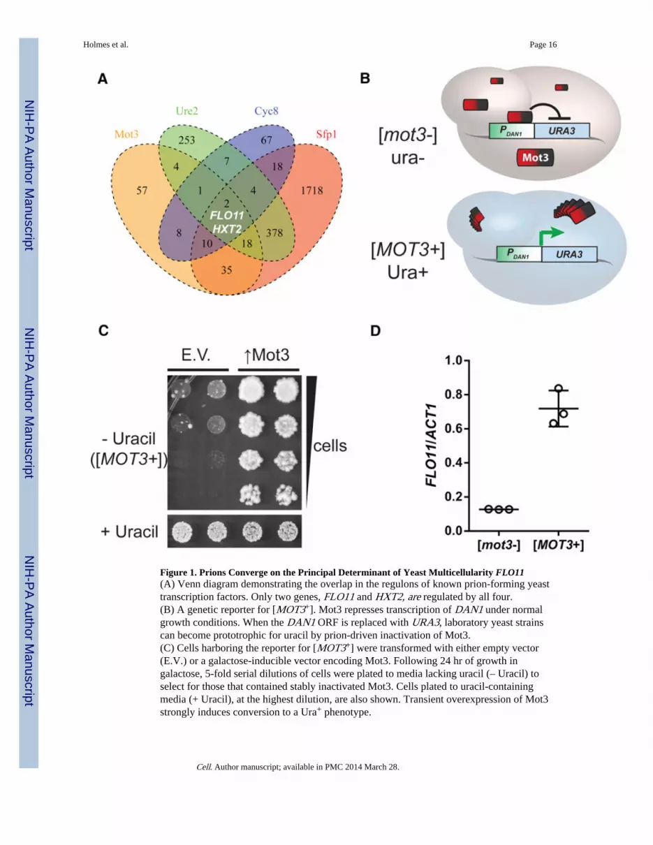
Figure 1. Prions Converge on the Principal Determinant of Yeast Multicellularity FLO11(A) Venn diagram demonstrating the overlap in the regulons of known prion-forming yeasttranscription factors. Only two genes, FLO11 and HXT2, are regulated by all four.(B) A genetic reporter for [MOT3+]. Mot3 represses transcription of DAN1 under normalgrowth conditions. When the DAN1 ORF is replaced with URA3, laboratory yeast strainscan become prototrophic for uracil by prion-driven inactivation of Mot3.(C) Cells harboring the reporter for [MOT3+] were transformed with either empty vector(E.V.) or a galactose-inducible vector encoding Mot3. Following 24 hr of growth ingalactose, 5-fold serial dilutions of cells were plated to media lacking uracil (– Uracil) toselect for those that contained stably inactivated Mot3. Cells plated to uracil-containingmedia (+ Uracil), at the highest dilution, are also shown. Transient overexpression of Mot3strongly induces conversion to a Ura+ phenotype.
Holmes et al. Page 16
Cell. Author manuscript; available in PMC 2014 March 28.
NIH
-PA Author Manuscript
NIH
-PA Author Manuscript
NIH
-PA Author Manuscript

(D) qRT-PCR using mRNA isolated from isogenic [MOT3+] and [mot3−] colonies revealsthat [MOT3+] cells have increased FLO11 expression. Values are normalized to ACT1.Error bars represent SD from triplicate colonies.See also Figure S1 and Tables S1 and S2.
Holmes et al. Page 17
Cell. Author manuscript; available in PMC 2014 March 28.
NIH
-PA Author Manuscript
NIH
-PA Author Manuscript
NIH
-PA Author Manuscript

Figure 2. [MOT3+] Enables Facultative Multicellular Growth(A) Schematics on the left depict multicellular phenotypes (dark cells) that are induced byeach of the common growth conditions indicated. On plates containing only proline as anitrogen source, [MOT3+] cells formed invasive filaments that penetrated the agar surface(top; invasive growth remains after dislodging surface cells under running water). On platescontaining only ethanol as a carbon source, [MOT3+] cells formed complex colonymorphologies (middle). The schematic for colony morphology is based on Váchová et al.(2011). When grown to saturation in liquid media, [MOT3+] cells exhibited a decreasedtendency to flocculate (bottom). Each of these behaviors of [MOT3+] cells was eliminated
Holmes et al. Page 18
Cell. Author manuscript; available in PMC 2014 March 28.
NIH
-PA Author Manuscript
NIH
-PA Author Manuscript
NIH
-PA Author Manuscript

by treating the cells transiently with GdHCl, an inhibitor of the prion-partitioning factorHsp104.(B) [MOT3+] cells form elaborate, biofilm-like structures when grown on semisolid ethanolmedia.Scale bars, 1 cm. See also Figure S2.
Holmes et al. Page 19
Cell. Author manuscript; available in PMC 2014 March 28.
NIH
-PA Author Manuscript
NIH
-PA Author Manuscript
NIH
-PA Author Manuscript

Figure 3. The N-Terminal Region Is Required for Prion-Mediated Inactivation of Mot3(A) A schematic of full-length Mot3 and two functionally distinct truncation mutants.Regions of high predicted prion-forming propensity are indicated in red. The C-terminalnonprion-like region of Mot3 contains two C2H2 zinc fingers, indicated in blue, which areinvolved in DNA binding. The prion-determining region, “PrD,” as initially defined byAlberti et al. (2009) encompasses the N-terminal 295 residues of the protein. A poly-asparagine (poly-N) tract stretches from residues 143–157. In the present study, weconstruct a variant, “ΔPrD,” that lacks much of the PrD including the poly-N tract. Anendogenous hexa-histidine motif is indicated in green.(B) Ectopic expression of ΔPrD (↑ΔPrD) from a constitutive promoter (TEF1) suppressesthe Ura+ phenotype of [MOT3+]. In contrast, [MOT3+] cells ectopically expressing full-length Mot3 (↑WT) remain Ura+. This is because WT Mot3, but not ΔPrD, accumulates asSDS-resistant aggregates, as visualized by SDD-AGE and immunoblotting against theendogenous hexa-histidine motif. Amyloids from endogenous Mot3 are too low abundanceto be detected in this exposure. An SDS-PAGE blot of lysates boiled in 2% LDSdemonstrates comparable expression of the Mot3 variants.
Holmes et al. Page 20
Cell. Author manuscript; available in PMC 2014 March 28.
NIH
-PA Author Manuscript
NIH
-PA Author Manuscript
NIH
-PA Author Manuscript

Figure 4. Prion-Mediated Inactivation of Mot3 Is Required but Is Not Sufficient to Convey theFull Spectrum of [MOT3+] Phenotypes(A) Ectopic expression of ΔPrD (↑ΔPrD) from a constitutive promoter (TEF1) suppressesthe complex colony morphology, invasion, and hypo-flocculation phenotypes of [MOT3+]cells. In contrast, [MOT3+] cells that ectopically express full-length Mot3 (↑WT) retain[MOT3+] phenotypes.(B) Genetic deletion of MOT3 confers some phenotypes that mimic those of [MOT3+],including biofilm formation and invasion. Δmot3 cells do not flocculate when grown tosaturation in liquid media.
Holmes et al. Page 21
Cell. Author manuscript; available in PMC 2014 March 28.
NIH
-PA Author Manuscript
NIH
-PA Author Manuscript
NIH
-PA Author Manuscript

(C) Genetic deletion of MOT3 is not sufficient to fully derepress the DAN1 promoter(PDAN1-URA3). Unlike [MOT3+] cells (which are Ura+), Δmot3 cells partition dynamicallybetween fully repressed (ura−) and derepressed (Ura+) states.(D) Ectopic expression of WT Mot3 stabilizes the derepressed (Ura+) state, whereas thenon-aggregating variant of Mot3, ΔPrD, restores the fully repressed (ura−) state. Expressionof PrD or empty vector has no effect. Duplicate transformants are shown, and serialdilutions were made onto the same plate. Bottom view is a western blot comparingexpression levels of the Mot3 variants.(E) In a hypothetical model for the gain of function of Mot3 prion conformers, Mot3interacts with another protein that corepresses transcription from the DAN1 promoter. In the[MOT3+] state, Mot3 prion particles sequester this protein, resulting in the full derepressionof the DAN1 promoter. In cells altogether lacking Mot3, this protein can still bind to andpartially repress the DAN1 promoter.Scale bars, 1 cm.
Holmes et al. Page 22
Cell. Author manuscript; available in PMC 2014 March 28.
NIH
-PA Author Manuscript
NIH
-PA Author Manuscript
NIH
-PA Author Manuscript

Figure 5. Environmental Conditions Govern Mot3 Prion Switching(A) The spontaneous switching of Mot3 to its prion state is increased by ethanol stress.Diploid [mot3−] cells, or as a negative control, WT/ΔPrD heterozygotes, were incubated for6 hr in media alone (mock), or media containing 12% ethanol (EtOH), prior to plating tomedia lacking uracil. Ura+ colonies were counted after 4 days and normalized by the numberof total colony-forming units. Data represent mean ± SD from three independentexperiments.(B) [mot3−] cells were transformed with either empty vector or a plasmid expressingHsp104 from a strong constitutive promoter (GPD). Transformants were inoculated to richmedia overnight, and then the fraction of [MOT3+] cells was determined as in (A).
Holmes et al. Page 23
Cell. Author manuscript; available in PMC 2014 March 28.
NIH
-PA Author Manuscript
NIH
-PA Author Manuscript
NIH
-PA Author Manuscript

(C) Passaging [MOT3+] cells under hypoxic conditions reverts them to [mot3−].(D) The effect of hypoxia is specific to [MOT3+]. A strain harboring prion states of threedifferent proteins ([MOT3+], [PSI+], and [RNQ+]) was treated transiently with either GdHClor hypoxia. Amyloids representing each prion state were detected by SDD-AGE. GdHCl,which targets the prion-partitioning factor Hsp104, eliminated all three prions, whereashypoxia specifically eliminated [MOT3+]. See also Figure S3.
Holmes et al. Page 24
Cell. Author manuscript; available in PMC 2014 March 28.
NIH
-PA Author Manuscript
NIH
-PA Author Manuscript
NIH
-PA Author Manuscript
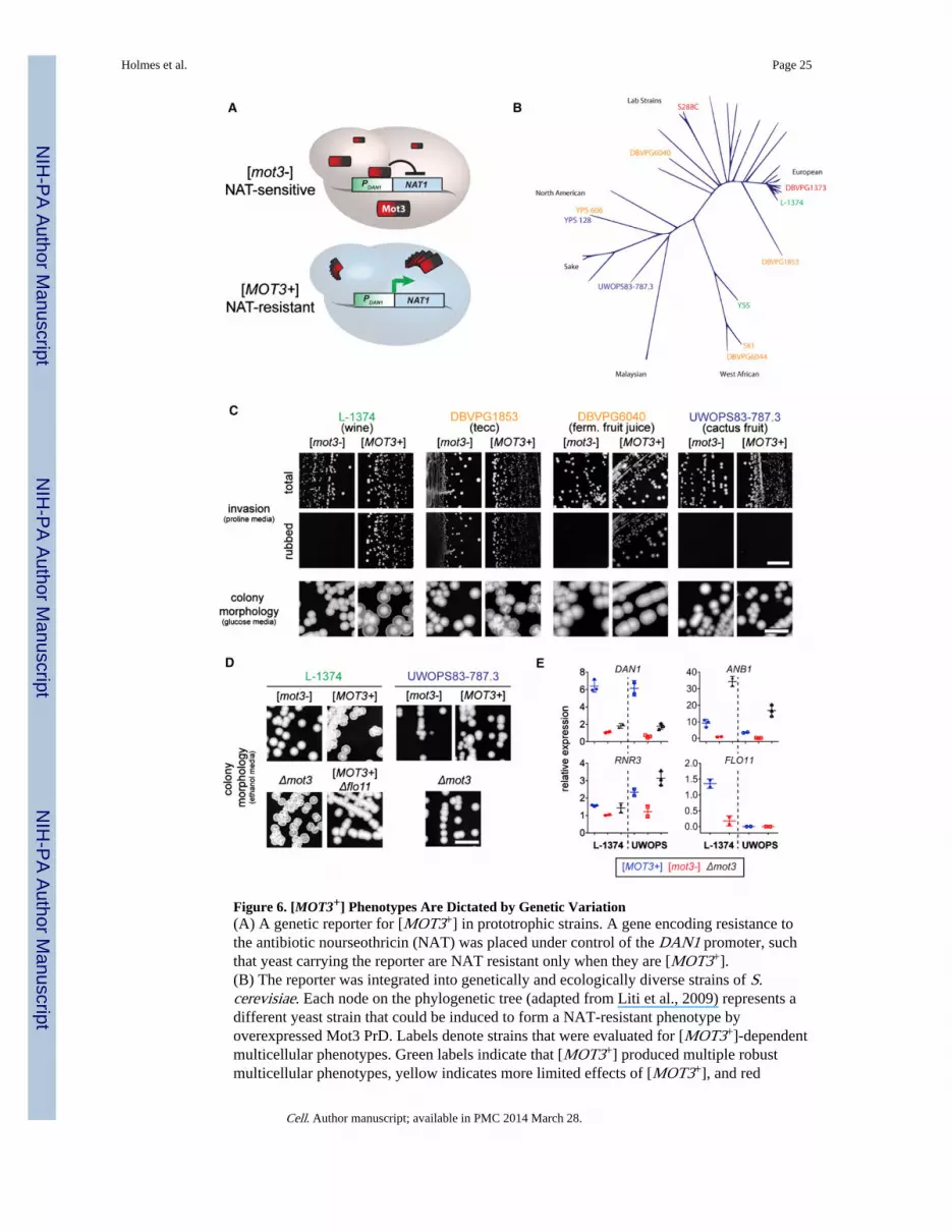
Figure 6. [MOT3+] Phenotypes Are Dictated by Genetic Variation(A) A genetic reporter for [MOT3+] in prototrophic strains. A gene encoding resistance tothe antibiotic nourseothricin (NAT) was placed under control of the DAN1 promoter, suchthat yeast carrying the reporter are NAT resistant only when they are [MOT3+].(B) The reporter was integrated into genetically and ecologically diverse strains of S.cerevisiae. Each node on the phylogenetic tree (adapted from Liti et al., 2009) represents adifferent yeast strain that could be induced to form a NAT-resistant phenotype byoverexpressed Mot3 PrD. Labels denote strains that were evaluated for [MOT3+]-dependentmulticellular phenotypes. Green labels indicate that [MOT3+] produced multiple robustmulticellular phenotypes, yellow indicates more limited effects of [MOT3+], and red
Holmes et al. Page 25
Cell. Author manuscript; available in PMC 2014 March 28.
NIH
-PA Author Manuscript
NIH
-PA Author Manuscript
NIH
-PA Author Manuscript

indicates no observable effect. The two strains labeled in blue exhibited [MOT3+]-dependent changes in flocculation.(C) [MOT3+] produces a different array of multicellular phenotypes in each strain, asdemonstrated by invasive behaviors and colony morphologies in four divergent strains.ferm., fermenting.(D) [MOT3+]-dependent morphological differences between L-1374 and UWOPS83-787.3are reproduced by deleting MOT3. [MOT3+]-dependent multicellularity in L-1374 requiresFLO11.(E) Demonstrative qRT-PCR analyses of the expression of Mot3-regulated genes forisogenic [MOT3+], [mot3−], and Δmot3 cells in L-1374 and UWOPS83-787.3. The directeffect of Δmot3 on FLO11 was not determined. Error bars represent SD from separateexperiments.Scale bars, 1 cm.See also Figure S4 and Tables S4 and S5.
Holmes et al. Page 26
Cell. Author manuscript; available in PMC 2014 March 28.
NIH
-PA Author Manuscript
NIH
-PA Author Manuscript
NIH
-PA Author Manuscript

Figure 7. Model for a Function for Mot3 Prion Switching in the Respiro-Fermentative Cycle ofWine YeastsYeast cells begin the cycle upon inoculation to glucose-rich grape must. Glucose isfermented to ethanol during the exponential growth phase. The ensuing ethanol stresstriggers prion conversion to [MOT3+], which, in conjunction with glucose exhaustion,drives some of the cells into a multicellular growth program that protects them from stressand/or increases metabolic efficiency. As the population respires ethanol and remainingsugars, oxygen levels decline resulting in the accelerated reappearance of [mot3−] cellswithin [MOT3+] subpopulations.
Holmes et al. Page 27
Cell. Author manuscript; available in PMC 2014 March 28.
NIH
-PA Author Manuscript
NIH
-PA Author Manuscript
NIH
-PA Author Manuscript


