Embed Size (px)
Citation preview
High-resolution high-speed panoramic cardiac imaging system
Dale W. Evertson (In Memoriam)1, Mark R. Holcomb2, Matthew D.C. Eames3,4, Mark-AnthonyP. Bray3,5, Veniamin Y. Sidorov3, Junkai Xu2, Holley Wingard1, Hana M. Dobrovolny6,7,Marcella C. Woods3, Daniel J. Gauthier6,8, and John P. Wikswo2,3,9,10
1 Department of Mechanical Engineering, Vanderbilt University
2 Department of Physics and Astronomy, Vanderbilt University
3 Department of Biomedical Engineering, Vanderbilt University
6 Department of Physics, Duke University
7 Center for Nonlinear and Complex Systems, Duke University
8 Department of Biomedical Engineering, Duke University
9 Department of Molecular Physiology and Biophysics, Vanderbilt University
10 Vanderbilt Institute for Integrative Biosystems Research and Education
AbstractA panoramic cardiac imaging system consisting of three high-speed CCD cameras has beendeveloped to image the surface electrophysiology of a rabbit heart via fluorescence imaging using avoltage-sensitive fluorescent dye. A robust, unique mechanical system was designed to accommodatethe three cameras and to adapt to the requirements of future experiments. A unified computer interfacewas created for this application – a single workstation controls all three CCD cameras, illumination,and stimulation, and the stepping motor rotates the heart. The geometric reconstruction algorithmswere adapted from a previous cardiac imaging system. We demonstrate the system by imaging apolymorphic cardiac tachycardia.
Author Contact Information Mark R. Holcomb, [email protected], Phone: (615) 343-4124, Fax: (615) 322-4977,Vanderbilt University, Department of Physics and Astronomy, 6301 Stevenson Center, Nashville, TN 37235-1807Matthew D.C. Eames, [email protected], Phone: (434) 243-6316, Fax: (434) 982-3870, University of Virginia, Department ofBiomedical Engineering, 415 Lane Rd. MR-5 Bldg, P.O. Box 800759, Charlottesville, VA 22908Mark-Anthony Bray, [email protected], Phone: (617) 495-3130, Fax: (617) 495-8534, 60 Oxford St, Rm 331, Cambridge, MA02138Veniamin Y. Sidorov, [email protected]., Phone: (615) 343-8170, Fax: (615) 322-4977, Vanderbilt University, Department ofBiomedical Engineering, 6301 Stevenson Center, Nashville, TN 37235-1631Junkai Xu, [email protected], Phone: (615) 343-7108-5, Fax: (615) 322-4977, Vanderbilt University, Department of Physicsand Astronomy, 6301 Stevenson Center, Nashville, TN 37235Holley Wingard, [email protected], Phone: (615) 322-2443, Fax: (615) 343-6687, Vanderbilt University, Department of MechanicalEngineering, 2301 Vanderbilt Place, Nashville, TN. 37235-1592Hana M. Dobrovolny, [email protected], Phone: (919) 660-2512, Fax: (919) 660-2525, Department of Physics, Duke University,Box 90305, Durham, NC, 27708Marcella C. Woods, [email protected]., Phone: (615) 343-1645, Fax: (615) 343-7919, Department of BiomedicalEngineering, Vanderbilt University, 5824 Stevenson Center, Nashville, TN 37235-1631Daniel J. Gauthier, [email protected], Phone: (919) 660-2511, Fax: (919) 660-2525, Department of Physics, Duke University. Box90305, Durham, NC 27708John P. Wikswo, [email protected]., Phone: (615) 343-4124, Fax: (615) 322-4977, Department of Physics, 6301 StevensonCenter, Nashville, TN 37235-18074Present Address, Department of Biomedical Engineering, University of Virginia5Present Address, Division of Engineering and Applied Sciences, Harvard University
NIH Public AccessAuthor ManuscriptIEEE Trans Biomed Eng. Author manuscript; available in PMC 2009 March 1.
Published in final edited form as:IEEE Trans Biomed Eng. 2008 March ; 55(3): 1241–1243. doi:10.1109/TBME.2007.912417.
NIH
-PA Author Manuscript
NIH
-PA Author Manuscript
NIH
-PA Author Manuscript

INTRODUCTIONCardiac tissue is an electrically excitable medium that can support either normal or abnormalpatterns of activation. The initiation of activation and its subsequent propagation bearsignificant importance in the design of pacemakers, defibrillators, and antiarrhythmic drugs.By studying the cardiac electrophysiology of rabbit hearts, researchers may extrapolate theacquired data and apply it to human heart electrophysiology. Panoramic cardiac opticalmapping using a voltage-sensitive dye to report the time-dependence of the transmembranepotential has been used previously to record and analyze cardiac electrical activity. Thistechnique, described in preliminary form by Lin et al. [1] and further expanded by Bray etal. [2], involves (1) imaging an excised, Langendorf-perfused rabbit heart from three points ofview, (2) numerically reconstructing the heart surface features, and (3) texture-mapping theacquired fluorescence images of the heart’s electrical activity onto the virtual surface. Theresult is a movie of the electrical activity across the entire 3-dimensional epicardial surface.Subsequently, panoramic systems for studying hearts have been developed by Kay et al. [4],and Qu et al. [5]. In addition to a more extended discussion than presented in thiscommunication, the supplement contains detailed descriptions of the surface reconstruction,and the custom kinematic mount, base plate, and computer control.
CAMERASThe system shown in Figure 1 uses three high-speed, low noise, CCD cameras spaced 120degrees around the heart on the horizontal plane. The three synchronized 14-bit CCD cameras(CardioCCD-SMQ, RedShirt Imaging) are used to image the electrical activity across thesurface of a heart. Each camera is capable of recording up to 2000, 3000, and 5000 fps atresolutions of 80 × 80, 40 × 40, and 26 × 26 pixels, respectively. We also have an alternateconfiguration which uses 128 × 128 pixel cameras running at 490 fps (DS-12-16K5H, Dalsa).All cameras are controlled by a single workstation (Precision 650, Dell) equipped with 2 GBof RAM. Both camera configurations use 12-mm C-mount lenses (LM12JCM, Kowa).
Qu et al. [5] make use of 16 × 16 element photodiode arrays (PDA) operating at 5000 fps in apanoramic system with similar experimental goals. The RedShirt cameras match the PDAframe rate while offering 250% more pixels. The 40 × 40 3000 fps mode is only 60% as fast,but offers 625% more pixels. Figure 2 shows raw (unfiltered) data from single pixels. Thesignal-to-noise ratio (SNR) for the RedShirt (~30) is slightly less than that of the PDAs but isfully acceptable for quantitative imaging of cardiac arrhythmias and fibrillation. Additionally,spatial filtering down to the resolution of PDAs would increase the SNR substantially. In thisapplication, the higher spatial resolution, along with the flexibility to change binning modeseasily with a software setting, makes the RedShirt an arguably superior choice over both PDAsand the Dalsa CCDs. Advanced complementary metal oxide semiconductor (CMOS) cameras,although expensive, may prove even better [6]. In contrast to PDA-based systems [4,5] we donot require the use of an additional video or CCD camera to obtain the high-resolution imagesrequired to create the wire frame model of the heart.
Each of the three cameras in the new system may be independently positioned and directed atthe target object. The camera mounts (PO80N, Newport) shown in Figure 3 allow easyadjustment of the pan, tilt, and roll of the camera to optimize the position of the heart in thefield-of-view.
ILLUMINATIONWe excite the voltage-sensitive dye with an illumination system comprising six high-intensitygreen LEDs (LXHL-LM5C, Luxeon) arranged about the heart and centered on the faces of avirtual cube aligned with its diagonal (1 1 1) axis oriented along the vertical axis of the system.
Evertson (In Memoriam) et al. Page 2
IEEE Trans Biomed Eng. Author manuscript; available in PMC 2009 March 1.
NIH
-PA Author Manuscript
NIH
-PA Author Manuscript
NIH
-PA Author Manuscript

We mounted the LEDs on CPU heat sinks and fans (SKU 275016, COMPUSA). A custombracket secures a lens (LXHL-NX05, Luxeon) to the front surface of each LED. The LEDs arepowered by three dual-mode power supplies (E3610A, Agilent). The shutter motors on acustom mount (Standard Servo Stock#: 900–00005, Parallax) are controlled by simple pulse-width modulation and open and close automatically before and after data acquisition. Theshutter system allows the LEDs to remain on continuously at steady-state intensity whilepreventing photobleaching by only exposing the heart to light during data acquisition. Thefluorescence response from the LED illumination is capable of saturating the cameras. Theentire LED illumination system, including power supplies, costs approximately $2,000,making it much more cost-effective than the $50,000 frequency-doubled Nd:YAG lasers(Verdi, Coherent) used in the previous system and also available for use with this system.
PERFUSION SYSTEMThe pumps and heat exchangers used in this experimental setup are identical to or are updatedversions of those used in our previous system [1,2]. For the purposes of this paper, the keyfunction of the perfusion system is to maintain the viability of the heart ex vivo while theexperiment is performed and data recorded. The perfusion system is also used to administerthe voltage-sensitive fluorescent dye, di-4-ANEPPS, (Molecular Probes Inc) to the cardiactissue.
ROTATION SYSTEMThe heat exchanger in the perfusion system is suspended from a computer-controlled steppingmotor (S57-102-MO, Compumotor), connected to a 20:1 gearhead (PG60-020-T01, Bayside)providing precise, 1° increments in the position for the sequence of 120 images necessary forcreating the wire-frame reconstruction of the heart surface features with voxels no larger than0.4 mm. The motor acceleration is chosen to minimize swinging.
ENCLOSUREA custom-built aluminum Faraday cage (Duck Welding, Nashville) provides bothelectromagnetic isolation, a light-tight enclosure for the system, thermal control, and a traywith drain for catching spills. The enclosure is fabricated from 5-mm-thick aluminum floorplate and has double-doors on three sides to ensure convenient access to the camera system.The Faraday enclosure is cemented to a concrete-block wall that is spanned by steel-reinforcedconcrete lintels to minimize vibration. A metal framework suspended from the ceiling supportsthe perfusion system pumps and power supplies and isolates their vibrations from the camerasystem.
RESULTSUsing the system just described, we conducted three demonstration experiments using rabbithearts: two using the RedShirt configuration and one using the Dalsa cameras. Figure 4 showsa sample of the results using the RedShirt cameras in their 1000 fps-80 × 80 pixel mode. Theheart was prepared as described previously [1]. Ten frames from a 45 ms interval of a rabbitheart in polymorphic tachycardia are shown. Every fifth frame is shown, which gives a 5 msinterval between images. The sequence shows a figure-of-eight reentry, which is a commonphenomenon observed in cardiac arrhythmias. We estimate the mean reconstruction error tobe on the order of 1.2 mm.
Evertson (In Memoriam) et al. Page 3
IEEE Trans Biomed Eng. Author manuscript; available in PMC 2009 March 1.
NIH
-PA Author Manuscript
NIH
-PA Author Manuscript
NIH
-PA Author Manuscript

DISCUSSIONWe have described and demonstrated a system which allows for both high spatial and temporalresolution of the transmembrane potential over the entire epicardial surface of a rabbit heart.Our implementation of the panoramic camera is designed to optimize the mechanical stabilityof the system, the accuracy of the reconstruction, and ease of use. Elsewhere we will reportthe use of this system to study the effects of global ischemia on propagation, and the virtualelectrode distribution from intracavity defibrillation strength shocks.
Supplementary MaterialRefer to Web version on PubMed Central for supplementary material.
AcknowledgementsThis work was supported in part by the National Institutes of Health (R01-HL58241) and the Vanderbilt Institute forIntegrative Biosystems Research and Education (VIIBRE). Hana Dobrovolny and Daniel Gauthier gratefullyacknowledge the financial support of the National Science Foundation (PHY-0243584) and the National Institutes ofHealth (1R01-HL-72831). We thank Richard Gray of the University of Alabama at Birmingham for sharing his twoRedShirt cameras for collaborative studies.
References1. Lin SF, Wikswo JP Jr. Panoramic optical imaging of electrical propagation in isolated heart. J Biomed
Opt 1999;4:200.2. Bray M-A, Lin SF, Wikswo JP Jr. Three-dimensional surface reconstruction and fluorescent
visualization of cardiac activation. IEEE Trans BME 2000;47:1383.3. Niem W. Robust and fast modeling of 3D natural objects from multiple views. SPIE Proc, Image and
Video Processing II 1994;2182:388–397.4. Kay M, Rogers J. Three-dimensional surface reconstruction and panoramic optical mapping of large
hearts. IEEE Trans BME 2004;51:1219.5. Qu F, Fritz A, Cheng Y, Nikolski V, Efimov IR. Panoramic Imaging of Electrical Activity in the Rabbit
Heart. PACE 2003;26:946.6. Tallini, et al. Imaging cellular signals in the heart in vivo: Cardiac expression of the high-signal Ca2
+ indicator GCaMP2. Proc Natl Acad Sci U S A 2006 March 21;103(12):4753–4758. [PubMed:16537386]
Evertson (In Memoriam) et al. Page 4
IEEE Trans Biomed Eng. Author manuscript; available in PMC 2009 March 1.
NIH
-PA Author Manuscript
NIH
-PA Author Manuscript
NIH
-PA Author Manuscript

Figure 1.System as seen from outside the Faraday cage through one of three sets of double doors.
Evertson (In Memoriam) et al. Page 5
IEEE Trans Biomed Eng. Author manuscript; available in PMC 2009 March 1.
NIH
-PA Author Manuscript
NIH
-PA Author Manuscript
NIH
-PA Author Manuscript

Figure 2.Single pixel pacing data. (A) Raw data (unfiltered) acquired at 3000 fps at a resolution of 40× 40 pixels. (B) Raw data (unfiltered) acquired at 5000 fps at a resolution of 26 × 26 pixels.LEDs were used for (A) and laser illumination (Verdi, Coherent) for (B). The SNR for A andB are 31 and 35, respectively, based on SNR = (S1 Amplitude)/(Standard deviation duringdiastolic interval). The pixels used were on the left ventricle, but from different hearts. Therecordings shown were taken shortly after the hearts were first stained, but are not atypicalsignals.
Evertson (In Memoriam) et al. Page 6
IEEE Trans Biomed Eng. Author manuscript; available in PMC 2009 March 1.
NIH
-PA Author Manuscript
NIH
-PA Author Manuscript
NIH
-PA Author Manuscript
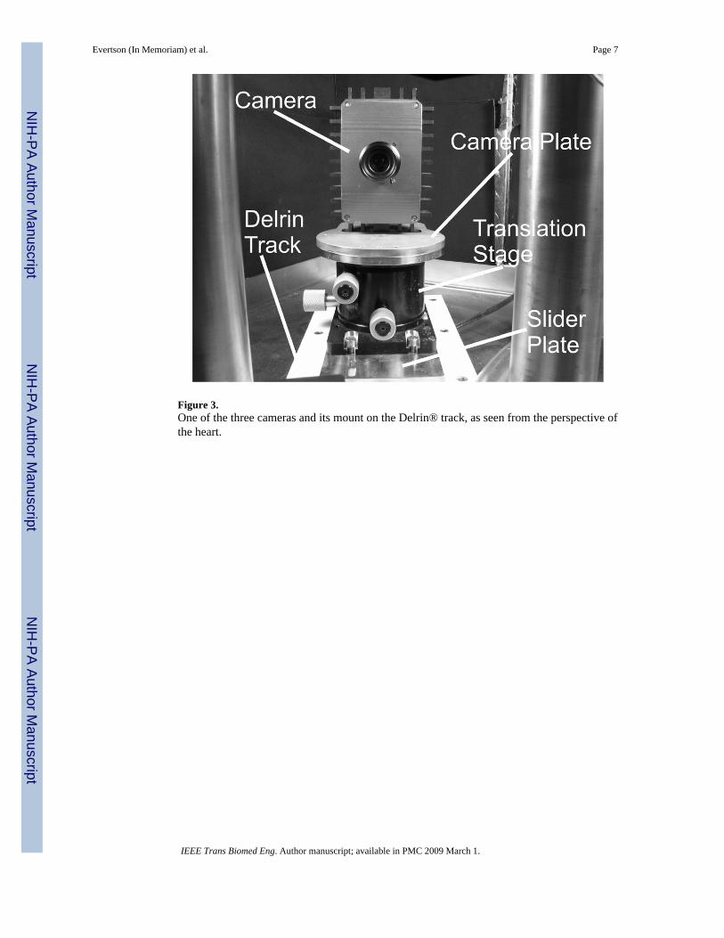
Figure 3.One of the three cameras and its mount on the Delrin® track, as seen from the perspective ofthe heart.
Evertson (In Memoriam) et al. Page 7
IEEE Trans Biomed Eng. Author manuscript; available in PMC 2009 March 1.
NIH
-PA Author Manuscript
NIH
-PA Author Manuscript
NIH
-PA Author Manuscript

Figure 4.Figure-of-eight reentry during polymorphic tachycardia. Every 5th frame of a 1000 fpssequence is shown. The excited tissue is red. The persistent red dot in the lower right of eachimage is an artifact due to the electrode.
Evertson (In Memoriam) et al. Page 8
IEEE Trans Biomed Eng. Author manuscript; available in PMC 2009 March 1.
NIH
-PA Author Manuscript
NIH
-PA Author Manuscript
NIH
-PA Author Manuscript


