Embed Size (px)
Citation preview

International Scholarly Research NetworkISRN Minimally Invasive SurgeryVolume 2012, Article ID 364285, 9 pagesdoi:10.5402/2012/364285
Research Article
High-Resolution Optical Imaging of Benign andMalignant Mucosa in the Upper Aerodigestive Tract:An Atlas for Image-Guided Surgery
Lauren L. Levy,1 Peter M. Vila,1 Richard W. Park,1 Richard Schwarz,2
Alexandros D. Polydorides,3 Marita S. Teng,1 Vivek V. Gurudutt,1 Eric M. Genden,1
Brett Miles,1 Sharmila Anandasabapathy,4 Ann M. Gillenwater,5
Rebecca Richards-Kortum,2 and Andrew G. Sikora1
1 Department of Otolaryngology-Head and Neck Surgery, Mount Sinai School of Medicine, New York, NY 10029, USA2 Department of Bioengineering, Rice University, Houston, TX 77030, USA3 Department of Pathology, Mount Sinai School of Medicine, New York, NY 10029, USA4 Division of Gastroenterology, Department of Medicine, Mount Sinai School of Medicine, New York, NY 10029, USA5 Division of Surgery, Head & Neck Surgery Department, University of Texas M.D. Anderson Cancer Center, Houston,TX 77030, USA
Correspondence should be addressed to Andrew G. Sikora, [email protected]
Received 18 June 2012; Accepted 9 July 2012
Academic Editors: M. Barczynski and K. J. Dedes
Copyright © 2012 Lauren L. Levy et al. This is an open access article distributed under the Creative Commons Attribution License,which permits unrestricted use, distribution, and reproduction in any medium, provided the original work is properly cited.
Background. High-resolution optical imaging provides real-time visualization of mucosa in the upper aerodigestive tract (UADT)which allows non-invasive discrimination of benign and neoplastic epithelium. The high-resolution microendoscope (HRME)utilizes a fiberoptic probe in conjunction with a tissue contrast agent to display nuclei and cellular architecture. This technologyhas broad potential applications to intraoperative margin detection and early cancer detection. Methods. Our group has createdan extensive image collection of both neoplastic and normal epithelium of the UADT. Here, we present and describe imagingcharacteristics of benign, dysplastic, and malignant mucosa in the oral cavity, oropharynx, larynx, and esophagus. Results. Thereare differences in the nuclear organization and overall tissue architecture of benign and malignant mucosa which correlate withhistopathologic diagnosis. Different anatomic subsites also display unique imaging characteristics. Conclusion. HRME allowsdiscrimination between benign and neoplastic mucosa, and familiarity with the characteristics of each subsite facilitates correctdiagnosis.
1. Introduction
Failure to obtain clear (tumor free) surgical margins is anadverse prognostic factor for patients with head and necksquamous cell carcinoma [1, 2]. Thus, during ablative cancersurgery complete removal of all malignant tissue is necessaryto maximize survival and decrease the chance of recurrence.However, unnecessary removal of normal tissue can leadto serious deficiencies in the ability to speak, swallow, orchew, with an overall decreased quality of life [3, 4]. Toobtain clear margins, the surgeon must discriminate betweenneoplastic and surrounding normal tissue during tumor
resection; currently, the extent of disease is defined usingvisual examination and palpation. Intraoperative “frozensection” pathological margins are often necessary to confirmcritical or questionable tumor margins. Although frozensections during ablative head and neck cancer surgery are avital adjunct to visual and tactile examination, the procedureis costly and time-consuming, and discrepancies betweenfrozen section margins and final pathology are common [5–7]. Thus, techniques for real-time visualization of tumormargins at the time of surgery have the potential to reducethe number and enhance the accuracy of frozen sectiondeterminations.

2 ISRN Minimally Invasive Surgery
(a)
Tissue sample
USB
Fiber optic probe
CCD camera
HRME internal configuration
Laptop computerwith image capture
10x objective lens
(b) (c)
Figure 1: The high resolution microendoscope (HRME). (a) External and internal appearance of the imaging device. (b) Simplifiedschematic diagram of the HRME. (c) In vivo use of the HRME on healthy mucosa of the lip.
Table 1: Demographics of imaging database.
Anatomical site(# of surgical specimens)
Number of sites withlisted pathological
diagnoses
Number ofimages incollection
Oral cavity (25)
Normal 35 195
Dysplasia 8 57
Cancer 41 394
Total 84 646
Oropharynx (29)
Normal 31 192
Dysplasia 9 51
Cancer 31 212
Total 71 455
Larynx (10)
Normal 21 112
Dysplasia 4 17
Cancer 19 226
Total 44 355
All sites (64)
Normal 87 499
Dysplasia 21 125
Cancer 91 832
Total 199 1,456
Image guided cancer surgery is an emerging area ofresearch, and several optical imaging modalities have beenproposed to improve intraoperative delineation of tumormargins [8]. These include wide-field imaging of tissueautofluorescence to delineate tumor margins based on lossof autofluorescence and high-resolution optical imaging toimage margins based on changes in tissue architecture and
cellular morphology [9–14]. Imaging of tissue autofluo-rescence during surgical resection has been suggested todecrease recurrence rate of oral cancers and currently a ran-domized, multicentre, double blind, and controlled surgicaltrial is underway to further validate these preliminary results[15, 16].
High-resolution images, such as those provided byconfocal microscopy provide the ability to spatially resolvemorphological changes in the epithelia that occur dur-ing neoplasia, including changes in nuclear morphologyand distribution [17, 18]. High-resolution confocal opticalimages can reveal morphologic details with similar qualityto that which can be seen in histological slides, but areobtained in a non-invasive manner and without the need forslide preparation and staining. However, despite their utilityand impressive resolution, confocal imaging devices arecomplex and expensive. As an alternative approach, we havepreviously described a portable high-resolution microendo-scope (HRME), which utilizes a flexible fiberoptic probe tointerrogate tissue treated with a topically applied fluorescentnuclear contrast agent. This system allows visualization ofepithelial architecture at video rate. This device (Figure 1)has been used to identify Barrett’s dysplasia in the esoph-agus, axillary lymph node metastasis in breast cancer, andneoplasia in resected oral squamous carcinoma specimenswith high sensitivity and specificity [19–21]. In a recentstudy, medical professionals were readily able to discriminatebenign and malignant images of the UADT following a brieftraining presentation, suggesting the potential utility of thisapproach for image-guided surgery in the head and neck[22].
In this paper, we describe the imaging characteristicsof benign mucosa and squamous cell carcinoma of theUADT, and present representative HRME images from exvivo surgical specimens and in vivo endoscopy as an atlas forimage-guided surgery.
ISRN Minimally Invasive Surgery 3
(a) (b)
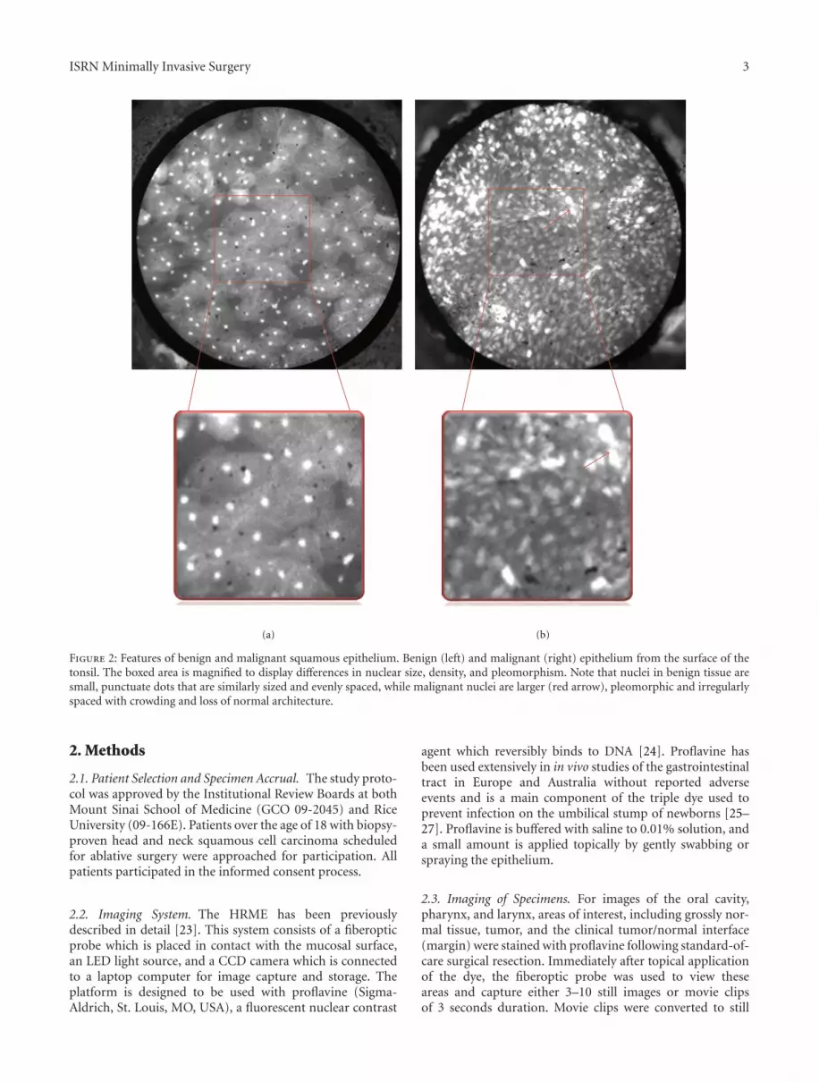
Figure 2: Features of benign and malignant squamous epithelium. Benign (left) and malignant (right) epithelium from the surface of thetonsil. The boxed area is magnified to display differences in nuclear size, density, and pleomorphism. Note that nuclei in benign tissue aresmall, punctuate dots that are similarly sized and evenly spaced, while malignant nuclei are larger (red arrow), pleomorphic and irregularlyspaced with crowding and loss of normal architecture.
2. Methods
2.1. Patient Selection and Specimen Accrual. The study proto-col was approved by the Institutional Review Boards at bothMount Sinai School of Medicine (GCO 09-2045) and RiceUniversity (09-166E). Patients over the age of 18 with biopsy-proven head and neck squamous cell carcinoma scheduledfor ablative surgery were approached for participation. Allpatients participated in the informed consent process.
2.2. Imaging System. The HRME has been previouslydescribed in detail [23]. This system consists of a fiberopticprobe which is placed in contact with the mucosal surface,an LED light source, and a CCD camera which is connectedto a laptop computer for image capture and storage. Theplatform is designed to be used with proflavine (Sigma-Aldrich, St. Louis, MO, USA), a fluorescent nuclear contrast
agent which reversibly binds to DNA [24]. Proflavine hasbeen used extensively in in vivo studies of the gastrointestinaltract in Europe and Australia without reported adverseevents and is a main component of the triple dye used toprevent infection on the umbilical stump of newborns [25–27]. Proflavine is buffered with saline to 0.01% solution, anda small amount is applied topically by gently swabbing orspraying the epithelium.
2.3. Imaging of Specimens. For images of the oral cavity,pharynx, and larynx, areas of interest, including grossly nor-mal tissue, tumor, and the clinical tumor/normal interface(margin) were stained with proflavine following standard-of-care surgical resection. Immediately after topical applicationof the dye, the fiberoptic probe was used to view theseareas and capture either 3–10 still images or movie clipsof 3 seconds duration. Movie clips were converted to still

4 ISRN Minimally Invasive Surgery
(a) (b)
(c)
∗
(d)
Figure 3: Oral cavity: representative images with corresponding histopathology (H&E original magnification 100x) of benign (top) andmalignant (bottom) mucosa. (a) Floor of mouth. (b) Tongue. (c) Mucosal lip. (d) Maxilla and overlying gingiva. ∗indicates keratinizingsquamous epithelium which appears hyperfluorescent on HRME. Red arrow indicates cotton fiber (artifact) present on the tissue followingapplication of proflavine.
images using Windows Movie Maker (Microsoft, Redmond,WA). A 3-mm punch biopsy of the imaged site was analyzedby conventional H&E histopathology by a board-certifiedpathologist.
In vivo Images of the esophagus were obtained duringendoscopy. The esophagus was sprayed with 1–3 ml of 0.01%
proflavine. The fiberoptic probe was then inserted throughthe biopsy channel of the endoscope. The probe was placed ingentle contact with the mucosa and video images obtained inreal time. A small dimple was made on the imaged site usingthe probe tip, and the imaged area was biopsied and analyzedby conventional H&E histopathology.

ISRN Minimally Invasive Surgery 5
(a) (b)
Figure 4: Oropharynx: representative images with corresponding histopathology (H&E original magnification 100x) of benign (top) andmalignant (bottom) mucosa. (a) Base of tongue. (b) Tonsil.
[t!]
(a) (b)
(c) (d)
Figure 5: Larynx: representative images with corresponding histopathology (H&E original magnification 100x) of benign (a and b) andmalignant (c and d) mucosa. (a), (b), and (c) are from the supraglottic larynx. (d) is from a glottic tumor. Arrow indicates hyperflourescentarea from proflavine staining.

6 ISRN Minimally Invasive Surgery
(a)
(b)
Figure 6: Characteristics of dysplasia. (a) Benign mucosa (left), high-grade dysplasia (center), and squamous cell carcinoma (right) onthe surface of the tonsil. (b) Dysplasia of the larynx (top) and floor of mouth (bottom) with corresponding histopathology (H&E originalmagnification 100x). Note enlarged and crowded nuclei with loss of normal cellular architecture.
HRME images were analyzed to identify imaging fea-tures of benign and malignant mucosa which correlatewith histopathological diagnosis, including nuclear size andshape, nuclear density, and overall tissue architecture.
3. Results
We generated an extensive library of images from varioussites in the upper aerodigestive tract including the oral cavity,oropharynx, larynx, and esophagus. From June 2009 to May2011, sixty four surgical specimens were imaged with theHRME. Over 1400 still images of benign, malignant, anddysplastic mucosa at various anatomical sites were obtained.Table 1 provides the breakdown by site of our currentimaging collection (Table 1).
For each anatomic site in the UADT, reproducibledifferences were observed in HRME images of benign andmalignant mucosa. In general, HRME images of benignmucosa are characterized by nuclei of consistent, regular
size, which are evenly spaced. This contrasts with malignantmucosa, in which the nuclei are enlarged and display crowd-ing with lack of organized tissue architecture, correspondingto increased cellularity found in cancerous tissue (Figure 2).
While differences in HRME images of benign and malig-nant tissue are generally consistent across anatomic sites,each mucosal site has a slightly different imaging appearance;thus, it is important for those interpreting HRME images tohave familiarity with each subsite of the UADT.
In the oral cavity (Figure 3), HRME images obtainedfrom the floor of mouth, tongue, and lip consistently displaythe previously described features of benign and malignantmucosa. However, images of heavily keratinized sites, likethe hard palate and gingiva (Figure 3(d)), can appearhyperfluorescent due to the affinity of proflavine for keratin.
Images of the oropharynx (base of tongue and tonsil),reliably display features of benign and malignant mucosa(Figure 4). Again, the nuclei of benign mucosa are punc-tate and regularly spaced, whereas images from malignant

ISRN Minimally Invasive Surgery 7
(a) (b)
(c)
∗
(d)
Figure 7: Esophagus. Representative images of (a) benign squamous epithelium; (b) high grade dysplasia; (c) adenocarcinoma; (d) Barrett’smetaplasia (H&E original magnification 100x). Notice glandular structures have imaging characteristics distinct from squamous epithelium,indicated by ∗.
mucosa show nuclei which are irregular, enlarged, andextremely disorganized. Characteristics of benign and malig-nant tissue from the larynx (Figure 5) are consistent withthose obtained from other sites. However, pseudostratifiedciliated columnar (respiratory) epithelium is a prominentfeature of this region and inexperienced observers mayconfuse the increased number of nuclei in respiratoryepithelium with dysplastic or cancerous mucosa.
Dysplasia is a common feature of UADT mucosaexposed to environmental carcinogens such as tobacco andalcohol, and dysplastic tissue has unique imaging features(Figure 6). Like malignant tissue, dysplastic mucosa alsocontains increased density of cell nuclei. However, dysplasticmucosa differs from malignant mucosa in that the tissuearchitecture is disorganized to varying degrees rather thanabsent. While it can be difficult to distinguish betweencancerous and dysplastic mucosa, the HRME character-istics of both pathologies are quite distinct from thoseof normal tissue. Thus, when choosing a biopsy site ordetermining surgical margins, abnormal mucosa is readilyidentified.
The fiberoptic probe can easily be delivered through aflexible endoscope and used during diagnostic or therapeuticendoscopy of the esophagus (Figure 7). The appearance ofbenign squamous epithelium in the esophagus is almostidentical to those of benign squamous epithelium at othersites of the UADT, displaying evenly spaced nuclei andintact cellular architecture (Figure 7(a)). Images of tis-sue with dysplasia or squamous cell carcinoma (Figures7(b) and 7(c)) feature both increased number of nuclei
and increased nuclear size. Interestingly, in the loweresophagus, the appearance of glandular mucosa is quitedistinct from squamous mucosa, thus making it possibleto identify the precancerous condition Barrett’s metaplasia(Figure 7(d)).
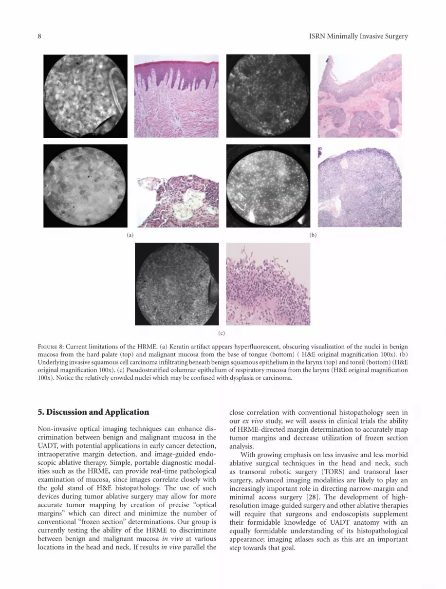
4. Current Limitations
Keratinized tissue (Figure 8) is a diagnostic challenge, sinceproflavine has an affinity for keratin which can obscure thevisualization of nuclei. The epithelium of both benign andcancerous tissue can contain keratin, which is a normalconstituent of the epithelium on the alveolar ridge andhard palate. However, ectopic keratinization can accompanyneoplastic transformation, and keratinization of normallynonkeratinized tissue is thus a potential diagnostic hallmarkof cancer. Submucosal tumor spread is another significantchallenge, since the depth of penetration of the fiberopticprobe is limited to approximately 25–50 micrometers (Fig-ure 8(b)). Therefore, images may be classified as normalwhen benign epithelium overlies tumor situated belowthis depth of penetration. Another potential confounder isrespiratory epithelium in the larynx, which has a greaterdensity of nuclei than typical squamous epithelium, thusmaking it more difficult to distinguish benign and malignantepithelium (Figure 8(c)). These limitations are active areasof ongoing research into strategies such as alternativecontrast agents, quantitative analysis of nuclear morphologyand pattern, and submucosal delivery of the fiberopticprobe.
8 ISRN Minimally Invasive Surgery
(a) (b)
(c)
Figure 8: Current limitations of the HRME. (a) Keratin artifact appears hyperfluorescent, obscuring visualization of the nuclei in benignmucosa from the hard palate (top) and malignant mucosa from the base of tongue (bottom) ( H&E original magnification 100x). (b)Underlying invasive squamous cell carcinoma infiltrating beneath benign squamous epithelium in the larynx (top) and tonsil (bottom) (H&Eoriginal magnification 100x). (c) Pseudostratified columnar epithelium of respiratory mucosa from the larynx (H&E original magnification100x). Notice the relatively crowded nuclei which may be confused with dysplasia or carcinoma.
5. Discussion and Application
Non-invasive optical imaging techniques can enhance dis-crimination between benign and malignant mucosa in theUADT, with potential applications in early cancer detection,intraoperative margin detection, and image-guided endo-scopic ablative therapy. Simple, portable diagnostic modal-ities such as the HRME, can provide real-time pathologicalexamination of mucosa, since images correlate closely withthe gold stand of H&E histopathology. The use of suchdevices during tumor ablative surgery may allow for moreaccurate tumor mapping by creation of precise “opticalmargins” which can direct and minimize the number ofconventional “frozen section” determinations. Our group iscurrently testing the ability of the HRME to discriminatebetween benign and malignant mucosa in vivo at variouslocations in the head and neck. If results in vivo parallel the
close correlation with conventional histopathology seen inour ex vivo study, we will assess in clinical trials the abilityof HRME-directed margin determination to accurately maptumor margins and decrease utilization of frozen sectionanalysis.
With growing emphasis on less invasive and less morbidablative surgical techniques in the head and neck, suchas transoral robotic surgery (TORS) and transoral lasersurgery, advanced imaging modalities are likely to play anincreasingly important role in directing narrow-margin andminimal access surgery [28]. The development of high-resolution image-guided surgery and other ablative therapieswill require that surgeons and endoscopists supplementtheir formidable knowledge of UADT anatomy with anequally formidable understanding of its histopathologicalappearance; imaging atlases such as this are an importantstep towards that goal.

ISRN Minimally Invasive Surgery 9
Acknowledgments
This work was supported by a grant from the Doris DukeCharitable Foundation to Mount Sinai School of Medicineto fund Clinical Research Fellows Lauren L. Levy andPeter M. Vila, a National Cancer Institute BioengineeringResearch Partnership (BRP) Grant 2R01CA103830-06A1and a competitive research grant from Intuitive Surgical Inc.
References
[1] J. A. Brennan, L. Mao, R. H. Hruban et al., “Molecularassessment of histopathological staging in squamous-cellcarcinoma of the head and neck,” The New England Journalof Medicine, vol. 332, no. 7, pp. 429–435, 1995.
[2] R. Haque, R. Contreras, M. P. McNicoll, E. C. Eckberg, and D.B. Petitti, “Surgical margins and survival after head and neckcancer surgery,” BMC Ear, Nose and Throat Disorders, vol. 6,article 2, 2006.
[3] A. Gamba, M. Romano, I. M. Grosso et al., “Psychosocialadjustment of patients surgically treated for head and neckcancer,” Head and Neck, vol. 14, no. 3, pp. 218–223, 1992.
[4] M. J. Dropkin, “Body image and quality of life after head andneck cancer surgery,” Cancer Practice, vol. 7, no. 6, pp. 309–313, 1999.
[5] C. Black, J. Marotti, E. Zarovnaya, and J. Paydarfar, “Criticalevaluation of frozen section margins in head and neck cancerresections,” Cancer, vol. 107, no. 12, pp. 2792–2800, 2006.
[6] L. J. DiNardo, J. Lin, L. S. Karageorge, and C. N. Powers,“Accuracy, utility, and cost of frozen section margins in headand neck cancer surgery,” Laryngoscope, vol. 110, no. 10, pp.1773–1776, 2000.
[7] R. A. Ord and S. Aisner, “Accuracy of frozen sections inassessing margins in oral cancer resection,” Journal of Oral andMaxillofacial Surgery, vol. 55, no. 7, pp. 663–671, 1997.
[8] S. Keereweer, H. J. C. M. Sterenborg, J. D. F. Kerrebijn, P. B. A.A. Van Driel, R. J. B. De Jong, and C. W. G. M. Lowik, “Image-guided surgery in head and neck cancer: current practice andfuture directions of optical imaging,” Head and Neck, vol. 34,pp. 120–126, 2011.
[9] D. Roblyer, R. Richards-Kortum, K. Sokolov et al., “Multi-spectral optical imaging device for in vivo detection of oralneoplasia,” Journal of Biomedical Optics, vol. 13, no. 2, ArticleID 024019, 2008.
[10] D. Roblyer, C. Kurachi, V. Stepanek et al., “Objective detectionand delineation of oral neoplasia using autofluorescenceimaging,” Cancer Prevention Research, vol. 2, no. 5, pp. 423–431, 2009.
[11] C. F. Poh, L. Zhang, D. W. Anderson et al., “Fluorescencevisualization detection of field alterations in tumor marginsof oral cancer patients,” Clinical Cancer Research, vol. 12, no.22, pp. 6716–6722, 2006.
[12] T. J. Muldoon, D. Roblyer, M. D. Williams, V. M. T. Stepanek,R. Richards-Kortum, and A. M. Gillenwater, “Noninvasiveimaging of oral neoplasia with a high-resolution fiber-opticmicroendoscope,” Head and Neck, vol. 34, no. 3, pp. 305–312,2012.
[13] A. L. Clark, A. M. Gillenwater, T. G. Collier, R. Alizadeh-Naderi, A. K. El-Naggar, and R. R. Richards-Kortum, “Con-focal microscopy for real-time detection of oral cavity neopla-sia,” Clinical Cancer Research, vol. 9, no. 13, pp. 4714–4721,2003.
[14] A. Clark, T. Collier, A. Lacy et al., “Detection of dysplasiawith near real time confocal microscopy,” Biomedical SciencesInstrumentation, vol. 38, pp. 393–398, 2002.
[15] C. F. Poh, C. E. MacAulay, L. Zhang, and M. P. Rosin, “Tracingthe “at-risk” oral mucosa field with autofluorescence: stepstoward clinical impact,” Cancer Prevention Research, vol. 2, no.5, pp. 401–404, 2009.
[16] C. F. Poh, J. S. Durham, P. M. Brasher et al., “CanadianOptically-guided approach for Oral Lesions Surgical (COOLS)trial: study protocol for a randomized controlled trial,” BMCCancer, vol. 11, article 462, Article ID 462, 2011.
[17] A. L. Clark, A. M. Gillenwater, T. G. Collier, R. Alizadeh-Naderi, A. K. El-Naggar, and R. R. Richards-Kortum, “Con-focal microscopy for real-time detection of oral cavity neopla-sia,” Clinical Cancer Research, vol. 9, no. 13, pp. 4714–4721,2003.
[18] M. W. White, M. Rajadhyaksha, S. Gonzalez, R. L. Fabian, andR. R. Anderson, “Noninvasive imaging of human oral mucosain vivo by confocal reflectance microscopy,” Laryngoscope, vol.109, no. 10, pp. 1709–1717, 1999.
[19] T. J. Muldoon, S. Anandasabapathy, D. Maru, and R. Richards-Kortum, “High-resolution imaging in Barrett’s esophagus:a novel, low-cost endoscopic microscope,” GastrointestinalEndoscopy, vol. 68, no. 4, pp. 737–744, 2008.
[20] K. J. Rosbach, D. Shin, T. J. Muldoon et al., “High-resolutionfiber optic microscopy with fluorescent contrast enhancementfor the identification of axillary lymph node metastases inbreast cancer: a pilot study,” Biomedical Optics Express, vol. 1,pp. 911–922, 2010.
[21] T. J. Muldoon, D. Roblyer, M. D. Williams, V. M. T. Stepanek,R. Richards-Kortum, and A. M. Gillenwater, “Noninvasiveimaging of oral neoplasia with a high-resolution fiber-opticmicroendoscope,” Head and Neck, vol. 34, no. 3, pp. 305–312,2012.
[22] P. Vila, C. Park, M. Pierce et al., “Discrimination of benignand neoplastic mucosa with a high-resolution microendo-scope (HRME) in head and neck cancer,” Annals of SurgicalOncology. In press.
[23] T. J. Muldoon, M. C. Pierce, D. L. Nida, M. D. Williams, A.Gillenwater, and R. Richards-Kortum, “Subcellular-resolutionmolecular imaging within living tissue by fiber microen-doscopy,” Optics Express, vol. 15, no. 25, pp. 16413–16423,2007.
[24] L. R. Ferguson and W. A. Denny, “Genotoxicity of non-covalent interactions: DNA intercalators,” Mutation Research,vol. 623, no. 1-2, pp. 14–23, 2007.
[25] A. L. Polglase, W. J. McLaren, S. A. Skinner, R. Kiesslich,M. F. Neurath, and P. M. Delaney, “A fluorescence confocalendomicroscope for in vivo microscopy of the upper- and thelower-GI tract,” Gastrointestinal Endoscopy, vol. 62, no. 5, pp.686–695, 2005.
[26] R. Kiesslich, J. Burg, M. Vieth et al., “Confocal laser endoscopyfor diagnosing intraepithelial neoplasias and colorectal cancerin vivo,” Gastroenterology, vol. 127, no. 3, pp. 706–713, 2004.
[27] P. A. Janssen, B. L. Selwood, S. R. Dobson, D. Peacock, and P.N. Thiessen, “To dye or not to dye: a randomized, clinical trialof a triple dye/alcohol regime versus dry cord care,” Pediatrics,vol. 111, no. 1, pp. 15–20, 2003.
[28] E. M. Genden, S. Desai, and C. K. Sung, “Transoral roboticsurgery for the management of head and neck cancer: apreliminary experience,” Head and Neck, vol. 31, no. 3, pp.283–289, 2009.

Submit your manuscripts athttp://www.hindawi.com
Stem CellsInternational
Hindawi Publishing Corporationhttp://www.hindawi.com Volume 2014
Hindawi Publishing Corporationhttp://www.hindawi.com Volume 2014
MEDIATORSINFLAMMATION
of
Hindawi Publishing Corporationhttp://www.hindawi.com Volume 2014
Behavioural Neurology
EndocrinologyInternational Journal of
Hindawi Publishing Corporationhttp://www.hindawi.com Volume 2014
Hindawi Publishing Corporationhttp://www.hindawi.com Volume 2014
Disease Markers
Hindawi Publishing Corporationhttp://www.hindawi.com Volume 2014
BioMed Research International
OncologyJournal of
Hindawi Publishing Corporationhttp://www.hindawi.com Volume 2014
Hindawi Publishing Corporationhttp://www.hindawi.com Volume 2014
Oxidative Medicine and Cellular Longevity
Hindawi Publishing Corporationhttp://www.hindawi.com Volume 2014
PPAR Research
The Scientific World JournalHindawi Publishing Corporation http://www.hindawi.com Volume 2014
Immunology ResearchHindawi Publishing Corporationhttp://www.hindawi.com Volume 2014
Journal of
ObesityJournal of
Hindawi Publishing Corporationhttp://www.hindawi.com Volume 2014
Hindawi Publishing Corporationhttp://www.hindawi.com Volume 2014
Computational and Mathematical Methods in Medicine
OphthalmologyJournal of
Hindawi Publishing Corporationhttp://www.hindawi.com Volume 2014
Diabetes ResearchJournal of
Hindawi Publishing Corporationhttp://www.hindawi.com Volume 2014
Hindawi Publishing Corporationhttp://www.hindawi.com Volume 2014
Research and TreatmentAIDS
Hindawi Publishing Corporationhttp://www.hindawi.com Volume 2014
Gastroenterology Research and Practice
Hindawi Publishing Corporationhttp://www.hindawi.com Volume 2014
Parkinson’s Disease
Evidence-Based Complementary and Alternative Medicine
Volume 2014Hindawi Publishing Corporationhttp://www.hindawi.com


