Embed Size (px)
Citation preview

Highly Sensitive Determination of DAO Activityby Oxidation of a Luminescence Reagent
I. Mayer & F. Pittner & M. Hermann & A. Missbichler
Received: 24 January 2007 /Accepted: 6 August 2007# Humana Press Inc. 2007
Abstract A highly sensitive method for measuring the activity of the enzyme diamineoxidase (DAO) independent of the type of substrate is described. The principle of the assayis to determine the amount hydrogen peroxide generated as a reaction product duringoxidation of diamines by DAO. PSatto™, a highly sensitive luminescence reagent, wasused to generate a signal depending on the hydrogen peroxide concentration based on theaction of horseradish peroxidase. DAO is specifically captured from a sample by anantibody immobilized to microwell plates, and the substrate is added to the bound enzyme.Various diamines were used as substrates; the peroxide produced is directly proportional tothe amount of DAO bound to the specific antibodies. With this very sensitive method, it ispossible to detect pmol amounts of generated hydrogen peroxide in plasma matrixcorresponding to the biological activity of DAO.
Keywords Diamine oxidase . Luminescence . Putrescine . Histamine .
Antibody . Purification
Introduction
Amine oxidases play an important role in the regulation of diamine levels in animals andplants. Degradation of biogenic amines is of eminent physiological importance; hence,amine oxidases represent a relatively conserved group of enzymes. Diamine oxidase (DAO;EC 1.4.3.6.) is one of the most prominent member of this group in mammals because itdegrades histamine and other biogenic amines like putrescine [1], which have variousregulative and modulating functions.
Appl Biochem BiotechnolDOI 10.1007/s12010-007-8023-0
I. Mayer (*) : A. MissbichlerSciotec Diagnostic Technologies GmbH, Simmeringer Haupstr. 24, 1110 Vienna, Austriae-mail: [email protected]
I. Mayer : F. PittnerDepartment of Biochemistry and Max F. Perutz Laboratories, University of Vienna, Vienna, Austria
M. HermannMax F. Perutz Laboratories, Medical University of Vienna, Vienna, Austria

DAO belongs to the class of copper-containing amine oxidases, which catalyze theoxidative deamination of primary amines by dioxygen to form aldehydes, ammonia, andhydrogen peroxide [2].
In many mammals, this enzyme is mainly responsible for the degradation of histamineingested with food [3, 4]. The structure, expression, activity, and function of porcine DAOwere analyzed in several studies [5–8].
Native DAO from porcine kidney is a homodimeric glycoprotein with subunits of arelative molecular mass of approximately 85 kDa [9, 10] linked by disulfide bonds [11], itcontains the active site cofactor topaquinone, formed posttranslationally by modification ofa conserved tyrosine residue and has a carbohydrate content of 11% [12].
As the biochemical functions of the intestine and kidney in omnivores are quite similar,many studies on DAO were performed using the porcine system [13, 14].The major sites ofexpression are the placenta, kidney, and intestine [15].
One fundamental function of DAO in the small intestine is to prevent the uptake ofingested histamine and other biogenic amines from the intestine into circulation. This is animportant step for the control of intestinal and blood concentrations of histamine. DAO as afirst barrier against orally ingested diamines is of vital importance for the effective degradationof this class of substances.
Experimental inhibition of DAO and food challenged with commercially availablecheese and wine in pigs induced anaphylactic reactions in each animal and death in 20% ofthe pigs [16, 17].
Normal human plasma levels range from 15 to 50 U/ml. Human plasma DAO is elevatedup to 500-fold during pregnancy [18] This elevated activity is caused by the production ofDAO in the placenta inhibiting unwanted contractions of the uterus initiated by highhistamine concentrations, e.g., after consumption of histamine-rich food. During pregnancy,the course of migraine is effected positively. Also, food-associated headache and allergicsymptoms are normally relieved in pregnancy with recurrence some weeks after delivery.
Remissions of symptoms concerted with the decrease of DAO activity indicate thathistamine is an important factor for the occurrence of these symptoms.
These findings confirm the importance of DAO activity in mammals. In the past, manyassays were developed to quantify DAO activity but most of them are not sensitive enoughfor detection of physiological concentrations, e.g., in plasma samples [19–22]. The mostcommon method for the determination of DAO activity in serum and plasma was firstdescribed by Tufvesson and Tryding [23]. This assay is based on the conversion ofradioactively labeled putrescine and remained the only relevant one for years. Even the firstcommercially available assay was based on this method [24]. Many laboratories try to avoidradioactivity because of legal restrictions and high costs for storage of radioactive waste.
Therefore, there is a need for a nonradioactive, highly sensitive assay for the quantificationof DAO activity in serum and plasma.
Reaction Scheme of Diamine Oxidase Catalyzed Reactions
As shown in Fig. 1, DAO catalyzes the oxidative deamination of primary amines bydioxygen to form aldehydes, ammonia, and hydrogen peroxide [25].
In the test system described in this study, the generated hydrogen peroxide acts assubstrate for a very sensitive luminescent reagent. Using horseradish peroxidase as aconverting enzyme, a luminescence signal directly proportional to DAO activity in thesample is generated. The test is easy to perform and even low activities of diamine oxidasecan be quantified with this single step assay.
Appl Biochem Biotechnol

Materials
Diamine oxidase from porcine kidney, horseradish peroxidase, hydrogen peroxide,putrescine, histamine, PMSF, caprylic acid, 4-nitrophenyl phosphate tablets, and all buffersubstances were obtained from Sigma.
HiTrap affinity columns, phenyl sepharose, and Q-sepharose were from Amersham,cellufine sulfate was from Millipore, NuPAGE™ 4–12% Bis–Tris gel was from Invitrogen,and luminescence substrate PSatto™ solution B was obtained from Lumigen Inc USA.
NUNC Maxisorp high-binding microwell plates were purchased from VWR Austria.
Methods
Purification of Diamine Oxidase from Porcine Kidney
Porcine kidneys were mixed with an equal volume (w/v) of 20 mM phosphate bufferpH 7.5, including 1 mM PMSF in a blender and homogenized by an ultraturrax. Thediamine oxidase was concentrated from the homogenate by fractionated ammonium sulfateprecipitation. Of the activity in the homogenate, >70% could be found in the pellet of 72%ammonium sulfate. This pellet was redissolved in PBS for further purification. The majorityof the proteins precipitated together with the enzyme could be separated from DAO byhydrophobic interaction chromatography (HIC) with phenyl sepharose. Fractions of 1 mlwere collected, and activity was determined. HIC fractions with the highest activity of DAOwere further purified on a QAE-column. DAO activity was quantified by radioextractionassay as described by Mayer et al. [24].
The last purification step was done on a cellulose sulfate column.Purity of the enzyme was proven by sodium dodecyl sulfate polyacrylamide gel
electrophoresis (SDS-PAGE) the protein was used for immunization of chickens.
Fig. 1 Scheme of the reaction catalyzed by copper containing amine oxidases
Appl Biochem Biotechnol

SDS-PAGE
Protein solution was mixed 1:2 (v/v) with 2x Laemmli buffer (0.5 M Tris–HCl; pH 6.8;10% w/v SDS, 0.1% bromophenolblue, 20% glycerol, 5% β-mercaptoethanol). NuPAGE™4–12% Bis–Tris gel was put in a chamber with MES-buffer (50 mM MES, 10% SDS,50 mM Tris, 0.8 mM EDTA). 10 μl of the sample was loaded onto the gel.
After electrophoresis, gel was stained with Coomassie blue.
Immunization of Laying Hens
Derco brown laying hens were purchased from Heindl (Vienna, Austria) and maintained onlayer’s mash with free access to water and feed under a daily light period of 16 h. Forprimary injection, the antigen was mixed with Freund’s complete adjuvant, and theresulting emulsion was applied by intramuscular injection. All subsequent injections werecarried out at intervals of 3 weeks each, this time using Freund’s incomplete adjuvant. Eggswere collected from second boost injection and pooled per week and per hen. Antibodieswere isolated from egg yolks. The concentration of specific antibodies in the yolks obtainedwas determined by enzyme linked immunosorbent assay (ELISA).
Purification of IgY
The egg yolks from the immunized hens were diluted with ninefold amount of deionizedaqua. After adjusting the pH to 4.9, yolk proteins were precipitated with caprylic acid.Antibodies (IgY) do not precipitate under these conditions. Antibodies from the supernatantwere further purified by ethanol precipitation. The pellets were finally suspended in PBSbuffer and the antibody solution was kept at −20 °C until further use.
Protein-Coating on Microtiter Strips
Protein solution (yolk antibody or DAO) was diluted in carbonate buffer (20 mM Na–carbonate; 0.1% Na–Acid; pH 9.6) to 1 μg protein/ml. Microtiter wells were incubated withcoating solution (200 μl per well) overnight at 4 °C, then the coating solution was pouredoff, and the well was incubated for 1 h at room temperature with 350 μl of blockingsolution (0.1% BSA; 5% sucrose in carbonate buffer). Finally, the blocking solution wasdiscarded and wells were dried and stored in an exsiccator.
Determination of Anti-DAO Titer of the Chicken Antibody
The IgY solution after ethanol precipitation was incubated on antigen-coated wellsovernight. Wells were washed five times with WPL buffer (10 mM Tris; 0.01% Brij 35)followed by 1 h incubation at 37 °C with anti-IgY conjugate diluted in APF buffer (10 mMHEPES, 100 mM guanidine, 0.1% gelatine, 0.05% Brij 35, 0.17% thimerosal). Afterwashing the wells again with WPL buffer, 100 μl of the substrate solution (4-nitrophenylphosphate=1 mg/ml) was added per well.
The absorption at 405 nm was measured after about 10 min incubation in the dark.
Purification of DAO-Specific IgY by Affinity Chromatography
DAO was coupled on a HiTrap NHS-activated Sepharose™ High Performance column fromAmersham as described in the instruction sheet. The IgY solution after ethanol precipitation
Appl Biochem Biotechnol
was diluted in 50 mM phosphate buffer pH 7.5 with 2% PEG before loading it onto theaffinity column. Elution of specific antibodies was achieved with 200 mM glycine at pH 2.2.The eluted fractions were neutralized immediately with an equal volume of 1M Tris pH 8.
Results
Characterization of PSatto™ Solution B
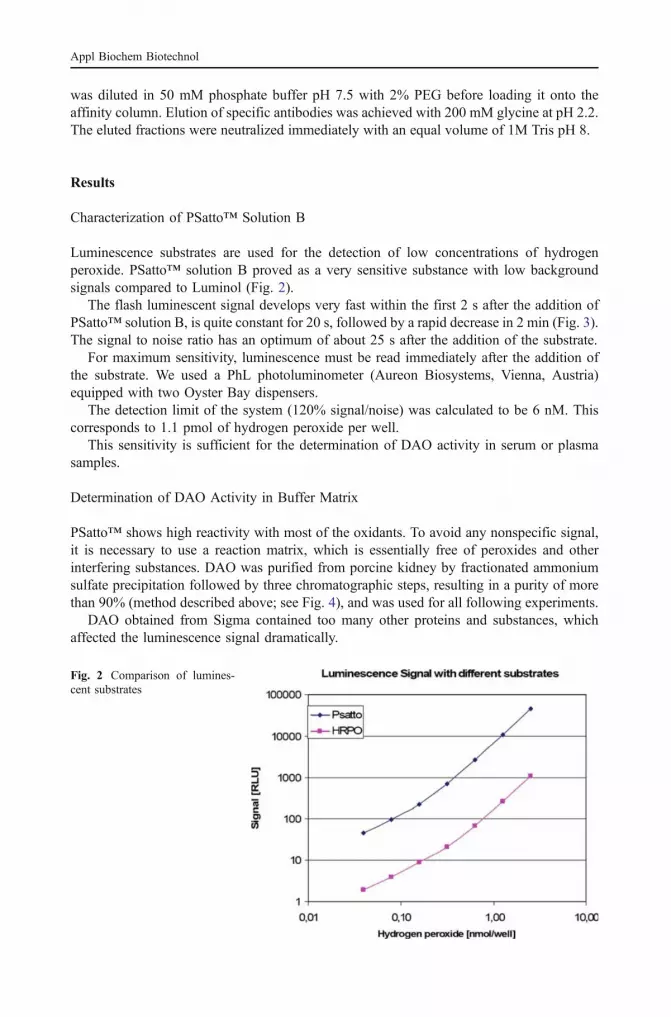
Luminescence substrates are used for the detection of low concentrations of hydrogenperoxide. PSatto™ solution B proved as a very sensitive substance with low backgroundsignals compared to Luminol (Fig. 2).
The flash luminescent signal develops very fast within the first 2 s after the addition ofPSatto™ solution B, is quite constant for 20 s, followed by a rapid decrease in 2 min (Fig. 3).The signal to noise ratio has an optimum of about 25 s after the addition of the substrate.
For maximum sensitivity, luminescence must be read immediately after the addition ofthe substrate. We used a PhL photoluminometer (Aureon Biosystems, Vienna, Austria)equipped with two Oyster Bay dispensers.
The detection limit of the system (120% signal/noise) was calculated to be 6 nM. Thiscorresponds to 1.1 pmol of hydrogen peroxide per well.
This sensitivity is sufficient for the determination of DAO activity in serum or plasmasamples.
Determination of DAO Activity in Buffer Matrix
PSatto™ shows high reactivity with most of the oxidants. To avoid any nonspecific signal,it is necessary to use a reaction matrix, which is essentially free of peroxides and otherinterfering substances. DAO was purified from porcine kidney by fractionated ammoniumsulfate precipitation followed by three chromatographic steps, resulting in a purity of morethan 90% (method described above; see Fig. 4), and was used for all following experiments.
DAO obtained from Sigma contained too many other proteins and substances, whichaffected the luminescence signal dramatically.
Fig. 2 Comparison of lumines-cent substrates
Appl Biochem Biotechnol

The stability of hydrogen peroxide in solution, which is influenced by varioussubstances and temperature, has to be considered.
As described in many papers, diamine oxidase shows high conversion rates mainly forthe aliphatic diamines putrescine and histamine [26–28].
We tested histamine and putrescine in different concentrations to determine substratepreferences and optimal substrate concentrations for our DAO preparation.
All reagents were diluted in 50 mM Tris pH 8.0. DAO was used in a serial dilution from19 to 500 U/ml. 1 Unit in our system corresponds to 70 µU/ml as specified by Sigma(defined as oxidation of 1 µmol Putrescin/hour at pH 7,2 at 37°C). 50 μl of enzymesolution were incubated with 50 μl of varying substrate concentrations for 1 h at 37 °C. Inadditional, 20 μl horseradish peroxidase (HRPO) and 50 μl of PSatto™ were added to thereaction mixture. Luminescence signal was integrated for 10 s after a lag time of 5 s. DAO-specific luminescence signal depended on substrate concentration. DAO activity isinhibited at a substrate concentration higher than about 30 μmol/l, independent of thesubstrate. Furthermore, conversion of putrescine is about four times more efficient thanhistamine (see Fig. 5).
Incubation Conditions
Different incubation conditions were tested to improve assay sensitivity: 100 μl DAO solutionin 50 mM Tris pH 8.0 was mixed with 50 μl putrescine (16 μM) and incubated at differenttemperatures for 30, 60 and 180 min. Subsequently, HRPO and PSatto™ were added with thedispenser. Finally, luminescence signal was integrated for 10 s after a delay time of 2 s.
Hydrogen peroxide is produced continuously during the action of the enzyme andaccumulates in the reaction mixture. Degradation of H2O2 produced during reaction timeshould be minimized until peroxidase and luminescence substrates are added. Diamineoxidase has a rather slow, but long-lasting activity. To obtain reliable signals with lowconcentrations of DAO, incubation has to be extended to at least 1 h. As demonstrated inFig. 6, shaking of the reaction mixture results in a dramatic decrease of signal because ofthe deterioration of H2O2. Although the working temperature of native porcine DAO inkidney is about 37 °C, maximal signal could be achieved at room temperature. Hydrogenperoxide is obviously not stable at higher temperatures. At 4 °C, enzyme activity is notsufficient; even after 3 h reaction time, the maximum signal could not be reached.
Fig. 3 Flash signal development
Appl Biochem Biotechnol

As shown in Fig. 7, maximum signal intensity is achieved after 7 h of incubation time.The maximum sensitivity of the system is already reached after 5 h of incubation time.After this period, background signal increases more quickly than the specific signal relatedto DAO activity. Under these assay conditions, a sensitivity of purified porcine DAO of2 U/ml (blank +2 SD) is achieved. The sensitivity of the system is not increased byincubation times longer than 5 h.
This assay allows to determine the activity of highly purified porcine DAO in a buffersystem. Based on this system, we developed an assay useful for clinical use. To purify DAOfrom a protein solution or plasma, we used a specific antibody coated on the microtiter well.
Production of DAO-Specific Antibody
Immunization of chicken was performed successfully with the purified enzyme (see above).IgY were purified using fractionated precipitation with caprylic acid and ethanol. Purified
Fig. 4 Purity of DAO after thelast chromatography step
Appl Biochem Biotechnol

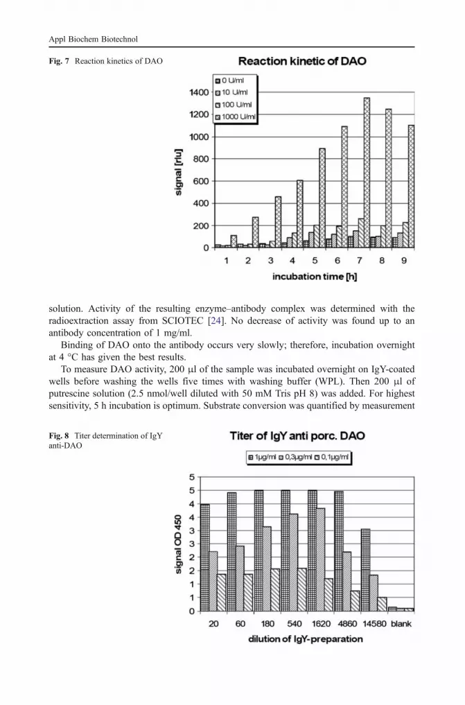
DAO was coated on NUNC Maxisorp microwell plates (1:0.3:1 μg/ml in 50 μM Na2CO3
pH 9.6; 200 μl per well, coating overnight at 4 °C). After blocking with 0.1% BSA in thesame buffer system, IgY preparation was incubated overnight at 4 °C in a serial dilution.Detection of specifically bound IgY was performed with rabbit anti-IgY-AP (Aves-Labs,USA). A highly specific titer of >1:20,000 could be found for DAO, as can be seen in Fig. 8.
IgY were further purified by an affinity chromatography column (HiTrap NHS-activated;Amersham Biosciences) loaded with DAO according to the manufacturers’ protocol. 500 ngof the affinity-purified antibody per well was coated on NUNC Maxisorp microtiter wells tocapture DAO out of different sample matrices. Strips were blocked as described before.
Measurement of DAO Activity after Binding on IgY
To determine any inhibition of the DAO activity by antibody binding to the active site ofthe enzyme, increasing concentrations of the purified antibody were added to the enzyme
Fig. 6 Optimization of incuba-tion conditions
Fig. 5 Reactivity of DAO withhistamine and putrescine
Appl Biochem Biotechnol
solution. Activity of the resulting enzyme–antibody complex was determined with theradioextraction assay from SCIOTEC [24]. No decrease of activity was found up to anantibody concentration of 1 mg/ml.
Binding of DAO onto the antibody occurs very slowly; therefore, incubation overnightat 4 °C has given the best results.
To measure DAO activity, 200 μl of the sample was incubated overnight on IgY-coatedwells before washing the wells five times with washing buffer (WPL). Then 200 μl ofputrescine solution (2.5 nmol/well diluted with 50 mM Tris pH 8) was added. For highestsensitivity, 5 h incubation is optimum. Substrate conversion was quantified by measurement
Fig. 8 Titer determination of IgYanti-DAO
Fig. 7 Reaction kinetics of DAO
Appl Biochem Biotechnol

of luminescence after the addition of 20 μl PSatto™ and 50 μl horseradish peroxidasesolution by a dispenser. For optimal sensitivity, the luminescence integral of the first 5 swas detected.
After isolation by antibody, the luminescence signal correlates with DAO activity of thesample, quantified by the SCIOTEC radioextraction assay. As shown in Fig. 9, it is possibleto measure a few DAO U/ml. The working range of this assay is between 2 and 250 DAOU/ml, slightly better than the radioextraction assay from SCIOTEC with measuring rangefrom 1.5 to 150 U/ml. Based on clinical data collected until now, a DAO activity of morethan 15 U/ml represents a healthy person. Activities below 15 U/ml are to be consideredpotentially histamine-intolerant.
Using a set of 24 plasma samples, a good correlation to the existing REA test system could beshown (Fig. 10). Therefore, this assay system is a useful alternative to the existing systems.Until now, no clinical data are available for DAO activities in tissues or other biological fluids.
Fig. 9 Typical calibration curve
Correlation DAO-REA
vs. Luminescence assay
R2 = 0,8815
0
5000
10000
15000
20000
25000
0 200 400 600 800 1000
Luminescence [rlu]
RE
A [
cp
m]
Fig. 10 Assay correlation with24 plasma samples
Appl Biochem Biotechnol

Discussion
Up to now, no highly sensitive nonradioactive method for the determination of porcineDAO was described. The aim of this work was to develop a very sensitive and simple assayfor research groups working with porcine DAO. Comparing different detection systems forhydrogen peroxide, the highly sensitive luminescence reagent PSatto™, performed best andattained the required sensitivity for hydrogen peroxide. The detection limit of PSatto™ forhydrogen peroxide, after verification by different experiments, was sufficient for thequantification of a 3 DAO U/ml.
DAO was captured from the sample by means of immunoaffinity-purified specificantibodies raised in hen using highly purified enzyme for the immunization. Theseantibodies were coated on microtiter wells for capturing DAO from the sample. Variationsof substrate, substrate concentration, incubation time, and temperature were made toidentify the best conditions for DAO.
Compared to histamine, putrescine generates a higher signal, this finding agrees withprior studies [17]. Substrate inhibition starts from 30 µM with putrescine and withhistamine; hence, 25 µM putrescine seemed to be the best condition. Because of the veryslow binding of DAO onto the antibody, an incubation overnight at 4 °C is preferable.
The final working protocol provides a less labor-intensive method to determine porcineDAO activity. The assay allows herewith to measure a huge amount of samplessimultaneous. This fact facilitates enormous testing of inhibitors and activators of DAO.
Because of the high relevance of DAO in connection with histamine intolerance, a lot ofstudies are necessary to characterize the clinical pattern of enzyme inhibition.
References
1. Buffoni, F. (1966). Pharmacological Reviews, 18, 1163–1199.2. Buffoni, F., & Ignesti, G. (2003). Inflammopharmacology, 11, 203–209.3. Aschenbach, J. R., Schwelberger, H. G., Ahrens, F., Fürll, B., & Gäbel, G. (2006). Scandinavian Journal
of Gastroenterology, 41, 712–719.4. Maintz, L., & Novak, N. (2007). American Journal of Clinical Nutrition, 85, 1185–1196.5. Sattler, J., Lorenz, W., Kubo, K., Schmal, A., Sauer, S., & Luben, L. (1989). Journal of Chromatography
B, Biomedical Sciences and Applications, 27, 212–214.6. Sattler, J., Hafer, D., Klotter, H. J., Lorenz, W., & Wagner, P. K. (1988). Agents Actions, 23, 361–365.7. Cubria, C., Alvarez-Bujidos, M., Negro, A., Balana-Fouce, R., & Ordonez, D. (1993). Comparative
Biochemistry and Physiology C, 105, 251–254.8. Schwelberger, H. G., & Bodner, E. (1997). Biochimica et Biophysica Acta, 1340, 152–164.9. Kluetz, D., & Schmidt, P. G. (1977). Biochemical and Biophysical Research Communications, 76,
40–45.10. Rinaldi, A., Vecchini, P., & Floris, G. (1982). Preparative Biochemistry, 12, 11–28.11. Wilfingseder, D., & Schwelberger, H. G. (2000). Journal of Chromatography B, Biomedical Sciences
and Applications, 737, 161–166.12. Shah, M. A., & Ali, R. (1988). Biochemical Journal, 253, 103–107.13. Biebl, M., Klocker, J., Perkmann, R., Kolbitsch, C., Klingler, P., Drasche, A., et al. (2002). Inflammation
Research, 51(1), 93–94.14. Ignesti, G. (2003). Journal of Enzyme Inhibition and Medicinal Chemistry, 18, 463–473.15. Klaus, A., Weiss, H., Nguyen, J. H., Margreiter, R., Obrist, P., & Schwelberger, H. G. (2003). Transplant
International, 16, 474–479.16. Sattler, J., Hafner, D., Klotter, H. J., Lorenz, W., & Wagner, P. K. (1988). Agents Actions, 23, 361–365.17. Sattler, J., Lorenz, W., Kubo, K., Schmal, A., Sauer, S., & Luben L. (1989). Agents Actions, 27, 212–214.18. Jarisch, R., & Wantke, F. (1996). International Archives of Allergy and Immunology, 110, 7–12.19. Nag, S., Saha, K., & Choudhuri, M. A. (2000). Plant Science, 157, 157–163.
Appl Biochem Biotechnol

20. Takagi, K., Nakao, M., Ogura, Y., Nabeshima, T., & Kunii, A. (1994). Clinica Chimica Acta, 226, 67–75.21. Houen, G. (1999). Acta Pathologica, Microbiologica et Immunologica Scandinavica, Supplementum, 96,
1–46.22. Ionescu, G., & Kiehl, R. (1988). Allergy, 43, 318–319.23. Tufvesson G., & Tryding, N. (1969). Scandinavian Journal of Clinical and Laboratory Investigation, 24,
163–168.24. Mayer, I., Missbichler, A., Wantke, F., Focke, M., Reichl, H., Winter, M., et al (2005). Allergologie, 28,
1–8.25. Bachrach, U. (1985). In: Mondovi (Ed.), Structure and Functions of Amine Oxidase (pp. 5–20). Boca
Raton: CRC Press.26. Bradsley, W. G., Crabbe, M. J. C., & Scott I. V. (1974). Biochemical Journal, 139, 169–181.27. Kufner, M. A., Ulrich, P., Raithel, M., & Schwelberger, H. G. (2001). Inflammation Research, 50(2), 96–97.28. Sasaki, A., Tsujikawa, T., Fujiyama, Y., & Bama, T. (2001). Journal of Gastroenterology and
Hepatology, 16(9), 986–990.
Appl Biochem Biotechnol


