Embed Size (px)
Citation preview
Original article
Hippocampal lipoprotein lipase regulates energybalance in rodents%
Alexandre Picard 1, Claude Rouch 1,2, Nadim Kassis 1,2, Valentine S. Moulle 1, Sophie Croizier 3,Raphael G. Denis 3, Julien Castel 1,2, Nicolas Coant 1, Kathryn Davis 4, Deborah J. Clegg 4,Stephen C. Benoit 5, Vincent Prevot 3, Sebastien Bouret 3,6, Serge Luquet 1,2,Herve Le Stunff 1, Celine Cruciani-Guglielmacci 1, Christophe Magnan 1,*
ABSTRACT
Brain lipid sensing is necessary to regulate energy balance. Lipoprotein lipase (LPL) may play a role in this process. We tested if hippocampal LPLregulated energy homeostasis in rodents by specifically attenuating LPL activity in the hippocampus of rats and mice, either by infusing apharmacological inhibitor (tyloxapol), or using a genetic approach (adeno-associated virus expressing Cre-GFP injected into Lpllox/lox mice).Decreased LPL activity by either method led to increased body weight gain due to decreased locomotor activity and energy expenditure, concomitantwith increased parasympathetic tone (unchanged food intake). Decreased LPL activity in both models was associated with increased de novoceramide synthesis and neurogenesis in the hippocampus, while intrahippocampal infusion of de novo ceramide synthesis inhibitor myriocincompletely prevented body weight gain. We conclude that hippocampal lipid sensing might represent a core mechanism for energy homeostasisregulation through de novo ceramide synthesis.
& 2013 The Authors. Published by Elsevier GmbH. All rights reserved.
Keywords Lipid sensing; Obesity; Ceramides; Parasympathetic nervous system; Energy expenditure
1. INTRODUCTION
The central nervous system (CNS) is a key player in the regulation of energybalance in mammals [1,2]. This process involves a combination of signalsarising from the periphery, including hormones and nutrients, which aredetected by specialized areas like the hypothalamus and brainstem [3–5].Since the work of Oomura et al. [6], there is a growing amount of evidenceto suggest that hypothalamic fatty acid sensing plays a role in the regulationof energy balance, including insulin secretion and action, hepatic glucoseproduction and food intake [7–10]. However the molecular mechanismsinvolved in this fatty acid sensing are still a matter of debate [8].Postprandial triglycerides (TG)-enriched particles are abundant lipid specieshydrolyzed by the lipoprotein lipase (LPL), and recent studies havehighlighted a role for neuronal LPL-mediated hydrolysis of TG particles inthe regulation energy balance [11,12].Otherwise, other areas beside the hypothalamus have been shown to beinvolved in the regulation of energy homeostasis. Among them, thehippocampus has also been described as a potential site for the regulationof feeding behavior and body weight homeostasis [13,14]. For example,hippocampal lesions are associated with increased body weight and food
http://dx.doi.org/10.1016/j.molmet.2013.11.002
Abbreviations: LPL, lipoprotein lipase; CNS, central nervous system; TG, triglycerides; AAV, adeno-assocSPT, serine palmitoyltransferase; CERS, ceramide synthase; SMPD1, acid sphingomyelin phosphodiester
%This is an open-access article distributed under the terms of the Creative Commons Attributioreproduction in any medium, provided the original author and source are credited.
1Universite Paris Diderot, Sorbonne Paris Cite, BFA, EAC 4413 CNRS, Case courrier 7126, 4, rue MaScientifique-CNRS EAC 4413, F-75205 Paris, France 3Jean-Pierre Aubert Research Center, DevelopmentMedicine, University of Texas Southwestern Medical Center, Dallas, TX, USA 5Department of Psychiatry,Children’s Hospital Los Angeles, University of Southern California, Los Angeles, USA
*Corresponding author. Fax: +33 1 57 27 77 96. Email: [email protected] (C. M
Received October 30, 2013 � Revision received November 7, 2013 � Accepted November 13, 2013
MOLECULAR METABOLISM ] (]]]]) ]]]–]]] & 2013 The Authors. Published by Elsevier GmbH. All rig
intake in ad libitum fed rats [15]. Hunger and satiety circulating signals suchas leptin or ghrelin were also shown to bind to hippocampus cells to regulatefood intake in rats [16]. Moreover the orexigenic peptide ghrelin was shownto modulate hippocampal dendritic spine and memory acquisition [17].Studies in both rodents and humans have reported that high fat/refined sugardiets impair hippocampal function [18]. Interestingly, LPL is highly expressedin the hippocampus [19,20] suggesting a potential role for this enzyme inbody weight regulation.The present work was aimed at studying whether TG hydrolysis by LPLspecifically in the dorsal hippocampus could represent a physiologicallyrelevant mechanism in energy homeostasis and body weight regulation. Tothis end, two different species and two experimental approaches were usedto locally decrease LPL activity in the hippocampus: (1) the infusion oftyloxapol, an inhibitor of LPL activity, into the hippocampus of rats and (2)the bilateral injection of an adeno-associated viral vector expressing a Cre-GFP fusion protein (AAV Cre-GFP) into the hippocampus of Lpllox/lox mice,leading to the specific deletion of the Lpl gene in the hippocampus of thesemice (“LPL Hip−/−” ). We observed using both techniques thathippocampal LPL inhibition led to both decreased locomotor activity andenergy expenditure and ultimately induced body weight gain. Importantly,
iated virus; GFP, green fluorescent protein; ANS, autonomic nervous system; RQ, respiratory quotient;ase 1; SPHK1, sphingosine kinase 1
n-NonCommercial-ShareAlike License, which permits non-commercial use, distribution, and
rie Andree Lagroua Weill-Halle, F-75205 Paris Cedex 13, France 2Centre National de la Rechercheand Plasticity of the Postnatal Brain, INSERM U837, 59045 Lille Cedex, France 4Department of InternalUniversity of Cincinnati, Cincinnati, OH, USA 6The Saban Research Institute, Neuroscience Program,
agnan).
� Available online Month XX, 2013
hts reserved. www.molecularmetabolism.com 1

Original article
food intake remained unchanged while the parasympathetic nervoussystem activity was increased suggesting a change in autonomic nervoussystem (ANS)-mediated peripheral energy handling. Inhibition of hippocam-pal LPL activity led to increased de novo ceramide biosynthesis, and aconcomitant increase in neurogenesis, while, conversely, local infusion ofmyriocin – a potent inhibitor of de novo ceramide biosynthesis – completelyprevented the metabolic changes in both mice and rats. Taken collectivelythese results highlight for the first time to our knowledge that nutritionallipid detection by LPL within hippocampus controls energy balance throughde novo ceramide synthesis pathway.
2. MATERIAL AND METHODS
The experimental protocol was approved by the institutional animal careand use committee of the Paris Diderot University (CEEA40).
2.1. Animal modelsTwo-month-old male Wistar rats (225–250 g, Charles River, l'Arbresle,France) and two-month-old Lpllox/lox mice (Jackson laboratory, strainB6.129S4-Lpltm1Ijg/J, no. 006503) were used. Littermates Lpl+/+ micewere used as controls. They were housed individually in stainless steelcages in a room maintained at 22±1 °C with lights on from 7 a.m. to7 p.m. They were given a standard laboratory diet (proteins 19.4%;carbohydrates 59.5%; lipids 4.6%; vitamins and minerals 16.5%) andwater ad libitum.
2.2. SolutionsOsmotic minipumps (Alzet® model 2004) were used to chronicallyinfuse different solutions specifically into the hippocampus, through acatheter connected to a depth-adjustable cannula (Alzet® brain infusionkit 1 for rats and Alzet® brain infusion kit 3 for mice). The vehiclesolution consisted of saline (Lavoisier). Tyloxapol solution (10 µg/day)was prepared by diluting 25 µL of tyloxapol (Sigma Aldrich # T8761)with 16.6 ml of saline. Myriocin solution (100 nM) was prepared bydiluting a myriocin mother solution (Sigma Aldrich # M117; 2 mg/ml inmethanol) 50,000 times in saline.
2.3. Viral productionAn adeno-associated virus, AAV Cre-GFP, was used in order to inducegenetic recombination within the hippocampus in Lpllox/lox mice. Theplasmid CBA.nls myc Cre.eGFP expressing the myc-nls-Cre-GFP fusionprotein was kindly provided by Richard Palmiter (University of Washing-ton, Seattle, USA). Adeno-associated viruses of the serotype 2/9 (AAV2/9) (6×10¹¹ vg/ml and 1.7×10⁸ pi/µl) were produced by the viralproduction facility of the UMR INSERM 1089 (Nantes, France).
2.4. Surgical proceduresBoth rats and mice were anesthetized with isoflurane and received a 10 µg/kg i.p. administration of xylazine. They were then placed on a stereotaxicframe. In rats, osmotic minipumps containing either vehicle, tyloxapol,myriocin, or both tyloxapol and myriocin were inserted subcutaneously andconnected to cannula implanted within the hippocampus (X : −3.5 mm; Y:−4.5 mm; Z: −3.5 mm; [21]). In Lpl+/+ and Lpllox/lox littermate mice, 1 µl of2/9 AAV Cre-GFP was injected per side (∼6×10⁸ particles/µl at a rate of0.20 µL/min for 5 min) bilaterally into the hippocampus (X : ±1 mm; Y:−2.06 mm; Z: −1.55 mm; [22]) to induce genetic recombination in floxedanimals. After viral injection, mice were implanted with osmotic minipumpscontaining myriocin or vehicle.
2 MOLECULAR METABOLISM ] (]]]]) ]]]–]]]
2.5. Measurement of body weight and body compositionIn rats, body weight was measured daily between 9 and 10 a.m. Inmice, body weight was measured weekly at the same time. For both,body mass composition (lean tissue mass, fat mass, free water and totalwater content) was analyzed before the indirect calorimetry studiesusing an Echo Medical Systems EchoMRI 100 (Whole Body CompositionAnalyzers, EchoMRI, Houston, USA) according to manufacturer's instruc-tions [23]. Briefly, awake animals were weighed before they wereplaced in a mouse holder and inserted in the MRI analyzer. Readings ofbody composition were obtained within 1 min. Body composition wasexpressed as a percentage of body weight.
2.6. Spontaneous alternation plus-maze taskBoth vehicle and tyloxapol-infused rats were used for memory testing.Animals were extensively handled for one week prior to testing. Animalswere placed into the center the center of the maze and allowed toexplore freely for 20 min while arm entries were recorded. Performancescores were calculated as described previously [24].
2.7. Indirect calorimetryIndirect calorimetry studies were performed in both rats and mice at day14 during 5 days; this period corresponded to the beginning ofincreased body weight gain compared to controls. Animals wereanalyzed for whole energy expenditure (kcal/h), oxygen consumptionand carbon dioxide production (VO2 and VCO2, where V is the volume),respiratory quotient (RQ=VCO2/VO2), food intake (g) and locomotoractivity (counts/hour) using calorimetric cages with bedding, food andwater (Labmaster, TSE Systems GmbH, Bad Homburg, Germany). Gasratio was determined using an indirect open-circuit calorimeter [25,26],which monitored O2 and CO2 concentrations by volume at the inlet portsof a tide cage with an airflow of 0.4 L/min, with regular comparisons toan empty reference cage. Whole energy expenditure was calculatedaccording to the Weir equation, using respiratory gas exchangemeasurements [27]. The flow was previously calibrated with a O2 andCO2 mixture of known concentrations (Air Liquide, S.A. France). Animalswere individually housed in a cage with lights on from 7 a.m. to 7 p.m.and an ambient temperature of 22±±1 °C. All animals were acclimatedto their cages for 48 h before experimental measurements. Dataregarding food and water consumption were collected every 40 min,and all ambulatory movements recorded during the entire experiment,with the aid of an automated online measurement system combininghighly sensitive feeding and drinking sensors and an infrared beam-based activity monitoring system. Gas and movement detection sensorswere operational during both light and dark phases, allowing forcontinuous recording. Animals were monitored for body weight andcomposition at the beginning and end of the experiment. Data analysiswas carried out with Excel XP using extracted raw values of VO2consumption, VCO2 production (in ml/h), and energy expenditure(kcal/h). Subsequently, each value was expressed either by whole leantissue mass extracted from the EchoMRI analysis.
2.8. Tissue collectionBrain tissues were dissected following the Glowinski and Iversentechnique [28] at day 28; total hippocampus, adjacent cortex andhypothalamus were immediately frozen.
2.9. LPL activity assayHeparin-releasable LPL activity was assayed in brain regions using aRoar LPL activity assay kit (RB-LPL, Roar Biomedical, Inc.). Briefly,tissues at day 28, then were lysed in 500 µL of assay buffer (150 mM
& 2013 The Authors. Published by Elsevier GmbH. All rights reserved. www.molecularmetabolism.com

NaCl, 10 mM Tris, 2 mM EDTA, pH 7.4) and incubated for 45 min at37 °C with an equivalent volume of heparin (100 U/ml). After incubation,samples were centrifuged for 10 min at 3000g, and aliquots (50 µL forrats and 10 µL for mice) of the aqueous phase deposited on LPLsubstrate emulsion in 96-well black microplates (VWR International, #25227-304) and incubated for 1 h at 37 °C. Finally, fluorescence wasread using a fluorimeter (370 nm excitation/450 nm emission) andcompared to a standard curve made using known concentrations of pre-hydrolyzed LPL substrate. LPL activity was expressed as nanomoles offree fatty acid produced per minute per gram of tissue.
2.10. Real-time quantitative PCRTotal RNA was isolated from the hippocampus dissected at day 28 at theend of the experimentation using RNeasy Lipid Tissue mini kit (Qiagen,Courtaboeuf, France). Real-time quantitative PCR was carried out in aLightCycler 1.5 detection system (Roche, Meylan, France) using theLight-Cycler FastStart DNA Master plus SYBR Green I kit (Roche). ThemRNA transcript level for each gene was normalized to levels of thehousekeeping gene TBP, which we have previously shown to beunaffected by LPL inhibition.
2.11. Measurement of ceramide levelsCeramide levels in tissues extracts collected at day 28 were measuredby the diacylglycerol kinase enzymatic method as previously described[29,30]. Briefly, aliquots of the chloroform phases from cellular lipidextracts were re-suspended in 7.5% (w/v) octyl-α-D-glucopyranoside/5 mM cardiolipin in 1 mM DETPAC/10 mM imidazole (pH 6.6). Theenzymatic reaction was started by the addition of 20 mM DTT, 0.88 U/ml Escherichia coli diacylglycerol kinase, 5 µCi/10 mM [γ-32P] ATP andthe reaction buffer (100 mM imidazole (pH 6.6), 100 mM NaCl, 25 mMMgCl2, and 2 mM EGTA). After incubation for 1 h at room temperature,lipids were extracted with chloroform/methanol/HCl (100:100:1, v/v) and1 M KCl. [γ-32P]-ceramide-1-phosphate was resolved by TLC withchloroform/acetone/methanol/acetic acid/water (10:4:3:2:1, v/v) andquantified with a FLA700 phosphorimager (Fuji, Japan). Known amountsof bovine ceramide standards were included with each assay. Eachmeasurement was done in duplicate. Ceramide levels were expressedas nanomoles per gram of tissue.
2.12. Parasympathetic firing-rate recordingsThe firing rate of the thoracic branch of vagus nerve along the carotidartery was recorded at the end of the experimentation at day 28 aspreviously described [31]. Recordings were carried out as follows:animals were anesthetized with isoflurane (Sanofi, Libourne, France).The vagus nerve, which lies close to the carotid artery, was dissectedfree of underlying tissues to a distance of approximately 5 mm. Thenerve was then covered with paraffin oil to prevent dehydration andcarefully placed on a pair of silver-wire recording electrodes (0.6-mmdiameter). The electrodes were connected to a high-impedance probeand action potentials were displayed and saved on a computer afterinitial amplification through a low-noise amplifier (BIO amplifier, ADInstrument, Rabalot, France). Unipolar nerve activity was recordedcontinuously for 15 min. Data were digitized with PowerLab/4spdigitizer. Signals were amplified 105 times and filtered using low- andhigh-frequency cut-offs of 100 and 1000 Hz, and monitored using theChart 4 computer program [32].
2.13. Statistical analysisEffects of treatments were assessed by either two-way ANOVA withBonferoni post hoc test or Student t-test (Figure 1A and B and Figure 2B).
MOLECULAR METABOLISM ] (]]]]) ]]]–]]] & 2013 The Authors. Published by Elsevier GmbH. All rig
A p-value lower than 0.05 was defined to be statistically significant.Statistical analysis were performed using GraphPad Prism 5®.
3. RESULTS
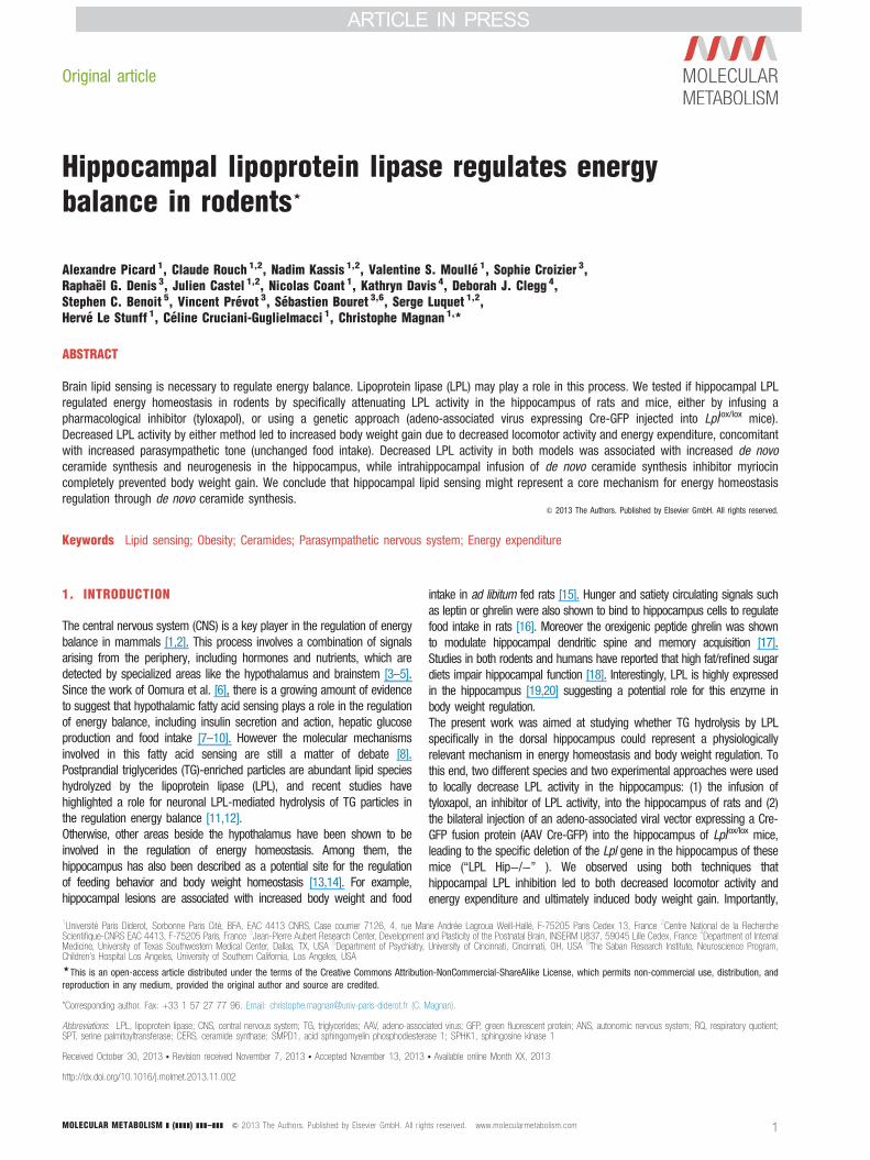
3.1. Decreased LPL activity in the hippocampus increases body weightgain in mice and rats without any change in food intakeLPL activity was specifically decreased in the hippocampus using bothpharmacological and genetic approaches. In rats, tyloxapol, an inhibitorof LPL activity, or vehicle was infused into the hippocampus for 28 daysusing a depth adjustable cannula (X: −3.5 mm; Y: −4.5 mm; Z:−3.5 mm [21]) connected to an osmotic minipump (Alzet). In thismodel, LPL activity was significantly decreased by approximately 25% inthe hippocampus of tyloxapol-infused animals compared to controls,while the hypothalamic and cortical LPL activity were unchanged (Figure1A). Importantly, there was no change in performance on a spontaneousalternation task in tyloxapol-infused rats compared to controls, indicatingthat there were no untoward effects of drug infusion such as ahippocampal lesion (percent alternation: 51%±9 vs. 65%±14 in vehiclegroup, N.S.). In the genetic approach, Lpl+/+ and Lpllox/lox mice receiveda single injection of Cre-GFP AAV into the hippocampus (X: ±1 mm;Y: −2.06 mm; Z: −1.55 mm [22]), yielding LPL Hip+/+ and LPL Hip−/−mice, respectively. In this model, we found a 33% decrease in LPLactivity (Figure 1B). In either of the paradigm, a decrease in LPL activityselectively in the hippocampus led to enhanced body weight gain ascompared to controls (Figure 1C and D). The time course of body weightgain was similar in both species, i.e. there was a significant increasecompared to controls beginning on day 14. Interestingly, this increase inbody weight gain was not related to a concomitant increase in foodintake (Figure 1E and F).
3.2. Selective decreased in hippocampal LPL activity induces de novoceramide biosynthesisIn the rat model, using real-time quantitative PCR, we observed changesin the expression of key enzymes involved in de novo ceramidesynthesis specifically in the hippocampus, following tyloxapol treatment(Figure 2A and B). The mRNA levels of both serine palmitoyltransferase3 (SPT3) and ceramide synthase 2 (CERS2) were increased, whereas theexpression of acid sphingomyelin phosphodiesterase (SMPD1) – a keyenzyme in the sphingomyelin hydrolysis pathway – was not affected bythe reduction in LPL activity, thus suggesting an activation of the denovo ceramide biosynthesis (Figure 2B). The expression of sphingosinekinase 1 (SPHK1) was also increased in tyloxapol-infused rats (Figure2B). The decrease of hippocampal LPL activity induced a concomitantincrease in hippocampal ceramide content in tyloxapol-treated rats(Figure 2C) whereas cortex ceramide content was unchanged (tyloxapol1.55±0.12 nmol/g vs. vehicle 1.68±0.24 nmol, NS). Hippocampalceramide content was also increased in LPL Hip−/− mice (Figure 2D).In rats, the infusion of the specific inhibitor of the SPTs, myriocin, completelyprevented the tyloxapol-induced increase in the ceramide content through theinhibition of de novo ceramide synthesis in the hippocampus and hadno effect on ceramide content in the cortex (vehicle+myriocin 1.33±0.18 nmol/g, tyloxapol+myriocin 1.46±0.19 nmol/l, NS vs. vehicle andNS vs. tyloxapol). These changes did not restore LPL activity (Figure 2C–F)therefore pointing at ceramide synthesis as a downstream mechanism. Inmice, myriocin infusion led to increased total ceramide content in LPL Hip+/+and LPL Hip−/− mice (Figure 2D), likely due to the compensatory stimulationof the sphingomyelin hydrolysis pathway as it has been already described[33]. However, myriocin also prevented any additional ceramide accumulation
hts reserved. www.molecularmetabolism.com 3
0.00
0.05
0.10
0 7 14 21 28
Daysvehicle tyloxapol
0
20
40
60
80
100
120
140
HIPPOCAMPUS CORTEX
LPL Hip +/+ LPL Hip -/-
0
0.5
1
1.5
2
2.5
3
3.5
4
0 7 14 21 28
LPL Hip +/+ LPL Hip -/-Days
Bod
y w
eigh
t gai
n (g
)
0
20
40
60
80
100
120
0 7 14 21 28
Daysvehicle tyloxapol
Bod
y w
eigh
t gai
n (g
)
0
10
20
30
40
50
60
HIPPOCAMPUS HYPOTHALAMUS CORTEX
Lpl a
ctiv
ity (n
mol
FFA
/min
/g)
vehicle tyloxapol
0.00
0.05
0.10
0.15
0.20
0 7 14 21 28
DaysLPL Hip +/+ LPLHip -/-
food
inta
ke/b
ody
wei
ght
(g/g
)Lp
l act
ivity
(nm
ol F
FA/m
in/g
)
food
inta
ke/b
ody
wei
ght
(g/g
)
Figure 1: Decreased LPL activity in the hippocampus increases body weight gain in mice and rats without affecting food intake. (A) LPL activity in the hippocampus, hypothalamus and cortex ofrats infused with vehicle or tyloxapol for 28 days, through an osmotic minipump connected to a cannula implanted in hippocampus; nZ6. FFA: free fatty acids. (B) LPL activity in the hippocampusand cortex of mice with a specific deletion of the Lpl gene in the hippocampus after AAV Cre-GFP injection (LPL Hip� /� mice) and their control littermates (LPL Hipþ /þ ); nZ3. Inset in (B):Cre-GFP fusion protein fluorescence in right hippocampus (coordinates X: þ1 mm; Y: �2.06 mm; Z: �1.55 mm, 20� magnification) 4 weeks after injection shows specific expression of theprotein within hippocampus with high fluorescence in CA1 pyramidal layer. (C and D) Body weight gain after the beginning of intrahippocampal tyloxapol infusion in rats (C) or AAV Cre-GFP injectionin Lpllox/lox mice (D); nZ6. (E and F) Time course of food intake reported to body weight in rats (E) and mice (F) during the 28 days of treatment; nZ6 for both rats and mice.
Original article
in LPL Hip−/− vs. LPL Hip+/+ mice (Figure 2D), again without anysignificant impact on LPL activity (Figure 2F).
3.3. Hippocampus de novo ceramide synthesis is required to mediateLPL action on energy balanceBlockade of de novo ceramide synthesis through myriocin infusion intothe hippocampus selectively prevented in both rats and mice the gain inbody weight associated with decreased LPL activity (Figure 3A and B).This was not due to changes in food intake (Figure 3C and D) or
4 MOLECULAR METABOLISM ] (]]]]) ]]]–]]]
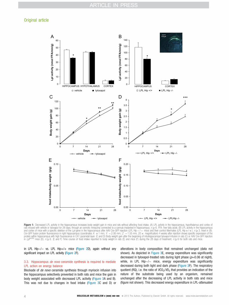
alterations in body composition that remained unchanged (data notshown). As depicted in Figure 3E, energy expenditure was significantlydecreased in tyloxapol-treated rats during light phase (p=0.06 at night),while, in LPL Hip−/− mice, energy expenditure was significantlydecreased during both light and dark phase (Figure 3F). The respiratoryquotient (RQ), i.e. the ratio of VCO2/VO2 that provides an indication of thenature of the substrate being used by an organism, remainedunchanged after the decreasing of LPL activity in both rats and mice(figure not shown). This decreased energy expenditure in LPL-attenuated
& 2013 The Authors. Published by Elsevier GmbH. All rights reserved. www.molecularmetabolism.com

0
10
20
30
40
50
60
HIPPOCAMPUS HYPOTHALAMUS CORTEX
)g /nim /AFFlo
mn(ytivi tcal pL
vehicle tyloxapolvehicle+myriocin tyloxapol+myriocin
0.0
0.2
0.4
0.6
0.8
1.0
1.2
1.4
1.6
1.8
2.0
vehicle tyloxapolvehicle+myriocin tyloxapol+myriocin
Cer
amid
e co
nten
t(nm
ol/g
)
*
**
020406080
100120140160180200
vehicle tyloxapol
Nor
mal
ized
gen
eex
pres
sion
(%of
con
trol
)
*
*
*
*
0
0.2
0.4
0.6
0.8
1
1.2
LPL Hip +/+ LPL Hip -/-LPL Hip +/+ myriocin LPL Hip-/- myriocin
Cer
amid
e co
nten
t (nm
ol/g
)
ns
*
*** ***ns
* *
0
20
40
60
80
100
120
140
HIPPOCAMPUS CORTEXLPL Hip +/+ LPL Hip -/-LPL Hip +/+ myriocin LPL Hip -/- myriocin
* *
)g /nim /AFFlo
mn(ytivi tcal pL
Figure 2: Decreased LPL activity is associated with increased hippocampal ceramide content through the activation of the de novo synthesis pathway. (A) Ceramide synthesis pathways: the denovo synthesis pathway that is inhibited by myriocin (left) and the sphingomyelin hydrolysis pathway (right). (B) Changes in gene expression of enzymes involved in ceramide synthesis pathways intyloxapol-treated rats vs. controls; nZ5. (C and D) Hippocampal ceramide content in tyloxapol-treated and control rats co-infused with myriocin or vehicle (C), and hippocampal ceramide content inLPL Hipþ /þ and LPL Hip� /� mice with or without myriocin treatment (D); nZ6 in rats and nZ3 in mice. (E and F) LPL activity in rats and mice in the four groups above; nZ6 and nZ3,respectively. *: po0.05, **: po0.01 vs. controls.
animals was correlated with a reduction in locomotor activity in both rats(Figure 3G) and mice (Figure 3H) during dark phase. Importantly,myriocin treatment blocked the effects of LPL attenuation on energyexpenditure as well as locomotor activity in both rats (Figure 3E and G)and mice (Figure 3F and H). Taken together, these results support theconcept that de novo ceramide synthesis is a core molecular mechan-ism relaying the action of hippocampal TG sensing onto body weightregulation.
MOLECULAR METABOLISM ] (]]]]) ]]]–]]] & 2013 The Authors. Published by Elsevier GmbH. All rig
3.4. Hippocampal attenuation of TG hydrolysis increasesparasympathetic nervous system activity in a ceramide dependentmechanismThe change in body weight was independent from feeding, we thereforeinvestigated to what extend non-food related modification in ANS outputcould be affected by hippocampus LPL knockdown.The activity of parasympathetic nervous system was recorded in vagusnerve along the carotid artery under basal conditions. Parasympathetic
hts reserved. www.molecularmetabolism.com 5

0
50
100
150
200
250
day nightLPL Hip +/+ LPL Hip -/-LPL Hip +/+ myriocin LPL Hip -/- myriocin
0
20
40
60
80
100
120
140
160
day nightvehicle tyloxapolvehicle+myriocin tyloxapol+myriocin
Ener
gy e
xpen
ditu
re
(kca
l/kgL
BM
/12h
)
0
20
40
60
80
100
120
0 7 14 21 28Days
vehicle tyloxapolvehicle+myriocin tyloxapol+myriocin
Bod
y w
eigh
t gai
n (g
)
0
10000
20000
30000
40000
day nightvehicle tyloxapolvehicle+myriocin tyloxapol+myriocin
Loco
cmot
or a
ctiv
ity
(
coun
ts/1
2h)
0
10000
20000
30000
40000
50000
day night
LPL Hip +/+ LPL Hip -/-LPL Hip +/+ myriocin LPL Hip -/- myriocin
0.06
00.5
11.5
22.5
33.5
4
0 7 14 21 28
LPL Hip +/+ LPL Hip -/-LPL Hip +/+ myriocin LPL Hip -/- myriocin
Days
Bod
y w
eigh
t gai
n (g
)
0.00
0.05
0.10
0 7 14 21 28Days
vehicle tyloxapolvehicle+myriocin tyloxapol+myriocin
food
inta
ke/b
ody
wei
ght (
g/g)
0.00
0.05
0.10
0.15
0.20
0 7 14 21 28Days
LPL Hip +/+ LPLHip -/-LPL Hip +/+ myriocin LPL Hip -/- myriocin
food
inta
ke/b
ody
wei
ght (
g/g)
En
ergy
exp
endi
ture
(k
cal/k
gLB
M/1
2h)
Loco
cmot
or a
ctiv
ity
(
coun
ts/1
2h)
Figure 3: Myriocin infusion into the hippocampus completely prevents increased body weight gain in both models. (A and B) Body weight gain over 28 days in tyloxapol-treated and control ratswith or without co-infusion with myriocin (A) and LPL Hipþ /þ and LPL Hip� /� mice with or without myriocin treatment (B); nZ6 each. (A and D) Time course of food intake reported to bodyweight in rats (E) and mice (F) during the 28 days of treatment; nZ6 for both rats and mice. (E and F) Energy expenditure in tyloxapol-treated and control rats with or without co-infusion withmyriocin (C) and LPL Hipþ /þ and LPL Hip� /� mice with or without myriocin treatment (D); nZ6 each. (G and H) Locomotor activity in the 4 groups of rats (E) and mice (F); nZ6 for both.*: po0.05, ***: po0.001; tyloxapol or LPL Hipþ /þ vs. controls. ##: po0.01; LPL Hip� /� myriocin vs. LPL Hipþ /þ .
Original article

nervous system activity was significantly increased in tyloxapol-treatedrats (Figure 4A and C) and LPL Hip−/− mice (Figure 4B and D) whencompared to their respective controls. Again we found that myriocintreatment normalized parasympathetic nervous system activity in bothmodels of LPL activity inhibition the hippocampus.
3.5. Selective decrease in hippocampal LPL activity increaseshippocampal neurogenesis in both mice and rats through ceramidesignalingAs shown in Supplementary Figures S1 and S2, the inhibition of hippocampalLPL activity led to an increase in neurogenesis, assessed by BrdU andneuronal marker co-labeling. Myriocin administration fully prevented theincreased neurogenesis in tyloxapol-treated rats (Figure S1) and LPL Hip−/−mice (Figure S2), while it had no effect on neurogenesis in controls.
4. DISCUSSION
Fatty acid sensing by the brain plays a key role in the regulation of energyhomeostasis by the central nervous system (CNS) [8,34]. Within the last
0.5 mV
10 sec
0.5 mV
10 sec
0.5 mV
10 sec
Vehicle
Tyloxapol
Vehicle + myriocin
Tyloxapol + myriocin
10 sec
0.5 mV
0
2
4
6
8
10
12
14
16
Even
ts/s
ec
vehicle tyloxapolvehicle+myriocin tyloxapol+myriocin
***
Figure 4: Increased body weight gain is related to increased parasympathetic nervous system acrecorded in tyloxapol-treated and control rats with or without co-infusion with myriocin (A) and LPL HQuantification of parasympathetic nervous system activity in the 4 groups of rats (C) and mice (D
MOLECULAR METABOLISM ] (]]]]) ]]]–]]] & 2013 The Authors. Published by Elsevier GmbH. All rig
decade, there is a growing evidence to demonstrate that hypothalamicfatty-acid-sensing is critical for the regulation of food intake as well as ofinsulin secretion, hepatic glucose production and lipogenesis [8]. Theintracerebroventricular infusion of oleic acid has been shown to decreaseboth food intake and hepatic glucose production [7,9]. However, the ideathat an increase in brain fatty acid levels could act as a satiety signal toinhibit feeding appears counterintuitive, given that plasma fatty acid levelsdo not rise substantially after food ingestion, but do rise significantlyduring fasting [35], a situation in which food intake would be expected toincrease. On the other hand, plasma levels of triglyceride-enrichedlipoproteins do rise after food ingestion and could represent physiologicallyrelevant signals to modulate energy balance.The TG-rich particles hydrolyzing enzyme LPL has recently beendemonstrated to play a role in the regulation of energy balance byneurons [11]. The role of LPL in the brain is to convert TG-richlipoproteins into fatty acids locally, thus providing a signal of themetabolic state to fatty-acid-sensitive neurons [12]. Mice lacking LPL inneurons (NEXLPL−/− mice) were shown to develop transient hyper-phagia and obesity further confirming a role for central TG hydrolysis inbody weight regulation [11]. However the precise structures that relay
0.3 mV
5 sec
0.3 mV
5 sec
0.3 mV
5 sec
0.3 mV
5 sec
Lpl HIP+/+
Lpl HIP -/-
Lpl HIP +/+ myriocin
Lpl HIP -/-myriocin
0
2
4
6
8
10
12
14
16
18
20
Even
ts/s
ec
LPL Hip +/+ LPL Hip -/-LPL Hip +/+ myriocin LPL Hip -/- myriocin
*
tivity and is prevented by myriocin treatment. (A and B) Parasympathetic nervous system activityipþ /þ and LPL Hip� /� mice with or without myriocin treatment (B); nZ4 each. (C and D)); nZ4 each. *: po0.05, ***: po0.001 vs. controls.
hts reserved. www.molecularmetabolism.com 7

Original article
the action of TG detection onto body weight remain elusive. In particular,the hippocampus exhibits Lpl mRNA levels and activity that are at least2.5 times higher than in the cortex, cerebellum or remaining brain [19]pointing at hippocampal TG hydrolysis as a potential mechanism linkingdaily variation in TG particles and energy homeostasis.In order to specifically address this hypothesis we used severalapproaches and rodent models to selectively alter hippocampal TGhydrolysis ability. The first approach relied on local infusion of the non-ionic detergent tyloxapol (oxyethylated t-octylphenol polymethylenepolymer, also named Triton WR 1339), the second approach relied onvirus-mediated genetic knock out of Lpl gene through bilateral injectionof AAV Cre-GFP into the hippocampus of Lpllox/lox mice.In both models, we demonstrated that dampening hippocampal LPLactivity in the hippocampus led to body weight gain. The body weightchanges were not due to change in food intake but rather involved adecreased locomotor activity and decreased energy expenditure as wellas an increased parasympathetic tone. The reduction of hippocampalLPL activity increases de novo ceramide synthesis, while the adminis-tration of myriocin, a de novo ceramide synthesis inhibitor, completelyprevented metabolic disorders.Among the physiological determinants that account for body weight gain,we predict that increased parasympathetic nervous system activity, whichis known to facilitate energy storage by reducing energy expenditure [36]come in addition to decreased locomotor-dependent energy expenditureto achieve body weight gain in a feeding-independent manner.Hippocampal lesion in rats leads to body weight gain when fed adlibitum [14]. It has recently been reported that leptin signaling in thehippocampus contributes to the inhibition of food-related memories elicitedby contextual stimuli [16]. In mice fed a high-fat diet, NMDA receptors in thehippocampus are desensitized, possibly accounting for the cognitive deficitsassociated with obesity [37]. Furthermore, in obese individuals, functionalmagnetic resonance imaging (fMRI) studies have documented a discrepancyin homeostatic and non-homeostatic integration of hunger related signalsthat results in the opposite stimulation profile of hypothalamus vs.sensorimotor, emotional and cognitive areas including the amygdala,hippocampus, insula and precentral gyrus [38]. These observations suggesta putative link between high fat diet, hippocampal dysfunction and alteredenergy balance. Our data support this hypothesis and provide a physiologicaland molecular mechanism by which daily variation of nutritional TG richparticles directly alters hippocampus activity and body weight homeostasis.As a major excitatory input to nucleus accumbens interneurons, thehippocampus is thought to participate in the striatal encoding of goaldirected behavior [39]. It is therefore formally possible that the reduction ofnocturnal locomotor activity, following intra-hippocampal knock down of TGhydrolysis, is an indirect consequence of decreased motivational output. Inthat line, Ben Zeev et al. have observed a 60% increase in hippocampal LPLactivity in rats deprived of food for 12 h when compared to fed rats,suggesting that hippocampal LPL activity during fasting could contribute tothe enhanced locomotor activity necessary for animals to search for food[19]. In the fed state, in which animals did not have to move to find food,hippocampal LPL activity was decreased. In addition, exercise andhippocampal function have also been previously shown to be linked [40–42].Brain areas controlling parasympathetic nervous system activity are locatedin the lateral hypothalamus, and the number of c-fos/orexin-immunor-eactive neurons in the perifornical lateral hypothalamus is positivelycorrelated with the amount of locomotion [43]. There are importantreciprocal connections between the hippocampus and the hypothalamus.Projections between the lateral hypothalamus and the hippocampal dentategyrus, have been documented by Wayner et al., who showed that long-term potentiation in hippocampal granule cells was inhibited by lateral
8 MOLECULAR METABOLISM ] (]]]]) ]]]–]]]
hypothalamic afferents [44]. A recent study has demonstrated thathippocampal BDNF levels were enhanced by hypothalamic stimulation,in proportion to the metabolic rate [45]. Thus, any changes in hippocampalactivity may have an impact on specific lateral hypothalamic neurons, inturn modifying parasympathetic activity and thus both locomotor activityand energy expenditure, and eventually, body weight.We identified de novo ceramide synthesis as a molecular mechanism bywhich altered hippocampal TG hydrolysis affected energy balance.Ceramide content was increased by hippocampal LPL inhibition whereasblockade of de novo ceramide synthesis completely rescue the physiologicaloutput of LPL knock down. By definition, a decrease in LPL activity wouldlimit the supply of fatty acids from outside the cell, likely triggering de novofatty acid synthesis in order to provide acyl-CoA to the ceramide synthesispathway. Consistently with this hypothesis, normal fat deposition in adiposetissue can occur in the complete absence of LPL, and conversely, if LPLactivity is increased by pharmacological means, increased fat storage doesnot necessarily follow [46]. For example, fat mass is preserved by theendogenous synthesis of lipids in mice lacking LPL in adipose tissue [47], aprocess that involves an increase in glucose uptake and the concomitantinduction of endogenous fatty acid and triglyceride production, probablythrough a mechanism involving SREBP-1 (sterol regulatory element-bindingprotein 1) [48]. Such a mechanism could also be involved in our study, withdecreased LPL activity leading to increased de novo synthesis of palmitoyl-CoA, in the hippocampus. This could in turn be incorporated into the denovo ceramide synthesis pathway, as described in other cells such asmonocytes [49]. The ceramides are known to act as an important cellularsignaling molecule, and recent literature pointed out that ceramides withdistinct acyls chains have different cellular functions [50]. In that view, it isformally possible that TG rich particles, which under normal condition enterthe brain only during postprandial period, would not provide the mainsubstrate for ceramide synthesis but rather act as signaling modulator ofhippocampal cell activity.The stimulation of de novo ceramide biosynthesis in the hippocampus couldaffect its function through changes in neuronal plasticity and neurogenesis.Hence, it has been reported that ceramide levels and/or composition areimportant for proper dendritic spine maturation in hippocampal neurons [51]and several studies have highlighted a role for lipids and dietary regulationin the control of neurogenesis in the hippocampus [52,53]. The relationshipbetween global metabolic efficiency and neuroplasticity in the hippocampushas also been demonstrated [54]. In our study, we observed that theincreased ceramide content associated with LPL activity knock downcorrelated positively with neurogenesis as evidenced by BrdU labeling(Supplementary Figures S1A and B and S2A and B). BrdU positive cellswere mainly putative neurons in rats (Figure S1C and D) and both putativeneurons and GFAP-positive astrocytes or neuronal precursors in mice(Figure S2C and D). Ceramide synthesis inhibition resulted in the oppositeconsequences. TG-mediated modulation of hippocampal neurogenesismight provide a cellular basis for long-term adaptive mechanism inresponse to nutritional changes.To conclude, our study highlights for the first time on the role of hippocampalLPL in the regulation of energy balance depending on de novo ceramidebiosynthesis pathway and the modulation of the parasympathetic nervoussystem activity.
ACKNOWLEDGMENTS
This work was supported by grants from the ANR (French National Agency for
Research): Lipobrain, Grant number: 11-BSV1-021 01; and other Grant sponsors:
CORDDIM Ile-de-France and European Foundation for the Study of Diabetes (EFSD).
& 2013 The Authors. Published by Elsevier GmbH. All rights reserved. www.molecularmetabolism.com

CONFLICT OF INTEREST
None declared.
APPENDIX A. SUPPLEMENTARY MATERIALS
Supplementary data associated with this article can be found in the online version at
http://dx.doi.org/10.1016/j.molmet.2013.11.002.
REFERENCES
[1] Luquet, S., Magnan, C., 2009. The central nervous system at the core of the
regulation of energy homeostasis. Frontiers in Bioscience (Scholar Edition)
1:448–465.
[2] Sanchez-Lasheras, C., Konner, A.C., Bruning, J.C., 2010. Integrative neurobiol-
ogy of energy homeostasis-neurocircuits, signals and mediators. Frontiers in
Neuroendocrinology 31 (1):4–15.
[3] Blouet, C., Schwartz, G.J., 2010. Hypothalamic nutrient sensing in the control of
energy homeostasis. Behavioural Brain Research 209 (1):1–12.
[4] Cowley, M.A., 2003. Hypothalamic melanocortin neurons integrate signals of
energy state. European Journal of Pharmacology 480 (1–3):3–11.
[5] Levin, B.E., Magnan, C., Dunn-Meynell, A., Le Foll, C., 2011. Metabolic sensing
and the brain: who, what, where, and how? Endocrinology 152 (7):2552–2557.
[6] Oomura, Y., Nakamura, T., Sugimori, M., Yamada, Y., 1975. Effect of free fatty
acid on the rat lateral hypothalamic neurons. Physiology and Behavior 14
(04):483–486.
[7] Lam, T.K., Pocai, A., Gutierrez-Juarez, R., Obici, S., Bryan, J., Aguilar-Bryan, L.,
et al., 2005. Hypothalamic sensing of circulating fatty acids is required for
glucose homeostasis. Nature Medicine 11 (3):320–327.
[8] Migrenne, S., Le Foll, C., Levin, B.E., Magnan, C., 2011. Brain lipid sensing and
nervous control of energy balance. Diabetes and Metabolism 37 (2):83–88.
[9] Obici, S., Feng, Z., Morgan, K., Stein, D., Karkanias, G., Rossetti, L., 2002.
Central administration of oleic acid inhibits glucose production and food intake.
Diabetes 51 (2):271–275.
[10] Cruciani-Guglielmacci, C., Hervalet, A., Douared, L., Sanders, N.M., Levin, B.E.,
Ktorza, A., et al., 2004. Beta oxidation in the brain is required for the effects of
non-esterified fatty acids on glucose-induced insulin secretion in rats.
Diabetologia 47 (11):2032–2038.
[11] Wang, H., Astarita, G., Taussig, M.D., Bharadwaj, K.G., DiPatrizio, N.V., Nave, K.A.,
et al., 2011. Deficiency of lipoprotein lipase in neurons modifies the regulation of
energy balance and leads to obesity. Cell Metabolism 13 (1):105–113.
[12] Wang, H., Eckel, R.H., 2012. Lipoprotein lipase in the brain and nervous system.
Annual Review of Nutrition.
[13] Davidson, T.L., Jarrard, L.E., 1993. A role for hippocampus in the utilization of
hunger signals. Behavioral and Neural Biology 59 (2):167–171.
[14] Davidson, T.L., Chan, K., Jarrard, L.E., Kanoski, S.E., Clegg, D.J., Benoit, S.C.,
2009. Contributions of the hippocampus and medial prefrontal cortex to energy
and body weight regulation. Hippocampus 19 (3):235–252.
[15] Davidson, T.L., Kanoski, S.E., Chan, K., Clegg, D.J., Benoit, S.C., Jarrard, L.E.,
2010. Hippocampal lesions impair retention of discriminative responding based
on energy state cues. Behavioral Neuroscience 124 (1):97–105.
[16] Kanoski, S.E., Hayes, M.R., Greenwald, H.S., Fortin, S.M., Gianessi, C.A., Gilbert,
J.R., et al., 2011. Hippocampal leptin signaling reduces food intake and
modulates food-related memory processing. Neuropsychopharmacology 36
(9):1859–1870.
[17] Diano, S., Farr, S.A., Benoit, S.C., McNay, E.C., da Silva, I., Horvath, B., et al.,
2006. Ghrelin controls hippocampal spine synapse density and memory
performance. Nature Neuroscience 9 (3):381–388.
MOLECULAR METABOLISM ] (]]]]) ]]]–]]] & 2013 The Authors. Published by Elsevier GmbH. All rig
[18] Francis, H.M., Stevenson, R.J., 2011. Higher reported saturated fat and refined
sugar intake is associated with reduced hippocampal-dependent memory and
sensitivity to interoceptive signals. Behavioral Neuroscience 125 (6):943–955.
[19] Ben-Zeev, O., Doolittle, M.H., Singh, N., Chang, C.H., Schotz, M.C., 1990.
Synthesis and regulation of lipoprotein lipase in the hippocampus. Journal of
Lipid Research 31 (7):1307–1313.
[20] Paradis, E., Clavel, S., Julien, P., Murthy, M.R., de Bilbao, F., Arsenijevic, D.,
et al., 2004. Lipoprotein lipase and endothelial lipase expression in mouse brain:
regional distribution and selective induction following kainic acid-induced lesion
and focal cerebral ischemia. Neurobiology of Disease 15 (2):312–325.
[21] Paxinos, G., Watson, C. (Eds.), 2005. The rat brain in stereotaxis coordinates.
Academic Press, San Diego, USA.
[22] Paxinos, G., Franklin, K. (Eds.), 2003. The mouse brain in stereotaxic coordinates:
compact, second edition Gulf Professional Publishing, San Diego, USA.
[23] Taicher, G.Z., Tinsley, F.C., Reiderman, A., Heiman, M.L., 2003. Quantitative
magnetic resonance (QMR) method for bone and whole-body-composition
analysis. Analytical and Bioanalytical Chemistry 377 (6):990–1002.
[24] McNay, E.C., Gold, P.E., 2001. Age-related differences in hippocampal
extracellular fluid glucose concentration during behavioral testing and following
systemic glucose administration. Journal of Gerontology Series A: Biological
Sciences and Medical Sciences 56 (2):B66–B71.
[25] Arch, J.R., Hislop, D., Wang, S.J., Speakman, J.R., 2006. Some mathematical
and technical issues in the measurement and interpretation of open-circuit
indirect calorimetry in small animals. International Journal of Obesity (London)
30 (9):1322–1331.
[26] Even, P.C., Mokhtarian, A., Pele., A., 1994. Practical aspects of indirect
calorimetry in laboratory animals. Neuroscience and Biobehavioral Reviews 18
(3):435–447.
[27] Weir, J.B., 1949. New methods for calculating metabolic rate with special
reference to protein metabolism. Journal of Physiology 109 (1–2):1–9.
[28] Glowinski, J., Iversen, L.L., 1966. Regional studies of catecholamines in the rat
brain. I. The disposition of [3H]norepinephrine, [3H]dopamine and [3H]dopa in
various regions of the brain. Journal of Neurochemistry 13 (8):655–669.
[29] Le Stunff, H., Galve-Roperh, I., Peterson, C., Milstien, S., Spiegel, S., 2002.
Sphingosine-1-phosphate phosphohydrolase in regulation of sphingolipid meta-
bolism and apoptosis. Journal of Cell Biology 158 (6):1039–1049.
[30] Veret, J., Coant, N., Berdyshev, E.V., Skobeleva, A., Therville, N., Bailbe, D.,
et al., 2011. Ceramide synthase 4 and de novo production of ceramides with
specific N-acyl chain lengths are involved in glucolipotoxicity-induced apoptosis
of INS-1 beta-cells. Biochemical Journal 438 (1):177–189.
[31] Magnan, C., Collins, S., Berthault, M.F., Kassis, N., Vincent, M., Gilbert, M.,
et al., 1999. Lipid infusion lowers sympathetic nervous activity and leads to
increased beta-cell responsiveness to glucose. Journal of Clinical Investigation
103 (3):413–419.
[32] Wang, R., Cruciani-Guglielmacci, C., Migrenne, S., Magnan, C., Cotero, V.E.,
Routh, V.H., 2006. Effects of oleic acid on distinct populations of neurons in the
hypothalamic arcuate nucleus are dependent on extracellular glucose levels.
Journal of Neurophysiology 95 (3):1491–1498.
[33] Car, H., Zendzian-Piotrowska, M., Prokopiuk, S., Fiedorowicz, A., Sadowska, A.,
Kurek, K., et al., 2012. Ceramide profiles in the brain of rats with diabetes
induced by streptozotocin. FEBS Journal 279 (11):1943–1952.
[34] Rasmussen, B.A., Breen, D.M., Lam, T.K., 2012. Lipid sensing in the gut, brain
and liver. Trends in Endocrinology and Metabolism 23 (2):49–55.
[35] Ruge, T., Hodson, L., Cheeseman, J., Dennis, A.L., Fielding, B.A., Humphreys, S.M.,
et al., 2009. Fasted to fed trafficking of Fatty acids in human adipose tissue reveals a
novel regulatory step for enhanced fat storage. Journal of Clinical Endocrinology and
Metabolism 94 (5):1781–1788.
[36] Peterson, H.R., Rothschild, M., Weinberg, C.R., Fell, R.D., McLeish, K.R., Pfeifer., M.
A., 1988. Body fat and the activity of the autonomic nervous system. New England
Journal of Medicine 318 (17):1077–1083.
hts reserved. www.molecularmetabolism.com 9

Original article
[37] Valladolid-Acebes, I., Merino, B., Principato, A., Fole, A., Barbas, C., Lorenzo, M.P.,
et al., 2012. High-fat diets induce changes in hippocampal glutamate metabolism
and neurotransmission. American Journal of Physiology – Endocrinology and
Metabolism 302 (4):E396–E402.
[38] Martin, L.E., Holsen, L.M., Chambers, R.J., Bruce, A.S., Brooks, W.M., Zarcone,
J.R., et al., 2010. Neural mechanisms associated with food motivation in obese
and healthy weight adults. Obesity (Silver Spring) 18 (2):254–260.
[39] Fidalgo, C., Conejo, N.M., Gonzalez-Pardo, H., Lazo, P.S., Arias, J.L., 2012. A
role for dorsal and ventral hippocampus in response learning. Neurosciences
Research 73 (3):218–223.
[40] Dietrich, M.O., Andrews, Z.B., Horvath, T.L., 2008. Exercise-induced synapto-
genesis in the hippocampus is dependent on UCP2-regulated mitochondrial
adaptation. Journal of Neuroscience 28 (42):10766–10771.
[41] Gobeske, K.T., Das, S., Bonaguidi, M.A., Weiss, C., Radulovic, J., Disterhoft, J.F.,
et al., 2009. BMP signaling mediates effects of exercise on hippocampal
neurogenesis and cognition in mice. PLoS One 4 (10):e7506.
[42] Santin, K., da Rocha, R.F., Cechetti, F., Quincozes-Santos, A., de Souza, D.F.,
Nardin, P., et al., 2011. Moderate exercise training and chronic caloric restriction
modulate redox status in rat hippocampus. Brain Research 1421:1–10.
[43] Li, F.W., Deurveilher, S., Semba, K., 2011. Behavioural and neuronal activation
after microinjections of AMPA and NMDA into the perifornical lateral hypotha-
lamus in rats. Behavioural Brain Research 224 (2):376–386.
[44] Wayner, M.J., Phelix, C.F., Armstrong, D.L., 1997. Lateral hypothalamic
stimulation inhibits dentate granule cell LTP: direct connections. Brain Research
Bulletin 43 (1):5–15.
[45] Ying, Z., Covalin, A., Judy, J., Gomez-Pinilla, F., 2012. Hypothalamic stimulation
enhances hippocampal BDNF plasticity in proportion to metabolic rate. Brain
Stimulation 5 (4):642–646.
10 MOLECULAR METABOLISM ] (]]]]) ]]]–]]]
[46] Fielding, B.A., Frayn, K.N., 1998. Lipoprotein lipase and the disposition of dietary
fatty acids. British Journal of Nutrition 80 (6):495–502.
[47] Weinstock, P.H., Levak-Frank, S., Hudgins, L.C., Radner, H., Friedman, J.M.,
Zechner, R., et al., 1997. Lipoprotein lipase controls fatty acid entry into adipose
tissue, but fat mass is preserved by endogenous synthesis in mice deficient in
adipose tissue lipoprotein lipase. Proceedings of the National Academy of
Sciences of the United States of America 94 (19):10261–10266.
[48] Wagner, E.M., Kratky, D., Haemmerle, G., Hrzenjak, A., Kostner, G.M., Steyrer,
E., et al., 2004. Defective uptake of triglyceride-associated fatty acids in adipose
tissue causes the SREBP-1c-mediated induction of lipogenesis. Journal of Lipid
Research 45 (2):356–365.
[49] Gao, D., Pararasa, C., Dunston, C.R., Bailey, C.J., Griffiths, H.R., 2012. Palmitate
promotes monocyte atherogenicity via de novo ceramide synthesis. Free Radical
Biology and Medicine 53 (4):796–806.
[50] Park, J.W., Park, W.J., Futerman, A.H., 2013. Ceramide synthases as potential
targets for therapeutic intervention in human diseases. Biochimica et Biophysica
Acta , Epub 2013/09/12.
[51] Carrasco, P., Sahun, I., McDonald, J., Ramirez, S., Jacas, J., Gratacos, E., et al.,
2004. Ceramide levels regulated by carnitine palmitoyl transferase 1C control
dendritic spine maturation and cognition. Journal of Biological Chemistry 287
(25):21224–21232.
[52] Jung, J.U., Ko, K., Lee, D.H., Chang, K.T., Choo, Y.K., 2009. The roles of
glycosphingolipids in the proliferation and neural differentiation of mouse
embryonic stem cells. Experimental and Molecular Medicine 41 (12):935–945.
[53] Lindqvist, A., Mohapel, P., Bouter, B., Frielingsdorf, H., Pizzo, D., Brundin, P.,
et al., 2006. High-fat diet impairs hippocampal neurogenesis in male rats.
European Journal of Neurology 13 (12):1385–1388.
[54] Stranahan, A.M., Mattson, M.P., 2008. Impact of energy intake and expenditure
on neuronal plasticity. Neuromolecular Medicine 10 (4):209–218.
& 2013 The Authors. Published by Elsevier GmbH. All rights reserved. www.molecularmetabolism.com


