Embed Size (px)
Citation preview
IntroductionStimulation of neovascularization is a therapeutic goalto rescue tissue from critical ischemia (1). Postnatalneovascularization was attributed mainly to angiogen-esis, a process that is mediated by proliferation, migra-tion, and remodeling of preexisting endothelial cells(2–4). However, recent studies provide increasing evi-dence that postnatal neovascularization does not relyexclusively on sprouting of preexisting vessels, but alsoinvolves bone marrow–derived circulating endothelialcells (5–7). A specific subset of leukocytes positive forCD34 was shown to home to sites of ischemia andexpress endothelial antigens (5, 8). Moreover, endothe-lial progenitor cells (EPCs) can be grown out of isolat-ed CD133+ or CD34+ cells in vitro (5, 9, 10) and make asignificant contribution to adult blood vessel forma-tion (8). Importantly, injection of isolated CD34+ cellsor cultivated EPCs accelerates the restoration of bloodflow in diabetic mice (11) and enhances neovascular-ization in vivo (7, 8, 12, 13). In addition, increased neo-vascularization induced by hematopoietic CD34-posi-tive cells or angioblasts was shown recently to improvecardiac function (14, 15). Therefore, the stimulation ofmobilization and/or differentiation of EPCs may pro-vide a useful novel therapeutic strategy to improve
postnatal angiogenesis. Proangiogenic growth factorssuch as VEGF (16) or GM-CSF therapy (17) augment-ed EPC levels. Moreover, VEGF165 gene therapyincreased the number of circulating differentiatedEPCs in patients with limb ischemia (18). However, themechanisms, which regulate EPC mobilization and dif-ferentiation, are not fully elucidated.
HMG-CoA reductase inhibitors (statins) have beendeveloped as lipid-lowering drugs and are well estab-lished to reduce morbidity and mortality from coronaryartery disease (19). Besides lipid-lowering, statins arecapable of reducing vascular inflammation (20), decreas-ing platelet aggregation and thrombus deposition (21),and increasing endothelium-derived nitric oxide pro-duction (22). Statins have recently been shown to stim-ulate the growth of new blood vessels in ischemic limbsof normocholesterolemic rabbits (23). To gain insightsinto the mechanism by which statins improve neovas-cularization, we investigated the effects of statins on cir-culating bone marrow–derived endothelial cells and elu-cidated the underlying signal transduction pathways.
The present study demonstrates that statins augmentEPC numbers in vitro and in mice. The increase in EPClevels induced by both statins and VEGF was mediatedvia the activation of the serine/threonine kinase Akt,
The Journal of Clinical Investigation | August 2001 | Volume 108 | Number 3 391
HMG-CoA reductase inhibitors (statins)increase endothelial progenitor cells via the PI 3-kinase/Akt pathway
Stefanie Dimmeler,1 Alexandra Aicher,1 Mariuca Vasa,1 Christiane Mildner-Rihm,1
Klaudia Adler,1 Michaela Tiemann,2 Hartmut Rütten,2 Stephan Fichtlscherer,1
Hans Martin,3 and Andreas M. Zeiher1
1Division of Molecular Cardiology, Department of Medicine IV, University of Frankfurt, Frankfurt, Germany 2Aventis Pharmaceuticals, Frankfurt, Germany 3Division of Hematology, Department of Medicine III, University of Frankfurt, Frankfurt, Germany
Address correspondence to: Stefanie Dimmeler, Molecular Cardiology, Department of Medicine IV, University of Frankfurt, Theodor Stern-Kai 7, 60590 Frankfurt, Germany. Phone: 49-69-6301-7440; Fax: 49-69-6301-7113; E-mail: [email protected].
Received for publication April 27, 2001, and accepted in revised form June 11, 2001.
HMG-CoA reductase inhibitors (statins) have been developed as lipid-lowering drugs and are well estab-lished to reduce morbidity and mortality from coronary artery disease. Here we demonstrate that statinspotently augment endothelial progenitor cell differentiation in mononuclear cells and CD34-positivehematopoietic stem cells isolated from peripheral blood. Moreover, treatment of mice with statinsincreased c-kit+/Sca-1+–positive hematopoietic stem cells in the bone marrow and further elevated thenumber of differentiated endothelial progenitor cells (EPCs). Statins induce EPC differentiation via thePI 3-kinase/Akt (PI3K/Akt) pathway as demonstrated by the inhibitory effect of pharmacological PI3Kblockers or overexpression of a dominant negative Akt construct. Similarly, the potent angiogenicgrowth factor VEGF requires Akt to augment EPC numbers, suggesting an essential role for Akt in reg-ulating hematopoietic progenitor cell differentiation. Given that statins are at least as potent as VEGFin increasing EPC differentiation, augmentation of circulating EPC might importantly contribute tothe well-established beneficial effects of statins in patients with coronary artery disease.
J. Clin. Invest. 108:391–397 (2001). DOI:10.1172/JCI200113152.
See related Commentary on pages 365–366.
which is known to play an important role in endothe-lial biology and angiogenesis (23, 24).
MethodsCell culture. Total mononuclear cells (MNCs) were iso-lated from blood of healthy human volunteers by den-sity gradient centrifugation with Biocoll separatingsolution (density 1.077 (Biochrom AG, Berlin, Ger-many). MNCs (4 × 10⁶) were plated in 0.5 ml endothe-lial basal medium (EBM) (CellSystems, St. Katharinen,Germany), with supplements (1 µg/ml hydrocortisone,3 µg/ml bovine brain extract, 30 µg/ml gentamicin, 50µg/ml amphotericin B, 10 µg/ml EGF, and 20% FCS)on fibronectin/gelatin–coated 24-well plates. After 3days of culture, MNCs were stimulated with humanrecombinant VEGF (Biomol Feinchemikalien GmbH,Hamburg, Germany) or activated simvastatin, mevas-tatin (Calbiochem-Novabiochem GmbH, Bad Soden,Germany) or atorvastatin for the respective timepoints. Atorvastatin was kindly donated byGoedecke/Parke-Davis (Freiburg, Germany). Mediumwas then changed, and adherent cells were washedwith medium and incubated with 2.4 µg/ml 1,1′-dioc-tadecyl-3,3,3′,3′-tetramethylindocarbocyanine–labeledacetylated LDL (DiLDL; Harbor Bio-Products, Nor-wood, Massachusetts, USA) for 1 hour. Cells were fixedin 2% paraformaldehyde and counterstained withFITC-labeled lectin from Ulex europaeus (Sigma RBI,Taufkirchen, Germany). Two to three independentinvestigators evaluated the number of EPCs per well bycounting three randomly selected high-power fields.
MNCs were transfected after 3 days of culture withpcDNA3.1.-GFP, pcDNA3.1 (empty vector), orpcDNA3.1.-myc-his dominant negative Akt (25) usingLipofectamine PLUS (Life Technologies, Karlsruhe,Germany). Then, 0.5 µg plasmid, 4 µl PLUS, and 150µl EBM medium were incubated for 15 minutes fol-lowed by a mixture of 150 µl EBM medium and 1 µlLipofectamine. Cells were washed once with EBMmedium and incubated with the mixture and 1 mlEBM medium for 3 hours. Then, 1 ml EBM mediumwas added, and cells were incubated for a further 3hours before medium was changed.
Isolation of CD34+ cells. Human CD34+ cells were iso-lated from leukapheresis products from chemotherapyand G-CSF–mobilized patients with non–Hodgkinlymphoma after informed consent. After incubationwith immunomagnetic CD34-microbeads (MiltenyiBiotec, Bergisch-Gladbach, Germany) for 30 minutes at4°C, cells were washed in PBS plus 0.5% BSA plus 2mM EDTA, filtered through a 40-µm cell strainer, andrun over a magnetic cell separation device (Auto-Macs;Miltenyi Biotec) for positive selection of CD34+ cells.Isolated CD34+ cells were cultured in fibronectin-coat-ed 24-well plates (Greiner Labortechnik GmbH, Frick-enhausen, Germany) at a density of 300,000 cells/0.5ml EBM medium for 48 hours.
Animal experiments. Age-matched C57BL/6 male mice(The Jackson Laboratory, Bar Harbor, Maine, USA)
were fed with a daily oral dose of 20 mg/kg simvastatinfor 3 weeks (n = 10 animals). Control mice (n = 15 ani-mals) were kept without simvastatin. Both groups ofmice were killed to obtain specimens of spleen andbone marrow. Spleens were mechanically minced usingsyringe plungers and laid over Ficoll to isolate MNCs(splenocytes). Splenocytes (8 × 10⁶) were seeded intofibronectin-coated 24-well plates in 0.5 ml EBM medi-um. After 48 hours, medium was removed and adher-ent cells were stained for Dil-Ac-LDL/lectin asdescribed above. Bone marrow was harvested by flush-ing femurs and tibias with RPMI-1640 medium con-taining 10% FCS and further analysed by FACS; c-kit/sca-1 double staining was only performed in asubset of animals.
FACS analysis. For FACS analysis of human EPCs, theadherent cells were gently scraped off using cell scrap-ers (Greiner Labortechnik GmbH) and washed in PBS.Cells were incubated in PBS plus 1% BSA plus 1%mouse serum in the presence of the following mAb’s.Staining of mouse anti-human VE-cadherin (SantaCruz Biotechnology Inc., Heidelberg, Germany) andKDR (Dianova, Hamburg, Germany) was visualizedusing RPE-conjugated goat anti-mouse F(ab′)2 (DAKOCorp., Hamburg, Germany). The anti-CD31 Ab wasdirectly linked to FITC. Rabbit anti-human vWF Ab(Calbiochem-Novabiochem GmbH) was used in com-bination with FITC-linked anti-rabbit secondary Ab.The secondary step reagent without primary Ab wasused as a negative staining control.
FACS staining of murine cells from bone marrow wasdone in PBS plus 1% BSA plus 1% rat/rabbit serum withthe following directly conjugated mAb’s: rat-anti mousec-kit (CD117)-FITC and sca-1-phycoerythrin (sca-1-PE)(BD Biosciences, Heidelberg, Germany), as well as theircorresponding isotype-matched FITC or PE-conjugat-ed rat immunoglobulins. Single- and two-color flowcytometric analyses were performed using a FACScanflow cytometer and Cell Quest software (BD Bio-sciences). Each analysis included at least 3,000 events.
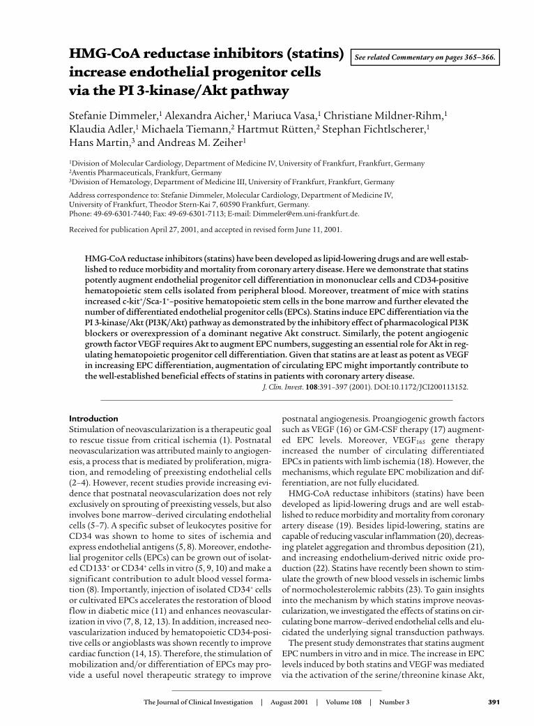
ResultsStatins increase EPCs in vitro. Incubation of isolatedhuman MNCs with atorvastatin dose and time depend-ently increased the number of differentiated, adherentEPCs from 2.1% ± 0.16% to 5% ± 0.35% of the totalnumber of MNCs in vitro (Figure 1, a and b). Similarly,the HMG-CoA reductase inhibitors mevastatin (1 µM)and simvastatin (1 µM) also augmented the number ofEPCs up to 288% ± 17% and 241% ± 102%, respectively(Figure 1c). EPCs were characterized as adherent cellsdouble positive for DiLDL-uptake and lectin binding(5, 18) (Figure 1c). The endothelial phenotype of theEPCs was additionally confirmed by documenting theexpression of well-established cell surface markers likevWF, VEGF-receptor 2 (KDR-receptor), VE-cadherin,and CD31 by fluorescence-activated cell sorting (FACS)and immunostaining (Figure 1d and data not shown).The expression pattern of these endothelial cell mark-
392 The Journal of Clinical Investigation | August 2001 | Volume 108 | Number 3
ers on EPCs was found to be comparable to thatobserved in human umbilical venous endothelial cells(HUVECs) (data not shown). In addition, the expres-sion and activity of the endothelial nitric oxide syn-thase in EPCs was demonstrated by immunostaining,Western blot analysis, and DAF-2 staining, respective-ly (data not shown).
Because the angiogenic growth factor VEGF is knownto augment the number of EPCs (16, 18), we comparedthe statin effect with VEGF. As illustrated in Figure 1e,human recombinant VEGF also stimulated the differ-entiation of EPCs with a maximal effect at 100 ng/ml.
The effect of statins appears to be at least equipotentcompared, with VEGF.
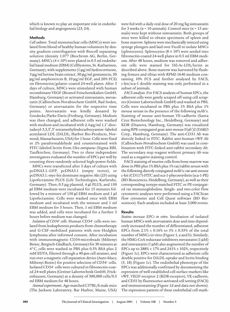
To exclude the possibility that the effect of VEGF andstatins is due to increased adherence of adult circulat-ing endothelial cells, we isolated CD34+ hematopoieticstem cells (HSCs), which presumably represent the EPCprecursor cells in humans (5, 12, 26). After separationof CD34-positive cells by immunomagnetic CD34microbeads from mobilized peripheral blood, the puri-ty of the population was 93.8% (range 82.4–99%) asanalysed by FACS (Figure 2a). Addition of VEGF oratorvastatin to the isolated CD34+ hematopoietic pro-
The Journal of Clinical Investigation | August 2001 | Volume 108 | Number 3 393
Figure 1 Statins increase the number of adherent EPCs. (a and b) MNCs were incubated with atorvastatin as indicated, and adherent DiLDL/lectin-positive cells were counted (13). Basal control values represent 388 ± 146 cells/mm2. The solvent DMSO had no effect on EPC differentiation.Data are mean ± SEM, n = 4–6. (c) MNCs were incubated with atorvastatin (1 µM), simvastatin (1 µM), or mevastatin (1 µM) for 24 hours,and adherent cells’ DiLDL uptake (red) and lectin binding (green) were assessed. Double positive cells appear yellow in the overlay. Repre-sentative images are shown from at least three experiments. (d) MNCs were incubated for 4 days. Adherent cells were analysed for expressionof KDR, CD31, VE-cadherin, and vWF by FACS. Dotted lines represent isotype controls. Similar expression patterns were observed after stim-ulation of MNCs with atorvastatin (1 µM, 24 hours) (data not shown). Representative images from n = 4 experiments are shown. (e) MNCswere incubated with VEGF for 24 hours and adherent DiLDL/lectin-positive cells were counted. Data are mean ± SEM, n = 3–6.
genitor cells significantly increased the number of dif-ferentiated EPCs (Figure 2b). Consistent with the effectobtained in MNCs, atorvastatin was at least as effectiveas VEGF (Figure 2b). Taken together, these datademonstrate that statins and VEGF induce differenti-ation of hematopoietic progenitor cells into EPCs.
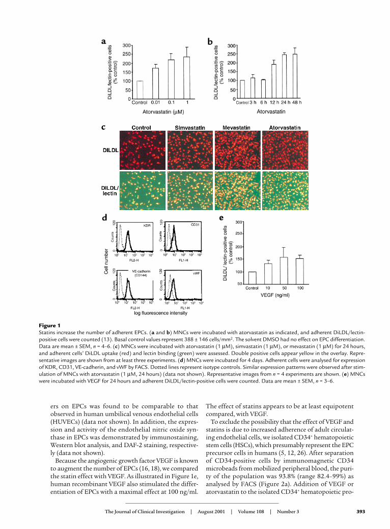
Statins increase EPC levels in vivo. To test the in vivo rel-evance of our findings, mice were fed with simvastatin(20 mg/kg daily) for 3 weeks, and EPC numbers weredetermined. As shown in Figure 3a, statin treatment ledto a more than twofold increase in DiLDL/lectin-posi-tive cells, thus extending the in vitro data. To gain fur-ther insights into the effect of statins, the number ofmultipotent bone marrow HSCs, which are character-ized by expression of c-kit (CD117) and sca-1 (26, 27),was analysed. The number of c-kithigh cells was pro-foundly increased in bone marrow of simvastatin-fedmice (Figure 3b). Moreover, c-kit/sca-1 double positivebone marrow cells were increased from 0.78% ± 0.3% incontrols to 8.2% ± 2.6% in simvastatin-treated mice (P ≤ 0.05). In contrast, the overall number of sca-1high
cells did not significantly change (37% ± 6% in controlversus 45% ± 14% in simvastatin-fed mice). These datasuggest that statins specifically augment multipotentc-kit/sca-1 HSCs in the bone marrow and concomi-tantly increase the number of differentiated EPCs inthe periphery in vivo.
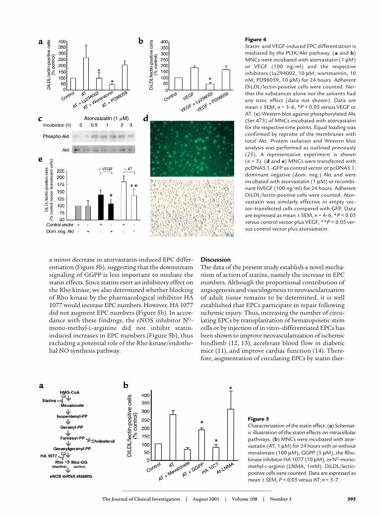
The effects of VEGF and statins on EPCs is mediated via thePI 3-kinase/Akt pathway. Next, we investigated themolecular mechanisms underlying the effects ofstatins on EPC differentiation. Since statins and VEGFhave both been shown to stimulate the PI 3-kinase/Akt(PI3K/Akt) pathway (23, 28), we used the pharmaco-logical PI3K-inhibitors Ly294002 and wortmannin toassess whether the PI3K-pathway is involved. Inhibi-
tion of the PI3K-pathway abolished statin- and VEGF-stimulated increase of EPCs (Figure 4, a and b). In con-trast, inhibition of another VEGF-stimulated kinasecascade, namely the MAP-kinases ERK-1 and -2, byPD98059 did not significantly alter the differentiationof EPCs (Figure 4, a and b), suggesting that a PI3Krather than a MAP-kinase pathway mediates theeffects of statins and VEGF.
To demonstrate that Akt is indeed an essential down-stream signaling event, Akt phosphorylation indicativefor Akt activity was detected by Western blot analysis.Statins and VEGF potently stimulated Akt phosphory-lation in isolated MNCs (Figure 4c and data notshown). The causal contribution of Akt for statin- andVEGF-induced EPC differentiation was finally demon-strated by transient transfection of the MNCs with adominant negative Akt construct. The transfection effi-ciency was about 50%, as determined by GFP-fluores-cent cells (Figure 4d). Overexpression of the dominantnegative Akt inhibited the increase of EPCs induced bystatins or VEGF (Figure 4e), clearly demonstrating thatAkt is causally involved in EPC differentiation.
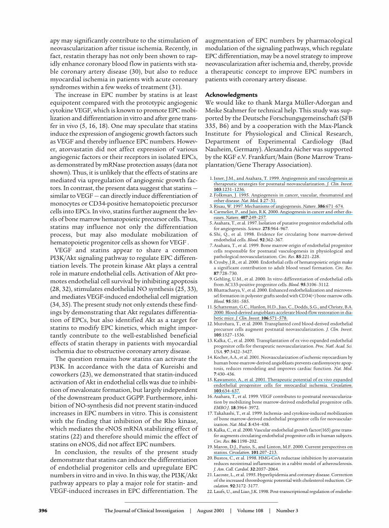
Finally, we attempted to characterize the molecularsignal transduction pathway of statins to modulateEPC differentiation. The inhibition of HMG-CoAreductase, which mediates the cholesterol-loweringeffect of statins, also reduces the formation of themetabolic products geranylgeranylpyrophosphat(GGPP) (22). Blockade of geranylgeranylation, in turn,abolishes Rho-kinase activation and increases eNOSmRNA stability (Figure 5a) (22, 29). Therefore, wedetermined the effect of mevalonate, the end productof the HMG-CoA reductase, and the further down-stream intermediate GGPP. Mevalonate reversed theinhibitory effect of statins, whereas GGPP led only to
394 The Journal of Clinical Investigation | August 2001 | Volume 108 | Number 3
Figure 2 Statins and VEGF induce differentiation of CD34+ cells. (a)CD34+ cells were isolated and purity of CD34+ cell fraction wasanalysed by FACS. Isotype control is represented by a dottedline and CD34 by a solid line. (b) CD34+ cells were incubatedwith atorvastatin (AT; 1 µM) or VEGF (100 ng/ml) for 24hours, and adherent DiLDL/lectin-positive cells were counted.Data are mean ± SEM, n = 4–7, *P < 0.05 versus control.
Figure 3Statins increase EPCs and HSCs in mice. (a) Micewere fed with simvastatin for 3 weeks. Spleno-cytes were isolated and incubated for 4 days.Adherent DiLDL/lectin-positive cells were count-ed. Data are mean ± SEM, *P < 0.05 versus con-trol. (b) Bone marrow cells were isolated and thenumber of c-kithigh cells was measured by FACS.Data are mean ± SEM, *P = 0.006 versus control(Mann-Whitney test).
a minor decrease in atorvastatin-induced EPC differ-entiation (Figure 5b), suggesting that the downstreamsignaling of GGPP is less important to mediate thestatin effects. Since statins exert an inhibitory effect onthe Rho kinase, we also determined whether blockingof Rho kinase by the pharmacological inhibitor HA1077 would increase EPC numbers. However, HA 1077did not augment EPC numbers (Figure 5b). In accor-dance with these findings, the eNOS inhibitor NG-mono-methyl-L-arginine did not inhibit statin-induced increases in EPC numbers (Figure 5b), thusexcluding a potential role of the Rho kinase/endothe-lial NO synthesis pathway.
DiscussionThe data of the present study establish a novel mecha-nism of action of statins, namely the increase in EPCnumbers. Although the proportional contribution ofangiogenesis and vasculogenesis to neovascularizationof adult tissue remains to be determined, it is wellestablished that EPCs participate in repair followingischemic injury. Thus, increasing the number of circu-lating EPCs by transplantation of hematopoietic stemcells or by injection of in vitro–differentiated EPCs hasbeen shown to improve neovascularization of ischemichindlimb (12, 13), accelerate blood flow in diabeticmice (11), and improve cardiac function (14). There-fore, augmentation of circulating EPCs by statin ther-
The Journal of Clinical Investigation | August 2001 | Volume 108 | Number 3 395
Figure 4 Statin- and VEGF-induced EPC differentiation ismediated by the PI3K/Akt pathway. (a and b)MNCs were incubated with atorvastatin (1 µM)or VEGF (100 ng/ml) and the respectiveinhibitors (Ly294002, 10 µM; wortmannin, 10nM; PD98059, 10 µM) for 24 hours. AdherentDiLDL/lectin-positive cells were counted. Nei-ther the substances alone nor the solvents hadany toxic effect (data not shown). Data aremean ± SEM, n = 3–6, *P < 0.05 versus VEGF orAT. (c) Western blot against phosphorylated Akt(Ser 473) of MNCs incubated with atorvastatinfor the respective time points. Equal loading wasconfirmed by reprobe of the membranes withtotal Akt. Protein isolation and Western blotanalysis was performed as outlined previously(25). A representative experiment is shown (n = 3). (d and e) MNCs were transfected withpcDNA3.1.-GFP as control vector or pcDNA3.1.dominant negative (dom. neg.) Akt and wereincubated with atorvastatin (1 µM) or recombi-nant hVEGF (100 ng/ml) for 24 hours. AdherentDiLDL/lectin-positive cells were counted. Ator-vastatin was similarly effective in empty vec-tor–transfected cells compared with GFP. Dataare expressed as mean ± SEM, n = 4–6, *P < 0.05versus control vector plus VEGF, **P < 0.05 ver-sus control vector plus atorvastatin.
Figure 5Characterization of the statin effect. (a) Schemat-ic illustration of the statin effects on intracellularpathways. (b) MNCs were incubated with ator-vastatin (AT; 1 µM) for 24 hours with or withoutmevalonate (100 µM), GGPP (5 µM), the Rho-kinase inhibitor HA 1077 (10 µM), or NG-mono-methyl-L-arginin (LNMA, 1mM). DiLDL/lectin-positive cells were counted. Data are expressed asmean ± SEM, P < 0.05 versus AT; n = 3–7.
apy may significantly contribute to the stimulation ofneovascularization after tissue ischemia. Recently, infact, restatin therapy has not only been shown to rap-idly enhance coronary blood flow in patients with sta-ble coronary artery disease (30), but also to reducemyocardial ischemia in patients with acute coronarysyndromes within a few weeks of treatment (31).
The increase in EPC number by statins is at leastequipotent compared with the prototypic angiogeniccytokine VEGF, which is known to promote EPC mobi-lization and differentiation in vitro and after gene trans-fer in vivo (5, 16, 18). One may speculate that statinsinduce the expression of angiogenic growth factors suchas VEGF and thereby influence EPC numbers. Howev-er, atorvastatin did not affect expression of variousangiogenic factors or their receptors in isolated EPCs,as demonstrated by mRNase protection assays (data notshown). Thus, it is unlikely that the effects of statins aremediated via upregulation of angiogenic growth fac-tors. In contrast, the present data suggest that statins —similar to VEGF — can directly induce differentiation ofmonocytes or CD34-positive hematopoietic precursorcells into EPCs. In vivo, statins further augment the lev-els of bone marrow hematopoietic precursor cells. Thus,statins may influence not only the differentiationprocess, but may also modulate mobilization ofhematopoietic progenitor cells as shown for VEGF .
VEGF and statins appear to share a commonPI3K/Akt signaling pathway to regulate EPC differen-tiation levels. The protein kinase Akt plays a centralrole in mature endothelial cells. Activation of Akt pro-motes endothelial cell survival by inhibiting apoptosis(28, 32), stimulates endothelial NO synthesis (25, 33),and mediates VEGF-induced endothelial cell migration(34, 35). The present study not only extends these find-ings by demonstrating that Akt regulates differentia-tion of EPCs, but also identified Akt as a target forstatins to modify EPC kinetics, which might impor-tantly contribute to the well-established beneficialeffects of statin therapy in patients with myocardialischemia due to obstructive coronary artery disease.
The question remains how statins can activate thePI3K. In accordance with the data of Kureishi andcoworkers (23), we demonstrated that statin-inducedactivation of Akt in endothelial cells was due to inhibi-tion of mevalonate formation, but largely independentof the downstream product GGPP. Furthermore, inhi-bition of NO-synthesis did not prevent statin-inducedincreases in EPC numbers in vitro. This is consistentwith the finding that inhibition of the Rho kinase,which mediates the eNOS mRNA stabilizing effect ofstatins (22) and therefore should mimic the effect ofstatins on eNOS, did not affect EPC numbers.
In conclusion, the results of the present studydemonstrate that statins can induce the differentiationof endothelial progenitor cells and upregulate EPCnumbers in vitro and in vivo. In this way, the PI3K/Aktpathway appears to play a major role for statin- andVEGF-induced increases in EPC differentiation. The
augmentation of EPC numbers by pharmacologicalmodulation of the signaling pathways, which regulateEPC differentiation, may be a novel strategy to improveneovascularization after ischemia and, thereby, providea therapeutic concept to improve EPC numbers inpatients with coronary artery disease.
AcknowledgmentsWe would like to thank Marga Müller-Adorgan andMeike Stahmer for technical help. This study was sup-ported by the Deutsche Forschungsgemeinschaft (SFB335, B6) and by a cooperation with the Max-PlanckInstitute for Physiological and Clinical Research,Department of Experimental Cardiology (BadNauheim, Germany). Alexandra Aicher was supportedby the KGF e.V. Frankfurt/Main (Bone Marrow Trans-plantation/Gene Therapy Association).
1. Isner, J.M., and Asahara, T. 1999. Angiogenesis and vasculogenesis astherapeutic strategies for postnatal neovascularization. J. Clin. Invest.103:1231–1236.
2. Folkman, J. 1995. Angiogenesis in cancer, vascular, rheumatoid andother disease. Nat. Med. 1:27–31.
3. Risau, W. 1997. Mechanisms of angiogenesis. Nature. 386:671–674.4. Carmeliet, P., and Jain, R.K. 2000. Angiogenesis in cancer and other dis-
eases. Nature. 407:249–257.5. Asahara, T., et al. 1997. Isolation of putative progenitor endothelial cells
for angiogenesis. Science. 275:964–967.6. Shi, Q., et al. 1998. Evidence for circulating bone marrow-derived
endothelial cells. Blood. 92:362–367.7. Asahara, T., et al. 1999. Bone marrow origin of endothelial progenitor
cells responsible for postnatal vasculogenesis in physiological andpathological neovascularization. Circ. Res. 85:221–228.
8. Crosby, J.R., et al. 2000. Endothelial cells of hematopoietic origin makea significant contribution to adult blood vessel formation. Circ. Res.87:728–730.
9. Gehling, U.M., et al. 2000. In vitro differentiation of endothelial cellsfrom AC133-positive progenitor cells. Blood. 95:3106–3112.
10. Bhattacharya, V., et al. 2000. Enhanced endothelialization and microves-sel formation in polyester grafts seeded with CD34(+) bone marrow cells.Blood. 95:581–585.
11. Schatteman, G.C., Hanlon, H.D., Jiao, C., Dodds, S.G., and Christy, B.A.2000. Blood-derived angioblasts accelerate blood-flow restoration in dia-betic mice. J. Clin. Invest. 106:571–578.
12. Murohara, T., et al. 2000. Transplanted cord blood-derived endothelialprecursor cells augment postnatal neovascularization. J. Clin. Invest.105:1527–1536.
13. Kalka, C., et al. 2000. Transplantation of ex vivo expanded endothelialprogenitor cells for therapeutic neovascularization. Proc. Natl. Acad. Sci.USA. 97:3422–3427.
14. Kocher, A.A., et al. 2001. Neovascularization of ischemic myocardium byhuman bone-marrow-derived angioblasts prevents cardiomyocyte apop-tosis, reduces remodeling and improves cardiac function. Nat. Med.7:430–436.
15. Kawamoto, A., et al. 2001. Therapeutic potential of ex vivo expandedendothelial progenitor cells for myocardial ischemia. Circulation.103:634–637.
16. Asahara, T., et al. 1999. VEGF contributes to postnatal neovasculariza-tion by mobilizing bone marrow-derived endothelial progenitor cells.EMBO J. 18:3964–3972.
17. Takahashi, T., et al. 1999. Ischemia- and cytokine-induced mobilizationof bone marrow-derived endothelial progenitor cells for neovascular-ization. Nat. Med. 5:434–438.
18. Kalka, C., et al. 2000. Vascular endothelial growth factor(165) gene trans-fer augments circulating endothelial progenitor cells in human subjects.Circ. Res. 86:1198–202.
19. Maron, D.J., Fazio, S., and Linton, M.F. 2000. Current perspectives onstatins. Circulation. 101:207–213.
20. Bustos, C., et al. 1998. HMG-CoA reductase inhibition by atorvastatinreduces neointimal inflammation in a rabbit model of atherosclerosis.J. Am. Coll. Cardiol. 32:2057–2064.
21. Lacoste, L., et al. 1995. Hyperlipidemia and coronary disease. Correctionof the increased thrombogenic potential with cholesterol reduction. Cir-culation. 92:3172–3177.
22. Laufs, U., and Liao, J.K. 1998. Post-transcriptional regulation of endothe-
396 The Journal of Clinical Investigation | August 2001 | Volume 108 | Number 3
lial nitric oxide synthase mRNA stability by Rho GTPase. J. Biol. Chem.273:24266–24271.
23. Kureishi, Y., et al. 2000. The HMG-CoA reductase inhibitor simvastatinactivates the protein kinase Akt and promotes angiogenesis in normoc-holesterolemic animals. Nat. Med. 6:1004–1010.
24. Dimmeler, S., and Zeiher, A.M. 2000. Akt takes center stage in angio-genesis signaling. Circ. Res. 86:4–5.
25. Dimmeler, S., et al. 1999. Activation of nitric oxide synthase in endothe-lial cells via Akt-dependent phosphorylation. Nature. 399:601–605.
26. Takakura, N., et al. 2000. A role for hematopoietic stem cells in promot-ing angiogenesis. Cell. 102:199–209.
27. Weissman, I.L. 2000. Stem cells: units of development, units of regener-ation, and units in evolution. Cell. 100:157–168.
28. Gerber, H.P., et al. 1998. Vascular endothelial growth factor regulatesendothelial cell survival through the phosphatidylinositol 3′-kinase/Aktsignal transduction pathway. Requirement for Flk-1/KDR activation. J.Biol. Chem. 273:30336–30343.
29. Laufs, U., and Liao, J.K. 2000. Targeting rho in cardiovascular disease.Circ. Res. 87:526–528.
30. Baller, D., et al. 1999. Improvement in coronary flow reserve determinedby positron emission tomography after 6 months of cholesterol-lower-ing therapy in patients with early stages of coronary atherosclerosis. Cir-culation. 99:2871–2875.
31. Schwartz, G.G., et al. 2001. Effects of atorvastatin on early recurrentischemic events in acute coronary syndromes: the MIRACL study. A ran-domized controlled trial. JAMA. 285:1711–1718.
32. Dimmeler, S., Assmus, B., Hermann, C., Haendeler, J., and Zeiher, A.M.1998. Fluid shear stress stimulates phosphorylation of Akt in humanendothelial cells: involvement in suppression of apoptosis. Circ. Res.83:334–342.
33. Fulton, D., et al. 1999. Regulation of endothelium-derived nitric oxideproduction by the protein kinase Akt. Nature. 399:597–601.
34. Dimmeler, S., Dernbach, E., and Zeiher, A.M. 2000. Phosphorylation ofthe endothelial nitric oxide synthase at Ser 1177 is required for VEGF-induced endothelial cell migration. FEBS Lett. 477:258–262.
35. Morales-Ruiz, M., et al. 2000. Vascular endothelial growth factor-stimu-lated actin reorganization and migration of endothelial cells is regulat-ed via the serine/threonine kinase Akt. Circ. Res. 86:892–896.
The Journal of Clinical Investigation | August 2001 | Volume 108 | Number 3 397


