Embed Size (px)
Citation preview
1 23
Journal of Industrial Microbiology& BiotechnologyOfficial Journal of the Society forIndustrial Microbiology ISSN 1367-5435Volume 38Number 9 J Ind Microbiol Biotechnol (2011)38:1311-1319DOI 10.1007/s10295-010-0912-5
Homologue expression of a #-xylosidasefrom native Aspergillus niger
A. Amaro-Reyes, B. E. García-Almendárez, D. G. Vázquez-Mandujano,S. Amaya-Llano, E. Castaño-Tostado,R. G. Guevara-González, et al.
1 23
Your article is protected by copyright and
all rights are held exclusively by Society for
Industrial Microbiology. This e-offprint is
for personal use only and shall not be self-
archived in electronic repositories. If you
wish to self-archive your work, please use the
accepted author’s version for posting to your
own website or your institution’s repository.
You may further deposit the accepted author’s
version on a funder’s repository at a funder’s
request, provided it is not made publicly
available until 12 months after publication.
ORIGINAL PAPER
Homologue expression of a b-xylosidase from nativeAspergillus niger
A. Amaro-Reyes • B. E. Garcıa-Almendarez • D. G. Vazquez-Mandujano •
S. Amaya-Llano • E. Castano-Tostado • R. G. Guevara-Gonzalez •
O. Loera • C. Regalado
Received: 26 July 2010 / Accepted: 11 November 2010 / Published online: 30 November 2010
� Society for Industrial Microbiology 2010
Abstract Xylan constitutes the second most abundant
source of renewable organic carbon on earth and is located
in the cell walls of hardwood and softwood plants in the
form of hemicellulose. Based on its availability, there is a
growing interest in production of xylanolytic enzymes
for industrial applications. b-1,4-xylan xylosidase (EC
3.2.1.37) hydrolyses from the nonreducing end of xylool-
igosaccharides arising from endo-1,4-b-xylanase activity.
This work reports the partial characterization of a purified
b-xylosidase from the native strain Aspergillus niger GS1
expressed by means of a fungal system. A gene encoding
b-xylosidase, xlnD, was successfully cloned from a native
A. niger GS1 strain. The recombinant enzyme was
expressed in A. niger AB4.1 under control of A. nidulans
gpdA promoter and trpC terminator. b-xylosidase was
purified by affinity chromatography, with an apparent
molecular weight of 90 kDa, and showed a maximum
activity of 4,280 U mg protein-1 at 70�C, pH 3.6. Half-life
was 74 min at 70�C, activation energy was 58.9 kJ mol-1,
and at 50�C optimum stability was shown at pH 4.0–5.0.
b-xylosidase kept residual activity[83% in the presence of
dithiothreitol (DTT), b-mercaptoethanol, sodium dodecyl
sulfate (SDS), ethylenediaminetetraacetate (EDTA), and
Zn2?. Production of a hemicellulolytic free xylosidase
showed some advantages in applications, such as animal
feed, enzymatic synthesis, and the fruit-juice industry
where the presence of certain compounds, high tempera-
tures, and acid media is unavoidable in the juice-making
process.
Keywords Aspergillus niger GS1 � b-xylosidase
activity � Thermostability � Homologue expression
Introduction
Xylan constitutes the second most abundant source of
renewable organic carbon on earth and is located in the cell
walls of hardwood and softwood plants in the form of
hemicellulose [28]. Endo-b-1,4-xylanases (EC 3.2.1.8) and
b-1,4-xylan xylosidase (EC 3.2.1.37) are key enzymes that
hydrolyze xylan into xylooligosaccharides [4, 27]. b-xy-
losidase hydrolyses the terminal xylose unit from the
nonreducing end of the xylooligosaccharides arising from
endo-1,4-b-xylanase activity [22]. This is important, as
b-xylosidase may relieve the end-product inhibition of
endoxylanases and is also effective in transglycosylation
reactions in which monosaccharide units or alcohols are
attached to or cleaved from xylose units [9]. There is a
growing interest in developing high-yield and low-cost
production of xylanolytic enzymes for industrial applica-
tions, such as bioconversion of agroindustrial residues to
biofuels, low-calorie sweeteners, and pharmacological
products [20]. The worldwide market of these enzymes is
A. Amaro-Reyes � B. E. Garcıa-Almendarez �D. G. Vazquez-Mandujano � S. Amaya-Llano �E. Castano-Tostado � C. Regalado (&)
DIPA, PROPAC. Facultad de Quımica, Universidad Autonoma
de Queretaro, C.U. Cerro de las Campanas s/n. Col. Las
Campanas, 76010 Qro. Queretaro, Mexico
e-mail: [email protected]; [email protected]
R. G. Guevara-Gonzalez
C.A Ingenierıa de Biosistemas, Facultad de Ingenierıa,
Universidad Autonoma de Queretaro, C.U. Cerro de las
Campanas s/n. Col. Las Campanas,
76010 Qro. Queretaro, Mexico
O. Loera
Dpto. de Biotecnologia, Universidad Autonoma
Metropolitana-Iztapalapa, 09340 Mexico, DF, Mexico
123
J Ind Microbiol Biotechnol (2011) 38:1311–1319
DOI 10.1007/s10295-010-0912-5
Author's personal copy
around 200 million US dollars per annum [14]. Therefore,
the search for strains with the generally recognized as safe
(GRAS) status that will grow in low-cost substrates that
will optimize xylanolytic enzyme production represents an
ultimate goal in this field of research. Filamentous fungi
are more attractive than bacteria as potential producers of
these enzymes because fungi secrete higher enzyme levels
into the culture medium [16]. Aspergillus niger is a sap-
rophytic fungus well known for its production and secre-
tion of a variety of hydrolytic enzymes, contributing to its
ability to degrade plant polysaccharides such as cellulose,
hemicellulose, pectin, starch, and inulin [31]. Several
studies have reported a variety of hemicellulolytic enzymes
induced with various hemicellulosic residues (corn cob,
sugar-cane bagasse, wheat bran, wheat straw, and rice
straw) as the main carbon source using Aspergillus species
[12, 18]. Nevertheless, some industrial applications require
xylanases and xylosidases free of cellulase, among other
hemicellulolytic activities, and enzymatic stability is also
required over a broad range of temperatures and pH values
[20].
A strain of Aspergillus was isolated from Mexican copra
paste that produces a variety of cell-wall-degrading
enzymes using different substrates upon solid-state fer-
mentation [23]. Molecular identification of this novel and
potentially useful Aspergillus strain and its xylanolytic
genes have not yet been reported. Natural inducers of
xylanolytic genes in Aspergillus may be products of xylan
degradation or transglycosylation processes, such as
D-xylose, xylobiose, xylotriose, and xylotetrose [27].
However, in the presence of readily metabolizable carbon
sources such as D-glucose, gene expression involved in the
use of less-favored carbon sources, such as the xylanolytic
system, is inhibited due to carbon catabolite repression
[21]. We describe the design of a system capable of pro-
ducing b-xylosidase even in presence of its repressor. The
purpose of our study was to constitutively express and
partially characterize a b-xylosidase from the native strain
A. niger GS1 by means of a fungal system.
Materials and methods
Materials
All chemicals were of analytical grade and were purchased
from Sigma (St. Louis, MO, USA), except as indicated.
Microorganisms and plasmid
Aspergillus niger GS1 (NCBI No. GU395669) was used as
a source of xylosidase gene (UAQ, Queretaro, Mexico).
Spores isolated from A. niger GS1 were stored in Tween 20
on silica gel at 4�C. Stock cultures were subcultured on
fresh sterile potato dextrose agar (PDA; Bioxon, Cu-
autitlan, Mexico) plates and incubated for 72–120 h at
30�C [23]. Escherichia coli JM109 genotype recA1,
endA1, gyrA96, thi, hsdR17 (rk-mK?), relA1, supE44,
D(lac-proAB) [F’, traD36, proAB, lacIqZDM15] (Promega,
Madison, WI, USA) was used to propagate vectors and was
cultured at 37�C in Luria–Bertani medium comprising
(g l-1): bacto-tryptone (Difco, Franklin Lakes, NJ, USA),
10; yeast extract (Difco), 5; sodium chloride (NaCl), 10;
supplemented with 100 lg ml-1 ampicillin. The pGEM-T
plasmid (Promega) was used as the subcloning vector, and
A. niger AB4.1 (pyrG-) strain [29] was used for homolo-
gous expression of xylosidase (xlnD) gene. Vector
pAN52.1 was used to construct the constitutive expression
vector pANJil. This vector contains the gpdA promoter
and the terminator region of the trpC gene (both from
A. nidulans) separated by BamHI and NcoI sites. Vector
pAB4.1 (pyrG) [29] was used as selection marker. Both
vectors were kindly provided by Dr. Punt (TNO, The
Netherlands).
Induction of b-xylosidase
Aspergillus niger GS1 spores were inoculated into PDA-
xylan slants (glucose 15 g l-1, oat spelts xylan 5 g l-1,
agar–agar (Bioxon) 15 g l-1, and potato infusion 0.5 l), pH
5.5–6.0, incubated at 30�C, for 72 h. Harvested spores
were then transferred to PDA-xylan slants increasing by
5 g l-1 the initial xylan concentration while decreasing
initial glucose concentration by 5 g l-1 until complete
replacement with xylan as main carbon source (g l-1)
(agar–agar, 15; oat spelts xylan, 20; and potato infusion,
0.5 l) was attained. Spores collected from the potato xylan
agar were seeded in basal xylan media supplemented with
yeast extract (oat spelts xylan 25 g l-1, yeast extract
5 g l-1). After incubation for 2–3 days, mycelia were
collected for RNA extraction using the RNeasy Plant Mini
Kit (Qiagen, Hamburg, Germany).
Molecular identification of A. niger GS1, and xlnD gene
Aspergillus niger GS1 was genetically identified by
sequencing the 26S ribosomal DNA (rDNA) (GBC-IPN,
Mexico). Collected mycelia from basal xylan media was
employed for RNA extraction. Forward (JilF 50-CCATG
GATGGCGCACTCAATGTCTCG-30), and reverse (JilR
50-CTGGATCCCTAGTGGTGATGGTGATGATGCTCCT
TCCCCGGCCAC-30) primers were designed using the
National Center for Biotechnical Information (NCBI)-
reported sequence (ANXM_001389379) for A. niger
b-1,4-xylan xylosidase. Bases coding for His-tag are shown
in italics.
1312 J Ind Microbiol Biotechnol (2011) 38:1311–1319
123
Author's personal copy
The RevertAidH Minus kit (Fermentas, Ontario, Can-
ada) was used to obtain xylosidase complementary DNA
(cDNA) from total RNA (tRNA) (2 lg) following the
manufacturer’s instructions. Polymerase chain reaction
(PCR) amplification of A. niger GS1 cDNA (100 ng) was
conducted using 400 mM primers; 10 mM deoxyribonu-
cleotide triphosphate (dNTP), 1.5 mM magnesium chloride
(MgCl2), 19 reaction buffer [200 mM Tris pH 8.4,
500 mM calcium chloride (KCl)] (Invitrogen, Carlsbad,
CA, USA), and 2 U recombinant Taq polymerase (Invit-
rogen) in a final volume of 50 ll. Reaction conditions were
5 min at 94�C, followed by 30 cycles of 30 s at 94�C, 30 s
at 65�C, 2.5 min at 72�C, and a final extension of 10 min at
72�C. The amplicon was ligated into pGEM-T vector,
followed by heat-shock cloning into E. coli JM109. Clones
containing the insert were directly identified by blue/white
color screening on indicator plates, and then from Mini-
prep (Qiagen) extraction, the isolated plasmid was sent for
sequencing (MCLab, San Francisco, CA, USA). After
sequence confirmation, the DNA open-reading frame was
cloned into the expression vector pAN52.1 (cloning sites
NcoI and BamHI) to obtain pANJil expression vector.
Aspergillus transformation
Fungal cotransformation was accomplished using the pro-
cedure reported by Sanchez and Aguirre [25] developed for
A. nidulans, with modifications. Spores from A. niger
AB4.1 were washed with 10 ml sterile distilled water, and
an inoculum of 8.6 9 106 spores ml-1 was added to 50 ml
of dextrose potato broth (Difco) supplemented with uridine
(2.5 g l-1), followed by incubation at 30�C in a rotary
shaker (300 rpm) for 15 h. Next, germinating spores (GTS)
were recovered by centrifugation at 4,0009g for 10 min at
4�C (Eppendorf, Mod. 5804R, Hamburg, Germany). GTS
were resuspended in 50 ml of ice-cold sterile water, cen-
trifuged again, resuspended in 25 ml of ice-cold pretreat-
ment buffer [1% yeast extract, 1% glucose; YED] plus
20 mM hydroxyethyl-1-piperazine ethanesulfonic acid
(HEPES) (adjusted to pH 8.0 with 100 mM Tris), and
incubated for 1 h at 30�C in a rotary shaker at 100 rpm.
After this incubation, GTS were centrifuged and resus-
pended in 1 ml (about 2.2 9 107 spores ml-1, final) of ice-
cold electroporation buffer [10 mM Tris–HCl(pH 7.3),
270 mM sucrose, 1 mM HEPES, 10 mM rubidium chloride
(RbCl), 10 mM lithium chloride (LiCl)], kept on ice, and
stored at -70�C. For electroporation, 2 lg of total DNA
(tDNA) (pANJil expression vector with pAB4.1 vector in a
10:1 volume ratio) was added to 50 ll of ice-cold GTS
suspension. The mixture was then incubated on ice for
15 min and transferred to a 0.2-cm cuvette. Electroporation
was performed using a MicroPulser Electroporation Appa-
ratus (Bio-Rad, Hercules, CA, USA), adjusting voltage to
1.4 kV and pulses lasting approximately 3.5 ms. After
electroporation, 600 ll of ice-cold YED was added to the
cuvette and the cell suspension transferred to a sterile 1.5-
ml tube, kept on ice for 10 min, and incubated at 30�C for
90 min in a rotary shaker at 100 rpm. Electroporated spores
(100 ll plate-1) were extended on sorbitol-containing
minimal agar (g l-1): glucose, 10; sorbitol, 218.64; sodium
nitrate (NaNO3), 6; KCl, 0.52, potassium dihydrogen
phosphate (KH2PO4), 1.52; trace elements: zinc sulfite
(ZnSO4 7H2O), 0.022; boric acid (H3BO3), 0.011; manga-
nese (II) chloride (MnCl2 4H2O), 0.005; ferrous sulfate
(FeSO4 7H2O), 0.005; cobalt(II) chloride (CoCl2 6H2O),
0.0017; copper(II) sulfate (CuSO4 5H2O), 0.0016; sodium
molybdate (Na2MoO4 H2O), 0.0015; sodium ethylenedia-
minetetraacetate (Na2EDTA) 0.05; without uridine. Spores
were incubated at 30�C, for 48 h. The stability of uridine
prototroph transformants was tested by velvet-replica plating
on selective minimal medium. In addition, a control was
transformed with the pyrG gene but without expression
vector.
Screening and production of b-xylosidase activity
Cotransformants were plated on the selective minimal
medium (without uridine) and incubated for 8 days at
30�C. Liquid cultures were inoculated in glucose-rich
medium (g l-l): glucose, 20; yeast extract, 0.5; NaNO3,
7.5; (NH4)2SO4, 1.5; KCl, 8.67; MgSO4 7H2O, 8.67; and
trace elements) with 2 9 106 spores ml-1 in 50-ml Falcon
tubes. To screen cotransformants, up to 20 individual
clones were inoculated into minimal medium and checked
daily for xylosidase activity for 4 days. For the activity
assay, a 1-ml aliquot of culture medium was collected, and
cells were removed by filtration through a 0.45-lm filter
(Millipore, Billerica, MA, USA). Positive cotransformants
were assayed through JilF–JilR amplification using geno-
mic DNA as template, obtained by the cetyl trimethylam-
monium bromide (CTAB) protocol [1].
Analytical methods
Protein and b-xylosidase activity
Soluble protein content was determined according to
Bradford [2] using bovine serum albumin (BSA) as stan-
dard. Xylosidase activity was measured by the p-nitro-
phenol method, as described by Pedersen et al. [18], using
2.5 mM p-nitrophenol-b-D-xylopyranoside (PNPX) in
50 mM acetate buffer and 100 ll of enzyme solution at pH
4.0 in a total reaction volume of 1 ml. After incubation at
50�C for 10 min, the reaction was stopped by adding 1 M
Na2CO3 (JT Baker, Phillipsburg, NJ, USA) to a final
concentration of 0.33 mM, and the released p-nitrophenol
J Ind Microbiol Biotechnol (2011) 38:1311–1319 1313
123
Author's personal copy
was measured spectrophotometrically at 400 nm. One unit
of b-xylosidase activity was calculated as the amount of
enzyme producing 1 lmol equivalent of p-nitrophenol
per min, as calculated from a standard curve. Endo-b-1,4-
xylanase and cellulase activities of the purified extract was
determined using 5 g l-1 xylan and carboxymethyl cellu-
lose as substrates, respectively, dissolved in 50 mM acetate
buffer, pH 5.5. The reaction mixture consisted of 100 ll of
enzyme solution, 400 ll of the corresponding substrate,
and incubated at 50�C for 10 min, followed by immersion
in ice-cold water. Released reducing sugars were quantified
according to Miller [15] using a xylose and a glucose
standard curve (1–10 mM). One activity unit (U) was
defined as the amount of enzyme that released 1 lmol of
xylose or glucose equivalents per minute at 50�C.
Sodium dodecyl sulfate polyacrylamide gel
electrophoresis (SDS–PAGE)
Sodium dodecyl sulfate polyacrylamide gel electrophoresis
(SDS–PAGE) was carried out using a 10% (w/v) T (%
acrylamide plus bis-acrylamide in gelling solution)
according to Laemmli [11], and protein bands were stained
with Coomassie Brilliant Blue R-250 [1]. b-xylosidase
activity was detected in the gel after electrophoresis by
cutting bands. Each band was washed three times with
high-performance liquid chromatography (HPLC)-grade
water, placed in a microtube containing 200 ll substrate
solution (PNPX), and incubated for 30 min at 50�C. The
reaction was stopped by adding 150 ll of 1 M Na2CO3
solution, and absorbance was measured at 400 nm.
Purification of the recombinant b-xylosidase
To purify the recombinant b-xylosidase, cotransformed
A. niger was cultivated in glucose-rich medium. Then, 600
ml of a 4-day-old culture medium was filtered through
Whatman No. 4 (Whatman International, Maidstone, UK)
and 0.2 lm membrane (Millipore) and concentrated using
a Centricon centrifugal filter unit with molecular mass
cutoff membrane of 10 kDa (Millipore). Concentrated
enzyme (2.0 ml) was applied into a diethylaminoethanol
(DEAE)-cellulose column (1.5 9 25 cm), equilibrated, and
washed with 50 mM acetate buffer, pH 3.9, at a flow rate of
25 ml h-1. Bound proteins were eluted using the same
washing buffer plus 1 M NaCl. Fractions of 1.5 ml were
collected and assayed for absorbance at 280 nm and
b-xylosidase activity, and those showing high activity were
pooled and concentrated using the Centricon units
(10 kDa). Concentrated fractions were then loaded onto a
Nickel (Ni) Sepharose 6 Fast Flow column (1 ml) (GE
Healthcare Bio-sciences, Uppsala, Sweden) following the
manufacturer’s manual.
Effect of temperature and pH on purified recombinant
b-xylosidase activity
To determine optimal temperature of purified recombinant
b-xylosidase, activity determinations were conducted using
65-ng aliquots of purified enzyme and incubating at
30–80�C. Activation energy (Ea) of recombinant b-xylos-
idase was calculated following an Arrhenius-type behavior
by plotting ln (activity) vs (absolute temperature, K)-1,
in the range of 30� to 60�C. The slope of this plot indicates
(–Ea R-1), where R is the universal gas constant. Optimal
pH was determined using same protein aliquots of the
enzyme in same total reaction volume, and activity was
measured using acetate buffer (50 mM) for pH values
3.6–5.0; 50 mM phosphate buffer was used for pH values
6.0–8.0. Experiments were conducted for three replicates.
Temperature and pH stability of the purified
recombinant b-xylosidase
Aliquots (65 ng) of purified recombinant b-xylosidase were
preincubated in the range of 50� to 70�C for 5, 60, and
120 min using an AccuBlock dry bath (Labnet, Edison, NJ,
USA). After cooling at 4�C, xylosidase activity was assayed.
The recombinant enzyme half-life was calculated by linear
regression analysis of specific activity at desired temperature
against time. The effect of pH on the purified recombinant
enzyme stability was studied by incubating in 50 mM acetate
buffer (pH 4.0, 5.0) and 50 mM phosphate buffer (pH 6.0), at
10 ± 2�C for 2 and 24 h, followed by activity determination.
Experiments were conducted for three replicates.
Effect of metal ions and chemical reagents
on recombinant b-xylosidase activity
The effects of several metal ion salts and chemicals on the
recombinant purified b-xylosidase were tested by measur-
ing enzyme activity in the presence of individual com-
pounds at a final concentration of 10 mM. These chemicals
have been shown to affect xylanolytic activity [6, 8], and
those used were CuSO4 5H2O, LiCl, ZnCl2, Na2EDTA,
SDS, dithiothreitol (DTT), and b-mercaptoethanol. After
preincubating for 10 min at room temperature (26 ± 2�C),
the residual enzyme activities were expressed as the per-
centage of enzyme activity without added chemical.
Results and discussion
Molecular identification of A. niger GS1 and xlnD gene
Aspergillus niger GS1 was isolated from copra paste. A
pure culture was obtained by single streaking on PDA and
1314 J Ind Microbiol Biotechnol (2011) 38:1311–1319
123
Author's personal copy
morphologically identified by microscopic visualization of
its reproductive structures and main characteristics [23].
This native Mexican fungal strain was genetically identi-
fied by sequencing the 26S rRNA gene. Sequence of this
gene has been extensively used to establish phylogenetic
and systematic relationships within fungi [13]. A partial
sequence of 552 bp was obtained and submitted to the
NCBI database, with accession No. GU395669. Blast
analysis against NCBI genome database showed 100%
similarity with A. niger CBS 513.88 clone An03
(NT_166520.1). In addition, using the nucleotide collec-
tion, a 99% identity was found for partial sequence of 26S
RNA genes of A. niger VTCC:F021 (GQ382274.1), and A.
niger VTCC:CF128 26 S (GQ382273.1). Thus, from both
morphological and molecular analysis, we conclude that
the isolated strain was A. niger. A single amplification band
was obtained from PCR products with an approximate size
of 2,433 bp when primers JilR and JilF were used (data not
shown). PCR product was inserted into pGEM-T vector,
and positive clones identified by sequencing showed up to
99% homology with A. niger CBS 513.88 xylosidase
(xlnD) (XM_0013819379) partial messenger RNA
(mRNA). Our sequence corresponded to a complete
structural gene coding for b-xylosidase, plus a six histidine
tag before the stop codon, which was submitted to NCBI
database, obtaining accession number GU585573. In
addition, our findings are in agreement with La Grange
et al. [10], who reported that b-xylosidase in A. niger is
coded by xlnD, containing an open reading frame of 2,412
nucleotides, which encodes a theoretical protein of 804
amino acids (85 kDa).
Expression of b-xylosidase gene from the gpdA
promoter
To avoid catabolic repression exerted by glucose over xlnD
expression, we expressed the b-xylosidase from A. niger
GS1 into pAN52-1 under the control of the strong consti-
tutive A. nidulans glyceraldehyde-3-phosphate dehydro-
genase (gpdA) promoter and trpC terminator. The gpdA
promoter is expressed efficiently in A. niger strains [7, 17].
The construction of pANJil included a DNA sequence
encoding the first 26 amino acids of the xlnD signal peptide
(ALA-QA), according to Bendtsen et al. [3]. The pANJil
was introduced into A. niger AB4.1 strain by cotransfor-
mation with the plasmid pAB4.1, which contains the
A. nidulans pyrG gene encoding orotidine 50-phosphate
carboxylase. Preliminary analysis of Pyr? regenerants from
these cotransformations identified up to 200 transformants.
From those transformants, 15 were streaked twice on
minimal plates and then screened for b-xylosidase activity
on minimal liquid media under glucose-rich conditions
(20 g l-1) that repress xlnD synthesis in the wild-type
strain. Two strains (gpd-Jil-1, -2) that were morphologi-
cally stable showed b-xylosidase activity and were cultured
for genomic DNA isolation, which was analyzed by PCR
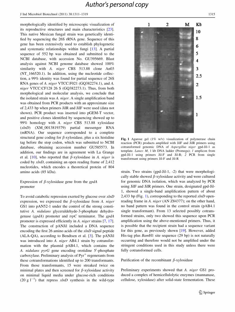
using JilF and JilR primers. One strain, designated gpd-Jil-
1, showed a single-band amplification pattern of about
2,433 bp (Fig. 1), corresponding to the reported xlnD open-
reading frame in A. niger (AN Z84377); on the other hand,
no band pattern was found in the control strain (pAB4.1
single transformant). From 13 selected possibly cotrans-
formed strains, only two showed this sequence upon PCR
amplification using the above-mentioned primers. Thus, it
is possible that the recipient strain had a sequence variant
for this gene, as previously shown [19]. However, added
His-tag plus BamH1 site sequence (29 bp) is not naturally
occurring and therefore would not be amplified under the
stringent conditions used in this study unless there were
fully cotransformed cells.
Purification of the recombinant b-xylosidase
Preliminary experiments showed that A. niger GS1 pro-
duced a complex of hemicellulolytic enzymes (mannanase,
cellulose, xylosidase) after solid-state fermentation. These
Fig. 1 Agarose gel (1% w/v) visualization of polymerase chain
reaction (PCR) products amplified with JilF and JilR primers using
cotransformed genomic DNA of Aspergillus niger gpd-Jil-1 as
template. Lanes: M, 1 kb DNA ladder (Promega); 1 amplicon from
gpd-Jil-1 using primers Jil-F and Jil-R; 2 PCR from single
transformant using primers Jil-F and Jil-R
J Ind Microbiol Biotechnol (2011) 38:1311–1319 1315
123
Author's personal copy
results indicate that hemicellulolytic enzyme expression is
subject to induction and carbon catabolic repression,
according to substrate composition used for fermentation
(data not shown). Therefore, the choice of an appropriate
system to constitutively provide hemicellulolytic free
b-xylosidase activity without being affected by catabolic
repression is of importance for processes such as modified
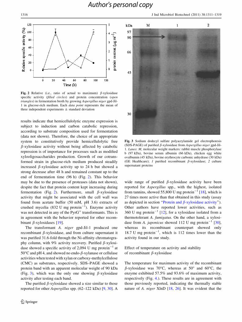
xylooligosaccharides production. Growth of our cotrans-
formed strain in glucose-rich medium produced steadily
increased b-xylosidase activity up to 24 h but showed a
strong decrease after 48 h and remained constant up to the
end of fermentation time (96 h) (Fig. 2). This behavior
may be due to the presence of proteases (data not shown),
despite the fact that protein content kept increasing during
fermentation (Fig. 2). Furthermore, small b-xylosidase
activity that might be associated with the cell wall was
found from acetate buffer (50 mM, pH 3.6) extracts of
crushed mycelia (832 U mg protein-1). Enzyme activity
was not detected in any of the PyrG? transformants. This is
in agreement with the behavior reported for other recom-
binant b-xylosidases [19].
The transformant A. niger gpd-Jil-1 produced one
recombinant b-xylosidase, and from culture supernatant it
was purified 31.6-fold through the Ni-affinity chromatogra-
phy column, with 9% activity recovery. Purified b-xylosi-
dase showed a specific activity of 2,094 U mg protein-1 at
50�C and pH 4, and showed no endo-b-xylanase or cellulase
activities when tested with xylan or carboxy-methylcellulose
(CMC) as substrates, respectively. SDS–PAGE showed a
protein band with an apparent molecular weight of 90 kDa
(Fig. 3), which was the only one showing b-xylosidase
activity after testing each band.
The purified b-xylosidase showed a size similar to those
reported for other Aspergillus spp. (62–122 kDa) [9, 30]. A
wide range of purified b-xylosidase activity have been
reported for Aspergillus spp., with the highest, isolated
from tannins, showed 55,800 U mg protein-1 [18], which is
27 times more active than that obtained in this study (assay
as depicted in section ‘‘Protein and b-xylosidase activity’’).
Other authors have reported lower activities, such as
360 U mg protein-1 [12], for a xylosidase isolated from a
thermotolerant A. fumigatus. On the other hand, a xylosi-
dase from A. japonicus showed 112 U mg protein-1 [30],
whereas its recombinant counterpart showed only
18.7 U mg protein-1, which is 112 times lower than the
activity found in our study.
Effect of temperature on activity and stability
of recombinant b-xylosidase
The temperature for maximum activity of the recombinant
b-xylosidase was 70�C, whereas at 50� and 60�C, the
enzyme exhibited 57.5% and 93.6% of maximum activity,
respectively (Fig. 4.). These results are in agreement with
those previously reported, indicating the thermally stable
nature of A. niger XlnD [18, 26]. It was evident that the
Fig. 2 Relative (i.e., ratio of actual to maximum) b-xylosidase
specific activity (filled circles) and protein concentration (opentriangles) in fermentation broth by growing Aspergillus niger gpd-Jil-
1 in glucose-rich medium. Each data point represents the mean of
three independent experiments ± standard deviation
Fig. 3 Sodium dodecyl sulfate polyacrylamide gel electrophoresis
(SDS-PAGE) of purified b-xylosidase from Aspergillus niger gpd-Jil-
1. Lanes: M, molecular weight markers: rabbit muscle phosphorylase
b (97 kDa), bovine serum albumin (66 kDa), chicken egg white
ovalbumin (45 kDa), bovine erythrocyte carbonic anhydrase (30 kDa)
(GE Healthcare); 1 purified recombinant b-xylosidase; 2 culture
supernatant proteins
1316 J Ind Microbiol Biotechnol (2011) 38:1311–1319
123
Author's personal copy

expression system used in this work was successful in
retaining protein thermal stability.
Thermostability studies showed that the purified enzyme
was stable at 50�C for 2 h, whereas a relatively long
enzyme half-life (74 min) was observed at 70�C (Fig. 5).
After incubation at 80�C for 10 min, purified xylosidase
showed activity slightly lower than that of the enzyme
heated at 50�C for same time (Fig. 4). This property may
be useful in applications where high temperatures are
required for short time, e.g., extrusion processes.
Optimal temperature of a purified b-xylosidase from
A. phoenicis was 75�C [24], in agreement with our results.
However, thermal stability was smaller than that shown by
our recombinant enzyme because residual activity after 1 h
at 65�, 70�, and 75�C significantly decreased (\30%, 25%,
and 0%, respectively). On the other hand, recombinant
b-xylosidase from A. niger showed 80% remaining activity
after incubating at 55�C for 2 h. However, thermal stability
was lower than that of our recombinant enzyme because
60% activity reduction was found by incubating at 60�C for
20 min, and after 2 h, activity decreased to \10% [10].
Only a few A. niger b-xylosidases are reported to be active
and stable at high temperatures [18, 26], whereas similar
findings are reported for A. phoenicis, which retained 100%
activity after 4 h at 60�C [20].
Following Arrhenius type behavior, a determination
coefficient (r2) of 0.97 was obtained, and from the slope, the
activation energy was 58.9 kJ mol-1. This value is similar to
that reported for a b-xylosidase from Thermoanaerobacter
ethanolicus, which was 69 kJ mol-1. Prediction electronic
tools for O-linked b-N-acetylglucosamine (O-GlNAc) and
O-b-GlcNAc attachment sites in eukaryotic protein
sequences, OGPET v. 1.0 (http//www.ogpet.utep.edu/ogpet/
index.php), and YinOYang v. 1.2 (http//www.cbs.dtu.dk/
services/YinOYang) respectively, using b-xylosidase amino
acid sequence analysis showed 14 potential glycosylation
sites. The carbohydrate moiety of b-xylosidase from most
Aspergillus spp. has been estimated to be between 10% and
47%, which promotes further enzyme stability under dena-
turing conditions without affecting catalytic activity [9].
Effect of pH on activity and stability of recombinant
b-xylosidase
The pH of maximum activity of purified recombinant
b-xylosidase was around 3.6 (Fig. 6). Activity decreased to
85.6, 74.9, and 44.4% of the optimum at pH values 4.0, 5.0,
and 6.0, respectively. This is in contrast with the results
found for b-xylosidase from A. niger IBT-3250, where
activity was similar in the range 3.0–5.0 [18]. The optimum
pH value was similar to that found for a b-xylosidase from
A. pulverulentus (2.5–3.5), which shows a versatile feature
in these strains, as the majority of other Aspergillus spp.
produce b-xylosidases with optimal pH between 4.0 and
6.0 [5]. Using the optimum pH (3.6) and temperature
(70�C), the recombinant b-xylosidase activity was found to
be as high as 4,280 U mg protein-1.
Results of stability of purified b-xylosidase toward pH
indicate that it was fairly stable after 2 h incubation at pH
values of 4.0 and 5.0 (Fig. 7). Nevertheless, the activity
slightly decreased at pH 6.0 (58.4% of its initial activity).
After 24 h of recombinant enzyme incubation at 10 ± 2�C,
the lowest relative activity was 52.1% at pH 6.0 (Fig. 7). On
the other hand, a b-xylosidase from A. phoenicis was stable
over the pH range 4.0–6.0 for 7 h at room temperature [24].
Fig. 4 Effect of temperature on activity of recombinant b-xylosidase.
The ordinate represents relative activity that is the ratio of the activity
to the activity found at optimal temperature (70�C, 3,272 U mg
protein-1) expressed as percentage. Each data point represents the
mean of three independent experiments ± standard deviation
Fig. 5 Thermostability of recombinant b-xylosidase at 50�C (filledsquare), 60�C (open square), and 70�C (gray shaded square). The
ordinate represents relative activity that is the ratio of the activity to
the initial activity (1,883 U mg protein-1) expressed as percentage.
Each data point represents the mean of three independent experi-
ments ± standard deviation
J Ind Microbiol Biotechnol (2011) 38:1311–1319 1317
123
Author's personal copy

In addition, a recombinant b-xylosidase from A. japonicus
retained [90% of its original activity between pH 2.0 and
7.0 when incubating at room temperature for 3 h [30].
Effect of metal ions and chemicals on recombinant
b-xylosidase activity
Effects of various metallic ions and other reagents on the
activity of purified b-xylosidase were investigated. As
shown in Table 1, activity was dramatically inhibited
(58.5% and 72.5% of residual activity) by Cu2? and Li?. A
slight inhibition (around 83%) was observed in the pres-
ence of EDTA, SDS, and b-mercaptoethanol, whereas
residual activity was 91.3% in the presence of DTT.
b-xylosidases from Emericella nidulans and A. nidulans
showed some resistance to SDS but high sensitivity to
Cu2? [8]. The author of that study also reported a stimu-
lating effect on residual activity when EDTA, DTT, and
Zn2? where added, which is in contrast to the behavior
reported here. An effect similar to the one found here was
noted by Rizzati [24], who used a purified b-xylosidase
from A. phoenicis, when Cu2?, EDTA, and b-mercap-
toethanol were added, but an opposite effect was observed
on activity when Zn2? was added.
Retention of main activity by b-xylosidase ([83%) in
the presence of reducing agents, detergents, and some salts,
such as DTT, b-mercaptoethanol, SDS, EDTA, and Zn?
show another remarkable feature of this enzyme, which
represents an advantage in industrial applications where the
presence of these compounds is unavoidable.
Conclusion
This work successfully obtained hemicellulolytic-free
xylosidase when growing A. niger gpd-Jil-1 using a single
carbon source. Recombinant b-xylosidase produced by
A. niger gpd-Jil-1 exhibited significant activity at high
temperatures, in acid media, and in the presence of reducing
agents. It is thus likely to have good potential in animal
feed, enzymatic synthesis, and the fruit-juice industry.
Acknowledgments Thanks are given to Consejo Nacional de
Ciencia y Tecnologıa for PhD grant to AAR.
Fig. 6 Effect of pH on recombinant b-xylosidase activity. Acetate
buffer (50 mM) was used for pH 3.6, 4.0, and 5.0; 50 mM phosphate
buffer was used for pH 6.0, 7.0, and 8.0. The ordinate represents
relative activity that is the ratio of the activity to the activity found at
optimal pH (2,694 U mg protein-1) expressed as percentage. Each
data point represents the mean of three independent experi-
ments ± standard deviation
Fig. 7 Effect of pH on recombinant b-xylosidase stability. The
enzyme was incubated at 10�C for 2 h and 24 h. Acetate buffer
(50 mM) was used for pH 4.0 (filled square) and pH 5.0 (opensquare), whereas 50 mM phosphate buffer was used for pH 6.0 (grayshaded). The ordinate represents relative activity that is the ratio of
the activity to initial activity (2,137 U mg protein-1). Each data pointrepresents the mean of three independent experiments ± standard
deviation
Table 1 Residual activity of recombinant b-xylosidase after incu-
bating for 10 min at 28�C with different additives (10 mM each)
Added chemical Residual activity (%)
No additive 100a
Cu 58.5 ± 11b
Li 71.3 ± 3.3
Zn 107 ± 14
EDTA 85.3 ± 9.7
SDS 85.3 ± 11
DTT 91.3 ± 11
b-mercaptoethanol 83.8 ± 8.5
Cu copper, Li lithium, Zn zinc, EDTA ethylenediaminetetraacetate,
SDS sodium dodecyl sulfate, DTT dithiothreitola 100% residual activity corresponded to 2,194 U mg protein-1.
Values represent mean of three independent experimentsb Unstable solution, showing small precipitate
1318 J Ind Microbiol Biotechnol (2011) 38:1311–1319
123
Author's personal copy
References
1. Ausubel FM, Brent R, Kingston RE, Moore DD, Seidman JG,
Smith JA, Struhl K (1999) Short protocols in molecular biology.
Wiley, New York, pp 2–11
2. Bradford MM (1976) A rapid and sensitive method for the
quantitation of microgram quantities of protein utilizing the
principle of protein-dye binding. Anal Biochem 72:248–254
3. Bendtsen JD, Nielsen H, von Heijne G, Brunak S (2004)
Improved prediction of signal peptides: signalP 3.0. J Mol Biol
340:783–795. doi:10.1016/j.jmb.2004.05.028
4. Collins T, Gerday C, Feller G (2005) Xylanases, xylanase fam-
ilies and extremophilic xylanases. FEMS Microbiol Rev 29:3–23
5. de Vries RP, Visser JAAP (2001) Aspergillus enzymes involved
in degradation of plant cell wall polysaccharides. Microbiol Mol
Biol Rev 65:497–522
6. Han Y, Chen H (2010) A b-xylosidase from cell wall of maize:
purification, properties and its use in hydrolysis of plant cell wall.
J Mol Catal B: Enzym 63:135–140
7. Kainz E, Gallmetzer A, Hatzl C, Nett JH, Li H, Schinko T,
Pachlinger R, Berger H, Reyes-Dominguez Y, Bernreiter A,
Gerngross T, Wildt S, Strauss J (2008) N-Glycan modification in
Aspergillus Species. Appl Environ Microbiol 74(4):1076–1086.
doi:10.1128/AEM.01058-07
8. Kumar S, Ramon D (1996) Purification and regulation of the
synthesis of a b-xylosidase from Aspergillus nidulans. FEMS
Microbiol Lett 135:287–293
9. Knob A, Terrasan CRF, Carmona EC (2010) b-Xylosidases from
filamentous fungi: an overview. World J Microbiol Biotechnol
26:389–407. doi:10.1007/s11274-009-0190-4
10. La Grange DC, Pretorius IS, Claeyssens M, van Zyl WH (2001)
Degradation of xylan to D-xylose by recombinant Saccharomycescerevisiae coexpressing the Aspergillus niger b-xylosidase (xlnD)
and the Trichoderma reesei xylanase II (xyn2) genes. Appl
Environ Microbiol 67(12):5512–5519. doi:10.1128/AEM.67.12.
5512-5519.2001
11. Laemmli UK (1970) Cleavage of structural proteins during the
assembly of the head of bacteriophage T4. Nature 227:680–685
12. Lenartovicz V, Marques Giatti, de Souza C, Guillen Moreira F,
Marina Peralta R (2003) Temperature and carbon source affect
the production and secretion of a thermostable b-xylosidase by
Aspergillus fumigatus. Process Biochem 38:1775–1780. doi:
10.1016/S0032-9592(02)00261-3
13. Linton CJ, Borman AM, Cheung C, Holmes AD, Szekely A,
Palmer MD, Bridge PD, Campbell CK, Johnson EM (2007)
Molecular identification of unusual pathogenic yeast isolates by
large ribosomal subunit gene sequencing: 2 years of experience at
the United Kingdom mycology reference laboratory. J Clin
Microbiol 45(4):1152–1158. doi:10.1128/JCM.02061-06
14. Liu C, Sun Z, Du J, Wang J (2008) Response surface optimiza-
tion of fermentation conditions for producing xylanase by
Aspergillus niger SL-05. J Ind Microbiol Biotechnol 35:703–711
15. Miller GL (1959) Use of dinitrosalicylic acid reagent for deter-
mination of reducing sugar. Anal Chem 31(3):426–428. doi:
10.1021/ac60147a030
16. Okafor U, Okochi V, Onyegeme-Okerenta B, Nwodo-Chinedu S
(2007) Xylanase production by Aspergillus niger ANL 301 using
agro-wastes. Afr J Biotechnol 6:1710–1714
17. Pachlinger R, Mitterbauer R, Adam G, Strauss J (2005) Meta-
bolically independent and accurately adjustable Aspergillus sp.
expression system. Appl Environ Microbiol 71(2):672–678. doi:
10.1128/AEM.71.2.672-678.2005
18. Pedersen M, Lauritzen HK, Frisvad JC, Meyer AS (2007) Iden-
tification of thermostable b-xylosidase activities produced by
Aspergillus brasiliensis and Aspergillus niger. Biotechnol Lett
29:743–748
19. Perez-Gonzalez JA, van Peij NNME, Bezoen A, Maccabe AP,
Ramon D, De Graaff LH (1998) Molecular cloning and transcrip-
tional regulation of the Aspergillus nidulans xlnD gene encoding a
b-xylosidase. Appl Environ Microbiol 64(4):1412–1419
20. Polizeli MLTM, Rizzatti ACS, Monti R, Terenzi HF, Jorge JA,
Amorim DS (2005) Xylanases from fungi: properties and
industrial applications. Appl Microbiol Biotechnol 67:577–591.
doi:10.1007/s00253-005-1904-7
21. Prathumpai W, McIntyre M, Nielsen J (2004) The effect of CreA in
glucose and xylose catabolism in Aspergillus nidulans. Appl
Microbiol Biotechnol 63:748–753. doi:10.1007/s00253-003-1409-1
22. Rasmussen LE, Sørensen HR, Vind J, Viksø-Nielsen A (2006)
Mode of action and properties of the b-xylosidases from Talar-omyces emersonii and Trichoderma reesei. Biotechnol Bioeng
94(5):871–876. doi:10.1002/bit.20908
23. Regalado C, Garcıa-Almendarez B, Venegas-Barrera L, Tellez-
Jurado A, Rodrıguez-Serrano G, Huerta-Ochoa S, Whitaker J
(2000) Production, partial purification and properties of-man-
nanases obtained by solid substrate fermentation of spent soluble
coffee wastes and copra paste using Aspergillus oryzae and
Aspergillus niger. J Sci Food Agric 80:1343–1350
24. Rizzatti A, Jorge J, Terenzi H, Rechia C, Polizeli M (2001)
Purification and properties of a thermostable extracellular b-D-
xylosidase produced by a thermotolerant Aspergillus phoenicis.
J Ind Microbiol Biotechnol 26:156–160
25. Sanchez O, Aguirre J (1996) Efficient transformation of Asper-gillus nidulans by electroporation of germinated conidia. Fungal
Genet Newsl 43:48–51
26. Selig MJ, Knoshaug EP, Decker SR, Baker JO, Himmel ME,
Adney WS (2008) Heterologous expression of Aspergillus nigerb-D-Xylosidase (XlnD): characterization on lignocellulosic sub-
strates. Appl Biochem Biotechnol 146:57–68
27. Shallom D, Shoham Y (2003) Microbial hemicellulases. Curr Op
Microbiol 6:219–228. doi:10.1016/S1369-5274(03)00056-0
28. Singh S, Madlala AM, Prior BA (2003) Thermomyces lanugi-
nosus: properties of strains and their hemicellulases. FEMS
Microbiol Rev 27:3–16
29. van Hartingsveldt W, Mattern IE, van Zeijl CMJ, Pouwels PH,
van den Hondel CAMJJ (1978) Development of a homologous
transformation system for Aspergillus niger based on the pyrG
gene. Mol Gen Genet 206:71–75
30. Wakiyama M, Yoshihara K, Hayashi S, Ohta K (2008) Purifi-
cation and properties of an extracellular b-xylosidase from
Aspergillus japonicus and sequence analysis of the encoding
gene. J Biosci Bioeng 106(4):398–404. doi:10.1263/jbb.106.398
31. Yuan XL, van der Kaaij RM, van den Hondel CAMJJ, Punt PJ, van
der Maarel MJEC, Dijkhuizen L, Ram AFJ (2008) Aspergillusniger genome-wide analysis reveals a large number of novel alpha-
glucan acting enzymes with unexpected expression profiles. Mol
Genet Genomics 279:545–561. doi:10.1007/s00438-008-0332-7
J Ind Microbiol Biotechnol (2011) 38:1311–1319 1319
123
Author's personal copy