Embed Size (px)
Citation preview

Soff and Jack L. ArbiserYe, Emma Murad, Wolfgang Dubiel, GeraldJ. Der, Traci Battle, David A. Frank, Keqiang Campbell, Baskaran Govindarajan, ChanningUshio-Fukai, Muhammad Waqas, Paul M. Xianhe Bai, Francesca Cerimele, Masuko
in Vivo and Tumor Growth VitroinNatural Product, Inhibits Angiogenesis
Honokiol, a Small Molecular WeightMechanisms of Signal Transduction:
doi: 10.1074/jbc.M302967200 originally published online June 19, 20032003, 278:35501-35507.J. Biol. Chem.
10.1074/jbc.M302967200Access the most updated version of this article at doi:
.JBC Affinity SitesFind articles, minireviews, Reflections and Classics on similar topics on the
Alerts:
When a correction for this article is posted•
When this article is cited•
to choose from all of JBC's e-mail alertsClick here
http://www.jbc.org/content/278/37/35501.full.html#ref-list-1
This article cites 41 references, 20 of which can be accessed free at
by guest on August 27, 2014
http://ww
w.jbc.org/
Dow
nloaded from
by guest on August 27, 2014
http://ww
w.jbc.org/
Dow
nloaded from

Honokiol, a Small Molecular Weight Natural Product, InhibitsAngiogenesis in Vitro and Tumor Growth in Vivo*
Received for publication, March 24, 2003, and in revised form, June 13, 2003Published, JBC Papers in Press, June 19, 2003, DOI 10.1074/jbc.M302967200
Xianhe Bai‡, Francesca Cerimele‡, Masuko Ushio-Fukai§,Muhammad Waqas§, Paul M. Campbell¶, Baskaran Govindarajan‡, Channing J. Der¶,Traci Battle�, David A. Frank�, Keqiang Ye**, Emma Murad‡, Wolfgang Dubiel‡‡,Gerald Soff§§, and Jack L. Arbiser‡¶¶
From the Departments of ‡Dermatology, §Cardiology, and **Pathology, Emory University School of Medicine, Atlanta,Georgia 30322, ¶Lineberger Cancer Center, University of North Carolina, Chapel Hill, North Carolina 27599,�Department of Adult Oncology, Dana Farber Cancer Institute, Harvard Medical School, Boston, Massachusetts 02115,‡‡Department of Surgery and Division of Molecular Biology, Medical Faculty Charite, Humboldt University,Berlin, Germany, and §§Northwestern University School of Medicine, Chicago, Illinois 60208
Natural products comprise a major source of smallmolecular weight angiogenesis inhibitors. We have usedthe transformed endothelial cell line SVR as an effectivescreen of natural product extracts to isolate anti-angio-genesis and anti-tumor compounds. Aqueous extracts ofMagnolia grandiflora exhibit potent activity in our SVRproliferation assays. We found that the small molecularweight compound honokiol is the active principle ofmagnolia extract. Honokiol exhibited potent anti-prolif-erative activity against SVR cells in vitro. In addition,honokiol demonstrated preferential inhibition of pri-mary human endothelial cells compared with fibro-blasts and this inhibition was antagonized by antibodiesagainst TNF�-related apoptosis-inducing ligand. In vivo,honokiol was highly effective against angiosarcoma innude mice. Our preclinical data suggests that honokiolis a systemically available and non-toxic inhibitor ofangiogenesis and should be further evaluated as a po-tential chemotherapeutic agent.
Angiogenesis inhibitors have been derived from a number ofsources, including cleaved proteins, monoclonal antibodies, andnatural products. Natural products contain a variety of chemo-preventive compounds that have been shown to prevent thedevelopment of malignancies (1, 2). We and others have discov-ered that some of these chemopreventive agents have anti-angiogenic activities, which may account in part for their che-mopreventive effects. These compounds include curcumin fromCurcuma longa, epicatechin gallate from tea, genistein fromsoybeans, and resveratrol from grapes and red wine (3–6).
These compounds exert anti-angiogenic and chemopreven-tive properties through a variety of mechanisms. Curcumininhibits angiogenesis by both direct effects on endothelium aswell as by inhibiting the COP9 signalosome-associated kinaseactivity, which regulates the degradation of the c-Jun oncogene
with consequent downstream effects on the synthesis of thepotent angiogenic factor, vascular endothelial growth factor(VEGF)1 (7, 8). Epicatechin gallate works in part through in-hibiting the activity of the 26 S proteasome, which may alsoregulate the synthesis of VEGF (4–9). Genistein and resvera-trol are broad spectrum protein kinase inhibitors that inhibittumor promotion (1, 2, 10, 11). However, few of these com-pounds actually exhibit activity against established tumorsin vivo.
We have developed a simple bioassay amenable to large-scale screening and fractionation of natural products, namelyinhibition of proliferation of the transformed endothelial cellline SVR (12). Using this bioassay on extracts of the seed coneof Magnolia grandiflora, we have shown that one of the activecomponents of this extract is the small molecule honokiol. Wedemonstrate that honokiol inhibits angiogenesis by interferingwith phosphorylation of VEGFR2 in human endothelial cells.In addition, honokiol inhibits the growth of transformed epi-thelial cells in vitro, thus demonstrating that it has both anti-angiogenic and anti-tumor activity. Honokiol is well toleratedand effective against sarcomas in mice, making it an attractivecandidate for clinical trials.
EXPERIMENTAL PROCEDURES
Extraction of Magnolia Grandiflora Seed Cones—Magnolia grandi-flora seed cones were collected and ground. The powdered magnoliacones (100 g) were extracted with 500 ml of boiling water for 30 min andthen allowed to cool to room temperature. The crude aqueous extractwas clarified using a 0.45-microfilter followed by ultrafiltration with3000 nominal molecular weight limits. The ultrafiltrate was lyophilizedand then reconstituted in distilled water to give a final concentration of500 mg/ml. The material was then fractionated by high pressure liquidchromatography, and fractions were lyophilized and reconstituted as 10mg/ml solutions. These fractions were tested on proliferation assays onSVR cells as described below. Honokiol and magnolol were obtainedfrom Wako Chemical Company (Tokyo, Japan), and unsubstituted bi-phenyls were obtained from Aldrich.
In Vitro Proliferation Assays—10,000 SVR cells were plated in 24-well dishes. The next day, the medium was replaced with fresh mediumcontaining the inhibitors or vehicle controls. Cells were incubated at37 °C for 72 h (12, 13), and cell number was determined in triplicateusing a Coulter Counter (Hialeah, FL). Immortalized and K-Ras trans-
* This work was supported by the American Skin Association, theAtorvastastin Research Award (Pfizer), and NIAMS, National Insti-tutes of Health Grant AR44947 (to J. L. A.), Emory Skin Disease Re-search Core Center P30, and National Institutes of Health GrantsAR42687 and AR02030 (to J. L. A.). The costs of publication of thisarticle were defrayed in part by the payment of page charges. Thisarticle must therefore be hereby marked “advertisement” in accordancewith 18 U.S.C. Section 1734 solely to indicate this fact.
¶¶ To whom correspondence should be addressed: Dept. of Dermatol-ogy, Emory University School of Medicine, WMB 5309, 1639 Pierce Dr.,Atlanta, GA 30322. Tel.: 404-727-5063; Fax: 404-727-0923; E-mail:[email protected].
1 The abbreviations used are: VEGF, vascular endothelial growthfactor; VEGFR, VEGF receptor; HUVEC, human umbilical vein endo-thelial cells; PBS, phosphate-buffered saline; PI 3-kinase, phosphati-dylinositol 3-kinase; MAPK, mitogen-activated protein kinase; MKK,MAPK kinase; ANOVA, analysis of variance; ERK, extracellular signal-regulated kinase; SAPK, stress-activated protein kinase; KDR,VEGFR2.
THE JOURNAL OF BIOLOGICAL CHEMISTRY Vol. 278, No. 37, Issue of September 12, pp. 35501–35507, 2003© 2003 by The American Society for Biochemistry and Molecular Biology, Inc. Printed in U.S.A.
This paper is available on line at http://www.jbc.org 35501
by guest on August 27, 2014
http://ww
w.jbc.org/
Dow
nloaded from

formed rat epithelial cells (RIEpZip and RIEpZipK-Ras12V) and fibro-blasts (NIH3T3 pZip and NIH3T3 pZipK-Ras12V) were maintained at37 °C, 10% CO2, in Dulbecco’s modified Eagle’s medium supplementedwith 5% fetal calf serum (RIE) or 10% calf serum (NIH3T3) (14, 15).Cells were plated at 105/well in six-well plates. Vector and Ras-trans-formed NIH3T3 and RIE cells were treated with either vehicle (20 �l ofMe2SO) or increasing concentrations (5, 10, 20, and 40 �g/ml) of hono-kiol (from a 2 mg/ml Me2SO stock) and observed for morphologychanges after 24 h.
Apoptosis Assays—SVR cells were plated at 125,000 cells/100-mmplate in 5% fetal bovine serum/Dulbecco’s modified Eagle’s medium.After 24 h, cells were treated with 10 �g/ml magnolol or honokiol or leftuntreated as control. At 18 h and 48 h of treatment, two plates percondition were analyzed. Adherent cells were washed with PBS, andthe cells were suspended with trypsin/EDTA treatment. Floating cellswere also collected by centrifugation of the conditioned medium, andthe total cell population was analyzed. Cell surface annexin V wasmeasured by flow cytometry using the ApoAlert annexin V kit (Clon-tech, Palo Alto, CA) as described by the manufacturer. The cells werewashed in 1� Binding Buffer by centrifugation and then resuspendedin 200 �l of 1� Binding Buffer containing annexin V (0.1 �g/ml) andpropidium iodide (0.5 �g/ml). After incubation at room temperature for15 min., the cells were analyzed by flow cytometry for the presence ofannexin V and propidium iodide.
Analysis of PI 3-Kinase and MAPK Signaling—SVR angiosarcomacells were cultured in low glucose Dulbecco’s modified Eagle’s mediumcontaining 10% fetal bovine serum. For experimental cultures, honokiolwas added from a 10 mg/ml stock solution made in Me2SO and used atfinal concentrations of 20–45 �g/ml as indicated. Cells were incubatedwith Honokiol for 1 h at 37 °C prior to harvesting cells for Western blotanalysis. The PI 3-kinase inhibitor, LY294002, and the MAPK kinase(MKK1) inhibitor, U0126 (Cell Signaling Laboratories, Beverly, MA)were used at final concentrations of 50 �M. Whole cell extracts wereprepared by lysing cells in buffer containing 50 mM Tris-Cl, pH 8.0, 250mM NaCl, 0.5% Nonidet P-40, 2 mM sodium orthovanadate, 1 mM
phenylmethylsulfonyl fluoride, and 2 �g/ml pepstatin. Protein concen-trations were determined by the Bradford assay (Bio-Rad). Equalamounts of protein (80 �g) were resolved by 10% SDS-polyacrylamidegel electrophoresis and transferred to nitrocellulose membrane. Blotswere incubated with an antibody specific for the phosphorylated form ofAkt (Ser-473), p44/42 MAPK (Thr-202/Tyr-204), or Src (Tyr-416) (CellSignaling Laboratories, Beverly, MA) using a 1:5,000 dilution of theantibodies. Blots were stripped and reprobed with antibodies that rec-ognizes unphosphorylated and phosphorylated Akt, p44/42 MAPK, orv-Src (Oncogene Research Products, San Diego, CA) using a 1:5,000dilution (Akt and p44/42 MAPK antibodies) or 1:40 dilution (v-Src) ofthe antibodies. Blots were incubated with horseradish peroxidase-con-jugated secondary antibodies (Calbiochem). Detection was performedusing the Renaissance chemiluminescent ECL kit (PerkinElmer LifeSciences) followed by autoradiography.
Lipid Kinase Assays—5 �g each of hemagglutinin-p85 and Myc-p110were cotransfected into human embryonic kidney 293 cells according topreviously published methods (16, 17). After 24 h, cells were treatedwith 10 �M honokiol or same volume of Me2SO as control for another24 h. After removal of the culture medium, cells were washed with 5 mlof ice-cold PBS twice, lysed in 0.5 ml of lysis Buffer A (50 mM Tris, pH7.4, 40 mM NaCl, 1 mM EDTA, 0.5% Triton X-100, 1.5 mM Na3VO4, 50mM NaF, 10 mM sodium pyrophosphate, 10 mM sodium �-glycerophos-phate, 1 mM phenylmethylsulfonyl fluoride, 5 mg/ml aprotinin, 1 mg/mlleupeptin, and 1 mg/ml pepstatin A) and was centrifuged for 10 min at14,000 � g at 4 °C. P110 was immunoprecipitated with anti-Myc anti-body from 500 �l of the supernatant. The immunoprecipitate waswashed with the following buffers: three times with Buffer B (PBS, 1%Nonidet P-40, and 1 mM dithiothreitol); twice with Buffer C (PBS, 0.5 M
LiCl, and 1 mM dithiothreitol); and twice with Buffer D (10 mM Tris-HCl, pH 7.4, 0.1 M NaCl, and 1 mM dithiothreitol). After washing,samples were aspirated completely and resuspended in 100 �l of kinasebuffer (40 mM Tris-HCl, 150 mM NaCl, 20 mM MgCl2, and 1 mM dithi-othreitol). 10 �l of propidium iodide substrate (2 mg/ml in HEPES, pH7.6, 1 mM EDTA, and 0.1% cholate) was added, and samples wereincubated at room temperature for 10 min. Reactions were initiated byadding 30 ml of reaction buffer (70 �M ATP in kinase buffer with 10 �Ciof [�-32P]ATP/reaction) to each sample. After 10-min incubation at roomtemperature, the mixture solubilized in 8 �l of 37% HCl was added andvortexed for a few seconds. 150 �l of 1:1 CHCl3:CH3OH was introducedand mixed and then centrifuged for 5 min. The bottom organic layer wasremoved into a fresh tube and air-dried overnight. The next morning 10 �lof methanol was added to dissolve the lipid, and then it was spotted onto
a TLC plate and the lipids were separated by 65:35 (v/v) 2-propanol, 2 M
acetic acid. After the TLC plate was dried, it was exposed to a film.VEGFR2 Phosphorylation Analysis—Human recombinant VEGF165
was purchased from R&D Systems (Minneapolis, MN). Anti-vascularendothelial growth factor R2 (KDR) antibody, anti-phosphotyrosine(PY99) antibody, and protein A-G-agarose were from Santa Cruz Bio-technology (Santa Cruz, CA). HUVECs were obtained from Emory SkinDiseases Research Center. Cells were grown on plates coated with 0.1%gelatin in EGM-MV BulletKit (Clonetics, San Diego, CA), 10% fetalbovine serum in endothelial basic medium with 12 �g/ml bovine brainextract, 1 �g/ml hydrocortisone, 1 �l/ml GA-1000, and human endothe-lial growth factor. Experiments were performed using cells betweenpassages 2 and 5. Growth-arrested HUVECs were stimulated withagonists at 37 °C, and cells were lysed with 500 �l of ice-cold lysisbuffer, pH 7.4 (in mM: 50 HEPES, 5 EDTA, and 50 NaCl), 1% TritonX-100, protease inhibitors (10 �g/ml aprotinin, 1 mM phenylmethylsul-fonyl fluoride, and 10 �g/ml leupeptin), and phosphatase inhibitors (inmM: 50 sodium fluoride, 1 sodium orthovanadate, and 10 sodium pyro-phosphate). For immunoprecipitation, cell lysates (600 �g) were precip-itated with antibody overnight at 4 °C and then incubated with 25 �l ofprotein A-G-agarose beads for 1.5 h at 4 °C. Cell immunoprecipitates(500 �g) were separated using SDS-polyacrylamide gel electrophoresisand transferred to nitrocellulose membranes, blocked overnight in PBScontaining 6% nonfat dry milk and 0.1% Tween 20, and incubated for1 h with primary antibodies as described previously (20). After incuba-tion with secondary antibodies, proteins were detected by ECL chemi-luminescence. The amount of KDR in each cell extract was assessed byimmunoblotting with anti-KDR antibody. Results were expressed asmean � S.E. Statistical significance was assessed by Student’s pairedtwo-tailed t test or analysis of variance on untransformed data followedby comparison of group averages by contrast analysis using the Super-ANOVA statistical program (Abacus Concepts, Berkeley, CA). A p valueof �0.05 was considered to be statistically significant.
Rac Activation Assay—HUVECs were grown to confluence and madequiescent in 0.5% fetal bovine serum for 12 h before stimulation withVEGF (20 ng/ml). Cells were lysed with ice-cold lysis buffer, pH 7.5,containing 25 mmol/liter HEPES, 150 mmol/liter NaCl, 1% IGEPALCA-630, 0.25% sodium deoxycholate, 10 mmol/liter MgCl2, 10% glyc-erol, 25 mmol/liter NaF, 1 mmol/liter EDTA, 1 mmol/liter sodium or-thovanadate, 10 �g/ml leupeptin, 10 �g/ml aprotinin, and 1 mmol/literphenylmethylsulfonyl fluoride. Activated (GTP-bound) Rac was affin-ity-precipitated with p21-activated kinase-1 protein binding domainpeptide, which binds only to Rac-GTP and not Rac-GDP. p21-Activatedkinase-1 protein binding domain-agarose (7.5 �g/mg cell lysate) wasadded, and the reaction mixture was gently rocked at 4 °C for 60 min.The agarose beads were collected by pulsing for 5 s in a microcentrifugeat 14,000 � g, and the beads were washed three times with 0.5 ml oflysis buffer. The agarose beads were resuspended in 40 �l of 1� SDSsample buffer and boiled for 5 min. The supernatant was separated bySDS-PAGE on a 12% gel, and the proteins were transferred to nitrocel-lulose membrane. After blocking for 1 h in PBS containing 5% nonfatdry milk and 0.1% Tween 20, the membrane was incubated with anti-Rac antibody (1:1000 dilution) overnight. After incubation with thesecondary antibody, Rac was detected by enhanced chemiluminescence.
Statistical Analysis—Results are expressed as mean � S.E. Statis-tical significance was assessed by Student’s paired two-tailed t test oranalysis of variance on untransformed data, followed by comparison ofgroup averages by contrast analysis, using the SuperANOVA statisticalprogram (Abacus Concepts, Berkeley, CA). A p value of �0.05 wasconsidered to be statistically significant.
TRAIL Inhibition Studies (Endothelial Proliferation, VEGFR2 Phos-phorylation, and Rac Activation)—Human dermal microvascular endo-thelial cells (Emory Skin Disease Research Center) were cultured in24-well plates with 10,000 cells/well for 24 h. Plates were washed byPBS, and 0.5 ml of fresh microvascular endothelial cell medium (18)with 0, 1, 6, or 9 �g/ml honokiol was added. Cells were incubated for 30min, and 30 �g of TRAIL antibody/well (Alexis 804–296-C100) or 30 �gof isotype IgG control antibody (sc-2050, Santa Cruz Biotechnology) wasadded to the control plate according to the method of Clarke et al. (19).30 �l of PBS was used as a vehicle control. The cells were incubated for48 h, and cells were counted using a Coulter Counter. To determinewhether TRAIL blockade could inhibit the effect of honokiol onVEGFR2 phosphorylation and Rac activation, HUVECs were treatedwith TRAIL antibody or mouse IgG (30 �g/ml) for 15 h before theaddition of honokiol (10 �g/ml) for 1 h in 0.5% fetal bovine serumcontaining cultured medium. Cells were then stimulated with VEGF(20 ng/ml) for 3 min. Cell lysates were assessed for measurement oftyrosine phosphorylation of VEGFR2 or Rac activity as described above.
Honokiol Inhibits Tumor Growth in Vivo35502
by guest on August 27, 2014
http://ww
w.jbc.org/
Dow
nloaded from

COP9 Signalosome-associated Kinase Assays—Kinase reaction wascarried out in a final volume of 20 �l in the presence of 1 �g ofrecombinant c-Jun and [�-32P]ATP and isolated COP9 signalosomefrom human erythrocytes. The reaction mixture was incubated for 60min at 37 °C. The complete reaction mixture was then separated bySDS-PAGE. The gel was dried and autoradiographed. Percent activitywas determined by densitometry. As negative controls, assays wereperformed in the absence of compounds, which represent 100% activity(7, 8). Compounds were tested in two concentrations (10 and 50 �M).
Kinase Inhibition Assays—Honokiol and magnolol were tested invitro for inhibitory activity against the following enzymes according tothe method of Cohen et al. (20): MKK1, MAPK2/ERK2, c-Jun N-termi-nal kinase/SAPK1c, SAPK2a/p38, SAPK2b/p38b2, SAPK3/p38g,SAPK4/p38d, MAPKAP-K1a, MAPKAP-K2, MSK1, PRAK, protein ki-nase A, protein kinase Ca, PDK1, protein kinase Ba, SGK, S6K1,GSK3b, ROCK-II, AMPK, CHK1, CK2, Phosphorylase kinase, Lck,CSK, CDK2/cyclin A, CK1, DYRK1a, and PP2a. We acknowledge theassistance of Dr. Philip Cohen of the University of Dundee with kinaseassays.
In Vivo Tumorigenesis—SVR (1 � 106) cells were injected into theflank of 6-week-old nude male mice obtained from Charles River Breed-ing Laboratories. When tumors became visible at approximately 1 weekafter inoculation, mice received 3 mg/day honokiol or vehicle controlsuspended in 20% Intralipid (Baxter Healthcare, Deerfield, IL) in atotal volume of 0.3 ml intraperitoneally. Tumor volume was measuredusing the formula (width2 � length) � 0.52 where width represents theshortest dimension (11). No weight loss or other toxicities were ob-served in honokiol or control mice.
RESULTS
Fractionation of Magnolia Extracts—Aqueous magnolia ex-tract displayed potent inhibitory effects on SVR cells (data notshown). High pressure liquid chromatography fractions of mag-nolia extracts corresponded to fractions known to contain mag-nolol and honokiol (21–23).
Effect of Purified Magnolia Compounds on SVR Prolifera-tion—Given the potential importance of natural products asanti-tumor and anti-angiogenesis agents, honokiol and magno-lol were tested for their effects on the survival and proliferationof SVR cells and a steep decline in cell number was seenbetween 4 and 8 �g/ml honokiol (Fig. 1A). A dose-dependentdecrease in cell number was seen at higher concentrations ofmagnolol, but given the higher potency of honokiol in ourproliferation assay, we chose to focus on honokiol. Both hono-kiol and magnolol are substituted hydroxybiphenyls, thus wetested the effect of non-substituted hydroxybiphenyls (Fig. 1B).The unsubstituted biphenyls are essentially inactive in theSVR bioassay, suggesting that the substitution is essentialfor bioactivity.
Effects of Magnolia Compounds on Apoptosis—SVR cellswere treated with magnolol and honokiol at 10 �g/ml. As notedabove, the cellular growth rates were reduced by both agents.At 18 h of honokiol treatment (10 �g/ml), there was a 2-foldincrease in the early apoptotic cells as measured by annexinV-positive, propidium iodide-negative. This further increasedto a 7.7-fold increase in early apoptotic cells by 48 h of treat-ment to 10.8% of total cells (Fig. 2). In contrast, magnolol atcomparable concentrations did not induce apoptosis as as-sessed by annexin V positivity. These data indicate that hono-kiol exerts much of its suppressive effect on SVR cells by theinduction of apoptosis.
Mechanistic Studies of Honokiol—Honokiol was found to ex-hibit inhibitory activity against the COP9 signalosome-associ-ated kinases of 13 �g/ml comparable to curcumin, an anti-angiogenic compound known to inhibit COP9 signalosomekinase activity (3, 8).
The phosphoinositol 3-kinase and p44/42 MAPK signaltransduction pathways are known to be important in cellgrowth and survival and may play a particularly important rolein angiogenesis (24, 25). Both Akt and p44/42 MAPK were
constitutively activated in SVR cells. Treatment of these cellswith the PI 3-kinase inhibitor LY294002 or the MKK inhibitorU0126 inhibited phosphorylation of Akt and p44/42 MAPK,respectively (Fig. 3A).
To determine whether honokiol could modulate these consti-tutively active signaling pathways involved in cell growth andsurvival, SVR cells were incubated with increasing amounts ofhonokiol in vitro and analyzed for changes in activated Akt andp44/42 MAPK. These dose response experiments demonstratedthat 30 �g/ml (112.5 �M) honokiol inhibited Akt phosphoryla-tion. Although incubation of SVR cells with lower concentra-tions of honokiol (2.7–13.3 �g/ml; i.e. 10–50 �M) for extendedtimes (2–24 h) did not affect p44/42 MAPK phosphorylation,
FIG. 1. A, inhibition of SVR proliferation by pure honokiol and mag-nolol and unsubstituted derivatives. SVR cells were treated andcounted using a Coulter Counter assay. The y axis represents cellnumber, whereas the x axis represents concentrations of test com-pounds. The green column represents magnolol, the dark blue columnrepresents honokiol, the pale blue column represents 4,4�-dihydroxybi-phenyl, the gray column represents 2,2�-dihydroxybiphenyl, and theblack column represents vehicle control (Me2SO). B, structure-functionrelationship of honokiol, magnolol, and parental dihydroxybiphenyls.
Honokiol Inhibits Tumor Growth in Vivo 35503
by guest on August 27, 2014
http://ww
w.jbc.org/
Dow
nloaded from
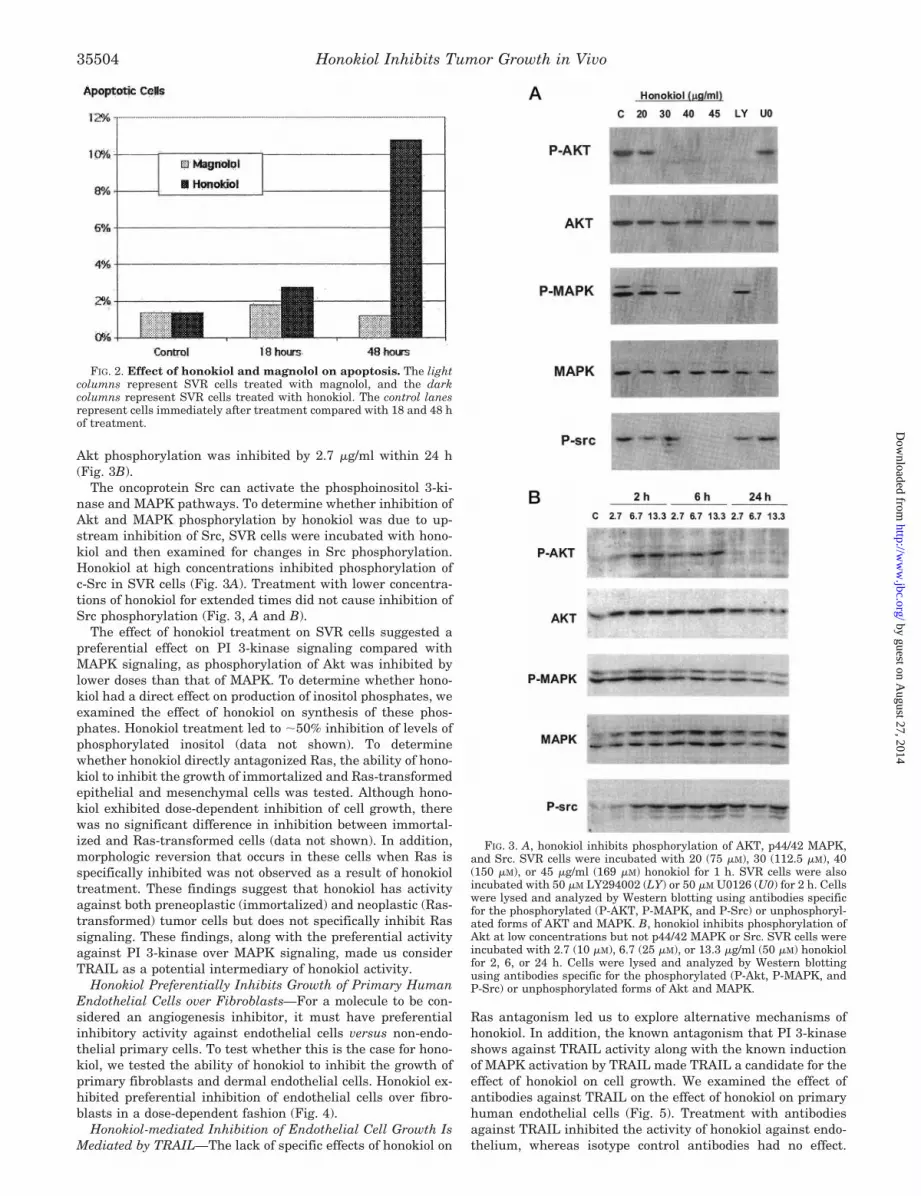
Akt phosphorylation was inhibited by 2.7 �g/ml within 24 h(Fig. 3B).
The oncoprotein Src can activate the phosphoinositol 3-ki-nase and MAPK pathways. To determine whether inhibition ofAkt and MAPK phosphorylation by honokiol was due to up-stream inhibition of Src, SVR cells were incubated with hono-kiol and then examined for changes in Src phosphorylation.Honokiol at high concentrations inhibited phosphorylation ofc-Src in SVR cells (Fig. 3A). Treatment with lower concentra-tions of honokiol for extended times did not cause inhibition ofSrc phosphorylation (Fig. 3, A and B).
The effect of honokiol treatment on SVR cells suggested apreferential effect on PI 3-kinase signaling compared withMAPK signaling, as phosphorylation of Akt was inhibited bylower doses than that of MAPK. To determine whether hono-kiol had a direct effect on production of inositol phosphates, weexamined the effect of honokiol on synthesis of these phos-phates. Honokiol treatment led to �50% inhibition of levels ofphosphorylated inositol (data not shown). To determinewhether honokiol directly antagonized Ras, the ability of hono-kiol to inhibit the growth of immortalized and Ras-transformedepithelial and mesenchymal cells was tested. Although hono-kiol exhibited dose-dependent inhibition of cell growth, therewas no significant difference in inhibition between immortal-ized and Ras-transformed cells (data not shown). In addition,morphologic reversion that occurs in these cells when Ras isspecifically inhibited was not observed as a result of honokioltreatment. These findings suggest that honokiol has activityagainst both preneoplastic (immortalized) and neoplastic (Ras-transformed) tumor cells but does not specifically inhibit Rassignaling. These findings, along with the preferential activityagainst PI 3-kinase over MAPK signaling, made us considerTRAIL as a potential intermediary of honokiol activity.
Honokiol Preferentially Inhibits Growth of Primary HumanEndothelial Cells over Fibroblasts—For a molecule to be con-sidered an angiogenesis inhibitor, it must have preferentialinhibitory activity against endothelial cells versus non-endo-thelial primary cells. To test whether this is the case for hono-kiol, we tested the ability of honokiol to inhibit the growth ofprimary fibroblasts and dermal endothelial cells. Honokiol ex-hibited preferential inhibition of endothelial cells over fibro-blasts in a dose-dependent fashion (Fig. 4).
Honokiol-mediated Inhibition of Endothelial Cell Growth IsMediated by TRAIL—The lack of specific effects of honokiol on
Ras antagonism led us to explore alternative mechanisms ofhonokiol. In addition, the known antagonism that PI 3-kinaseshows against TRAIL activity along with the known inductionof MAPK activation by TRAIL made TRAIL a candidate for theeffect of honokiol on cell growth. We examined the effect ofantibodies against TRAIL on the effect of honokiol on primaryhuman endothelial cells (Fig. 5). Treatment with antibodiesagainst TRAIL inhibited the activity of honokiol against endo-thelium, whereas isotype control antibodies had no effect.
FIG. 2. Effect of honokiol and magnolol on apoptosis. The lightcolumns represent SVR cells treated with magnolol, and the darkcolumns represent SVR cells treated with honokiol. The control lanesrepresent cells immediately after treatment compared with 18 and 48 hof treatment.
FIG. 3. A, honokiol inhibits phosphorylation of AKT, p44/42 MAPK,and Src. SVR cells were incubated with 20 (75 �M), 30 (112.5 �M), 40(150 �M), or 45 �g/ml (169 �M) honokiol for 1 h. SVR cells were alsoincubated with 50 �M LY294002 (LY) or 50 �M U0126 (U0) for 2 h. Cellswere lysed and analyzed by Western blotting using antibodies specificfor the phosphorylated (P-AKT, P-MAPK, and P-Src) or unphosphoryl-ated forms of AKT and MAPK. B, honokiol inhibits phosphorylation ofAkt at low concentrations but not p44/42 MAPK or Src. SVR cells wereincubated with 2.7 (10 �M), 6.7 (25 �M), or 13.3 �g/ml (50 �M) honokiolfor 2, 6, or 24 h. Cells were lysed and analyzed by Western blottingusing antibodies specific for the phosphorylated (P-Akt, P-MAPK, andP-Src) or unphosphorylated forms of Akt and MAPK.
Honokiol Inhibits Tumor Growth in Vivo35504
by guest on August 27, 2014
http://ww
w.jbc.org/
Dow
nloaded from

Thus, the activity of honokiol activity is mediated in partby TRAIL.
Honokiol Inhibits VEGF-induced KDR Autophosphorylationin Human Endothelial Cells—The mitogenic and chemotacticeffects of VEGF on endothelial cells are mainly mediatedthrough the VEGFR2 tyrosine kinase, KDR. Because we havepreviously demonstrated that reactive oxygen species are in-volved in VEGF-induced KDR autophosphorylation in endothe-lial cells (26), we next examined the effect of honokiol on thisresponse in cultured HUVEC. As shown in Fig. 4, VEGF in-duces a 3.8-fold increase (p � 0.05) in phosphorylation of KDRin HUVECs at the peak of 5 min (Fig. 6A) and honokiol signif-icantly inhibited VEGF-induced response in a dose-dependentmanner. Treatment of endothelial cells with honokiol in thepresence of TRAIL antibodies did not antagonize the effect ofhonokiol on phosphorylation of VEGFR2 (data not shown).
Honokiol Inhibits VEGF-induced Rac1 Activation in HumanEndothelial Cells—We have previously demonstrated thatRac1 activation is required for VEGF-induced production ofreactive oxygen species derived from NAD(P)H oxidase andsubsequent KDR autophosphorylation in HUVEC (26). Be-
cause honokiol inhibited VEGF-induced KDR autophosphoryl-ation, we next examined whether this effect is mediatedthrough the inhibition of Rac1. As shown in Fig. 6B, honokiol(10 �g/ml) that almost completely blocked KDR autophospho-rylation dramatically reduced VEGF-stimulated Rac1 activitywithout affecting its basal levels. These results suggest thathonokiol may act as an anti-oxidant mainly through inhibition ofRac1, a critical component of NAD(P)H oxidase, in endothelialcells. Treatment of endothelial cells with honokiol in the presenceof TRAIL antibodies did not antagonize the effect of honokiol onactivation of Rac1 (data not shown). Prior studies have suggestedthe induction of reactive oxygen both prior to and following phos-phorylation of KDR (26). Both possibilities may be true as in-creased levels of reactive oxygen have been shown previously toaugment phosphorylation of receptors. This may be due in part tooxidative inactivation of protein tyrosine phosphatases, whichexhibit an active cysteine residue that can be inactivated byreactive oxygen (27, 28).
Honokiol Exhibits Anti-tumor Activity in Mice—To deter-mine whether honokiol exhibited anti-tumor activity in vivo,mice were inoculated with 1 � 106 SVR angiosarcoma cells
FIG. 4. Honokiol preferentially inhibits endothelial prolifera-tion compared with fibroblast proliferation. The x axis repre-sents dosage of honokiol, whereas the y axis represents cell number.The color scale at bottom shows the doses of honokiol used inmicrograms/milliliter.
FIG. 5. Honokiol inhibition of endothelial proliferation isTRAIL-dependent. 104/well microvascular endothelial cells were cul-tured in 24-well plates for 24 h. The next day, cells were washed by PBSand pretreated with 0.5 ml/well fresh MEC medium with 0, 1, 6, or 9�g/ml honokiol for 30 min before addition of TRAIL or isotype controlantibody (30 �g/well). Cells were incubated for 48 h after the addition ofreagents and were counted with a Coulter Counter. The green barsrepresent endothelial cells treated with honokiol alone, the dark bluebars represent cells treated with honokiol and TRAIL antibody, and thelight blue bars represent cells treated with honokiol and isotype controlantibody. The differences in honokiol-treated endothelium in the pres-ence or absence of TRAIL antibody are significant (p � 0.05).
FIG. 6. A, effect of honokiol on VEGF-induced KDR autophosphoryl-ation in HUVECs. HUVECs were preincubated with vehicle or honokiol(5 and 10 �g/ml) for 60 min and then stimulated with 20 ng/ml VEGFfor 5 min. Lysates were immunoprecipitated (IP) with anti-phospho-tyrosine (pTyr) antibody followed by immunoblotting (IB) with anti-KDR antibody (top panel). Bottom panel represents averaged data ex-pressed as fold change over basal (the ratio in untreated cells was set to1). Values are the means � S.E. for three independent experiments. *,p � 0.05 for increase in phosphorylation by VEGF in the presence ofinhibitor versus VEGF alone. B, effect of honokiol on VEGF-induced Racactivation. HUVECs were preincubated with vehicle or honokiol (10�g/ml) for 60 min and then stimulated with 20 ng/ml VEGF for 3 min.Rac activity was measured by p21-activated kinase-1-protein bindingdomain affinity precipitation as described under “Experimental Proce-dures.” Top, representative immunoblot of GTP-bound Rac. Bottom,densitometric analysis (mean � S.E.) of immunoblots from three exper-iments expressed as fold increase over control. *, p � 0.01 comparedwith VEGF alone.
Honokiol Inhibits Tumor Growth in Vivo 35505
by guest on August 27, 2014
http://ww
w.jbc.org/
Dow
nloaded from

subcutaneously and treated with honokiol or vehicle when tu-mors became clinically evident. Honokiol treatment led to�50% inhibition of tumor growth compared with vehicle con-trol (p � 0.05) (Fig. 7).
DISCUSSION
We describe the isolation of a systemically active inhibitor oftumor growth through a rapid bioassay of proliferation of SVR-transformed endothelium. We have previously shown that SVRcells accurately predict in vivo responses of two known an-giogenesis inhibitors, trinitrophenylnucleotide 1470 and 2-methoxyestradiol (12, 29). In addition, curcumin, a naturalproduct not previously known to be anti-angiogenic, wasdemonstrated to have anti-angiogenic activity on both immor-talized and primary endothelial cultures (3, 30).
A number of chemopreventive agents have been isolated andcharacterized primarily through activities against skin andcolon cancer promotion protocols. These include tea polyphe-nols, curcumin, and caffeic acid phenethyl ester (6, 31–33).Although many of these agents have potent chemopreventiveactivities, their activity against established tumors is not po-tent. One possible explanation is that high concentrations of drugare achievable at the skin and colon while sustained systemicconcentrations cannot be achieved. This is clearly the case withcurcumin in which previous studies have shown that it is poorlyabsorbed from the gastrointestinal tract and, even when system-ically administered, is rapidly cleared by hepatic metabolism (1).We have found that honokiol, unlike curcumin, can inhibit
growth of an established tumor when administered systemically.Our fractionation process using inhibition of transformed
SVR endothelial cell proliferation as a bioassay yielded a frac-tion that contains magnolol and honokiol, which are substi-tuted biphenols. Magnolol and honokiol are closely related, buthonokiol appeared to have enhanced activity in the SVR inhi-bition assay. Substitution of the hydroxylated biphenyl is re-quired for activity, because the 2,2�- and 4,4�-dihydroxybiphe-nyl had no effect on proliferation of SVR cells in vitro.Consistent with increased activity of honokiol against SVRcells, honokiol is more effective in the induction of apoptosisthan magnolol.
To determine the mechanisms of activity of honokiol, weexamined its effect on expression and phosphorylation of keysignal transduction pathways. Activation of both MAPK andAkt was inhibited, indicating that a mechanism of activity isprobably upstream of these pathways and possibly at the levelof Src. Inhibition of Akt phosphorylation occurred at an earliertime point and at lower doses than inhibition of MAPK phos-phorylation, indicating a preferential inhibition of PI 3-kinasesignaling. A potential explanation for the diverging regulationof MAPK and PI 3-kinase resulting from honokiol treatmentmay stem from the involvement of TRAIL as demonstrated inhuman endothelial cells. The preferential inhibition of PI 3-ki-nase signaling over MAPK signaling coupled with preferentialactivity of honokiol against multiple neoplastic cells and endo-thelium while having little effect on primary fibroblasts isconsistent with TRAIL activation (34–36). Although TRAILpromotes apoptosis, TRAIL also stimulates activation of MAPK(37). The lack of effect on MAPK may result from the combinedeffect of high constitutive levels of MAPK activation in SVRcells and MAPK up-regulation by TRAIL.
Honokiol demonstrated inhibition of VEGFR2/Flk/KDR au-tophosphorylation in human endothelial cells. In addition,honokiol treatment resulted in blockade of VEGF-induced Racactivation (38). Because Rac is required for VEGF-inducedendothelial migration and proliferation (39, 40), the blockade ofVEGFR2 autophosphorylation and subsequent Rac activationindicate potential for honokiol as an anti-angiogenic agent thatfunctions in human cells in addition to anti-tumor activity.TRAIL blockade did not antagonize the effect of honokiol onphosphorylation of VEGFR2 or activation of Rac1. This may bethe result of TRAIL affecting signaling downstream ofVEGFR2/Rac1 or through an independent pathway.
Given that polyphenols, including curcumin and epicatechingallate, both target proteasomes, we examined the effect ofhonokiol on the COP9 signalosome. Honokiol is an effectiveinhibitor of COP9 signalosome kinase activity.
In conclusion, we demonstrate the utility of the SVR bioas-say in isolating an active principle in a natural product andcharacterize its mechanism of action. The active principle,honokiol, has been previously described as a component of aJapanese herbal medicine “saiboku-to” and of the Chinese med-icine “houpo” and has been shown to have anxiolytic propertiesin mice (21–23, 41, 42). We have demonstrated for the first timethat honokiol has potent anti-angiogenic and anti-tumor prop-erties in vitro and is systemically active against aggressiveangiosarcoma in vivo. In addition, honokiol is well tolerated bythe host animal in therapeutically beneficial doses, making itan attractive candidate for further preclinical testing as ananti-neoplastic agent.
REFERENCES
1. Rao, C. V., Rivenson, A., Simi, B., and Reddy, B. S. (1995) Ann. N. Y. Acad. Sci.768, 201–204
2. Lin, J. K., Chen, Y. C., Huang, Y. T., and Lin-Shiau, S. Y. (1997) J. Cell.Biochem. 28/29, (suppl.) 39–48
3. Arbiser, J. L., Klauber, N., Rohan, R., van Leeuwen, R., Huang, M. T., Fisher,C., Flynn, E., and Byers, H. R. (1998) Mol. Med. 4, 376–383
FIG. 7. Effect of honokiol on in vivo growth of SVR angiosar-coma in nude mice. The y axis represents tumor volume. Column 1represents vehicle-treated mice (n � 3), and column 2 represents hono-kiol-treated mice (n � 3).
Honokiol Inhibits Tumor Growth in Vivo35506
by guest on August 27, 2014
http://ww
w.jbc.org/
Dow
nloaded from

4. Cao, Y., and Cao, R. (1999) Nature 398, 3815. Huang, M. T., Lysz, T., Ferraro, T., Abidi, T. F., Laskin, J. D., and Conney,
A. H. (1991) Cancer Res. 51, 813–8196. Huang, M. T., Lou, Y. R., Ma, W., Newmark, H. L., Reuhl, K. R., and Conney,
A. H. (1994) Cancer Res. 54, 5841–58477. Pollmann, C., Huang, X., Mall, J., Bech-Otschir, D., Naumann, M., and Dubiel,
W. (2001) Cancer Res. 61, 8416–84218. Bech-Otschir, D., Kraft, R., Huang, X., Henklein, P., Kapelari, B., Pollmann,
C., and Dubiel, W. (2001) EMBO J. 20, 1630–16399. Nam, S., Smith, D. M., and Dou, Q. P. (2001) J. Biol. Chem. 276, 13322–13330
10. Mukhopadhyay, D., Tsiokas, L., Zhou, X. M., Foster, D., Brugge, J. S., andSukhatme, V. P. (1995) Nature 375, 577–581
11. Gupta, S., Hastak, K., Ahmad, N., Lewin, J. S., and Mukhtar, H. (2001) Proc.Natl. Acad. Sci. U. S. A. 98, 10350–10355
12. Arbiser, J. L., Panigrathy, D., Klauber, N., Rupnick, M., Flynn, E., Udagawa,T., and D’Amato, R. J. (1999) J. Am. Acad. Dermatol. 40, 925–929
13. LaMontagne, K. R., Jr., Moses, M. A., Wiederschain, D., Mahajan, S., Holden,J., Ghazizadeh, H., Frank, D. A., and Arbiser, J. L. (2000) Am. J. Pathol.157, 1937–1945
14. Oldham, S. M., Clark, G. J., Gangarosa, L. M., Coffey, R. J., Jr., and Der, C. J.(1996) Proc. Natl. Acad. Sci. U. S. A. 93, 6924–6928
15. Pruitt, K., Pestell, R. G., and Der, C. J. (2000) J. Biol. Chem. 275,40916–40924
16. Serunian, L. A., Auger, K. R., and Cantley, L. C. (1991) Methods Enzymol. 198,78–87
17. Ye, K., Aghdasi, B., Luo, H. R., Moriarity, J. L., Wu, F. Y., Hong, J. J., Hurt,K. J., Bae, S. S., Suh, P. G., and Snyder, S. H. (2002) Nature 415, 541–544
18. Daneker, G. W., Lund, S. A., Caughman, S. W., Swerlick, R. A., Fischer, A. H.,Staley, C. A., and Ades, E. W. (1998) In Vitro Cell Dev. Biol. Anim. 34,370–377
19. Clarke, P., Meintzer, S. M., Gibson, S., Widmann, C., Garrington, T. P.,Johnson, G. L., and Tyler, K. L. (2000) J. Virol. 8135–8139
20. Alessi, D. R., Cuenda, A., Cohen, P., Dudley, D. T., and Saltiel, A. R. (1995)J. Biol. Chem. 270, 27489–27494
21. Kuribara, H., Stavinoha, W. B., and Maruyama, Y. (1999) J. Pharm. Pharma-col. 51, 97–103
22. Maruyama, Y., Kuribara, H., Morita, M., Yuzurihara, M., and Weintraub, S. T.(1998) J. Nat. Prod. 61, 135–138
23. Fujita, M., Itokawa, H., and Sashida, Y. (1973) Yakugaku Zasshi 93, 429–43424. Sun, M., Wang, G., Paciga, J. E., Feldman, R. I., Yuan, Z. Q., Ma, X. L., Shelley,
S. A., Jove, R., Tsichlis, P. N., Nicosia, S. V., and Cheng, J. Q. (2001) Am. J.Pathol. 159, 431–437
25. Hutchinson, J., Jin, J., Cardiff, R. D., Woodgett, J. R., and Muller, W. J. (2001)Mol. Cell. Biol. 21, 2203–2212
26. Ushio-Fukai, M., Tang, Y., Fukai, T., Dikalov, S. I., Ma, Y., Fujimoto, M.,Quinn, M. T., Pagano, P. J., Johnson, C., and Alexander, R. W. (2002) Circ.Res. 91, 1160–1167
27. Pieri, L., Dominici, S., Del Bello, B., Maellaro, E., Comporti, M., Paolicchi, A.,and Pompella, A. (2003) Biochim. Biophys. Acta 1621, 76–83
28. Finkel, T. (2000) FEBS Lett. 476, 52–5429. Arbiser, J. L., Moses, M. A., Fernandez, C. A., Ghiso, N., Cao, Y., Klauber, N.,
Frank, D., Brownlee, M., Flynn, E., Parangi, S., Byers, H. R., and Folkman,J. (1997) Proc. Natl. Acad. Sci. U. S. A. 94, 861–866
30. Thaloor, D., Singh, A. K., Sidhu, G. S., Prasad, P. V., Kleinman, H. K., andMaheshwari, R. K. (1998) Cell Growth Differ. 9, 305–312
31. Samaha, H. S., Kelloff, G. J., Steele, V., Rao, C. V., and Reddy, B. S. (1997)Cancer Res. 57, 1301–1305
32. Dinkova-Kostova, A. T., and Talalay, P. (1999) Carcinogenesis 20, 911–91433. Fahey, J. W., and Talalay, P. (1999) Food Chem. Toxicol. 37, 973–97934. Hersey, P., and Zhang, X. D. (2001) Nat. Rev. Cancer 1, 142–15035. LaVallee, T. M., Zhan, X. H., Johnson, M. S., Herbstritt, C. J., Swartz, G.,
Williams, M. S., Hembrough, W. A., Green, S. J., and Pribluda, V. S. (2003)Cancer Res. 63, 468–475
36. Schneider, P., Thome, M., Burns, K., Bodmer, J. L., Hofmann, K., Kataoka, T.,Holler, N., and Tschopp, J. (1997) Immunity 7, 831–836
37. Tran, S. E., Holmstrom, T. H., Ahonen, M., Kahari, V. M., and Eriksson, J. E.(2001) J. Biol. Chem. 276, 16484–16490
38. Zeng, H., Dvorak, H. F., and Mukhopadhyay, D. (2001) J. Biol. Chem. 276,26969–26979
39. Colavitti, R., Pani, G., Bedogni, B., Anzevino, R., Borrello, S., Waltenberger, J.,and Galeotti, T. (2002) J. Biol. Chem. 277, 3101–3108
40. Zeng, H., Zhao, D., and Mukhopadhyay, D. (2002) J. Biol. Chem. 277,4003–4009
41. Kuribara, H., Kishi, E., Hattori, N., Okada, M., and Maruyama, Y. (2000)J. Pharm. Pharmacol. 52, 1425–1429
42. Ai, J., Wang, X., and Nielsen, M. (2001) Pharmacology 63, 34–41
Honokiol Inhibits Tumor Growth in Vivo 35507
by guest on August 27, 2014
http://ww
w.jbc.org/
Dow
nloaded from