Embed Size (px)
Citation preview

Hsp70 promotes TNF-mediated apoptosisby binding IKK� and impairing NF-�Bsurvival signalingRuiqiong Ran,1,6 Aigang Lu,1 Lu Zhang,2 Yang Tang,1 Hongyan Zhu,1 Huichun Xu,1 Yuxin Feng,2
Chun Han,3 Guoping Zhou,4 Alan C. Rigby,4 and Frank R. Sharp1
1Departments of Neurology, Pediatric Neurology and the Neurosciences Program, 2Department of Cell Biology,Neurobiology and Anatomy, and 3Cincinnati Children’s Hospital and Medical Center, University of Cincinnati, Cincinnati,Ohio 45267, USA; 4Department of Medicine, Beth Israel Deaconess Medical Center, Harvard Medical School, Boston,Massachusetts 02115, USA
The major heat shock protein, Hsp70, can protect against cell death by directly interfering with mitochondrialapoptosis pathways. However, Hsp70 also sensitizes cells to certain apoptotic stimuli like TNF. Little isknown about how Hsp70 enhances apoptosis. We demonstrate here that Hsp70 promotes TNF killing byspecifically binding the coiled-coil domain of I�B kinase � (IKK�) to inhibit IKK activity and consequentlyinhibit NF-�B-dependent antiapoptotic gene induction. An IKK� mutant, which interacts with Hsp70,competitively inhibits the Hsp70–IKK� interaction and relieves heat-mediated NF-�B suppression. Depletionof Hsp70 expression with RNA interference rescues TNF-mediated cell death. Although TNF may or may notbe sufficient to trigger apoptosis on its own, TNF-triggered apoptosis was initiated or made worse when Hsp70expression increased to high levels to disrupt NF-�B signaling. These results provide significant novel insightsinto the molecular mechanism for the pro-apoptotic behavior of Hsp70 in death-receptor-mediated cell death.
[Keywords: Hsp70; NF-�B; IKK�; TNF; apoptosis; death receptor signaling]
Received January 21, 2004; revised version accepted March 30, 2004.
Even though heat shock proteins are known to inhibitvarious types of apoptosis, some studies show that heator elevated heat shock protein 70 (Hsp70) also potenti-ates cell death following specific stimuli. For instance,overexpression of HSF1 (heat shock transcription factor1, one transcription factor that controls the heat shockresponse) enhances Fas-induced cell apoptosis (Xia et al.2000). Elevated Hsp70 promotes TCR/CD3- and Fas/Apo-1/CD95-mediated Jurkat T-cell apoptosis (Liossis etal. 1997). Heat increases cell death following some che-motherapeutics and radiation (Dewey 1984), and heatshock also sensitizes AML cells and endothelial cells toapoptosis (Chant et al. 1996; DeMeester et al. 1998).Hsp70 inhibits cellular proliferation (Maheswaran et al.1998). Moreover, it is also well established that heatshock or elevated Hsp70 alters the regulation of signal-ing cascades and transcription factors and potently sen-sitizes tumors to radiation (Curry et al. 1999). These re-sults imply that heat or Hsp70 could disable a cell sur-vival signal under the appropriate circumstances.
NF-�B is sequestered in the cytoplasm in an inactivecomplex with I�B proteins (Huxford et al. 1998; Malek et
al. 2001). Activation of the NF-�B-mediated signal is ini-tiated through degradation of phosphorylated I�B. Vari-ous stimuli activate the I�B kinase (IKK) complex, whichin turn phosphorylates I�B, leading to NF-�B activation(Chen et al. 1995; Zandi et al. 1997). NF-�B is critical forsurvival of most cells through the induction of antiapo-ptotic genes (Beg et al. 1995; Beg and Baltimore 1996;Van Antwerp et al. 1996; Wang et al. 1998; Zong et al.1999; Rudolph et al. 2000). IKK plays a central role inmediating NF-�B activation. IKK is composed of twocatalytic subunits, IKK� and IKK�, which can directlyphosphorylate I�B. IKK� (also called NEMO) is an abso-lutely essential regulatory component of the IKK com-plex that is necessary for NF-�B activation (Rothwarf etal. 1998; Yamaoka et al. 1998; Makris et al. 2002).
A number of studies have also shown that heat shockor elevated Hsp70 suppresses NF-�B activity (Feinsteinet al. 1997; Guzhova et al. 1997; Curry et al. 1999; An-dres et al. 2002; Malhotra and Wong 2002). Althoughthese studies imply the possibility that Hsp70 impairsNF-�B signaling, the exact molecular basis of the Hsp70and NF-�B interaction is still enigmatic.
This study therefore examined the mechanism bywhich Hsp70 interacts with NF-�B and might promoteapoptosis. To do this, we studied several cell lines (Cos-1, Hela, 293 cells) that do not apparently undergo apo-
6Corresponding author.E-MAIL [email protected]; FAX (513) 558-7009.Article and publication are at http://www.genesdev.org/cgi/doi/10.1101/gad.1188204.
1466 GENES & DEVELOPMENT 18:1466–1481 © 2004 by Cold Spring Harbor Laboratory Press ISSN 0890-9369/04; www.genesdev.org

ptosis when exposed to TNF alone. However, whenthese cells are exposed to TNF in combination withHsp70 overexpression or heat shock, they die via apopto-sis. Here, we demonstrate that Hsp70 associates directlyand specifically with IKK� and blocks the formation ofthe IKK complex—possibly by inhibiting oligomeriza-tion of IKK�. This inhibits TNF-triggered NF-�B activa-tion and subsequently prevents NF-�B-dependent anti-apoptotic gene expression. This work demonstrates howHsp70 promotes rather than inhibits TNF-mediated celldeath.
Results
Hsp70 inhibits NF-�B activity
To determine whether Hsp70 itself specifically sup-pressed NF-�B activity, we cotransfected Cos-1 cellswith an NF-�B reporter and the indicated constructs. Inthis transfection assay, Hsp70 and I�B�DN (I�B� domi-nant negative) suppressed NF-�B reporter gene activity,whereas Hsp70C (C terminus of Hsp70) and Hsp70N (Nterminus of Hsp70) failed to do so (Fig. 1A). HSF1 alsoinhibited NF-�B activity. However, this inhibition wasabrogated by cotransfection with Hsp70AS (antisenseHsp70), indicating that HSF1 did not directly affect NF-�B activity (Fig. 1A). Hsp90 did not affect NF-�B activityin resting cells but enhanced NF-�B activity when cellswere treated with TNF (Fig. 1A).
To demonstrate the specificity of the Hsp70 inhibi-tion, we did similar experiments with AP-1- and HSF-dependent reporters. It was shown that Hsp70 did notsignificantly affect activation of AP-1, whereas bothHsp70 and Hsp70C efficiently inhibited HSF activity(Fig. 1B). This is consistent with findings that Hsp70 caninhibit HSF1 transcriptional activity (Abravaya et al.1992; Shi et al. 1998). An additional control experimentshowed that Hsp70 had similar effects on the refolding of�-gal and luciferase, as measured by their enzymatic ac-tivities (data not shown). Therefore, in all of the trans-fection assays shown, the elevated Hsp70 did not artifi-cially decrease or increase the luciferase readings whennormalized to �-gal.
To determine whether physiologically elevated levelsof Hsp70 inhibit NF-�B activity, we established Hsp70-inducible cell lines using the nonintegrating plasmidpMEP4 under the control of a Zn2+-regulated metallo-thionein promoter. Hsp70 dose-dependently inhibitedNF-�B activity (Fig. 1C). The modest reduction ofNF-�B activity in Hsp70C-expressing cells (Fig. 1C,lanes 2–4) likely occurred because of Zn2+-mediated in-duction of endogenous Hsp70 (Fig. 1C,D). Cells treatedwith TNF alone, in the absence of ZnCl2, had a relativeluciferase activity for NF�B-luc of 100 ± 8 (see resultsfollowing), which is similar to the relative luciferase ac-tivity levels for the cells stably expressing His-Hsp70Cin the presence of ZnCl2 (Fig. 1C, lanes 2–4) and for His-Hsp70 cells treated with low levels of ZnCl2 (Fig. 1C,lane 6).
Hsp70 did not directly interfere with the function ofthe NF-�B activation domain using a cotransfection as-say with plasmid encoding Gal4 (DNA-binding domain)-p65 (activation domain) chimeric protein, a Gal-4 re-porter, and Hsp70 construct (data not shown). Next, toanalyze the effect of Hsp70 on NF-�B DNA-binding ac-tivity, we performed a gel mobility shift assay using ap65 probe and nuclear extracts isolated from Cos-1 cells.Heat, Hsp70, I�B�DN, and HSF1 inhibited NF-�B DNAbinding (Fig. 1E, lanes 1–8). The effects of HSF1 were nota direct effect on the NF-�B activity because Hsp70 an-tisense (Hsp70AS) blocked the effect, showing that it isHSF1 induction of Hsp70 that inhibits NF-�B DNA bind-ing (Fig. 1E, lane 8). Cellular localization data showedthat Hsp70 inhibited TNF-induced translocation of p65from the cytoplasm to the nucleus (Fig. 1F).
To test whether Hsp70 and HSF1 directly affect theNF-�B DNA-binding activity, we translated p65, Hsp70,and HSF1 proteins in vitro and performed the aforemen-tioned gel mobility shift assays. Hsp70 and HSF1 failedto directly influence the NF-�B DNA binding (Fig. 1G).These negative data are shown because they help ruleout the possibility that Hsp70 or HSF1 directly regulatesNF-�B DNA-binding activity. In order to further rule outthe possibility that Hsp70 directly interacts with NF-�B(p65), we constructed NLS/p65 plasmids with forced p65nuclear localization that was independent of I�B� degra-dation. Cos-1 cells were cotransfected with NLS/p65,NF-�B reporter, and Hsp70 constructs. NF-�B reporteractivity analysis showed that Hsp70 did not suppressNF-�B activity (data not shown). This confirmed the ear-lier findings that Hsp70 did not directly interact withNF-�B (p65).
Hsp70 inhibits TNF-induced phosphorylation of I�B�
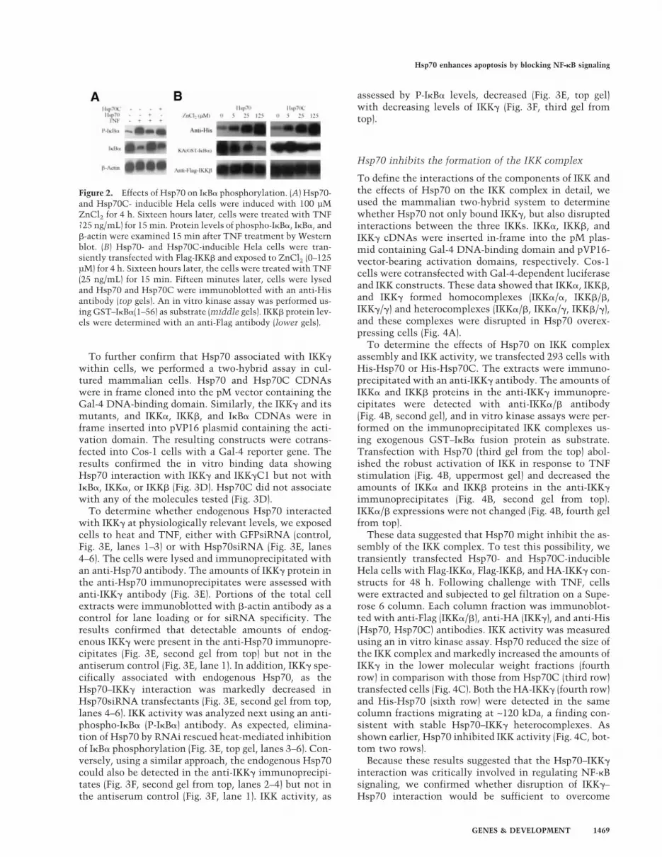
Because Hsp70 inhibited NF-�B activity without directlybinding NF-�B, this indicated that Hsp70 impaired NF-�B signaling upstream of NF-�B (p65). Because activationof NF-�B requires phosphorylation of I�B, we examinedthe effects of Hsp70 on the phosphorylation of I�B usingspecific antibodies to examine phosphorylated and un-phosphorylated I�B� proteins. The results showed thatHsp70 inhibited TNF-induced I�B� phosphorylation(Fig. 2A).
These data suggested either that Hsp70 directly inhib-ited I�B activity, or that Hsp70 directly bound I�B� tomask its phosphorylation site, or that Hsp70 acceleratedI�B� dephosphorylation. In order to differentiate be-tween these possibilities, several experiments were per-formed. First, pull-down data, immunoprecipitationdata, and two-hybrid experiments showed that Hsp70did not interact with I�B� (see data below). Second,Hsp70- and Hsp70C-inducible Hela cells were tran-siently transfected with Flag-IKK� and Flag-IKK�. Theeffect of Hsp70 on IKK activity was assayed using an invitro kinase assay with exogenous GST–I�B� (1–56) asthe substrate. Hsp70 reduced IKK activity in a dose-de-pendent manner (Fig. 2B). Neither Hsp70 nor Hsp70Caltered IKK� expression (Fig. 2B).
Hsp70 enhances apoptosis by blocking NF-�B signaling
GENES & DEVELOPMENT 1467

Hsp70 directly interacts with IKK�
On the basis of these observations, we consideredwhether Hsp70 directly interacted with one or morecomponents of the IKK complex and inhibited IKK ac-tivity. To determine which molecule(s) of the IKK com-plex might be the primary target of Hsp70, we searchedfor a direct interaction between the three IKKs andHsp70 using a beads pull-down assay. 35S-labeled IKK�
was retained on Hsp70-, but not on Hsp70C- conjugatedbeads. IKK� and IKK� did not associate with Hsp70 orwith Hsp70C (Fig. 3A).
To map which region within IKK� was responsible forthis interaction, we constructed various deletions ofIKK� (Fig. 3B). The in vitro beads pull-down assayshowed that only the IKK� and IKK�C1, which containedthe coiled-coil motif, interacted with Hsp70 (Fig. 3C), sug-gesting that Hsp70 bound to the coiled-coil motif of IKK�.
Figure 1. Hsp70 inhibits NF-�B activa-tion. (A) Cos-1 cells were transfected withthe indicated plasmids and the NF-�B re-porter. Twenty-four hours after transfec-tion, cells were treated with TNF (25 ng/mL) or vehicle for 15 min. Luciferase ac-tivity was measured 24 h later. (B) Effectsof Hsp70 on Ap-1- and HSF-1-dependenttransactivation. Cos-1 cells were cotrans-fected with an AP-1 or HSF-1 luciferasereporter along with the indicated vectors.Twenty-four hours later, cells were treatedwith TNF or vehicle for 15 min. After 24h, luciferase activities were measured. (C)Hsp70- and Hsp70C-inducible cells weretransiently transfected with an NF-�B-de-pendent luciferase reporter and thentreated with different concentrations ofZnCl2 for 4 h to induce His-tagged Hsp70and His-tagged Hsp70C expression. At 16h after ZnCl2 treatment, cells were treatedwith TNF (25 ng/mL) or vehicle for 15min. NF-�B activity was analyzed 24 h af-ter TNF treatment. Hsp70 and Hsp70C ex-pression levels were examined with ananti-His antibody. �-Actin served as thelane-loading control. (D) Immunoblots ofexogenous and endogenous Hsp70 expres-sion in Hsp70 stable cells were treatedwith ZnCl2 (for 4 h). Sixteen hours later,cell lysates were immunoblotted withanti-Hsp70 antibody. (E) Cos-1 cells weretransfected with the indicated constructsand selected cells treated with TNF (25 ng/mL) for 15 min or heated for 30 min at45°C. Twenty-four hours later, nuclear ex-tracts were prepared and subjected to a gelshift assay using a 32P-labeled p65 probe.(NS) Nonspecific binding. (F) Hsp70- orHsp70C-inducible Hela cells were treatedwith ZnCl2 (100 µM) or vehicle for 4 h.Sixteen hours later, cells were treated withTNF (25 ng/mL) or vehicle for 15 min.Cells were stained 15 min later with ananti-p65 antibody and a TRITC-conju-gated second antibody. Nuclei were visu-alized with Hoechst. (G) Gel shift assaywith in vitro translated Hsp70, HSF1, andp65 proteins and a 32P-labeled p65 probe.(NS) Nonspecific binding.
Ran et al.
1468 GENES & DEVELOPMENT
To further confirm that Hsp70 associated with IKK�within cells, we performed a two-hybrid assay in cul-tured mammalian cells. Hsp70 and Hsp70C CDNAswere in frame cloned into the pM vector containing theGal-4 DNA-binding domain. Similarly, the IKK� and itsmutants, and IKK�, IKK�, and I�B� CDNAs were inframe inserted into pVP16 plasmid containing the acti-vation domain. The resulting constructs were cotrans-fected into Cos-1 cells with a Gal-4 reporter gene. Theresults confirmed the in vitro binding data showingHsp70 interaction with IKK� and IKK�C1 but not withI�B�, IKK�, or IKK� (Fig. 3D). Hsp70C did not associatewith any of the molecules tested (Fig. 3D).
To determine whether endogenous Hsp70 interactedwith IKK� at physiologically relevant levels, we exposedcells to heat and TNF, either with GFPsiRNA (control,Fig. 3E, lanes 1–3) or with Hsp70siRNA (Fig. 3E, lanes4–6). The cells were lysed and immunoprecipitated withan anti-Hsp70 antibody. The amounts of IKK� protein inthe anti-Hsp70 immunoprecipitates were assessed withanti-IKK� antibody (Fig. 3E). Portions of the total cellextracts were immunoblotted with �-actin antibody as acontrol for lane loading or for siRNA specificity. Theresults confirmed that detectable amounts of endog-enous IKK� were present in the anti-Hsp70 immunopre-cipitates (Fig. 3E, second gel from top) but not in theantiserum control (Fig. 3E, lane 1). In addition, IKK� spe-cifically associated with endogenous Hsp70, as theHsp70–IKK� interaction was markedly decreased inHsp70siRNA transfectants (Fig. 3E, second gel from top,lanes 4–6). IKK activity was analyzed next using an anti-phospho-I�B� (P-I�B�) antibody. As expected, elimina-tion of Hsp70 by RNAi rescued heat-mediated inhibitionof I�B� phosphorylation (Fig. 3E, top gel, lanes 3–6). Con-versely, using a similar approach, the endogenous Hsp70could also be detected in the anti-IKK� immunoprecipi-tates (Fig. 3F, second gel from top, lanes 2–4) but not inthe antiserum control (Fig. 3F, lane 1). IKK activity, as
assessed by P-I�B� levels, decreased (Fig. 3E, top gel)with decreasing levels of IKK� (Fig. 3F, third gel fromtop).
Hsp70 inhibits the formation of the IKK complex
To define the interactions of the components of IKK andthe effects of Hsp70 on the IKK complex in detail, weused the mammalian two-hybrid system to determinewhether Hsp70 not only bound IKK�, but also disruptedinteractions between the three IKKs. IKK�, IKK�, andIKK� cDNAs were inserted in-frame into the pM plas-mid containing Gal-4 DNA-binding domain and pVP16-vector-bearing activation domains, respectively. Cos-1cells were cotransfected with Gal-4-dependent luciferaseand IKK constructs. These data showed that IKK�, IKK�,and IKK� formed homocomplexes (IKK�/�, IKK�/�,IKK�/�) and heterocomplexes (IKK�/�, IKK�/�, IKK�/�),and these complexes were disrupted in Hsp70 overex-pressing cells (Fig. 4A).
To determine the effects of Hsp70 on IKK complexassembly and IKK activity, we transfected 293 cells withHis-Hsp70 or His-Hsp70C. The extracts were immuno-precipitated with an anti-IKK� antibody. The amounts ofIKK� and IKK� proteins in the anti-IKK� immunopre-cipitates were detected with anti-IKK�/� antibody(Fig. 4B, second gel), and in vitro kinase assays were per-formed on the immunoprecipitated IKK complexes us-ing exogenous GST–I�B� fusion protein as substrate.Transfection with Hsp70 (third gel from the top) abol-ished the robust activation of IKK in response to TNFstimulation (Fig. 4B, uppermost gel) and decreased theamounts of IKK� and IKK� proteins in the anti-IKK�immunoprecipitates (Fig. 4B, second gel from top).IKK�/� expressions were not changed (Fig. 4B, fourth gelfrom top).
These data suggested that Hsp70 might inhibit the as-sembly of the IKK complex. To test this possibility, wetransiently transfected Hsp70- and Hsp70C-inducibleHela cells with Flag-IKK�, Flag-IKK�, and HA-IKK� con-structs for 48 h. Following challenge with TNF, cellswere extracted and subjected to gel filtration on a Supe-rose 6 column. Each column fraction was immunoblot-ted with anti-Flag (IKK�/�), anti-HA (IKK�), and anti-His(Hsp70, Hsp70C) antibodies. IKK activity was measuredusing an in vitro kinase assay. Hsp70 reduced the size ofthe IKK complex and markedly increased the amounts ofIKK� in the lower molecular weight fractions (fourthrow) in comparison with those from Hsp70C (third row)transfected cells (Fig. 4C). Both the HA-IKK� (fourth row)and His-Hsp70 (sixth row) were detected in the samecolumn fractions migrating at ∼120 kDa, a finding con-sistent with stable Hsp70–IKK� heterocomplexes. Asshown earlier, Hsp70 inhibited IKK activity (Fig. 4C, bot-tom two rows).
Because these results suggested that the Hsp70–IKK�interaction was critically involved in regulating NF-�Bsignaling, we confirmed whether disruption of IKK�–Hsp70 interaction would be sufficient to overcome
Figure 2. Effects of Hsp70 on I�B� phosphorylation. (A) Hsp70-and Hsp70C- inducible Hela cells were induced with 100 µMZnCl2 for 4 h. Sixteen hours later, cells were treated with TNF?25 ng/mL) for 15 min. Protein levels of phospho-I�B�, I�B�, and�-actin were examined 15 min after TNF treatment by Westernblot. (B) Hsp70- and Hsp70C-inducible Hela cells were tran-siently transfected with Flag-IKK� and exposed to ZnCl2 (0–125µM) for 4 h. Sixteen hours later, the cells were treated with TNF(25 ng/mL) for 15 min. Fifteen minutes later, cells were lysedand Hsp70 and Hsp70C were immunoblotted with an anti-Hisantibody (top gels). An in vitro kinase assay was performed us-ing GST–I�B�(1–56) as substrate (middle gels). IKK� protein lev-els were determined with an anti-Flag antibody (lower gels).
Hsp70 enhances apoptosis by blocking NF-�B signaling
GENES & DEVELOPMENT 1469

Hsp70-mediated NF-�B suppression. Because Hsp70 di-rectly bound both IKK� and IKK�C1, but not IKK�C2(Fig. 3C,D), we used IKK�C1 to selectively disrupt theinteraction between IKK� and Hsp70 by competing forbinding to Hsp70. Therefore, 293 cells were transfectedwith HA-IKK� and increasing amounts of HA-IKK�C1 orHA-IKK�C2. Cells were exposed to TNF to activate NF-�B and exposed to heat shock (45°C) for 30 min to induceendogenous Hsp70, and cell extracts were immuno-precipitated with anti-Hsp70 antibody. Appreciableamounts of HA-IKK� protein could be detected withanti-HA antibody in the anti-Hsp70 immunoprecipitates(Fig. 4D, top gel, lanes 3–5) but not in the antiserumcontrol (Fig. 4D, top gel, lane 1). Another portion of thesecells was transfected with an NF-�B reporter gene forNF-�B activity analysis. Immunoprecipitation showedthat Hsp70 interacted with IKK� and that IKK�C1 dis-placed IKK� from Hsp70 immunocomplex (Fig. 4D, topgel, lanes 3–5,7–9). IKK�C1-mediated IKK� release fromthe Hsp70–IKK� complex was accompanied by gradualrestoration of NF-�B activity (Fig. 4D, upper panel, lanes
7–9), whereas IKK�C2 had no such effect (Fig. 4D, upperpanel, lanes 3–5).
Hsp70 precludes IKK� oligomerization,which is required for NF-�B activation
These data showed that IKK� formed homo-oligomers(Fig. 4A) and that the coiled-coil motif of IKK� was theHsp70-binding site (Fig. 3B,C), suggesting that thecoiled-coil region serves as an effector domain to regu-late the IKK complex via its oligomerization. If this con-clusion is correct, the coiled-coil deletion mutant ofIKK� should not self-associate and should suppress NF-�B signaling. We tested this by transfecting Cos-1 cellswith IKK�CC (the coiled-coil deletion mutant of IKK�,Fig. 3B) and either Gal4- or NF-�B-dependent reporters.The IKK�CC failed to interact with itself or IKK� andIKK�, even on TNF treatment (Fig. 5A). In addition, theIKK�CC, like Hsp70, inhibited TNF-mediated NF-�B ac-tivation (Fig. 5B). These effects of Hsp70 and IKK�CC onsuppressing TNF-mediated NF-�B activation are specific
Figure 3. Hsp70 interacts with IKK�. (A)Hsp70 or Hsp70C was immobilized on Hisbeads and incubated with 35S-labeledIKK�, IKK�, or IKK�. After extensivewashing, bound proteins were analyzed bySDS-PAGE. (B) Diagram of IKK� and thetruncation mutants used in this study. (C)His beads alone or His-Hsp70 immobilizedon beads were incubated with 35S-labeledIKK� and its truncation mutants. After ex-tensive washing, the bound proteins wereanalyzed by SDS-PAGE. (D) Two-hybridassays for protein–protein interactions inliving cells. Cos-1 cells were transientlytransfected with the Gal4-reporter to-gether with the indicated constructs, andluciferase activity was measured 24 hlater. (E) Interaction of endogenous Hsp70and IKK�. 293 cells were transfected withincreasing amounts of Hsp70 siRNA(lanes 4–6) or control GFP–siRNA (lanes1–3). After 48 h, cells were heated at 45°Cfor 30 min. Sixteen hours later, cells weretreated for 15 min with TNF or vehicle.Cell lysates were immunoprecipitatedwith anti-Hsp70 antibody (IP) and thenimmunoblotted with anti-IKK� antibody(IB). The IKK activities were detected us-ing the whole-cell extracts with anti-phos-pho-I�B� antibody (IB, top gel). Hsp70 wasdetected with an anti-Hsp70 antibody(third gel from top). (F) Coimmunoprecipi-tation of endogenous IKK� and Hsp70. 293cells were transfected with increasingamounts of IKK� siRNA (lanes 3–6). After48 h, the cells were treated with vehicle orTNF (25 ng/mL) for 15 min. Cell lysateswere immunoprecipitated with anti-IKK�
antibody and then immunoblotted with anti-Hsp70 antibody. The whole-cell lysates were immunoblotted with anti-IKK� and anti-�-actin antibodies. IKK activities were detected using the whole-cell extracts with anti-phospho-I�B� antibody (top gel).
Ran et al.
1470 GENES & DEVELOPMENT

because our previous data showed that neither Hsp70Cnor Hsp70N affected NF-�B activity (Fig. 1A). These datasuggested that IKK� oligomerization via the coiled-coilmotif was indispensable for IKK activity.
To further examine the effects of Hsp70 on IKK� oligo-merization and its regulation of IKK activity, we trans-fected 293 cells with HA-IKK�CC or HA-IKK�. Some ofthe cells were cotransfected with Hsp70 or Hsp70C. Allcells were metabolically labeled with 35S-methionineand some were then briefly exposed to TNF. In vivochemical cross-linking experiments in 293 cell extractswere performed using the homobifunctional cross-linkerethyleneglycol-bis-succinimidylsuccinate (EGS), thenwere immunoprecipitated with anti-HA antibody orwith anti-IKK�/� antibody (for IKK�-depleted extracts).Some extracts were immunodepleted with an anti-IKK�antibody prior to immunoprecipitation. To analyze the
oligomeric state of IKK�, we analyzed the cross-linked,immunoprecipitated complexes by SDS-PAGE. Wereadily detected the different IKK� oligomeric stateswith masses of >250 kDa (Fig. 5C, lane 3), which was notthe case for IKK�CC transfected cells (Fig. 5C; lanes 1,2).The IKK� multimers containing the IKK�/� complexesmay be necessary for basal NF-�B activity (Fig. 5C, lane3). TNF treatment resulted in formation of the high-mo-lecular-weight IKK complex (Fig. 5C, lanes 4,7), whereasimmunodepletion of IKK� and transfection with Hsp70markedly decreased the amount of the high-molecular-weight IKK complex (Fig. 5C, lanes 5,6). To determinewhether the catalytic activity of the IKK complex re-quired IKK� oligomerization, we retrieved IKK com-plexes from the transfected cells by immunoprecipita-tion with anti-HA/IKK� antibody or anti-IKK�/� anti-body and examined them for in vitro kinase activity.
Figure 4. Hsp70 disrupts formation of the IKK complex. (A) Two-hybrid experiment. Cos-1 cells were transfected with Gal4-Luctogether with the indicated constructs in the figure and in Materials and Methods. Cells were subjected to luciferase analysis 24 h later.(B) Hsp70 disrupts the IKK complex. 293 cells were transfected with His-Hsp70 or His-Hsp70C. After transfection for 48 h, the cellswere treated with vehicle or with TNF (25 ng/mL) for 15 min. After immunoprecipitation (IP) with anti-IKK� antibody, the IKKactivity was analyzed by in vitro kinase assay using GST–I�B� (1–56) as substrate (top gel). The level of IKK�/� present in theimmunocomplexes was immunoblotted with anti-IKK�/� antibody (second gel from top). Expression of Hsp70, Hsp70C, and IKK�/�
are shown using anti-His and IKK�/� antibodies from whole-cell extracts (third and fourth gels). (C) Hsp70 or Hsp70C stable Hela cellswere transiently transfected with Flag-IKK�, IKK�, and HA-IKK�. After 48 h, cells were lysed and applied to a Superose 6 column. Thefractions were subjected either to immunoblotting (top six gels) or in vitro kinase assays using the GST–I�B� (1–56) as substrate (lowesttwo gels). (D) 293 cells were transfected with the same amounts of HA-IKK� (second gel) and increasing amounts of HA-IKK�C1 (lanes7–9) or IKK�C2 (lanes 3–5) constructs. Twenty-four hours later, the cells were heated at 45°C for 30 min. After 16 h, the cells weretreated for 15 min with TNF or vehicle. The cell lysates were immunoprecipitated with anti-Hsp70 antibody and then immunoblottedwith anti-HA-IKK� antibody (top gel). The total expression levels of IKK�, IKK�C1, IKK�C2, and Hsp70 in the whole-cell lysates wereimmunoblotted with the appropriate antibodies. A portion of these cells was transiently transfected with an NF-�B-dependent reporterand Luciferase assays (top panel), performed as described in Materials and Methods 24 h after TNF treatment.
Hsp70 enhances apoptosis by blocking NF-�B signaling
GENES & DEVELOPMENT 1471

Figure 5. Hsp70 inhibits the IKK� oligomerization required for NF-�B activation. (A) Two-hybrid experiment for protein interactions. Cos-1 cells were transiently transfected witha Gal-4-dependent reporter and the indicated constructs and then subjected to luciferase analysis 24 h later. (B) IKK�CC and Hsp70 inhibit TNF-induced (25 ng/mL) NF-�Bactivation. Cos-1 cells were transfected with NF-�B-Luc together with either IKK�CC or Hsp70 and then subjected to luciferase analysis 24 h later. (C) IKK� formed oligomers.293 cells expressing HA-IKK� (lanes 3–7) or HA-IKK�CC (lanes 1,2) were labeled with 35S-methionine. Some of the cells were transfected with Hsp70 (lane 6) or Hsp70C (lane 7)expression vectors. Cells were treated with vehicle or TNF (25 ng/mL). Cells were lysed and the extracts were incubated with cross-linker (EGS). After cross-linking, one batchhad IKK� immunodepleted using IKK� antibody (lane 5). IKK� was immunoprecipitated with HA antibody (lanes 1–4,6,7), and the IKK� immunodepleted extracts were immu-noprecipitated with an anti-IKK�/� antibody (lane 5). The pellets were washed and analyzed by SDS-PAGE. IKK activity (KA) in the immunoprecipitates was determined withGST–I�B� as substrate (bottom gel). (D) Hsp70 inhibits the IKK� oligomerization in vitro. 35S-labeled IKK� and IKK�CC were incubated with or without purified Hsp70 and thecross-linking agent (EGS). The proteins were analyzed by SDS-PAGE. (E) IKK� or IKK complexes can be visualized in vivo and are inhibited by Hsp70 or heat. GFP–IKK�,GFP–IKK�CC, or RFP–Hsp70 were expressed alone (panel a) or together (panels b,c) in Hela cells. Nontransfected cells were exposed either to vehicle (panel d), TNF (25 ng/mLfor 15 min; panel e), or TNF (25 ng/mL for 15min) plus heat (30 min at 45°C; panel f). They were then fixed and double immunostained with anti-IKK� and anti-IKK�/� antibodies,and fluorescent signals were visualized by confocal microscopy.
Ran
etal.
1472G
ENES
&D
EVELO
PMEN
T

Only IKK� transfected cells that were treated with TNFhad high IKK activity (Fig. 5C, lanes 4,7). IKK activitywas greatly decreased in TNF-treated cells by IKK�CCtransfection (Fig. 5C, lane 2), by Hsp70 cotransfection(Fig. 5C, lane 6), and following IKK� depletion (Fig. 5C,lane 5). These results indicate that inducible IKK activityis critically dependent on IKK� or its oligomerization.These in vivo data also suggest that overexpression ofHsp70 but not Hsp70C blocks formation of the IKK com-plex and favors formation of Hsp70–IKK� heterodimers(Fig. 5C, lane 6).
Therefore, we next determined whether Hsp70 wouldblock IKK� oligomerization in vitro and whether IKK�oligomerization was dependent on any other proteins.35S-labeled HA-IKK� protein and purified Hsp70 werecross-linked with EGS followed by anti-HA antibody im-munoprecipitation. Cross-linked, immunoprecipitatedIKK� yielded additional species with molecular weightscorresponding to IKK� dimers, trimers, and tetramers,with these multimers being dose-dependently inhibitedby addition of Hsp70 (Fig. 5D). This occurred becauseHsp70 associated with monomeric IKK� to form het-erodimers (Fig. 5D, lanes 3–5). These in vitro experi-ments support the idea that Hsp70 inhibited formationof the IKK complex by blocking oligomerization of IKK�.Although these data show that Hsp70 is sufficient toprevent IKK� oligomerization in vitro, future studieswill be needed to determine whether Hsp70 blocks IKK�oligomerization in vivo.
To further confirm the effects of Hsp70 on IKK� or IKKcomplex in living cells, we transfected GFP/IKK� (GFP,green fluorescent protein), GFP/IKK�CC, and RFP/Hsp70 (RFP, red fluorescent protein) into Hela cells asreporters and examined their subcellular localization us-ing confocal microscopy. Expression of GFP/IKK� dem-onstrated multifocal, punctate regions of staining. Theycould be macromolecular foci composed of either IKK�oligomeric complexes and/or complexes of IKK� withother proteins. This contrasted with IKK�CC expression,which showed diffuse, uniform fluorescence (Fig. 5E,panel a). These findings were observed in >95% of thecells in which GFP/IKK� or GFP/IKK�CC was expressed.Cotransfection of GFP/IKK� and RFP/Hsp70 resulted ina dramatic redistribution of IKK� from discrete macro-molecular foci to uniform fluorescence (Fig. 5E, panelc)—the findings being observed in >60% of cells in whichboth GFP/IKK� and RFP/Hsp70 were coexpressed (insome cells, the levels of Hsp70 expression may be notsufficient for the redistribution of IKK�). In nontrans-fected cells, double labeling with antibodies to IKK� andIKK�/� again showed macromolecular foci in TNF-treated cells (Fig. 5E, panel e), whereas there were noIKK�/IKK foci in control (no TNF treatment, Fig. 5E,panel d) or heat-shocked (Fig. 5E, panel f) cells (Fig. 5E)—the findings again being observed in >95% of cells. Thesedata represent a clear in vivo demonstration of the abil-ity of Hsp70 to inhibit formation of these IKK� immu-nostained foci. The data are consistent with Hsp70 in-terfering with the formation of IKK� oligomers (dimers,trimers, tetramers), and/or with Hsp70 interfering with
the association of IKK� with other proteins that make upthe IKK macromolecular complex.
Hsp70 promotes TNF-mediated cell death
Because Hsp70 suppression of NF-�B activity could ad-versely affect cell survival, we investigated the effects ofHsp70 on cell survival following treatment with severalstressors. We compared the apoptotic behavior of controlHela cells to I�B�DN (I�B�DN-dominant negative usedto block NF-�B activation) and Hsp70 stable Hela cells.In many of the experiments, the cells were exposed toTNF for 4 h and/or heating to 45°C for 30 min. Cell deathwas detected by examination of the nuclear morphol-ogy using Hoechst staining. Normal cell survival wasobserved for cells with vector alone, vector + TNF,Hsp70C + TNF, or Hsp70, or when heated alone (Fig.6A). Cell death, manifested by nuclei with intensely con-densed and occasionally fragmented morphology, wasobserved with cells treated with TNF plus any of thefollowing three treatments: overexpressing I�B�DN,overexpressing Hsp70, or heating (Fig. 6A).
Quantitative evaluation of Hela cell survival showedthat treatment of Hela cells with LPS, PMA, staurospo-rine (STS), and serum deprivation decreased cell survivalfrom 100% to 40% or less. In contrast, nearly 95% of thecells survived when treated with TNF alone (Fig. 6B, leftpanel). Following expression of Hsp70, however, cell sur-vival following LPS, PMA, STS, and serum deprivationincreased to 60%, whereas TNF treatment plus Hsp70decreased survival from 95% (Fig. 6B, left panel) to ∼50%(Fig. 6B, middle panel). Expression of Hsp70C combinedwith TNF treatment was associated with nearly normal95% cell survival (Fig. 6B, right panel), similar to the cellsurvival with TNF treatment alone (Fig. 6B, left panel).
In a second experiment designed to examine the ef-fects of NF-�B and dose-dependent changes of Hsp70 andHsp70C on cell survival, treatment of control Hela cells(empty vector) with TNF plus 0.01–0.25 mM ZnCl2 de-creased cell survival from 100% to roughly 80% (Fig.6C). In cells stably expressing I�B�DN, increasing induc-tion of I�B�DN with increasing doses of ZnCl2 decreasedcell survival from 73% down to 22% after TNF treat-ment; and, in cells stably expressing Hsp70, increasinginduction of Hsp70 with increasing doses of ZnCl2 de-creased cell survival from 75% down to 35% after TNFtreatment. Importantly, cells stably expressingHsp70C—which does not block NF-�B activation—hadnear normal survival with increasing doses of ZnCl2from 78% up to 95%. It is notable that for both the Helacells stably expressing the vector pMEP4 and for the cellsstably expressing Hsp70C, increasing doses of ZnCl2 ac-tually improved cell survival by a small but significantamount in both cases. The explanation for this is notclear, because increasing ZnCl2 doses induce endog-enous Hsp70 to a modest degree (Fig. 1D). It is possiblethat the ZnCl2 induces additional heat shock or otherstress proteins that protect the Hela cells by a smallamount in all of the experiments shown. In the case ofthe vector and Hsp70C-expressing cells, the small pro-
Hsp70 enhances apoptosis by blocking NF-�B signaling
GENES & DEVELOPMENT 1473

Figure 6. Effects of Hsp70 and NF-�B on cell survival. (A) Hsp70-, Hsp70C-, and I�B�DN-inducible Hela cells were treated with ZnCl2 (100 µM) for 4 h. Sixteen hours later, thecells were treated with vehicle or TNF (50 ng/mL) for 4 h. Nuclei were stained with Hoechst. (B) Hsp70- or Hsp70C-inducible Hela cells had Hsp70 (middle panel) or Hsp70C (rightpanel) induced with ZnCl2 (100 µM) for 4 h. Sixteen hours later, all cells were treated with the indicated agents. Cell viability was assessed 24 h later using MTT. (C) I�B�DN-,Hsp70-, or Hsp70C-inducible Hela cells, or cells with empty vector (pMEP4), were treated with increasing concentrations of ZnCl2 for 4 h. Sixteen hours later, cells were treatedwith vehicle or TNF (50 ng/mL). Cell viability was assessed 24 h later using MTT. (D) NF-�B rescues Hsp70–TNF-induced cell apoptosis. Hsp70-inducible Hela cells weretransiently transfected with an NF-�B/p65 expression vector. Twenty-four hours later, the cells were treated with ZnCl2 (100 µM) for 4 h to induce Hsp70. TNF (50 ng/mL) orvehicle was then added and cell viability assessed 24 h later using MTT. (E) IKK� overcomes TNF–Hsp70-induced cell apoptosis. Hsp70-inducible Hela cells were transientlytransfected with IKK� or IKK�CC or empty vector. Twenty-four hours later, the cells were treated with vehicle or with ZnCl2 (100 µM) for 4 h to induce Hsp70. Sixteen hourslater, TNF (50 ng/mL) or vehicle was added and cell viability assessed 24 h after that using MTT. (F) I�B�DN-, Hsp70-, or Hsp70C-inducible Hela cells were treated with ZnCl2
(100 µM) for 4 h. Sixteen hours later, cells were then treated with vehicle or TNF (50 ng/mL). Twenty-four hours later, Western blots were performed on cell extracts withantibodies to PARP, I�B�, and �-actin. NF-�B activity was assessed in cells under the same conditions using a luciferase assay (bottom panel). (G) The effects of Hsp70–IKK�
interactions on IKK activity and cell survival. HA-IKK� or Hsp70 siRNA (lanes 8,9) or GFPsiRNA (lanes 1–4,6,7) were transiently transfected into 293 cells. After 48 h, the cellswere treated either with vehicle, with TNF (50 ng/mL), and/or with heat (30 min at 45°C). Cells were lysed immediately, and the lysates immunoblotted with anti-phospho-I�B�
(top gel), anti-Hsp70 (second gel down), anti-IKK� (third gel down), and �-actin antibodies. The survival of similarly treated cells was determined 24 h later using MTT. (H) Hsp70inhibits FLIP and IAP-2 expression. 293 cells were transiently transfected with His-Hsp70- or HA-IKK�-expressing constructs. Forty-eight hours later, cells were treated withvehicle or TNF, and FLIP and IAP-2 expression were assayed by immunoblots 24 h later.
Ran
etal.
1474G
ENES
&D
EVELO
PMEN
T

tective effects of these ZnCl2-induced stress proteins areobserved; however, in the I�B�DN and Hsp70-expressingcells, the combined lethal effects of increasing amountsof I�B�DN and Hsp70 plus TNF treatment lead to sig-nificant cell death and the small protective effects ofZnCl2 are overwhelmed.
These results suggest that elevated Hsp70 sensitizedcells to TNF killing by interfering with NF-�B signaling.If this were the case, activation of NF-�B would be ex-pected to decrease apoptosis following combined expo-sure to Hsp70 and TNF. Hsp70 zinc-inducible cells weretransiently transfected with NLS/p65 construct. Indeed,increasing levels of p65 improved cell survival followingzinc induction of His-Hsp70 and TNF treatment (Fig.6D). To provide further evidence for this, we reversed theimproved cell survival obtained with increasing levels ofIKK� by the IKK� coiled-coil deletion mutant, IKK�CC(Fig. 6E).
To examine the mechanism by which Hsp70 inhibi-tion of NF-�B signaling leads to cell death, we deter-mined whether elevated Hsp70 favored TNF-mediatedcaspase activation via inhibiting the NF-�B pathway.The effects of Hsp70 and I�B�DN on caspase-3 process-ing were examined. The results showed that Hsp70 andI�B�DN, but not Hsp70C, decreased TNF-induced NF-�B activity (Fig. 6F, lower panel). In addition, Hsp70 andI�B�DN significantly enhanced TNF-induced PARPcleavage (Fig. 6F, topmost gel).
Enforced expression of p65/NF-�B or IKK� did not en-tirely rescue Hsp70-mediated cell death (Fig. 6C,D). Thismight be due to the low efficiency of transient transfec-tion in Hela cells (∼30%, data not shown). Additionally,Hsp70AS did not appear to block Hsp70 function effi-ciently (Fig. 1A,E). We therefore performed additionalstudies in 293 cells, where much higher rates of trans-fection could be obtained (∼90%–95% rate of transfec-tion, data not shown). The 293 cells were transfectedwith IKK� and Hsp70 siRNA or control GFPsiRNA for 2d and then exposed to heat (30 min at 45°C) and/or TNF(50 ng for 15 min). IKK� activity was detected using ananti-P-I�B� antibody. Cell survival was measured usingMTT 24 h after heat and/or TNF treatment. Treatmentwith GFPsiRNA alone, TNF plus GFPsiRNA, or heatshock plus GFPsiRNA did not affect cell survival (Fig.6G, lanes 1–3). The combination of TNF treatment andheat shock, however, decreased cell survival to ∼30%(Fig. 6G, lanes 4,7). Overexpression of HA-IKK� (con-firmed with HA-IKK� antibody; Fig. 6G, third gel fromtop, lanes 5,6) enhanced I�B� phosphorylation (Fig. 6G,top gel, lanes 5,6) and increased cell survival to near con-trol levels of 90% (Fig. 6G, top panel, lanes 5,6). Simi-larly, transfection of cells with Hsp70 siRNA almost re-versed heat-shock-mediated IKK inhibition (Fig. 6G, topgel, lanes 7–9) and increased cell survival levels to nearcontrol levels of 90% despite combined heat shock andTNF treatment (Fig. 6G, top panel, lanes 7–9). Lanes 4and 7 in Figure 6G are replicates of each experiment andare not duplicates.
Because Hsp70 inhibits NF-�B transactivation and pro-motes caspase activation during TNF signaling (Fig. 6F),
it is proposed that Hsp70 would block endogenous NF-�B-dependent antiapoptotic gene expression. Indeed, theresults show that expression levels of cFLIP and IAP-2,two antiapoptotic proteins (Fig. 6H, upper two gels), de-creased in TNF-treated 293 cells with increasing levelsof His-Hsp70 protein. This Hsp70-mediated decrease ofcFLIP and IAP-2 expression was IKK� dependent becauseincreasing IKK� levels significantly reversed this (Fig.6H, last three lanes).
Discussion
This study demonstrates how Hsp70 negatively regu-lates NF-�B, thus establishing the first direct, mechanis-tic link between Hsp70 and the NF-�B signaling cascade.The Hsp70 decrease of NF-�B activity tips the balancefrom cell survival to cell death following TNF/death re-ceptor stimulation (Fig. 7). The ability of Hsp70 to in-hibit NF-�B signaling may contribute to the enhancedsensitivity of heated cells to chemotherapeutic agentsand radiation treatment, and it helps explain at leastsome of the previous reports that demonstrate the pro-apoptotic effects of heat shock and Hsp70.
Hsp70 inhibits NF-�B activation by binding IKK�
Previous studies have shown that Hsp70 and heat shockmodulate NF-�B function, but the mechanism by whichHsp70 inhibits NF-�B remained unclear (Feinstein et al.1997; Guzhova et al. 1997; Curry et al. 1999; Andres et
Figure 7. Proposed pro-apoptotic role of Hsp70 in TNF-medi-ated apoptosis. TNF simultaneously activates caspase-8 andNF-�B (Micheau and Tschopp 2003). Hsp70 directly targets thecoiled-coil domain of IKK� and impairs TNF-triggered NF-�Bactivation. This occurs by binding of Hsp70 to IKK�, whichprevents formation of the IKK complex and blocks TNF-trig-gered NF-�B activation. Additionally, Hsp70 cannot blockcaspase-8-mediated cell apoptosis (Beere et al. 2000). Therefore,once Hsp70 levels are sufficient to significantly block the for-mation of the IKK complex, this plays an important role inshifting cells from survival to death in response to TNF chal-lenge.
Hsp70 enhances apoptosis by blocking NF-�B signaling
GENES & DEVELOPMENT 1475

al. 2002; Malhotra et al. 2002). In the present study, weclarify the mechanism by which Hsp70 inhibits NF-�B.Our data demonstrate that Hsp70 directly binds to IKK�and this inhibits formation of the IKK complex andblocks NF-�B activation.
IKK� contains several distinct domains that are in-volved in regulating IKK activity (Rothwarf et al. 1998;May et al. 2000; Yamamoto et al. 2001). The interactionof IKK� with IKK� and IKK� is critical for the assemblyof the high-molecular-weight IKK complex that activatesNF-�B, and IKK� appears to function as an adaptor pro-tein to increase the interactions of key factors requiredfor NF-�B activation (Yamaoka et al. 1998). IKK� is anessential component of the IKK signalsome, as demon-strated in IKK�-deficient or IKK� mutant cells, which areunable to trigger the NF-�B response to a wide array ofstimuli (Rothwarf et al. 1998; May et al. 2000; Rudolphet al. 2000; Makris et al. 2002). The large 700–900-kDaIKK complex does not form in cells lacking IKK� (Ya-maoka et al. 1998). A variety of proteins that interactwith IKK�, including RIP, A20, Tax, CIKS, vCLAP, andCYLD are involved in regulating NF-�B signaling (Jin etal. 1999; Leonardi et al. 2000; Poyet et al. 2000, 2001;Zhang et al. 2000; Brummelkamp et al. 2003). We pos-tulate and verify the possibility that Hsp70 binds IKK� tohamper IKK activation. Our results show that Hsp70–IKK� interaction plays a key role in NF-�B signaling.
It has been well characterized that the coiled-coil do-main of IKK� is responsible for IKK� oligomerization,which is critical for activating IKK activity (Poyet et al.2000). The results of this study show that Hsp70 specifi-cally binds the coiled-coil domain of IKK�. These find-ings agree, at least in part, with those of Agou et al.(2002) who have shown that IKK� (NEMO) binding viathe coiled-coil domain to Hsp70 prevents incorrect in-terdomain pairing reactions. In the present study, wesuggest that excess Hsp70 binding to IKK� may preventIKK� self-association that is critical for the formation ofthe high-molecular-weight IKK complex. Hsp70 bindingto the coiled-coil domain of IKK� might lead to a chap-erone-dependent change in the conformation of IKK�. Inthis case, IKK� does not form oligomers when bound toHsp70, and it is rendered inaccessible to the IKKs andprevents assembly of the IKK complex. Because IKK ac-tivity is markedly impaired in cells that express IKK�CCor Hsp70, and enforced oligomerization of IKK� was ableto activate NF-�B (Poyet et al. 2000), these data indicatethat IKK� oligomerization is absolutely essential forTNF-induced NF-�B activation (Tegethoff et al. 2003).
Although strong evidence is provided that Hsp70 tar-geting to IKK� plays a negative role in NF-�B signaling,the data do not rule out the possibility that Hsp70 mayinteract with other components of the IKK complex. Thegel filtration data show that Hsp70 is detected in frac-tions in addition to the IKK� fractions (200–500 kDa).Future studies will be required to examine the possibil-ity that Hsp70 could have other targets in this pathway.Interestingly, we found that IKK� can be directly visual-ized in macromolecular foci in living cells, which is inline with previous reports (Poyet et al. 2000; Heussler et
al. 2002). Moreover, overexpression of Hsp70 or heattreatment significantly suppresses these macromolecu-lar foci. These findings could suggest that Hsp70 inhibitsthese foci by preventing IKK� oligomerization or Hsp70suppresses these foci by inhibiting IKK� binding to otherproteins in the IKK complex.
Although Hsp70 did not bind directly to either IKK� orIKK� (Fig. 3A), Hsp70 still influenced the formation ofIKK� and IKK� hetero- and homocomplexes (Fig. 4A). Itis likely that Hsp70 indirectly regulates the assembly ofthe IKK complex via interacting with IKK�. BecauseIKK� is a component of the IKK� and IKK� hetero- andhomocomplexes (Yamaoka et al. 1998; Mercurio et al.1999), the formation of these complexes may be IKK�dependent. Several reports demonstrate that IKK� is re-quired to facilitate the interactions of the IKK complexas a whole and/or that it influences individual compo-nents of the IKK complex (Mercurio et al. 1999; Poyet etal. 2000; Yamamoto et al. 2001). The exact mechanismremains to be elucidated.
The finding that transfection of Hsp70 siRNA into 293cells restores IKK activity is strong evidence, along withthe other experiments shown, that Hsp70 directly regu-lates IKK activity. However, it is notable that completeelimination of Hsp70 expression with Hsp70 siRNA (Fig.3E, lane 6) did not increase IKK activity over the non-heat-shock control, where moderate amounts of Hsp70were present (Fig. 3E, lane 2). This suggests that otherheat shock proteins could also down-regulate IKK activ-ity. Indeed, it has recently been shown that Hsp27 bindsIKK� and inhibits NF-�B activity (Park et al. 2003). Thesituation is even more complex because Hsp90 can alsobind the kinase domain of IKK� or IKK� to form part ofthe ∼900-kDa IKK complex (Chen et al. 2002). However,Hsp90 binding increased TNF-mediated NF-�B activa-tion, as shown in the present study (Fig. 1A). Therefore,although heat shock down-regulates NF-�B activity (Fig.4D), this is likely due to a complex interaction of Hsp70–IKK� and Hsp27–IKK� to down-regulate IKK activity,whereas Hsp90–IKK� and Hsp90–IKK� interactions up-regulate IKK activity. Because heat shock down-regu-lates NF-�B activation (Fig. 4D), the effects of Hsp70 andHsp27 on down-regulation must overwhelm the up-regu-lation by Hsp90. In addition, the current studies demon-strate that although low levels of Hsp70 do not appear toaffect IKK activity a great deal, high-level Hsp70 expres-sion significantly blocks IKK activity and markedly de-creases NK-�B activity.
Although the data show that Hsp70 specifically bindsthe coiled-coil motif of IKK�, the regions of Hsp70 re-sponsible for this binding are less clear. Although theATP-binding domain of Hsp70 might be expected to bindIKK�, our data show that neither the N-terminal nor theC-terminal domains of Hsp70 significantly affect NF-�Bactivity (Fig. 1A). This result is similar to a recent studyin which it was shown that full-length Hsp70 bindsApaf-1, whereas neither Hsp70C nor Hsp70N could beshown to interact with Apaf-1 (Ravagnan et al. 2001).The results of our study and that of Ravagnan et al.(2001) could suggest that full-length Hsp70 is essential
Ran et al.
1476 GENES & DEVELOPMENT

for the interaction with some molecules such as IKK�and Apaf-1. This is not surprising because the ATP-bind-ing domains and the peptide-binding domains of Hsp70are functionally coupled each other and probably essen-tial for the complete repertoire of physiological effects ofthe molecule.
Hsp70 promotes apoptosis by blockingNF-�B-dependent gene expression
The discovery that Hsp70 suppressed NF-�B activationprovides the first clear explanation for the pro-apoptoticeffect of Hsp70 on cell survival. TNF applied to the threetypes of cells examined in this study activated NF-�B butdid not produce apparent apoptosis, a finding consistentwith the recent report (Micheau and Tschopp 2003). TNFapplied to the same three cell types overexpressingHsp70 in this study, however, led to the failure to acti-vate NF-�B, decreased expression of NF-�B antiapoptoticgenes such as c-FLIP and IAP-2, and activated caspase-3-dependent cleavage of PARP. These findings are in starkcontrast with the effects of Hsp70 on mitochondrial-me-diated apoptosis, in which Hsp70 inhibits cell apoptosisby interfering with Apaf-1 and activation of caspase-3-mediated apoptotic pathways (Beere et al. 2000). Hsp70appears to facilitate apoptosis that is initiated by TNFactivation of its death receptors, with very high levels ofHsp70 protein being required to sensitize cells to TNFkilling. These results are also generally consistent with astudy that demonstrated increased cell death followinginhibition of antiapoptotic genes (Goyal et al. 2000).
The death-promoting effect reported here for Hsp70 isat variance with the commonly described protective ef-fect of Hsp70. However, some reports show that heatshock can also increase susceptibility to death, as occursfor NK or LAK cells (Jaattela 1990; Fujieda et al. 1995). Inacute myeloid leukemia, apoptosis correlated with in-creased Hsp70 levels (Chant et al. 1996). A pro-apoptoticfunction of Hsp70 itself has been described after TCR/CD3 or CD95 activation in Jurkat cells overexpressingHsp70 (Liossis et al. 1997). Hsp70 was found to acceler-ate the caspase-activated DNase and DNA fragmenta-tion in TCR-stimulated T-cell apoptosis (Liu et al. 2003).It has been known for some time that heat producesradiosensitization, in which prior heat shock increasescell death in tumors produced by radiation (Dewey andFreeman 1980; Dewey 1994). The adenovirus E1A sensi-tizes tumor cells to lysis by macrophages through nitricoxide- and TNF-�-dependent mechanisms, despite up-regulation of Hsp70 (Miura et al. 2003). These data, to-gether with our results, demonstrate that Hsp70 poten-tiates TNF-mediated cell apoptosis. Although Hsp70generally prevents cell death, Hsp70 can promote celldeath when it is overexpressed in a cell that is also ex-posed to TNF and possibly other death-receptor mol-ecules.
There is a report that TNF mediates susceptibility toheat-induced apoptosis by TNF-induced inhibition ofHsp70 expression (Schett et al. 2003). However, this isinconsistent with the finding that TNF receptor I is re-
quired for induction of macrophage heat shock protein70 (Heimbach et al. 2001). In addition, TNF inducesHsp70 expression in cardiac myocytes (Nakano et al.1996). Therefore, although TNF could down-regulateHsp70 in selected cells in certain circumstances (Schettet al. 2003), our data and other studies do not find thatTNF inhibits HSF1/Hsp70 induction in most cells(Schett et al. 1998; Heimbach et al. 2001). Instead, ourdata show that TNF-induced cell death is mediated or atleast potentiated by Hsp70 down-regulation of NF-�Bsignaling in most cells. The differences in these resultsmight relate to differences in NF-�B-mediated gene ex-pression in different cell types, where NF-�B mediatescell survival in neuronal and most other cells, whereasNF-�B may mediate cell death in at least some cell types.
Although most studies, including the present one,have shown that TNF and heat shock work synergisti-cally to kill cells, the potential for sensitizing cells toTNF killing by manipulating Hsp70 may not be appli-cable to all cells, as evidenced by at least one report(Jaattela et al. 1998). The discrepancy between thesestudies could be due to several experimental variablesthat have not been controlled for. For instance, recentdata show that Hsp70 only temporarily protects againstTNF-mediated cell apoptosis and this protection is lostafter 16 h (Gabai et al. 2002). Although one study hasshown that Hsp70 inhibited TNF-induced ME180 celldeath (Jaattela et al. 1998), another has found that Hsp70did not impede TNF-mediated ME180 cell apoptosis(Ravagnan et al. 2001). It is certainly possible that el-evated Hsp70 might protect cells from TNF-induced celldeath, particularly during early stages. However, veryhigh levels of Hsp70 do not favor cell survival when cellsare exposed to TNF challenge, at least in this study (Fig.6A,C,G). In addition, the current study shows that theHsp70 effect on IKK� is dose related, with lower Hsp70levels seemingly having little effect on IKK activity orNF-�B activation (Figs. 3E, 4D). Only very high levels ofHsp70 protein decrease IKK activity (Fig. 4B), and thehighest Hsp70 protein levels are required for the greatestTNF-induced apoptosis (Fig. 6B,C,G). It is worth empha-sizing that even in the current study, Hsp70 enhancesTNF-induced apoptosis, but this did not occur for otherstimuli (Fig. 6B). Instead, Hsp70 provided modest protec-tion against LPS-, PMA-, STS-, and serum deprivation-induced cell death (Fig. 6B). Taken together, these find-ings indicate that the overexpression of Hsp70 is capableof either promoting or inhibiting apoptosis, dependingon the nature of the stimulus. Finally, even our datasuggest that zinc induction of other heat shock and otherstress proteins may protect, at least to some degree,against TNF-induced cell death (Fig. 6C).
Our unique findings help explain a number of impor-tant and puzzling phenomena that heat shock or el-evated Hsp70 potentiate TNF-, FasL- and radiation-me-diated cell death. It is likely that this Hsp70–NF-�B in-teraction is involved in a variety of disease conditions.Clinical studies over a number of years have shown thatthere is often an advantage for using heat combined withradiation or cytotoxic drugs to enhance tumor cell kill-
Hsp70 enhances apoptosis by blocking NF-�B signaling
GENES & DEVELOPMENT 1477

ing (Connor et al. 1977; Miller et al. 1977; Dewey andFreeman 1980; Dewey 1984; Curry et al. 1999). We haveidentified the structural requirement for the interactionof Hsp70 with IKK� and shown that the coiled-coil do-main of IKK� is necessary for TNF-triggered, Hsp70-de-pendent decrease of NF-�B activation. The coiled-coildomain of IKK� thus appears to be an attractive target fordrug development. Drugs like Hsp70 that target thecoiled-coil domain of IKK� might decrease NF-�B acti-vation during death-receptor stimulation and be clini-cally useful for enhancing tumor cell death or control-ling inflammation.
Materials and methods
Cell culture
Hela, Cos-1 (ATCC), and 293 (Invitrogen) cells were grown inDMEM medium (Gibco) supplemented with 10% (v/v) fetal calfserum and 2 mM L-glutamine (Gibco). Hela Zn2+-inducible celllines were generated by transfection with pMEP4, pMEP4/His-Hsp70, pMEP4/His-Hsp70C, and pMEP4/I�B�DN constructs.The transfected cells were selected with hygromycin (Gibco) for2 wk. The selected clones were incubated in medium with Zn2+
for 4 h to induce gene expression that was confirmed by Westernblot 24 h later.
Cell viability assays
The MTT assay was used to assess cell viability and was per-formed according to the directions in the manufacturer’s in-struction manual (Sigma).
Plasmids and reagents
The following constructs were used in various experiments inthis study: pCMV/p65, pCMV4/I�B�DN (I�B� dominant nega-tive), pCR-Flag/IKK�, pCR-Flag/IKK�, pRC-HA/IKK�, pRC-Flag/IKK�, pRC-HA/IKK�, GST/I�B� (1–56), and pcDNA3/HSF1 (see Acknowledgments). Other plasmids were obtainedcommercially: pBluescript/Hsp90, pAG153/Hsp70 (ATCC);pSV/�-galactosidase (Promega); Cal4-Luc, NF-�B-Luc (Strata-gene); and pMEP4 (Invitrogen). The mammalian two-hybrid sys-tem and pEGFP and pDsRed2 were obtained from Clontech andpCruz HA was obtained from Santa Cruz.
The other plasmids were constructed by PCR or appropriaterestrictive-enzyme-digested methods: pcDNA3/Hsp90, pcDNA3/Hsp70 antisense, pcDNA3/I�B�DN, pcDNA3/His-Hsp70N(amino acids 1–420), pcDNA3/His-Hsp70C (amino acids 420–640), pMEP4/I�B�DN, pMEP4/His-Hsp70, pMEP4/His-Hsp70C(amino acids 420–640), pM/IKK�, pVP16/IKK�, pM/IKK�,pVP16/IKK�, pM/IKK�, pVP16/IKK�, pM/IKK�CC (deletedamino acids 260–320, see Fig. 3B), pVP16/IKK�CC, pM/I�B�,pVP16/I�B�, pCruz/IKK�, pCruz/IKK�N (amino acids 1–100),pCruz/IKK�C1 (amino acids 250–419), pCruz/IKK�C2 (aminoacids 320–419), pCruz/IKK�CC, pM/p65AD, NLS/p65, GFP/IKK�, GFP/IKK�CC, RFP/HSP70, pRSET/Hsp70, pRSET/Hsp70C (amino acids 420–640).
Antibodies used included anti-His, phospho-I�B� (Cell Sig-naling), �-actin and TRITC-conjugated IgG, Flag (Sigma), HA,I�B�, IKK�/� (which recognized both IKK� and IKK�), IKK�,p65, c-FLIP, IAP-2 (Santa Cruz), and PARP (Oncogene). Chemi-cals used included STS, TNF, LPS, PMA, protein G agarosebeads, and Hoechst (Sigma). The TNT kit that was used for in
vitro translation and the Luciferase kits were from Promega.The SuperFect transfection kit was from Qiagen. The gel filtra-tion column kit was from Amersham Pharmacia Biotech. The[�32P]ATP (3000 mCi/nmole) and [35S]methionine were fromDupont/NEN and the cross-linker EGS was from Pierce Inc.
Transfection and gene silencing assays
Hsp70 siRNA, IKK� siRNA, and GFP siRNAs were generated asfollows. The recombinant dice enzyme was used to cleave invitro transcribed dsRNA into 22-bp siRNA according to theinstructions supplied by the Dicer siRNA Generation Kit (GeneTherapy Systems, Inc).
The following primers were used for Hsp70: 5�-primer, 5�-GCGTAATACGACTCACTATAGGGAGAATGCCCCCAGCTACGTGGCCTTC-3�; and 3�-primer, 5�-GCGTAATACGACTCACTATAGGGAGATAAAGCTTGGCGTCGCGCAGAGC-3�.
The following primers were used for IKK�: 5�-primer, 5�-GCGTAATACGACTCACTATAGGGAGAATGTGCACCTGCCTTCAGAACAG-3�; and 3�-primer, 5�-GCGTAATACGACTCACTATAGGGAGATAACTGGAAGTCCGCCTTGGTAG-3�.
The following primers were used for GFP: 5�-primer, 5�-GCGTAATACGACTCACTATAGGGAGAATGAAGCTGACCTGAAGTTCATC-3�; and 3�-primer, 5�-GCGTAATACGACTCACTATAGGGAGATAATGATCGCGCGCTTCTCGTTG-3�.
Mammalian two-hybrid assays
IKK�, IKK�, IKK�, IKK� mutants, I�B�, Hsp70, Hsp70N,Hsp70C, and other constructs were in-frame inserted into themammalian two-hybrid vectors pVP16 and pM, respectively.Cos-1 cells were cotransfected with these constructs and theGal4-Luc reporter gene and equal amounts of �-gal plasmid asan internal control. Luciferase activity was measured 24 h aftertransfections.
Reporter gene assays
Hela cell stable cell lines, 293 cells, and Cos-1 cells were seededinto 24-well plates. On the following day, cells were transfectedusing the SurpFect kit. Each well was transfected with 50 ngNF-�B-, AP-1-, or HSF-1-dependent luciferase reporters, 25 ngpSV/�-galactosidase as an internal control, and the desired con-structs as described in the figures or in their legends. Twenty-four hours posttransfection, the cells were stimulated for 15min with TNF. Twenty-four hours later, the cell extracts weresubjected to luciferase assays.
Electrophoretic mobility shift assay
EMSA was performed as previously described (Lu et al. 2002).The 32P-labeled oligonucleotide AGTTGGGGACTTTCCCAGG was used as the probe. Probe (2.5 ng), nuclear proteins (5 µg),or the indicated amounts of in vitro translated proteins wereused as described in the figure legends.
In vitro protein interactions
Three microliters of 35S-labeled-IKK�, IKK�, IKK�, and IKK�
mutants and 0.25 µM of Hsp70 or Hsp70C previously immobi-lized on beads were incubated in binding buffer for 3 h at 4°C.The mixtures were washed five times with wash buffer to re-move nonspecifically bound proteins and then analyzed by SDS-PAGE and autoradiography.
Ran et al.
1478 GENES & DEVELOPMENT

Gel filtration chromatography
Gel filtration chromatography was carried out on a Superose 6column. Hsp70 or Hsp70C stably expressing cells were tran-siently transfected with Flag-tagged IKK�, Flag-tagged IKK�,and HA-tagged IKK�. Cells (2 × 10⁷) were washed with PBS.Cells were collected, and S100 extracts were prepared as previ-ously described (Poyet et al. 2000). The cell extracts were loadedonto a Superose 6 column. The column was precalibrated withthe molecular mass marker proteins dextran (2000 kDa), thyro-globulin (670 kDa), �-amylase (230 kDa), and bovine serum al-bumin (66 kDa). Proteins were eluted from the column andanalyzed by SDS-PAGE followed by Western blotting for thedifferent IKK subunits and Hsp70 and Hsp70C using appropriateantibodies. IKK activity of each fraction was measured using anin vitro kinase assay.
Immunoprecipitation, immunoblotting, and kinase assays
Immunoprecipitation studies were performed as described byChen et al. (2002). Briefly, cells were collected and lysed in 500µL lysis buffer. Cleared cell extracts were incubated in 2 µg/500µL of the appropriate antibodies and 10 µL of protein-G agarosebeads for 3 h at 4°C. After the incubation, the precipitated com-plexes were washed five times and samples were boiled for 5min in loading buffer, applied to SDS-PAGE, and analyzed byWestern blotting using appropriate antibodies. IKK�/� was im-munoprecipitated from cell extracts with the appropriate anti-bodies, as indicated in the figure legends. In vitro kinase assay ofthe immunocomplexes was performed as described (Chen et al.2002).
Chemical cross-linking of proteins
293 cells were cultured in DMEM without methionine for 1 hand then labeled with 500 µCi of 35S-methionine per 60 mmdish for 6 h. The cells were lysed in lysis buffer. Cross-linkingusing EGS and immunoprecipitation were performed as re-cently described (Tegethoff et al. 2003). Cross-linking of in vitrotranslated IKK� was performed using the same methods.
Confocal microscopy
Microscopy was performed using a Zeiss LSM-510 confocal mi-croscope.
Acknowledgments
We thank Dr. G. Ghosh, Dr. H. Nakano, Dr. D.V. Goeddel, Dr.A. Lin, and Dr. S.S. Makarov for providing essential plasmidsand Dr. Christianna Sample for providing technical assistancewith the confocal microscopy. We thank Dr. D.L. Vaux for criti-cally reading the manuscript and for valuable suggestions. Wealso thank the National Institutes of Health (NS38743,NS42774, NS43252) and the American Heart Association(Bugher Award) for supporting these studies. We also thank thereviewers of this manuscript and the Genes & Developmentjournal for providing many helpful and insightful commentsthat have substantially improved the final version of this work.
The publication costs of this article were defrayed in part bypayment of page charges. This article must therefore be herebymarked “advertisement” in accordance with 18 USC section1734 solely to indicate this fact.
References
Abravaya, K., Myers, M.P., Murphy, S.P., and Morimoto, R.I.1992. The human heat shock protein hsp70 interacts with
HSF, the transcription factor that regulates heat shock geneexpression. Genes & Dev. 6: 1153–1164.
Agou, F., Ye, F., Goffinont, S., Courtois, G., Yamaoka, S., Israel,A. and Veron, M. 2002. NEMO trimerizes through its coiled-coil C-terminal domain. J. Biol. Chem. 277: 17464–17475.
Andres, D., Diez-Fernandez, C., Castrillo, A., and Cascales, M.2002. Relationship between the activation of heat shock fac-tor and the suppression of nuclear factor–�B activity in rathepatocyte cultures treated with cyclosporine A. Biochem.Pharmacol. 64: 247–256.
Barkett, M. and Gilmore, T.D. 1999. Control of apoptosis byRel/NF-�B transcription factors. Oncogene 18: 6910–6924.
Beere, H.M., Wolf, B.B., Cain, K., Mosser, D.D., Mahboubi, A.,Kuwana, T., Tailor, P., Morimoto, R.I., Cohen, G.M., andGreen, D.R. 2000. Heat-shock protein 70 inhibits apoptosisby preventing recruitment of procaspase-9 to the Apaf-1apoptosome. Nat. Cell Biol. 2: 469–475.
Beg, A.A. and Baltimore, D. 1996. An essential role for NF-�B inpreventing TNF-�-induced cell death. Science 274: 782–784.
Beg, A.A., Sha, W.C., Bronson, R.T., Ghosh, S., and Baltimore,D. 1995. Embryonic lethality and liver degeneration in micelacking the RelA component of NF-� B. Nature 376: 167–170.
Brummelkamp, T.R., Nijman, S.M., Dirac, A.M., and Bernards,R. 2003. Loss of the cylindromatosis tumour suppressor in-hibits apoptosis by activating NF-�B. Nature 424: 797–801.
Chant, I.D., Rose, P.E., and Morris, A.G. 1996. Susceptibility ofAML cells to in vitro apoptosis correlates with heat shockprotein 70 (hsp 70) expression. Br. J. Haematol. 93: 898–902.
Chen, Z., Hagler, J., Palombella, V.J., Melandri, F., Scherer, D.,Ballard, D., and Maniatis, T. 1995. Signal-induced site-spe-cific phosphorylation targets I � B � to the ubiquitin–protea-some pathway. Genes & Dev. 9: 1586–1597.
Chen, G., Cao, P., and Goeddel, D.V. 2002. TNF-induced re-cruitment and activation of the IKK complex require Cdc37and Hsp90. Mol. Cell 9: 401–410.
Connor, W.G., Gerner, E.W., Miller, R.C., and Boone, M.L.1977. Prospects for hyperthermia in human cancer therapy.Part II: Implications of biological and physical data for ap-plications of hyperthermia to man. Radiology 123: 497–503.
Curry, H.A., Clemens, R.A., Shah, S., Bradbury, C.M., Botero,A., Goswami, P., and Gius, D. 1999. Heat shock inhibitsradiation-induced activation of NF-�B via inhibition of I-�Bkinase. J. Biol. Chem. 274: 23061–23067.
DeMeester, S.L., Buchman, T.G., Qiu, Y., Dunnigan, K., Hotch-kiss, R.S., Karl, L.E., and Cobb, J.P. 1998. Pyrrolidine dithio-carbamate activates the heat shock response and therebyinduces apoptosis in primed endothelial cells. Shock 10: 1–6.
Dewey, W.C. 1984. Interaction of heat with radiation and che-motherapy. Cancer Res. 44: 4714s-4720s.
———. 1994. Arrhenius relationships from the molecule andcell to the clinic. Int. J. Hyperthermia 10: 457–483.
Dewey, W.C. and Freeman, M.L. 1980. Rationale for use of hy-perthermia in cancer therapy. Ann. N. Y. Acad. Sci. 335:372–378.
Feinstein, D.L., Galea, E., and Reis, D.J. 1997. Suppression ofglial nitric oxide synthase induction by heat shock: Effectson proteolytic degradation of I�B-�. Nitric Oxide 1: 167–176.
Fujieda, S., Noda, I., Saito, H., Hoshino, T., and Yagita, M. 1995.Heat shock enhances the susceptibility of tumor cells tolysis by lymphokine-activated killer cells. Arch. Otolaryn-gol. Head Neck Surg. 121: 1009–1014.
Gabai, V.L., Mabuchi, K., Mosser, D.D., and Sherman, M.Y.2002. Hsp72 and stress kinase c-jun N-terminal kinase regu-late the bid-dependent pathway in tumor necrosis factor-induced apoptosis. Mol. Cell. Biol. 22: 3415–3424.
Hsp70 enhances apoptosis by blocking NF-�B signaling
GENES & DEVELOPMENT 1479

Goyal, L., McCall, K., Agapite, J., Hartwieg, E., and Steller, H.2000. Induction of apoptosis by Drosophila reaper, hid andgrim through inhibition of IAP function. EMBO J. 19: 589–597.
Guzhova, I.V., Darieva, Z.A., Melo, A.R., and Margulis, B.A.1997. Major stress protein Hsp70 interacts with NF-�B regu-latory complex in human T-lymphoma cells. Cell StressChaperones 2: 132–139.
Harhaj, E.W. and Sun, S.C. 1999. IKK� serves as a docking sub-unit of the I�B kinase (IKK) and mediates interaction of IKKwith the human T-cell leukemia virus Tax protein. J. Biol.Chem. 274: 22911–22914.
Heimbach, J.K., Reznikov, L.L., Calkins, C.M., Robinson, T.N.,Dinarello, C.A., Harken, A.H., and Meng, X. 2001. TNF re-ceptor I is required for induction of macrophage heat shockprotein 70. Am. J. Physiol. Cell Physiol. 281: C241–C247.
Heussler, V.T., Rottenberg, S., Schwab, R., Kuenzi, P., Fernan-dez, P.C., McKellar, S., Shiels, B., Chen, Z.J., Orth, K., Wal-lach, D., et al. 2002. Hijacking of host cell IKK signalosomesby the transforming parasite Theileria. Science 298: 1033–1036.
Huxford, T., Huang, D.B., Malek, S., and Ghosh, G. 1998. Thecrystal structure of the I�B&apha;/NF-�B complex revealsmechanisms of NF-�B inactivation. Cell 95: 759–770.
Jaattela, M. 1990. Effects of heat shock on cytolysis mediated byNK cells, LAK cells, activated monocytes and TNFs � and �.Scand. J. Immunol. 31: 175–182.
Jaattela, M., Wissing, D., Kokholm, K., Kallunki, T., and Ege-blad, M. 1998. Hsp70 exerts its anti-apoptotic functiondownstream of caspase-3-like proteases. EMBO J. 17: 6124–6134.
Jin, D.Y., Giordano, V., Kibler, K.V., Nakano, H., and Jeang, K.T.1999. Role of adapter function in oncoprotein-mediated ac-tivation of NF-�B. Human T-cell leukemia virus type I Taxinteracts directly with I�B kinase �. J. Biol. Chem. 274:17402–17405.
Leonardi, A., Chariot, A., Claudio, E., Cunningham, K., andSiebenlist, U. 2000. CIKS, a connection to I� B kinase andstress-activated protein kinase. Proc. Natl. Acad. Sci. 97:10494–10499.
Li, Y., Kang, J., Friedman, J., Tarassishin, L., Ye, J., Kovalenko,A., Wallach, D., and Horwitz, M.S. 1999. Identification of acell protein (FIP-3) as a modulator of NF-�B activity and as atarget of an adenovirus inhibitor of tumor necrosis factor�-induced apoptosis. Proc. Natl. Acad. Sci. 96: 1042–1047.
Liossis, S.N., Ding, X.Z., Kiang, J.G., and Tsokos, G.C. 1997.Overexpression of the heat shock protein 70 enhances theTCR/CD3- and Fas/Apo-1/CD95-mediated apoptotic celldeath in Jurkat T cells. J. Immunol. 158: 5668–5675.
Liu, Q.L., Kishi, H., Ohtsuka, K., and Muraguchi, A. 2003. Heatshock protein 70 binds caspase-activated DNase and en-hances its activity in TCR-stimulated T cells. Blood 102:1788–1796.
Lu, A., Ran, R., Parmentier-Batteur, S., Nee, A., and Sharp, F.R.2002. Geldanamycin induces heat shock proteins in brainand protects against focal cerebral ischemia. J. Neurochem.81: 355–364.
Makris, C., Roberts, J.L., and Karin, M. 2002. The carboxyl-terminal region of I�B kinase � (IKK�) is required for full IKKactivation. Mol. Cell. Biol. 22: 6573–6581.
Malek, S., Chen, Y., Huxford, T., and Ghosh, G. 2001. I�B�, butnot I�B�, functions as a classical cytoplasmic inhibitor ofNF-�B dimers by masking both NF-�B nuclear localiza-tion sequences in resting cells. J. Biol. Chem. 276: 45225–45235.
Malhotra, V. and Wong, H.R. 2002. Interactions between the
heat shock response and the nuclear factor–� B signalingpathway. Crit. Care Med. 30: S89–S95.
Malhotra, V., Eaves-Pyles, T., Odoms, K., Quaid, G., Shanley,T.P., and Wong, H.R. 2002. Heat shock inhibits activation ofNF-�B in the absence of heat shock factor-1. Biochem. Bio-phys. Res. Commun. 291: 453–457.
May, M.J., D’Acquisto, F., Madge, L.A., Glockner, J., Pober, J.S.,and Ghosh, S. 2000. Selective inhibition of NF-�B activationby a peptide that blocks the interaction of NEMO with theI�B kinase complex. Science 289: 1550–1554.
Mercurio, F., Murray, B.W., Shevchenko, A., Bennett, B.L.,Young, D.B., Li, J.W., Pascual, G., Motiwala, A., Zhu, H.,Mann, M., et al. 1999. I�B kinase (IKK)-associated protein 1,a common component of the heterogeneous IKK complex.Mol. Cell. Biol. 19: 1526–1538.
Micheau, O. and Tschopp, J. 2003. Induction of TNF receptorI-mediated apoptosis via two sequential signaling com-plexes. Cell 114: 181–190.
Miller, R.C., Connor, W.G., Heusinkveld, R.S., and Boone, M.L.1977. Prospects for hyperthermia in human cancer therapy.Part I: Hyperthermic effects in man and spontaneous animaltumors. Radiology 123: 489–495.
Miura, T.A., Morris, K., Ryan, S., Cook, J.L., and Routes, J.M.2003. Adenovirus E1A, not human papillomavirus E7, sen-sitizes tumor cells to lysis by macrophages through nitricoxide- and TNF-�-dependent mechanisms despite up-regula-tion of 70-kDa heat shock protein. J. Immunol. 170: 4119–4126.
Mosser, D.D., Caron, A.W., Bourget, L., Denis-Larose, C., andMassie, B. 1997. Role of the human heat shock protein hsp70in protection against stress-induced apoptosis. Mol. Cell.Biol. 17: 5317–5327.
Nakano, M., Knowlton, A.A., Yokoyama, T., Lesslauer, W., andMann, D.L. 1996. Tumor necrosis factor-�-induced expres-sion of heat shock protein 72 in adult feline cardiac myo-cytes. Am. J. Physiol. 270 (4 Pt 2): H1231–H1239.
Park, K.J., Gaynor, R.B., and Kwak, Y.T. 2003. Heat shock pro-tein 27 association with the I � B kinase complex regulatestumor necrosis factor �-induced NF-� B activation. J. Biol.Chem. 278: 35272–35278.
Poyet, J.L., Srinivasula, S.M., Lin, J.H., Fernandes-Alnemri, T.,Yamaoka, S., Tsichlis, P.N., and Alnemri, E.S. 2000. Activa-tion of the I� B kinases by RIP via IKK�/NEMO-mediatedoligomerization. J. Biol. Chem. 275: 37966–37977.
Poyet, J.L., Srinivasula, S.M., and Alnemri, E.S. 2001. vCLAP, acaspase-recruitment domain-containing protein of equineHerpesvirus-2, persistently activates the I� B kinasesthrough oligomerization of IKK�. J. Biol. Chem. 276: 3183–3187.
Ravagnan, L., Gurbuxani, S., Susin, S.A., Maisse, C., Daugas, E.,Zamzami, N., Mak, T., Jaattela, M., Penninger, J.M., Gar-rido, C., et al. 2001. Heat-shock protein 70 antagonizes ap-optosis-inducing factor. Nat. Cell Biol. 3: 839–843.
Rothwarf, D.M., Zandi, E., Natoli, G., and Karin, M. 1998. IKK-�is an essential regulatory subunit of the I�B kinase complex.Nature 395: 297–300.
Rudolph, D., Yeh, W.C., Wakeham, A., Rudolph, B., Nallaina-than, D., Potter, J., Elia, A.J., and Mak, T.W. 2000. Severeliver degeneration and lack of NF-�B activation in NEMO/IKK�-deficient mice. Genes & Dev. 14: 854–862.
Saleh, A., Srinivasula, S.M., Balkir, L., Robbins, P.D., and Al-nemri, E.S. 2000. Negative regulation of the Apaf-1 apopto-some by Hsp70. Nat. Cell Biol. 2: 476–483.
Schett, G., Redlich, K., Xu, Q., Bizan, P., Groger, M., Tohidast-Akrad, M., Kiener, H., Smolen, J., and Steiner, G. 1998. En-hanced expression of heat shock protein 70 (hsp70) and heat
Ran et al.
1480 GENES & DEVELOPMENT

shock factor 1 (HSF1) activation in rheumatoid arthritis sy-novial tissue. Differential regulation of hsp70 expression andhsf1 activation in synovial fibroblasts by proinflammatorycytokines, shear stress, and antiinflammatory drugs. J. Clin.Invest. 102: 302–311.
Schett, G., Steiner, C.W., Xu, Q., Smolen, J.S., and Steiner, G.2003. TNF� mediates susceptibility to heat-induced apopto-sis by protein phosphatase-mediated inhibition of the HSF1/hsp70 stress response. Cell Death Differ. 10: 1126–1136.
Tegethoff, S., Behlke, J., and Scheidereit, C. 2003. Tetramericoligomerization of I�B kinase � (IKK�) is obligatory for IKKcomplex activity and NF-�B activation. Mol. Cell. Biol. 23:2029–2041.
Van Antwerp, D.J., Martin, S.J., Kafri, T., Green, D.R., andVerma, I.M. 1996. Suppression of TNF-�-induced apoptosisby NF-�B. Science 274: 787–789.
Wang, C.Y., Mayo, M.W., Korneluk, R.G., Goeddel, D.V., andBaldwin Jr., A.S. 1998. NF-�B antiapoptosis: Induction ofTRAF1 and TRAF2 and c-IAP1 and c-IAP2 to suppresscaspase-8 activation. Science 281: 1680–1683.
Xia, W., Voellmy, R., and Spector, N.L. 2000. Sensitization oftumor cells to fas killing through overexpression of heat-shock transcription factor 1. J. Cell Physiol. 183: 425–431.
Yamamoto, Y., Kim, D.W., Kwak, Y.T., Prajapati, S., Verma, U.,and Gaynor, R.B. 2001. IKK�/NEMO facilitates the recruit-ment of the I�B proteins into the I�B kinase complex. J. Biol.Chem. 276: 36327–36336.
Yamaoka, S., Courtois, G., Bessia, C., Whiteside, S.T., Weil, R.,Agou, F., Kirk, H.E., Kay, R.J., and Israel, A. 1998. Comple-mentation cloning of NEMO, a component of the I�B kinasecomplex essential for NF-�B activation. Cell 93: 1231–1240.
Zandi, E., Rothwarf, D.M., Delhase, M., Hayakawa, M., andKarin, M. 1997. The I�B kinase complex (IKK) contains twokinase subunits, IKK� and IKK�, necessary for I�B phos-phorylation and NF-�B activation. Cell 91: 243–252.
Zhang, S.Q., Kovalenko, A., Cantarella, G., and Wallach, D.2000. Recruitment of the IKK signalosome to the p55 TNFreceptor: RIP and A20 bind to NEMO (IKK�) upon receptorstimulation. Immunity 12: 301–311.
Zong, W.X., Edelstein, L.C., Chen, C., Bash, J., and Gelinas, C.1999. The prosurvival Bcl-2 homolog Bfl-1/A1 is a directtranscriptional target of NF-�B that blocks TNF�-inducedapoptosis. Genes & Dev. 13: 382–387.
Hsp70 enhances apoptosis by blocking NF-�B signaling
GENES & DEVELOPMENT 1481


