Embed Size (px)
Citation preview

B R A I N R E S E A R C H 1 3 6 7 ( 2 0 1 1 ) 9 4 – 1 0 2
ava i l ab l e a t www.sc i enced i r ec t . com
www.e l sev i e r . com/ loca te /b ra i n res
Research Report
Human dental pulp stem cells protect mouse dopaminergicneurons against MPP+ or rotenone
Claudia Nestia,d,1, Carla Pardinib,1, Serena Barachinia, Delfo D'Alessandroa,Gabriele Sicilianob, Luigi Murrib, Mario Petrinic, Francesca Vaglinib,⁎aRRMR/CUCCS (Rete Regionale di Medicina Rigenerativa/Center for the Clinical Use of Stem Cells), ItalybDepartment of Neurosciences, University of Pisa, ItalycDepartment of Oncology, Transplants and Advanced Technologies, University of Pisa, ItalydStella Maris Scientific Institute, Pisa, Italy
A R T I C L E I N F O
⁎ Corresponding author. Department of Neuro050 2218717.
E-mail address: [email protected] (F.Abbreviations: α-MEM, minimum essenti
hamster ovary cells; CNS, central nervous sycells; FBS, fetal bovine serum; GAG, glycosaneurotrophic factor; MPTP, 1-methyl-4-phensolution; PD, Parkinson's disease; RT-PCR, re1 Drs. Nesti and Pardini contributed equally
0006-8993/$ – see front matter © 2010 Publisdoi:10.1016/j.brainres.2010.09.042
A B S T R A C T
Article history:Accepted 11 September 2010Available online 18 September 2010
Parkinson's disease (PD) is a neurodegenerative disorder characterized by the progressive deathof substantia nigra dopaminergic neurons that results in a regional loss of striatal dopamine(DA) levels. Dental pulp contains ex vivo-expandable cells called dental pulp stemcells (DPSCs),with the capacity to differentiate into multiple cell lineages. More interestingly, due to theirembryonic origin, DPSCs express neurotrophic factors such as brain-derived neurotrophicfactor, nerve growth factor and glial cell-derived neurotrophic factor. The aim of the presentstudywas to investigate theneuroprotectiveeffects ofDPSCsagainstMPP+ (2.5, 5, and10 μM)androtenone (0.25, 0.5 and 1 μM) in an in vitromodel of PD, using an indirect co-culture systemwithmesencephalic cell cultures. When mesencephalic cultures were challenged with MPP+ orrotenone, in the presence ofDPSCs a statistically significant protective effectwas observed at allthe tested doses in terms of DA uptake. DPSCs protective effect on DA neurons was alsoconfirmedby immunocytochemistry: an increasednumberof spared tyrosinehydroxylase (TH)+
cells was observed in co-culture conditions compared to controls, and neurons showed longerprocesses in comparisonwithmesencephalic cells grownwithout DPSCs. In conclusion, the co-culture with DPSCs significantly attenuated MPP+ or rotenone-induced toxicity in primarycultures of mesencephalic neurons. Considering that the direct contact between the two celltypes was prevented, it can be speculated that neuroprotection could be due to soluble factorssuchasBDNFandNGF, releasedbyDPSCs. BlockingBDNFandNGFwithneutralizing antibodies,the neuroprotecting effect of DPSCs was completely abolished. Therefore DPSCs can be viewedas possible candidates for studies on cell-based therapy in neurodegenerative disorders.
© 2010 Published by Elsevier B.V.
Keywords:Dental pulp cellNeuroprotectionParkinson's disease modelMPP+
RotenoneMesencephalic culture
science, Section of Pharmacology University of Pisa, Via Roma 55, 56126, Pisa, Italy. Fax: +39
Vaglini).al medium alpha modification; BDNF, brain-derived neurotrophic factor; CHO, Chinesestem; DA, dopamine; DMEM, Dulbecco's Modified Eagle Medium; DPSCs, dental pulp stemminoglycan; GAPDH, glyceraldehyde 3-phosphate dehydrogenase; GDNF, glial cell-derivedyl-1,2,3,6-tetrahydropyridine; NGF, nerve growth factor; PBS, phosphate-buffered saline
verse transcription-polymerase chain reaction; TH, tyrosine hydroxylaseto this work.
hed by Elsevier B.V.

95B R A I N R E S E A R C H 1 3 6 7 ( 2 0 1 1 ) 9 4 – 1 0 2
1. Introduction
Dental pulp stem cells (DPSCs) reside in the central cavity of theteeth containing the pulp tissue and are a source of progenitor/stem cells that can proliferate and differentiate into multiplecell lineages in vitro (Gronthos et al., 2000, 2002). Interestingly,DPSCs originate from the cranial neural crest and have neuralcharacteristics (Chai et al., 2000) such as the expression ofneurotrophins. These diffusible peptides, secreted by neuronsand neuron-supporting cells, serve as growth factors for thedevelopment, maintenance, repair and survival of specificneuronal populations: in particular, brain-derived neurotrophicfactor (BDNF), nerve growth factor (NGF) and glial cell-derivedneurotrophic factor (GDNF) produced by DPSCs have beenshown to have crucial influence over neurons in the centralnervous system (CNS), such as motor neurons and DA neuronsof the substantia nigra (Oppenheimet al., 1992, 1995; Gash et al.,1996; Nosrat et al., 2001). Therefore DPSCs may represent apromising source in cell therapy for neurological disorders.
Parkinson's disease (PD) is a degenerative disorder charac-terized by the loss of substantia nigraDAneurons that leads to areduction in striatal DA levels. Thus, a critical objective of PDtreatment is to increase DA levels in the striatum: in this view,stem cell therapy may be beneficial because the release ofneurotrophic factors may be locally enhanced, supporting DAneurons. Among the factors that have been implicated inneuronal degeneration in PD are mitochondrial dysfunction,oxidative stress, the activity of excitotoxins, deficient neuro-trophic support, and immune mechanisms (Lang and Lozano,1998). Very recently it has been shown that parkin, a protein–ubiquitin E3 ligase linked to PD, stabilizes microtubules and itsability to attenuate microtubule depolimerization was abrogat-ed in B-lymphocytes and fibroblasts derived from PD patientswithparkinmutations suchasexon4mutation (Renet al., 2009).
Fig. 1 – Differentiation of DPSCs towards osteoblast and chondrobby the deposition of amineralizedmatrix indicated by von Kossa(original magnification 20×). B: Chondrogenesis is evaluated by Achondrogenic medium (original magnification 40×). The cells aresulphurated and non-sulphurated glycosaminoglycans.
The aim of this study was to investigate for the first timethe neuroprotective effects of human DPSCs on DA neuronsagainst MPP+, the toxic metabolite of 1-methyl-4-phenyl-1,2,3,6-tetrahydropyridine (MPTP) and rotenone, neurotoxinscommonly used to induce in vitro models of PD. The principalmechanism of action of both neurotoxins is the impairment ofmitochondrial complex I activity; furthermore MPP+ androtenone are able to inhibit microtubule polymerization(Cappelletti et al., 2001; Jiang et al., 2006). Interestingly thiseffect is reached at concentrations of MPP+ and rotenonesimilar to those required to induce neurotoxicity in neuronalcells (Cappelletti et al., 1999).
In the present study we show, using a functional assay tomeasureDAuptake, that humanDPSCs are able to attenuate theselective toxicity of MPP+ and rotenone against mesencephalicDA neurons co-cultured indirectly (using porous cell cultureinserts).
2. Results
2.1. Characterization of DPSCs
An adherent cell population, with fibroblast-like morphology,was generated from human dental pulps. By 20–25 days, thesecultures reached approximately 80% of confluence and wereexpanded and maintained in a continuous culture for 10passages over a period of 4 months.
Considering thatmultilineagedifferentiation is instrumentalin defining a stem/progenitor cell, DPSCs differentiation abilityhas also been investigated. After 21 days in osteogenic differ-entiation medium, the cells showed morphological changeswith respect to undifferentiated DPSCs, and the deposition of acalcium matrix was observed by von Kossa staining (Fig. 1-A).Chondro-induced DPSCs stained with Alcian Blue were positive
last phenotype. A: Osteogenic differentiation is demonstratedstaining on DPSCs cultured for 21 days in osteogenic mediumlcian Blue staining on DPSCs treated for 3 weeks withpositive at pH 1 and pH 2.5 indicating the presence of both

Fig. 2 – RT-PCR analysis of DPSCs and CHO cells. A: DPSCs expressmRNAs for BDNF (147 bp), NGF (174 bp), and GDNF (335 bp) inculture (lanes 1, 3, and 5), unlike CHO cells, in which the expression is almost absent (lanes 2, 4, and 6). B: Expression of GAPDH(347 bp), loading control, in DPSCs (1) and CHO cells (2). M: size marker. BDNF: brain-derived neurotrophic factor; NGF: nervegrowth factor; GDNF: glial cell-derived neurotrophic factor; GAPDH: glyceraldehyde 3-phosphate dehydrogenase.
96 B R A I N R E S E A R C H 1 3 6 7 ( 2 0 1 1 ) 9 4 – 1 0 2
at pH 1 and pH 2.5 (Fig. 1-B) indicating the presence of bothsulphurated and non-sulphurated glycosaminoglycans (GAGs).Due to their embryonic origin, DPSCs express genes related toneural cells. Expression of neurotrophins has been evaluated inDPSCs compared to CHO cells. As shown in Fig. 2, DPSCsexpressedhigh levelsof all theneurotrophins tested (NGF,GDNFand in particular BDNF), while almost no expression wasdetected in CHO cells. The expression of the neurotrophinswas normalized with the housekeeping gene glyceraldehyde3-phosphate dehydrogenase (GAPDH).
In order to evaluate theamountofBDNFandNGFproducedbyDPSCs in supernatants, quantitative enzyme-linked immuno-sorbent assays (ELISA) were performed. After seven days incultures, the production of BDNF and NGF was 3.2×10⁻⁴±0.9×10⁻⁴ and 3.6×10⁻⁴±1.1×10⁻⁴ pg/cell, respectively (averagevalues from three independent experiments). These resultsindicated that there is an effective release of neurotrophins byDPSCs in the culture medium.
2.2. Neuroprotective effect of DPSCs
When mesencephalic cultures were challenged with MPP+ orrotenone, in the presence of DPSCs a statistically significantprotective effect was observed at all the tested doses in termsof [3H] dopamine ([3H] DA) uptake. In fact, the treatment with2.5, 5, and 10 μM of MPP+ caused a reduction of [3H] DA uptakein the control cultures from 49.9±1.3 to 38.3±1.4 to 19.6±1.2%,whereas when DPSCs were present, the fall in [3H] DA uptakewas significantly less pronounced (64.3±1.7, 52.5±1.0, 28.3±1.1%, respectively; p<0.001), as shown in Fig. 3-A. Rotenonetreatment produced analogous results: [3H] DA uptake inmesencephalic cultures alone was 33.0±2.4, 22.4±2.9 and14.2±2.1% at 0.25, 0.5 and 1 μM respectively; whereas, whenmesencephalic cells were grown in co-culture with DPSCs weobserved a significant increase in [3H] DA uptake (59.3±1.8,37.7±1.9, 28.4±1.4%; p<0.001) (Fig. 3-B).
When mesencephalic cultures were grown with CHO cellsinstead of DPSCs, [3H] DA uptake was not influenced by co-culture conditions and the toxic effect of MPP+ and rotenonewas quite similar to that observed on mesencephalic culturesalone (Figs. 3-A, B).
Comparing the effects of the two different cell typescultured with mesencephalic cells, a significantly higher
protective effect was exerted by DPSCs rather than CHO cells(Figs. 3-A, B).
To determine if the neurotrophic effect was due to growthfactors released by DPSCs, the specific antibodies to BDNF andNGF were added to the culture medium. At DIV 6 the cultureswere treated with the neurotoxins with the same schedulepreviously described, and 24 h later the [3H] DA uptake wasperformed. Our data show that blocking of BDNF andNGFwithspecific antibodies (1 μg/ml each) dramatically abolished theDPSCs-induced neuroprotective effect. When mesencephaliccultures were treated with 2.5 or 5 μM MPP+, [3H] DA uptakewas 45.3±2.1 and 36.7±2.3%, whereas antibodies to BDNF andNGFwere added to themedium the [3H] DA uptake fell to 38.1±2.3 and 31.5±1.1% respectively (p<0.001) (Fig. 4-A). Whenrotenone (0.25 and 0.5 μM) was added to the mesencephaliccultures, similar results were obtained; when the cells weregrown in the presence of antibodies against growth factors, weobserved a significant decrease in [3H] DA uptake (from 31.5 ±2.4 to 19.3±2.6% and from 26.1±3.3 to 17.7±2.3% without andwith DPSCs respectively; p<0.001) (Fig. 4-B).
In order to directly test the protective effect of neurotrophicfactors, BDNF (50 ng/ml) and NGF (25 ng/ml) were added to themedium: when mesencephalic cultures were treated withMPP+ or rotenone a protective effect was observed at all thetested doses. In fact the treatment with 2.5 and 5 μM of MPP+
caused a reduction of [3H] DA uptake in the control culturesfrom 48.3±1.5 to 35.2±1.3% respectively; while in the presenceof neurotrophic factors the percentages were quite similar tothose obtained with stem cells (60.2±1.8 and 47.5±1.1%respectively). When mesencephalic cultures were treatedwith 0.25 or 0.5 μM of rotenone, [3H] DA uptake was 30.3±2.1and 20.7±1.9% vs 54.3±2.3 and 35.5±1.7% respectively whenBDNF and NGF were added to the medium.
Untreated controls (mesencephalic cells alone, mesence-phalic cells co-cultured with stem cells, with CHO cells or withneurotrophins) were not significantly different for [3H] DAuptake. Disintegration for minute (dpm) of mesencephalic cellsalone were 21751±1531; when grown in the presence of DPSCswere 22936±1108. Each control condition (i.e. cellswithout toxintreatments) was assessed as 100% uptake and was used tocalculate thepercentageofDAuptakeafter different treatments.
Direct microscope observation showed the normal aspectof a mesencephalic culture containing polygonal-shaped

Fig. 3 – Effects of DPSCs co-culture onmesencephalic cell cultures treatedwithMPP+ or rotenone evaluated as [3H] DA uptake. AtDIV 6 mesencephalic cultures were treated with different doses of MPP+ or rotenone; 24 h later a [3H] DA uptake test wasperformed. Values represent means±S.E. Statistical analysis consisted of ANOVA Scheffe's F-test. **p<0.01; ***p<0.001.
97B R A I N R E S E A R C H 1 3 6 7 ( 2 0 1 1 ) 9 4 – 1 0 2
neurons with their axon processes; in the same cultureconditions, DPSCs presence produced mesencephalic cellswith longer and more elaborated neurites, as also shown bytyrosine hydroxylase (TH) immunoreactivity (Figs. 5-A, B).
Immunofluorescence analysis on mesencephalic cells withan anti-TH antibody showed that the treatment with increasingdoses of MPP+ resulted in TH+ neurons with progressivelydamaged fibres. In particular, at MPP+ higher dose (10 μM), thespared TH+ neurons had a roundish-shaped body and werewithout fibres (Fig. 5-C). In co-cultureconditions,DPSCsexertedamild protective effect as shown by the presence of longerprocesses in DA neurons (Fig. 5-D). Similar findings wereobserved in the rotenone neurotoxicity model: treatment with1 μM of rotenone induced the fragmentation of neuronalprocesses (Fig. 5-E), but co-culture with DPSCs partially pre-vented this phenomenon (Fig. 5-F). The relative percentages ofTH+ cells after neurotoxins treatment are reported in Table 1,whereas in Fig. 6 themeasurements of neurite length are shown.
No morphological changes and no protective effects havebeen observedwhenmesencephalic cultures were grownwithCHO cells instead of DPSCs (data not shown).
Therefore, immunocytochemistry data confirmed theDPSC protective effect on DA neurons observed by thefunctional analysis of [3H] DA uptake: an increased numberof TH+ cells with preserved morphology was observed in co-culture conditions compared to controls.
3. Discussion
Parkinson's disease is a major neurodegenerative disordercharacterized by progressive and substantial loss of DAneuronsin the substantia nigra compacta, resulting indebilitatingmotorsigns including tremors, bradykinesia and rigidity. Because theavailable therapies remain up to now symptomatic, and areunlikely to prevent further neurodegenerative progression, cell

Fig. 4 – Effects of antibody anti-growth factors (BDNF andNGF) onmesencephalic cells treatedwith neurotoxins and co-culturedwith DPSCs. At DIV 3 antibodies against BDNF and NGF were added at the culture medium; at DIV 6 the cultures were treatedwith different doses of MPP+ or rotenone; 24 h later a [3H] DA uptake test was performed. Values represent means±S.E.Statistical analysis consisted of ANOVA Scheffe's F-test. ***p<0.001.
98 B R A I N R E S E A R C H 1 3 6 7 ( 2 0 1 1 ) 9 4 – 1 0 2
replacement strategies are of interest to develop. Bone marrowmesenchymal stem cells have received most attention(Hellmannetal., 2006) andrecently thediscoveryof apopulationof stem cells in the dental pulp has been studied as a newapproach for autologous transplantation therapy for repair andregeneration for the CNS. These cells called DPSCs are thoughtto be derived from migrating neural crest cells during develop-ment (Chai et al., 2000) and display high proliferation rates andmultipotentiality (Zhang et al., 2006). Ex vivo-expanded DPSCswere shown to constitutively express nestin, an earlymarker ofneural precursor cells, and glial fibrillary acid protein (GFAP), anantigen characteristic of glial cells. Furthermore, NGF, BDNF,and GDNF transcripts are found in vivo in the dental pulp at thetime of the onset of dental pulp innervation, and are expressedby primary cultures of DPSCs in vitro (Nosrat et al., 1998;Lillesaar et al., 2001).
Mesencephalic cell cultures, containingTH+cells, represent auseful tool to study basic mechanisms involved in the growthand the development of DA neurons. Many of the features thatcharacterize theseneurons in vivo are retainedwhen these cellsare dissociated in culture. Moreover, neurotoxins for the nigro-striatal DA neurons like MPP+ or rotenone induce DA cell deathin mesencephalic cell cultures (Sanchez-Ramos et al., 1988;Bennett et al., 1993; Vaglini et al., 1995). Besides microtubulepolymerization, oxidative damage and inhibition of glycolysis(Cappelletti et al., 2005; Mazzio et al., 2003), themost commonlyaccepted mechanism of action of MPP+, the toxic metabolite ofMPTP, is the inhibition of complex I of the mitochondrialrespiratory chain (Gerlach et al., 1991). It has also been reportedthat inhibition of the activity of complex I plays a pivotal role inrotenone-induced cell death (Greenamyre et al., 1999; Betarbetet al., 2000; Fiskum et al., 2003).

Fig. 5 – Immunofluorescence micrographs of primary mesencephalic cultures grown with/without DPSCs and challenged withneurotoxins. When neuronal cells were grown with DPSCs (B), they are very similar to control cultures (A), with long andelaborated neurites, as shown by tyrosine hydroxylase (TH) immunoreactivity. The protective effect of DPSCs after 10 μMMPP+
(D) or 1 μM rotenone (F) treatment was evaluated for TH+ cells in comparison with mesencephalic cells alone (C, E). Themorphology of the surviving cells is partially preserved, showing longer processes in DA neurons (D, F). Magnification: 20×.
99B R A I N R E S E A R C H 1 3 6 7 ( 2 0 1 1 ) 9 4 – 1 0 2
The aim of the present study was to investigate theneuroprotective potential of human adult DPSCs in an invitro model of PD. The treatment of ventral mesencephalic
Table 1 – Number of TH+ cells/total cell number (%).
Treatment TH+ Cells (%)
Control 100±0.5MPP+ (10 μM) 22±2.2*Rotenone (1 μM) 18±4.0*MPP+ (10 μM) and DPSCs 40±2.6**Rotenone (1 μM) and DPSCs 36±1.2**
The protective effect of DPSCs after MPP+ 10 μM or rotenone 1 μMtreatment was evaluated for TH immunoreactivity in comparisonwith mesencephalic cells alone. The number of TH+ cells wasdetermined in 10 non-overlapping fields; the average number ofTH+ cells in control cultures was 8 cells/field. Values representmeans±S.E. Statistical analysis consisted of ANOVA Scheffe'sF-test in three independent experiments. *Significantly differentfrom control with p<0.05. **Significantly different from MPP+ orrotenone with p<0.01.
neurons with MPP+ or rotenone induced reproducible anddose-dependent DA neuron cell death. This toxic effect wasattenuated by the co-culturewith humanDPSCs as revealed byDA uptake. On the other hand, when CHO cells were presentinstead of DPSCs, no neuroprotection could be observed. Aspreviously mentioned, both neurotoxins are known to inhibitmitochondrial complex I activity, but, more recently, it hasbeen shown that they also interfere with the formation ofmicrotubules from tubulin (Cappelletti et al., 1999; Ren et al.,2005). This effect may be quite relevant to the mechanism ofDA neurodegeneration because excess of tubulin monomersmay be toxic to cells. Considering that in our experimentalprotocol the direct contact between the two cell types wasprevented,we can postulate that neuroprotection could be dueto soluble factors released by DPSCs. In fact neurotrophicfactors such as NGF, BDNF and GDNF, aswell as their receptorshave been localized by immunocytochemistry studies in DAneurons in the substantia nigra (Nishio et al., 1998; Walker etal., 1998; Chauhan et al., 2001). By RT-PCR analysis weconfirmed that high levels of BDNF, NGF and GDNF transcriptswere constitutively expressed by DPSCs. Moreover in our handthe blockade of BDNF and NGF in culture medium abolished

Fig. 6 – Quantitative analysis of neurite length of tyrosinehydroxylase-positive (TH+) neurons. The length of DAneurons neurites was determined in 10 non-overlappingfields using ImageJ software, measuring all the neurites thatbranched off the cell bodies. Black bars: mesencephalic cells;white bars: mesencephalic cells co-cultured with DPSCs.Values represent means±SD; t-test: *P<0.05; **P<0.01;***P<0.001.
100 B R A I N R E S E A R C H 1 3 6 7 ( 2 0 1 1 ) 9 4 – 1 0 2
the neuroprotective effects of DPSC co-cultures in the presenceof MPP+ or rotenone. Furthermore the direct addiction of BDNFand NGF to mesencephalic cells significantly attenuated thetoxic effect induced by increasing doses ofMPP+ or rotenone, inthe same fashion as described for DPSCs.
Neurotrophins regulate development, maintenance andfunctionof thenervous system.They regulate cell fatedecisions,axon growth, dendrite pruning, the patterning of innervationsand the expression of proteins crucial for normal neuronalfunction. In the mature nervous system, these proteins alsocontrol synaptic function and synaptic plasticity, while continu-ing to modulate neuronal survival (Levy et al., 2005).
In conclusion, our results indicate that dental-pulp-derivedneurotrophic factors provide neuroprotection for DA neuronsagainst MPP+ or rotenone toxicity in vitro.
Before DPSCs could be considered a valuable tool forautologous cell-based therapies, further in vivo studies arenecessary in order to exclude a possible tumorigenic potentialand to clarify whether they retain the ability to produceneurotrophic factors after transplantation.
4. Experimental procedures
The experiments described in this article were formallyapproved by the Committee for Scientific Ethics of theUniversity of Pisa.
4.1. Isolation and characterization of human DPSCs
Human dental pulps were obtained from molars of healthysubjects, 18–35 years of age, after informed consent. Radicular
dental pulps were obtained, using a Gracey curette, fromhealthy and non-carious teeth. Pulp tissue explants wereplaced in tissue flasks containing Minimum Essential Mediumalpha modification (α-MEM, Sigma Aldrich, Germany) supple-mented with 20% fetal bovine serum (FBS, Sigma), 100 IU/mlpenicillin (Pharmacia&UpJohn SpA, Italy), 100 IU/ml strepto-mycin (Bristol-Myers Squibb SpA, Italy) and 2 mM L-glutamine(Cambrex Bioscience Inch., Baltimore).
4.2. Differentiation assays on DPSCs
For differentiation assays, DPSCs were seeded in 6-well plates(CoStar Group) for 3 weeks in the appropriate medium contain-ing osteogenic or chondrogenic differentiation supplements. Inorder to evaluate the presence of mineralized matrix in osteo-induced cells and GAGs in chondro-induced cells, cytochemicalanalyses were performed as described (Barachini et al., 2009).
4.3. Reverse transcription-polymerase chain reaction(RT-PCR) analysis
Total RNA was isolated from cell cultures using GenEluteMammalian Total RNA Miniprep Kit (Sigma Aldrich, Germany)according to the manufacturer's protocol. The RNA concentra-tion wasmeasured in a spectrophotometer at 260 nm. Identicalamounts of RNA were reverse transcribed into cDNA usingTranscriptor First Strand cDNA Synthesis kit (Roche, Germany).cDNAwassubsequentlyamplifiedbypolymerasechainreaction(PCR)at 94 °C for 30 s, 59 °C for30 s (58 °C for 60 s forGAPDH), and72 °C for 30 s for 30 cycles (25 cycles for GAPDH), after initialdenaturation at 94 °C for 5 min. Primers used for amplificationwere: 5′-AGAGGCTTGACATCATTGGCTG-3′ and 5′-CAAAGG-CACTTGACTACTGAGCATC-3′ amplifying a product of 147 bpcorresponding to BDNF; 5′-ATACAGGCGGAACCACACTCAG-3′and 5′-GTCCACAGTAATGTTGCGGGTC-3′ amplifying a productof 174 bp corresponding to NGF; 5′-CACCAGATAAACAAATGG-CAGTGC-3′ and 5′-CGACAGGTCATCATCAAAGGCG-3′ amplify-ing a product of 335 bp corresponding to GDNF; 5′-GCCAAAAGGGTCATCATCTCTG-3′ and 5′-CATGCCAGT-GAGCTTCCCGT-3′amplifyingaproductof 347 bpcorrespondingto GAPDH, the housekeeping gene, used as internal standard.Each RT-PCR was performed at least twice, using RNA obtainedfrom different extractions. The PCR products were loaded on a2.5% agarose gel and stained with ethidium bromide.
4.4. ELISA quantification of BDNF and NGF
Liquid culture supernatants from three distinct DPSC cultureswerecollected induplicatesand frozenat−80 °C.Then,BDNFandNGF concentration was assessed using two ELISA kits (producedby Promega, USA and KOMA Biotech, Korea, respectively),following the manufacturers' protocols. For both neurotrophins,standards and samples from supernatants were added to theplate in triplicate and the absorbance was measured at 450 nmusing a microplate reader (Model 550, Bio Rad Laboratories).
4.5. Mesencephalic cell cultures
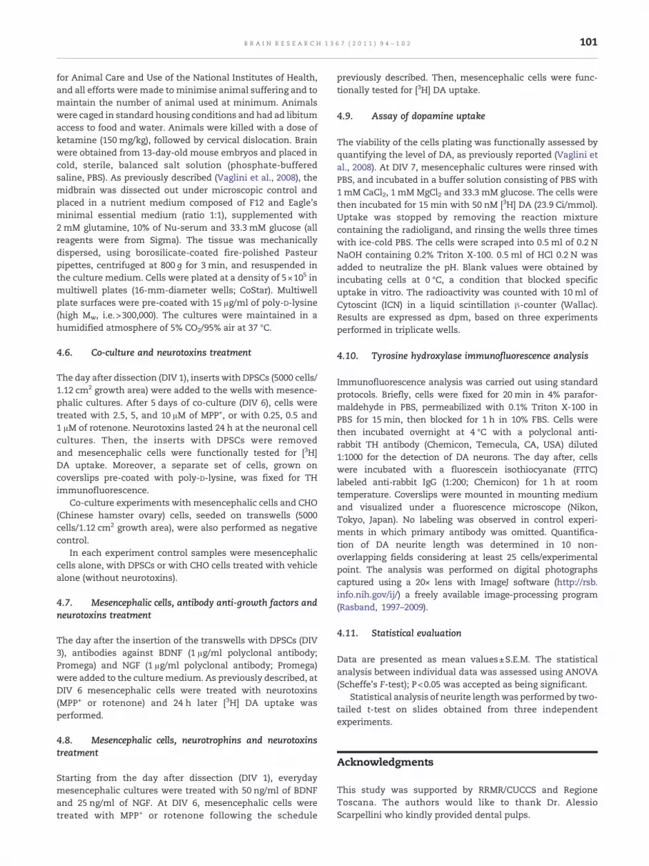
Timed pregnant CD1 mice were obtained from our laboratory.The animals were treated in accordance with the Guidelines
101B R A I N R E S E A R C H 1 3 6 7 ( 2 0 1 1 ) 9 4 – 1 0 2
for Animal Care and Use of the National Institutes of Health,and all efforts were made to minimise animal suffering and tomaintain the number of animal used at minimum. Animalswere caged in standard housing conditions and had ad libitumaccess to food and water. Animals were killed with a dose ofketamine (150 mg/kg), followed by cervical dislocation. Brainwere obtained from 13-day-old mouse embryos and placed incold, sterile, balanced salt solution (phosphate-bufferedsaline, PBS). As previously described (Vaglini et al., 2008), themidbrain was dissected out under microscopic control andplaced in a nutrient medium composed of F12 and Eagle'sminimal essential medium (ratio 1:1), supplemented with2 mM glutamine, 10% of Nu-serum and 33.3 mM glucose (allreagents were from Sigma). The tissue was mechanicallydispersed, using borosilicate-coated fire-polished Pasteurpipettes, centrifuged at 800 g for 3 min, and resuspended inthe culture medium. Cells were plated at a density of 5×10⁵ inmultiwell plates (16-mm-diameter wells; CoStar). Multiwellplate surfaces were pre-coated with 15 μg/ml of poly-D-lysine(high Mw, i.e.>300,000). The cultures were maintained in ahumidified atmosphere of 5% CO2/95% air at 37 °C.
4.6. Co-culture and neurotoxins treatment
The day after dissection (DIV 1), inserts with DPSCs (5000 cells/1.12 cm2 growth area) were added to the wells with mesence-phalic cultures. After 5 days of co-culture (DIV 6), cells weretreated with 2.5, 5, and 10 μM of MPP+, or with 0.25, 0.5 and1 μM of rotenone. Neurotoxins lasted 24 h at the neuronal cellcultures. Then, the inserts with DPSCs were removedand mesencephalic cells were functionally tested for [3H]DA uptake. Moreover, a separate set of cells, grown oncoverslips pre-coated with poly-D-lysine, was fixed for THimmunofluorescence.
Co-culture experiments with mesencephalic cells and CHO(Chinese hamster ovary) cells, seeded on transwells (5000cells/1.12 cm2 growth area), were also performed as negativecontrol.
In each experiment control samples were mesencephaliccells alone, with DPSCs or with CHO cells treated with vehiclealone (without neurotoxins).
4.7. Mesencephalic cells, antibody anti-growth factors andneurotoxins treatment
The day after the insertion of the transwells with DPSCs (DIV3), antibodies against BDNF (1 μg/ml polyclonal antibody;Promega) and NGF (1 μg/ml polyclonal antibody; Promega)were added to the culturemedium. As previously described, atDIV 6 mesencephalic cells were treated with neurotoxins(MPP+ or rotenone) and 24 h later [3H] DA uptake wasperformed.
4.8. Mesencephalic cells, neurotrophins and neurotoxinstreatment
Starting from the day after dissection (DIV 1), everydaymesencephalic cultures were treated with 50 ng/ml of BDNFand 25 ng/ml of NGF. At DIV 6, mesencephalic cells weretreated with MPP+ or rotenone following the schedule
previously described. Then, mesencephalic cells were func-tionally tested for [3H] DA uptake.
4.9. Assay of dopamine uptake
The viability of the cells plating was functionally assessed byquantifying the level of DA, as previously reported (Vaglini etal., 2008). At DIV 7, mesencephalic cultures were rinsed withPBS, and incubated in a buffer solution consisting of PBS with1 mM CaCl2, 1 mM MgCl2 and 33.3 mM glucose. The cells werethen incubated for 15 min with 50 nM [3H] DA (23.9 Ci/mmol).Uptake was stopped by removing the reaction mixturecontaining the radioligand, and rinsing the wells three timeswith ice-cold PBS. The cells were scraped into 0.5 ml of 0.2 NNaOH containing 0.2% Triton X-100. 0.5 ml of HCl 0.2 N wasadded to neutralize the pH. Blank values were obtained byincubating cells at 0 °C, a condition that blocked specificuptake in vitro. The radioactivity was counted with 10 ml ofCytoscint (ICN) in a liquid scintillation β-counter (Wallac).Results are expressed as dpm, based on three experimentsperformed in triplicate wells.
4.10. Tyrosine hydroxylase immunofluorescence analysis
Immunofluorescence analysis was carried out using standardprotocols. Briefly, cells were fixed for 20 min in 4% parafor-maldehyde in PBS, permeabilized with 0.1% Triton X-100 inPBS for 15min, then blocked for 1 h in 10% FBS. Cells werethen incubated overnight at 4 °C with a polyclonal anti-rabbit TH antibody (Chemicon, Temecula, CA, USA) diluted1:1000 for the detection of DA neurons. The day after, cellswere incubated with a fluorescein isothiocyanate (FITC)labeled anti-rabbit IgG (1:200; Chemicon) for 1 h at roomtemperature. Coverslips were mounted in mounting mediumand visualized under a fluorescence microscope (Nikon,Tokyo, Japan). No labeling was observed in control experi-ments in which primary antibody was omitted. Quantifica-tion of DA neurite length was determined in 10 non-overlapping fields considering at least 25 cells/experimentalpoint. The analysis was performed on digital photographscaptured using a 20× lens with ImageJ software (http://rsb.info.nih.gov/ij/) a freely available image-processing program(Rasband, 1997–2009).
4.11. Statistical evaluation
Data are presented as mean values±S.E.M. The statisticalanalysis between individual data was assessed using ANOVA(Scheffe's F-test); P<0.05 was accepted as being significant.
Statistical analysis of neurite lengthwas performed by two-tailed t-test on slides obtained from three independentexperiments.
Acknowledgments
This study was supported by RRMR/CUCCS and RegioneToscana. The authors would like to thank Dr. AlessioScarpellini who kindly provided dental pulps.

102 B R A I N R E S E A R C H 1 3 6 7 ( 2 0 1 1 ) 9 4 – 1 0 2
R E F E R E N C E S
Barachini, S., Trombi, L., Danti, S., D'Alessandro, D., Battolla, B.,Legitimo, A., Nesti, C., Mucci, I., D'Acunto, M., Cascone, M.G.,Lazzeri, L., Mattii, L., Consolini, R., Petrini, M., 2009.Morpho-functional characterization of human mesenchymalstem cells from umbilical cord blood for potential uses inregenerative medicine. Stem Cells Dev. 18, 293–305.
Bennett, B.A., Hyde, C.E., Pecora, J.R., Clodfelter, J.E., 1993. Differingneurotoxic potencies of methamphetamine, mazindol, andcocaine on mesencephalic cultures. J. Neurochem. 60,1444–1452.
Betarbet, R., Sherer, T.B., MacKenzie, G., Garcia-Osuna, M., Panov,A.V., Greenamyre, J.T., 2000. Chronic systemic pesticideexposure reproduces features of Parkinson's disease. Nat.Neurosci. 3, 1301–1306.
Cappelletti, G., Maggioni, M.G., Maci, R., 1999. Influence of MPP+ onthe state of tubulin polymerisation in NGF-differentiated PC12cells. J. Neurosci. Res. 56, 28–35.
Cappelletti, G., Pedrotti, B., Maggioni, M.G., Maci, R., 2001. Tubulinpolymerisation is directly affected by MPP+ in vitro. Cell Biol.Int. 25, 981–984.
Cappelletti, G., Surrey, T., Maci, R., 2005. The parkinsonismproducing neurotoxin MPP+ affects microtubule dynamics byacting as a destabilising factor. FEBS Lett. 579, 4781–4786.
Chai, Y., Jiang, X., Ito, Y., Bringas Jr., P., Han, J., Rowitch, D.H.,Soriano, P., McMahon, A.P., Sucov, H.M., 2000. Fate of themammalian cranial neural crest during tooth and mandibularmorphogenesis. Development 127, 1671–1679.
Chauhan, N.B., Siegel, G.J., Lee, J.M., 2001. Depletion of glial cellline-derived neurotrophic factor in substantia nigra neurons ofParkinson's disease brain. J. Chem. Neuroanat. 21, 277–288.
Fiskum, G., Starkov, A., Polster, B.M., Chinopoulos, C., 2003.Mitochondrial mechanisms of neural cell death andneuroprotective interventions in Parkinson's disease. Ann. NYAcad. Sci. 991, 111–119.
Gash, D.M., Zhang, Z., Ovadia, A., Cass, W.A., Yi, A., Simmerman,L., Russell, D., Martin, D., Lapchak, P.A., Collins, F., Hoffer, B.J.,Gerhardt, G.A., 1996. Functional recovery in parkinsonianmonkeys treated with GDNF. Nature 380, 252–255.
Gerlach, M., Riederer, P., Przuntek, H., Youdim, M.B., 1991. MPTPmechanisms of neurotoxicity and their implications forParkinson's disease. Eur. J. Pharmacol. 208, 273–286.
Greenamyre, J.T., MacKenzie, G., Peng, T.I., Stephans, S.E., 1999.Mitochondrial dysfunction in Parkinson's disease. Biochem.Soc. Symp. 66, 85–97.
Gronthos, S., Mankani, M., Brahim, J., Robey, P.G., Shi, S., 2000.Postnatal human dental pulp stem cells (DPSCs) in vitro and invivo. Proc. Natl. Acad. Sci. USA 97, 13625–13630.
Gronthos, S., Brahim, J., Li, W., Fisher, L.W., Cherman, N., Boyde,A., DenBesten, P., Robey, P.G., Shi, S., 2002. Stem cell propertiesof human dental pulp stem cells. J. Dent. Res. 81, 531–535.
Hellmann, M.A., Panet, H., Barhum, Y., Melamed, E., Offen, D.,2006. Increased survival and migration of engraftedmesenchymal bone marrow stem cells in6-hydroxydopamine-lesioned rodents. Neurosci. Lett. 395,124–128.
Jiang, Q., Yan, Z., Feng, J., 2006. Neurotrophic factorsstabilize microtubules and protect against rotenone
toxicity on dopaminergic neurons. J. Biol. Chem. 281,29391–29400.
Lang, A.E., Lozano, A.M., 1998. Parkinson's disease: first of twoparts. N. Engl. J. Med. 339, 1044–1053.
Levy, Y.S., Gilgun-Sherki, Y., Melamed, E., Offen, D., 2005.Therapeutic potential of neurotrophic factors inneurodegenerative diseases. BioDrugs 19, 97–127.
Lillesaar, C., Eriksson, C., Fried, K., 2001. Rat tooth pulp cells elicitneurite growth from trigeminal neurones and express mRNAsfor neurotrophic factors in vitro. Neurosci. Lett. 308, 161–164.
Mazzio, E., Yoon, K.J., Soliman, K.F., 2003. Acetyl-L-carnitinecytoprotection against 1-methyl-4-phenylpyridinium toxicityin neuroblastoma cells. Biochem. Pharmacol. 66, 297–306.
Nishio, T., Furukawa, S., Akiguchi, I., Sunohara, N., 1998. Medialnigral dopamine neurons have rich neurotrophin support inhumans. NeuroReport 9, 2847–2851.
Nosrat, C.A., Fried, K., Ebendal, T., Olson, L., 1998. NGF, BDNF, NT3,NT4 and GDNF in tooth development. Eur. J. Oral Sci. 106 (Suppl1), 94–99.
Nosrat, I.V., Widenfalk, J., Olson, L., Nosrat, C.A., 2001. Dental pulpcells produce neurotrophic factors, interact with trigeminalneurons in vitro, and rescue motoneurons after spinal cordinjury. Dev. Biol. 238, 120–132.
Oppenheim, R.W., Yin, Q.W., Prevette, D., Yan, Q., 1992.Brain-derived neurotrophic factor rescues developing avianmotoneurons from cell death. Nature 360, 755–757.
Oppenheim, R.W., Houenou, L.J., Johnson, J.E., Lin, L.F., Li, L., Lo, A.C.,Newsome, A.L., Prevette, D.M., Wang, S., 1995. Developingmotorneurons rescued from programmed and axotomy-induced celldeath by GDNF. Nature 373, 344–346.
Rasband, W.S., 1997–2009. ImageJ. U. S. National Institutes ofHealth, Bethesda, Maryland, USA. http://rsb.info.nih.gov/ij/.
Ren, Y., Liu, W., Jiang, H., Jiang, Q., Feng, J., 2005. Selectivevulnerability of dopaminergic neurons to microtubuledepolymerization. J. Biol. Chem. 280, 34105–34112.
Ren, Y., Jiang, H., Yang, F., Nakaso, K., Feng, J., 2009. Parkin protectsdopaminergic neurons against microtubule-depolymerizingtoxins by attenuating microtubule-associated protein kinaseactivation. J. Biol. Chem. 284, 4009–4017.
Sanchez-Ramos, J., Michel, P., Weiner, W.J., Hefti, F., 1988.Selective destruction of cultured dopaminergic neurons fromfetal rat mesencephalon by 1-methyl-4-phenylpyridinium:cytochemical and morphological evidence. J. Neurochem. 50,1934–1944.
Vaglini, F., Pardini, C., Maggio, R., Corsini, G.U., 1995. Role ofexcitatory amino acids in diethyldithiocarbamate-induced celldeath in mesencephalic cultures. Brain Res. 674, 127–132.
Vaglini, F., Pardini, C., Viaggi, C., Caramelli, A., Corsini, G.U., 2008.Apomorphine offers new insight into dopaminergic neuronvulnerability in mesencephalic cultures. Neuropharmacology55, 737–742.
Walker, D.G., Beach, T.G., Xu, R., Lile, J., Beck, K.D., McGeer, E.G.,McGeer, P.L., 1998. Expression of the proto-oncogene Ret, acomponent of the GDNF receptor complex, persists in humansubstantia nigra neurons in Parkinson's disease. Brain Res. 792,207–217.
Zhang, W., Walboomers, X.F., Shi, S., Fan, M., Jansen, J.A., 2006.Multilineage differentiation potential of stem cells derivedfrom human dental pulp after cryopreservation. Tissue Eng. 12,2813–2823.


