Embed Size (px)
Citation preview

www.elsevier.com/locate/jim
Journal of Immunological Methods 289 (2004) 179–190
Research paper
Human tonsillar tissue block cultures differ from autologous
tonsillar cell suspension cultures in lymphocyte subset activation
and cytokine gene expression
Bettina Gigera, Athos Bonanomia, Bernhard Odermattb, Kristin Ladella,Roberto F. Speckc, Dejan Kojica, Christoph Bergera, Felix K. Nigglid, David Nadala,*
aDivision of Infectious Diseases, University Children’s Hospital of Zurich, Steinwiesstrasse 75, CH-8032 Zurich, SwitzerlandbDepartment of Pathology, University Hospital of Zurich, Zurich, Switzerland
cDivision of Infectious Diseases and Hospital Hygiene, Department of Internal Medicine,
University Children’s Hospital of Zurich, Zurich, SwitzerlanddDivision of Oncology, University Children’s Hospital of Zurich, Zurich, Switzerland
Received 13 October 2003; received in revised form 29 March 2004; accepted 19 April 2004
Available online 31 May 2004
Abstract
Lymphoid tissues cultured either as tissue blocks or as cell suspensions are used to study the behaviour of immune cells
within their habitat. The preservation of tissue structures in tissue blocks, which is considered to be a major advantage, has been
poorly defined. We characterised the morphological evolution of tissue cultures from human palatine tonsils and compared their
lymphocyte subsets and the constitutive cytokine gene expression to those in autologous tonsillar single-cell suspension cultures
over time, and after adding cyclosporin A (CsA) to mimic the situation in individuals treated with immunosuppressive drugs.
Density and morphology of follicles were conserved up to 4 days, during which tissue cultures exhibited similar cell viability as
suspension cultures, but a significantly less frequent increase of CD95 expression in T cells, smaller variation of the proportion
of CD4+ cells and better CD21+/CD23� B-cell survival. Treatment with cyclosporin A at higher concentrations resulted in
superior histologic preservation of lymphoid tissue structures and seemed to further prevent the expression of CD95 by CD3+
cells and the activation in tissue culture of CD21+ cells. Constitutive gene expression levels of the stromal cytokines interleukin
(IL)-1h and interleukin-6 in tissue culture were significantly higher than those in suspension cultures. These results suggest that
tonsillar tissue cultures preserve their structure only for a limited time, during which they more closely reflect processes in vivo,
including a state of iatrogenic immunosuppression, than do their cell suspension counterparts.
D 2004 Elsevier B.V. All rights reserved.
Keywords: Tonsillar tissue block cultures; Cell suspension cultures; In vitro model; Cytokines; Lymphocyte subsets; Cyclosporin A; Interleukin-
1; Interleukin-6; Transforming growth factor-h
0022-1759/$ - see front matter D 2004 Elsevier B.V. All rights reserved.
doi:10.1016/j.jim.2004.04.015
Abbreviations: CsA, cyclosporin A; FITC, fluorescein isothiocyanate; HE, haematoxylin–eosin; IFN, interferon; IL, interleukin; TCSC,
tonsillar cell suspension culture; TGF, transforming growth factor; TTBC, tonsillar tissue block culture; PE, phycoerythrin.
* Corresponding author. Tel.: +41-1-266-7562; fax: +41-1-266-7157.
E-mail address: [email protected] (D. Nadal).

B. Giger et al. / Journal of Immunological Methods 289 (2004) 179–190180
1. Introduction
Immune responses, such as activation or differen-
tiation of B lymphocytes in germinal centres, are
tightly regulated by signals from different interacting
cell populations (Abbas and Lichtman, 2003). Cul-
tures of cell suspensions, often used to mimic in vivo
situations, probably reflect the situation in vivo only
to a limited degree since they lack tissue structures
essential for the development of immune responses.
This drawback, i.e., the likelihood of missing impor-
tant steps, could be overcome by culturing lymphoid
tissue blocks that preserve organ tissue structure
thereby retaining the original microenvironment, as
reported for cultures using tissue samples from spleen,
cervix or tumours (Hoffmann et al., 1995; Collins et
al., 2000; Au et al., 2002).
The survival of cells in tissue cultures from spleen
is comparable to counterparts cultured in single-cell
suspensions (Hoffmann et al., 1995). Immunoglobulin
production in these tissue cultures starts more
promptly and reaches higher levels than in cell sus-
pension cultures (Hoffmann et al., 1995). Moreover,
marked differences in the cytokine secretion profile
between tissue cultures and cell suspension cultures
have been observed (Skibinski et al., 1997; Skibinski
and James, 1997).
Tonsils are candidate organs for tissue culture since
they are more readily obtainable than spleen or other
secondary lymphoid organs. Tissue cultures from
human palatine tonsils have been employed to study
lymphocyte activation and differentiation (Ferro et al.,
1993) and immune processes during infection with the
human immunodeficiency virus (Glushakova et al.,
1995; Blauvelt et al., 2000; Grivel et al., 2001; Penn et
al., 2001; Bounou et al., 2002) and the human
herpesvirus 6 (Grivel et al., 2001, 2003). However,
the morphological evolution of these tissue cultures
has been characterised to only a limited degree.
This work aimed to describe the evolution of tissue
structures in tonsillar tissue block cultures (TTBCs)
and compare lymphocyte subsets in TTBCs with
those in autologous tonsillar cell suspension cultures
(TCSCs) over time. Furthermore, by adding cyclo-
sporin A (CsA) to the cultures, the situation in
individuals treated with immunosuppressive drugs
could be mimicked. Since the cytokine milieu in vivo
results from the interaction of many different cells
within the microenvironment of preserved tissue
structure, we also examined the expression of cyto-
kine genes in TTBCs versus TCSCs.
2. Materials and methods
2.1. Tissue samples
The palatine tonsils which were used were surgi-
cally removed from children because of hyperplasia at
the University Children’s Hospital of Zurich. The
organs were immediately wrapped in sterile gauze
soaked with phosphate-buffered saline pH 7.4 (Invi-
trogen, Basel, Switzerland), placed in a sterile airtight
container and transported to the laboratory within 4 h.
The institutional ethics committee approved the study.
2.2. Preparation of tonsillar tissue block and cell
suspension cultures
From each tonsil, TTBCs and TCSCs were pre-
pared in parallel. For TTBCs, part of the tonsil was cut
manually in phosphate-buffered saline into 2-mm3
blocks that were set up in sponge cultures (Jenkinson
et al., 1982) as follows: each well of a 6-well plate
contained a gelatine sponge (Gelfoam, 1 cm thick,
Pharmacia & Upjohn, Dubendorf, Switzerland)
soaked with medium and covered with a membrane
(Isopore, 4 Am, Millipore, Volketswil, Switzerland).
On each membrane, six tissue blocks were placed and
medium was added to reach the level of the mem-
brane. For TCSCs, the other part of the tonsil was
disintegrated with a scalpel tip in fresh medium and
filtered through a 70-Am cell strainer (Falcon, Fisher,
Wohlen, Switzerland). Cells were counted using the
0.4% trypan blue (Sigma, Buchs, Switzerland) exclu-
sion test and cultured in 24-well plates, each well
containing 2� 106 viable cells in 1-ml medium. Both
TTBCs and TCSCs were kept in a humidified incu-
bator at 37 jC and 5% CO2. Every second day, half of
the medium was replaced.
2.3. Culture media and treatments
TTBCs were set up with Yssel’s medium (Gemini
Bio-Products, Woodland, CA, USA) and RPMI 1640
medium (Invitrogen), and the latter was used also for

Fig. 1. Rating of morphological evolution of TTBCs over time.
Tissue blocks of the same tonsil were cultured in RPMI 1640 or
Yssel’s medium. The rating scale presented in Table 1 was applied to
sections of TTBCs from eight organs. The evolution of the average
score with standard error bars for 8 days comparing the two media is
shown.
B. Giger et al. / Journal of Immunological Methods 289 (2004) 179–190 181
TCSCs. Both media contained HEPES buffer, L-glu-
tamine, 1 mM sodium pyruvate, 1 mM nonessential
amino acids, 15% fetal bovine serum, 100 U/ml
penicillin, 100 Ag/ml streptomycin sulfate and 2.5
Ag/ml amphotericin B (all from Invitrogen). For some
experiments, 1, 5 or 25 mg/l CsA (Novartis, Basel,
Switzerland) was added. All cultures for each condi-
tion were set up at least in duplicate.
2.4. Assessment of tissue structure preservation in
tonsillar tissue block cultures
Tissue blocks were harvested daily until day 8,
fixed in 4% buffered formalin (Sigma), embedded in
Paraplast, sectioned and stained in haematoxylin–
eosin (HE) solution (Fluka, Buchs, Switzerland).
Density and morphology of lymphoid follicles, tissue
consistency and necrosis were rated using the scale
listed in Table 1.
2.5. Immunohistochemical phenotyping of lymphocyte
subsets
Immunohistochemistry was performed on paraffin
sections from tissues using the Ventana Benchmark
automated staining system (Ventana Medical Systems,
Table 1
Histological rating criteria for tonsillar tissue block cultures
Parameter Score
Lymphoid follicles: density and morphology
High density
(more than 5 per low power optical field)
4
High density, loss of thickness of the follicle
wall
3
Low density (1–3), only small number of
follicles
2
No lymphoid follicles 1
Tissue consistency
Compact throughout the tissue 4
Tissue loosening in the germinal centres 3
Tissue loosening throughout the section 2
Several small dissolved spots, mainly in
the centre of the tissue
1
Necrosis
< 20% of tissue area 4
20–40% 3
>40–60% 2
>60% 1
Tucson, AZ, USA). For antigen retrieval, slides were
heated with cell conditioner 1 (mild protocol). For
CD68, enzymatic predigestion with protease 1 was
performed for 2 min. Primary antibodies against the
following antigens were applied: CD3 (affinity puri-
fied rabbit antibodies, dilution 1/80; DakoCytomation,
Glostrup, Denmark), CD4 (clone 1F6, dilution 1/10;
Novocastra, Newcastle upon Tyne, UK), CD8 (clone
C8/144B, dilution 1/100; Dako, Zug, Switzerland),
CD20 (clone L26, dilution 1/400; Dako), CD23 (clone
1B12, dilution 1/30; Novocastra) and CD68 (clone
PG-M1, dilution 1/50; Dako). Primary antibodies
were detected with the Ventana iVIEW DAB detec-
tion kit, yielding a brown reaction product. For CD4,
CD8 and CD23, the signal was enhanced with the
Ventana amplification kit. Slides were counterstained
with haematoxylin.
2.6. Assessment of cell viability and relative distribu-
tion of lymphocyte subsets
Tissue blocks were disintegrated with a scalpel tip
in staining buffer (phosphate-buffered saline with 5%

Fig. 2. Representative paraffin sections of TTBCs at days 0, 1, 2, 3 and 4 of culture, respectively. HE-stained sections are shown at two
different magnifications. Sections immunostained for the macrophage marker CD68 are shown at the higher magnification. Note involution of
the follicular structure, loosening of the cell aggregation density and decrease of number and size of the macrophages (CD68+ cells) over
time.
Fig. 3. Cell viability in tonsillar tissue block cultures (TTBCs) and
tonsillar cell suspension cultures (TCSCs). Cells from four replicate
cultures were stained with 7-amino-actinomycin D (7-AAD) to
quantify nonviable cells by FACS and therefore to determine the
cell survival shortly after excision and during 4 days in culture. The
data are presented as average percentages of viable cells for
autologous TTBCs and TCSCs with standard deviation.
B. Giger et al. / Journal of Immunological Methods 289 (2004) 179–190182
fetal bovine serum and 0.1% sodium azide; Sigma)
and filtered through a 70-Am cell strainer (Falcon) to
obtain single-cell suspensions. Pellets of TCSCs were
resuspended and washed with staining buffer. Single
cells (2.5� 105) were double-stained with a fluores-
cein isothiocyanate (FITC)-labelled and a phycoery-
thrin (PE)-labelled mouse monoclonal antibodies (all
from BD Biosciences, Basel, Switzerland, if not stated
differently) at a dilution of 1/10, for 30 min at 4 jC in
the dark following the instructions of the manufactur-
er. FITC-conjugated anti-mouse IgG1 (FITC-IgG1)
with PE-conjugated anti-mouse IgG1 (PE-IgG1),
FITC-IgG1 with PE–HLA-ABC and FITC-CD45
with PE-IgG1 served as isotype controls. 7-Amino-
actinomycin D (7-AAD) was used to identify nonvi-
able cells. B cells versus T cells were quantified by
FITC-CD20 with PE-CD3. Activated cells prone to
apoptosis were monitored by FITC-CD95 with PE-
CD3 or with PE-CD20, respectively. T helper lym-
phocytes versus cytotoxic T lymphocytes were distin-
guished with FITC-CD4 and PE-CD8. Resting and
activated B cells were identified using FITC-CD21
(Dako) with PE-CD23. After staining, the cells were
washed with staining buffer, fixed with 3% parafor-
maldehyde for 20 min and washed again. The percen-
tages of positive cells and mean fluorescence intensity
were analysed using FACS Calibur (BD Biosciences)
equipped with a 488- and 635-nm laser for double-

B. Giger et al. / Journal of Immunological Methods 289 (2004) 179–190 183
colour analysis, gating on the lymphocyte population,
as defined by forward and side light scatter. Data were
recorded and analysed with Cell Quest software (BD
Biosciences).
2.7. Assessment of cytokine gene expression
RNA extractions and real-time quantitative poly-
merase chain reactions (PCRs; TaqMank) for human
interleukin (IL)-1h, IL-2, IL-6, IL-10, IL-12, IL-15,interferon (IFN)-g, transforming growth factor (TGF)-
h and the endogenous reference, the housekeeping
gene hydroxymethylbilane synthase, were performed
Fig. 4. In situ localisation of lymphocyte subsets in TTBCs over time. A
cultured in RPMI 1640 medium were stained with antibodies against the
CD23 or the macrophage marker CD68.
following the supplier’s instructions (Applied Biosys-
tems, Foster City, CA, USA) and as described previ-
ously (Bonanomi et al., 2003). All reactions were
performed in duplicate.
2.8. Statistics
For comparison between groups, either the v2 test
or the two-tailed Fisher’s exact test was used. The
Mann–Whitney U-test was used for comparison of
meanF standard errors of the mean values between
groups. Values of p < 0.05 were considered statistically
significant.
t each day in culture, paraffin sections from a single tissue block
T cell markers CD3, CD4 and CD8, the B cell markers CD20 and

Table 2
Relative distribution of lymphocyte subsets in tonsillar tissue block
cultures and tonsillar cell suspension cultures on day 4 compared to
day 0
Phenotype Variation p Values
TTBC TCSC
CD3+/CD95� 23.47F 6.67 17.21F 7.38 ns
CD3+/CD95+ � 1.3F 1.6 4.5F 1.9 0.019
CD4+/CD8� 2.9F 2.1 6.4F 5.0 ns
CD4�/CD8+ 5.6F 0.7 3.8F 1.3 ns
CD20+/CD95� � 21.0F 8.7 � 14.8F 7.4 ns
CD20+/CD95+ � 5.9F 2.2 � 4.7F 3.1 ns
CD21+/CD23� � 2.7F 4.9 � 8.8F 2.6 ns
CD21+/CD23+ � 28.0F 5.1 � 22.4F 6.8 ns
Values are presented as meansF S.E.M. of 12 tonsils; ns: not
significant.
B. Giger et al. / Journal of Immunological Methods 289 (2004) 179–190184
3. Results
3.1. Morphological evolution of TTBCs
The number and morphology of follicles over time
were similar in Yssel’s medium and RPMI 1640
medium, showing a decrease from day 1 to day 7
when all follicles had disappeared (Fig. 1). The
preservation of the tissue consistency differed slightly
between both media in the first 3 days in favour of
Yssel’s medium, but was similar thereafter. Necrosis
was augmented more rapidly after day 3 using RPMI
1640 medium compared to Yssel’s medium. However,
the sponges used to support the TTBCs disintegrated
in Yssel’s medium but not in RPMI 1640 medium and
impeded preparation of tissue sections after day 3.
Thus, based on the morphological rating, both RPMI
1640 medium and Yssel’s medium can be used to
establish TTBCs, but due to structural changes of the
tissue, including involution of the follicular structure,
loosening of the cell aggregation density and a de-
crease in the number and size of the macrophages over
time (Fig. 2), potential functional advantages of
TTBCs over TCSCs could only be expected within
the first 4 days of culture. Therefore, we limited
cultures to 4 days and preferred RPMI 1640 medium
to Yssel’s medium for the better preservation of the
sponges and better comparison with TCSCs.
3.2. Cell viability and lymphocyte subsets
Staining with 7-AAD showed that in both TTBCs
and in TCSCs, the viability of gated lymphocytes was
comparable on days 0 (84.9 F 4.1% versus
88.1F 0.4%), 3 and 4 of culture, when it declined
to 38.6F 2.5% versus 29.9F 8.3%. However, viabil-
ity was lower in TTBCs than that of TCSCs on days 1
and 2 (Fig. 3).
Immunohistochemical staining of TTBC tissue
sections from day 0 (Fig. 4) showed typical well-
demarcated secondary follicles with well-developed
germinal centres. Follicle mantle zones and germinal
centres stained for CD20. Cells within the follicular
mantle zone also expressed CD23, although the most
prominent staining for this marker was seen on
follicular dendritic cells in the apical light zones of
germinal centres. CD3+ cells (mostly CD4+ and, to a
lesser extent, CD8+) localised preferentially in the T
zones. Numerous macrophages expressing CD68
were found in germinal centres and scattered in the
surrounding areas. This regular distribution of the
different cell subsets lasted until day 4 in TTBCs.
However, immunostaining started to fade on days 3
and 4 (Fig. 4) and became completely diffused there-
after (not shown), reflecting the decay of tissue
structure observed in HE-stained sections.
Table 2 and Fig. 5 show the variations in lympho-
cyte subsets in TTBCs and TCSCs on day 4 of culture
compared to the frequencies on day 0. The proportion
of CD3+/CD95� cells increased in TTBCs and
TCSCs, whereas the proportion of CD20+ cells de-
creased. The differences between TTBCs and autolo-
gous TCSCs were within the 10% range, except for
two pairs with CD3+/CD95� cells in TTBCs higher
by 17.2% and 36.8%, respectively, than in TCSCs
(Fig. 5). Only 2 out of 12 TTBCs, but 11 out of 12
TCSCs showed an increase of CD3+/CD95+ cells
(Fig. 5; p < 0.001). Accordingly, the average change
of CD3+/CD95+ cells compared to baseline in TTBCs
was lower than that of TCSCs ( p = 0.019). The
variation of the relative CD4+ cell increase in TTBCs
was smaller than that of TCSCs (Table 2). In three
(25%) TCSCs, a relative increase of the CD4+ cell
percentage greater than 20% compared to baseline
was observed. These three TCSCs and a fourth one
showed CD4+ cell percentages between 14.6% and
25.5% greater than autologous TTBCs. Finally, an
increase in the percentage of resting, mature B cells
(CD21+/CD23�) was noted in four TTBCs and one
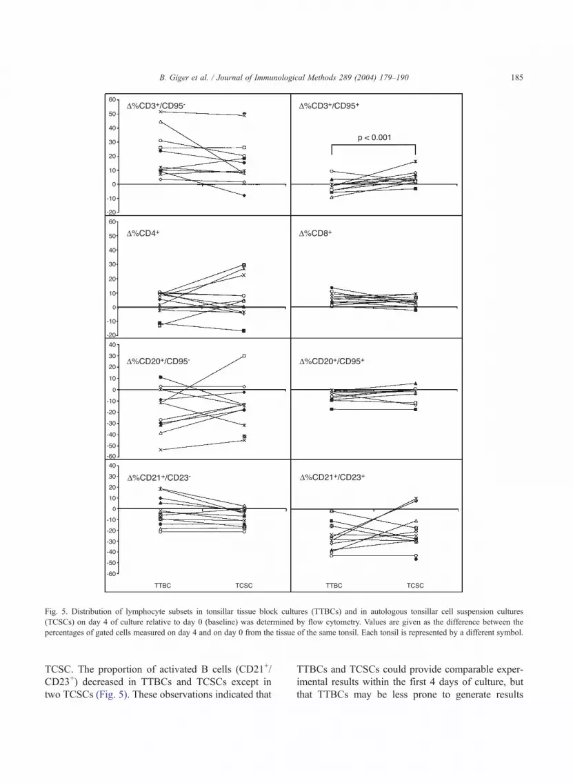
Fig. 5. Distribution of lymphocyte subsets in tonsillar tissue block cultures (TTBCs) and in autologous tonsillar cell suspension cultures
(TCSCs) on day 4 of culture relative to day 0 (baseline) was determined by flow cytometry. Values are given as the difference between the
percentages of gated cells measured on day 4 and on day 0 from the tissue of the same tonsil. Each tonsil is represented by a different symbol.
B. Giger et al. / Journal of Immunological Methods 289 (2004) 179–190 185
TCSC. The proportion of activated B cells (CD21+/
CD23+) decreased in TTBCs and TCSCs except in
two TCSCs (Fig. 5). These observations indicated that
TTBCs and TCSCs could provide comparable exper-
imental results within the first 4 days of culture, but
that TTBCs may be less prone to generate results

B. Giger et al. / Journal of Immunological Methods 289 (2004) 179–190186
influenced by T cells activated to susceptibility for
apoptosis.
3.3. Treatment with cyclosporin A
Treatment of TTBCs with CsA at concentrations of
1 or 5 mg/l did not show significant differences in
tissue preservation compared to controls (Fig. 6). At
day 4, the number of follicles and the tissue consis-
tency had decreased and the amount of necrosis
Fig. 6. In situ localisation of lymphocyte subsets in TTBCs on day 2 of cu
sections from TTBCs from two different donors are shown. CsA denotes
increased as seen in untreated TTBCs (total score
for each condition 5). By contrast, using CsA at 25
mg/l showed less decay of tissue consistency and
increase in necrosis, and the density of follicles
remained stable (Fig. 6) until day 4 of culture (total
score 10).
In general, CsA resulted in an increase of the
proportion of CD3+/CD95� cells, although with var-
iations of up to 50%. At higher doses, CsA seemed to
reduce the proportion of CD3+/CD95+ cells in TTBCs
lture following no treatment or treatment with CsA. Representative
cyclosporin A.

B. Giger et al. / Journal of Immunological Methods 289 (2004) 179–190 187
but not in TCSCs (Fig. 7). The range of variation of
CD4+ cells seemed to diminish with increasing con-
centrations of CsA. The proportion of CD21+/CD23�
cells in TTBCs was either increased or reduced,
whereas in TCSCs, it was always reduced. Finally,
the percentages of CD21+/CD23+ cells were invariably
reduced in TTBCs, but not in TCSCs where increases
were seen (Fig. 7). Thus, treatment with CsA seemed
to prevent the expression of CD95 by CD3+ cells and
the activation of CD21+ cells in TTBCs. This sug-
gested that CsA treatment of TTBCs could better
Fig. 7. Distribution of lymphocyte subsets in tonsillar tissue block cultures
with 1, 5 or 25 mg/l cyclosporin A relative to nontreated cultures (baselin
between the percentages of gated cells obtained from cultures treated with
single-cell suspensions from the same tonsil. TTBC, tonsillar tissue block
mimic states of diminished T cell activation with
ensuing activation of B cells as in iatrogenic immuno-
suppression than did similar treatment of TCSCs.
3.4. Constitutive expression of cytokine genes
There were no differences between TTBCs and
TCSCs in the constitutive expression of IL-2, IFN-g,
IL-10, IL-12 and IL-15 (data not shown). By contrast,
the constitutive expression levels of IL-1h and IL-6
were higher in TTBCs than in autologous TCSCs
and autologous tonsillar cell suspension cultures on day 4 of culture
e) determined by flow cytometry. Values are given as the difference
cyclosporin A (CsA) and untreated cultures of tonsillar tissues or
culture; TSCS, tonsillar cell suspension culture.

Fig. 8. Cytokine gene expression in tonsillar tissue block cultures
versus autologous tonsillar cell suspension cultures. mRNA was
extracted from cultures on day 2 and reverse-transcribed. Expres-
sion of IL-1h, IL-6 and transforming growth factor (TGF)-h was
quantified by real-time polymerase chain reaction. Cycle threshold
values were normalised to the housekeeping gene hydroxymethyl-
bilane synthase and calibrated on the calibrator sample with CARTA
(Bonanomi et al., 2003). The results are expressed as F log2(expression) relative to the calibrator sample. Horizontal bars
indicate mean values. TTBC, tonsillar tissue block culture; TSCS,
tonsillar cell suspension culture.
B. Giger et al. / Journal of Immunological Methods 289 (2004) 179–190188
( p < 0.005 and p < 0.01, respectively), whereas the
levels of TGF-h in TTBCs and TCSCs were within
the same range (Fig. 8). This indicated that relevant
differences in cytokine-driven activities exist between
immune cells within intact tissue structures and im-
mune cells deprived of their natural habitat.
4. Discussion
The evolution of TTBCs with regard to morpho-
logical appearance, distributions of lymphocyte sub-
sets, reaction to CsA and cytokine gene expression
have been studied for the first time. Lymphoid tissue
was well preserved until day 4 of culture. High doses
of CsA prevented disintegration of lymphoid fol-
licles. Compared to autologous TCSCs, TTBCs
showed less T cells activated to express CD95 and
exhibited contrasting constitutive cytokine gene ex-
pression patterns.
Although Yssel’s medium seemed to prevent cell
decay better until day 3, RPMI 1640 medium was
preferred since it did not provoke disintegration of the
sponges used to nest the tissue blocks and thus
allowed better processing for histology. Our results
contrast with those of (Glushakova et al., 1995) who
cultured TTBCs for 10–26 days and reported that the
key elements of tissue architecture, including well-
defined germinal centres, were preserved even into the
fourth week of culture. However, immunohistochem-
ical data were not presented. Another study of TTBCs
addressing histology, although less systematically
than the present study, obtained acceptable viability
of cells until day 4 in one experiment and until day 7
in another experiment (Ferro et al., 1993). Studies
using human spleen for functional experiments during
a period of 3–7 days did not investigate histology
(Hoffmann et al., 1995; Skibinski et al., 1997).
During the first 4 days of culture, a similar
relative increase of T cells paralleled by a decrease
of B cells was observed in TTBCs and TCSCs. This
low survival of B cells was expected based on the
great number of naive B cells in lymphoid follicles.
Naive B cells die within 48 h in the absence of
specific antigen stimulation (Liu, 1992). This was
reflected by the increasing amount of necrosis over
time within the follicles seen in the tissue sections
(Figs. 1 and 2).
Significantly less frequent and less numeric
increases in the proportion of T cells activated to
express CD95 were observed in TTBCs compared to
TCSCs. This suggested that T cells in TTBCs expe-
rienced fewer stimuli-provoking expression of CD95
and susceptibility to the cell death program than was
the case for T cells in TCSCs. One reason could be
that cutting the tonsils into pieces results in fewer cell
stimulatory events than disruption to obtain single
cells. Also, T cells within TTBCs remain sessile,
whereas in TCSCs, they are obviously mixed with
the cellular components of all the distinct lymphoid
tissue areas. Thus, T cells in TCSCs are more likely
exposed to foreign and self-antigenic stimuli usually
located in different areas of the organ. The smaller
range of variation in the frequencies of CD4+ T cells
compared to baseline in TTBCs versus TCSCs is in
agreement with a more controlled degree of stimula-
tion in TTBCs than in TCSCs. Thus, the study of
specified T cell stimulatory events in TTBCs would be
expected to be less confounded by cell manipulation-
induced in vitro effects and therefore give results
closer to reality.

B. Giger et al. / Journal of Immunological Methods 289 (2004) 179–190 189
Treatment of TTBCs with CsA at high doses
prevented disintegration of lymphoid follicles. Thus,
CsA at these concentrations seemed to penetrate
TTBCs in amounts sufficient to exert an effect. A
clear-cut dose-related effect of CsA on the distribution
of lymphocyte subsets was neither seen in TTBCs nor
in TCSCs, indicating that CsA exhibited functional
changes rather than numeric changes in immune cells.
Indeed, CsA interferes with the inducible transcription
of cytokine genes in T cells, B cells and other immune
cells (Shaw et al., 1995; Matsuda and Koyasu, 2000;
Tajima et al., 2003).
The significantly higher levels of constitutive gene
expression for the pro-inflammatory cytokines IL-1hand IL-6 in TTBCs, compared to TCSCs, were
remarkable, and notably no differences were found
for the cytokines IL-2, IFN-g, IL-10, IL-12 and IL-15.
Major differences in the spontaneous cytokine secre-
tion profile have been noted between tissue cultures
and cell suspension cultures of human spleen (Ski-
binski et al., 1997). Similar to our findings, the
stromal cytokines IL-1h, IL-6, IL-8 and IL-11 were
detected in supernatants of tissue block cultures at
significantly higher concentrations than in superna-
tants of cell suspension cultures (Skibinski et al.,
1997). These differences could be partly attributable
to the loss of key cell types while preparing TCSCs,
such as stromal or follicular dendritic cells, major
sources of these pro-inflammatory cytokines. A more
plausible reason could be the disruption of vital cell–
cell and cell–matrix interactions during preparation of
the cell suspensions. Finally, removal of key cells or
of important mediators during washing of the cell
suspensions could also lead to differences in compar-
ison to TTBCs.
TTBCs differed at least in two remarkable aspects
from TCSCs, namely, in a less frequent increase in
CD95 expression in T cells, which indicates augment-
ed susceptibility to apoptosis in TCSCs, and in
cytokine gene expression. Both aspects may be
interconnected and depend on various factors, of
which cell-to-cell interaction seems to be a crucial
one. For the cross talk between immune cells, the
microenvironment of a lymphoid organ is substantial,
as can be concluded from the distribution of lympho-
cyte subsets with different functions to different areas
of the organ. Thus, in vitro investigations of immune-
related events, especially those expected to occur in
secondary lymphoid organs, using TTBCs may more
closely reflect in vivo processes than when using
TCSCs. We have documented that TTBCs preserve
the organ structure for a limited time. Within the
framework of 4 days, they may be more appropriate
than TCSCs for the study of immune responses to
infectious agents naturally invading the nasopharynx
and effects of immunosuppressive compounds on
these responses and the promotion of pathologic
lymphoproliferation as induced by Epstein–Barr vi-
rus. Also, mimicking a state of immunosuppression in
ex vivo lymphoid tissue, e.g., using CsA, may be
more accurate in TTBCs than in TCSCs. Finally,
TTBCs may make it possible to identify anatomically
the sites of specific processes and to pinpoint them at
the single-cell level, e.g., by employing in situ hybrid-
isation or in situ PCR.
Acknowledgements
This work was supported by SWISS BRIDGE
Foundation, the Cancer League of the Kanton of
Zurich, the Swiss National Foundation (grants #32-
53982.98 and #3339-64124.00), the EMDO-Stiftung
and the Sassella-Stiftung.
References
Abbas, A., Lichtman, A., 2003. Cellular and Molecular Immunol-
ogy. Saunders, Philadelphia, PA.
Au, J.L., Jang, S.H., Wientjes, M.G., 2002. Clinical aspects of drug
delivery to tumors. J. Control. Release 78, 81.
Blauvelt, A., Glushakova, S., Margolis, L.B., 2000. HIV-infected
human Langerhans cells transmit infection to human lymphoid
tissue ex vivo. AIDS 14, 647.
Bonanomi, A., Kojic, D., Giger, B., Rickenbach, Z., Jean-Richard-
Dit-Bressel, L., Berger, C., Niggli, F.K., Nadal, D., 2003. Quan-
titative cytokine gene expression in human tonsils at excision
and during histoculture assessed by standardized and calibrated
real-time PCR and novel data processing. J. Immunol. Methods
283, 27.
Bounou, S., Leclerc, J.E., Tremblay, M.J., 2002. Presence of host
ICAM-1 in laboratory and clinical strains of human immunode-
ficiency virus type 1 increases virus infectivity and CD4(+)-T-
cell depletion in human lymphoid tissue, a major site of repli-
cation in vivo. J. Virol. 76, 1004.
Collins, K.B., Patterson, B.K., Naus, G.J., Landers, D.V., Gupta, P.,
2000. Development of an in vitro organ culture model to study
transmission of HIV-1 in the female genital tract. Nat. Med. 6,
475.

B. Giger et al. / Journal of Immunological Methods 289 (2004) 179–190190
Ferro, L.M., Weedon, H.M., Flego, L.R., Beroukas, D., Zola, H.,
1993. An organ fragment culture model to study lymphocyte
activation in human lymphoid tissue. Immunobiology 188, 51.
Glushakova, S., Baibakov, B., Margolis, L.B., Zimmerberg, J.,
1995. Infection of human tonsil histocultures: a model for
HIV pathogenesis. Nat. Med. 1, 1320.
Grivel, J.C., Ito, Y., Faga, G., Santoro, F., Shaheen, F., Malnati,
M.S., Fitzgerald, W., Lusso, P., Margolis, L., 2001. Suppression
of CCR5—but not CXCR4—tropic HIV-1 in lymphoid tissue
by human herpesvirus 6. Nat. Med. 7, 1232.
Grivel, J.C., Santoro, F., Chen, S., Faga, G., Malnati, M.S., Ito, Y.,
Margolis, L., Lusso, P., 2003. Pathogenic effects of human her-
pesvirus 6 in human lymphoid tissue ex vivo. J. Virol. 77, 8280.
Hoffmann, P., Skibinski, G., James, K., 1995. Organ culture of
human lymphoid tissue: I. Characteristics of the system.
J. Immunol. Methods 179, 37.
Jenkinson, E.J., Franchi, L.L., Kingston, R., Owen, J.J., 1982. Ef-
fect of deoxyguanosine on lymphopoiesis in the developing
thymus rudiment in vitro: application in the production of chi-
meric thymus rudiments. Eur. J. Immunol. 12, 583.
Liu, Y.-J.e.a., 1992. Germinal centres in T-cell-dependent antibody
responses. Immunol. Today 13, 17.
Matsuda, S., Koyasu, S., 2000. Mechanisms of action of cyclospor-
ine. Immunopharmacology 47, 119.
Penn, M.L., Myers, M., Eckstein, D.A., Liegler, T.J., Hayden, M.,
Mammano, F., Clavel, F., Deeks, S.G., Grant, R.M., Goldsmith,
M.A., 2001. Primary and recombinant HIV type 1 strains resis-
tant to protease inhibitors are pathogenic in mature human lym-
phoid tissues. AIDS Res. Hum. Retrovir. 17, 517.
Shaw, K.T., Ho, A.M., Raghavan, A., Kim, J., Jain, J., Park, J.,
Sharma, S., Rao, A., Hogan, P.G., 1995. Immunosuppressive
drugs prevent a rapid dephosphorylation of transcription fac-
tor NFAT1 in stimulated immune cells. Proc. Natl. Acad. Sci.
U. S. A. 92, 11205.
Skibinski, G., James, K., 1997. The use of tissue slices in immu-
nological investigations. Arch. Immunol. Ther. Exp. (Warsz) 45,
411.
Skibinski, G., Hoffmann, P., Radbruch, A., James, K., 1997. Organ
culture of human lymphoid tissue: II. Marked differences in
cytokine production and proliferation between slice and suspen-
sion cultures of human spleen. J. Immunol. Methods 205, 115.
Tajima, K., Amakawa, R., Ito, T., Miyaji, M., Takebayashi, M.,
Fukuhara, S., 2003. Immunomodulatory effects of cyclosporin
A on human peripheral blood dendritic cell subsets. Immunol-
ogy 108, 321.


