Embed Size (px)
Citation preview

Hydrophobic Compounds Reshape Membrane DomainsJonathan Barnoud1,2, Giulia Rossi3, Siewert J. Marrink4, Luca Monticelli1,2*
1 IBCP, CNRS UMR 5086, Lyon, France, 2 Universite Claude Bernard Lyon I, Lyon, France, 3 Dept of Physics, University of Genoa, Genoa, Italy, 4 Groningen Biomolecular
Sciences and Biotechnology Institute and Zernike Institute for Advanced Materials, University of Groningen, Groningen, The Netherlands
Abstract
Cell membranes have a complex lateral organization featuring domains with distinct composition, also known as rafts,which play an essential role in cellular processes such as signal transduction and protein trafficking. In vivo, perturbations ofmembrane domains (e.g., by drugs or lipophilic compounds) have major effects on the activity of raft-associated proteinsand on signaling pathways, but they are difficult to characterize because of the small size of the domains, typically belowoptical resolution. Model membranes, instead, can show macroscopic phase separation between liquid-ordered and liquid-disordered domains, and they are often used to investigate the driving forces of membrane lateral organization. Studies inmodel membranes have shown that some lipophilic compounds perturb membrane domains, but it is not clear whichchemical and physical properties determine domain perturbation. The mechanisms of domain stabilization anddestabilization are also unknown. Here we describe the effect of six simple hydrophobic compounds on the lateralorganization of phase-separated model membranes consisting of saturated and unsaturated phospholipids and cholesterol.Using molecular simulations, we identify two groups of molecules with distinct behavior: aliphatic compounds promotelipid mixing by distributing at the interface between liquid-ordered and liquid-disordered domains; aromatic compounds,instead, stabilize phase separation by partitioning into liquid-disordered domains and excluding cholesterol from thedisordered domains. We predict that relatively small concentrations of hydrophobic species can have a broad impact ondomain stability in model systems, which suggests possible mechanisms of action for hydrophobic compounds in vivo.
Citation: Barnoud J, Rossi G, Marrink SJ, Monticelli L (2014) Hydrophobic Compounds Reshape Membrane Domains. PLoS Comput Biol 10(10): e1003873. doi:10.1371/journal.pcbi.1003873
Editor: Bert L. de Groot, Max Planck Institute for Biophysical Chemistry, Germany
Received May 19, 2014; Accepted July 25, 2014; Published October 9, 2014
Copyright: � 2014 Barnoud et al. This is an open-access article distributed under the terms of the Creative Commons Attribution License, which permitsunrestricted use, distribution, and reproduction in any medium, provided the original author and source are credited.
Data Availability: The authors confirm that all data underlying the findings are fully available without restriction. All data necessary to repeat all simulations inthe paper have been deposited on FigShare. DOI:10.6084/m9.figshare.1144384
Funding: This work was performed using HPC resources from GENCI-CINES (Grants No. 2012-076353 and No. 2013-076353), HPC-Europa2 (Contract No. 228398),and the PRACE-2IP (FP7 RI-283493) project NANODROPS. LM acknowledges funding by the Institut National de la Sante et de la Recherche Medicale (INSERM), JBis funded by iViv (Universite Paris Diderot, France). The funders had no role in study design, data collection and analysis, decision to publish, or preparation of themanuscript.
Competing Interests: The authors have declared that no competing interests exist.
* Email: [email protected]
Introduction
Biological membranes are both chemically and structurally
heterogeneous. The constituent lipids can self-organize in domains
[1], which differ in chemical composition and in physical
properties, including structural, dynamic, and elastic properties.
Domains have a functional role in cells: membrane proteins
partition preferentially to one specific domain (or to domain
boundaries) and carry out their function correctly only when in the
appropriate environment – as expressed by the raft concept [1].
Membrane lateral organization is involved in biological processes
such as membrane fusion [2,3], signal transduction [4], protein
trafficking [5], and viral infection [6,7]. Alterations of the
membrane lateral organization have been identified in pathologies
like allergies and the Alzheimer disease [8], and have been linked
to the mechanism of action of general anesthetics [9,10].
Understanding the determinants of domain stability in vivo is
therefore of paramount importance in biomedical sciences. Yet,
characterization of raft domains in vivo is challenging because of
the small size of the domains, which are typically smaller than
optical resolution [11]. In model systems (i.e., vesicles), instead,
domains are usually larger and can even coalesce to yield
macroscopic phase separation. For this reason, model systems
are often used to study membrane lateral organization [11].
Among model systems, the most frequently used are ternary
mixtures of cholesterol and two lipids with different melting
temperatures, as they show liquid-ordered (Lo) – liquid-disordered
(Ld) phase coexistence, similar to cell membranes [12].
Compounds with sufficiently high affinity for membranes can
modulate biological function by virtue of membrane-mediated
effects [13–15], including the alteration of membrane lateral
organization. Recent studies have shown that, in model mem-
branes, phase coexistence is affected by a variety of compounds.
For instance, some lipids [16], vitamin E [17], and n-alcohols [10]
destabilize phase separation in ternary lipid mixtures. On the
contrary, transmembrane helical peptides [18], benzyl alcohol
[17], and polystyrene [19] stabilize phase separation. It is unclear
which chemical or physical properties of the solutes determine
stabilization or destabilization of phase separation. Systematic
studies on the effect of solutes on membrane lateral organization
are lacking. Moreover, the mechanisms of stabilization and
destabilization of domains are not understood.
In the present report, we describe the effect of different
hydrophobic compounds on lipid mixing in phase-separated
membranes. Hydrophobic compounds partition largely to the
interior of lipid membranes, hence they do affect many membrane
properties. Hydrophobic compounds are extremely common in
commercial products and in the environment; for instance, they
PLOS Computational Biology | www.ploscompbiol.org 1 October 2014 | Volume 10 | Issue 10 | e1003873

are used as fuels in combustion engines, as solvents in industrial
processes, and as scaffolds in drugs. Also, they are building blocks
for many industrial polymers and they are found in the
atmosphere as pollutants (e.g., in products of incomplete
combustion of fossil fuels). We determine the effect of hydrophobic
compounds on phase separation using coarse-grained (CG)
molecular dynamics (MD) simulations of Lo-Ld phase-separated
lipid membranes. We focus on six different hydrophobic solutes
covering a wide range of sizes and a variety of chemical structures:
cyclohexane, octane, hexadecane, benzene, C60 fullerene and
polystyrene. All solutes partition to the interior of the membrane
but, remarkably, they show very different lateral distributions. We
identify two distinct groups with different lateral distributions, and
we show that they have opposite effects on lipid mixing. Finally,
we determine the mechanism of action for both groups of
molecules.
Results
Lipid mixing: Effect of aromatic vs. aliphatic solutesWe used the MARTINI coarse-grained (CG) force field [20,21]
to simulate model membranes consisting of dipalmitoyl-phospha-
tidylcholine (DPPC), dilinoleyl-phosphatidylcholine (DLiPC), and
cholesterol, at 42:28:30 molar ratio. At a temperature of 295 K, in
the absence of solutes, the membrane showed phase separation
into a liquid-ordered (Lo) domain, comprising mostly DPPC and
cholesterol, and a liquid-disordered (Ld) domain, comprising
mostly DLiPC, as reported previously [22]. Due to periodic
boundary conditions used in the simulations, the domains
organized in stripes along one axis of the box (Fig. 1). These
stripes persisted during the simulation, yet the interfaces were
dynamic and lipid molecules exchanged between the Lo and Ld
phase.
We then carried out simulations of the same membrane in the
presence of six different hydrophobic solutes: octane, hexadecane,
cyclohexane, benzene, fullerene, and polystyrene. These solutes
are chemically diverse, as they include linear alkanes of different
size, a cyclic alkane, a common aromatic hydrocarbon, a well-
known carbon nanoparticle, and a common industrial polymer.
For each solute, we performed two simulations at low solute
concentration (3.3% solute/lipid molar ratio) and two simulations
at high solute concentration; due to the very different molecular
weight of the solutes, for the high concentration we chose to use a
common solute/lipid mass ratio of 4.8%. For polystyrene, we only
considered simulations at high concentration. Additional simula-
tions where performed for some solutes (see Table 1 for the
complete list of simulations). Partition of the six solutes to the
interior of lipid membranes is thermodynamically highly favor-
able, as shown in several previous studies [19,23–25], but the time
scales for permeation depend largely on the size of the particles:
small molecules penetrate within a few nanoseconds [24] while
large polymer particles require microseconds or more [19]. Since
our interest was not in the kinetics of permeation but in the effect
of the solutes on the membrane, we decided to start all simulations
with the solutes placed inside the membrane, homogeneously
distributed in the membrane plane.
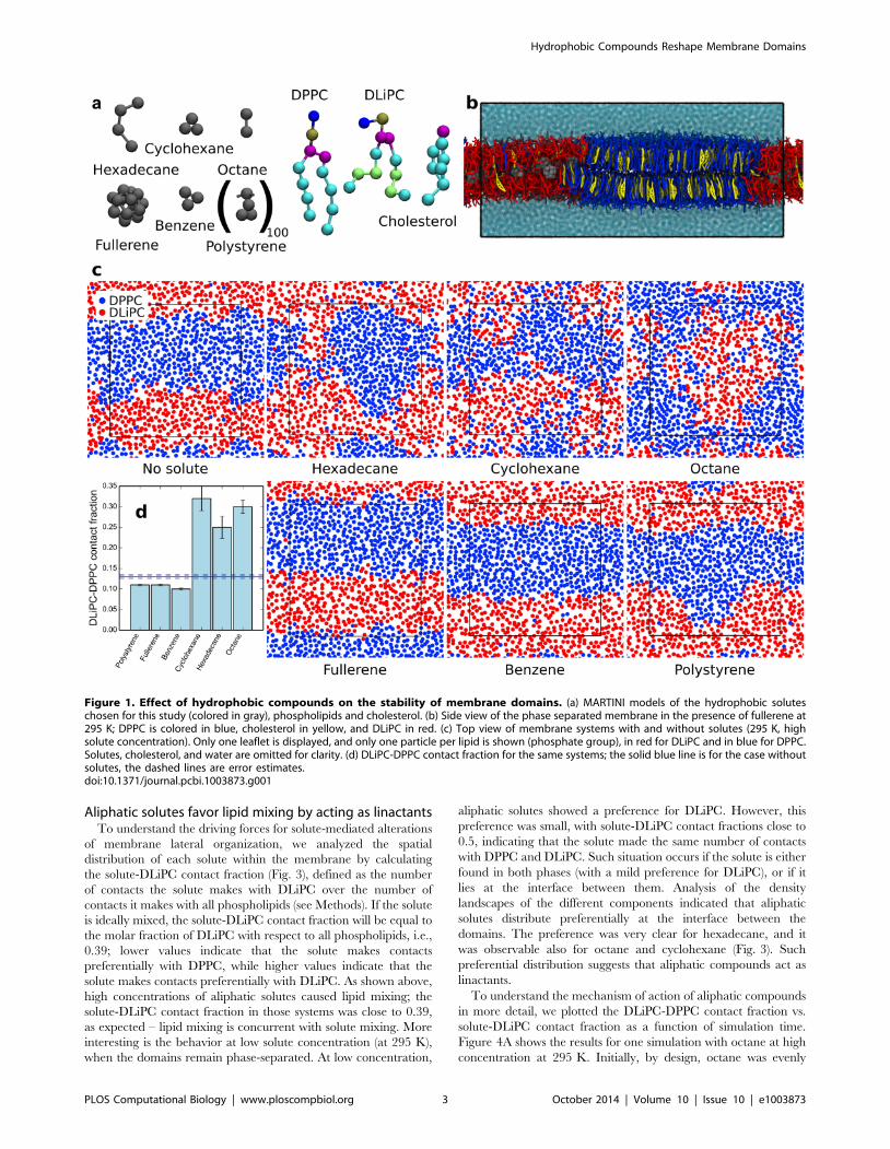
To quantify phase separation, we calculated the DLiPC-DPPC
contact fraction, fmix, defined as the fraction of DLiPC-DPPC contacts
over the total number of contacts of DLiPC with all phospholipids
(therefore not including cholesterol; see Methods). The DLiPC-DPPC
contact fraction will tend to 0 at complete phase separation and will
reach 0.61 at ideal mixing (equaling the DPPC molar fraction with
respect to phospholipids only). In the absence of solutes, fmix was
0.1360.004, indicating strong phase separation. Addition of a small
concentration of hydrophobic solutes had a minor effect on the
DLiPC-DPPC contact fraction (0.11,fmix,0.15, depending on solute
type; see Table 1). Yet two trends were distinguishable: octane,
hexadecane, and cyclohexane caused an increase in lipid mixing,
while benzene, fullerene, and polystyrene caused a slight decrease in
lipid mixing. These trends were more evident at high solute
concentration: fmix reached 0.25–0.32 with the first group of
compounds, and decreased to 0.10–0.11 with the second group (see
Table 1). Visual inspection of the trajectories showed significant
mixing (although not ideal mixing) in the presence of octane,
hexadecane or cyclohexane, while domains were clearly separated in
the presence of benzene, fullerene, or polystyrene (Fig. 1).
The demixing effect induced by benzene, fullerene, and
polystyrene appeared weaker than the striking mixing effect of
octane, hexadecane, and cyclohexane. This is because the
reference membrane was already phase-separated at 295 K. To
assess domain stabilization by benzene, fullerene, and polystyrene,
we carried out simulations at higher temperature. At the
temperature of 325 K the system without any solute was no
longer phase-separated, with fmix = 0.3460.02 (Fig. 2). Remark-
ably, the membrane remained clearly phase-separated in the
presence of benzene, fullerene, and polystyrene, and the increase
in lipid mixing at higher temperature was minor (Fig. 2). The
DLiPC-DPPC contact fraction was only 0.2060.004 in the
presence of fullerene at 325 K, and even less with benzene and
polystyrene (Table 1). In contrast, in the presence of cyclohexane,
lipids were already rather mixed at 295 K (at high concentration)
and they mixed more at 325 K.
In summary, we observe a strong effect of all hydrophobic
molecules on the stability of domains in phase-separated
membranes, and we identify two groups of compounds with
opposite effects on domain stability. The only obvious chemical
property common within each group appears to be aromaticity (or
the lack of it): all aromatic compounds promote lipid demixing,
while all aliphatic compounds promote lipid mixing. What are,
then, the mechanisms leading to such different effects on phase
separation?
Author Summary
Cell membranes consist of a variety of lipids and proteinswith inhomogeneous lateral distribution, forming domainswith distinct composition and properties. These domainsplay a fundamental role in a number of biologicalprocesses, and perturbing them can have importanteffects on cellular functions. Some chemicals with highaffinity for lipid membranes perturb membrane domains,but the link between properties of the chemicals anddomain perturbation is not understood. The mechanismsof domain perturbation are also not understood. In thepresent work we use molecular simulations of modelmembranes to understand the driving forces and themechanisms of domain perturbation by different chemi-cals. We explore the effect of six hydrophobic compounds,all of them rather simple and common but with differentsize, shape, and properties. We find that all hydrophobiccompounds alter the stability of domains, but not all ofthem in the same way. We identify two groups ofcompounds with opposite effects: aromatic compoundsstabilize domains, while aliphatic compounds destabilizethem. Simulations also allow us to visualize, for the firsttime, the mechanism of domain perturbation – which isvery difficult to assess experimentally. Our findings onmodel membranes suggest possible mechanisms of actionfor hydrophobic chemicals in living cells.
Hydrophobic Compounds Reshape Membrane Domains
PLOS Computational Biology | www.ploscompbiol.org 2 October 2014 | Volume 10 | Issue 10 | e1003873
Aliphatic solutes favor lipid mixing by acting as linactantsTo understand the driving forces for solute-mediated alterations
of membrane lateral organization, we analyzed the spatial
distribution of each solute within the membrane by calculating
the solute-DLiPC contact fraction (Fig. 3), defined as the number
of contacts the solute makes with DLiPC over the number of
contacts it makes with all phospholipids (see Methods). If the solute
is ideally mixed, the solute-DLiPC contact fraction will be equal to
the molar fraction of DLiPC with respect to all phospholipids, i.e.,
0.39; lower values indicate that the solute makes contacts
preferentially with DPPC, while higher values indicate that the
solute makes contacts preferentially with DLiPC. As shown above,
high concentrations of aliphatic solutes caused lipid mixing; the
solute-DLiPC contact fraction in those systems was close to 0.39,
as expected – lipid mixing is concurrent with solute mixing. More
interesting is the behavior at low solute concentration (at 295 K),
when the domains remain phase-separated. At low concentration,
aliphatic solutes showed a preference for DLiPC. However, this
preference was small, with solute-DLiPC contact fractions close to
0.5, indicating that the solute made the same number of contacts
with DPPC and DLiPC. Such situation occurs if the solute is either
found in both phases (with a mild preference for DLiPC), or if it
lies at the interface between them. Analysis of the density
landscapes of the different components indicated that aliphatic
solutes distribute preferentially at the interface between the
domains. The preference was very clear for hexadecane, and it
was observable also for octane and cyclohexane (Fig. 3). Such
preferential distribution suggests that aliphatic compounds act as
linactants.
To understand the mechanism of action of aliphatic compounds
in more detail, we plotted the DLiPC-DPPC contact fraction vs.
solute-DLiPC contact fraction as a function of simulation time.
Figure 4A shows the results for one simulation with octane at high
concentration at 295 K. Initially, by design, octane was evenly
Figure 1. Effect of hydrophobic compounds on the stability of membrane domains. (a) MARTINI models of the hydrophobic soluteschosen for this study (colored in gray), phospholipids and cholesterol. (b) Side view of the phase separated membrane in the presence of fullerene at295 K; DPPC is colored in blue, cholesterol in yellow, and DLiPC in red. (c) Top view of membrane systems with and without solutes (295 K, highsolute concentration). Only one leaflet is displayed, and only one particle per lipid is shown (phosphate group), in red for DLiPC and in blue for DPPC.Solutes, cholesterol, and water are omitted for clarity. (d) DLiPC-DPPC contact fraction for the same systems; the solid blue line is for the case withoutsolutes, the dashed lines are error estimates.doi:10.1371/journal.pcbi.1003873.g001
Hydrophobic Compounds Reshape Membrane Domains
PLOS Computational Biology | www.ploscompbiol.org 3 October 2014 | Volume 10 | Issue 10 | e1003873

Ta
ble
1.
List
of
sim
ula
tio
ns
pe
rfo
rme
d.
Mo
lecu
le#
mo
lsT
em
pe
ratu
re(K
)M
ass
rati
o(%
)M
ola
rra
tio
(%)
Du
rati
on
(ms)
*D
LiP
C-D
PP
Cco
nta
ctfr
act
ion
(fm
ix)
So
lute
-DL
iPC
con
tact
fra
ctio
nC
ho
lest
ero
l-D
LiP
Cco
nta
ctfr
act
ion
No
ne
02
95
00
30
+10
0.1
36
0.0
04
-0
.116
0.0
15
No
ne
03
05
00
10
0.2
06
0.0
17
-0
.136
0.0
03
No
ne
03
15
00
10
0.2
56
0.0
29
-0
.166
0.0
10
No
ne
03
25
00
10
0.3
46
0.0
17
-0
.206
0.0
04
Oct
ane
64
29
50
.56
3.2
92
4+2
00
.156
0.0
04
0.5
16
0.0
06
0.1
16
0.0
03
Oct
ane
27
62
95
2.4
31
4.2
07
0.2
46
0.0
05
0.4
46
0.0
03
0.1
46
0.0
02
Oct
ane
54
72
95
4.8
12
8.1
42
5+1
50
.306
0.0
16
0.4
16
0.0
02
0.1
56
0.0
10
Oct
ane
74
32
95
6.5
33
8.2
26
0.3
36
0.0
13
0.3
96
0.0
01
0.1
66
0.0
03
He
xad
eca
ne
64
29
51
.12
3.2
92
8+1
90
.146
0.0
06
0.5
26
0.0
12
0.1
16
0.0
03
He
xad
eca
ne
27
62
95
4.8
11
4.2
01
5+2
10
.256
0.0
27
0.4
36
0.0
13
0.1
36
0.0
06
He
xad
eca
ne
54
72
95
9.5
32
8.1
41
00
.316
0.0
04
0.4
26
0.0
03
0.1
56
0.0
01
He
xad
eca
ne
74
32
95
12
.95
38
.22
10
0.3
06
0.0
09
0.4
46
0.0
13
0.1
46
0.0
05
Cyc
loh
exa
ne
64
29
50
.41
3.2
91
5+1
00
.146
0.0
10
0.5
26
0.0
11
0.1
06
0.0
02
Cyc
loh
exa
ne
27
62
95
1.7
91
4.2
06
0.2
56
0.0
03
0.4
46
0.0
20
0.1
36
0.0
15
Cyc
loh
exa
ne
54
72
95
3.5
42
8.1
41
00
.286
0.0
02
0.4
26
0.0
01
0.1
26
0.0
01
Cyc
loh
exa
ne
74
32
95
4.8
13
8.2
22
2+1
90
.326
0.0
30
0.4
06
0.0
02
0.1
36
0.0
10
Cyc
loh
exa
ne
74
33
25
4.8
13
8.2
22
0+2
00
.486
0.0
04
0.3
66
0.0
01
0.2
46
0.0
02
Be
nze
ne
64
29
50
.38
3.2
92
0+2
10
.126
0.0
03
0.9
36
0.0
04
0.0
96
0.0
04
Be
nze
ne
80
02
95
4.8
14
1.1
52
4+1
30
.106
0.0
03
0.9
06
0.0
04
0.0
36
0.0
01
Be
nze
ne
80
03
25
4.8
14
1.1
52
0+1
30
.166
0.0
03
0.8
26
0.0
03
0.0
66
0.0
01
Be
nze
ne
**8
00
32
54
.81
41
.15
70
.176
0.0
11
0.7
96
0.0
07
0.0
86
0.0
03
Be
nze
ne
***
80
03
25
4.8
14
1.1
54
.50
.186
0.0
21
0.8
06
0.0
19
0.0
76
0.0
09
Fulle
ren
e6
42
95
3.5
53
.29
23
+20
0.1
16
0.0
03
0.9
76
0.0
01
0.0
86
0.0
02
Fulle
ren
e8
72
95
4.8
34
.48
23
+18
0.1
16
0.0
02
0.9
66
0.0
07
0.0
76
0.0
02
Fulle
ren
e8
73
25
4.8
34
.48
25
+21
0.2
06
0.0
04
0.9
56
0.0
02
0.1
16
0.0
07
Po
lyst
yre
ne
62
95
4.8
10
.31
10
+10
0.1
16
0.0
02
0.9
46
0.0
01
0.0
66
0.0
04
Po
lyst
yre
ne
63
25
4.8
10
.31
10
+10
0.1
56
0.0
01
0.9
46
0.0
01
0.0
76
0.0
01
*W
he
n2
nu
mb
ers
are
rep
ort
ed
,th
ey
refe
rto
2in
de
pe
nd
en
tsi
mu
lati
on
so
fth
esa
me
syst
em
.**
Mo
dif
ied
forc
efi
eld
,w
ith
stro
ng
er
inte
ract
ion
sb
etw
ee
nch
ole
ste
rol
and
aro
mat
ics
(SC
4-S
C1
inte
ract
ion
set
toe
=3
.5kJ
/mo
l,in
ste
ado
fe
=3
.11
kJ/m
ol)
.**
*Si
mu
lati
on
star
tin
gfr
om
am
ixe
dm
em
bra
ne
(sta
rtin
gco
nfi
gu
rati
on
take
nfr
om
the
last
fram
eo
fth
esi
mu
lati
on
inth
ep
rese
nce
of
cycl
oh
exa
ne
at3
25
K).
do
i:10
.13
71
/jo
urn
al.p
cbi.1
00
38
73
.t0
01
Hydrophobic Compounds Reshape Membrane Domains
PLOS Computational Biology | www.ploscompbiol.org 4 October 2014 | Volume 10 | Issue 10 | e1003873

distributed in the membrane, and the membrane was phase-
separated (point 1 in the figure). During the first 40 ns, octane
moved to the interface without affecting phase separation (1R2).
Once octane molecules reached the interface, they remained there
while the lipids started mixing (2R3). After about 300 ns, the
domains started to become blurry (3). While phase separation
disappeared, octane mixed as well (3R4). Finally, both the lipids
and the solute were mostly mixed (4). A very similar behavior was
observed also with hexadecane and cyclohexane, although the
detailed kinetics was different (Fig. S1). The sequence of events
indicates clearly that all aliphatic compounds act as linactants, first
moving towards the Ld-Lo interface and then destabilizing phase
separation.
While the general mechanism of action was similar for all
aliphatic compounds (Fig. S1), the kinetics of mixing depended on
the nature and on the concentration of the solute: cyclohexane
induced mixing faster than octane and hexadecane; also, in the
presence of cyclohexane the transition (2R3) started before the
solute-DLiPC contact fraction reached 0.5, i.e., before all the
solute reached the interface. On the contrary, the time scale for
mixing was longer in simulations with hexadecane; compared to
octane and cyclohexane, hexadecane showed a higher affinity for
unsaturated lipids.
Despite differences in the kinetics of lipid mixing, the extent of
lipid mixing was remarkably similar for all aliphatic solutes at all
concentrations, once concentrations were expressed as molar
fractions (Fig. 4B). This indicates that the chemical potential of
each lipid in the Lo and Ld phase did not depend on the type of
aliphatic solute. In other words, the thermodynamics of lipid
mixing was surprisingly independent of the nature of the solute.
Aromatic compounds favor phase separation byredistributing cholesterol
In contrast to aliphatic compounds, aromatic solutes such as
benzene, fullerene, and polystyrene, stabilized the Lo-Ld phase
separation. How did aromatics stabilize phase separation? To
understand the underlying mechanism, we analyzed solute
distribution in the membrane. Solute-DLiPC contact fractions
for all aromatic compounds were close to 1, indicating a strong
preference for the Ld phase, as also confirmed by density
landscapes (Fig. 5). An obvious potential mechanism to promote
phase separation involves changes in the properties of the Ld
phase, where aromatics lie. For example, thinning of the Ld phase
would lead to an increase in thickness mismatch between the Ld
and Lo domains, favoring phase separation. However, we found
that all aromatic solutes caused an increase in the thickness of the
Ld domain (Fig. S3). As a result, the thickness mismatch between
the two phases was actually reduced by these solutes, not
increased. The largest reduction in thickness mismatch was
observed in case of polystyrene, amounting to about 0.2 nm.
Clearly changes in the thickness of the Ld phase cannot explain the
effect of aromatic compounds.
An alternative hypothesis is that aromatic solutes compete with
the (small) fraction of cholesterol that resides in the Ld phase.
Visual inspection of the trajectories suggested that aromatic solutes
replaced cholesterol in the Ld phase (Fig. 5). Cholesterol-DLiPC
contact fraction showed that few cholesterol molecules partitioned
to the Ld phase, both with and without added solutes. Yet, in the
presence of aromatic compounds, the presence of cholesterol in
the DLiPC-rich phase was significantly reduced, particularly at
high temperature (see Table 1). We conclude that aromatic
solutes, by partitioning into the Ld domain, provide an additional
driving force for cholesterol to enter the Lo phase. As a result, the
difference in order between the domains increases even further,
and domain segregation becomes stronger.
Since the mechanism of action of aromatic compounds
appeared to involve the displacement of cholesterol from the Ld
phase, we verified that this result does not depend strongly on the
particular choice of the cholesterol-aromatic interaction. We
carried out additional simulations with a modified force field, in
which the strength of cholesterol-aromatic interaction was
increased (see Methods for the details). We found that phase
separation was about the same as with the original force field (Fig.
S4): in the presence of benzene, DLiPC-DPPC contact fraction
was 0.1760.01, very similar to the contact fraction obtained with
the regular force field in the same conditions (0.16). Moreover,
solute-DLiPC and cholesterol-DLiPC contact fractions calculated
with the original and with the modified force field were very
similar (Table 1). Overall, our results indicate that phase
separation and the mechanism of its stabilization by aromatics
are robust with respect to reasonable variations in cholesterol-
aromatic interaction.
Discussion
Membrane lateral organization has paramount importance in
cellular processes such as signaling, protein trafficking, and viral
infection. Perturbations of membrane lateral organization can
affect a large number of processes vital to the cell. Changes in
domain structure can be brought about by modifications in
membrane composition or by the addition of molecules that
dissolve in the membrane. The effect of a few small molecules
(alcohols [9,10], surfactants [17], anesthetics [9,10]) on membrane
lateral organization has been studied experimentally in model
systems. It has been observed that some molecules stabilize
Figure 2. Thermal stabilization of phase separation. Snapshotsof a single leaflet from simulations of systems with high concentrationof benzene or cyclohexane, and with no solute, at 325 K. Colors are thesame as in Fig. 1. Bottom right: DLiPC-DPPC contact fraction as afunction of temperature from simulations without solutes and with highconcentration of solutes. Thermal stabilization of phase separation isevident for fullerene, benzene, and polystyrene.doi:10.1371/journal.pcbi.1003873.g002
Hydrophobic Compounds Reshape Membrane Domains
PLOS Computational Biology | www.ploscompbiol.org 5 October 2014 | Volume 10 | Issue 10 | e1003873

domains, while others destabilize them, but results are sparse, so
it has not been possible to pinpoint the chemical or physical
properties determining stabilization and destabilization of do-
mains. Moreover, little is known on the mechanisms of lipid
domain reshaping. Both the thermodynamics and the mecha-
nisms of domain reshaping are difficult to study in living cells
because of the small size of the domains and their highly dynamic
nature.
Here we studied the effect of a set of common hydrophobic
molecules on the lateral organization of model lipid membranes,
consisting of saturated and unsaturated phospholipids, and
cholesterol. Such membranes display clear phase separation
between Ld and Lo phases at room temperature, and lipid mixing
at higher temperatures – both experimentally and in MARTINI
CG simulations. Our simulations predict that common hydropho-
bic compounds have major effects on lipid mixing in model
membranes. Based on their effect on phase separation, the
hydrophobic molecules selected for our study can be divided in
two groups: (1) octane, hexadecane, and cyclohexane distribute
preferentially at domain boundaries and destabilize phase
separation; (2) benzene, fullerene, and polystyrene, instead,
partition largely to the Ld phase and stabilize phase separation.
These predictions can be tested directly with experiments on
model systems. Considering the diversity of the chemical structures
used in our study, our conclusions are likely to be valid in a general
way for purely hydrophobic compounds.
Figure 3. Aliphatic compounds act as linactants. Localization of DLiPC lipids and aliphatic solutes (averaged over the last 100 ns of simulationat low solute concentration, 295 K) expressed as relative density dDLiPC= dDLiPCzdDPPCð Þ½ � and normalized solute density dsolute=dMAX
solute
� �. Right
panels: snapshots from the same simulations, side view. Only one particle per lipid is shown; phosphate group is colored in red for DLiPC and in bluefor DPPC, cholesterol hydroxyl group is in yellow, solutes in gray.doi:10.1371/journal.pcbi.1003873.g003
Figure 4. Mechanism of action and thermodynamics oflinactants. (a) Mechanism of action of octane on phase-separatedmembranes: lipid mixing (expressed as DLiPC-DPPC contact fraction) vs.solute distribution (expressed as solute-DLiPC contact fraction) as afunction of simulation time (represented with a color scale, from greento blue). The vertical solid line marks the solute-DLiPC contact fractionof 0.5 (solute mostly at the interface). The vertical dashed line marks asolute-DLiPC contact fraction of 0.39 (ideal mixing of the solute). Thehorizontal solid line indicates the DLiPC-DPPC contact fraction in theabsence of solute. The numbers in red circles refer to specific timesduring the simulation: (1) t = 0 ns, (2) t = 40 ns, (3) t = 300 ns, (4)t = 20 ms. (b) Lipid mixing (expressed as DLiPC-DPPC contact fraction) asa function of linactant molar fraction at 295 K.doi:10.1371/journal.pcbi.1003873.g004
Hydrophobic Compounds Reshape Membrane Domains
PLOS Computational Biology | www.ploscompbiol.org 6 October 2014 | Volume 10 | Issue 10 | e1003873

The persistence of phase separation in the presence of aromatic
compounds at high temperature raises questions on the possibility
that the systems might be trapped in metastable states. Based on the
analysis of contact fractions, convergence requires about 1 ms in all
simulated systems. Since our sampling is generally at least one order
of magnitude longer, we expect phase separation to be well
converged in all simulations. Yet, to guarantee that our simulations
overcome potential metastable states, we repeated one simulation
with benzene (high concentration, high temperature) starting from a
well-mixed membrane (see Methods for details). Again, we observed
phase separation within about 1 ms, and the contact fractions
converged rapidly to the same values calculated in the original set of
simulations (see Table 1). We conclude that persistence of phase
separation in simulations with aromatic compounds is not due to
limited sampling or the presence of metastable states.
Coarse-grained simulations provide both equilibrium and time-
dependent distributions of all species in a membrane; therefore
they can be used to shed light on the mechanism of action of the
different compounds – which is more difficult to access experi-
mentally. For the first group of molecules (octane, hexadecane,
and cyclohexane), we found that the mechanism of action is the
one typical for linactants: those compounds tend to accumulate in
the Ld-Lo interface region, which leads to a destabilization of the
phase boundary [26]. For the second group (benzene, fullerene,
and polystyrene), crowding of the Ld phase prevents cholesterol
from entering it, causing enrichment in cholesterol in the Lo phase,
particularly at high temperature. Cholesterol distribution in the Lo
phase has been associated to phase stabilization [27].
One of the goals of our study was to understand which chemical
and physical properties of hydrophobic molecules determine their
effect on domain stability. The compounds used in our study differ
in several ways. Octane and hexadecane differ only in size, and they
differ from the other compounds for the absence of ring structures.
Hexadecane and cyclohexane are smaller than fullerene and
polystyrene but bigger than benzene. Clearly the difference in
domain remodeling behavior does not depend on the size of the
solute. Nor it depends on cyclic nature of the compounds: both
benzene and cyclohexane are cyclic (and of very similar size), but
they have opposite effects on domain stabilization. Instead, the main
discriminant between the two groups of compounds is aromaticity.
The stronger affinity of aromatic compounds for the Ld phase can
be explained by p-p interactions between aromatic rings and double
bonds in unsaturated acyl chains – which are captured by the force
field in an effective way, through more attractive Lennard-Jones
interactions. Aliphatic compounds, on the contrary, have higher
affinity for saturated acyl chains (as expected based on experimental
partitioning data [28]) but the dense packing of the Lo phase
prevents mixing of these solutes with the Lo phase, as shown before
for transmembrane peptides [29]. Dense packing of the Lo phase
appears to be responsible for preferential partitioning of aliphatic
compounds at domain boundaries.
Together with the current study, there is a growing body of
evidence indicating that small molecules can have a pronounced
effect on lipid phase behavior. Like octane, hexadecane, and
cyclohexane, also amphipathic molecules such as palmitoyloleoyl-
phosphatidylcholine (POPC) [16] and vitamin E [17] distribute at
the Lo-Ld interface and destabilize domains. Stabilization of phase
separation has also been reported, for instance, in our previous
work on polystyrene fragments of varying sizes [19], but also for
transmembrane peptides [18] and for less hydrophobic solutes
such as benzyl alcohol [17]. Exclusion of cholesterol from the Ld
phase due to crowding is likely to be the underlying mechanism of
domain stabilization in all of these cases.
ConclusionsIn conclusion, we showed that relatively small concentrations of
six different hydrophobic compounds have a major impact on lipid
domain stability in model membranes. Aliphatic compounds
behave like linactants, accumulating at the interface between
liquid-ordered and liquid-disordered domains and promoting lipid
mixing, while aromatic compounds partition preferentially to
liquid-disordered domains and stabilize phase separation. Both
stabilization and destabilization of lipid domains can have an
important impact on biological function. For example, it has been
shown that, in vitro, raft-disrupting drugs can inhibit various
cellular signaling pathways, including apoptitic pathways [30].
More studies are needed to understand how the complex interplay
between lipids, proteins, and drugs affects signaling pathways invivo. Nevertheless, our results on model systems shed light on the
driving forces and the mechanisms of domain perturbation, and
can be used to guide the rational design of drugs modulating phase
separation. Knowledge of how hydrophobic molecules affect phase
separation can also help understanding the side effects of drugs,
and suggest possible mechanisms behind the toxicity of hydro-
phobic pollutants, such as hydrocarbons, air-borne carbon
nanoparticles and nanoplastics.
Methods
System setupWe carried out all MD simulations at the coarse-grained (CG)
level using the MARTINI force field [20,21,31]. The MARTINI
Figure 5. Mechanism of action of aromatic compounds. (a) Lipidand solute lateral distribution at 325 K, with high concentration ofsolute, expressed as relative DLiPC density normalized solute density.Aromatic solutes co-localize with unsaturated lipids. (b) Close-up viewof the membrane centered on the Ld phase, in a system without solute(left) and in the presence of benzene (gray); cholesterol molecules arehighlighted in orange. Benzene is found approximately at the samelocation as cholesterol.doi:10.1371/journal.pcbi.1003873.g005
Hydrophobic Compounds Reshape Membrane Domains
PLOS Computational Biology | www.ploscompbiol.org 7 October 2014 | Volume 10 | Issue 10 | e1003873

force field is widely used for a large variety of membrane processes,
including domain formation, as reviewed in refs [32] and [33]. We
carried out simulations of lipid mixtures in water, containing 540
DLiPC, 828 DPPC, and 576 cholesterol molecules, as well as
21,880 water particles. The membrane was originally formed
through self-assembly, by Risselada et al. [22] In the absence of
solutes, at 295 K, the membranes yield phase separation and
display a liquid-ordered (Lo) and a liquid-disordered (Ld) phase,
which form stripes across the periodic box (Fig. 1).
We then simulated the same model membrane in the presence
of six different hydrophobic solutes: octane, hexadecane,
cyclohexane, benzene, C60 fullerene, and polystyrene. We
carried out the simulations at two solute concentrations. For
the lower concentration, we used a constant solute:membrane
molar ratio of 3.3% (i.e. 64 solute molecules). For the higher
concentration, we used a constant mass ratio of 4.8% (based on
real molecular masses). Except for the simulations with
polystyrene, simulations started with the solute evenly distribut-
ed, on a grid, at the center of the membrane. The simulation
time was between 6 and 30 ms (see Table 1). For polystyrene, we
used simulations from a previous work by Rossi et al. [19]; in this
case, the system contained 6 chains of 100 styrene residues each
(PS100). PS100 chains formed compact clusters in water but
dissolved once in the membrane interior, on a time scale of
about 10 ms. In addition to the simulations above, some systems
were simulated also at higher temperature or with additional
solute concentrations. Simulations at higher temperatures were
usually started from the same starting configurations used in
simulations at lower temperature. One additional simulation was
carried out in the presence of benzene starting with lipids
completely mixed; in this case, the starting configuration was
taken from the simulation with cyclohexane at 325 K, which
showed a very high degree of mixing. Considering all simula-
tions, the total sampling was over 680 ms. A list of all simulations
performed is reported in Table 1.
Simulation parametersThe MARTINI [20,21] force field was used in all simulations.
For simulations with fullerene, we used the fullerene model
developed by Monticelli [24,34]. For simulations with polysty-
rene, we used the model by Rossi et al. [35]. One simulation was
carried out with a modified force field, in which the strength of
cholesterol-aromatic interaction was increased; namely, the SC4-
SC1 interaction was increased from 3.1 kJ/mol to e= 3.5 kJ/
mol, while leaving all other interactions unchanged. Non-bonded
interactions were calculated with a cut-off of 1.2 nm, on which
we applied a shift function, starting at 0.9 nm for Van der Waals
interactions and at 0 nm for Coulomb interactions. Charges
were screened with a relative dielectric constant erel = 15. A
neighbor list (with a cut-off of 1.3 nm) was updated every 10
steps.
Simulations were run in the NPT ensemble. Pressure was
coupled to 1 bar using a semi-isotropic barostat and the Parrinello-
Rahman algorithm [36] (time constant of 4 ps and compressibility
of 4.561025 bar21). The temperature was coupled using the
Bussi-Donadio-Parrinello thermostat [37] (time constant of 2 ps).
We carried out most simulations at 295 K, and some additional
ones at higher temperatures: 305 K, 315 K, and 325 K (see
Table 1). We used the leapfrog integrator and an integration time
step of 20 fs. The time step was reduced to 15 fs in simulations at
temperatures of 315 K or higher, and 18 fs in all simulations with
polystyrene. All simulations were carried out using the GRO-
MACS software package (v4.5) [38].
Simulation analysisContact fraction. As a metric for phase separation, we used
DLiPC-DPPC contact fractions, defined as:
fmix~cDLiPC{DPPC
cDLiPC{DPPCzcDLiPC{DLiPC
where c is the number of contacts between the two lipid species in
subscript. Contacts were calculated only between PO4 beads of
the lipids. We used a distance threshold of 1.1 nm, like in previous
work by Domanski [18].
Solute and cholesterol lateral distribution were quantified by
calculating solute-DLiPC and cholesterol-DLiPC contact fraction,
respectively. These contact fractions are defined as:
fmix~cX{DLiPC
cX{DLiPCzcX{DPPC
where X is either solute or cholesterol, and c is the number of
contacts between the species in subscript. Contacts between
cholesterol and DLiPC were calculated using only the PO4 bead
of lipids and the ROH bead of cholesterol, with a distance
threshold of 1.1 nm. For the solute-DLiPC contact fraction, we
used all particles of the lipids and the solute, and a distance
threshold of 0.8 nm. Averaging was done over the last 5 ms of each
simulation, and errors were estimated by block averaging as
implemented in GROMACS [38].
Density landscapes. We used two kinds of density land-
scapes to visualize the density of the different molecules in the
plane of the membrane: the partial density landscape, and the
DLiPC density fraction landscape. The partial density landscape
was defined as the density of a given molecule calculated on a grid
placed in the plane of the membrane (XY plane). The X and Y
dimensions were divided in 50 bins each so the grid cells were
about 0.460.4 nm. We averaged densities over the last 0.5 ms of
the simulations. The DLiPC density fraction was defined as the
fraction of DLiPC density over the total density of PC lipids for
each cell; therefore, it can assume values between 0 (DPPC is the
only lipid in that cell) and 1 (DLiPC is the only lipid type in that
cell). The main interfaces are located where the DLiPC density
fraction is 0.5. Landscapes were calculated using an in-house
software freely available from our website (http://perso.ibcp.fr/
luca.monticelli, see also ref [39]).
Thickness calculations. Membrane thickness was calculat-
ed as the distance between the average positions of PO4 beads of
the two leaflets on a grid. The X and Y dimensions were divided in
50 bins each, so the grid cells were about 0.460.4 nm2. For each
cell, the thickness was averaged over the last 0.5 ms for each
trajectory. The thickness of a phase in simulations with stripe
domains was defined as the most frequent local thickness in the
thickness landscape. Thicknesses were calculated with an in-house
software freely available on our website (http://perso.ibcp.fr/luca.
monticelli/).
Supporting Information
Figure S1 Mechanism of action of linactants. Lipid
mixing (as DLiPC-DPPC contact fraction) as a function of solute
phase distribution (as solute-DLiPC contact fraction) along time
(color scale, from green to blue). The vertical plain line marks a
solute-DLiPC contact fraction of 0.5, that is the value expected
when the solute is at the Lo-Ld interface. The vertical dashed line
marks a solute-DLiPC contact fraction of 0.39, that is the
estimated value for ideal mixing. The horizontal solid line is the
Hydrophobic Compounds Reshape Membrane Domains
PLOS Computational Biology | www.ploscompbiol.org 8 October 2014 | Volume 10 | Issue 10 | e1003873

DLiPC-DPPC contact fraction in the reference simulation, in the
absence of solute.
(TIFF)
Figure S2 Mechanism of action of aromatic com-pounds. (a) Solute distribution (expressed as solute-DLiPC
contact fraction) for aromatic compounds at different concentra-
tions and temperatures. ‘‘L’’ stands for low concentration and ‘‘H’’
stands for high concentration. All aromatic compounds show a
strong preference for unsaturated lipids. (b) Cholesterol distribu-
tion (expressed as cholesterol-DLiPC contact fraction) at 295 K.
The vertical solid line indicates the value observed in the absence
of solute; the dashed lines represent error estimates.
(TIFF)
Figure S3 Difference in thickness between Lo and Ld
phases. Left panels: histograms of membrane thickness in the
absence and in the presence of different solutes. Thicknesses are
calculated separately for the DLiPC and the DPPC components,
based on the distance (along the bilayer normal) between
phosphate groups in each leaflet. Line colors are the same as in
the right panels. Right panels: difference in thickness between the
DLiPC-rich and the DPPC-rich phases, in the absence and in the
presence of different solutes. Numbers in parentheses indicate
different replicas of the simulations.
(TIFF)
Figure S4 Robustness of the results. Snapshots of a single
leaflet from simulations of systems with high concentration of
benzene at 325 K, carried out with the original MARTINI force
field (left panel) and the modified force field (right panel). Colors
are the same as in Fig. 1: DPPC is colored in blue and DLiPC in
red.
(TIFF)
Author Contributions
Conceived and designed the experiments: LM SJM. Performed the
experiments: JB GR. Analyzed the data: JB GR LM. Contributed
reagents/materials/analysis tools: JB LM. Wrote the paper: JB GR SJM
LM.
References
1. Simons K, Ikonen E (1997) Functional rafts in cell membranes. Nature 387:
569–572.2. Chamberlain LH, Burgoyne RD, Gould GW (2001) SNARE proteins are highly
enriched in lipid rafts in PC12 cells: Implications for the spatial control ofexocytosis. Proceedings of the National Academy of Sciences of the United
States of America 98: 5619–5624.
3. Lang T, Bruns D, Wenzel D, Riedel D, Holroyd P, et al. (2001) SNAREs areconcentrated in cholesterol-dependent clusters that define docking and fusion
sites for exocytosis. EMBO J 20: 2202–2213.4. Simons K, Ikonen E (2000) Cell biology - How cells handle cholesterol. Science
290: 1721–1726.5. Lundbaek JA, Andersen OS, Werge T, Nielsen C (2003) Cholesterol-induced
protein sorting: An analysis of energetic feasibility. Biophysical Journal 84: 2080–
2089.6. Suomalainen M (2002) Lipid Rafts and Assembly of Enveloped Viruses. Traffic
3: 705–709.7. Suzuki T, Suzuki Y (2006) Virus Infection and Lipid Rafts. Biological and
Pharmaceutical Bulletin 29: 1538–1541.
8. Simons K, Ehehalt R (2002) Cholesterol, lipid rafts, and disease. Journal ofClinical Investigation 110: 597–603.
9. Weinrich M, Worcester DL (2013) Xenon and Other Volatile AnestheticsChange Domain Structure in Model Lipid Raft Membranes. Journal of Physical
Chemistry B.
10. Gray E, Karslake J, Machta BB, Veatch SL (2013) Liquid General AnestheticsLower Critical Temperatures in Plasma Membrane Vesicles. Biophysical
Journal 105: 2751–2759.11. Simons K, Vaz WLC (2004) Model Systems, Lipid Rafts, and Cell Membranes.
Annual Review of Biophysics and Biomolecular Structure 33: 269–295.12. Veatch SL, Keller SL (2003) Separation of liquid phases in giant vesicles of
ternary mixtures of phospholipids and cholesterol. Biophysical Journal 85: 3074–
3083.13. Ingolfsson HI, Andersen OS (2010) Screening for Small Molecules’ Bilayer-
Modifying Potential Using a Gramicidin-Based Fluorescence Assay. Assay andDrug Development Technologies 8: 427–436.
14. Lucio M, Lima JLFC, Reis S (2010) Drug-Membrane Interactions: Significance
for Medicinal Chemistry. Current Medicinal Chemistry 17: 1795–1809.15. Sikkema J, Debont JAM, Poolman B (1995) Mechanisms of membrane toxicity
of hydrocarbons. Microbiological Reviews 59: 201–222.16. Schafer LV, Marrink SJ (2010) Partitioning of Lipids at Domain Boundaries in
Model Membranes. Biophysical Journal 99: L91–L93.17. Muddana HS, Chiang HH, Butler PJ (2012) Tuning Membrane Phase
Separation Using Nonlipid Amphiphiles. Biophysical Journal 102: 489–497.
18. Domanski J, Marrink SJ, Schafer LV (2012) Transmembrane helices can inducedomain formation in crowded model membranes. Biochimica et Biophysica
Acta-Biomembranes 1818: 984–994.19. Rossi G, Barnoud J, Monticelli L (2014) Polystyrene Nanoparticles Perturb Lipid
Membranes. Journal of Physical Chemistry Letters 5: 241–246.
20. Marrink SJ, de Vries AH, Mark AE (2004) Coarse grained model forsemiquantitative lipid simulations. Journal of Physical Chemistry B 108: 750–
760.
21. Marrink SJ, Risselada HJ, Yefimov S, Tieleman DP, de Vries AH (2007) The
MARTINI force field: Coarse grained model for biomolecular simulations.Journal of Physical Chemistry B 111: 7812–7824.
22. Risselada HJ, Marrink SJ (2008) The molecular face of lipid rafts in modelmembranes. Proceedings of the National Academy of Sciences of the United
States of America 105: 17367–17372.
23. MacCallum JL, Tieleman DP (2006) Computer simulation of the distribution ofhexane in a lipid bilayer: spatially resolved free energy, entropy, and enthalpy
profiles. Journal of the American Chemical Society 128: 125–130.24. Wong-Ekkabut J, Baoukina S, Triampo W, Tang IM, Tieleman DP, et al. (2008)
Computer simulation study of fullerene translocation through lipid membranes.Nature Nanotechnology 3: 363–368.
25. Barnoud J, Rossi G, Monticelli L (2014) Lipid membranes as solvents for carbon
nanoparticles. Physical Review Letters 112: 068102.26. Trabelsi S, Zhang S, Lee TR, Schwartz DK (2008) Linactants: Surfactant
analogues in two dimensions. Physical Review Letters 100: 037802.27. Silvius JR (2003) Role of cholesterol in lipid raft formation: lessons from lipid
model systems. Biochimica et Biophysica Acta-Biomembranes 1610: 174–183.
28. Abraham MH, Whiting GS, Fuchs R, Chambers EJ (1990) Thermodynamics ofsolute transfer from water to hexadecane. Journal of the Chemical Society-
Perkin Transactions 2: 291–300.29. Schafer LV, de Jong DH, Holt A, Rzepiela AJ, de Vries AH, et al. (2011) Lipid
packing drives the segregation of transmembrane helices into disordered lipid
domains in model membranes. Proceedings of the National Academy ofSciences of the United States of America 108: 1343–1348.
30. George KS, Wu S (2012) Lipid raft: A floating island of death or survival.Toxicology and Applied Pharmacology 259: 311–319.
31. Monticelli L, Kandasamy SK, Periole X, Larson RG, Tieleman DP, et al. (2008)The MARTINI coarse-grained force field: Extension to proteins. Journal of
Chemical Theory and Computation 4: 819–834.
32. Marrink SJ, Tieleman DP (2013) Perspective on the Martini model. ChemicalSociety Reviews 42: 6801–6822.
33. Baoukina S, Mendez-Villuendas E, Bennett WFD, Tieleman DP (2013)Computer simulations of the phase separation in model membranes. Faraday
Discussions 161: 63–75.
34. Monticelli L (2012) On Atomistic and Coarse-Grained Models for C60Fullerene. Journal of Chemical Theory and Computation 8: 1370–1378.
35. Rossi G, Monticelli L, Puisto SR, Vattulainen I, Ala-Nissila T (2011) Coarse-graining polymers with the MARTINI force-field: polystyrene as a benchmark
case. Soft Matter 7: 698–708.36. Parrinello M, Rahman A (1981) Polymorphic Transitions in Single Crystals - a
New Molecular Dynamics Method. Journal of Applied Physics 52: 7182–7190.
37. Bussi G, Donadio D, Parrinello M (2007) Canonical sampling through velocityrescaling. Journal of Chemical Physics 126: 014101.
38. Hess B, Kutzner C, van der Spoel D, Lindahl E (2008) GROMACS 4:Algorithms for highly efficient, load-balanced, and scalable molecular simula-
tion. Journal of Chemical Theory and Computation 4: 435–447.
39. Castillo N, Monticelli L, Barnoud J, Tieleman DP (2013) Free energy ofWALP23 dimer association in DMPC, DPPC, and DOPC bilayers. Chemistry
and Physics of Lipids 169: 95–105.
Hydrophobic Compounds Reshape Membrane Domains
PLOS Computational Biology | www.ploscompbiol.org 9 October 2014 | Volume 10 | Issue 10 | e1003873


