Embed Size (px)
Citation preview
International Scholarly Research NetworkISRN PharmacologyVolume 2011, Article ID 505247, 7 pagesdoi:10.5402/2011/505247
Research Article
Hypolipidemic and Antiobesity-Like Activity of StandardisedExtract of Hypericum perforatum L. in Rats
Gulam Mohammed Husain,1 Shyam Sunder Chatterjee,2, 3 Paras Nath Singh,1
and Vikas Kumar1
1 Pharmacology Research Laboratory, Department of Pharmaceutics, Institute of Technology, Banaras Hindu University,Varanasi 221 005, India
2 Pharmacology Research Laboratories, Dr. Willmar Schwabe GmbH & Co. KG, Karlsruhe, Germany3 Stettiner Straße 1, 76138 Karlsruhe, Germany
Correspondence should be addressed to Vikas Kumar, [email protected]
Received 31 January 2011; Accepted 13 March 2011
Academic Editor: E. E. El-Fakahany
Copyright © 2011 Gulam Mohammed Husain et al. This is an open access article distributed under the Creative CommonsAttribution License, which permits unrestricted use, distribution, and reproduction in any medium, provided the original work isproperly cited.
Hypericum perforatum is known to have diverse medicinal uses for centuries. The antidepressant activity of Hypericum perforatumis widely accepted and proved in both animal and clinical studies. Present study was undertaken to investigate the effect ofHypericum perforatum in a battery of animal models for metabolic disorder. Hypericum is tested for hypolipidemic activity innormal rats, antiobesity activity in high-fat-diet induced obese rats, and fructose-fed rats. Hypericum was orally administered assuspension in 0.3% carboxymethyl cellulose at the doses of 100 and 200 mg/kg body weight for 15 consecutive days. Hypericumsignificantly lowered total cholesterol and low-density cholesterol in normal rats. Hypericum significantly inhibited weight gainin high-fat-fed rats. In fructose-fed rats, Hypericum normalised the dyslipidemia induced by fructose feeding and improved theinsulin sensitivity. Taken together, Hypericum could be the antidepressant therapy of choice for patients suffering from comorbiddiabetes and obesity.
1. Introduction
Hypericum perforatum L. (Family: Clusiaceae), also calledSt. John’s wort, is widely distributed in Europe, Asia,North Africa, and North America. In India, Hypericumperforatum is found in the western Himalayas at altitudesof 3000–10,500 feet [1]. Hypericum perforatum is widelyused as complementary and alternative medicine by patientssuffering from a range of CNS disorders [2–7]. The antide-pressant potential of Hypericum is widely accepted. It isnow well established that prolonged treatment with syntheticantidepressant drugs markedly increased the risk of weightgain and obesity [8]. Moreover, synthetic antidepressantdrugs are also reported to increase the risk of development oftype 2 diabetes [9]. Recently, Hypericum perforatum extractand hyperforin, a major bioactive constituent of Hypericumperforatum, have been reported to protect cytokine-induced
β-cell injury, thereby improving β-cell function and survival[10] which could be potentially valuable for the preventionor limitation of beta-cell loss, observed in diabetes. A coupleof studies from our laboratory found a significant anti-hyperglycemic activity of Hypericum perforatum extract indiabetic rats [11, 12]. Therefore, we propose that Hypericumcould be the antidepressant therapy of choice for patientssuffering from comorbid diabetes or obesity. In view ofputative antiobesity activity, Hypericum perforatum will gaina new perspective as an antidepressant therapy.
Animal models are useful tools for obesity researchas they readily gain weight when fed with high-fat diets[13]. The rats fed with high fat develop obesity, hyper-phagia, hyperleptinemia, hyperinsulinemia, hyperglycemia,and hypertriglyceridemia [14]. The physiological aspects ofthis model replicate many of the features observed withthe human obesity syndrome [15]. Therefore, the high fat
2 ISRN Pharmacology
fed model has a good translation potential to extrapolateanimal data for clinical studies. Rats, maintained on high-fructose diet, develop an acute hypertriglyceridemia andinsulin resistance [16–18]. Fructose is more lipogenic thanglucose or starch and induces moderate obesity and severaladverse metabolic effects, including hypertriglyceridemia,hyperinsulinemia, and hypertension in rodents [19]. Fruc-tose bypasses the phosphofructokinase regulatory step andenters the pathway of glycolysis or gluconeogenesis atthe triose phosphate level, resulting in increased hepatictriglyceride production [20] and insulin resistance [21].The abnormalities and the disease progression in fructose-fed rats resemble the human condition of metabolic syn-dromei hence, this model also has good predictive validity.In the present communication, hypolipidemic activity ofHypericum perforatum was assessed in normal rats, and twovalidated models were used to assess the antiobesity activity,that is high-fat-fed model and fructose-fed model.
2. Materials and Methods
2.1. Animals. Adult Charles Foster rats (150 ± 10 g) wereobtained from the Central Animal House of Institute ofMedical Sciences, Banaras Hindu University, Varanasi, India.The animals were housed in groups of six in polypropylenecages at an ambient temperature of 25 ± 1◦C and 45–55% relative humidity, with a 12 : 12 h light/dark cycle.Animals were provided with commercial food pellets andwater ad libitum, except if otherwise stated. All the animalswere acclimatized to laboratory conditions for at least oneweek before using them for the experiments. Principles oflaboratory animal care guidelines (NIH publication number85–23, revised 1985) were followed. Prior approval from theInstitutional Animal Ethics Committee was obtained (Letterno. Dean/2009-10/693).
2.2. Plant Extract. Dried hydroalcoholic (50%) extract ofwhole plant of Hypericum perforatum L. was used in thepresent study. Standardised extract was procured fromIndian Herbs Research and Supply Co. Ltd., Saharanpur,India. The extract of Hypericum perforatum (HpE) wasstandardised by HPLC to contain not less than 3.00%hyperforin and 0.3% hypericinsi thus, the extract used inthe present study was similar to the extracts of Hypericumpreviously studied in our laboratory [2, 4–6].
2.3. Administration of Plant Extract. HpE was suspendedin 0.3% carboxymethyl cellulose (CMC) and administeredorally through orogastric tube at the doses of 100 and200 mg/kg of body weight per day for 15 consecutive days.Doses are selected based on the earlier studies from ourlaboratory on the same extract [2, 4–6, 11].
2.4. Hypolipidemic Activity in Normal Rats. Rats were main-tained on normal pellet diet (NPD) and orally treatedwith HpE 100 and 200 mg/kg for a period of 15 days.Control group was treated with CMC throughout the 15days of study. Clofibrate 100 mg/kg served as standard
hypolipidemic drug and was orally administered for 15days [18]. Blood samples were collected on the 15th dayafter appropriate fasting under ether anaesthesia, for theestimation of total cholesterol (TC), HDL cholesterol (HDL-C), LDL cholesterol (LDL-C), and triglyceride (TG) as inour earlier study [12]. Total cholesterol estimation was basedon the hydrolysis of cholesterol esters by cholesterol esteraseto free cholesterol and fatty acids. The free cholesterol wasthen oxidized by cholesterol oxidase to cholest-4-en-3-onewith the simultaneous production of hydrogen peroxide.The hydrogen peroxide produced was coupled with 4-aminoantipyrine and phenol, in the presence of peroxidase,to yield a chromogen with maximum absorbance at 505 nm.The absorbance of coloured dye was proportional to the totalcholesterol concentration present in the sample. For HDLcholesterol estimation, LDL cholesterol, VLDL cholesteroland chylomicron fractions were precipitated by the additionof polyethylene glycol 6000. After centrifugation, the HDLfraction remained in the supernatant and was analysed inthe same manner as mentioned in the total cholesterolestimation. For triglycerides estimation, triglycerides werehydrolysed by lipoprotein lipase to produce glycerol andfree fatty acid. In the presence of glycerol kinase andadenosine triphosphate, glycerol was phosphorylated toglycerol-3-phsophate and adenosine diphosphate. Glycerol-3-phosphate was further oxidised by glycerol-3-phosphateoxidase to yield dihydroxyacetone phosphate and H2O2.H2O2 was then coupled with 4-aminoantipyrine and 4-chlorophenol in the presence of peroxidase to produce redquinoneimine dye. The absorbance of coloured dye wasmeasured at 505 nm and was proportional to triglyceridesconcentration present in the sample. LDL cholesterol wascalculated using Friedewald’s equation [22].
2.5. Fructose-Induced Hypertriglyceridemia and Insulin Resis-tance. Rats were maintained on normal pellet diet (NPD)and 20% fructose in drinking water for 15 days [16–18].Control rats were given NPD and normal drinking waterthroughout the study period. Rats were randomly dividedinto different groups as follows:
Group-I: NPD + normal drinking water + 0.3% CMC(16th to 30th day),
Group-II: NPD + 20% fructose water + 0.3% CMC(16th to 30th day),
Group-III: NPD + normal drinking water + 0.3%HpE 100 mg/kg (16th to 30th day),
Group-IV: NPD + normal drinking water + HpE200 mg/kg (16th to 30th day),
Group-V: NPD + 20% fructose water + HpE100 mg/kg (16th to 30th day),
Group-VI: NPD + 20% fructose water + HpE200 mg/kg (16th to 30th day).
The administration of herbal extract was started from the16th day and was continued up to the 30th day of exper-iment. Group-I (normal control) and Group-II (fructosecontrol) rats were given equal volume of vehicle (0.3% CMC
ISRN Pharmacology 3
suspension) for the same duration. The body weight of ratswas recorded on the 1st day and subsequently once a week,likewise food and water intake was also recorded on a weeklybasis [18]. Blood samples were withdrawn from retroorbitalvenous plexus on the 30th day after appropriate fasting,and plasma TC, HDL-C, LDL-C, TG, and glucose wereestimated using biochemical kits. Glucose was estimated byglucose oxidase/peroxidase method as in our earlier studies[11, 12]. Briefly, glucose was converted to gluconic acid andH2O2 in the presence of glucose oxidase. Subsequently, ina peroxidase-catalysed reaction, the oxygen liberated wasaccepted by the chromogen system to give a red-colouredquinine-imine compound. The absorbance of red colourwas measured at 505 nm and was directly proportional toglucose concentration. Plasma insulin level was estimatedusing enzyme-linked immunosorbent assay (ELISA kit; DRGDiagnostics, GmbH, Germany).
2.6. High-Fat-Diet-Induced Obesity. Rats were maintainedon an NPD for one week before the start of experiment.After one week, rats were randomly assigned into normal andobese groups and fed with NPD and HFD, respectively, for15 days. High-fat diet was made as described by Srinivasan etal. [23]. After 15 days, the HFD-fed rats showing significantweight gain compared to the NPD rats were again dividedinto three groups (six rats in each group): the HFD controlgroup fed with HFD only, the HFD + HpE (100 mg/kg)group, and the HFD + HpE (200 mg/kg) group. HpE treat-ment was started from the 16th day up to day 30. Controlgroup rats were provided with NPD for the entire 30 daysof study. Control group and HFD control group were givenequal volume of 0.3% CMC suspension from the 16th to the30th day.
The body weight was recorded on day one and then ona weekly basis. Average food intake was recorded on the 1st,15th, and the 30th day. Fasting blood samples were collectedon 30th day from the retro-orbital venous plexus underlight ether anesthesia. Plasma glucose, insulin, TC, LDL-C,HDL-C, and TG level were estimated. On the 30th day, afterthe collection of blood sample, animals were sacrificed andmesenteric, perirenal, and epididymal fat pads were isolatedand weighed [24].
2.7. Statistical Analysis. Data was expressed as mean ±standard error of mean (SEM) for each group (n = 6).Statistical analysis was performed by one-way analysis ofvariance (ANOVA) followed by the Student-Newman-Keulstest. GraphPad InStat (version 3.06) software was used forstatistical analysis.
3. Results
3.1. Hypolipidemic Activity in Rats. 15 days of oral adminis-tration with HpE resulted in significant decrease of plasmatotal cholesterol (F(3, 20) = 19.69; P < .001) and LDL-Clevel compared to vehicle-treated control group. Clofibratetreatment decreased total cholesterol, LDL-C (F(3, 20) =18.56; P < .001), and triglyceride with simultaneous increase
in HDL-C compared to control group (F(3, 20) = 10.37;P < .001). Results are summarised in Table 1. There was nostatistically significant difference in body weight of differenttreatment group throughout the study (data not shown).
3.2. Fructose-Induced Hypertriglyceridemia and
Insulin Resistance in Rats
3.2.1. Blood Glucose and Insulin Level in Fructose-Fed Rats.HpE administration in rats maintained on normal pelletdiet and normal drinking water did not significantly changeplasma glucose or insulin level compared to vehicle-treatednormal control rats. Fructose-fed rats showed a markedincrease in plasma glucose level along with a significantincrease in plasma insulin level compared to vehicle-treatednormal control rats. Both doses of HpE significantly inhib-ited the increase in plasma glucose caused by fructose feeding(F(5, 30) = 54.11; P < .001). HpE 200 mg/kg significantlydecreased plasma insulin level compared to fructose-fedcontrol group (F(5, 30) = 60.39; P < .001). Results arepresented in Table 2.
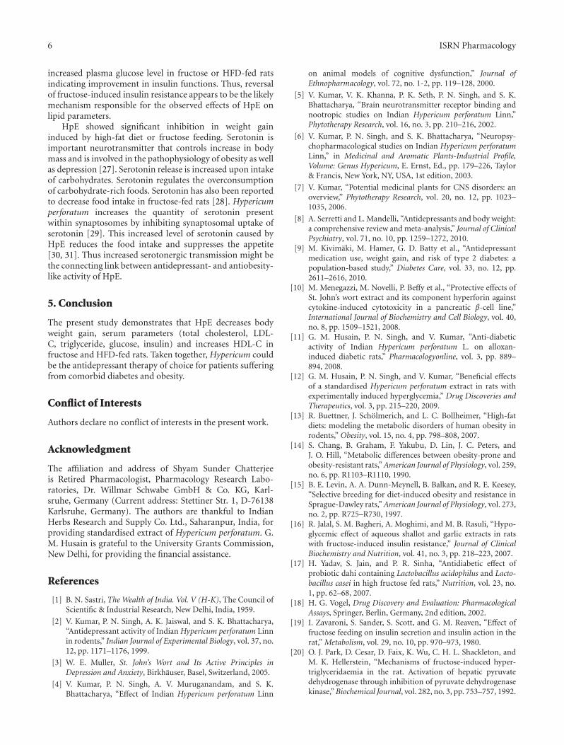
3.2.2. Effect of HpE on Lipid Parameters in Fructose-FedRats. Fructose feeding to rats resulted in impairment innormal lipid profile leading to increased total cholesterol,LDL-C, and triglyceride level while HDL-C was decreased.Triglyceride level of fructose-fed rats increased up to 3times higher than vehicle-treated normal rats. HpE dosedependently and significantly decreased total cholesterol(F(3, 20) = 13.05; P < .01) and TG level (F(3, 20) = 77.06;P < .001) while HDL-C was increased compared to vehicle-treated fructose fed rats (F(3, 20) = 10.66; P < .01). Resultsare summarized in Table 3.
3.2.3. Effect of HpE on Body Weight in Fructose-Fed Rats.Fructose-fed rats significantly gained weight compared tonormal rats (F(5, 30) = 8.18; P < .001). The oraladministration of HpE for 15 days did not alter the bodyweight of rats compared to vehicle-treated control rats.HpE administration reduced body weight gain inducedby fructose feeding; however, data remained statisticallyinsignificant (Table 2).
3.3. High-Fat-Diet-Induced Obesity
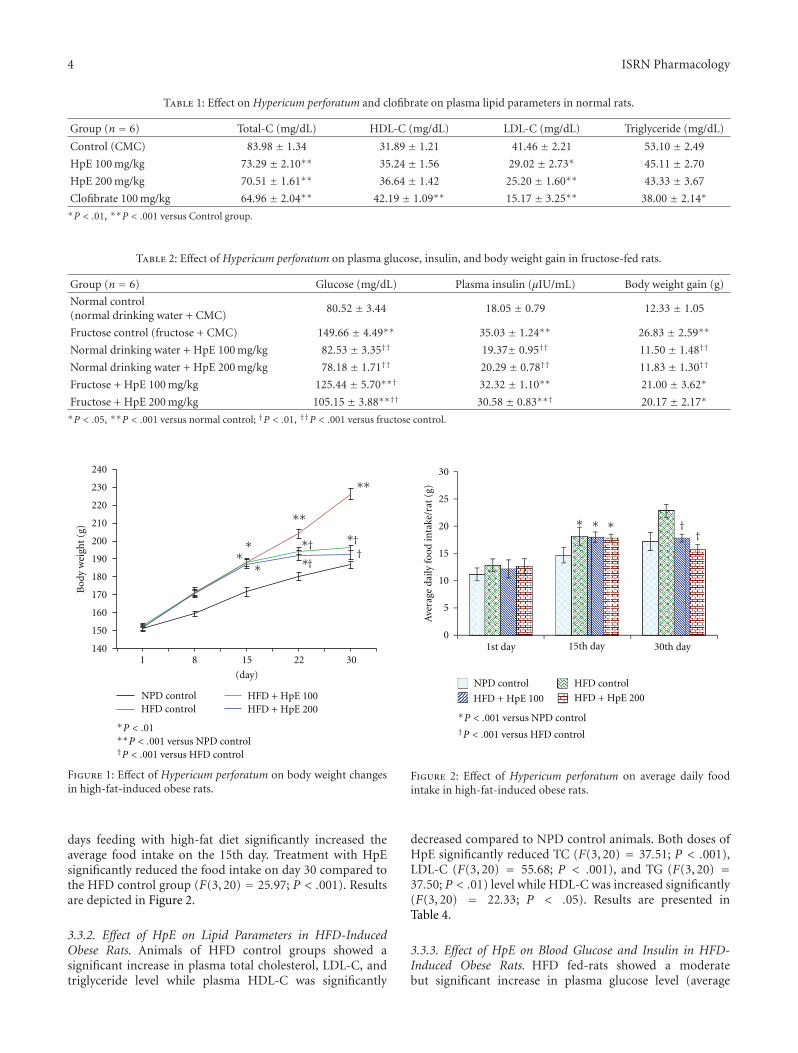
3.3.1. Effect of HpE on Body Weight and Food Intake inHFD-Induced Obese Rats. There was no significant differ-ence in body weight of different treatment groups at thecommencement of study. Animals fed with high-fat dietshowed significant increase in body weight compared tothose fed with NPD. HpE dose dependently and significantlyinhibited the increase in body weight induced by high-fat diet (F(3, 20) = 61.21; P < .001). On day 30, therewas no statistically significant difference in body weight of200 mg/kg HpE-treated HFD-fed rats and NPD control rats(Figure 1). The average daily food intake of all the groupswas the same at the commencement of study; however, 15
4 ISRN Pharmacology
Table 1: Effect on Hypericum perforatum and clofibrate on plasma lipid parameters in normal rats.
Group (n = 6) Total-C (mg/dL) HDL-C (mg/dL) LDL-C (mg/dL) Triglyceride (mg/dL)
Control (CMC) 83.98 ± 1.34 31.89 ± 1.21 41.46 ± 2.21 53.10 ± 2.49
HpE 100 mg/kg 73.29 ± 2.10∗∗ 35.24 ± 1.56 29.02 ± 2.73∗ 45.11 ± 2.70
HpE 200 mg/kg 70.51 ± 1.61∗∗ 36.64 ± 1.42 25.20 ± 1.60∗∗ 43.33 ± 3.67
Clofibrate 100 mg/kg 64.96 ± 2.04∗∗ 42.19 ± 1.09∗∗ 15.17 ± 3.25∗∗ 38.00 ± 2.14∗
∗P < .01, ∗∗P < .001 versus Control group.
Table 2: Effect of Hypericum perforatum on plasma glucose, insulin, and body weight gain in fructose-fed rats.
Group (n = 6) Glucose (mg/dL) Plasma insulin (μIU/mL) Body weight gain (g)
Normal control(normal drinking water + CMC)
80.52 ± 3.44 18.05 ± 0.79 12.33 ± 1.05
Fructose control (fructose + CMC) 149.66 ± 4.49∗∗ 35.03 ± 1.24∗∗ 26.83 ± 2.59∗∗
Normal drinking water + HpE 100 mg/kg 82.53 ± 3.35†† 19.37± 0.95†† 11.50 ± 1.48††
Normal drinking water + HpE 200 mg/kg 78.18 ± 1.71†† 20.29 ± 0.78†† 11.83 ± 1.30††
Fructose + HpE 100 mg/kg 125.44 ± 5.70∗∗† 32.32 ± 1.10∗∗ 21.00 ± 3.62∗
Fructose + HpE 200 mg/kg 105.15 ± 3.88∗∗†† 30.58 ± 0.83∗∗† 20.17 ± 2.17∗
∗P < .05, ∗∗P < .001 versus normal control; †P < .01, ††P < .001 versus fructose control.
Figure 1: Effect of Hypericum perforatum on body weight changesin high-fat-induced obese rats.
days feeding with high-fat diet significantly increased theaverage food intake on the 15th day. Treatment with HpEsignificantly reduced the food intake on day 30 compared tothe HFD control group (F(3, 20) = 25.97; P < .001). Resultsare depicted in Figure 2.
3.3.2. Effect of HpE on Lipid Parameters in HFD-InducedObese Rats. Animals of HFD control groups showed asignificant increase in plasma total cholesterol, LDL-C, andtriglyceride level while plasma HDL-C was significantly
0
5
10
15
20
25
30
1st day 15th day 30th day
Ave
rage
daily
food
inta
ke/r
at(g
)
NPD control HFD control
HFD + HpE 100 HFD + HpE 200
P < .001 versus NPD control
P < .001 versus HFD control
Figure 2: Effect of Hypericum perforatum on average daily foodintake in high-fat-induced obese rats.
decreased compared to NPD control animals. Both doses ofHpE significantly reduced TC (F(3, 20) = 37.51; P < .001),LDL-C (F(3, 20) = 55.68; P < .001), and TG (F(3, 20) =37.50; P < .01) level while HDL-C was increased significantly(F(3, 20) = 22.33; P < .05). Results are presented inTable 4.
3.3.3. Effect of HpE on Blood Glucose and Insulin in HFD-Induced Obese Rats. HFD fed-rats showed a moderatebut significant increase in plasma glucose level (average
ISRN Pharmacology 5
Table 3: Effect on Hypericum perforatum on plasma lipid parameters in fructose-fed rats.
Group (n = 6) Total-C (mg/dL) HDL-C (mg/dL) LDL-C (mg/dL) Triglyceride (mg/dL)
Normal control(normal drinking water + CMC)
81.68 ± 2.36 36.21 ± 1.41 35.98 ± 1.65 47.48 ± 3.15
Fructose control (fructose + CMC) 117.12 ± 5.12∗∗∗ 27.38 ± 1.35∗∗∗ 62.04 ± 4.89∗∗ 138.52 ± 4.88∗∗∗
Fructose + HpE 100 mg/kg 106.01 ± 4.35∗∗ 30.12 ± 0.92∗∗ 57.25 ± 5.57∗∗ 93.24 ± 4.12∗∗∗††
Fructose + HpE 200 mg/kg 96.99 ± 4.25∗† 33.69 ± 1.01† 47.21 ± 4.84 80.50 ± 4.80∗∗∗††
∗P < .05, ∗∗P < .01, ∗∗∗P < .001 versus normal control; †P < .01, ††P < .001 versus fructose control.
Table 4: Effect of Hypericum perforatum on plasma lipid parameters in high-fat-induced obese rats.
Group (n = 6) Total-C (mg/dL) HDL-C (mg/dL) LDL-C (mg/dL) Triglyceride (mg/dL)
Normal control (NPD + CMC) 53.60 ± 2.27 34.65 ± 0.92 33.55 ± 3.07 53.60 ± 2.27
HFD control (HFD + CMC) 105.03 ± 4.87∗∗ 24.62 ± 0.79∗∗ 116.49 ± 6.68∗∗ 105.03 ± 4.87∗∗
HFD + HpE 100 mg/kg 86.27 ± 2.51∗∗†† 28.24 ± 0.89∗∗† 80.72 ± 3.83∗∗††† 86.27 ± 2.51∗∗††
HFD + HpE 200 mg/kg 80.42 ± 3.61∗∗††† 30.19 ± 0.93∗††† 62.85 ± 4.21∗∗††† 80.40 ± 3.61∗∗†††
∗P < .01, ∗∗P < .001 versus normal control; †P < .05, ††P < .01, †††P < .001 versus HFD control.
NPD control HFD control
HFD + HpE 100 HFD + HpE 200
0
1
2
3
4
5
6
7
8
9
Mesenteric Perirenal Epididymal Total
Ave
rage
daily
food
inta
ke/r
at(g
)
∗†
P < .01 P < .001 versus Normal control
P < .01 P < .001 versus HFD control
Figure 3: Effect of Hypericum perforatum on various adipose tissuesin high-fat-induced obese rats.
plasma glucose level = 130 mg/dL) a long with a significantincrease in plasma insulin level compared to vehicle-treatednormal control rats. HpE dose dependently and significantlyinhibited the increase in plasma glucose caused by high-fatdiet (F(3, 20) = 34.01; P < .001) along with a simultaneousdecrease in plasma insulin level (F(3, 20) = 88.02; P < .001)compared to vehicle-treated HFD control group (Table 5).
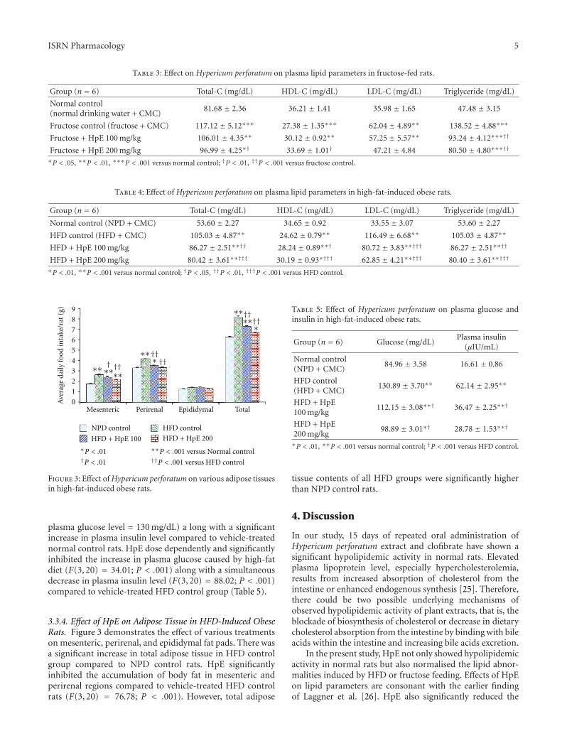
3.3.4. Effect of HpE on Adipose Tissue in HFD-Induced ObeseRats. Figure 3 demonstrates the effect of various treatmentson mesenteric, perirenal, and epididymal fat pads. There wasa significant increase in total adipose tissue in HFD controlgroup compared to NPD control rats. HpE significantlyinhibited the accumulation of body fat in mesenteric andperirenal regions compared to vehicle-treated HFD controlrats (F(3, 20) = 76.78; P < .001). However, total adipose
Table 5: Effect of Hypericum perforatum on plasma glucose andinsulin in high-fat-induced obese rats.
Group (n = 6) Glucose (mg/dL)Plasma insulin
(μIU/mL)
Normal control(NPD + CMC)
84.96 ± 3.58 16.61 ± 0.86
HFD control(HFD + CMC)
130.89 ± 3.70∗∗ 62.14 ± 2.95∗∗
HFD + HpE100 mg/kg
112.15 ± 3.08∗∗† 36.47 ± 2.25∗∗†
HFD + HpE200 mg/kg
98.89 ± 3.01∗† 28.78 ± 1.53∗∗†
∗P < .01, ∗∗P < .001 versus normal control; †P < .001 versus HFD control.
tissue contents of all HFD groups were significantly higherthan NPD control rats.
4. Discussion
In our study, 15 days of repeated oral administration ofHypericum perforatum extract and clofibrate have shown asignificant hypolipidemic activity in normal rats. Elevatedplasma lipoprotein level, especially hypercholesterolemia,results from increased absorption of cholesterol from theintestine or enhanced endogenous synthesis [25]. Therefore,there could be two possible underlying mechanisms ofobserved hypolipidemic activity of plant extracts, that is, theblockade of biosynthesis of cholesterol or decrease in dietarycholesterol absorption from the intestine by binding with bileacids within the intestine and increasing bile acids excretion.
In the present study, HpE not only showed hypolipidemicactivity in normal rats but also normalised the lipid abnor-malities induced by HFD or fructose feeding. Effects of HpEon lipid parameters are consonant with the earlier findingof Laggner et al. [26]. HpE also significantly reduced the
6 ISRN Pharmacology
increased plasma glucose level in fructose or HFD-fed ratsindicating improvement in insulin functions. Thus, reversalof fructose-induced insulin resistance appears to be the likelymechanism responsible for the observed effects of HpE onlipid parameters.
HpE showed significant inhibition in weight gaininduced by high-fat diet or fructose feeding. Serotonin isimportant neurotransmitter that controls increase in bodymass and is involved in the pathophysiology of obesity as wellas depression [27]. Serotonin release is increased upon intakeof carbohydrates. Serotonin regulates the overconsumptionof carbohydrate-rich foods. Serotonin has also been reportedto decrease food intake in fructose-fed rats [28]. Hypericumperforatum increases the quantity of serotonin presentwithin synaptosomes by inhibiting synaptosomal uptake ofserotonin [29]. This increased level of serotonin caused byHpE reduces the food intake and suppresses the appetite[30, 31]. Thus increased serotonergic transmission might bethe connecting link between antidepressant- and antiobesity-like activity of HpE.
5. Conclusion
The present study demonstrates that HpE decreases bodyweight gain, serum parameters (total cholesterol, LDL-C, triglyceride, glucose, insulin) and increases HDL-C infructose and HFD-fed rats. Taken together, Hypericum couldbe the antidepressant therapy of choice for patients sufferingfrom comorbid diabetes and obesity.
Conflict of Interests
Authors declare no conflict of interests in the present work.
Acknowledgment
The affiliation and address of Shyam Sunder Chatterjeeis Retired Pharmacologist, Pharmacology Research Labo-ratories, Dr. Willmar Schwabe GmbH & Co. KG, Karl-sruhe, Germany (Current address: Stettiner Str. 1, D-76138Karlsruhe, Germany). The authors are thankful to IndianHerbs Research and Supply Co. Ltd., Saharanpur, India, forproviding standardised extract of Hypericum perforatum. G.M. Husain is grateful to the University Grants Commission,New Delhi, for providing the financial assistance.
References
[1] B. N. Sastri, The Wealth of India. Vol. V (H-K), The Council ofScientific & Industrial Research, New Delhi, India, 1959.
[2] V. Kumar, P. N. Singh, A. K. Jaiswal, and S. K. Bhattacharya,“Antidepressant activity of Indian Hypericum perforatum Linnin rodents,” Indian Journal of Experimental Biology, vol. 37, no.12, pp. 1171–1176, 1999.
[3] W. E. Muller, St. John’s Wort and Its Active Principles inDepression and Anxiety, Birkhauser, Basel, Switzerland, 2005.
[4] V. Kumar, P. N. Singh, A. V. Muruganandam, and S. K.Bhattacharya, “Effect of Indian Hypericum perforatum Linn
on animal models of cognitive dysfunction,” Journal ofEthnopharmacology, vol. 72, no. 1-2, pp. 119–128, 2000.
[5] V. Kumar, V. K. Khanna, P. K. Seth, P. N. Singh, and S. K.Bhattacharya, “Brain neurotransmitter receptor binding andnootropic studies on Indian Hypericum perforatum Linn,”Phytotherapy Research, vol. 16, no. 3, pp. 210–216, 2002.
[6] V. Kumar, P. N. Singh, and S. K. Bhattacharya, “Neuropsy-chopharmacological studies on Indian Hypericum perforatumLinn,” in Medicinal and Aromatic Plants-Industrial Profile,Volume: Genus Hypericum, E. Ernst, Ed., pp. 179–226, Taylor& Francis, New York, NY, USA, 1st edition, 2003.
[7] V. Kumar, “Potential medicinal plants for CNS disorders: anoverview,” Phytotherapy Research, vol. 20, no. 12, pp. 1023–1035, 2006.
[8] A. Serretti and L. Mandelli, “Antidepressants and body weight:a comprehensive review and meta-analysis,” Journal of ClinicalPsychiatry, vol. 71, no. 10, pp. 1259–1272, 2010.
[9] M. Kivimaki, M. Hamer, G. D. Batty et al., “Antidepressantmedication use, weight gain, and risk of type 2 diabetes: apopulation-based study,” Diabetes Care, vol. 33, no. 12, pp.2611–2616, 2010.
[10] M. Menegazzi, M. Novelli, P. Beffy et al., “Protective effects ofSt. John’s wort extract and its component hyperforin againstcytokine-induced cytotoxicity in a pancreatic β-cell line,”International Journal of Biochemistry and Cell Biology, vol. 40,no. 8, pp. 1509–1521, 2008.
[11] G. M. Husain, P. N. Singh, and V. Kumar, “Anti-diabeticactivity of Indian Hypericum perforatum L. on alloxan-induced diabetic rats,” Pharmacologyonline, vol. 3, pp. 889–894, 2008.
[12] G. M. Husain, P. N. Singh, and V. Kumar, “Beneficial effectsof a standardised Hypericum perforatum extract in rats withexperimentally induced hyperglycemia,” Drug Discoveries andTherapeutics, vol. 3, pp. 215–220, 2009.
[13] R. Buettner, J. Scholmerich, and L. C. Bollheimer, “High-fatdiets: modeling the metabolic disorders of human obesity inrodents,” Obesity, vol. 15, no. 4, pp. 798–808, 2007.
[14] S. Chang, B. Graham, F. Yakubu, D. Lin, J. C. Peters, andJ. O. Hill, “Metabolic differences between obesity-prone andobesity-resistant rats,” American Journal of Physiology, vol. 259,no. 6, pp. R1103–R1110, 1990.
[15] B. E. Levin, A. A. Dunn-Meynell, B. Balkan, and R. E. Keesey,“Selective breeding for diet-induced obesity and resistance inSprague-Dawley rats,” American Journal of Physiology, vol. 273,no. 2, pp. R725–R730, 1997.
[16] R. Jalal, S. M. Bagheri, A. Moghimi, and M. B. Rasuli, “Hypo-glycemic effect of aqueous shallot and garlic extracts in ratswith fructose-induced insulin resistance,” Journal of ClinicalBiochemistry and Nutrition, vol. 41, no. 3, pp. 218–223, 2007.
[17] H. Yadav, S. Jain, and P. R. Sinha, “Antidiabetic effect ofprobiotic dahi containing Lactobacillus acidophilus and Lacto-bacillus casei in high fructose fed rats,” Nutrition, vol. 23, no.1, pp. 62–68, 2007.
[18] H. G. Vogel, Drug Discovery and Evaluation: PharmacologicalAssays, Springer, Berlin, Germany, 2nd edition, 2002.
[19] I. Zavaroni, S. Sander, S. Scott, and G. M. Reaven, “Effect offructose feeding on insulin secretion and insulin action in therat,” Metabolism, vol. 29, no. 10, pp. 970–973, 1980.
[20] O. J. Park, D. Cesar, D. Faix, K. Wu, C. H. L. Shackleton, andM. K. Hellerstein, “Mechanisms of fructose-induced hyper-triglyceridaemia in the rat. Activation of hepatic pyruvatedehydrogenase through inhibition of pyruvate dehydrogenasekinase,” Biochemical Journal, vol. 282, no. 3, pp. 753–757, 1992.
ISRN Pharmacology 7
[21] T. Nakagawa, H. Hu, S. Zharikov et al., “A causal role foruric acid in fructose-induced metabolic syndrome,” AmericanJournal of Physiology, vol. 290, no. 3, pp. F625–F631, 2006.
[22] W. T. Friedewald, R. I. Levy, and D. S. Fredrickson, “Estima-tion of the concentration of low-density lipoprotein choles-terol in plasma, without use of the preparative ultracen-trifuge,” Clinical Chemistry, vol. 18, no. 6, pp. 499–502, 1972.
[23] K. Srinivasan, B. Viswanad, L. Asrat, C. L. Kaul, and P.Ramarao, “Combination of high-fat diet-fed and low-dosestreptozotocin-treated rat: a model for type 2 diabetes andpharmacological screening,” Pharmacological Research, vol. 52,no. 4, pp. 313–320, 2005.
[24] L. K. Han, Y. Kimura, and H. Okuda, “Anti-obesity effects ofnatural products,” in Studies in Natural Products Chemistry,Atta-ur-Rahman, Ed., vol. 30, pp. 79–110, 2005.
[25] E. Ikonen, “Mechanisms for cellular cholesterol transport:defects and human disease,” Physiological Reviews, vol. 86, no.4, pp. 1237–1261, 2006.
[26] H. Laggner, S. Schreier, M. Hermann et al., “The maincomponents of St John’s Wort inhibit low-density lipoproteinatherogenic modification: a beneficial “side effect” of an OTCantidepressant drug?” Free Radical Research, vol. 41, no. 2, pp.234–241, 2007.
[27] H. P. Chudasama and P. A. Bhatt, “Evaluation of anti-obesityactivity of duloxetine in comparison with sibutramine alongwith its anti-depressant activity: an experimental study inobese rats,” Canadian Journal of Physiology and Pharmacology,vol. 87, no. 11, pp. 900–907, 2009.
[28] S. H. Hsiao, H. H. Chung, Y. C. Tong, and J. T. Cheng,“Chronic fluoxetine administration desensitizes the hyper-glycemia but not the anorexia induced by serotonin in ratsreceiving fructose-enriched chow,” Neuroscience Letters, vol.404, no. 1-2, pp. 6–8, 2006.
[29] W. E. Muller, “Effects of hypericum extract (Ll 160) inbiochemical models of antidepressant activity,” Pharmacopsy-chiatry, vol. 30, supplement 2, pp. 102–107, 1997.
[30] A. G. Brasswell and A. J. Ahmed, “Method for controllingweight with Hypericum perforatum and Garcinia cambogia,”US patent no. 5911992, 1999.
[31] I. Yegorova and D. Jiang, “Compositions and methods forregulating metabolism and balancing body weight,” US patentno. 6399089, 2002.

Submit your manuscripts athttp://www.hindawi.com
PainResearch and TreatmentHindawi Publishing Corporationhttp://www.hindawi.com Volume 2014
The Scientific World JournalHindawi Publishing Corporation http://www.hindawi.com Volume 2014
Hindawi Publishing Corporationhttp://www.hindawi.com
Volume 2014
ToxinsJournal of
VaccinesJournal of
Hindawi Publishing Corporation http://www.hindawi.com Volume 2014
Hindawi Publishing Corporationhttp://www.hindawi.com Volume 2014
AntibioticsInternational Journal of
ToxicologyJournal of
Hindawi Publishing Corporationhttp://www.hindawi.com Volume 2014
StrokeResearch and TreatmentHindawi Publishing Corporationhttp://www.hindawi.com Volume 2014
Drug DeliveryJournal of
Hindawi Publishing Corporationhttp://www.hindawi.com Volume 2014
Hindawi Publishing Corporationhttp://www.hindawi.com Volume 2014
Advances in Pharmacological Sciences
Tropical MedicineJournal of
Hindawi Publishing Corporationhttp://www.hindawi.com Volume 2014
Medicinal ChemistryInternational Journal of
Hindawi Publishing Corporationhttp://www.hindawi.com Volume 2014
AddictionJournal of
Hindawi Publishing Corporationhttp://www.hindawi.com Volume 2014
Hindawi Publishing Corporationhttp://www.hindawi.com Volume 2014
BioMed Research International
Emergency Medicine InternationalHindawi Publishing Corporationhttp://www.hindawi.com Volume 2014
Hindawi Publishing Corporationhttp://www.hindawi.com Volume 2014
Autoimmune Diseases
Hindawi Publishing Corporationhttp://www.hindawi.com Volume 2014
Anesthesiology Research and Practice
ScientificaHindawi Publishing Corporationhttp://www.hindawi.com Volume 2014
Journal of
Hindawi Publishing Corporationhttp://www.hindawi.com Volume 2014
Pharmaceutics
Hindawi Publishing Corporationhttp://www.hindawi.com Volume 2014
MEDIATORSINFLAMMATION
of


