Embed Size (px)
Citation preview
IntroductionCardiac myocyte cell death by apoptosis accompaniesheart disease of both ischemic and nonischemic origin(reviewed in refs. 1–3). It has been demonstrated in themyocardium from failing human hearts (4–7), in patientswith arrhythmogenic right ventricular dysplasia (8), andin association with myocardial infarction, both withinthe infarcted area itself and in the surrounding viable tis-sue (5, 9–12). In animal models, increased apoptosisaccompanies pacing-induced dilated cardiomyopathy(13, 14), pressure-overload hypertrophy (15, 16), hyper-tension (17), hibernating myocardium (18), and bothphases of ischemia and reperfusion (3). The precise initi-ating stimuli, signaling pathways, and mechanisms ofapoptosis are not understood.
A number of factors may contribute to ischemia-mediated cell death (19–21). Hypoxia causes inhibitionof oxidative phosphorylation and a switch to glycolyt-ic metabolism, resulting in decreased levels of high-energy phosphates, increased lactic acid production,and lower intracellular pH ([pH]i) (22–24). If ATP israpidly depleted, necrosis will occur because of passiveloss of transmembrane ion gradients, followed by cell
swelling and loss of membrane integrity (19–21, 25).Unlike necrosis, apoptosis is an active, energy-consum-ing process that must be executed in the presence ofsufficient cellular ATP (26), and one in which mem-brane integrity is preserved until very late stages. It iswidely accepted that apoptosis accounts for some ofthe cell death observed after myocardial infarction,although the relative contributions of necrosis andapoptosis are unclear (27, 28). Initiating signals forapoptosis of ischemic heart cells may be related to cel-lular energy levels, osmotic/ionic changes, altered geneexpression, altered Ca2+ handling, oxidative stress, andaccumulation of waste metabolites that are normallycleared from the interstitial space.
Recently, it has been suggested that proton pumpsand pH regulation may play a role in apoptosis signal-ing (29–32). Ischemic cardiac myocytes generate excessH+ through increased anaerobic metabolism, nethydrolysis of ATP, and CO2 retention (33). These pro-tons are extruded from the myoplasm to the interstitialspace by the combined action of 3 major ion-specificmembrane transporters, including the Na+/H+
exchanger, the Na+/HCO3– cotransporter, and the vac-
The Journal of Clinical Investigation | August 1999 | Volume 104 | Number 3 239
Hypoxia-activated apoptosis of cardiac myocytes requiresreoxygenation or a pH shift and is independent of p53
Keith A. Webster,1,2 Daryl J. Discher,1 Shari Kaiser,2 Olga Hernandez,1
Barbara Sato,2 and Nanette H. Bishopric1
1Department of Molecular and Cellular Pharmacology, University of Miami Medical Center, Miami, Florida 33136, USA2Pharmaceutical Discovery Division, SRI International, Menlo Park, California, 94025, USA
Address correspondence to: Keith A. Webster, Department of Molecular and Cellular Pharmacology, Rosenstiel Medical Science Building, Room 6038, University of Miami Medical Center, 1600 NW 10th Avenue, Miami, Florida 33136, USA. Phone: (305) 243-6779; Fax: (305) 243-6082; E-mail: [email protected].
A preliminary report of this work has appeared in abstract form (1998. Circulation. 98:I743).
Received for publication November 23, 1998, and accepted in revised form June 24, 1999.
Ischemia and reperfusion activate cardiac myocyte apoptosis, which may be an important featurein the progression of ischemic heart disease. The relative contributions of ischemia and reperfusionto apoptotic signal transduction have not been established. We report here that severe chronichypoxia alone does not cause apoptosis of cardiac myocytes in culture. When rapidly contractingcardiac myocytes were exposed to chronic hypoxia, apoptosis occurred only when there was adecrease in extracellular pH ([pH]o). Apoptosis did not occur when [pH]o was neutralized. Additionof acidic medium from hypoxic cultures or exogenous lactic acid stimulated apoptosis in aerobicmyocytes. Hypoxia-acidosis–mediated cell death was independent of p53: equivalent apoptosisoccurred in cardiac myocytes isolated from wild-type and p53 knockout mice, and hypoxia causedno detectable change in p53 abundance or p53-dependent transcription. Reoxygenation of hypox-ic cardiac myocytes induced apoptosis in 25–30% of the cells and was also independent of p53 by thesame criteria. Finally, equivalent levels of apoptosis, as demonstrated by DNA fragmentation, wereinduced by ischemia-reperfusion, but not by ischemia alone, of Langendorff-perfused hearts fromwild-type and p53 knockout mice. We conclude that acidosis, reoxygenation, and reperfusion, butnot hypoxia (or ischemia) alone, are strong stimuli for programmed cell death that is substantiallyindependent of p53.
J. Clin. Invest. 104:239–252 (1999).
uolar proton ATPase (30, 34, 35). Increased activity ofthe Na+/H+ exchanger can cause Ca2+ overload becausethe elevated intracellular Na+ is subsequentlyexchanged for Ca2+ via the Na+/Ca2+ exchanger (36).Inhibition of Na+/H+ exchange has been shown to pro-tect against ischemic injury, possibly by preventing thisincrease in Ca2+ (37, 38). Conversely, inhibition of thevacuolar ATPase promotes apoptosis, in part by shift-ing the proton load toward the Na+/H+ transporter andthus increasing Ca2+ uptake, and in part by reducingthe myocyte capacity to control [pH]i (29–31). Acidosishas been shown to correlate with apoptosis in a num-ber of other systems (39–41), but its role, if any, inischemic cardiac myocyte apoptosis is not understood.
There are a number of important and controversialquestions about the mechanism of cardiac myocyteapoptosis during ischemia (32). One concerns the initi-ating signals for cell death. Is hypoxia alone sufficientto induce apoptosis, or does it require a combination ofhypoxia and acidosis or accumulation of other extra-cellular waste metabolites? Another question relates tothe role of p53. Increased activity of p53 protein hasbeen reported to accompany apoptosis in response tovolume overload and mechanical stretch (42). In thesemodels, increased levels of angiotensin II inducedexpression of p53, promoting changes in the balancebetween Bcl-2 and Bax in favor of apoptosis (14, 42).Hypoxia has been shown to induce p53 in transformedcells (43) and both p53 and Fas in cardiac myocytes (44,45). p53 mRNA transcript levels can also be induced incardiac myocytes by inhibition of vacuolar ATPase withbafilomycin A (31). However, these studies did notestablish a direct cause-and-effect relationship betweenenhanced p53 expression and hypoxia-mediated apop-tosis of cardiac myocytes. Ischemia has been shown toinduce apoptosis equally well in wild-type and p53-defi-cient mice (46).
The goal of the present study was to evaluate the sep-arate effects of hypoxia, pH, and reoxygenation onapoptosis in actively contracting cardiac myocytes. Weprovide evidence that severe chronic hypoxia is not suf-ficient to cause apoptosis; however, hypoxia associat-ed with acidosis or reoxygenation caused extensiveapoptosis that was independent of changes in theactivity of p53. The results with isolated myocytes invitro were reproduced in an adult mouse model ofischemia and reperfusion.
MethodsReagents. Antibodies to p53, p21, Bax, Bcl-2, and actinwere from Santa Cruz Biotechnology Inc. (Santa Cruz,California, USA), and anti-Bak was from LXR Biotech-nology (Richmond, California, USA). Anti–sarcomericmyosin antibody (MF-20) was obtained from the Devel-opmental Studies Hybridoma Bank at the University ofIowa (Ames, Iowa USA). HOECHST 33342 and propidiumiodide (PI) dyes were purchased from Calbiochem-Nov-abiochem Corp. (San Diego, California, USA). Ad-p53and AdDN-p53 were obtained from F. Graham (Micro-
bix Inc., Ontario, Ontario, Canada). Plasmids p21-Lucand p21-M–Luc, containing the wild-type and deletedp21 promoters, respectively, were provided by W. El-Deiry (Howard Hughes Medical Institute, Philadelphia,Pennsylvania, USA). Other plasmids have been describedpreviously (47). All other reagents were purchased fromSigma Chemical Co. (St. Louis, Missouri, USA).
Cell culture. All procedures involving animals were per-formed in accordance with University of Miami and SRIInternational guidelines for the care and use of animals.Methods for primary culture of neonatal rat cardiacmyocytes have been described previously (48, 49). Inbrief, enriched cultures of myocyte and nonmyocytecells were obtained from 1- to 2-day-old rats by stepwisetrypsin dissociation, and were plated at a density of 4 ×10⁶ cells/60-mm dish or on 2-well glass dishes (NalgeNunc International, Naperville, Illinois, USA) at a den-sity of 4 × 10⁵ cells/cm2, in MEM supplemented with 5%FCS, penicillin, and streptomycin (MEM + 5% FCS).After 3–5 days, cells were rinsed 3 times in MEM andtransferred to a defined serum-free DMEM/M-199 (4:1)medium supplemented with transferrin, vitamin B12,and insulin. The final cultures contained more than95% cardiac myocytes, contracting at greater than 200beats per minute (bpm). Bromodeoxyuridine (BrdU; 0.1mM) was included in the medium for the first 3 daysafter plating to inhibit fibroblast growth. In some exper-iments, nonmyocytes from the same cultures (∼ 95%fibroblasts, with small percentages of smooth muscleand endothelial cells) were used. These cells were usedat passages 2 or 3, maintained in MEM + 5% serum asdescribed previously (49), and transferred to serum-freeDMEM/M-199 (4:1) before experiments.
Hypoxia and reoxygenation. Cultures were placed inserum-free DMEM/M-199 (4:1) containing 3.8 g/L glu-cose and transferrin, insulin, and vitamin B12, 24 hoursbefore exposure to hypoxia. Details of our methods forexposing cells to hypoxia by incubation in an environ-mental chamber have been described previously(48–50). Oxygen inside the chamber was continuouslymonitored with an oxygen electrode (ControlsKatharobic, Philadelphia, Pennsylvania, USA), and con-tractility was monitored by edge detection as describedpreviously (48, 49). The chamber oxygen concentrationwas maintained at greater than 10 mmHg. For replace-ment of the culture medium under hypoxia, freshmedium was incubated under hypoxia at room tem-perature for 24 hours; the medium was brought to37°C; and the pO2 was measured before adding to thedishes. Cultures exposed only to hypoxia (withoutreoxygenation) were lysed under hypoxia using ice-colddeoxygenated buffers. For reoxygenation, plates wereremoved from the chamber and reoxygenated byreplacing the medium with oxygenated medium andincubating under 21% O2 (air/5% CO2). Cells were har-vested and lysed for apoptosis, Western blots, or bio-chemical assays as described later here.
Quantitative analysis of apoptotic nuclei. Cells were exam-ined for morphological evidence of apoptosis or necro-
240 The Journal of Clinical Investigation | August 1999 | Volume 104 | Number 3
sis after staining with the fluorescent DNA-bindingdyes HOECHST 33342 and PI as described previously(51). Treated and control cell monolayers grown onuncoated Nunc 2-well coverslip dishes were rinsed withPBS, stained with 5 µg/mL HOECHST 33342 and 5µg/mL PI for 15 minutes, and viewed ×400 on a ZeissIM fluorescence microscope (Carl Zeiss Inc., Thorn-wood, New York, USA). Cells were scored as apoptoticif they exhibited unequivocal nuclear chromatin con-densation and/or fragmentation; PI-stained cells withnormal nuclear morphology were scored as necrotic. Atthe later time points (72 hours of hypoxia-acidosis),cells with condensed nuclei that costained with PI werealso scored as apoptotic. In some experiments, apop-totic nuclei were localized within cardiac myocytes bystaining with an mAb against sarcomeric myosin (MF-20). Cells were fixed in ice-cold methanol, rinsed, andstained with the anti-myosin antibody and HOECHST
33342, followed by an FITC-tagged anti-mouse IgGsecondary antibody. Cells were imaged and pho-tographed on a Zeiss IM inverted-phase fluorescencemicroscope using a mounted Contax 35-mm cam-era(Yashica, Tokyo, Japan) and ASA 400 Kodak colortransparency film(Eastman Kodak Co., Rochester, NewYork, USA). To quantitate apoptosis, an average of 400nuclei from random fields were analyzed, and apop-totic cell counts were expressed as a percentage of thetotal number of nuclei counted.
Analysis of DNA fragmentation. Cells were lysed for 5hours at 37°C in a buffer containing 100 mM NaCl, 10mM Tris-Cl (pH 8.0), 5 mM EDTA, 0.5% SDS, and 1µg/mL proteinase K; proteins were precipitated with0.8 M NaCl; and DNA was extracted with phenol/chlo-roform and precipitated with an equal volume of iso-propanol. The resulting washed pellet was resuspend-ed in Tris-EDTA buffer and treated with 100 µg/mL ofDNase-free RNase for 30 minutes at 37°C. The DNAcontent was quantitated by spectrophotometry at260/280 nm. Samples (5 µg) were subjected to elec-trophoresis in 2% agarose gels and were imaged byethidium bromide staining and digital photography.In some cases, the extent of DNA fragmentation wasquantified by densitometry of subchromosomal DNAfragments on digitized images using Adobe Photo-shop 4.0 (Adobe Systems Inc., Mountain View, Cali-fornia, USA) for Macintosh.
Western blot analysis. For detection of p53, Bax, Bak,Bcl2, and actin proteins, cells were harvested in ice-coldlysis buffer (50 mM Tris-HCl [pH 7.5], 150 mM NaCl,1% Triton X-100, and 50 mM NaF) with freshly added1 mM Na3VO4, 0.5 mM DTT, 1 mM PMSF, 10 µg/mLleupeptin, and 2 µg/mL aprotinin. Cells were triturat-ed with a pipette tip 10 times, and the resulting lysateswere centrifuged at 14,000 g for 10 minutes to removecell debris. Protein content was determined using aPierce BCA kit (Pierce Chemical Co., Rockford, Illinois,USA). Equal amounts of protein (10–200 µg) were frac-tionated on 12% or 15% SDS-polyacrylamide gels andwere electroblotted to nitrocellulose (Bio-Rad Labora-
tories Inc., Hercules, California, USA). Blots werestained with Ponceau red to monitor the transfer ofproteins. Membranes were blocked for 1 hour at roomtemperature with 5% nonfat milk in TBS (25 mM Tris,137 mM NaCl, and 2.7 mM KCl) containing 0.05%Tween-20, and were incubated with specific antibodiesfor 2–4 hours in the same buffer. After washing, theblots were incubated for 1 hour with 1:7,500 dilutionof horseradish peroxidase–conjugated (HRP-conjugat-ed) anti-rabbit IgG or HRP-conjugated donkey anti-goat IgG, and were viewed using an enhanced chemilu-minescence detection system (Pierce Chemical Co.).
Northern blots. Northern blot procedures were exactlyas described previously (47, 52). Full-length rat p21 andp53 probes were generated by RT-PCR on rat templatemRNA using primers derived from the publishedsequences (53, 54).
Electrophoretic gel mobility shift. Nuclear extracts wereprepared from confluent plates as described previous-ly (55). Sequences of the oligonucleotide probes (sensestrands) were as follows: p53 wild-type, TACAGAA-CATGTCTAAGCATGCTGGGG; p53 mutant, TACA-GAATCTGTCTAAGC ATGCTGGGG; HRE competi-tor, AAAGAGAGGCGGGGCTGGCTGGG (47).Gel-purified double-stranded oligonucleotides wereend labeled with [32P]ATP using T4 polynucleotidekinase (Promega Biotech, Promega Corp., Madison,Wisconsin, USA) and [γ-32P]ATP (Du Pont NENResearch Products, Boston, Massachusetts, USA).Equal amounts of radioactive probe (1.5 × 10⁴ to 2.5 ×10⁴ cpm) were added to binding reactions that con-tained 8 µg of nuclear extract protein in 20 µL of abuffer containing 4 mM Tris (pH 7.8), 12 mM HEPES(pH 7.9), 60 mM KCl, 30 mM NaCl, 0.1 mM EDTA, and1 µg poly(dI-dC) (Amersham Pharmacia Biotech, Pis-cataway, New Jersey, USA). Reactions were incubatedfor 20 minutes at 22°C before separating on nondena-turing 5% polyacrylamide gels at 4°C. CompetitorDNAs were added in 200-fold excess immediatelybefore the radioactive probe. Proteins were determinedwith a Pierce BCA kit.
Transient expression. Cardiac myocytes were trans-fected the day after isolation using calcium phos-phate as described previously (47, 49). Plates (60 mm)were transfected with 9 µg of the test plasmid (p21-Luc, p21-M–Luc, pα-MHC–HRE-Luc, and Rous sar-coma virus-Luc [RSV-Luc]) and 1 µg of pTK-RN(Promega Corp.) as the internal control to correct forvariations in transfection efficiency. For adenoviralinfections, transfected cells were infected with virusat 10 plaque-forming units (PFU) per cell the dayafter transfection. Plates were exposed to hypoxia orair after an additional 2–3 days. Adenoviruses infect-ed 100% of the cells under these conditions, as deter-mined by X-gal staining after infection with Ad–β-gal, and wild-type and mutant p53 viruses generatedequivalent levels of protein expression by Westernblots (data not shown; see Figure 7). Cultures wereharvested after treatments and lysed for luciferase
The Journal of Clinical Investigation | August 1999 | Volume 104 | Number 3 241
assays. Equal amounts of protein were assayed forexpression of luciferase using the Promega dual-luciferase reporter assay system according to themanufacturer’s protocol. Protein was assayed using aBio-Rad assay kit.
p53–/– knockout mice. Mice heterozygous for a disrup-tion in the p53 gene locus (56) were a kind gift from R.Kitsis (Albert Einstein College of Medicine, Bronx,New York, USA), and were backcrossed into C57BL/6wild-type mice. Offspring of heterozygous pairs weregenotyped by PCR analysis of tail DNA by PCR withprimers X6.5 (5′-ACAGCGTGGTACCTTAT-3′) and X7(5′-TATACTCAGAGCCGGCCT-3′) to amplify the endogenous allele, and primers neo 18.5 (5′-TCCTCGTGCTTTACGGTATC-3′) and X7 to amplifythe disrupted allele. Homozygous offspring were bred
to produce F1 litters of p53–/– pups, which were used togenerate neonatal mouse cardiac myocyte culturesexactly as already described here for the neonatal rat.Wild-type C57BL/6 litters were used as controls.
Langendorff perfusions. Mouse hearts were perfused bythe Langendorff method essentially as described previ-ously (57). Briefly, C57BL/6 or p53 knockouts, asdescribed here (25–35 g body weight), were anesthetizedwith pentobarbital (60 mg/kg intraperitoneally). Thechests were opened, and the hearts were rapidly excisedand placed in iced Krebs-Henseleit buffer (KHB) (4°C).Thymic and fatty tissues were carefully trimmed toreveal the ascending aorta, which was then cannulatedwith a blunted 20-gauge needle. During transfer to theLangendorff apparatus, the cannula was perfused toavoid air entry. The heart was attached to the apparatus
242 The Journal of Clinical Investigation | August 1999 | Volume 104 | Number 3
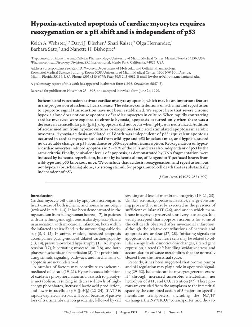
Figure 1Contributions of waste metabolic buildup to apoptosis induced by chronic hypoxia. (a and b) Parallel cultures of cardiac myocytes wereexposed to hypoxia as described in Methods. In a, the medium was replaced with fresh hypoxic medium every 12 hours; in b, there was nomedium replacement. Cultures were harvested at the indicated times and processed for DNA fragmentation. (c) Intracellular ATP, mediumglucose, and [pH]o were measured in parallel cultures as described in Methods; results are means of 3 separate experiments. Open circlesare results from cultures without medium replacement; closed circles, with medium replacement. (d–f) Typical fields of myocytes stainedwith HOECHST 33342 and anti-myosin antibody as described in Methods. (g) Quantitations of HOECHST-stained condensed nuclei, alsodescribed in Methods. At 24 and 48 hours, less than 2% of cells were PI positive (scored as necrotic) under any condition; at 72 hours, hypox-ia-acidic cultures had more PI-positive cells, and these were scored as apoptotic if they contained condensed nuclei. Costaining populationswere not distinguished from PI-excluding cells at this stage. Results are representative of at least 3 experiments.
and retrograde perfused at constant pressure (80mmHg) with a flow rate of 2–4 mL/min. The time fromexcision of the heart to commencement of perfusiondid not exceed 5 minutes. The perfusion buffer consistsof a phosphate-free KHB as follows (mM): 118 NaCl, 24KCl, 2.5 CaCl2, 1.2 MgSO4, 0.5 EDTA, 25 NaHCO3, 0.5pyruvate, and 10 glucose. The KHB buffer was equili-brated at 37°C with 5% CO2/95% O2 (pH 7.4), and theLangendorff apparatus was housed in a temperature-controlled (37°C) and humidified incubator. Any heartsin which coronary flow was excessive (>5 mL/min;indicative of an aortic tear), that contracted at morethan 300 bpm, or that maintained persistent arrhyth-mias after stabilization were excluded from furtherstudy. Before experimentation, all hearts were exposedto an initial 30-minute stabilization period. For con-trols, hearts were perfused for 3 additional hours;ischemia consisted of no flow for the periods indicated;ischemia-reperfusion entailed a 20-minute no-flow withreperfusion for the indicated times. At the end of eachexperiment, hearts were frozen at –95°C.
Isolation of DNA from perfused hearts. The left ventriclewas removed from frozen hearts, sliced into approxi-mately 2-mm squares, and digested for 16 hours at37°C in a lysis buffer containing 10% SDS, 10 mM Tris-HCl (pH 7.8), 1 mM EDTA, and 200 µg/mL proteinaseK. The DNA was purified by twice extracting with phe-nol/chloroform and precipitating with isopropanol.The DNA pellet was rinsed with 70% ethanol and resus-pended in Tris-EDTA buffer. DNA samples were sub-jected to electrophoresis as described earlier here.
Biochemical assays. ATP and glucose were measured asdescribed previously (48, 49), and pH was measureddirectly in the culture medium using an Orion meterwith a micro Ross electrode (Orion Research Inc., NewSouth Wales, Australia).
Statistical analysis. Results are expressed as mean ±SEM. Differences between means were evaluated by 2-tailed Student’s t test. ANOVA was carried out usingInStat 2.0 (GraphPad Software for Science Inc., SanDiego, California, USA) for Macintosh.
ResultsCorrelation of hypoxia-mediated apoptosis with change inextracellular pH. We reported previously that rapidlycontracting cardiac myocytes remained fully viable andcontractile during culture under severe hypoxia for upto 6 days (48). Glycolysis was induced approximately10-fold within 1 hour, and there was no evidence ofmajor cell loss. The intracellular ATP of hypoxicmyocytes dropped to about 70% of control plates, anda slightly reduced contractility correlated with lowerintracellular cAMP in the hypoxic cultures. Theseresults contrast with more recent reports of significantcardiac myocyte cell loss by apoptosis after 48–72 hoursof exposure to an equivalent degree of hypoxia (44, 45).To investigate this apparent discrepancy, we exposedcultures of myocytes to 2 different hypoxic regimensand measured the levels of apoptosis by DNA ladders
and HOECHST staining. In the first set, the cultures weremaintained continuously under hypoxia, and the medi-um was replaced twice daily with fresh hypoxic medi-um to prevent the buildup of waste metabolites (48). Inthe second set, the cultures were maintained in paral-lel, but the medium was not replaced. The cultures werealso monitored for ATP, glucose, and extracellular pH([pH]o) as described previously (48, 58) and in Methods.The results are shown in Figure 1, a and b. There wasno evidence of DNA fragmentation under the first setof conditions (Figure 1a), but cultures subjected tohypoxia without medium changes showed significantDNA laddering after 48 hours and extensive ladderingafter 72 hours (Figure 1b). In agreement with our pre-
The Journal of Clinical Investigation | August 1999 | Volume 104 | Number 3 243
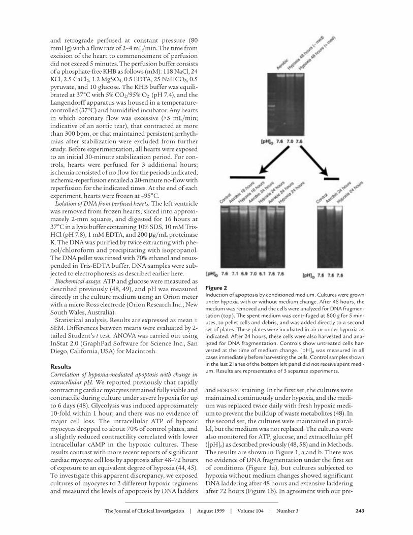
Figure 2Induction of apoptosis by conditioned medium. Cultures were grownunder hypoxia with or without medium change. After 48 hours, themedium was removed and the cells were analyzed for DNA fragmen-tation (top). The spent medium was centrifuged at 800 g for 5 min-utes, to pellet cells and debris, and was added directly to a secondset of plates. These plates were incubated in air or under hypoxia asindicated. After 24 hours, these cells were also harvested and ana-lyzed for DNA fragmentation. Controls show untreated cells har-vested at the time of medium change. [pH]o was measured in allcases immediately before harvesting the cells. Control samples shownin the last 2 lanes of the bottom left panel did not receive spent medi-um. Results are representative of 3 separate experiments.
vious observations, when the medium was replacedtwice daily, ATP levels dropped to about 75% of aerobiccontrol levels after 24 hours and remained stable there-after; [pH]o did not change, and glucose levelsremained high (Figure 1c). Under these conditions, themyocytes continued to contract for the duration of theexperiment, as reported previously, and there was nosignificant loss of total DNA or protein (data notshown; ref. 48). When the medium was not replaced,intracellular ATP levels were sustained up to 48 hoursbut dropped dramatically at 72 hours, coincident withthe loss of most of the cells by apoptosis. Glucosedeclined progressively over the 72-hour period but was
not depleted, and [pH]o declined steadily to a finalvalue of about 6.0 (Figure 1c). Under these conditions,contractions ceased before 48 hours (data not shown).
Cardiac myocytes were grown on glass coverslips andwere fixed and double stained with HOECHST 33342 andanti-myosin antibody (Figure 1, d–f). In cultures grownaerobically, abundant myofilaments with clear cross-striations were evident with the myosin stain (Figure 1d,right panel). As indicated by the white arrows, most ofthe nuclei were oval, and there was sparse evidence ofcondensation or internal fragmentation. In this field, 29nuclei were scored normal and 1 was condensed; 25nuclei were localized within cells that were myosin pos-
244 The Journal of Clinical Investigation | August 1999 | Volume 104 | Number 3
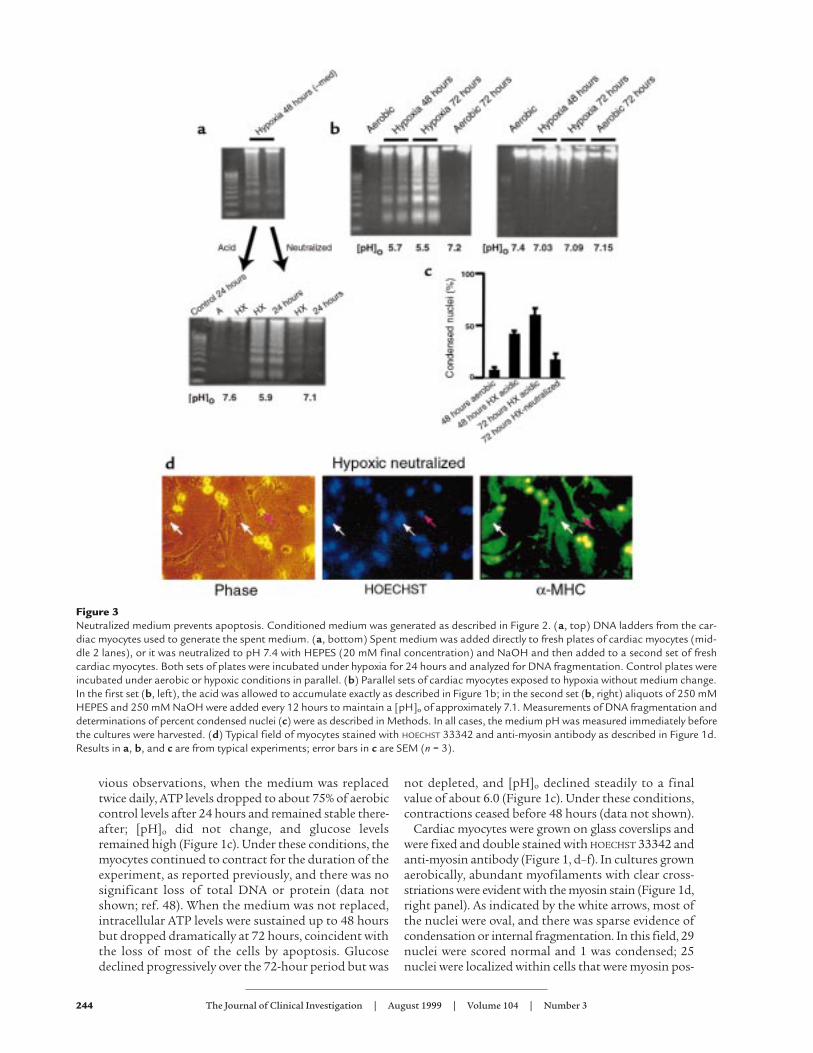
Figure 3Neutralized medium prevents apoptosis. Conditioned medium was generated as described in Figure 2. (a, top) DNA ladders from the car-diac myocytes used to generate the spent medium. (a, bottom) Spent medium was added directly to fresh plates of cardiac myocytes (mid-dle 2 lanes), or it was neutralized to pH 7.4 with HEPES (20 mM final concentration) and NaOH and then added to a second set of freshcardiac myocytes. Both sets of plates were incubated under hypoxia for 24 hours and analyzed for DNA fragmentation. Control plates wereincubated under aerobic or hypoxic conditions in parallel. (b) Parallel sets of cardiac myocytes exposed to hypoxia without medium change.In the first set (b, left), the acid was allowed to accumulate exactly as described in Figure 1b; in the second set (b, right) aliquots of 250 mMHEPES and 250 mM NaOH were added every 12 hours to maintain a [pH]o of approximately 7.1. Measurements of DNA fragmentation anddeterminations of percent condensed nuclei (c) were as described in Methods. In all cases, the medium pH was measured immediately beforethe cultures were harvested. (d) Typical field of myocytes stained with HOECHST 33342 and anti-myosin antibody as described in Figure 1d.Results in a, b, and c are from typical experiments; error bars in c are SEM (n = 3).
itive. At 72 hours, the unfed hypoxic cultures (Figure 1e)still stained strongly with myosin antibody, but therewas clear deterioration of the myofilaments, and cross-striations were no longer clearly visible. In the fieldshown, 22 nuclei were scored condensed (examples areindicated by the arrows), and 14 were normal; only 2nuclei in this field were localized to myosin-negativecells. In contrast, cells that received medium replace-ment still exhibited myofilaments with intact cross-stri-ations after 72 hours of hypoxia (Figure 1f, right panel,arrow at far right), and most of the nuclei were normal.In this field, 27 nuclei were scored normal and 3 con-densed; 2 nuclei were localized to nonmyocytes. Whitearrows indicate normal cardiac myocyte nuclei; the pinkarrow indicates a condensed nucleus.
These data were quantitated as shown in Figure 1g.The bar graphs indicate the percentage of apoptoticnuclei scored for each condition. Control aerobic cul-tures contained 5–7% apoptotic cells, similar to previ-ous reports (51). This increased to 44% after 48 hours ofhypoxia with metabolite buildup, and to 60% after 72hours of hypoxia. In hypoxic cultures without metabo-lite buildup, the apoptotic index was significantly lower(11 ± 1.5% at 72 hours). This slight increase in apopto-sis over aerobic myocytes could be due to transitory aci-dosis under these conditions or to other factors associ-ated with hypoxic incubation. Except for the 72-hourhypoxia-acidosis condition, necrotic cells (PI-positivenoncondensed nuclei) were less than 5% of the totalcells counted (see Methods; data not shown).
These results suggest either that proapoptotic factorsaccumulate in the medium during hypoxia or that vitalcomponents are depleted. To test these possibilities,medium from 48-hour hypoxic cultures that were justbeginning to show signs of DNA laddering (Figure 2,top) was transferred to fresh cardiac myocytes. Thesecells were incubated for 24 hours under either hypoxicor aerobic conditions. Apoptosis was monitored byDNA fragmentation. Control plates received mediumfrom hypoxic cells that underwent medium replace-ment (+ med). A typical experiment is shown in Figure2. In this case, significant apoptosis was apparent inboth aerobic and hypoxic cultures 24 hours after expo-sure to the spent medium. Glucose and ATP levels weremaintained in all cultures (data not shown). The [pH]o
in the aerobic plates remained stable at 7.0, whereas the[pH]o under hypoxia dropped to 6.1. More DNA frag-mentation appeared in the sample from the 24-hourhypoxic plate, correlating with the lower [pH]o. In con-trol plates (Figure 2, bottom right), there was onlyslight DNA laddering at 24 hours, and the [pH]o
remained high in these cultures.The induction of apoptosis of fresh cardiac myocytes
by spent medium could be due to the accumulation ordepletion of factors in this medium. Obvious candi-dates for proapoptotic factors are the protons extrudedfrom the hypoxic cells. To determine whether this wasthe case, the pH of the spent medium from 48-hourhypoxic cultures was readjusted to 7.6 with NaOH and
HEPES before adding back to fresh cardiac myocytes.Apoptosis was again monitored in the recipient cells asdescribed in Figure 2. These results are shown in Figure3a. As before, acidic spent medium from hypoxicmyocytes again caused extensive apoptosis after 24hours (Figure 3a, bottom, lanes 4 and 5). In contrast,minimal DNA fragmentation was detected in the con-trol cells (normal medium, lanes 2 and 3) and in sam-ples from cells exposed to the same spent medium afterpH neutralization (lanes 6 and 7). This suggests that anacidic pH is important for the induction of nuclear frag-mentation by spent medium from hypoxic cultures. Italso suggests that acidic [pH]o may directly induceapoptosis. To test this hypothesis directly, parallel car-diac myocyte cultures were again exposed to hypoxia asdescribed in Figure 1. In the first set, the medium wasnot replaced and became acidic (conditions were thesame as described in Figure 1b). In the second set of cul-tures, the medium was not replaced, but [pH]o wasmaintained higher than 7.0 by adding predeterminedamounts of HEPES and NaOH at 12-hour intervals.The results are shown in Figure 3b. In the absence ofadditional buffer, [pH]o dropped to 5.7 at 48 hours, andthere was extensive apoptosis at both 48 hours and 72hours. In the cultures with neutralized medium, DNAfragmentation was significantly reduced.
These results were confirmed by quantitation ofHOECHST-stained condensed nuclei (Figure 3c). Cardiacmyocytes were cultured for 72 hours under hypoxiawithout medium change, with or without the additionof sufficient buffer and alkali to neutralize the pH every12 hours. These cells were fixed and double stained withHOECHST 33342 and MF-20 (Figure 1). Examples areshown in Figure 3d. Hypoxic, pH-neutralized myocytes
The Journal of Clinical Investigation | August 1999 | Volume 104 | Number 3 245
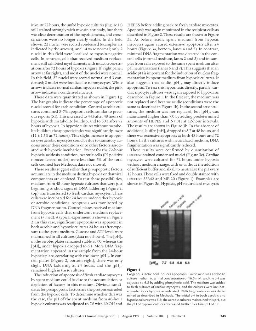
Figure 4Exogenous lactic acid induces apoptosis. Lactic acid was added toculture medium to a final concentration of 16.5 mM, and the pH wasadjusted to 6.8 by adding phosphoric acid. The medium was addedto fresh cultures of cardiac myocytes, and the cultures were incubat-ed under air or hypoxia as indicated. DNA fragmentation was deter-mined as described in Methods. The initial pH in both aerobic andhypoxic cultures was 6.8; the aerobic cultures maintained this pH, butthe pH of hypoxic cultures decreased further to a final pH of 5.8.
exhibited some degree of myofilament deterioration,but cross-striations were still apparent, and most of thenuclei were normal (compare with Figure 1e). In thefield shown, 25 nuclei were scored normal and 3 wereapoptotic; 1 nucleus was localized to a nonmyocyte.
Induction of apoptosis by extracellular lactic acid. Theresults described here demonstrate that low [pH]o iscritical for the induction of apoptosis by hypoxia in thismodel. They do not show that acidosis can activateapoptosis independently of other factors that mayaccumulate or disappear from the extracellular medi-um during exposure to hypoxia. To test the effect ofacidosis directly, we added lactic acid to fresh cardiacmyocyte cultures at similar concentrations to thoseseen in conditioned medium after 72 hours of hypoxia(48), and adjusted the pH to 6.8 with phosphoric acid.These cultures were exposed to aerobic or hypoxic con-ditions as before. As shown in Figure 4, the addition of
acid induced DNA laddering in both aerobic andhypoxic cardiac myocytes. The fragmentation inducedby exogenous acid was not as pronounced as thatcaused by 24 hours of conditioned medium (Figure 2).This could be due to a number of factors, including thesite of generation of the acid or additional proapop-totic components in the conditioned medium. Howev-er, there was more DNA fragmentation in the acidifiedaerobic cultures than in either the aerobic or hypoxiccontrols, even though the hypoxia control culturesachieved the same final pH. These results confirm thatextracellular acidosis can induce significant apoptosisindependently of hypoxia in cardiac myocytes.
Hypoxia-acidosis–mediated apoptosis is independent ofchanges in p53. The results described here demonstratethat extracellular acidosis, and possibly other metabo-lites, induces apoptosis of cardiac myocytes independ-ently of hypoxia. Because p53 has been implicated in
246 The Journal of Clinical Investigation | August 1999 | Volume 104 | Number 3
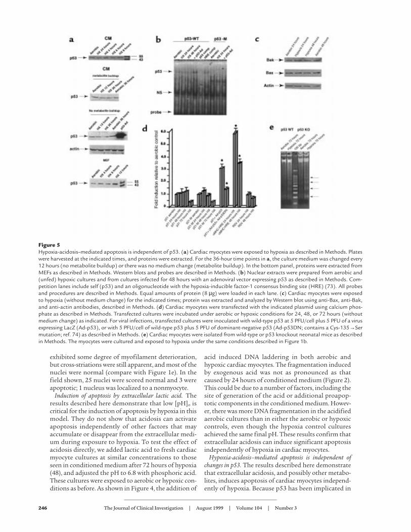
Figure 5Hypoxia-acidosis–mediated apoptosis is independent of p53. (a) Cardiac myocytes were exposed to hypoxia as described in Methods. Plateswere harvested at the indicated times, and proteins were extracted. For the 36-hour time points in a, the culture medium was changed every12 hours (no metabolite buildup) or there was no medium change (metabolite buildup). In the bottom panel, proteins were extracted fromMEFs as described in Methods. Western blots and probes are described in Methods. (b) Nuclear extracts were prepared from aerobic and(unfed) hypoxic cultures and from cultures infected for 48 hours with an adenoviral vector expressing p53 as described in Methods. Com-petition lanes include self (p53) and an oligonucleotide with the hypoxia-inducible factor-1 consensus binding site (HRE) (73). All probesand procedures are described in Methods. Equal amounts of protein (8 µg) were loaded in each lane. (c) Cardiac myocytes were exposedto hypoxia (without medium change) for the indicated times; protein was extracted and analyzed by Western blot using anti-Bax, anti-Bak,and anti-actin antibodies, described in Methods. (d) Cardiac myocytes were transfected with the indicated plasmid using calcium phos-phate as described in Methods. Transfected cultures were incubated under aerobic or hypoxic conditions for 24, 48, or 72 hours (withoutmedium change) as indicated. For viral infections, transfected cultures were inoculated with wild-type p53 at 5 PFU/cell plus 5 PFU of a virusexpressing LacZ (Ad-p53), or with 5 PFU/cell of wild-type p53 plus 5 PFU of dominant-negative p53 (Ad-p53DN; contains a Cys-135→Sermutation; ref. 74) as described in Methods. (e) Cardiac myocytes were isolated from wild-type or p53 knockout neonatal mice as describedin Methods. The myocytes were cultured and exposed to hypoxia under the same conditions described in Figure 1b.
numerous pathways of apoptosis in other systems, aswell as in cardiac myocytes subjected to stretch,angiotensin II, and hypoxia (42–44), we asked whetherp53 was involved in initiating the signaling pathwaysmediated by hypoxia-acidosis. Four assays for p53activity are shown in Figure 5, a–d. Figure 5a shows typ-ical Western blots of proteins from cardiac myocytesand mouse embryo fibroblasts (MEFs) subjected toincreasing periods of hypoxia (as in Figure 1, a and b).The p53 levels in the myocytes were very low at all timesand did not change after short- or long-term exposureto hypoxia, with or without metabolic buildup. Infec-tion with a p53 adenovirus caused a large accumula-tion of p53 (Figure 5a, third panel). As a positive con-trol, p53 was measured in MEFs exposed to hypoxia(Figure 5a, bottom panel). In agreement with a previ-ous report (43), p53 protein was elevated approximate-ly 4-fold in these cells after 12 hours.
Changes in the activity of p53 may occur independ-ently of net synthesis, e.g., through phosphorylation ornuclear translocation (59, 60). To determine whetherthere were changes in the activity of nuclear p53, DNAbinding was measured by electrophoretic mobility shiftsusing a p53 binding site oligonucleotide and nuclearextracts from aerobic and hypoxic cardiac myocytes (asdescribed in Methods). Previous reports have demon-strated the weak but specific nature of p53 binding bythis assay (42, 43). The arrow in Figure 5b indicates the
p53-specific binding complex. This band was competedby excess cold probe (lane 5), but not by excess of anon–p53 binding site probe (HRE, lane 6), and it wasnot present when the labeled probe contained a p53binding site mutation (lanes 7–10). There was nochange in the specific p53 binding complex betweenaerobic and hypoxic nuclear extracts. In agreement witha previous report (44), infection with a p53 adenoviruscaused an increase of p53 complex binding (lane 4).
Changes in the expression of p53 target genes Baxand Bcl2 have been implicated in apoptosis caused bypressure overload, angiotensin II, and stretch (42).Increased expression of Bcl2 and Fas proteins and nochange of Bax were reported in ischemic myocardial tis-sue (61). Western blot assays were used to determinewhether chronic hypoxia caused changes of Bax, Bcl2,or Bak. As shown in Figure 5c, exposure of myocytes tohypoxia for 24 or 48 hours caused no apparent changesin Bak or Bax; Bcl-2 protein levels were too low todetect, consistent with a previous report (62).
To assay for changes in function of p53, we measuredthe effects of chronic hypoxia on the expression of atransfected p21 promoter (43, 44). As shown in Figure5d, there was no significant effect of hypoxia on theexpression of luciferase from either wild-type ormutant p21 promoters (in p21-M, the p53 bindingsites of the wild-type p21 promoter are deleted). Theslight trend toward increased p21 promoter activity
The Journal of Clinical Investigation | August 1999 | Volume 104 | Number 3 247
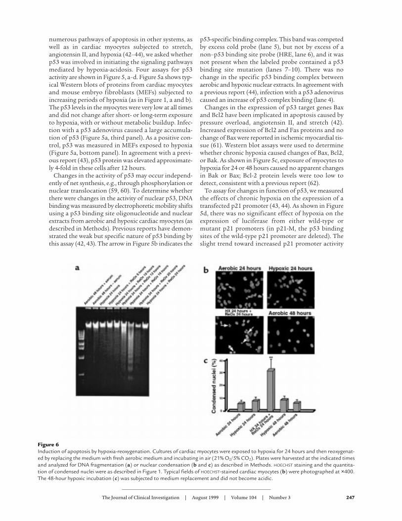
Figure 6Induction of apoptosis by hypoxia-reoxygenation. Cultures of cardiac myocytes were exposed to hypoxia for 24 hours and then reoxygenat-ed by replacing the medium with fresh aerobic medium and incubating in air (21% O2/5% CO2). Plates were harvested at the indicated timesand analyzed for DNA fragmentation (a) or nuclear condensation (b and c) as described in Methods. HOECHST staining and the quantita-tion of condensed nuclei were as described in Figure 1. Typical fields of HOECHST-stained cardiac myocytes (b) were photographed at ×400.The 48-hour hypoxic incubation (c) was subjected to medium replacement and did not become acidic.
after 48 hours of hypoxia (1.2 ± 0.3–fold), although dif-fering from the trends of the α-MHC and RSV pro-moters at this time point, was not significant whencompared with the p21 aerobic control or the p21-Mpromoter at 48 hours of hypoxia (0.9 ± 0.15). As a pos-itive control, p21-Luc–transfected cells were infectedwith a p53 adenovirus (p53-Ad); in this case, expressionof the p21 promoter was induced by about 3-fold.When the p21-Luc–transfected cultures were infectedwith equal amounts (5 PFU/cell) of Ad-p53 and an ade-noviral vector containing a dominant-negative mutantp53 (Ad-p53DN), the induction was quenched (Figure5d, lane 10). Expression of the hypoxia-responsive vec-tor pα-MHC–HRE-Luc (lanes 11 and 12) confirmedthat the culture conditions used here were sufficient toactivate hypoxia-regulated genes, whereas there was noeffect on the expression of the RSV promoter (47, 63).
Finally, we detected no change in endogenous p53 orp21 transcripts by Northern blots of RNA from cardiacmyocytes exposed to hypoxia or hypoxia-acidosis for 6,12, 24, or 36 hours, compared with aerobic controls(data not shown).
These results indicate that p53 activity in cardiacmyocytes does not change during the exposure of thesecells to chronic hypoxia and suggest that the ensuingapoptosis is independent of changes in p53 activity. Toconfirm this, cardiac myocytes were isolated from wild-type and p53 knockout mice and were subjected tochronic hypoxia-acidosis (without medium change) asdescribed in Figure 1b. As shown in Figure 5e, althoughthe levels of apoptosis were lower than previously seenwith rat cardiac myocytes, there was clear genomic
DNA fragmentation in both wild-type and knockoutmouse cells; the arrows in the figure indicate fragmentsof about 200-bp increments, highly characteristic ofapoptotic genomic DNA (2). No fragmentation wasdetected in the control (aerobic) samples. Thereappeared to be slightly more DNA loaded in the knock-out lanes, and more intense fragmentation; densito-metric scanning indicated approximately equivalentratios of fragmented/intact DNA in the 2 samples (n =2; data not shown). The reason for lower levels of apop-tosis in mouse versus rat cardiac myocytes is probablyrelated to the slightly decreased contractility and slow-er rate of acid production in the mouse cultures (datanot shown). In other experiments, rat myocytes treatedwith Ad-p53DN, as described in Figure 5d, displayedidentical DNA fragmentation as the controls afterexposure to chronic hypoxia-acidosis (data not shown).Together, these results strongly suggest that hypoxia-acidosis–mediated apoptosis of cardiac myocytes isindependent of p53 activity.
Reoxygenation of hypoxic cardiac myocytes induces apoptosis.The results reported here show that chronic hypoxiaalone does not cause apoptosis of cardiac myocytes. Ourprevious results indicated that chronic hypoxia causedloss of glutathione and mediated a stress response thatincluded the induction of c-Jun NH2-terminal kinase(JNK) activity when the cultures were reoxygenated (64).These conditions mimic the changes in oxygen tensionthat occur during ischemia and reperfusion. We there-fore asked whether reoxygenation of hypoxic culturesalso induced apoptosis. These results are shown in Fig-ure 6, a and b. There was no evidence of apoptosis after24 hours of hypoxia, in agreement with the results in Fig-ure 1. However, DNA laddering was apparent 8–12 hoursafter reoxygenation. Maximal DNA fragmentationoccurred 16–24 hours after reoxygenation and thendeclined to baseline levels. Figure 6b shows representa-tive fields of HOECHST-stained nuclei. Arrows in the bot-tom left panel indicate examples of condensed nucleithat were scored positive. In this field, 12 nuclei werescored condensed and 15 were normal. Figure 6c showsthe quantitation of condensed nuclei under the differ-ent conditions, determined as described in Figure 1;approximately 30% of the cells became apoptotic at thepeak time of 16–24 hours after reoxygenation.
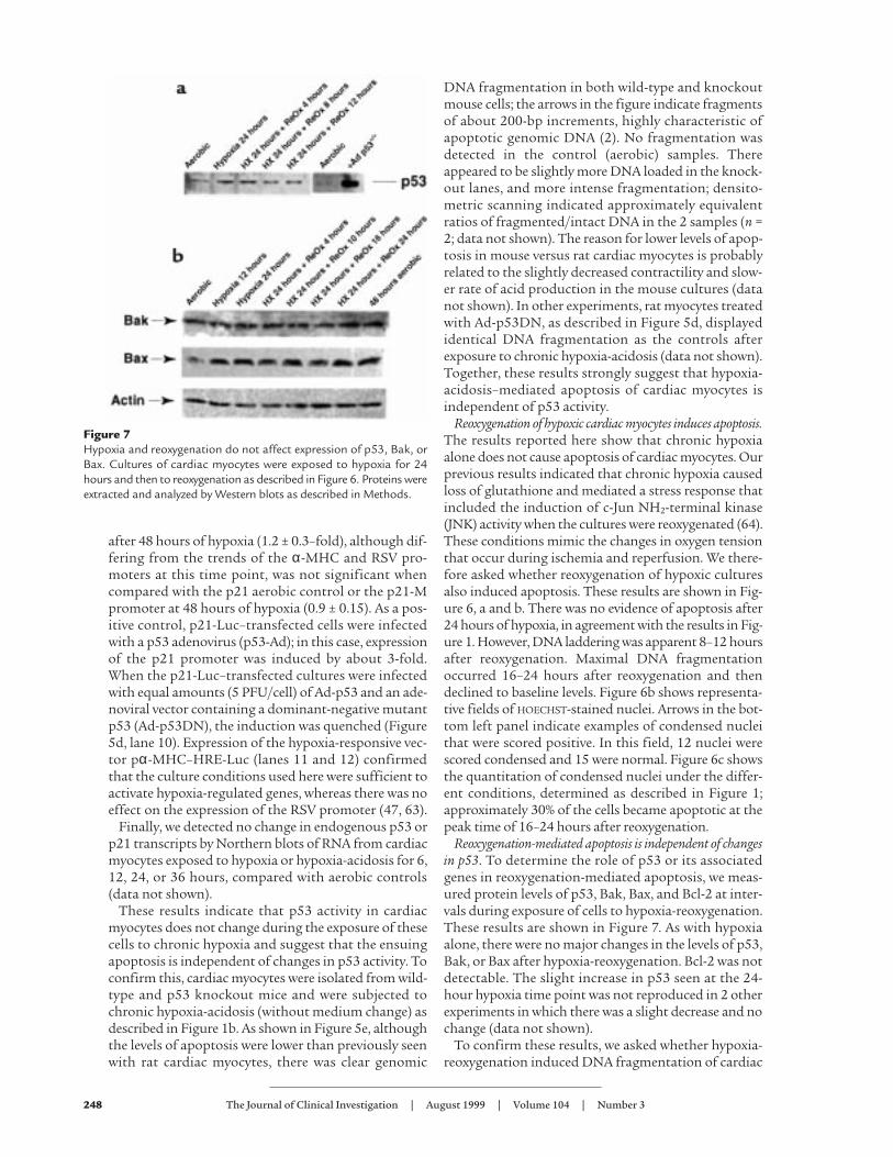
Reoxygenation-mediated apoptosis is independent of changesin p53. To determine the role of p53 or its associatedgenes in reoxygenation-mediated apoptosis, we meas-ured protein levels of p53, Bak, Bax, and Bcl-2 at inter-vals during exposure of cells to hypoxia-reoxygenation.These results are shown in Figure 7. As with hypoxiaalone, there were no major changes in the levels of p53,Bak, or Bax after hypoxia-reoxygenation. Bcl-2 was notdetectable. The slight increase in p53 seen at the 24-hour hypoxia time point was not reproduced in 2 otherexperiments in which there was a slight decrease and nochange (data not shown).
To confirm these results, we asked whether hypoxia-reoxygenation induced DNA fragmentation of cardiac
248 The Journal of Clinical Investigation | August 1999 | Volume 104 | Number 3
Figure 7Hypoxia and reoxygenation do not affect expression of p53, Bak, orBax. Cultures of cardiac myocytes were exposed to hypoxia for 24hours and then to reoxygenation as described in Figure 6. Proteins wereextracted and analyzed by Western blots as described in Methods.
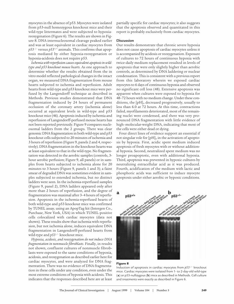
myocytes in the absence of p53. Myocytes were isolatedfrom p53-null homozygous knockout mice and theirwild-type littermates and were subjected to hypoxia-reoxygenation (Figure 6). The results are shown in Fig-ure 8. DNA internucleosomal cleavage peaked earlierand was at least equivalent in cardiac myocytes fromp53–/– versus p53+/+ animals. This confirms that apop-tosis mediated by either hypoxia-reoxygenation orhypoxia-acidosis does not require p53.
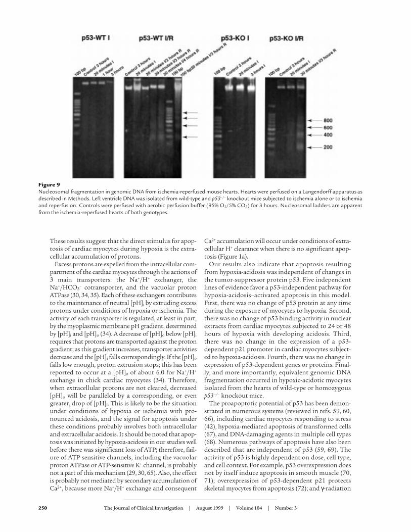
Ischemia with reperfusion causes equivalent apoptosis in wild-type and p53 knockout mouse hearts. As one approach todetermine whether the results obtained from the invitro model reflected pathological changes in the intactorgan, we measured DNA fragmentation from mousehearts subjected to ischemia and reperfusion. Adulthearts from wild-type and p53 knockout mice were per-fused by the Langendorff technique as described inMethods. Previous studies demonstrated that DNAfragmentation induced by 24 hours of permanentocclusion of the coronary artery (ischemia alone)occurred at equivalent levels in wild-type and p53knockout mice (46). Apoptosis induced by ischemia andreperfusion of Langendorff-perfused mouse hearts hasnot been reported previously. Figure 9 compares nucle-osomal ladders from the 2 groups. There was cleargenomic DNA fragmentation in both wild-type and p53knockout cells subjected to 20 minutes of ischemia and3 hours of reperfusion (Figure 9, panels 2 and 4, respec-tively). DNA fragmentation in the knockout hearts wasat least equivalent to that in the wild-type. No fragmen-tation was detected in the aerobic samples (controls, 3-hour aerobic perfusion; Figure 9, all panels) or in sam-ples from hearts subjected to ischemia alone for 20minutes to 3 hours (Figure 9, panels 1 and 3); a faintsmear of degraded DNA was sometimes evident in sam-ples subjected to extended ischemia, but no distinctladders were seen. In the ischemia-reperfused samples(Figure 9, panel 2), DNA ladders appeared only aftermore than 2 hours of reperfusion, and the degree offragmentation was maximal after 3–4 hours of reperfu-sion. Apoptosis in the ischemia-reperfused hearts ofboth wild-type and p53 knockout mice was confirmedby TUNEL assay, using an ApopTag kit (Intergen Co.,Purchase, New York, USA) in which TUNEL-positivecells colocalized with cardiac myocytes (data notshown). These results show that ischemia with reperfu-sion, but not ischemia alone, induces equivalent DNAfragmentation in Langendorff-perfused hearts fromwild-type and p53–/– knockout mice.
Hypoxia, acidosis, and reoxygenation do not induce DNAfragmentation in nonmuscle fibroblasts. Finally, in resultsnot shown, confluent cultures of nonmuscle fibrob-lasts were exposed to the same conditions of hypoxia,acidosis, and reoxygenation as described earlier here forcardiac myocytes, and were analyzed for DNA frag-mentation. There was no evidence of DNA fragmenta-tion in these cells under any condition, even under themost extreme conditions of hypoxia with acidosis. Thisindicates that the responses described here are at least
partially specific for cardiac myocytes; it also suggeststhat the apoptosis observed and quantitated in thisreport is probably exclusively from cardiac myocytes.
DiscussionOur results demonstrate that chronic severe hypoxiadoes not cause apoptosis of cardiac myocytes unless itis accompanied by acidosis or reoxygenation. Exposureof cultures to 72 hours of continuous hypoxia withtwice-daily medium replacement resulted in levels ofapoptosis that were only slightly higher than aerobiccontrols, as determined by DNA laddering or nuclearcondensation. This is consistent with a previous reportfrom this laboratory wherein we exposed cardiacmyocytes to 6 days of continuous hypoxia and observedno significant cell loss (48). Extensive apoptosis wasapparent when cultures were exposed to hypoxia for48–72 hours with no medium change. Under these con-ditions, the [pH]o decreased progressively, usually toless than 6.0 at 72 hours. At this time, contractionsfailed, myofilaments deteriorated, most of the remain-ing nuclei were condensed, and there was very pro-nounced DNA fragmentation with little evidence ofhigh–molecular-weight DNA, indicating that most ofthe cells were either dead or dying.
Four direct lines of evidence support an essential ifnot singular role for [pH]o in the activation of apopto-sis by hypoxia. First, acidic spent medium inducedapoptosis of fresh myocytes with or without addition-al hypoxia. Second, neutralized spent medium was nolonger proapoptotic, even with additional hypoxia.Third, apoptosis was prevented in hypoxic cultures byneutralizing extracellular acid as it was produced.Fourth, acidification of the medium with lactic andphosphoric acids was sufficient to induce myocyteapoptosis under either aerobic or hypoxic conditions.
The Journal of Clinical Investigation | August 1999 | Volume 104 | Number 3 249
Figure 8Induction of apoptosis in cardiac myocytes from p53–/– knockoutmice. Cardiac myocytes were isolated from 1- to 2-day-old wild-type(a) or p53-nullizygous (b) mice as described in Methods. Cell cultureand treatments were exactly as described in Figure 6.
These results suggest that the direct stimulus for apop-tosis of cardiac myocytes during hypoxia is the extra-cellular accumulation of protons.
Excess protons are expelled from the intracellular com-partment of the cardiac myocytes through the actions of3 main transporters: the Na+/H+ exchanger, theNa+/HCO3
– cotransporter, and the vacuolar protonATPase (30, 34, 35). Each of these exchangers contributesto the maintenance of neutral [pH]i by extruding excessprotons under conditions of hypoxia or ischemia. Theactivity of each transporter is regulated, at least in part,by the myoplasmic membrane pH gradient, determinedby [pH]i and [pH]o (34). A decrease of [pH]o below [pH]i
requires that protons are transported against the protongradient; as this gradient increases, transporter activitiesdecrease and the [pH]i falls correspondingly. If the [pH]o
falls low enough, proton extrusion stops; this has beenreported to occur at a [pH]o of about 6.0 for Na+/H+
exchange in chick cardiac myocytes (34). Therefore,when extracellular protons are not cleared, decreased[pH]o will be paralleled by a corresponding, or evengreater, drop of [pH]i. This is likely to be the situationunder conditions of hypoxia or ischemia with pro-nounced acidosis, and the signal for apoptosis underthese conditions probably involves both intracellularand extracellular acidosis. It should be noted that apop-tosis was initiated by hypoxia-acidosis in our studies wellbefore there was significant loss of ATP; therefore, fail-ure of ATP-sensitive channels, including the vacuolarproton ATPase or ATP-sensitive K+ channel, is probablynot a part of this mechanism (29, 30, 65). Also, the effectis probably not mediated by secondary accumulation ofCa2+, because more Na+/H+ exchange and consequent
Ca2+ accumulation will occur under conditions of extra-cellular H+ clearance when there is no significant apop-tosis (Figure 1a).
Our results also indicate that apoptosis resultingfrom hypoxia-acidosis was independent of changes inthe tumor-suppressor protein p53. Five independentlines of evidence favor a p53-independent pathway forhypoxia-acidosis–activated apoptosis in this model.First, there was no change of p53 protein at any timeduring the exposure of myocytes to hypoxia. Second,there was no change of p53 binding activity in nuclearextracts from cardiac myocytes subjected to 24 or 48hours of hypoxia with developing acidosis. Third,there was no change in the expression of a p53-dependent p21 promoter in cardiac myocytes subject-ed to hypoxia-acidosis. Fourth, there was no change inexpression of p53-dependent genes or proteins. Final-ly, and more importantly, equivalent genomic DNAfragmentation occurred in hypoxic-acidotic myocytesisolated from the hearts of wild-type or homozygousp53–/– knockout mice.
The proapoptotic potential of p53 has been demon-strated in numerous systems (reviewed in refs. 59, 60,66), including cardiac myocytes responding to stress(42), hypoxia-mediated apoptosis of transformed cells(67), and DNA-damaging agents in multiple cell types(68). Numerous pathways of apoptosis have also beendescribed that are independent of p53 (59, 69). Theactivity of p53 is highly dependent on dose, cell type,and cell context. For example, p53 overexpression doesnot by itself induce apoptosis in smooth muscle (70,71); overexpression of p53-dependent p21 protectsskeletal myocytes from apoptosis (72); and γ-radiation
250 The Journal of Clinical Investigation | August 1999 | Volume 104 | Number 3
Figure 9Nucleosomal fragmentation in genomic DNA from ischemia-reperfused mouse hearts. Hearts were perfused on a Langendorff apparatus asdescribed in Methods. Left ventricle DNA was isolated from wild-type and p53–/– knockout mice subjected to ischemia alone or to ischemiaand reperfusion. Controls were perfused with aerobic perfusion buffer (95% O2/5% CO2) for 3 hours. Nucleosomal ladders are apparentfrom the ischemia-reperfused hearts of both genotypes.
The Journal of Clinical Investigation | August 1999 | Volume 104 | Number 3 251
activation of p53 may mediate apoptosis or cell-cyclewithdrawal, depending on the cell type (68). A previousstudy reported that p53 was induced in neonatal car-diac myocytes in response to hypoxia (44). Although wecannot account for the apparent discrepancies betweenthis report and our observations, they may be related todifferences in the models and the pleiotropic and con-ditional properties of p53. The previous study suggest-ed that hypoxia-mediated changes in p53 correlatedwith increased apoptosis, although it did not demon-strate that p53 was required for apoptosis.
Also, to our knowledge, we present the first definitiveevidence that reoxygenation is a direct and distinctstimulus for apoptosis of cardiac myocytes. Nucleoso-mal fragmentation was evident 8–12 hours after reoxy-genation and peaked at 16–24 hours in different exper-iments. Quantitation of condensed nuclei by HOECHST
staining indicated that approximately 30% of myocyteswere apoptotic after 1 cycle of hypoxia-reoxygenation.This clearly demonstrates that reoxygenation of hypox-ic cardiac myocytes is extremely stressful, and it sup-ports our previous report of an intense induction ofJNK activity under the same conditions (64). Apopto-sis caused by hypoxia-reoxygenation was also inde-pendent of p53 by the same criteria described earlierhere for hypoxia-acidosis–mediated apoptosis.
We found no evidence for modulation of the expres-sion of the Bcl-2 family members Bak and Bax inhypoxia-acidosis– or hypoxia-reoxygenation–mediatedapoptosis. Unchanged levels of Bax protein have alsobeen reported in ischemic myocardial tissue (61).Although this does not exclude a role for posttransla-tional modification of these proteins, the results sup-port our conclusion that neither of the apoptosis path-ways described here is dependent on p53.
Although it is not possible to extrapolate all ofthese results to conditions in vivo, it is noteworthythat hearts from p53 knockout mice show the samedegree of apoptosis as wild-type hearts subjected tochronic ischemia (46) or to ischemia and reperfusion(Figure 9). Therefore, in the mouse heart, p53 is clear-ly not required for apoptosis in response to eitherischemia or reperfusion. This does not mean thatapoptosis in response to other stresses is independ-ent of p53, or that p53 will not contribute toischemia-mediated apoptosis under some circum-stances, because ischemia is a complex stress. Ourresults suggest that hypoxia per se is not a signal forapoptosis, because cardiac myocytes can remain com-pletely viable for extended times under severe hypox-ia, provided they have sufficient glycolytic substrateand waste metabolite removal. There may be directmolecular parallels between the in vitro models ofhypoxia, acidosis, and reoxygenation described hereand ischemia in the intact adult heart. Myocardialtissue subjected to severe chronic ischemia by coro-nary artery occlusion is likely to be subjected to vary-ing degrees of both hypoxic stress and acidosis,whereas ischemia-reperfused tissue experiences
hypoxia-reoxygenation. All of these conditions cancause apoptosis in a p53-null cellular environment.
AcknowledgmentsThis work was supported by the National Institutes ofHealth (HL-44578 to K. Webster; HL-49891 to N.H.Bishopric), by the Cigarette and Tobacco Surtax of theState of California through the Tobacco-Related Dis-ease Research Program of the University of California(1RT-402 to K.Webster), by an Established InvestigatorAward from the American Heart Association (to N.H.Bishopric), and, in part, by a grant from the MiamiHeart Research Institute. We acknowledge the excellentcontributions of J. Zang and P. Andreka in breeding andcharacterizing the p53 knockout mice, and we thank M.Javadpour and N. Maulik for their helpful suggestionsin setting up Langendorff-perfused mouse hearts.
1. Haunstetter, A., and Izumo, S. 1998. Apoptosis: basic mechanisms andimplications for cardiovascular disease. Circ. Res. 82:1111–1129.
2. MacLellan, W.R., and Schneider, M.D. 1997. Death by design: pro-grammed cell death in cardiovascular biology and disease. Circ. Res.81:137–144.
3. Fliss, H., and Gattinger, D. 1996. Apoptosis in ischemic and reperfusedrat myocardium. Circ. Res. 79:949–956.
4. Olivetti, G., et al. 1997. Apoptosis in the failing human heart. N. Engl. J.Med. 336:1131–1141.
5. Olivetti, G., et al. 1994. Acute myocardial infarction in humans is asso-ciated with activation of programmed myocyte cell death in the surviv-ing portion of the heart. J. Mol. Cell. Cardiol. 28:2005–2016.
6. Kajstura, J., et al. 1998. Myocyte proliferation in end-stage cardiac fail-ure in humans. Proc. Natl. Acad. Sci. USA. 95:8801–8805.
7. Narula, J., et al. 1996. Apoptosis in myocytes in end-stage heart failure.N. Engl. J. Med. 335:1182–1189.
8. Mallat, Z., et al. 1996. Evidence of apoptosis in arrhythmogenic rightventricular dysplasia. N. Engl. J. Med. 335:1190–1196.
9. Misao, J., et al. 1996. Expression of bcl-2 protein, an inhibitor of apop-tosis, and Bax, an accelerator of apoptosis, in ventricular myocytes ofhuman hearts with myocardial infarction. Circulation. 94:1506–1512.
10. Itoh, G., et al. 1995. DNA fragmentation of human infarcted myocardialcells demonstrated by the nick end labeling method and DNA agarosegel electrophoresis. Am. J. Pathol. 146:1325–1331.
11. Saraste, A., et al. 1997. Apoptosis in human acute myocardial infarction.Circulation. 95:320–323.
12. Cheng, W., et al. 1996. Programmed myocyte cell death affects the viablemyocardium after infarction in rats. Exp. Cell Res. 226:316–327.
13. Kajstura, J., et al. 1995. The cellular basis of pacing-induced dilated car-diomyopathy: myocyte cell loss and myocyte cellular reactive hypertro-phy. Circulation. 92:2306–2317.
14. Leri, A., et al. 1998. Pacing-induced heart failure in dogs enhances theexpression of p53 and p53-dependent genes in ventricular myocytes. Cir-culation. 97:194–203.
15. Gottlieb, R.A., Burleson, K.O., Kloner, R.A., Babior, B.M., and Engler, R.L.1994. Reperfusion injury induces apoptosis in rabbit cardiomyocytes. J.Clin. Invest. 94:1612–1628.
16. Teiger, E., et al. 1996. Apoptosis in pressure overload-induced hearthypertrophy in the rat. J. Clin. Invest. 97:2891–2897.
17. Hamet, P., et al. 1996. The time window of apoptosis: a new componentin the therapeutic strategy for cardiovascular remodeling. J. Hypertens.Suppl. 14:S65–S70.
18. Chen, C., et al. 1997. Myocardial cell death and apoptosis in hibernatingmyocardium. J. Am. Coll. Cardiol. 30:1407–1412.
19. Buja, L.M., Eigenbrodt, M.L., and Eigenbrodt, E.H. 1993. Apoptosis andnecrosis: basic types and mechanisms of cell death. Arch. Pathol. Lab. Med.117:1208–1214.
20. Majno, G., and Joris, S. 1995. Apoptosis, oncosis, and necrosis: anoverview of cell death. Am. J. Pathol. 146:3–15.
21. Reimer, K.A., and Ideker, R.E. 1987. Myocardial ischemia and infarction:anatomic and biochemical substrates for ischemic cell death and ven-tricular arrhythmias. Hum. Pathol. 18:462–475.
22. Allen, D.G., and Orchard, C.H. 1987. Myocardial contractile functionduring ischemia and hypoxia. Circ. Res. 60:153–168.
23. Allen, D.G., Lee, J.A., and Smith, G.L. 1989. The consequences of simu-lated ischaemia on intracellular Ca2+ and tension in isolated ferret ven-tricular muscle. J. Physiol. 410:297–323.
48. Webster, K.A., and Bishopric, N.H. 1992. Molecular regulation of cardiacmyocyte adaptations to chronic hypoxia. J. Mol. Cell. Cardiol. 24:741–751.
49. Webster, K.A., Discher, D., and Bishopric, N.H. 1993. Induction andnuclear accumulation of fos and jun proto-oncogenes in hypoxia cardiacmyocytes. J. Biol. Chem. 268:16852–16859.
50. Bishopric, N.H., Simpson, P.C., and Ordahl, C.P. 1987. Induction of theskeletal actin gene in alpha1-adrenoceptor mediated hypertrophy of ratcardiac myocytes. J. Clin. Invest. 80:1194–1199.
51. Bishopric, N.H., Zeng, G., Sato, B., and Webster, K.A. 1997. AdenovirusE1A inhibits cardiac myocyte-specific gene expression through its aminoterminus. J. Biol. Chem. 272:20584–20594.
52. Webster, K.A., Muscat, G.E.O., and Kedes, L. 1988. Adenovirus E1Aproducts suppress myogenic differentiation and inhibit transcriptionfrom muscle-specific promoters. Nature. 332:553–561.
53. Soussi, T., de Fromentel, C.C., Breugnot, C., and May, E. 1988.Nucleotide sequence of a cDNA encoding the rat p53 nuclear oncopro-tein. Nucleic Acids Res. 16:11383.
54. Belinski, S.A., Middleton, S.K., Picksley, S.M., Hahn, F.F., and Niku, K.1996. Analysis of the K-ras and p53 pathways in x-ray–induced lungtumors in the rat. Radiat. Res. 145:449–456.
55. Wu, X., Bishopric, N.H., Discher, D.J., Murphy, B.J., and Webster, K.A.1996. Physical and functional sensitivity of zinc finger transcription fac-tors to redox change. Mol. Cell. Biol. 16:1035–1046.
56. Jacks, T., et al. 1994. Tumor spectrum analysis in p53-mutant mice. Curr.Biol. 4:1–7.
57. Summeray, M.S., and Yellon, D.M. 1998. Characterisation and valida-tion of a murine model of global ischaemia-reperfusion injury. Mol. Cell.Biochem. 186:61–68.
58. Hoppe-Seyler, F., and Butz, K. 1992. Activation of human papillomavirustype 18 E6-E7 oncogene expression by transcription factor Sp1. NucleicAcids Res. 20:6701–6706.
59. Bellamy, C.O.C. 1997. p53 and apoptosis. Br. Med. Bull. 53:522–538.60. Canman, C.E., and Kastan, M.B. 1997. Role of p53 in apoptosis. Adv.
Pharmacol. 41:429–460.61. Kajstura, J., Cheng, W., Reiss, K., and Clark, W.A. 1996. Apoptotic and
necrotic myocyte cell death are independent contributing variables ofinfarct size. Lab. Invest. 74:86–107.
62. Wang, L., Ma, W., Markovich, R., Chen, J., and Wang, P.H. 1998. Regula-tion of cardiomyocyte apoptotic signaling by insulin-like growth factor.Circ. Res. 83:516–522.
63. Prentice, H., et al. 1997. Regulated expression of a foreign gene targetedto the ischemic myocardium. Cardiovasc. Res. 35:567–574.
64. Laderoute, K.R., and Webster, K.A. 1997. Hypoxia/reoxygenation stimu-lates jun kinase activity through redox signaling in cardiac myocytes.Circ. Res. 80:336–344.
65. Forgac, M. 1989. Structure and function of vacuolar class of ATP-driv-en proton pumps. Physiol. Rev. 69:765–796.
66. Ko, L.J., and Prives, C. 1996. p53: puzzle and paradigm. Genes Dev.10:1054–1072.
67. Graeber, T.G., et al. 1996. Hypoxia-mediated selection of cells withdiminished apoptotic potential in solid tumors. Nature. 379:88–91.
68. Agarwal, M.L., Taylor, W.R., Chernov, M.V., Chernova, O.B., and Stark,G.R. 1998. The p53 network. J. Biol. Chem. 273:1–4.
69. Steller, H. 1995. Mechanisms and genes of cellular suicide. Science.267:1445–1449.
70. Bennett, M.R., Littlewood, T.D., Schwartz, S.M., and Weissberg, P.L.1997. Increased sensitivity of human vascular smooth muscle cells fromatherosclerotic plaques to p53-mediated apoptosis. Circ. Res. 81:591–599.
71. Bennet, M.R., Evan, G.I., and Schwartz, S.M. 1995. Apoptosis of rat vas-cular smooth muscle cells is regulated by p53-dependent and -inde-pendent pathways. Circ. Res. 77:266–273.
72. Wang, J., Guo, K., Wills, K.N., and Walsh, K. 1997. Rb functions to inhib-it apoptosis during myocyte differentiation. Cancer Res. 57:351–354.
73. Hu, J., Discher, D.J., Bishopric, N.H., and Webster, K.A. 1998. Hypoxiaregulates expression of the endothelin-1 gene through a proximal hypox-ia-inducible factor-1 binding site on the antisense strand. Biochem. Bio-phys. Res. Commun. 245:894–899.
74. Bacchetti, S., and Hraham, F.L. 1993. Inhibition of cell proliferation byan adenovirus vector expressing the human wild type p53 protein. Int. J.Oncol. 3:781–788.
252 The Journal of Clinical Investigation | August 1999 | Volume 104 | Number 3
24. Neely, J.R., and Grotyohann, L.W. 1984. Role of glycolytic products indamage to ischemic myocardium. Circ. Res. 55:816–824.
25. Hochachka, P.W., Buck, L.T., Doll, C.J., and Land, S.C. 1996. Unifying the-ory of hypoxia tolerance: molecular/metabolic defense and rescue mech-anisms for surviving oxygen lack. Proc. Natl. Acad. Sci. USA. 93:9493–9498.
26. Buja, L.M., and Entman, M.L. 1998. Modes of myocardial cell injury andcell death in ischemic heart disease. Circulation. 98:1355–1357.
27. Kajstura, J., et al. 1996. Apoptotic and necrotic myocyte cell deaths are inde-pendent contributing variables of infarct size in rats. Lab. Invest. 74:86–107.
28. Ohno, M., et al. 1998. “Apoptotic” myocytes in infarct area in rabbithearts may be oncotic myocytes with DNA fragmentation: analysis byimmunogold electron microscopy combined with in situ nick end-label-ing. Circulation. 98:1422–1430.
29. Gottlieb, R.A., Gruol, D.L., Zhu, J.Y., and Engler, R.L. 1996. Precondi-tioning in rabbit cardiomyocytes. J. Clin. Invest. 97:2391–2398.
30. Karwatowska-Prokopczuk, E., Nordberg, J.A., Li, H.L., Engler, R.L., andGottlieb, R.A. 1998. Effect of vacuolar proton ATPase on pHi, Ca2+, andapoptosis in neonatal cardiac myocytes during metabolicinhibition/recovery. Circ. Res. 82:1139–1144.
31. Long, X., et al. 1998. Enhanced expression of p53 and apoptosis inducedby blockade of the vacuolar proton ATPase in cardiomyocytes. J. Clin.Invest. 101:1453–1461.
32. Anversa, P., and Kajstura, J. 1998. Myocyte cell death in the diseasedheart. Circ. Res. 82:1231–1233.
33. Dennis, S.C., Gevers, W., and Opie, L.H. 1991. Protons in ischemia: wheredo they come from; where do they go? J. Mol. Cell. Cardiol. 23:1077–1086.
34. Lazdunski, M., Frelin, C., and Vigne, P. 1985. The sodium/hydrogenexchange system in cardiac cells: its biochemical and pharmacologicalproperties and its role in regulating internal concentrations of sodiumand internal pH. J. Mol. Cell. Cardiol. 17:1029–1042.
35. Lagadic-Gossmann, D., Buckler, K.J., and Vaughan-Jones, R.D. 1992.Role of bicarbonate in pH recovery from intracellular acidosis in theguinea-pig ventricular myocyte. J. Physiol. 458:361–384.
36. Pierce, G.N., and Czubryt, M.P. 1995. The contribution of ionic imbalanceto ischemia/reperfusion-induced injury. J. Mol. Cell. Cardiol. 27:53–63.
37. Shimada, Y., Hearse, D.J., and Avkiran, M. 1996. Impact of extracellularbuffer composition on cardioprotective efficacy of Na+/H+ exchangeinhibitors. Am. J. Physiol. 270:H692–H700.
38. Bond, J.M., Chacon, E., Herman, B., and Lemasters, J.J. 1993. Intracellu-lar pH and Ca2+ homeostasis in the pH paradox of reperfusion injuryto rat neonatal cardiac myocytes. Am. J. Physiol. 265:C129–C137.
39. Li, J., and Eastman, A. 1995. Apoptosis in an interleukin-2–dependentcytotoxic T lymphocyte cell line is associated with intracellular acidifi-cation. J. Biol. Chem. 270:3203–3211.
40. Gottlieb, R.A., et al. 1995. Cell acidification in apoptosis: granulocyte colony-stimulating factor delays programmed cell death in neutrophils by up-reg-ulating the vacuolar H+ ATPase. Proc. Natl. Acad. Sci. USA. 92:5965–5968.
41. Perez-Sala, D., Collado-Escobar, D., and Mollinedo, F. 1998. Intracellu-lar alkalinization suppresses lovastatin-induced apoptosis in HL-60 cellsthrough the inactivation of a pH-dependent endonuclease. J. Biol. Chem.270:6235–6242.
42. Leri, A., et al. 1998. Stretch-mediated release of angiotensin II inducesmyocyte apoptosis by activating p53 that enhances the local renin-angiotensin system and decreases the Bcl-2-to-Bax protein ratio. J. Clin.Invest. 101:1326–1342.
43. Graeber, T.G., et al. 1994. Hypoxia induces accumulation of p53 proteinbut activation of a G1-phase checkpoint by low-oxygen is independentof p53 status. Mol. Cell. Biol. 14:6264–6277.
44. Long, X., et al. 1997. p53 and the hypoxia-induced apoptosis of culturedneonatal rat cardiac myocytes. J. Clin. Invest. 99:2635–2643.
45. Tanaka, M., et al. 1994. Hypoxia induces apoptosis with enhancedexpression of Fas antigen messenger RNA in cultured neonatal rat car-diomyocytes. Circ. Res. 75:426–433.
46. Bialik, S., et al. 1997. Myocyte apoptosis during acute myocardial infarc-tion in the mouse localizes to hypoxic regions but occurs independent-ly of p53. J. Clin. Invest. 100:1363–1372.
47. Discher, D.J., Bishopric, N.H., Wu, X., Peterson, C.A., and Webster, K.A.1998. Hypoxia regulates b-enolase and pyruvate kinase-M promoters bymodulating Sp1/Sp3 binding to a conserved GC element. J. Biol. Chem.273:26087–26093.


