Embed Size (px)
Citation preview
McPheat, Olov Wiklund, Bertil G. Ohlsson and Lillemor Mattsson HulténSvensson, Lena M.S. Carlsson, Ann-Cathrine Jönsson-Rylander, Göran I. Hansson, William
Ellen Knutsen Rydberg, Alexandra Krettek, Christina Ullström, Karin Ekström, Per-ArneMacrophages
Hypoxia Increases LDL Oxidation and Expression of 15-Lipoxygenase-2 in Human
Print ISSN: 1079-5642. Online ISSN: 1524-4636 Copyright © 2004 American Heart Association, Inc. All rights reserved.
Greenville Avenue, Dallas, TX 75231is published by the American Heart Association, 7272Arteriosclerosis, Thrombosis, and Vascular Biology
doi: 10.1161/01.ATV.0000144951.08072.0b2004;
2004;24:2040-2045; originally published online September 9,Arterioscler Thromb Vasc Biol.
http://atvb.ahajournals.org/content/24/11/2040World Wide Web at:
The online version of this article, along with updated information and services, is located on the
http://atvb.ahajournals.org/content/suppl/2004/11/04/24.11.2040.DC1.htmlData Supplement (unedited) at:
http://atvb.ahajournals.org//subscriptions/
at: is onlineArteriosclerosis, Thrombosis, and Vascular Biology Information about subscribing to Subscriptions:
http://www.lww.com/reprints
Information about reprints can be found online at: Reprints:
document. Question and AnswerPermissions and Rightspage under Services. Further information about this process is available in the
which permission is being requested is located, click Request Permissions in the middle column of the WebCopyright Clearance Center, not the Editorial Office. Once the online version of the published article for
can be obtained via RightsLink, a service of theArteriosclerosis, Thrombosis, and Vascular Biologyin Requests for permissions to reproduce figures, tables, or portions of articles originally publishedPermissions:
by guest on December 9, 2013http://atvb.ahajournals.org/Downloaded from by guest on December 9, 2013http://atvb.ahajournals.org/Downloaded from by guest on December 9, 2013http://atvb.ahajournals.org/Downloaded from by guest on December 9, 2013http://atvb.ahajournals.org/Downloaded from by guest on December 9, 2013http://atvb.ahajournals.org/Downloaded from by guest on December 9, 2013http://atvb.ahajournals.org/Downloaded from by guest on December 9, 2013http://atvb.ahajournals.org/Downloaded from by guest on December 9, 2013http://atvb.ahajournals.org/Downloaded from
Hypoxia Increases LDL Oxidation and Expression of15-Lipoxygenase-2 in Human Macrophages
Ellen Knutsen Rydberg, Alexandra Krettek, Christina Ullstrom, Karin Ekstrom, Per-Arne Svensson,Lena M.S. Carlsson, Ann-Cathrine Jonsson-Rylander, Goran I. Hansson, William McPheat,
Olov Wiklund, Bertil G. Ohlsson, Lillemor Mattsson Hulten
Objective—Macrophage-mediated oxidation of low-density lipoprotein (LDL) by enzymes, such as the lipoxygenases, isconsidered of major importance for the formation of oxidized LDL during atherogenesis. Macrophages have beenidentified in hypoxic areas in atherosclerotic plaques.
Methods and Results—To investigate the role of hypoxia in macrophage-mediated LDL oxidation, we incubated humanmonocyte-derived macrophages with LDL under normoxic (21% O2) or hypoxic (0% O2) conditions. The results showedthat hypoxic macrophages oxidized LDL to a significantly higher extent than normoxic cells. Interestingly, the mRNAand protein expression of 15-lipoxygenase-2 (15-LOX-2) as well as the activity of this enzyme are elevated inmacrophages incubated at hypoxia. Both the unspliced 15-LOX-2 and the spliced variant 15-LOX-2sv-a are found inmacrophages. In addition, 15-LOX-2 was identified in carotid plaques in some macrophage-rich areas but was onlyexpressed at low levels in nondiseased arteries.
Conclusions—In summary, these observations show for the first time that 15-LOX-2 is expressed in hypoxic macrophagesand in atherosclerotic plaques and suggest that 15-LOX-2 may be one of the factors involved in macrophage-mediatedLDL oxidation at hypoxia. (Arterioscler Thromb Vasc Biol. 2004;24:2040-2045.)
Key Words: atherosclerosis � macrophages � hypoxia � oxidized-LDL � 15-lipoxygenase-2
An early phenomenon in atherosclerosis is the retention,oxidation, and accumulation of low-density lipoprotein
(LDL) in the vessel wall.1,2 Oxidized LDL (oxLDL), one of thekey players in atherogenesis, attracts monocytes to the vesselwall where they differentiate into macrophages.3,4 Oxidation ofLDL mediated by macrophages is considered to be of majorimportance for the formation of oxLDL within the atheroscle-rotic plaque. Enzymes involved in this process are 15-lipoxy-genase (15-LOX),5,6 myeloperoxidase (MPO),7 and NADPHoxidase.8 Macrophages in the arterial wall take up oxLDLthrough scavenger receptors and accumulate oxLDL as choles-terol esters, which results in foam cell formation.
The thickness of the arterial wall increases as the athero-sclerotic plaque develops. This leads to an impaired diffusion,which results in oxygen and nutrient deficiency in the deeperportion of the arterial intima and in atherosclerotic plaques.Simultaneously, oxygen consumption by cells within theplaque rises,9,10 which could be because of the increasednumber of energy-consuming foam cells.10 In healthy tissues,oxygen tension is 20 to 70 mm Hg (2.5% to 9% O2).However, in diseased tissue, eg, in atherosclerotic plaques,inadequate perfusion may reduce O2 tension to below
10 mm Hg (�1% O2) in some regions.11 Results from ourlaboratory have previously shown that areas of hypoxia occurwithin atherosclerotic plaques in cholesterol-fed rabbits.12
Hypoxia may lead to retention of macrophages in these areas,because it has been shown that macrophage migration isreduced by hypoxia.13
The role of hypoxia in the development of atheroscleroticplaques is not known. In this study, we have explored theeffect of hypoxia on macrophage-mediated LDL oxidationand the expression of enzymes, which could be involved inthis process. We found that hypoxia increases macrophage-mediated LDL oxidation but also the mRNA, protein expres-sion, and activity of 15-LOX-2 in macrophages. This enzymewas also identified in atherosclerotic plaques. These findingssuggest that 15-LOX-2 could be an enzyme involved inhypoxia-induced LDL oxidation in atherosclerotic plaques.
Materials and MethodsMacrophage PreparationHuman mononuclear cells were isolated from buffy coats, obtainedfrom the blood bank at Sahlgrenska University Hospital, Hospital,Goteborg, Sweden, and isolated using Ficoll-Paque discontinuous
Original received March 23, 2004; final version accepted July 18, 2004.From the Wallenberg Laboratory for Cardiovascular Research (E.K.R., A.K., C.U., K.E., O.W., B.G.O., L.M.H.); the Research Center for
Endocrinology & Metabolism (P.-A.S., L.M.S.C.), Sahlgrenska University Hospital, Goteborg, Sweden; and Molecular Pharmacology (A.-C.J.-R, W.M.)and DMPK, Bioanalytical Chemistry (G.I.H.) AstraZeneca R&D, Molndal, Sweden.
Correspondence to Ellen K. Rydberg, Wallenberg Laboratory for Cardiovascular Research, Sahlgrenska University Hospital, SE 413 45 Goteborg,Sweden. E-mail [email protected]
© 2004 American Heart Association, Inc.
Arterioscler Thromb Vasc Biol. is available at http://www.atvbaha.org DOI: 10.1161/01.ATV.0000144951.08072.0b
2040 by guest on December 9, 2013http://atvb.ahajournals.org/Downloaded from
gradient centrifugation (Amersham Pharmacia Biotech).14 Cellswere seeded at a density of 1.25�106 cells per mL and cultured aspreviously described.15 Macrophages in 6-well plates (1.5 mL perwell) were used to study mRNA expression and LDL oxidation.Cells plated on Petri dishes with a diameter of 10 cm (8 mL per dish)were used for Western blot analyses.
Macrophage ExperimentsMacrophages were incubated with or without 50 �g/mL LDL undernormoxic (21% O2) or hypoxic (0% O2) conditions. For details onLDL preparation, please refer to the online Methods, available athttp://atvb.ahajournals.org. For hypoxia, the medium was equili-brated to 0% O2 with 5% CO2 and 95% N2, and macrophages wereincubated in a humid incubator at 37°C with a constant flow of 5%CO2 and 95% N2. After incubation, the cells were immediatelyharvested in a hypoxic chamber and collected in the different lysisbuffers. Normoxic cells were incubated under normal cell cultureconditions at 37°C with 21% O2, 5% CO2, and 74% N2. Total cellprotein extracts were harvested in 0.2 mol/L NaOH, and proteinconcentrations were determined using the Bradford assay.16 Potentialcytotoxic effects of the different culture conditions were measured aslactate dehydrogenase leakage in a Cobas-BIO autoanalyzer. Lactatedehydrogenase leakage from cells was �13%, indicating that thecells were viable under the culture conditions used.
LDL OxidationLDL from media incubated with macrophages for 24 hours atnormoxia or at hypoxia was reisolated by sequential centrifugation(density�1.019 to 1.063 g/mL), and 20 �g of the LDL wascharacterized after electrophoresis on a 0.5% agarose gel. Theprotein was visualized after staining with Coomassie brilliant blue.Further characterization of LDL was done as described17 andexpressed as thiobarbituric acid reactive substances (TBARS; nmolmalondialdehyde [MDA] equivalents per mg LDL). LDL oxidationwas also analyzed by the formation of conjugated dienes measured asabsorbance at 234 nm in the medium.18
DNA Microarray Analysis andReal-Time RT-PCRTotal RNA was isolated with the RNeasy kit (Qiagen) frommacrophages incubated at normoxia or hypoxia for 24 hours. RNAwas analyzed as described in the online Methods.
Splice Variants of 15-LOX-215-LOX-2 exists as 3 splice variants (15-LOX-2sv-a/b/c). Comparedwith the unspliced form, 15-LOX-2sv-a and 15-LOX-2sv-b areshorter variants caused by deletions of exons, whereas the 15-LOX-2sv-c contains an additional 80-bp segment.19 The Taqman reversetranscriptase reaction kit, with random hexamer primers (AppliedBiosystems), was used to synthesize cDNA from RNA of bothnormoxic and hypoxic macrophages. To identify the splice variants,the polymerase chain reaction (PCR) was performed and analyzed asdescribed in the online Methods.
Tissue SamplesFresh surgical specimens of human carotid atherosclerotic plaquesand nondiseased internal mammary arteries were obtained fromsurgery, according to protocols approved by the Ethical ResearchCommittee at Sahlgrenska University Hospital.
Western BlotMacrophages incubated under hypoxic or normoxic conditions for 8,24, or 48 hours were harvested in lysis buffer (0.15 mol/L NaCl,10 mmol/L Tris�HCl, pH 7.2, 2 mmol/L EDTA, and 1% TritonX-100) with protease inhibitors (Complete Mini; Roche Diagnos-tics). Tissue extracts of surgical specimens were prepared as previ-ously described.20 15-LOX-2 positive control tissue extracts fromprostate glands were a generous gift from Dr Scott B. Shappell at theDepartment of Pathology, Vanderbilt University School of Medi-
cine, Nashville, Tenn. Protein concentrations were determined withthe BC Assay (Optima, Biosite), and 50 �g protein (cell lysates) or40 �g protein (tissue extracts) were separated on an 8% SDS-PAGEunder nonreducing conditions and transferred to polyvinylidenefluoride membranes (BioRad) as described.21 Immunoreactive bandswere visualized with rabbit anti–15-LOX-2 (1:1000; Oxford Bio-medical Research, Oxford, Mich) and peroxidase-conjugated swineanti-rabbit immunoglobulin (IgG; 1:3000; DAKO, Carpinteria,Calif) using the enhanced chemiluminescence detection system(Amersham Pharmacia Biotech). Bands were densitometrically ana-lyzed with ImageQuant 5.0 Software (Academic Computing HealthSciences).
ImmunohistochemistrySerial formalin-fixed and paraffin-embedded sections of humancarotid atherosclerotic plaques and nondiseased internal mammaryartery were analyzed by immunohistochemistry after high tempera-ture antigen unmasking. Sections were stained with rabbit polyclonalanti–15-LOX-2 (1:150; Oxford Biomedical Research, Oxford,Mich), mouse monoclonal anti-human CD68 (Ki-M6; 1:100; BMABiomedical AG, Augst, Switzerland), and mouse monoclonal anti-human �-actin (1:2000; Cedarlane Laboratories Ltd, Ontario, Can-ada). Proteins were visualized with the ABC (avidin-biotin-peroxidase complex) method (Vector Laboratories, Petersborough,UK) using donkey anti-rabbit IgG (Jackson ImmunoResearch Lab-oratories Inc, West Grove, Penn) and donkey anti-mouse IgG(Jackson ImmunoResearch Laboratories Inc, West Grove, Penn) assecondary antibodies. Hematoxylin was used for nucleus staining. Asnegative controls for the 15-LOX-2 stainings, 2 plaque sections wereeither stained with PBS or rabbit IgG instead of primary antibody.
15-LOX-2 Enzyme ActivityMacrophages treated with normoxia or hypoxia for 24 hours werecollected in PBS supplemented with protease inhibitor (CompleteMini; Roche Diagnostics). The cells were lysed by freezing (�80°C)and thawing 5� before 100 �mol/L arachidonic acid (AA; CaymanChemical) was added and incubated for 30 minutes at roomtemperature. The cell reaction mixture was stored at �80°C untilliquid chromatography/mass spectrometry (LC/MS) analysis. AAwas also incubated in PBS for 30 minutes. Five mL of methyl-tert-butyl ether/hexan (50:50, v/v) were added to the cell homogenate(250 �L). The extraction was performed at pH�3 for 15 minutes atroom temperature. The organic phase was separated (1500g, 5minutes) and evaporated under nitrogen, the residue was reconsti-tuted in 100 �L of the mobile phase consisting of methanol/water(50:50, v/v) with acetic acid (0.1%), and 10 �L was injected into anLC/MS system. The chromatography was performed on a 5 �mThermo column (HyPURITY C18, 50�2.1 mm) at a flow rate of 0.3mL/min. Single ion monitoring was performed on a Platform LCZ(Micromass) in electrospray ionization negative mode using ionmass-to-charge ratio (m/z) 319.2. Quantification was performedagainst external standard (15-HETE) and deuterated internal stan-dard (15-HETE-d8) from Cayman Chemicals.
Statistical AnalysisResults are shown as mean�SD. Differences between groups wereassessed with Student 2-tailed paired t test. P�0.05 was consideredstatistically significant.
ResultsHypoxia Increases Macrophage-MediatedLDL OxidationLDL oxidation was studied by incubating LDL with macro-phages under normoxic or hypoxic conditions. LDL in cellculture media incubated with hypoxic macrophages for 24hours had increased electrophoretic mobility compared withLDL in media from normoxic cells (Figure IA, availableonline at http://atvb.ahajournals.org). Furthermore, TBARS
Rydberg et al Hypoxia Increases LDL Oxidation by Macrophages 2041
by guest on December 9, 2013http://atvb.ahajournals.org/Downloaded from
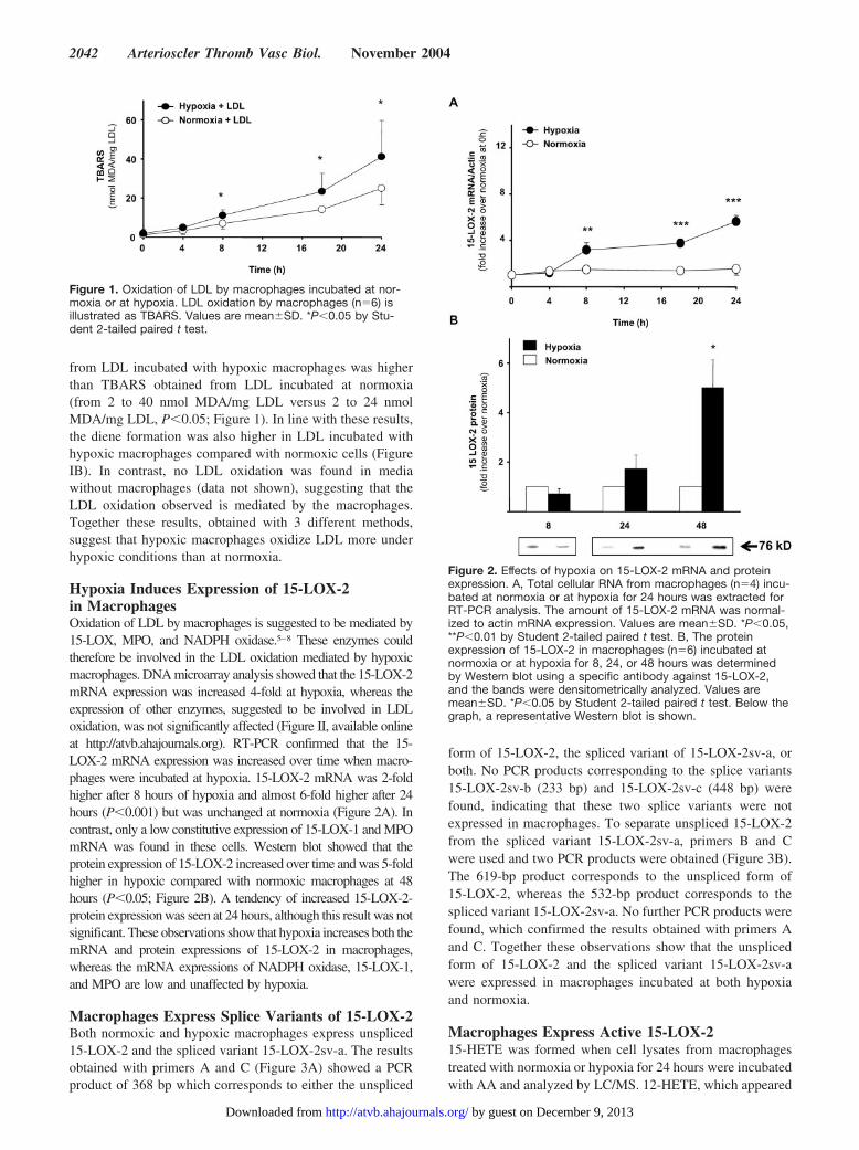
from LDL incubated with hypoxic macrophages was higherthan TBARS obtained from LDL incubated at normoxia(from 2 to 40 nmol MDA/mg LDL versus 2 to 24 nmolMDA/mg LDL, P�0.05; Figure 1). In line with these results,the diene formation was also higher in LDL incubated withhypoxic macrophages compared with normoxic cells (FigureIB). In contrast, no LDL oxidation was found in mediawithout macrophages (data not shown), suggesting that theLDL oxidation observed is mediated by the macrophages.Together these results, obtained with 3 different methods,suggest that hypoxic macrophages oxidize LDL more underhypoxic conditions than at normoxia.
Hypoxia Induces Expression of 15-LOX-2in MacrophagesOxidation of LDL by macrophages is suggested to be mediated by15-LOX, MPO, and NADPH oxidase.5–8 These enzymes couldtherefore be involved in the LDL oxidation mediated by hypoxicmacrophages. DNA microarray analysis showed that the 15-LOX-2mRNA expression was increased 4-fold at hypoxia, whereas theexpression of other enzymes, suggested to be involved in LDLoxidation, was not significantly affected (Figure II, available onlineat http://atvb.ahajournals.org). RT-PCR confirmed that the 15-LOX-2 mRNA expression was increased over time when macro-phages were incubated at hypoxia. 15-LOX-2 mRNA was 2-foldhigher after 8 hours of hypoxia and almost 6-fold higher after 24hours (P�0.001) but was unchanged at normoxia (Figure 2A). Incontrast, only a low constitutive expression of 15-LOX-1 and MPOmRNA was found in these cells. Western blot showed that theprotein expression of 15-LOX-2 increased over time and was 5-foldhigher in hypoxic compared with normoxic macrophages at 48hours (P�0.05; Figure 2B). A tendency of increased 15-LOX-2-protein expression was seen at 24 hours, although this result was notsignificant. These observations show that hypoxia increases both themRNA and protein expressions of 15-LOX-2 in macrophages,whereas the mRNA expressions of NADPH oxidase, 15-LOX-1,and MPO are low and unaffected by hypoxia.
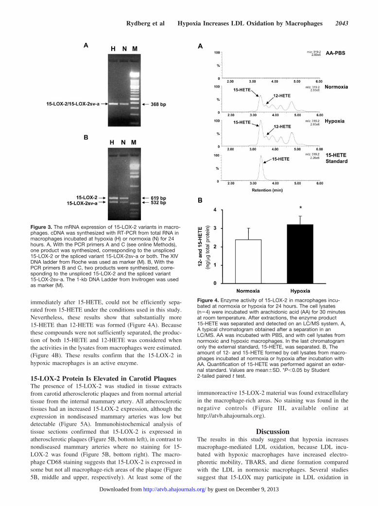
Macrophages Express Splice Variants of 15-LOX-2Both normoxic and hypoxic macrophages express unspliced15-LOX-2 and the spliced variant 15-LOX-2sv-a. The resultsobtained with primers A and C (Figure 3A) showed a PCRproduct of 368 bp which corresponds to either the unspliced
form of 15-LOX-2, the spliced variant of 15-LOX-2sv-a, orboth. No PCR products corresponding to the splice variants15-LOX-2sv-b (233 bp) and 15-LOX-2sv-c (448 bp) werefound, indicating that these two splice variants were notexpressed in macrophages. To separate unspliced 15-LOX-2from the spliced variant 15-LOX-2sv-a, primers B and Cwere used and two PCR products were obtained (Figure 3B).The 619-bp product corresponds to the unspliced form of15-LOX-2, whereas the 532-bp product corresponds to thespliced variant 15-LOX-2sv-a. No further PCR products werefound, which confirmed the results obtained with primers Aand C. Together these observations show that the unsplicedform of 15-LOX-2 and the spliced variant 15-LOX-2sv-awere expressed in macrophages incubated at both hypoxiaand normoxia.
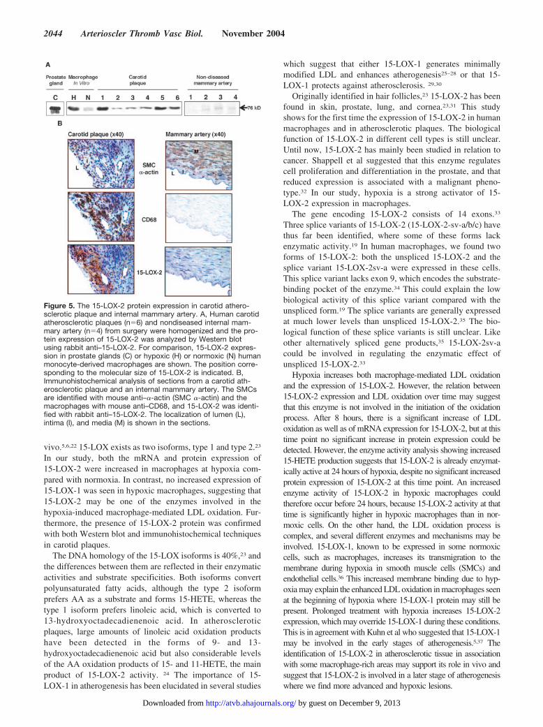
Macrophages Express Active 15-LOX-215-HETE was formed when cell lysates from macrophagestreated with normoxia or hypoxia for 24 hours were incubatedwith AA and analyzed by LC/MS. 12-HETE, which appeared
Figure 1. Oxidation of LDL by macrophages incubated at nor-moxia or at hypoxia. LDL oxidation by macrophages (n�6) isillustrated as TBARS. Values are mean�SD. *P�0.05 by Stu-dent 2-tailed paired t test.
Figure 2. Effects of hypoxia on 15-LOX-2 mRNA and proteinexpression. A, Total cellular RNA from macrophages (n�4) incu-bated at normoxia or at hypoxia for 24 hours was extracted forRT-PCR analysis. The amount of 15-LOX-2 mRNA was normal-ized to actin mRNA expression. Values are mean�SD. *P�0.05,**P�0.01 by Student 2-tailed paired t test. B, The proteinexpression of 15-LOX-2 in macrophages (n�6) incubated atnormoxia or at hypoxia for 8, 24, or 48 hours was determinedby Western blot using a specific antibody against 15-LOX-2,and the bands were densitometrically analyzed. Values aremean�SD. *P�0.05 by Student 2-tailed paired t test. Below thegraph, a representative Western blot is shown.
2042 Arterioscler Thromb Vasc Biol. November 2004
by guest on December 9, 2013http://atvb.ahajournals.org/Downloaded from
immediately after 15-HETE, could not be efficiently sepa-rated from 15-HETE under the conditions used in this study.Nevertheless, these results show that substantially more15-HETE than 12-HETE was formed (Figure 4A). Becausethese compounds were not sufficiently separated, the produc-tion of both 15-HETE and 12-HETE was considered whenthe activities in the lysates from macrophages were estimated.(Figure 4B). These results confirm that the 15-LOX-2 inhypoxic macrophages is an active enzyme.
15-LOX-2 Protein Is Elevated in Carotid PlaquesThe presence of 15-LOX-2 was studied in tissue extractsfrom carotid atherosclerotic plaques and from normal arterialtissue from the internal mammary artery. All atherosclerotictissues had an increased 15-LOX-2 expression, although theexpression in nondiseased mammary arteries was low butdetectable (Figure 5A). Immunohistochemical analysis oftissue sections confirmed that 15-LOX-2 is expressed inatherosclerotic plaques (Figure 5B, bottom left), in contrast tonondiseased mammary arteries where no staining for 15-LOX-2 was found (Figure 5B, bottom right). The macro-phage CD68 staining suggests that 15-LOX-2 is expressed insome but not all macrophage-rich areas of the plaque (Figure5B, middle and upper, respectively). At least some of the
immunoreactive 15-LOX-2 material was found extracellularyin the macrophage-rich areas. No staining was found in thenegative controls (Figure III, available online athttp://atvb.ahajournals.org).
DiscussionThe results in this study suggest that hypoxia increasesmacrophage-mediated LDL oxidation, because LDL incu-bated with hypoxic macrophages have increased electro-phoretic mobility, TBARS, and diene formation comparedwith the LDL in normoxic macrophages. Several studiessuggest that 15-LOX may participate in LDL oxidation in
Figure 3. The mRNA expression of 15-LOX-2 variants in macro-phages. cDNA was synthesized with RT-PCR from total RNA inmacrophages incubated at hypoxia (H) or normoxia (N) for 24hours. A, With the PCR primers A and C (see online Methods),one product was synthesized, corresponding to the unspliced15-LOX-2 or the spliced variant 15-LOX-2sv-a or both. The XIVDNA ladder from Roche was used as marker (M). B, With thePCR primers B and C, two products were synthesized, corre-sponding to the unspliced 15-LOX-2 and the spliced variant15-LOX-2sv-a. The 1-kb DNA Ladder from Invitrogen was usedas marker (M).
Figure 4. Enzyme activity of 15-LOX-2 in macrophages incu-bated at normoxia or hypoxia for 24 hours. The cell lysates(n�4) were incubated with arachidonic acid (AA) for 30 minutesat room temperature. After extractions, the enzyme product15-HETE was separated and detected on an LC/MS system. A,A typical chromatogram obtained after a separation in anLC/MS. AA was incubated with PBS, and with cell lysates fromnormoxic and hypoxic macrophages. In the last chromatogramonly the external standard, 15-HETE, was separated. B, Theamount of 12- and 15-HETE formed by cell lysates from macro-phages incubated at normoxia or hypoxia after incubation withAA. Quantification of 15-HETE was performed against an exter-nal standard. Values are mean�SD. *P�0.05 by Student2-tailed paired t test.
Rydberg et al Hypoxia Increases LDL Oxidation by Macrophages 2043
by guest on December 9, 2013http://atvb.ahajournals.org/Downloaded from
vivo.5,6,22 15-LOX exists as two isoforms, type 1 and type 2.23
In our study, both the mRNA and protein expression of15-LOX-2 were increased in macrophages at hypoxia com-pared with normoxia. In contrast, no increased expression of15-LOX-1 was seen in hypoxic macrophages, suggesting that15-LOX-2 may be one of the enzymes involved in thehypoxia-induced macrophage-mediated LDL oxidation. Fur-thermore, the presence of 15-LOX-2 protein was confirmedwith both Western blot and immunohistochemical techniquesin carotid plaques.
The DNA homology of the 15-LOX isoforms is 40%,23 andthe differences between them are reflected in their enzymaticactivities and substrate specificities. Both isoforms convertpolyunsaturated fatty acids, although the type 2 isoformprefers AA as a substrate and forms 15-HETE, whereas thetype 1 isoform prefers linoleic acid, which is converted to13-hydroxyoctadecadienenoic acid. In atheroscleroticplaques, large amounts of linoleic acid oxidation productshave been detected in the forms of 9- and 13-hydroxyoctadecadienenoic acid but also considerable levelsof the AA oxidation products of 15- and 11-HETE, the mainproduct of 15-LOX-2 activity. 24 The importance of 15-LOX-1 in atherogenesis has been elucidated in several studies
which suggest that either 15-LOX-1 generates minimallymodified LDL and enhances atherogenesis25–28 or that 15-LOX-1 protects against atherosclerosis. 29,30
Originally identified in hair follicles,23 15-LOX-2 has beenfound in skin, prostate, lung, and cornea.23,31 This studyshows for the first time the expression of 15-LOX-2 in humanmacrophages and in atherosclerotic plaques. The biologicalfunction of 15-LOX-2 in different cell types is still unclear.Until now, 15-LOX-2 has mainly been studied in relation tocancer. Shappell et al suggested that this enzyme regulatescell proliferation and differentiation in the prostate, and thatreduced expression is associated with a malignant pheno-type.32 In our study, hypoxia is a strong activator of 15-LOX-2 expression in macrophages.
The gene encoding 15-LOX-2 consists of 14 exons.33
Three splice variants of 15-LOX-2 (15-LOX-2-sv-a/b/c) havethus far been identified, where some of these forms lackenzymatic activity.19 In human macrophages, we found twoforms of 15-LOX-2: both the unspliced 15-LOX-2 and thesplice variant 15-LOX-2sv-a were expressed in these cells.This splice variant lacks exon 9, which encodes the substrate-binding pocket of the enzyme.34 This could explain the lowbiological activity of this splice variant compared with theunspliced form.19 The splice variants are generally expressedat much lower levels than unspliced 15-LOX-2.35 The bio-logical function of these splice variants is still unclear. Likeother alternatively spliced gene products,35 15-LOX-2sv-acould be involved in regulating the enzymatic effect ofunspliced 15-LOX-2.33
Hypoxia increases both macrophage-mediated LDL oxidationand the expression of 15-LOX-2. However, the relation between15-LOX-2 expression and LDL oxidation over time may suggestthat this enzyme is not involved in the initiation of the oxidationprocess. After 8 hours, there is a significant increase of LDLoxidation as well as of mRNA expression for 15-LOX-2, but at thistime point no significant increase in protein expression could bedetected. However, the enzyme activity analysis showing increased15-HETE production suggests that 15-LOX-2 is already enzymat-ically active at 24 hours of hypoxia, despite no significant increasedprotein expression of 15-LOX-2 at this time point. An increasedenzyme activity of 15-LOX-2 in hypoxic macrophages couldtherefore occur before 24 hours, because 15-LOX-2 activity at thattime is significantly higher in hypoxic macrophages than in nor-moxic cells. On the other hand, the LDL oxidation process iscomplex, and several different enzymes and mechanisms may beinvolved. 15-LOX-1, known to be expressed in some normoxiccells, such as macrophages, increases its transmigration to themembrane during hypoxia in smooth muscle cells (SMCs) andendothelial cells.36 This increased membrane binding due to hyp-oxia may explain the enhanced LDL oxidation in macrophages seenat the beginning of hypoxia where 15-LOX-1 protein may still bepresent. Prolonged treatment with hypoxia increases 15-LOX-2expression, which may override 15-LOX-1 during these conditions.This is in agreement with Kuhn et al who suggested that 15-LOX-1may be involved in the early stages of atherogenesis.5,37 Theidentification of 15-LOX-2 in atherosclerotic tissue in associationwith some macrophage-rich areas may support its role in vivo andsuggest that 15-LOX-2 is involved in a later stage of atherogenesiswhere we find more advanced and hypoxic lesions.
Figure 5. The 15-LOX-2 protein expression in carotid athero-sclerotic plaque and internal mammary artery. A, Human carotidatherosclerotic plaques (n�6) and nondiseased internal mam-mary artery (n�4) from surgery were homogenized and the pro-tein expression of 15-LOX-2 was analyzed by Western blotusing rabbit anti–15-LOX-2. For comparison, 15-LOX-2 expres-sion in prostate glands (C) or hypoxic (H) or normoxic (N) humanmonocyte-derived macrophages are shown. The position corre-sponding to the molecular size of 15-LOX-2 is indicated. B,Immunohistochemical analysis of sections from a carotid ath-erosclerotic plaque and an internal mammary artery. The SMCsare identified with mouse anti–�-actin (SMC �-actin) and themacrophages with mouse anti–CD68, and 15-LOX-2 was identi-fied with rabbit anti–15-LOX-2. The localization of lumen (L),intima (I), and media (M) is shown in the sections.
2044 Arterioscler Thromb Vasc Biol. November 2004
by guest on December 9, 2013http://atvb.ahajournals.org/Downloaded from
Hypoxic areas are found in atherosclerotic lesions, but therole of hypoxia in the development of atherosclerotic plaquesis not known. This study shows that macrophage-mediatedLDL oxidation is significantly higher at hypoxia than atnormoxia and that hypoxia significantly increases the levelsof both mRNA and protein of the active form of 15-LOX-2.Interestingly, this is the first report of 15-LOX-2 expressionin atherosclerotic plaques. These findings suggest that hyp-oxia, by increasing macrophage-mediated LDL oxidation,may contribute to an enhanced development of atherosclero-sis, and that 15-LOX-2 may be one of the factors involved inthis hypoxia-induced LDL oxidation.
AcknowledgmentsThis work was supported by the Swedish Heart Lung Foundation(grant no. 20020385 to L.M.H.), the Swedish Society of Medicine(grant no. 2002-684 to L.M.H.), the Swedish Research Council(grants nos. 13488 and K2003-71P-14816-01A to O.W. and A.K,respectively), and the Swegene foundation (grant to P.-A.S.). We aregrateful for the technical assistance of Margareta Jernås (ResearchCenter for Endocrinology & Metabolism), Kristina Skålen (Wallen-berg Laboratory), Gun-Britt Forsberg, and Anne-Christine Andreas-son (AstraZeneca R&D).
References1. Skalen K, Gustafsson M, Rydberg EK, Hulten LM, Wiklund O, Innerarity
TL, Boren J. Subendothelial retention of atherogenic lipoproteins in earlyatherosclerosis. Nature. 2002;417:750–754.
2. Li AC, Glass CK. The macrophage foam cell as a target for therapeuticintervention. Nat Med. 2002;8:1235–1242.
3. Steinberg D, Witztum JL. Lipoproteins and atherogenesis. Currentconcepts. JAMA. 1990;264:3047–3052.
4. Yla-Herttuala S, Palinski W, Rosenfeld ME, Parthasarathy S, Carew TE,Butler S, Witztum JL, Steinberg D. Evidence for the presence of oxida-tively modified low density lipoprotein in atherosclerotic lesions of rabbitand man. J Clin Invest. 1989;84:1086–1095.
5. Kuhn H, Belkner J, Zaiss S, Fahrenklemper T, Wohlfeil S. Involvementof 15-lipoxygenase in early stages of atherogenesis. J Exp Med. 1994;179:1903–1911.
6. Folcik VA, Nivar-Aristy RA, Krajewski LP, Cathcart MK. Lipoxygenasecontributes to the oxidation of lipids in human atherosclerotic plaques.J Clin Invest. 1995;96:504–510.
7. Podrez EA, Abu-Soud HM, Hazen SL. Myeloperoxidase-generatedoxidants and atherosclerosis. Free Radic Biol Med. 2000;28:1717–1725.
8. Aviram M, Rosenblat M, Etzioni A, Levy R. Activation of NADPHoxidase required for macrophage-mediated oxidation of low-densitylipoprotein. Metabolism. 1996;45:1069–1079.
9. Morrison AD, Clements RS Jr, Winegrad AI. Effects of elevated glucoseconcentrations on the metabolism of the aortic wall. J Clin Invest. 1972;51:3114–3123.
10. Bjornheden T, Bondjers G. Oxygen consumption in aortic tissue fromrabbits with diet-induced atherosclerosis. Arteriosclerosis. 1987;7:238–247.
11. Lewis JS, Lee JA, Underwood JC, Harris AL, Lewis CE. Macrophageresponses to hypoxia: relevance to disease mechanisms. J Leukoc Biol.1999;66:889–900.
12. Bjornheden T, Levin M, Evaldsson M, Wiklund O. Evidence of hypoxicareas within the arterial wall in vivo. Arterioscler Thromb Vasc Biol.1999;19:870–876.
13. Turner L, Scotton C, Negus R, Balkwill F. Hypoxia inhibits macrophagemigration. Eur J Immunol. 1999;29:2280–2287.
14. Ohlsson BG, Englund MC, Karlsson AL, Knutsen E, Erixon C, SkribeckH, Liu Y, Bondjers G, Wiklund O. Oxidized low density lipoproteininhibits lipopolysaccharide-induced binding of nuclear factor-kappaB toDNA and the subsequent expression of tumor necrosis factor-� andinterleukin-1� in macrophages. J Clin Invest. 1996;98:78–89.
15. Rydberg EK, Salomonsson L, Hulten LM, Noren K, Bondjers G, WiklundO, Bjornheden T, Ohlsson BG. Hypoxia increases 25-hydroxycholester-ol-induced interleukin-8 protein secretion in human macrophages. Ath-erosclerosis. 2003;170:245–252.
16. Bradford MM. A rapid and sensitive method for the quantitation ofmicrogram quantities of protein utilizing the principle of protein-dyebinding. Anal Biochem. 1976;72:248–254.
17. Ohkawa H, Ohishi N, Yagi K. Assay for lipid peroxides in animal tissuesby thiobarbituric acid reaction. Anal Biochem. 1979;95:351–358.
18. Esterbauer H, Gebicki J, Puhl H, Jurgens G. The role of lipid peroxidationand antioxidants in oxidative modification of LDL. Free Radic Biol Med.1992;13:341–390.
19. Tang S, Bhatia B, Maldonado CJ, Yang P, Newman RA, Liu J, ChandraD, Traag J, Klein RD, Fischer SM, Chopra D, Shen J, Zhau HE, ChungLW, Tang DG. Evidence that arachidonate 15-lipoxygenase 2 is anegative cell cycle regulator in normal prostate epithelial cells. J BiolChem. 2002;277:16189–16201.
20. Galis ZS, Sukhova GK, Lark MW, Libby P. Increased expression ofmatrix metalloproteinases and matrix degrading activity in vulnerableregions of human atherosclerotic plaques. J Clin Invest. 1994;94:2493–2503.
21. Krettek A, Sukhova GK, Libby P. Elastogenesis in human arterialdisease: a role for macrophages in disordered elastin synthesis. Arte-rioscler Thromb Vasc Biol. 2003;23:582–587.
22. Yla-Herttuala S, Rosenfeld ME, Parthasarathy S, Glass CK, Sigal E,Witztum JL, Steinberg D. Colocalization of 15-lipoxygenase mRNA andprotein with epitopes of oxidized low density lipoprotein in macro-phage-rich areas of atherosclerotic lesions. Proc Natl Acad Sci U S A.1990;87:6959–6963.
23. Brash AR, Boeglin WE, Chang MS. Discovery of a second 15S-lipoxy-genase in humans. Proc Natl Acad Sci U S A. 1997;94:6148–6152.
24. Waddington E, Sienuarine K, Puddey I, Croft K. Identification andquantitation of unique fatty acid oxidation products in human athero-sclerotic plaque using high-performance liquid chromatography. AnalBiochem. 2001;292:234–244.
25. Sigari F, Lee C, Witztum JL, Reaven PD. Fibroblasts that overexpress15-lipoxygenase generate bioactive and minimally modified LDL. Arte-rioscler Thromb Vasc Biol. 1997;17:3639–3645.
26. Harats D, Shaish A, George J, Mulkins M, Kurihara H, Levkovitz H,Sigal E. Overexpression of 15-lipoxygenase in vascular endotheliumaccelerates early atherosclerosis in LDL receptor-deficient mice. Arte-rioscler Thromb Vasc Biol. 2000;20:2100–2105.
27. Cyrus T, Witztum JL, Rader DJ, Tangirala R, Fazio S, Linton MF, FunkCD. Disruption of the 12/15-lipoxygenase gene diminishes atherosclero-sis in apo E-deficient mice. J Clin Invest. 1999;103:1597–1604.
28. George J, Afek A, Shaish A, Levkovitz H, Bloom N, Cyrus T, Zhao L,Funk CD, Sigal E, Harats D. 12/15-Lipoxygenase gene disruptionattenuates atherogenesis in LDL receptor-deficient mice. Circulation.2001;104:1646–1650.
29. Shen J, Herderick E, Cornhill JF, Zsigmond E, Kim HS, Kuhn H,Guevara NV, Chan L. Macrophage-mediated 15-lipoxygenase expressionprotects against atherosclerosis development. J Clin Invest. 1996;98:2201–2208.
30. Serhan CN, Jain A, Marleau S, Clish C, Kantarci A, Behbehani B, ColganSP, Stahl GL, Merched A, Petasis NA, Chan L, Van Dyke TE. Reducedinflammation and tissue damage in transgenic rabbits overexpressing15-lipoxygenase and endogenous anti-inflammatory lipid mediators.J Immunol. 2003;171:6856–6865.
31. Chanez P, Bonnans C, Chavis C, Vachier I. 15-lipoxygenase: a Janusenzyme? Am J Respir Cell Mol Biol. 2002;27:655–658.
32. Shappell SB, Boeglin WE, Olson SJ, Kasper S, Brash AR. 15-lipoxygen-ase-2 (15-LOX-2) is expressed in benign prostatic epithelium and reducedin prostate adenocarcinoma. Am J Pathol. 1999;155:235–245.
33. Furstenberger G, Marks F, Krieg P. Arachidonate 8(S)-lipoxygenase.Prostaglandins Other Lipid Mediat. 2002;68–69:235–243.
34. Kilty I, Logan A, Vickers PJ. Differential characteristics of human 15-li-poxygenase isozymes and a novel splice variant of 15S-lipoxygenase. EurJ Biochem. 1999;266:83–93.
35. Lopez AJ. Alternative splicing of pre-mRNA: developmental conse-quences and mechanisms of regulation. Annu Rev Genet. 1998;32:279–305.
36. Zhu D, Medhora M, Campbell WB, Spitzbarth N, Baker JE, Jacobs ER.Chronic hypoxia activates lung 15-lipoxygenase, which catalyzes produc-tion of 15-HETE and enhances constriction in neonatal rabbit pulmonaryarteries. Circ Res. 2003;92:992–1000.
37. Kuhn H, Heydeck D, Hugou I, Gniwotta C. In vivo action of 15-lipox-ygenase in early stages of human atherogenesis. J Clin Invest. 1997;99:888–893.
Rydberg et al Hypoxia Increases LDL Oxidation by Macrophages 2045
by guest on December 9, 2013http://atvb.ahajournals.org/Downloaded from
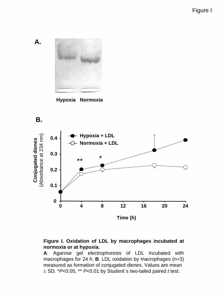
Figure I
A.
Hypoxia Normoxia
B.
0
0.1
0.2
0.3
0.4
0 4 8 12 16 20 24
Time (h)
Con
juga
ted
dien
es(A
bsor
banc
eat
234
nm
)
** *
Normoxia + LDLHypoxia + LDL
Figure I. Oxidation of LDL by macrophages incubated at normoxia or at hypoxia.A. Agarose gel electrophoresis of LDL incubated with macrophages for 24 h. B. LDL oxidation by macrophages (n=3) measured as formation of conjugated dienes. Values are mean ± SD. *P<0.05, ** P<0.01 by Student´s two-tailed paired t test.
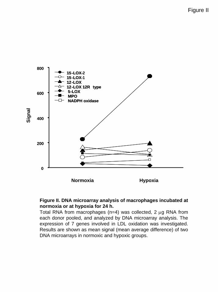
Figure II
0
200
400
600
80015-LOX 215-LOX 112-LOX12-LOX 12R type
MPONADPH oxidase
5-LOX
0
200
400
600
800
Normoxia Hypoxia
Sign
al
15-LOX-15-LOX-12-LOX12-LOX 12R type
MPONADPH oxidase
5-LOX
Figure II. DNA microarray analysis of macrophages incubated at normoxia or at hypoxia for 24 h.Total RNA from macrophages (n=4) was collected, 2 µg RNA from each donor pooled, and analyzed by DNA microarray analysis. The expression of 7 genes involved in LDL oxidation was investigated. Results are shown as mean signal (mean average difference) of two DNA microarrays in normoxic and hypoxic groups.
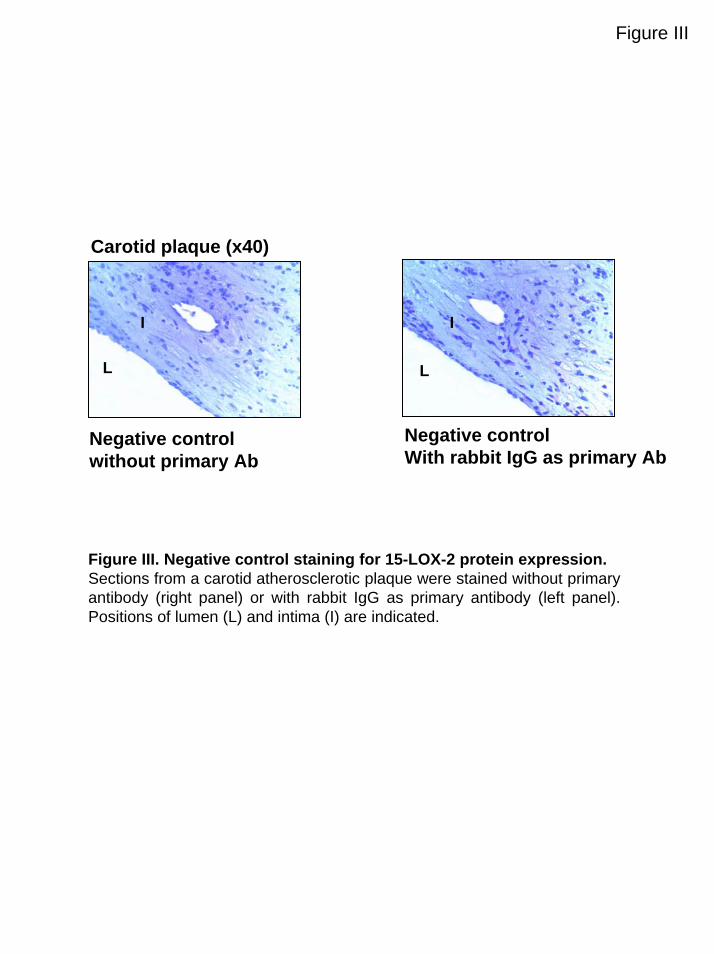
Figure III
Carotid plaque (x40)
Negative controlwithout primary Ab
L
I I
L
Negative controlWith rabbit IgG as primary Ab
Figure III. Negative control staining for 15-LOX-2 protein expression.Sections from a carotid atherosclerotic plaque were stained without primary antibody (right panel) or with rabbit IgG as primary antibody (left panel). Positions of lumen (L) and intima (I) are indicated.
METHODS SUPPLEMENT MS ID#: ATVB/2004/016196, Rydberg et al.
LDL Preparation
Human LDL (d = 1.019 to 1.063 g/mL) was prepared by sequential centrifugation of plasma
from healthy fasted male donors in the presence of EDTA (0.2% w/v) at 4°C. For each
experiment LDL from 2 donors were pooled, filtered through 0.22-µm sterile filters, stored at
4°C, and used within 1 week. Before use, LDL was separated from EDTA over two
consecutive PD-10 columns (Amersham Pharmacia Biotech).
DNA Microarray Analysis
Total RNA was isolated with the RNeasy kit (Qiagen) from macrophages incubated at
normoxia or hypoxia for 24 h. Two µg RNA from 4 donors incubated at normoxia and 4
donors at hypoxia was pooled respectively and these 2 pools were used for the target
preparation and analyzed on duplicate DNA microarrays (Hu95A; Affymetrix). Target
preparation, DNA microarray hybridization, and scanning were performed as described 1.
Scanned output files were analyzed with GeneChip 3.1 software (Affymetrix) and globally
scaled to an average intensity of 500. Potential candidate genes encoding proteins involved in
LDL oxidation and lipoxygenase-related genes were selected based on information from
current literature 2-6. The probe sets corresponding to these genes were designed using the
Netaffix database (www.affymetrix.com). The mean average signal difference was compared
between normoxic and hypoxic groups.
Real-Time RT-PCR
Real-time RT-PCR was performed with a TaqMan RT-PCR kit (Applied Biosystems) to
detect mRNA expression of 15-lipoxygenase type 1 (15-LOX-1), 15-LOX-2, and MPO. The
1
expression was normalized to actin mRNA expression using a pre-developed TaqMan assay
reagent kit, with primers and the probe purchased from Applied Biosystems. The actin probe
was 5′-labeled with VIC® and 3′-labeled with tetramethylrhodamine (TAMRA).
Oligonucleotide primers and probes for the different genes of interest were designed with the
Primer Express 1.5 software and purchased from Applied Biosystems. These probes were 5′-
labeled with 5-carboxyfluorescein (FAM) and 3′-labeled with TAMRA. The following
primers and probes were used. Human 15-LOX-2 (GenBank No. U78294): forward primer,
5′-GGCCTCATTGTTGGGTCCT-3′; reverse primer, 5′-TGCCGTGATCCACCA AGAA-3′;
probe, FAM-5′-AGCCCTTCTCTAGCTCAGCCTGCAAGC-3′-TAMRA. Human 15-LOX-1
(GenBank No. M23892): forward primer, 5′-TGAGCGATTTCTGGAAGACA AGA-3′;
reverse primer, 5′-ATTTAGAGAGTCTTTGTATAGCGAGGTC-3′; probe, FAM-5′-
CCTTGGCCAGCGAAACCTCAAAGTC-3′-TAMRA. Human MPO (GenBank No.
M19507): forward primer, 5′-CAGGACAAATACCGCACCATC-3′; reverse primer, 5′-
CACAAAGGC ACGGTTGGAG-3′; probe, FAM-5′-
ACCGGGATGTGCAACAACAGACGC-3′-TAMRA. The RT reaction was performed with a
Gene Amp PCR system 9700 (Applied Biosystems). PCR amplification for all genes was
performed for 40 cycles on an ABI PRISM 7700 sequence detection system (Applied
Biosystems).
Splice Variants of 15-Lipoxygenase-2
The Taqman reverse transcriptase reaction kit, with random hexamer primers (Applied
Biosystems), was used to synthesize cDNA from RNA of normoxic or hypoxic treated
macrophages. To identify the splice variants 15-LOX-2sv-b (233 bp) and 15-LOX-2sv-c (448
bp), the PCR was performed using the forward primer A (5′-
CAGGCTACTACTACCGTGATG-3′) corresponding to position 1477-1498 and the reverse
2
primer C (5′-TATGAAGCCCTCGCATGTTG-3′) corresponding to position 1844-1825 of the
15-LOX-2 cDNA sequence (GenBank No.U78294). Using these primers, the unspliced form
of 15-LOX-2 and the splice variant 15-LOX-2sv-a result in two PCR products with equal
sizes (368 bp). Therefore, another PCR was performed to separate the unspliced 15-LOX-2
from the splice variant 15-LOX-2sv-a by using the forward primer B (5′-
CCTGGCTACCCTGCGTCA-3′) corresponding to position 1226-1243 together with the
reverse primer C. In this reaction, the unspliced 15-LOX-2 and the splice variant 15-LOX-
2sv-a, 15-LOX-2sv-b and 15-LOX-2sv-c would correspond to PCR products of 619 bp, 532
bp, 397 bp and 699 bp, respectively. The PCR products were separated on a 2% agarose gel
and visualized by ethidium bromide staining.
REFERENCES
1. Gabrielsson BG, Johansson JM, Jennische E, Jernas M, Itoh Y, Peltonen M, Olbers T,
Lonn L, Lonroth H, Sjostrom L, Carlsson B, Carlsson LM, Lonn M. Depot-specific expression of fibroblast growth factors in human adipose tissue. Obes Res. 2002;10:608-616.
2. Kuhn H, Belkner J, Zaiss S, Fahrenklemper T, Wohlfeil S. Involvement of 15-lipoxygenase in early stages of atherogenesis. J Exp Med. 1994;179:1903-1911.
3. Folcik VA, Nivar-Aristy RA, Krajewski LP, Cathcart MK. Lipoxygenase contributes to the oxidation of lipids in human atherosclerotic plaques. J Clin Invest. 1995;96:504-510.
4. Podrez EA, Abu-Soud HM, Hazen SL. Myeloperoxidase-generated oxidants and atherosclerosis. Free Radic Biol Med. 2000;28:1717-1725.
5. Aviram M, Rosenblat M, Etzioni A, Levy R. Activation of NADPH oxidase required for macrophage-mediated oxidation of low-density lipoprotein. Metabolism. 1996;45:1069-1079.
6. Furstenberger G, Marks F, Krieg P. Arachidonate 8(S)-lipoxygenase. Prostaglandins Other Lipid Mediat. 2002;68-69:235-243.
3


