Embed Size (px)
Citation preview

HPB Surgery, 1997, Vol. 10, pp. 221-227Reprints available directly from the publisherPhotocopying permitted by license only
(C) 1997 OPA (Overseas Publishers Association)Amsterdam B.V. Published in The Netherlands
by Harwood Academic PublishersPrinted in India
Iatrogenic Biliary Strictures" Surgical Experience with 39Patients
LUIZ ROHDE, MRIOSRGIO BORGES DA COSTA, LUIS ROBERTO WENDT, OLY CORLETAandMARCELO FERREIRA
Department of Surgery, Medical School of the Federal University ofRio Grande Do Sul, Hospital De Clinicas, Porto Alegre, Brazil
(Received April 1995)
The authors report their experience with surgicaltreatment of 39 patients with biliary strictures ofiatrogenic origin. Patients were grouped according tothe level of obstruction as described by Bismuth, andthe type of repair was based on this classification. Atotal of 45 operations were performed, including thosefor recurrent strictures: 22 hepaticojejunostomies, 10Hepp and Couinad’s operations, 6 choledochojejuno-stomies, 3 separate right and left hepaticojejuno-stomies, 1 hepaticojejunostomy with mucosal graft(Smith’s technique), 1 intrahepatic cholangiojejuno-stomy (Longmire’s technique), 1 choledochoduode-nostomy and 1 choledochoplasty. Results wereconsidered good if the patient was free of symptoms,jaundice or episodes of cholangitis, with serum alka-line phosphatase less than two-times the normal value.Minimum follow-up period of two years (obtained in35 patients) was required to evaluate the results. Goodresults were obtained in 26 of those 30 patients (87%)who underwent only one biliary reconstruction, and in3 of those 5 (60%) with more than one repair. Overall, 29patients (83% of those 35) presented good results. Thecomplexity of the surgical treatment of biliary stric-tures imposes the adoption of measures to preventlesions to the bile duct. Factors related to the progno-sis that must be emphasized are surgeons’ individualexperience and skills, location of the stricture anddiameter of the anastomosis.
Keywords: Biliary strictures, biliary obstruction, bileduct injuries, bilioenteric anastomosis
INTRODUCTION
Recent studies show that accidental surgicallesions of the biliary ducts occur in 0.2% of opencholecystectomies [1,2] and that this percentageis higher with the laparoscopic technique [3,4].These lesions can result in bile peritonitis,biliaryfistula, biliary tract strictures or an associationof these complications.Repeated surgical procedures at the hepatic
hilum makes the treatment of biliary stricturesa great challenge, especially the higher ones, de-manding skill and experience from the surgeon.Many surgical procedures have been described totreat these lesions [5-8], but the limited number ofcases treated by the same surgeon makes the stand-ardization of the reconstruction technique dif-ficult. The classification of the biliary strictures
proposed by Bismuth [9] has been adopted to des-cribe these lesions. However, the important of its usein choosing the treatment has not been emphasized.
This study reports the experience of the Liverand Biliary Tract Surgical Group of the Hospital deClnicas de Porto Alegre (HCPA) with the surgicaltreatment of iatrogenic biliary strictures in a 13
Correspondence to: Professor Luiz Rohde, Av. Palmeira 740, CEP 90470-300, Porto Alegre, Brazil.
221
222 L. ROHDE et al.
year period, emphasizing the choice of tech-nique according to the stricture level.
PATIENTS AND METHODS
(ERCP) in 12 patients. Six of those 12 had bothERCP and PTC.
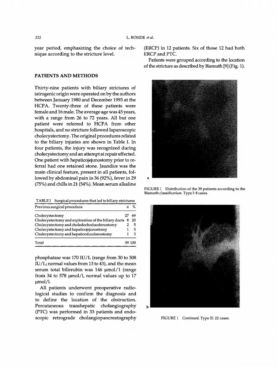
Patients were grouped according to the locationof the stricture as described by Bismuth [9] (Fig. 1).
Thirty-nine patients with biliary strictures ofiatrogenic origin were operated onby the authorsbetween January 1980 and December 1993 at theHCPA. Twenty-three of these patients werefemale and 16 male. The average age was 45 years,with a range from 26 to 72 years. All but one
patient were referred to HCPA from otherhospitals, and no stricture followed laparoscopiccholecystectomy. The original procedures relatedto the biliary injuries are shown in Table I. Infour patients, the injury was recognized duringcholecystectomy and an attempt at repair effected.One patient with hepaticojejunostomy prior to re-ferral had one retained stone. Jaundice was themain clinical feature, present in all patients, fol-lowed by abdominal pain in 36 (92%), fever in 29(75%) and chills in 21 (54%). Mean serum alkaline
TABLE Surgical procedures that led to biliary strictures
Previoussurgicalprocedure n %
Cholecystectomy 27 69Cholecystectomy and exploration of thebiliary ducts 8 20Cholecystectomy and choledochoduodenostomy 2 5Cholecystectomy and hepaticojejunostomy 1 3Cholecystectomy and hepaticoduodenostomy 1 3
FIGURE Distribution of the 39 patients according to theBismuth classification. Type I: 8 cases.
Total 39 100
phosphatase was 170 IU/L (range from 30 to 508IU/L; normal values from 13 to 43), and the meanserum total bilirrubin was 146 blmol/1 (rangefrom 34 to 578 lmol/1, normal values up to 17wnol/1.
All patients underwent preoperative radio-
logical studies to confirm the diagnosis andto define the location of the obstruction.Percutaneous transhepatic cholangiography(PTC) was performed in 33 patients and endo-scopic retrograde cholangiopancreatography FIGURE 1 Continued. Type II: 22 cases.

IATROGENIC BILIARY STRICTURES 223
FIGURE 1 Continued. Type III" 7 cases
FIGURE 1 Continued. Type IV: 2 cases
The initial surgical repair of these 39 patients isshown in Table II. All biliary-enteric anastomo-sis were performed with one-layer of inter-
rupted stitches of chromic catgut 3-0 or
TABLE II Reconstructions used to treat the different typesof strictures
Type Technique of reconstruction
IIIII
IV
Choledocojejunostomy* 6Choledochoduodenostomy 1Choledocoplasty 1Hepaticojejunostomy* 22Hepp & Couinaud* 6Hepaticojejunostomy* R and L 1Hepaticojejunostomy* R and L 1Hepaticojejunostomy*withmucosalgraft (Smith) 1
Total 39
*Roux-en-Y reconstruction;R=right; L=left.
poligalactin (Vycril) 3-0, and those usingjejunun were constructed in the Roux-en-Y fash-ion. Stents were used in 20 patients and left in
place from 16 to 75 days (average of 56 days).Results were considered good if the patient
was free of symptoms, jaundice or episodes ofcholangitis[10] with serum phosphatase less thantwice the normal value [2,11,12]. Minimum fol-low-up period of two yearswas required to eval-uate the results.
RESULTS
A total of 45 biliary-enteric anastomosis were
performed. Thirty-four patients underwent onlyone procedure. Five patients required more thanone repair due to re-stenosis. In 4 of those 5patients, stenotic hepaticojejunostomies were fol-lowed by Hepp and Couinaud’s operations [6],one of which subsequently strictured and was
repaired by Longmire’s intrahepatic cholangio-jejeunostomy [7]. The other patient with a strictureafter Hepp and Couinaud’s anastomosis requireda separate right and left hepaticojejunostomy forrepair. This patient has postoperative sepsis andwas the only death of this series of 39 patients(2.5%). Nonlethal postoperative complicationswere wound infection (5 cases), external biliaryfistula (4 cases with spontaneous closure), sub-hepatic abscess (1 case treated by percutaneous

224 L. ROHDE et al.
drainage) and intra-abdominal bleeding (1 casewhich demanded reoperation).
Reoperations after failure of the initial repairare shown in Table III. Thirty-five patients hada minimum follow-up period of 2 years, and themean follow up period in this group was37 months. Good results ’were obtained in 26 ofthose 30 patients (87%) who underwent onlyone biliary reconstruction, and in 3 of those 5with more than one repair. Overall, 29 patients(83% of the 35) presented good results (Fig. 2).Previous repairs in those 4 patients with un-
satisfactory results who underwent only one
TABLE III Reoperations after failure of the initial biliaryrepair based on Bismuth classification
Type n Original surgery Treatment (reoperation)
II 4 Hepaticojejunostomy Hepp and CouinaudIII 1 Hepp and Couinaud HJ* isolated R and L
1 Hepp and Couinaud LongmireiV 0
*HJ=hepaticojejunostomy; R=right; L=left.
39 patients opertted
30 (one operati0,n) (more than one operation)
26goodesudeath biliary
(sepsis) cirrhosis
29 good results (83%)
cases with episodic cholangitis and cases with alkaline phosplmtase persistently elevated.
FIGURE 2 Clinical result related to the number of surgicalprocedures.
operation were one choledochoplasty, onecholedochoduodenostomy, one separate rightand left hepaticojejunostomy and one Smith’smucosal-graft [8]. The two patients with persis-tently elevated alkaline phosphatase did notundergo liver biopsy to rule out the develop-ment of cirrhosis.
DISCUSSION
The great majority of the benign biliary stricturesare secondary to accidental surgical trauma, andthe operation that most often leads to these le-sions is cholecystectomy [12,13]. In this series, allreferred patients have been submitted previouslyto open cholecystectomy, one third of them withassociated bile duct exploration and four had un-
dergone biliodigestive anastomosis. The author’sexperience in the treatment of biliary stricturesinduced by laparoscopic cholecystectomy beganonly after December 1993.Videolaparoscopic cholecystectomy has be-
come the treatment of choice of symptomaticcholelithiasis in many institutions, and a bile ductlesion is considered its most important complica-tion [14]. The effect of this new approach in theincidence of biliary lesions is a cause of debate. Ina North-American study of 1518 videolaparo-scopic cholecystectomies [15], it has beenobserved that the frequency of intraoperativeductal lesions is greater in the first 13 case of thesurgeon (2.2%), decreasing to levels comparableto open cholecystectomy with the increase of thesurgeon’s experience. In a study of 77,7604 casestreated in many institutions, Deziel and co-work-ers [16] observed that the mean rate of bile ductinjury was 0.65% in hospitals performing 100 orfewer cases compared with 0.42% in centers thathas performed more than 100 cases. However,these figures are well above those reported in
open cholecystectomy. Recent publications de-monstrate rates of bile duct injury from 0.16 to0,2% [1,17]. Some authors suggest that laparo-scopic cholecystectomy is associated with a in-
herently higher risk of bile duct injury than

IATROGENIC BILIARY STRICTURES 225
open cholecystectomy [3,4,16,18,19]. Besides, therewas a seemingly increased number of patientswith accidental biliary lesions referred to someinstitutions [4,14,18]. Measures to prevent biliaryinjury during laparoscopic cholecys-tectomy havebeen proposed by some authors [9-21].
Intraoperative recognition of the bile ductinjury during the original operation occurred in
only four patients of this series (10%). Most au-
thors show intraoperative recognition rates dur-ing surgery of 12-22%, but in some series theserates reach up to 37-42% [22, 23]. One possibleexplanation for the low frequency of early recog-nition of the biiiary injuries in this and otherseries could be the fact that, in many cases, thebile duct wall is not sectioned, but the bloodvessels are damaged. Northover and Terblanche[24] showed that the arterial supply of the supra-duodenal duct is axial, with the main vesselsbeing located at the 3 o’clock and 9 o’ clock posi-tions along the lateral and medial borders of theduct. These vessels are easily damaged, and thereare reported cases confirming as ischemic basisfor biliary strictures [25]. In order to avoidischemic lesions, some authors advise minimumdissection of the proximal bile duct stump duringreconstruction [5;26].
Thirty-three of 39 cases of the present serieswere investigated radiologically by percuta-neous transhepatic cholangiography (PTC). Thisis considered the method of choice for radiologi-cal investigation in this setting because ensuesvisualization of the stricture location and theanatomy of the proximal ducts [10,12]. In addi-tion, placing a catheter during the procedure al-lows preoperative external biliary drainage andfacilitates intraoperative location of the bile ductproximal to the obstruction [10,13,27].
Transanastomotic tubes have been used by thevast majority of authors [5,8,10,13,22,23,28-31],although some use them selectively [9,11,12].How long tubes should be kept in place is con-troversial. In general, it is recommended tomaintain tubes for 3 to 6 months. In most complex
cases, up to 12 months of stenting can be neces-
sary [22,32,33]. The authors who use stents se-
lectively consider that they are unnecessary if atension-free anastomosis with wide stoma andgood blood supply is accomplished. However,they use tubes in case of higher and more
complex strictures, specially those with unsuc-cessful previous repairs. In this study,transanastomotic tubes were used selectivelyand were preferentially exteriorized throughthe excluded jejunal loop (like a Witzel
jejunostomy) (Fig. 3), and exceptionally by thetranshepatic route (Fig. 4). Data from this study(and other studies in which the selective use oftubes is adopted) don’t permit valid conclu-sions concerning the influence of stents in out-come, since they were used only in complicatedcases.
FIGURE 3 Cholangiography of a right and left hepaticojejuno-stomy with good result.

226 L. ROHDE et al.
FIGURE 4 Cholangiography of a hepaticojejunostomy withmucosal graft (Smith’s technique).
Caution is necessary to compare the results ofsurgical reconstruction from different institu-tions. Differences related to the location of thestricture, the moment of the repair (when theinjury occurs or later on), the number ofpreviousattempts of repairs, the presence of complications(biliary cirrhosis, portal hypertension, cholangitis),as well as diverse definitions of"good results" canlead to conflicting conclusions. One of the criteria todefine "good results" in this study was alkalinephosphatase levelsless than two times the normalvalues. Although it canbe considered a rigid crite-ria, the authors think that it is valid since persist-ently elevated alkaline phosphatase levels denotesome degree of cholestasis, and patients with thisfeature are at increased risk of developingcholangitis or biliary cirrhosis later on [11,12].There was only one confirmed case of biliary cir-rhosis (a patient with more than one repair). Twoof the patients with one repair had persistentlyelevated alkaline phosphatase. Hepatobiliary
scintigraphy (IDA scan) in one of these patientsshowed a delayed radionuclide passage to thejejunal loop indicating biliary stricture. Noneof these two patients underwent liver biopsiesto investigate concomitant cirrhosis. The 83% ofgood results presented in this study is accept-able, since the literature shows figures from 70to 90% [10].The Bismuth classification has been used as
a pattern to the description of biliary strictures.
Analysis of the results of treatments based on thisclassfication has been used in the last years [2, 26,30,33] avoiding comparison between groups of pa-tients with different levels of complexity. Basedon the results of this and other sutides, the authorssuggest the following standardization of the sur-
gical treatment of iatrogenic bile strictures:
a) Type I: Roux-en-Y choledochojejunostomyor hepaticojejunostomy;
b) Type II: Roux-en-Y hepaticojejunostomy;c) Type III: Roux-en-Y hepaticojejunostomyby
Hepp and Couinaud’s technique;d) Type IV: repair according to the case (sepa-
rate right and left hepaticojejunostomy orSmith’s mucosal graft).
The length of choledochus and hepatic ductdepends on the level of union of the cystic ductwith the biliary tract: the higher the union be-tween cystic duct and choledochus, the shorterhepatic duct is, making hepaticojejunostomy moredifficult.Choledochoduodenostomywas performed only
once to hasten the operation of a high-risk elderlypatient. Choledochoplasty was indicated onlyonce too. Both techniques are not good for biliarystricture repair, and resulted in bad outcome inthis series. The risk ofpoor results increases whenthe initial repair is inappropriate, the stricture is
high and more than one reconstruction is neces-sary [27,33]. The occurence of two re-stenosis in6 Hepp and Couinaud’s operations does notmean that this technique is not valuable, butindeed denotes the complexity of the cases inwhich it was used (type III strictures).The surgical treatment of iatrogenic biliary
strictures is a great challenge. Even in experienced

IATROGENIC BILIARY STRICTURES 227
hands, significant morbidity and bad long-termresults are frequent. Consequently, it is of ut-most important to adopt routine measures toprevent accidental intraoperative bile dict in-
jury [14,19-21]. During the operative repair,some principles of biliary reconstruction mustbe kept in mind, such as the importance of per-forming a wide tension-free anastomosis withmucosa-mucosa apposition in all circumferenceand with a good blood supply [26,27].
References[1] Roslyn, J.J., Binns, G.S., Hughes, E.F.X., Kirkwood,
K.S., Zinner, M.J. and Cates, J.A. (1993). Opencholecystectomy: a contemporary analysis of 42, 474patients. Annals of Surgery, 218,129-137.
[2] Raute, M., Podlech, P., Jaschke, W., Manegold, B.C.,Trade, M. and Chir, B. (1993). Management of bile ductinjuries and strictures following cholecystectomy. WorldJournal of Surgery, 17, 553-562.
[3] Crist, D.W. and Gadacz, T.R. (1993). Complications oflaparoscopic surgery. Surgical Clinics of North America,73, 265-289.
[4] Moosa, A.R., Easter, D.W., VanSonnenberg, E., Casola, Gand D’Agostino H. (1992). Laparoscopic injuries to thebile duct: a cause for concern. Annals of Surgery, 215,203-208.
[5] Bolton, J.S., Braasch, J.W. and Rossi, RL (1980). Manage-ment ofbenign biliary strictures.Surgical Clinics ofNorthAmerica, 60, 313-332.
[6] Hepp, and Couinaud, C. (1956). L’abord et l’utilizationdu canal hepatique gauche dans les reparations de lavoie biliaire principale. Presse Mddical, 64, 947-948.
[7] Longmire, W.P. and Sandford, M.C. (1948). Intrahepaticcholangiojejunostomy withy partial hepatectomyfor biliary obstruction. Surgery, 128, 330-347.
[8] Wexler, M.J. and Smith, R. (1975). Jejunal mucosal graft:a sutureless technic for repair of high bile duct stric-tures. American Journal of Surgery, 129, 204-211.
[9] Bismuth, H. (1983). Postoperative strictures of the biliarytract. In: The Bitiary Tract. Clinical Surgery International,edited by L.H. Blumgart, 5, 209-218. Edinburgh:Chruchill Livingstone.
[10] Lillemoe, K.D., Pitt, H.A. and Cameron, J.L. (1990).Postoperative bile duct strictures. Surgical ClinicsofNorth America, 70, 1355-1380.
[11] Pellegrini, C.A., Thomas, M.J. and Way, L.W. (1984).Recurrent biliary stricture: patterns of recurrence andoutcome of surgical therapy. American Journal of Surgery,147,175-179.
[12] Innes, J.T., Ferrara, J.J. and Carey, L.C. (1988). Biliaryreconstruction without transtransanastomotic stent.American Surgeon, 54, 27-30.
[13] Roslyn, J.J. and Tompkins, R.K. (1991). Reoperation forbiliary strictures. Surgical Clinics of North America, 71,109-116.
[14] Davidoff, A.M., Pappas, T.N. and Murray, E.A., et al.(1992). Mechanisms of major biliary injury duringlaparoscopic cholecystectomy. Annals of Surgery, 215,196-202.
[15] The Southern Surgeons Club (1991). A prospective
analysis of 1, 518 laparoscopic cholecystectomies per-formed by SouthernU.S. surgeons.New EnglandJournal ofMedicine, 324,1073-1078.
[16] Deziel, D.J., Millikan, K.W., Economou, S.G., Doolas,A., Ko, S.T. and Aifan, M.C. (1993). Complications oflaparoscopic cholecystectomy: a national survey of 4,292hospitals and analysis of 77,604 cases.American Journal ofSurgery, 165, 9-14.
[17] Morgenstern, L., Wong, L. and Berci, G. (1992). Twelvehundred open cholecystectomies before the laparo-scopic era: a standard for comparison. Archives ofSurgery, 127, 400-403.
[18] Peters, J.H., Gibbons, G.D., Innes, J.T., et al. (1991).Complications of laparoscopic cholecystectomy.Surgery, 110, 769-778.
[19] Rossi, R.L., Schirmer, W.J., Braasch, J.W., Sansers, L.B.and Munson, JL. (1992). Laparoscopic bile duct injuries:risk factors, recognition and repair. Archives ofSurgery,127, 596-602.
[20] Hunter, J.G. (1991). Avoidance ofbile duct injury duringlaparoscopic cholecystetomy. American Journal of Sur-gery, 162, 71-76.
[21] Way, L. (1992). Bile duct injury during laparoscopiccholecystectomy (editorial). Annals ofSurgery, 215,195.
[22] Browder, W., Dowling, J.B., Koontz, K.K. and Litwin,M.S. (1987). Early management of operative injuries ofthe extrahepatic biliary tract. Annals of Surgery, 205,649-658.
[23] Schulz, F., Ffigger, R., Herbst, F. and Huk, I. (1990). Thetherapy of iatrogenic lesions of the bile duct. Hepato-Gastroenterology, 37 (suppl II), 149-155.
[24] Northover, J.M.A. and Terblanche, J. (1979). A new lookat the arterial supply of the bile duct in man and itssurgical implications. British Journal of Surgery, 66,379-384.
[25] Terblanche, J., Allison, H. and Northover, J.M. (1983). Anischemic basis for biliary strictures.Surgery, 94,52-57.
[26] Csendes, A., Diaz, C., Burdiles, P., Nava, O., Yarmuch,J., Maluenda, F. and Fernandez E. (1992). Indicationsand results of hepaticojejunostomy in benign stricturesof biliary tract. Hepato-Gastroenterol, 39, 333-336.
[27] Moosa, A.R. (1990). Bile duct injury: some myths andrealities. In Progress in Hepatic, Biliary and PancreaticSurgery, edited by J.S. Najarian and J.P. Delaney, pp.173-181. Chicago: Year Book Medical Publishers.
[28] Millis, J.M., Tompkins, R.K., Zinner, M.J., Longmire Jr,W.P. and Roslyn, J.J. (1992). Management of bile ductstrictures: an evolving strategy.Archives ofSurgery, 127,1077-1094.
[29] Mufioz, R., Cardenas, S. (1990). Thirty years’ experiencewith biliary tract reconstruction by hepaticoenteros-tomy and transhepatic T tube. American Journal okfSurgery, 159, 405-410.
[30] Pereira-Lima. (1992). Biliary reconstruction in benignpostoperative stricture with transhepatic tubes.Ameri-can Journal of Surgery, 164,124-128.
[31] Pitt, H.A., Miyamoto, T., and Parapatis, S.K., et al.(1982). Factors influencing outcome in patients withpostoperative biliary strictures. American Journal ofSurgery, 144,14-21.
[32] Pitt, H.A., Kaufman, S.L., Coleman, J., White, R.I. andCameron JL. (1989). Benign postoperative biliary stric-tures: operate or dilate? Annals ofSurgery, 210, 417-427.
[33] Blumgart, L.H., Kellyer, C.J. and Benjamin, I.S. (1994).Bile duct stricture following cholecystectomy: criticalfactors in management. British Journal of Surgery, 71,836-843.


