Embed Size (px)
Citation preview
Molecular Biology of the CellVol. 20, 3170–3177, July 15, 2009
Id3 Is a Direct Transcriptional Target of Pax7 in QuiescentSatellite CellsDeepak Kumar,* Jennifer L. Shadrach,†‡ Amy J. Wagers,†‡ and Andrew B. Lassar*
*Department of Biological Chemistry and Molecular Pharmacology, Harvard Medical School, Boston, MA02115; †Section on Developmental and Stem Cell Biology, Joslin Diabetes Center, Boston, MA 02115; and‡Department of Stem Cell and Regenerative Biology, Harvard University, and Harvard Stem Cell Institute,Cambridge, MA 02138
Submitted December 9, 2008; Revised March 26, 2009; Accepted May 13, 2009Monitoring Editor: Marianne Bronner-Fraser
Pax7 is a key regulator of skeletal muscle stem cells and is required along with Pax3 to generate skeletal muscleprecursors. We have identified a collection of genes induced by either Pax3 or Pax7 in C2C12 muscle cells. Two notablePax3/7 targets are the inhibitory helix-loop-helix (HLH) proteins inhibitor of DNA binding (Id) 2 and Id3, both of whichare coordinately expressed with Pax7 in quiescent satellite cells and are induced in quiescent C2C12 myogenic cells afterectopic expression of either Pax3 or Pax7. Ectopic Pax7 activates expression of a luciferase reporter driven by the Id3promoter, and maximal induction of this reporter requires a conserved Pax7 binding site located upstream of the Id3 gene.Chromatin immunoprecipitation indicated that Pax7 is bound upstream of the Id3 promoter in quiescent satellite cells. Inaddition, short hairpin RNA-mediated knockdown of Pax7 expression in cultured satellite cells coordinately decreasedboth Id2 and Id3 expression. Together, these findings indicate that Id3 is a direct transcriptional target for Pax7 inquiescent satellite cells, and they suggest that Pax7 acts to block premature differentiation of quiescent satellite cells byinducing the expression of Id2 and Id3, which in turn may act to block either the precocious induction of myogenic basic(b)HLH proteins, the activity of myogenic bHLH proteins, or both.
INTRODUCTION
Pax3 and Pax7 are two closely related transcription factorsthat are expressed in the dermomyotome, and they havebeen shown to be essential for generation of all fetal trunkmusculature (Relaix et al., 2005). Pax3/7-expressing dermo-myotomal cells have recently been shown to give rise toskeletal muscle progenitors, termed satellite cells, in theadult (Gros et al., 2005; Kassar-Duchossoy et al., 2005; Relaixet al., 2005; Schienda et al., 2006). Satellite cells are a smallpopulation of myogenic progenitors that reside between thesarcolemma and basal lamina of the muscle fiber, and theyplay a crucial role in postnatal muscle growth and regener-ation (reviewed in Zammit et al., 2006). Following skeletalmuscle injury or exercise-induced activation, the normallyquiescent Pax7-expressing satellite cells proliferate exten-sively and up-regulate expression of MyoD (Smith et al.,2001; Yan et al., 2003; Montarras et al., 2005) and Myf-5(Conboy and Rando, 2002; Yan et al., 2003), before differen-tiation into skeletal muscle.
Pax3 and Pax7 are members of the Pax transcription factorfamily and contain both paired (PD) and homeodomain(HD) DNA binding motifs. Pax3 and Pax7 can bind to DNAsequences containing either a consensus paired domainbinding site (GTCAC A/G C/G A/T T/C) or a homeodo-main binding site (ATTA) (Chalepakis et al., 1994; Chalepakisand Gruss, 1995). Pax3 and Pax7 activate MyoD expression(Maroto et al., 1997; Tajbakhsh et al., 1997; Relaix et al., 2003,2004), and recently Pax3 has been shown to directly bindsequences that regulate the expression of either MyoD inC2C12 cells (Hu et al., 2008) or Myf-5 during development ofhypaxial muscle (Bajard et al., 2006). Although it is clear thatPax3/7 can directly induce the expression of Myf5 (Bajard etal., 2006), MyoD (Hu et al., 2008), and fibroblast growthfactor receptor 4 (Lagha et al., 2008), other relevant targetsfor Pax3/7 in satellite cells have yet to be identified. Toidentify potential targets for Pax3/7 in satellite cells, weexamined the transcriptional profile of genes induced bythese transcription factors in the C2C12 muscle cell line. Ofthe genes identified, we found that a subset were also ex-pressed in quiescent satellite cells and therefore they couldpotentially be direct targets of Pax7 in these cells. In thisreport, we focus on two such putative Pax3/7 transcrip-tional targets, inhibitor of DNA binding (Id) 2 and Id3,which we found to be expressed in quiescent satellite cells.We report that Pax3/7 can drive expression of both Id2 andId3 in C2C12 cells under low serum conditions, that the Id3promoter contains a conserved Pax3/7 binding site, and thatPax3/7 can activate expression of a reporter constructdriven by the Id3 promoter. In addition, we demonstratethat Pax7 is normally bound to the Id3 promoter in quiescentsatellite cells and that short hairpin RNA (shRNA)-mediatedknockdown of Pax7 expression in cultured satellite cells
This article was published online ahead of print in MBC in Press(http://www.molbiolcell.org/cgi/doi/10.1091/mbc.E08–12–1185)on May 20, 2009.
Address correspondence to: Andrew B. Lassar ([email protected]).
Abbreviations used: C1R, complement component 1 R subunit;ChIP, chromatin immunoprecipitation; CSM4B, CD45� Sca-1�
Mac-1� CXCR4� �-1 integrin�; EMSA, electrophoretic mobilityshift assay; HD, homeo domain; Id, inhibitor of DNA binding;ITM2A, integral membrane protein 2A; MGP, matrix gla protein;PD, paired domain; PTGIS, prostaglandin I2 (prostacyclin)synthase.
3170 © 2009 by The American Society for Cell Biology http://www.molbiolcell.org/content/suppl/2009/05/20/E08-12-1185.DC1.htmlSupplemental Material can be found at:
coordinately decreases both Id2 and Id3 expression. To-gether, these findings suggest that Id2 and Id3 are bothtranscriptional targets of Pax7 and that Id3 is a direct tran-scriptional target of Pax7 in satellite cells. Because the Idgene family contains key negative regulators of positivelyacting basic helix-loop-helix (bHLH) proteins (reviewed inRuzinova and Benezra, 2003; Perk et al., 2005), our findingssuggest that Pax7-induced expression of Id family membersin quiescent satellite cells may act to block myogenic bHLHfunction in these cells.
MATERIALS AND METHODS
AntibodiesAnti-FLAG and anti-tubulin antibodies were purchased from Sigma-Aldrich(St. Louis, MO). Anti-hemagglutinin (HA) antibody was purchased fromSanta Cruz Biotechnology (Santa Cruz, CA). Anti-Pax3 and anti-Pax7 anti-bodies were obtained from Developmental Studies Hybridoma Bank (Uni-versity of Iowa, Iowa City, IA). Anti-Id3 antibody was generously supplied byRobert Benezra (Memorial Sloan Kettering Cancer Center, New York, NY).
Cell Culture and Transient TransfectionCSM4B cells were isolated from 2-mo-old C57BL/6 mice as described previously(Cerletti et al., 2008). The cells were grown in F-12 media supplemented with 20%horse serum, 50 U/ml penicillin, and 50 �g/ml streptomycin. C2C12 cells weregrown in DMEM supplemented with 10% fetal bovine serum, 50 U/ml penicillin,and 50 �g/ml streptomycin. For differentiation, the media were changed toDMEM supplemented with 2% horse serum, 50 U/ml penicillin, and 50 �g/mlstreptomycin. FuGENE 6 (Roche Diagnostics, Indianapolis, IN) was used fortransient transfection per the manufacturer’s directions.
PlasmidsMouse Pax3 (Maroto et al., 1997) or Pax7d (Seale et al., 2004) cDNAs werepolymerase chain reaction (PCR) amplified to remove stop codons and clonedin a retroviral vector (pOZ-FH-C-puro). pOZ-FH-C-puro vector was made bymodifying the pOZ-FH-C vector (Nakatani and Ogryzko, 2003). A PCR-amplified puromycin resistance gene (isolated from pBabe-puro) was clonedin place of interleukin-2R� using NcoI and BamHI site. pGL3-Id3(�934)-firefly luciferase and pGL3-Id3(�517)-firefly luciferase were made by PCRamplification of mouse Id3 proximal promoter fragments �934 to �13 and�517 to �13. These fragments were then ligated to KpnI–BglII cut pGL3-basicvector (Promega, Madison, WI). The HD and PD mutant Id3 reporters weremade by PCR mutagenesis. The HD site (AATTAA) was mutated to an XhoIsite (CTCGAG), whereas PD site (GTCACAAGAT) was mutated to create aNheI site (TTGCTAGCCC). The lentiviral vector expressing green fluorescentprotein (GFP) shRNA was obtained from Addgene (Cambridge, MA) (depos-ited by Robert Weinberg, Massachusetts Institute of Technology, Cambridge,MA). The lentivirus packaging vector (psPAX2) and envelope vector(pMD2.G) were also obtained from Addgene (deposited by Didier Trono,École Polytechnique Fédérale de Lausanne, Lausanne, Switzerland). The len-tiviral vector expressing Pax7 shRNA was purchased from Open Biosystems(Huntsville, AL). The sequence of the Pax7 shRNA target site was GCTGTT-GATTACCTGGCCAAA.
Generation of Pax3- or Pax7-expressing C2C12 CellsMurine retroviruses expressing either Pax3 or Pax7 were made as described inNakatani and Ogryzko (2003). C2C12 cells were infected with either pOZ-FH-C-puro or with pOZ-Pax3-Flag/HA-puro or pOZ-Pax7-Flag/HA-puro. Twenty-four hours after infection, stable polyclones were selected using puromycin.
Reverse Transcriptase (RT)-PCRRNA was harvested from samples using the RNeasy mini kit (QIAGEN,Valencia, CA) per the manufacturer’s instructions. For semiquantitative re-verse transcriptase-PCR, reverse transcription and PCR analysis were carriedout as described previously (Munsterberg et al., 1995). The primers used forPCR are described in Supplemental Table 2. The RT-quantitative (q)PCR wascarried out using ABI Prism 7700 and SYBR Premix Ex Taq kit (Takara BioUSA, Madison, WI) per the manufacturer’s instructions.
ImmunostainingSingle myofibers were isolated from intact limb muscle and immunostainedas described in Cerletti et al. (2008). In brief, myofibers were permeabilizedwith 0.2% Triton X-100 for 20 min, washed with phosphate-buffered saline,and then blocked for 1 h with M.O.M. Ig blocking reagent (Vector Laborato-ries, Burlingame, CA)/milk and 2% goat serum. Subsequently, myofiberswere blocked with an avidin/biotin blocking kit (Vector Laboratories). Themyofibers were then incubated overnight at 4°C with primary antibodies
against Pax7 (mouse anti-Pax7) and Id3 (rabbit anti-Id3). After washing, themyofibers were incubated with goat anti-mouse Alexa 594 (Invitrogen, Carls-bad, CA) for Pax7 and biotinylated anti-rabbit IgG (Vector Laboratories)followed by streptavidin-Alexa 488 (Invitrogen) for Id3. Nuclei were stainedwith 4,6-diamidino-2-phenylindole (Vector Laboratories). Fluorescence im-ages were acquired using an BX60 microscope with DPManager software(Olympus Optical, Center Valley, PA).
Electrophoretic Mobility Shift Assay (EMSA)C2C12 nuclear extract was prepared using nuclear extract preparation kit(Active Motif, Carlsbad, CA) per the manufacturer’s instructions. To make theprobe, two complementary oligonucleotides, 5�-GCTTCACCGCAATTAATTT-TTTCCCCCTCTGGTCACAAGATAATTCCTGA-3� and 5�-TCAGGAATTATC-TTGTGACCAGAGGGGGAAAAAATTAATTGCGGTGAAGC-3�, containing theHD and PD binding element of the Id3 promoter were annealed and radiolabeledusing [�-32P]dATP. The nuclear extracts were incubated with radiolabeled DNAprobe for 30 min at 25°C in a reaction mixture containing 10 mM Tris, pH 7.9, 50mM NaCl, 2 mM MgCl2, 1 mM EDTA, 1 mM dithiothreitol, 5% glycerol (vol/vol), and 100 ng/ml poly(dI-dC). DNA–protein complexes were fractionated ina 6% nondenaturing polyacrylamide gel. For antibody interaction studies, nu-clear extract was preincubated with antibody for 15 min.
Chromatin Immunoprecipitation (ChIP) AssayChIP with C212 cells was carried out using the ChIP assay kit (Millipore,Billerica, MA) per the manufacturer’s directions. ChIP on isolated CSM4Bcells was performed as described in Attema et al. (2007). The immunoprecipi-tated DNA was recovered using a PCR purification kit (QIAGEN) and wasused as template in PCR with specific primers spanning the HD and PDbinding site of Id3 promoter. PCR products were run on 1% agarose gel andvisualized by ethidium bromide staining. Primers used for amplification ofmouse Id3 promoter were 5�-CCGGGCATACATTTAGTTCCT-3� and 5�-TCTCTCTCTCCTCTCTCTCTCTCAA-3�.
RESULTS
Identification of Pax3/7 Transcriptional Targets in C2C12CellsTo identify the transcriptional targets of Pax3 and Pax7, weinfected C2C12 cells with either a retrovirus (pOZ-FH-C-puro) that encodes puromycin resistance, or with retrovi-ruses programmed to encode both puromycin resistance andeither HA/FLAG-tagged Pax3 or HA/FLAG-tagged Pax7(pOZ-Pax3-FLAG/HA-puro or pOZ-Pax7-FLAG/HA-puro,respectively). Puromycin-resistant polyclones of C2C12 cellsthat stably expressed either Pax3 or Pax7 were selected.Because the parental C2C12 cells used in our laboratoryexpress either no or only trace levels of endogenous Pax3and Pax7 (Figure 2A), these cells are a good cellular contextto evaluate the effects of exogenous Pax3/7 expression in acellular background that is devoid of these proteins. Weperformed DNA microarray profiling by using the MouseExpression Array 430_2.0 (Affymetrix, Santa Clara, CA) toidentify genes whose expression was induced by either orboth of these Pax genes in C2C12 cells cultured under lowserum conditions. Of a total of 39,000 genes analyzed, weidentified 156 whose expression was induced threefold orgreater by either Pax3 or Pax7 (Supplemental Table 1).Eleven of these Pax3/7-inducible genes have been observedpreviously to be induced by Pax7 in C2C12 cells (McKinnellet al., 2008) (Supplemental Table 1). In addition, 28 of thegenes we identified were shown previously to be expressedat higher levels in quiescent versus activated satellite cells(Fukada et al., 2007) (Supplemental Table 1), correlating withthe greater level of expression of Pax7 in quiescent versusactivated satellite cells (Figure 1A). The HLH inhibitor Id3was one of the genes most highly induced by Pax7 in C2C12cells cultured in low serum, showing a 33-fold induction anda high absolute level of expression (3168 U). Although Id2was also induced by Pax7, Id2 displayed both a lower -foldinduction (5-fold) and a lower absolute level of expression(542 U). Both these genes had been observed previously tobe induced by Pax7 in C2C12 cells (McKinnell et al., 2008).
Id3 Is a Transcriptional Target of Pax7
Vol. 20, July 15, 2009 3171
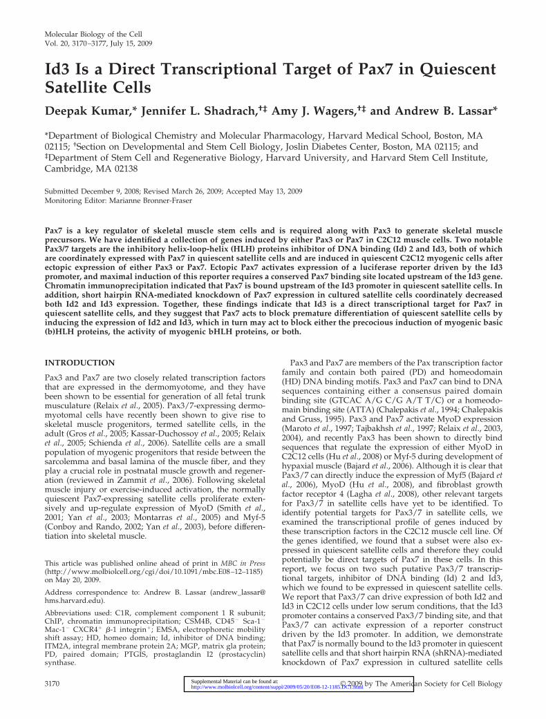
Because it was not clear whether the Pax3/7-induciblegenes that we had identified in C2C12 cells were indeedphysiological targets of Pax3/7, we decided to focus ouranalysis on the putative Pax3/7 target genes that were alsoexpressed in quiescent satellite cells, which constitutivelyexpress endogenous Pax7. We purified CD45�, Sca-1�, Mac-1�, CXCR4�, and �-1 integrin� (CSM4B) myofiber-associ-ated satellite cells by fluorescence-activated cell sorting(FACS) (Cerletti et al., 2008) from the skeletal muscle of2-mo-old mice. Greater than 90% of CSM4B cells have beenshown to express Pax7 after immediate isolation, and theyefficiently differentiate into skeletal muscle both in vitro andin vivo (Cerletti et al., 2008). We assayed by RT-PCR theexpression of Id2, Id3, and some other putative Pax7 targetsthat we had identified in the sorted CSM4B population. Thisanalysis identified several putative Pax7 target genes thatare specifically expressed in quiescent Pax7� CSM4B cells,including Id2 and Id3, integral membrane protein 2A(ITM2A), the chemokine receptor CXCR4, Bone Morpho-genic Protein 4 (BMP4), the sugar binding protein chond-rolectin, complement component 1 R subunit (C1R), pros-taglandin I2 (prostacyclin) synthase (PTGIS), and matrixGla protein (MGP). RT-PCR analysis of gene expression inimmediately harvested versus cultured CSM4B cells indi-cated that all these putative Pax7 targets are expressed infreshly isolated CSM4B cells (which express high levels ofPax7), and that in many cases their expression declinesduring culture of these cells (Figure 1A), in parallel withreduced Pax7 expression, induced proliferation, and even-
tual differentiation of these cells into myotubes (Cerletti etal., 2008). Id2 and Id3 were robustly expressed in bothfreshly isolated Pax7�, MyoD� CSM4B cells and in prolif-erating Pax7�, MyoD� CSM4B cultures; expression of Id2and Id3 declined and was eventually lost in differentiatedcultures of Pax7�, MyoD� CSM4B-derived myoblasts (Fig-ure 1A, lanes 1–6). In contrast to Id2 and Id3, Id1 expressionwas not detected in either freshly isolated or culturedCSM4B cells (data not shown). Although the Id genes havebeen linked previously to cellular proliferation in at leastsome cellular contexts (Benezra et al., 1990), in CSM4B-derived myoblasts Id2 and Id3 are expressed in both quies-cent and proliferating cells, indicating that their expressioncan be uncoupled from the cell cycle in this cell type.
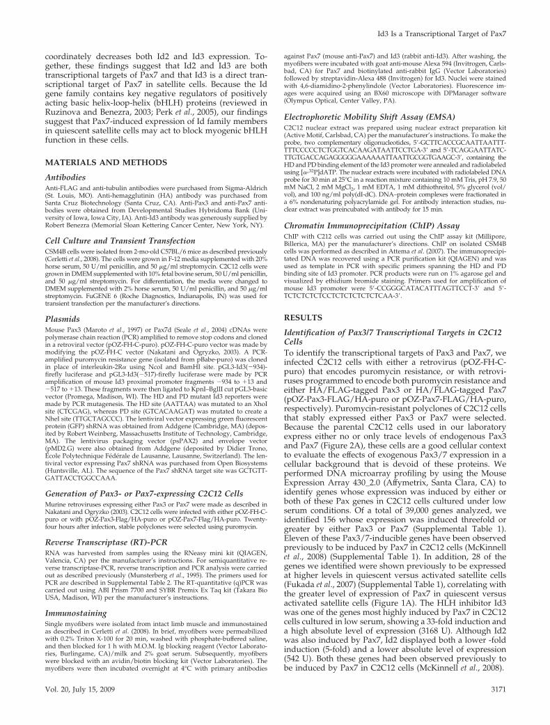
Pax3 or Pax7 Induces the Expression of Id FamilyMembers in C2C12 Cells and Id3 Protein Is Detectable inQuiescent Satellite CellsTo further analyze the ability of Pax3 or Pax7 to induceexpression of Id proteins, we evaluated expression of Id1,Id2, and Id3 by RT-PCR in either parental C2C12 cells or inpolyclones of C2C12 cells programmed to express exoge-nous Pax3 or Pax7. Both the parental C2C12 cells and thePax3/Pax7-expressing C2C12 polyclones expressed approx-imately equivalent levels of Id1, Id2, and Id3 transcriptswhen the cells were cultured under high serum conditions(i.e., 10% fetal bovine serum) (Figure 2A, lanes 1–3). Instriking contrast, when the cells were cultured under lowserum conditions for 3 d (i.e., 2% horse serum), parental
Figure 1. Id2, Id3, and other putative Pax7 target genes are ex-pressed in quiescent satellite cells. (A) CSM4B cells were isolatedfrom the skeletal muscle of 2-mo-old mice. RT-PCR analysis of geneexpression in either freshly isolated CSM4B cells or in cells culturedfor designated days in vitro is displayed. (B–E) Id3 protein is ap-parent in satellite cells. Myofibers (and associated satellite cells)were isolated and immunostained with anti-Pax7 and anti-Id3.Pax7�, Id3� satellite cell nucleus is indicated by the arrow. Al-though Id3 staining in the nucleus is specific (and confined to thePax7� nucleus; arrow), apparent Id3 staining in the myotube is dueto autofluorescence.
Figure 2. Id2 and Id3 are induced in serum-starved C2C12 cells byeither Pax3 or Pax7. (A) RT-PCR analysis of gene expression ineither parental C2C12 cells (control) or in C2C12 polyclones express-ing exogenous Pax3-HA or Pax7-HA. Cells were either cultured in10% fetal calf serum (high serum) or in 2% horse serum (low serum)for 3 d. (B–W) Immunostaining for Id3 or Pax3/7-HA expression ineither parental C2C12 cells or C2C12 polyclones expressing exoge-nous Pax3-HA or Pax7-HA, grown in either high or low serumconditions.
D. Kumar et al.
Molecular Biology of the Cell3172
C2C12 cells down-regulated expression of Id1, Id2, and Id3,whereas the expression of these genes was maintained in theC2C12 polyclones programmed to express either Pax3 orPax7 (Figure 2A, lanes 4–6).
Consistent with our analysis of Id3 transcript levels, wefound that Id3 protein is apparent in discrete nuclear inclu-sions in proliferating C2C12 cells (Figure 2B), but it is notobserved in quiescent C2C12 cells cultured in low serumconditions (Figure 2E). These findings are consistent withprior work indicating that Id family members are specifi-cally expressed in proliferating cells and are down-regulatedduring the process of skeletal muscle differentiation and cellcycle withdrawal (Benezra et al., 1990). In contrast to theparental C2C12 cells, where Id3 only accumulates in divid-ing cells, Id3 protein is apparent both throughout the nu-cleus and in nuclear inclusions in C2C12 polyclones express-ing either exogenous Pax3 or Pax7 in high serum and atslightly attenuated levels in low serum conditions (Figure 2,I, M, Q, and U). These findings indicate that Pax3 or Pax7can induce the expression of Id2 and Id3 specifically in cellsthat are cultured under low serum conditions. To evaluate
whether Id3 protein accumulates in quiescent satellite cells,we immunostained freshly dissected myofibers with theirassociated satellite cells for both Pax7 and Id3. Id3 proteinwas specifically detected in Pax7-expressing quiescent sat-ellite cells located on the periphery of the myofiber (Figure 1,B–E). These data are consistent with the high level of expres-sion of Id3 transcripts that are detectable in CSM4B cellsisolated by FACS (Figure 1A, lane 1).
Pax7/Pax3 Can Bind to the Id3 Promoter and Induce theTranscriptional Activity of an Id3 Luciferase ReporterTo examine whether Id3 is directly regulated by Pax3 orPax7, we analyzed the proximal promoter of the mouse Id3gene for putative PD and HD binding sites. Interestingly, weidentified putative PD and HD binding sites upstream of theId3 proximal promoter in several mammalian species, in-cluding mouse, rat, human, orangutan, dog, and horse (Fig-ure 3A). To determine whether Pax3/7 bind to Id3 regula-tory sequences, we monitored whether Pax3/7 present innuclear extracts made from C2C12 cells programmed to
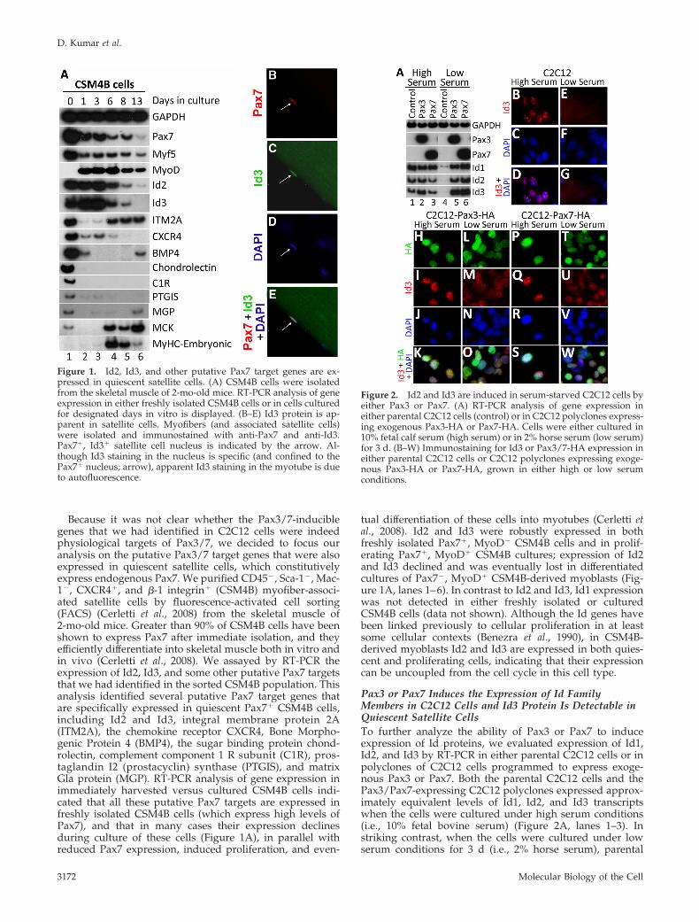
Figure 3. A conserved Pax3/7 binding site ispresent upstream of the Id3 promoter. (A)Conserved HD and PD binding sites arepresent upstream of the Id3 promoter in sev-eral organisms. (B) EMSA using an oligo en-coding the HD/PD binding sites upstream ofthe mouse Id3 gene with nuclear extractsmade from parental C2C12 cells or polyclonesexpressing Pax3-FLAG or Pax7-FLAG. Anti-Pax3, anti-Pax7, anti-FLAG, or anti-tubulinwere added to the EMSA as indicated. Pax3/7gel shift is indicated by the black arrow; anti-body supershift is indicated by the white arrow.(C) Serum factors affect Pax3 or Pax7 mediatedinduction of the Id3 reporter. C2C12 cells werecotransfected with pGL3-Id3(�934)-firefly lucif-erase and pSV40-Renilla-luciferase. In addition,the cells were cotransfected with either Pax3 orPax7 expression vectors, as indicated, andgrown in presence of either high or low serum.The relative luciferase units (RLU) of pGL3-Id3-firefly luciferase and pSV40-Renilla-luciferaseare displayed. (D) Pax3 and Pax7 induction ofan Id3 reporter requires both HD and PD bind-ing sites in the Id3 promoter. C2C12 cells werecotransfected with either pGL3-Id3(�934)-fire-fly luciferase (containing the wild-type Id3 pro-moter) or mutated versions of this reporter lack-ing HD and PD binding sites as shown, pluspSV40-Renilla-luciferase. In addition, the cellswere cotransfected with expression vehicles en-coding either Pax3 or Pax7, as indicated. TheRLUs of pGL3-Id3-firefly luciferase and pSV40-Renilla-luciferase is displayed.
Id3 Is a Transcriptional Target of Pax7
Vol. 20, July 15, 2009 3173
express these proteins would interact in vitro with an oligo-nucleotide containing the putative Pax3/7 binding site lo-cated upstream of the Id3 promoter. Nuclear extracts de-rived from either parental C2C12 cells or such cells infectedwith a retrovirus encoding either Pax3-FLAG or Pax7-FLAGwere incubated with a radiolabeled oligomer containing theputative Pax3/7 binding site in the Id3 promoter (Figure 3B).The Pax3/7-DNA protein complexes were visualized byEMSA. Nuclear extracts derived from C2C12 cells pro-grammed to express either FLAG-tagged Pax3 or FLAG-tagged Pax7 gave rise to a new DNA–protein complex onthe Id3 oligo (Figure 3B, lanes 3 and 7, black arrow desig-nates new gel shift) that was not present in extracts derivedfrom parental C2C12 cells (Figure 3B, lane 2). Importantly,these DNA–protein complexes were shifted to a lower elec-trophoretic mobility by inclusion of either anti-Pax3, anti-Pax7, or anti-FLAG antibodies (Figure 3B, lanes 4, 5, 8, and9; hollow arrow designates antibody supershifted complex).In contrast, a control anti-tubulin antibody did not affect themobility of the Pax3/7-DNA complexes (Figure 3B, lanes 6and 10). These findings indicate that Pax3 and Pax7 can bindto a conserved sequence located just upstream of the Id3promoter in vitro.
To evaluate the importance of the putative Pax3/7 bind-ing site located upstream of the Id3 promoter, we con-structed a reporter vehicle containing 934 base pairs up-stream of the Id3 transcriptional initiation site (�934 to �13)driving expression of firefly luciferase (Yeh and Lim, 2000).Forced expression of either Pax3 or Pax7 in C2C12 cellscotransfected with this Id3-luciferase reporter, resulted in an8- to 10-fold increase in luciferase activity, indicating thatboth Pax3 and Pax7 can induce expression of a reporterdriven by the Id3 promoter (Figure 3C). Because we notedthat Id3 protein was decreased in C2C12 cells programmedto express either Pax3 or Pax7 in low serum conditions(Figure 2, 1, M, Q, and U), we evaluated whether serumfactors would influence the ability of either Pax3 or Pax7 toinduce expression of the Id3-luciferase reporter. Induction ofthe Id3 reporter by either Pax3 or Pax7 was greatest in high
serum conditions (Figure 3C), suggesting that serum factorsmay indeed enhance the ability of these transcription factorsto induce maximal Id3 expression. To investigate whethereither the PD or HD binding sites were necessary for Pax3/7to induce the expression of the Id3 reporter, we mutatedthese binding sites individually or in combination. Mutationof only the PD binding site resulted in about a 50% reductionin the expression of the Id3 reporter in response to eithercotransfected Pax3 or Pax7, whereas mutation or deletion ofboth the PD and HD binding sites resulted in approximatelyan 80% reduction in the induction of this reporter by Pax3/Pax7 (Figure 3D).
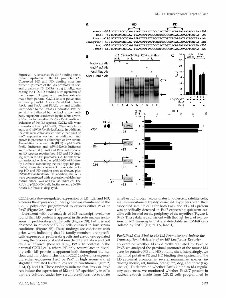
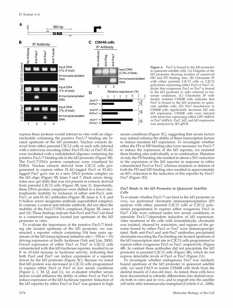
Pax7 Binds to the Id3 Promoter in Quiescent SatelliteCellsTo evaluate whether Pax3/7 can bind to the Id3 promoter invivo, we performed chromatin immunoprecipitation (IP)analysis with either parental C2C12 cells or C2C12 poly-clones programmed to express either exogenous Pax3 orPax7. Cells were cultured under low serum conditions, tostimulate Pax3/7-dependent induction of Id3 expression.After treatment of the cells with formaldehyde, chromatinwas isolated, sheared by sonication, and regions of the ge-nome bound by either Pax3 or Pax7 were immunoprecipi-tated. Both anti-Pax3 and anti-Pax7 antibodies precipitatedchromatin encoding the Pax binding site located upstream ofthe Id3 transcription start site in C2C12 cells programmed toexpress either exogenous Pax3 or Pax7, respectively (Figure4B). In contrast these antibodies did not precipitate the Id3promoter in parental C2C12 cells (Figure 4B), which do notexpress detectable levels of Pax3 or Pax7 (Figure 2A).
To investigate whether endogenous Pax7 was similarlybound upstream of the Id3 promoter in quiescent satellitecells, we used FACS to isolate CSM4B cells fresh from theskeletal muscle of 2-mo-old mice. As stated, these cells havebeen documented to robustly differentiate into skeletal mus-cle both in vitro and in vivo, and to engraft into the satellitecell niche after intramuscular transplant (Cerletti et al., 2008).
Figure 4. Pax7 is bound to the Id3 promoterin quiescent satellite cells. (A) Diagram of theId3 promoter showing location of conservedHD and PD binding sites. (B) Chromatin IPwith either parental C2C12 cells or C2C12polyclones expressing either Pax3 or Pax7, in-dicate that exogenous Pax3 or Pax7 is boundto the Id3 promoter in cells cultured in lowserum conditions. (C) Chromatin IP withfreshly isolated CSM4B cells indicates thatPax7 is bound to the Id3 promoter in quies-cent satellite cells. (D) Pax7 knockdown inCSM4B cells significantly decreases Id2 andId3 expression. CSM4B cells were infectedwith lentivirus expressing either GFP shRNAor Pax7 shRNA. Pax7, Id2, and Id3 expressionwas analyzed by RT-qPCR.
D. Kumar et al.
Molecular Biology of the Cell3174
Coupled with the fact that �90% of sorted CSM4B cellsexpress high levels of Pax7, whereas �5% express MyoD(Cerletti et al., 2008), it seems likely that this population ofcells consists predominantly of quiescent satellite cells.Freshly isolated CSM4B cells, which express high levels ofId3 (Figure 1A), were purified and immediately cross-linkedfor chromatin IP analysis. Notably anti-Pax7 was able toimmunoprecipitate chromatin encoding the Pax binding sitelocated upstream of the Id3 promoter in these freshly iso-lated Pax7� satellite cells (Figure 4C).
shRNA-mediated Knockdown of Pax7 Expression inSatellite Cells Significantly Reduces the Expression of Id2and Id3To examine whether Pax7 was necessary to maintain theexpression of either Id2 or Id3 in satellite cells, we infectedCSM4B cells, isolated from skeletal muscle tissue, with len-tiviruses programmed to express shRNAs directed againsteither green fluorescent protein (shGFP; as a control) or Pax7(shPax7). Four days after infection, RNA was isolated fromthe infected CSM4B cells, and RT-qPCR was used to assaygene expression. Expression of Pax7 was reduced to 40% ofcontrol levels in cells expressing Pax7 shRNA (Figure 4D).Notably, the expression of Id2 and Id3 was similarly re-duced to 61 and 52% of control levels, respectively, in cellsexpressing Pax7 shRNA (Figure 4D). The reduction in ex-pression of both Id2 and Id3 following knockdown of Pax7expression suggests that Pax7 is necessary to maintain highlevel expression of both Id2 and Id3 in satellite cells.
DISCUSSION
Ectopic expression of either Pax3 or Pax7 in C2C12 cells ledto the induction of Id1, Id2, and Id3, specifically when thesecells were cultured under low serum conditions. Althoughthese genes are both expressed in proliferating C2C12 cellsgrown in 10% fetal bovine serum, their expression is signif-icantly attenuated when these cells are cultured in low se-rum conditions (2% horse serum) for 3 d. In striking contrast,in C2C12 cells programmed to express either exogenous Pax3or Pax7, expression of these genes is maintained in lowserum-containing medium. Although the Id genes have pre-viously been linked to cellular proliferation in at least somecellular contexts (Benezra et al., 1990), in satellite cells Id2and Id3 are expressed in both quiescent and proliferatingcells, indicating that their expression can be uncoupled fromthe cell cycle in this cell type. shRNA-mediated knockdownof Pax7 expression in purified satellite cells leads to a sig-nificant loss in the expression of both Id2 and Id3 in acti-vated satellite cells in vitro, suggesting that Pax7 is neces-sary to maintain high level expression of these Id familymembers in satellite cells. Because there are conservedpaired domain and homeodomain binding sites located up-stream of the mouse, rat, human, orangutan, dog, and horseId3 genes, which are necessary for efficient induction of anId3-luciferase reporter by either Pax3 or Pax7, we proposethat Id3 is a direct transcriptional target of Pax3/7. Consis-tent with this notion, we have documented that Pax3 andPax7 bind to chromatin containing this region of the Id3gene in C2C12 cells programmed to express either Pax3 orPax7 and that endogenous Pax7 is similarly bound to thisregion of the Id3 gene in quiescent satellite cells. Together,our findings suggest that Id2 and Id3 are transcriptionaltargets of Pax7 in satellite cells.
Previous work has indicated that �90% of CSM4B cellsisolated from skeletal muscle express Pax7 and that suchcells exhibit the functional characteristics of quiescent skel-
etal muscle stem cells or satellite cells (Cerletti et al., 2008).When freshly isolated, this cell population displays robustexpression of Pax7, Myf5, and both Id2 and Id3, yet it doesnot express appreciable levels of Id1. Culturing these cells invitro leads to both their entry into the cell cycle (Cerletti etal., 2008), induction of MyoD expression, and subsequentloss of Pax7 expression (Figure 1A). The expression of Id2and Id3 eventually declines in these cultures as well, inparallel with loss of Pax7 expression and induction of skel-etal muscle differentiation. Previous work has indicated thatexpression of Id1 is high in proliferating C2C12 cells andsignificantly decreases when these cells are induced to dif-ferentiate in low serum-containing medium (Benezra et al.,1990). The induction of Id genes by Pax3/Pax7 in C2C12cells cultured in low serum, or expression of Id2 and Id3 inquiescent satellite cells, suggests that the expression of Idfactors can be divorced from cellular proliferation. Interest-ingly, however, maximal induction of the Id3-luciferase re-porter by cotransfected Pax3/7 required the presence ofhigh serum.
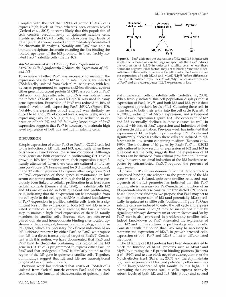
Chromatin IP analysis demonstrated that Pax7 binds to aconserved binding site adjacent to the promoter of the Id3gene in freshly isolated, quiescent satellite cells, and mu-tagenesis of the Id3 promoter has indicated that this Pax7binding site is necessary for Pax7-mediated induction of anId3-promoter-luciferase construct in transfected C2C12 cells.Based upon these findings, we propose that Pax7 may act tomaintain the expression of Id3 (and potentially Id2) specif-ically in quiescent satellite cells (outlined in Figure 5). Oncesatellite cells are induced to enter the cell cycle and expressMyoD, expression of Id2/3 may be maintained either bysignaling pathways downstream of serum factors and/or byPax7 that is also expressed in proliferating satellite cells.Indeed knockdown of Pax7 attenuated the expression ofboth Id2 and Id3 in cultures of proliferating satellite cells.Consistent with the notion that Pax7 may be necessary tomaintain the expression of Id2/3 in growth arrested cells,expression of both Pax7 and Id2/3 is lost in differentiatedmyotubes.
The Id family of HLH proteins have been demonstrated toblock the function of bHLH proteins such as MyoD andMyf5, by titrating their E protein binding partners (Benezraet al., 1990); and to also block negative autoregulation of theNotch effector Hes1 (Bai et al., 2007) and thereby maintainhigh level expression of Hes1 and potentially other membersof the hairy/enhancer of split family. In this light, it isinteresting that quiescent satellite cells express relativelyrobust levels of both Id2 and Id3 (this study) and several
Figure 5. Pax7 activates the expression of Id2 and Id3 in quiescentsatellite cells. Based on our findings we speculate that Pax7 inducesthe expression of Id2/3 in quiescent satellite cells, where thesedominant-negative HLH factors may act to block premature differ-entiation of these cells. In activated satellite cells, Pax7 may inducethe expression of both Id2/3 and MyoD/Myf5 before differentia-tion. In differentiated myotubes, MyoD/Myf5 represses expressionof Pax7 and as a consequence Id2/3 expression is lost.
Id3 Is a Transcriptional Target of Pax7
Vol. 20, July 15, 2009 3175
members of the hairy/enhancer of split family, includingHeyl (Fukada et al., 2007), Hes1 and Hey1 (Wagers, unpub-lished observations). It is possible that high-level expressionof Id2 and Id3 in quiescent satellite cells acts to either blockthe activity of Myf5 that is readily expressed (at least at theRNA level) in at least 90% of quiescent satellite cells (Kuanget al., 2007) or to maintain the expression of the Notch-induced transcriptional repressors Hes1, Hey1, and/orHeyl, which in turn may block expression of MyoD andMyf5. Thus, by inducing the expression of Id2 and Id3, Pax7may act to block the premature differentiation of quiescentsatellite cells. In addition, Pax7 may also block skeletal mus-cle differentiation by targeting myogenic bHLH proteins fordegradation (Olguin et al., 2007). In contrast, in activatedsatellite cells, Pax7 may act to directly induce the expressionof Myf5 (Bajard et al., 2006) and MyoD (Hu et al., 2008),which in turn will compete with Id proteins for E proteindimerization. In this scenario, Pax7 serves two roles in sat-ellite cells: blocking their differentiation via Id2/3 inductionduring quiescence and promoting their differentiation viaMyf5/MyoD induction during activation (Figure 5). Inter-estingly, Id family members have been implicated in bothpromoting self-renewal and blocking differentiation of he-matopoietic (Jankovic et al., 2007) and neural (Robert Ben-ezra, personal communication) stem cells, and our worksuggests that they may play a similar role in satellite cells aswell. Although mice deleted for Id3 are viable and show nodiscernible muscle phenotype (Pan et al., 1999), mice lackingboth Id2 and Id3 are embryonic lethal (Robert Benezra,personal communication). Because quiescent satellite cellsexpress both Id2 and Id3 (Figure 1), it is possible that thelack of a skeletal muscle phenotype in mice lacking only Id3(Pan et al., 1999) is due to functional redundancy with Id2.Therefore, to determine whether Id2 and Id3 are indeednecessary to block differentiation of quiescent satellite cells,it will be necessary to conditionally delete both these genesin this cell type.
In addition to Id2 and Id3, we noted that several othergenes induced by Pax7 in C2C12 cells were also specificallyexpressed in quiescent satellite cells. These genes includeITM2A, the chemokine receptor CXCR4, BMP4, the sugarbinding protein chondrolectin, C1R, PTGIS, and MGP. In-terestingly, the expression of many of these Pax7 induciblegenes was greatest in quiescent satellite cells and expressionof these genes was extinguished during either proliferationor differentiation of these cells. CXCR4 has already beendemonstrated to play a crucial role in migration of skeletalmuscle cells during embryogenesis (Vasyutina et al., 2005). Itwill be interesting to determine the role of both Id2/Id3 andthese other putative Pax7 transcriptional targets in quiescentsatellite cells.
ACKNOWLEDGMENTS
We thank Robert Lim for generously supplying the Id3 promoter; MichaelRudnicki for the Pax7 cDNA; Hyung-song Nam, Yvette Chin, and RobertBenezra for characterizing and supplying the anti-Id3 rabbit monoclonalantibody (now available from CalBioreagents, San Mateo, CA); and RobertBenezra for thoughtful comments on our work. This work was funded byNational Institutes of Health grant GM-054879 (to A.B.L.) and grants from theHarvard Stem Cell Institute, Jain Foundation, and Beckman Foundation (toA.J.W.). D. K. was funded by a fellowship from the Muscular DystrophyAssociation.
REFERENCES
Attema, J. L., Papathanasiou, P., Forsberg, E. C., Xu, J., Smale, S. T., andWeissman, I. L. (2007). Epigenetic characterization of hematopoietic stem cell
differentiation using miniChIP and bisulfite sequencing analysis. Proc. Natl.Acad. Sci. USA 104, 12371–12376.
Bai, G., Sheng, N., Xie, Z., Bian, W., Yokota, Y., Benezra, R., Kageyama, R.,Guillemot, F., and Jing, N. (2007). Id sustains Hes1 expression to inhibitprecocious neurogenesis by releasing negative autoregulation of Hes1. Dev.Cell 13, 283–297.
Bajard, L., Relaix, F., Lagha, M., Rocancourt, D., Daubas, P., and Buckingham,M. E. (2006). A novel genetic hierarchy functions during hypaxial myogenesis:Pax3 directly activates Myf5 in muscle progenitor cells in the limb. GenesDev. 20, 2450–2464.
Benezra, R., Davis, R. L., Lockshon, D., Turner, D. L., and Weintraub, H.(1990). The protein Id: a negative regulator of helix-loop-helix DNA bindingproteins. Cell 61, 49–59.
Cerletti, M., Jurga, S., Witczak, C. A., Hirshman, M. F., Shadrach, J. L.,Goodyear, L. J., and Wagers, A. J. (2008). Highly efficient, functional engraft-ment of skeletal muscle stem cells in dystrophic muscles. Cell 134, 37–47.
Chalepakis, G., Goulding, M., Read, A., Strachan, T., and Gruss, P. (1994).Molecular basis of splotch and Waardenburg Pax-3 mutations. Proc. Natl.Acad. Sci. USA 91, 3685–3689.
Chalepakis, G., and Gruss, P. (1995). Identification of DNA recognition se-quences for the Pax3 paired domain. Gene 162, 267–270.
Conboy, I. M., and Rando, T. A. (2002). The regulation of Notch signalingcontrols satellite cell activation and cell fate determination in postnatal myo-genesis. Dev. Cell 3, 397–409.
Fukada, S., Uezumi, A., Ikemoto, M., Masuda, S., Segawa, M., Tanimura, N.,Yamamoto, H., Miyagoe-Suzuki, Y., and Takeda, S. (2007). Molecular signa-ture of quiescent satellite cells in adult skeletal muscle. Stem Cells 25, 2448–2459.
Gros, J., Manceau, M., Thome, V., and Marcelle, C. (2005). A common somiticorigin for embryonic muscle progenitors and satellite cells. Nature 435, 954–958.
Hu, P., Geles, K. G., Paik, J. H., DePinho, R. A., and Tjian, R. (2008). Code-pendent activators direct myoblast-specific MyoD transcription. Dev. Cell 15,534–546.
Jankovic, V., Ciarrocchi, A., Boccuni, P., DeBlasio, T., Benezra, R., and Nimer,S. D. (2007). Id1 restrains myeloid commitment, maintaining the self-renewalcapacity of hematopoietic stem cells. Proc. Natl. Acad. Sci. USA 104, 1260–1265.
Kassar-Duchossoy, L., Giacone, E., Gayraud-Morel, B., Jory, A., Gomes, D.,and Tajbakhsh, S. (2005). Pax3/Pax7 mark a novel population of primitivemyogenic cells during development. Genes Dev. 19, 1426–1431.
Kuang, S., Kuroda, K., Le Grand, F., and Rudnicki, M. A. (2007). Asymmetricself-renewal and commitment of satellite stem cells in muscle. Cell 129,999–1010.
Lagha, M., Kormish, J. D., Rocancourt, D., Manceau, M., Epstein, J. A., Zaret,K. S., Relaix, F., and Buckingham, M. E. (2008). Pax3 regulation of FGFsignaling affects the progression of embryonic progenitor cells into the myo-genic program. Genes Dev. 22, 1828–1837.
Maroto, M., Reshef, R., Munsterberg, A. E., Koester, S., Goulding, M., andLassar, A. B. (1997). Ectopic Pax-3 activates MyoD and Myf-5 expression inembryonic mesoderm and neural tissue. Cell 89, 139–148.
McKinnell, I. W., Ishibashi, J., Le Grand, F., Punch, V. G., Addicks, G. C.,Greenblatt, J. F., Dilworth, F. J., and Rudnicki, M. A. (2008). Pax7 activatesmyogenic genes by recruitment of a histone methyltransferase complex. Nat.Cell Biol. 10, 77–84.
Montarras, D., Morgan, J., Collins, C., Relaix, F., Zaffran, S., Cumano, A.,Partridge, T., and Buckingham, M. (2005). Direct isolation of satellite cells forskeletal muscle regeneration. Science 309, 2064–2067.
Munsterberg, A. E., Kitajewski, J., Bumcrot, D. A., McMahon, A. P., andLassar, A. B. (1995). Combinatorial signaling by Sonic hedgehog and Wntfamily members induces myogenic bHLH gene expression in the somite.Genes Dev. 9, 2911–2922.
Nakatani, Y., and Ogryzko, V. (2003). Immunoaffinity purification of mam-malian protein complexes. Methods Enzymol. 370, 430–444.
Olguin, H. C., Yang, Z., Tapscott, S. J., and Olwin, B. B. (2007). Reciprocalinhibition between Pax7 and muscle regulatory factors modulates myogeniccell fate determination. J. Cell Biol. 177, 769–779.
Pan, L., Sato, S., Frederick, J. P., Sun, X. H., and Zhuang, Y. (1999). Impairedimmune responses and B-cell proliferation in mice lacking the Id3 gene. Mol.Cell Biol. 19, 5969–5980.
Perk, J., Iavarone, A., and Benezra, R. (2005). Id family of helix-loop-helixproteins in cancer. Nat. Rev. Cancer 5, 603–614.
D. Kumar et al.
Molecular Biology of the Cell3176
Relaix, F., Polimeni, M., Rocancourt, D., Ponzetto, C., Schafer, B. W., andBuckingham, M. (2003). The transcriptional activator PAX3-FKHR rescues thedefects of Pax3 mutant mice but induces a myogenic gain-of-function pheno-type with ligand-independent activation of Met signaling in vivo. Genes Dev.17, 2950–2965.
Relaix, F., Rocancourt, D., Mansouri, A., and Buckingham, M. (2004). Divergentfunctions of murine Pax3 and Pax7 in limb muscle development. Genes Dev. 18,1088–1105.
Relaix, F., Rocancourt, D., Mansouri, A., and Buckingham, M. (2005). A Pax3/Pax7-dependent population of skeletal muscle progenitor cells. Nature 435, 948–953.
Ruzinova, M. B., and Benezra, R. (2003). Id proteins in development, cell cycleand cancer. Trends Cell Biol. 13, 410–418.
Schienda, J., Engleka, K. A., Jun, S., Hansen, M. S., Epstein, J. A., Tabin, C. J.,Kunkel, L. M., and Kardon, G. (2006). Somitic origin of limb muscle satelliteand side population cells. Proc. Natl. Acad. Sci. USA 103, 945–950.
Seale, P., Ishibashi, J., Scime, A., and Rudnicki, M. A. (2004). Pax7 is necessaryand sufficient for the myogenic specification of CD45�:Sca1� stem cells frominjured muscle. PLoS Biol. 2, E130.
Smith, H. K., Maxwell, L., Rodgers, C. D., McKee, N. H., and Plyley, M. J.(2001). Exercise-enhanced satellite cell proliferation and new myonuclearaccretion in rat skeletal muscle. J. Appl. Physiol. 90, 1407–1414.
Tajbakhsh, S., Rocancourt, D., Cossu, G., and Buckingham, M. (1997). Rede-fining the genetic hierarchies controlling skeletal myogenesis: Pax-3 andMyf-5 act upstream of MyoD. Cell 89, 127–138.
Vasyutina, E., Stebler, J., Brand-Saberi, B., Schulz, S., Raz, E., and Birchmeier,C. (2005). CXCR4 and Gab1 cooperate to control the development of migrat-ing muscle progenitor cells. Genes Dev. 19, 2187–2198.
Yan, Z., Choi, S., Liu, X., Zhang, M., Schageman, J. J., Lee, S. Y., Hart, R., Lin,L., Thurmond, F. A., and Williams, R. S. (2003). Highly coordinated generegulation in mouse skeletal muscle regeneration. J. Biol. Chem. 278, 8826–8836.
Yeh, K., and Lim, R. W. (2000). Genomic organization and promoter analysisof the murine Id3 gene. Gene 254, 163–171.
Zammit, P. S., Partridge, T. A., and Yablonka-Reuveni, Z. (2006). The skeletalmuscle satellite cell: the stem cell that came in from the cold. J. Histochem.Cytochem. 54, 1177–1191.
Id3 Is a Transcriptional Target of Pax7
Vol. 20, July 15, 2009 3177


