Embed Size (px)
Citation preview

Identification of a High-Affinity Ligand That Exhibits CompleteAryl Hydrocarbon Receptor Antagonism□S
Kayla J. Smith, Iain A. Murray, Rachel Tanos, John Tellew, Anthony E. Boitano,William H. Bisson, Siva K. Kolluri, Michael P. Cooke, and Gary H. PerdewCenter for Molecular Toxicology and Carcinogenesis, Department of Veterinary and Biomedical Sciences, Pennsylvania StateUniversity, University Park, Pennsylvania (K.J.S., I.A.M., R.T., G.H.P.); Genomics Institute of the Novartis Research Foundation,San Diego, California (J.T., A.E.B., M.P.C.); Pharmaceutical Biochemistry Group, School of Pharmaceutical Sciences, Universityof Geneva, Geneva, Switzerland (W.H.B.); and Cancer Biology Laboratory, Department of Environmental and MolecularToxicology, Environmental Health Sciences Center, Oregon State University, Corvallis, Oregon (S.K.K.)
Received December 18, 2010; accepted March 16, 2011
ABSTRACTThe biological functions of the aryl hydrocarbon receptor (AHR)can be delineated into dioxin response element (DRE)-dependentor -independent activities. Ligands exhibiting either full or partialagonist activity, e.g., 2,3,7,8-tetrachlorodibenzo-p-dioxin and�-naphthoflavone, have been demonstrated to potentiate bothDRE-dependent and -independent AHR function. In contrast, therecently identified selective AHR modulators (SAhRMs), e.g., 1-allyl-3-(3,4-dimethoxyphenyl)-7-(trifluoromethyl)-1H-indazole (SGA360),bias AHR toward DRE-independent functionality while displayingantagonism with regard to ligand-induced DRE-dependent transcrip-tion. Recent studies have expanded the physiological role of AHR toinclude modulation of hematopoietic progenitor expansion and im-munoregulation. It remains to be established whether such physio-logical roles are mediated through DRE-dependent or -independentpathways. Here, we present evidence for a third class of AHR ligand,“pure” or complete antagonists with the capacity to suppress both
DRE-dependent and -independent AHR functions, which may facil-itate dissection of physiological AHR function with regard to DRE ornon-DRE-mediated signaling. Competitive ligand binding assays to-gether with in silico modeling identify N-(2-(1H-indol-3-yl)ethyl)-9-isopropyl-2-(5-methylpyridin-3-yl)-9H-purin-6-amine (GNF351) as ahigh-affinity AHR ligand. DRE-dependent reporter assays, in con-junction with quantitative polymerase chain reaction analysis of AHRtargets, reveal GNF351 as a potent AHR antagonist that demon-strates efficacy in the nanomolar range. Furthermore, unlike manycurrently used AHR antagonists, e.g., �-naphthoflavone, GNF351 isdevoid of partial agonist potential. It is noteworthy that in a model ofAHR-mediated DRE-independent function, i.e., suppression of cyto-kine-induced acute-phase gene expression, GNF351 has the capac-ity to antagonize agonist and SAhRM-mediated suppression ofSAA1. Such data indicate that GNF351 is a pure antagonist with thecapacity to inhibit both DRE-dependent and -independent activity.
IntroductionThe aryl hydrocarbon receptor (AHR) is a ligand-activated
transcription factor, which is found in the cytoplasm in itslatent form bound to heat shock protein 90, and translocatesinto the nucleus upon ligand-mediated activation (Beischlag
et al., 2008). Once inside the nucleus, it binds to the AHRnuclear translocator (ARNT), which displaces heat shock pro-tein 90, and this complex binds to dioxin response elements(DRE) on its direct target genes. Binding to DRE sequencesleads to transcription, which was first described for genesthat encode for phase I metabolic enzymes, such as CYP1A1/1A2. These enzymes are responsible for the conversion of anumber of carcinogens [e.g., benzo(a)pyrene] from procar-cinogens into genotoxic intermediates. The most potent pro-totypic exogenous agonist for the AHR is 2,3,7,8-tetrachlo-rodibenzo-p-dioxin (TCDD), a highly toxic environmentalpollutant. Thus the AHR was originally associated with toxic
This work was supported by the National Institutes of Health NationalInstitute of Environmental Health Sciences [Grant ES04869].
Article, publication date, and citation information can be found athttp://jpet.aspetjournals.org.
doi:10.1124/jpet.110.178392.□S The online version of this article (available at http://jpet.aspetjournals.org)
contains supplemental material.
ABBREVIATIONS: AHR, aryl hydrocarbon receptor; ARNT, AHR nuclear translocator; SAhRM, selective AHR modulator; TCDD, 2,3,7,8-tetrachlorodibenzo-p-dioxin; DRE, dioxin response element; TH, T helper; DMSO, dimethyl sulfoxide; IL, interleukin; PCR, polymerase chainreaction; TPA, 12-O-tetradecanoylphorbol-13-acetate; PAL, 2-azido-3-[125I]iodo-7,8-dibromodibenzo-p-dioxin; GNF351, N-(2-(1H-indol-3-yl)ethyl)-9-isopropyl-2-(5-methylpyridin-3-yl)-9H-purin-6-amine; SGA360, 1-allyl-3-(3,4-dimethoxyphenyl)-7-(trifluoromethyl)-1H-indazole; �NF,�-naphthoflavone; MNF, 3�-methoxy-4�-nitroflavone; TMF, 6,2�,4�-trimethoxyflavone; CH-223191, 2-methyl-2H-pyrazole-3-carboxylic acid(2-methyl-4-o-tolylazo-phenyl)-amide; SR1, 4-(2-(2-(benzo[b]thiophen-3-yl)-9-isopropyl-9H-purin-6-ylamino)ethyl)phenol; I3S, 3-indoxyl-sulfate;WAY-69916, 4-[1-allyl-7-(trifluoromethyl)-1H-indazol-3yl]benzene-1.
0022-3565/11/3381-318–327$25.00THE JOURNAL OF PHARMACOLOGY AND EXPERIMENTAL THERAPEUTICS Vol. 338, No. 1Copyright © 2011 by The American Society for Pharmacology and Experimental Therapeutics 178392/3696987JPET 338:318–327, 2011 Printed in U.S.A.
318

responses at both the cellular and whole-organism level.However, in recent years the AHR has been shown to play animportant role in an array of physiological processes. Exam-ination of the physiological role of the AHR was greatlyfacilitated by the development of Ahr-null mice, leading tothe observation of multiple phenotypic defects including im-mune system dysfunction, reduced reproductive success, andaltered liver vascular development (Schmidt and Bradfield,1996). Further studies have implicated the AHR in addi-tional physiological roles, such as anti-inflammatory end-points, and T cell differentiation (Quintana et al., 2008; Patelet al., 2009). The activation of the AHR leads to the stimu-lation of a T cell population that secretes interleukin (IL)-17,thus generating a proinflammatory autoimmune potential(Kimura et al., 2008; Veldhoen et al., 2008). The critical rolethat the AHR plays in this process was underscored by theability of the AHR antagonist 2-methyl-2H-pyrazole-3-carbox-ylic acid (2-methyl-4-o-tolylazo-phenyl)-amide (CH-223191) toattenuate TH17 cell development in vivo and subsequent secre-tion of IL-17 and IL-22 (Veldhoen et al., 2009). Another biolog-ical endpoint that is influenced by AHR activity is the expan-sion of human hematopoietic stem cells in cell culture (Boitanoet al., 2010). The presence of the AHR antagonist StemRegenin1 [4-(2-(2-(benzo[b]thiophen-3-yl)-9-isopropyl-9H-purin-6-ylamino)ethyl)phenol; SR1] leads to ex vivo expansion of CD34� cells thatmaintain an undifferentiated phenotype and retain the ability toengraft immunodeficient mice. These studies underscore the po-tential of AHR antagonists as therapeutic agents.
This interest in the physiological processes regulated bythe AHR has also led to an increased interest in differenti-ating between classes of AHR ligand and their effects onAHR-mediated transcriptional activity, to modulate possiblebeneficial roles of the AHR, while inhibiting its potentiallytoxic effects. A distinct class of ligands has recently beencharacterized, which are able to bind to the AHR and fail toactivate the DRE-mediated responses, yet are able to represscytokine-induced acute-phase gene expression. These com-pounds, classified as selective AHR modulators (SAhRMs)1,are interesting in a therapeutic sense, in that the effects ofDRE-mediated AHR activity would be repressed while thepotentially beneficial anti-inflammatory properties would beretained (Murray et al., 2010c). Two distinct compounds havebeen characterized as SAhRM, SGA360, and 3�,4�-dimethoxy�-naphthoflavone (�NF); collectively, they have been shownto repress a variety of cytokine-induced acute-phase genes,including SAA1, CRP, LBP, C3, C1S, and C1R (Murray et al.,2010b, 2011). Others also use the term SAhRM in anothercontext, that of a compound that may be used therapeuticallyin the treatment of breast cancer through AHR-estrogenreceptor � cross-talk, this compound exhibits partial agonistactivity (Safe and McDougal, 2002). However, in this articlethe use of the term SAhRM will adhere to the definition inthe footnote. After the discovery of this class of compounds, itwas hypothesized that a class of AHR antagonists may exist,which not only inhibits the DRE response, but also fails toexhibit SAhRM activity. Though a number of AHR antago-nists are known and have been used in past studies, thesecompounds were characterized only in the context of antag-
onism of an agonist and thus may only antagonize DRE-mediated AHR activity. Also whether these AHR antagonistsexhibit SAhRM activity remains to be explored.
This article establishes that N- [2-(3H-indol-3-yl)ethyl]-9-isopropyl-2-(5-methyl-3-pyridyl)purin-6-amine (GNF351) isan AHR ligand that functions as a “pure antagonist.”2 Wehave found that this compound displays antagonist activityat a lower concentration than most previously cited AHRantagonists, exhibits no AHR agonist activity, and antago-nizes both the DRE-mediated and acute-phase gene repres-sion activities of the AHR. These findings will prove valuabletoward further characterization of the AHR and its ability tobe activated by various classes of ligands, as well as yieldingfurther insight into its possible role as a therapeutic agent.
Materials and MethodsMaterials. GNF351 was acquired from the Genomics Institute of
the Novartis Research Foundation (San Diego, CA). TCDD waskindly provided by Dr. Stephen Safe (Texas A&M University, CollegeStation, TX). 1-Allyl-3-(3,4-dimethoxyphenyl)-7-(trifluoromethyl)-1H-indazole (SGA360) was synthesized as described previously(Murray et al., 2010b). �NF and 6,2�,4�-trimethoxyflavone (TMF)were acquired from Indofine Chemicals (Hillsborough, NJ). 3�-Me-thoxy-4�-nitroflavone (MNF) was a kind gift from Dr. T. Gasiewicz(University of Rochester, Rochester, NY). Resveratrol (3,5,4�-trihy-droxy-trans-stilbene) was purchased from Biomol (Hamburg, Ger-many). 2-Methyl-2H-pyrazole-3-carboxylic acid (2-methyl-4-o-tolyl-azo-phenyl)-amide) (CH-223191) was purchased from ChembridgeCorporation (San Diego, CA). Human recombinant IL-1B was ac-quired from PeproTech (Rocky Hill, NJ).
Cell Culture. Huh7 cells, a human hepatoma-derived cell line, aswell as the stable reporter cell lines HepG2 40/6 and H1L1.1c2, weremaintained in �-minimal essential medium (Sigma, St. Louis, MO),supplemented with 8% fetal bovine serum (HyClone Laboratories,Logan, UT), 100 units/ml penicillin, and 100 �g/ml streptomycin(Sigma). Cells were grown in a humidified incubator at 37°C, with anatmospheric composition of 95% air and 5% CO2. The human hepa-toma-derived reporter line HepG2 40/6 contains the stably inte-grated pGudluc 6.1 DRE-driven reporter (Long et al., 1998), whereasthe murine hepatoma-derived reporter line H1L1.1c2, which wasoriginally obtained from Dr. M. Denison (University of California,Davis, CA) contains the stably integrated pGudluc 1.1 vector (Gar-rison et al., 1996).
Ligand-Binding Assays. Binding assays were conducted as de-scribed previously (Flaveny et al., 2009). In brief, the AHR photoaf-finity ligand 2-azido-3-[125I]iodo-7,8-dibromodibenzo-p-dioxin (PAL)was synthesized as described previously (Poland et al., 1986). Togenerate hepatic cytosol samples, mouse livers from B6.Cg-Ahrtm3.1 Bra
Tg (Alb-cre, Ttr-AHR)1GHP “humanized” AHR mice were homoge-nized with MENG buffer (25 mM MOPS, 2 mM EDTA, 0.02% NaN3,and 10% glycerol, pH 7.4) with 20 mM sodium molybdate and pro-tease inhibitors (Sigma). Samples were centrifuged for 1 h at100,000g. Binding assays were conducted in the dark except for thephoto-cross-linking of PAL. Next, 0.21 pmol (8 � 105 cpm/tube) ofPAL (a saturating quantity) was combined with 150 �g of the hepaticcytosolic protein sample. This combination was then incubated withincreasing concentrations of SR1 or GNF351 at room temperaturefor 20 min. These samples were then photolyzed (402 nm) at 8-cmdistance for 4 min, after which 1% charcoal/dextran (final concen-
1 The term SAhRM is defined as an AHR ligand that exhibits essentially noagonist activity with regard to DRE-mediated transcription yet is capable ofrepressing cytokine-mediated acute-phase gene expression.
2 The term “pure antagonist” is defined as an AHR ligand that exhibits noagonist activity with regard to DRE-mediated transcription, fails to facilitatenon-DRE-dependent suppression of gene expression and thus not a SAhRM,and exhibits competitive inhibition of both agonist- and SAhRM-dependentsignaling. However, whether a pure antagonist will block all non-DRE-medi-ated AHR activity will require further studies.
Redefining Ah Receptor Antagonism 319

tration) was incubated at 4°C for 5 min. The samples were thencentrifuged at 3000g for 10 min to remove remaining unbound PAL.Samples were then subjected to gel electrophoresis on an 8% tricine-polyacrylamide gel, after which they were transferred to a polyvi-nylidene difluoride membrane and visualized by autoradiography.Radioactive bands were cut from the membrane and quantified bygamma-counting.
Cell-Based Luciferase Reporter Assay. Reporter cell lines usedin luciferase reporter assays were grown in six-well plates and treatedwith AHR ligands dissolved in DMSO (0.1% final concentration) andincubated for 4 h. For antagonism experiments the antagonist wasadded 5 min before the addition of TCDD. Lysis buffer [25 mM Tris-phosphate, pH 7.8, 2 mM dithiothreitol, 2 mM 1,2-diaminhocyclo-hexane-N,N,N�,N�-tetraacetic acid, 10% (v/v) glycerol, and 1% (v/v) Tri-ton X-100] was then added to each well. The activity of each sample wasmeasured using a TD-20e luminometer using luciferase assay substrate(Promega, Madison, WI) as suggested by the manufacturer.
RNA Isolation and Reverse Transcription. mRNA was iso-lated from cell cultures using TRI reagent according to the manu-facturer’s specifications (Sigma). RNA was converted to cDNA usingthe High-Capacity cDNA Archive Kit (Applied Biosystems, FosterCity, CA).
Real-Time Quantitative PCR. Sequences of primers used forquantitative PCR have been described previously (Murray et al.,2010b). PerfeCTa SYBR Green SuperMix for iQ (Quanta Biosciences,Gaithersburg, MD) was used to determine mRNA levels, and anal-ysis was conducted using MyIQ software, in conjunction with aMyIQ-single-color PCR detection system (Bio-Rad Laboratories, Her-cules, CA).
Acute-Phase Gene Repression Assay. A human hepatoma-derived cell line (Huh7) was pretreated for 1 h with AHR ligands andincubated at 37°C in a cell culture incubator. After 1 h, IL-1� andIL-6 were added to the appropriate wells at a concentration of 2ng/ml for each cytokine. The cells were incubated for an additional6 h, followed by removal of the media from the cells and 1 ml of TRIreagent was added per well. Quantitative PCR was performed on thesamples, with the levels of SAA1 transcripts normalized to L13a.
Mouse Ear Edema Assay. Mouse ear edema assays were con-ducted as described previously (Murray et al., 2010b). In brief,6-week-old male C57BL6/J mice (wild type) were anesthetized. Then,1.5 �g of 12-O-tetradecanoylphorbol-13-acetate (TPA) in 50 �l ofhigh-performance liquid chromatography-grade acetone (Sigma) wasapplied directly to the right ear, followed by application of the testcompounds. The left ear received vehicle only. After a 6-h treatmentperiod, the mice were euthanized by carbon dioxide asphyxiation. Toquantify levels of inflammation, edema thickness was measuredusing a micrometer.
AHR Modeling and Ligand Docking. Ligand binding modelingwas conducted as described previously (Bisson et al., 2009).
Statistical Analysis. Data were analyzed using one-way analysisof variance with Tukey’s multiple comparison post test using Prism(version 5.01) software (GraphPad Software, Inc., San Diego, CA) todetermine statistical significance between treatments. Data repre-sent the mean change in a given endpoint � S.E.M. (n � 3/treatmentgroup) and were analyzed to determine significance.
ResultsGNF351 Is an AHR Ligand. A screen conducted to iden-
tify compounds with the capacity to expand CD34� hemato-poietic stem cells in vitro identified SR1, which elicits itsactivity through antagonism of the AHR (Boitano et al.,2010). In fact, SR1 is a potent AHR antagonist that exhibitsspecies selectivity in that it inhibits human AHR but notmouse or rat AHR. Medicinal chemistry optimization wasused to synthesize GNF351, a closely related analog of SR1that also displayed potent AHR antagonist activity. The
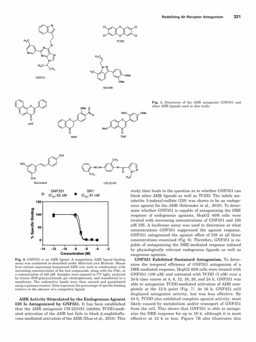
structure of GNF351, as well as other AHR agonists andantagonists used or discussed in this study, are found inFig. 1. To establish that GNF351 is a direct ligand for theAHR, a ligand competition binding assay using the PAL wasperformed. Figure 2 demonstrates that GNF351 is capable ofcompeting with the photoaffinity ligand for binding to thehuman AHR and has a relative affinity for the AHR similarto that of SR1. These data demonstrate that GNF351 has arelatively high affinity for the receptor.
GNF351 Does Not Activate AHR-Dependent DRE-Mediated Transcription. Because many AHR antagonistsalso show some degree of agonist activity at higher concen-trations, it was necessary to determine whether this was truefor GNF351. To determine whether the compound is a partialagonist for the AHR, a transcriptional response assay wasconducted using a stable human hepatoma-derived cell linecontaining the pGudluc 6.1 DRE-driven reporter (HepG2 40/6). Upon treatment with GNF351 for 4 h, no significantagonist activity was observed for 100 nM to 10 �M GNF351treatments compared with vehicle (Fig. 3A). To determinethe effect of GNF351 on levels of endogenous AHR-mediatedgene expression, quantitative PCR was performed on HepG240/6 cells treated for 4 h with DMSO, TCDD (5 nM), orincreasing concentrations of GNF351 (100 nM, 1 �M, and 10�M). TCDD dramatically induced CYP1A1 mRNA levels,whereas in contrast GNF351 failed to exhibit induction ofCYP1A1 even at be highest concentration of 10 �M (Fig. 3B).Indeed, constitutive levels diminished to below basal activity,although this effect was not statistically significant. Theseresults confirmed those generated with the reporter assaysystem. In addition, these observations suggest that long-term treatment with GNF351 should be an effective means toinhibit basal transcriptional activity of the AHR.
GNF351 Antagonizes Ligand-Mediated AHR Tran-scriptional Activity. HepG2 40/6 cells were treated withGNF351 in combination with TCDD for 4 h to determinewhether GNF351 inhibits the potent agonist effect seen withTCDD treatment. As the concentration of GNF351 increased,the AHR DRE-mediated response was antagonized in a dose-dependent manner (Fig. 4A). To determine whether this ef-fect is species-specific, H1L1.1.1c2 cells were also treatedwith increasing concentrations of GNF351 in combinationwith TCDD. Figure 4B shows that GNF351 antagonizes theagonist response in a mouse cell line in a dose-dependentmanner, although it took a higher concentration of GNF351to give the same antagonistic effect as that seen in the HepG240/6 cells. This was not unexpected, considering that themurine AHR has a 10-fold higher affinity for TCDD. Takingthis into account it would seem that the affinity of GNF351for the mouse and human AHR are similar. QuantitativePCR was conducted with HepG2 40/6 cells treated with ve-hicle, TCDD (2 nM), or a combination of GNF351 (100 nM)and TCDD (2 nM) for 4 h. Figure 5 shows that levels oftranscribed CYP1A1, CYP1A2, and AHRR decreased withthe combined treatment. These three genes have previouslybeen shown to be AHR-responsive (Beischlag et al., 2008).The data further show that the antagonistic effect of GNF351is not limited to one specific AHR-dependent gene. Consider-ing that a 4-h treatment was able to decrease constitutiveAHR target gene expression, it is likely that further repres-sion would be observed with a longer GNF351 treatment.
320 Smith et al.
AHR Activity Stimulated by the Endogenous AgonistI3S Is Antagonized by GNF351. It has been establishedthat the AHR antagonist CH-223191 inhibits TCDD-medi-ated activation of the AHR but fails to block �-naphthofla-vone-mediated activation of the AHR (Zhao et al., 2010). This
study then leads to the question as to whether GNF351 canblock other AHR ligands as well as TCDD. The indole me-tabolite 3-indoxyl-sulfate (I3S) was shown to be an endoge-nous agonist for the AHR (Schroeder et al., 2010). To deter-mine whether GNF351 is capable of antagonizing the DREresponse of endogenous agonists, HepG2 40/6 cells weretreated with increasing concentrations of GNF351 and 100nM I3S. A luciferase assay was used to determine at whatconcentrations GNF351 suppressed the agonist response.GNF351 antagonized the agonist effect of I3S at all threeconcentrations examined (Fig. 6). Therefore, GNF351 is ca-pable of antagonizing the DRE-mediated response inducedby physiologically relevant endogenous ligands as well asexogenous agonists.
GNF351 Exhibited Sustained Antagonism. To deter-mine the temporal efficiency of GNF351 antagonism of aDRE-mediated response, HepG2 40/6 cells were treated withGNF351 (100 nM) and cotreated with TCDD (5 nM) over a24-h time course at 4, 8, 12, 16, 20, and 24 h. GNF351 wasable to antagonize TCDD-mediated activation of AHR com-pletely at the 12-h point (Fig. 7). At 16 h, GNF351 stilldisplayed antagonist activity, but was less effective. By24 h, TCDD also exhibited complete agonist activity, mostlikely caused by metabolism and/or transport of GNF351from the cell. This shows that GNF351 is able to antago-nize the DRE response for up to 16 h, although it is mosteffective at 12 h or less. Figure 7B also illustrates this
Fig. 1. Structures of the AHR antagonist GNF351 andother AHR ligands used in this study.
Fig. 2. GNF351 is an AHR ligand. A competition AHR ligand bindingassay was conducted as described under Materials and Methods. Mouseliver cytosol expressing humanized AHR was used in combination withincreasing concentrations of the test compounds, along with the PAL, ata concentration of 420 pM. Samples were exposed to UV light, analyzedby tricine SDS-polyacrylamide gel electrophoresis, and transferred to amembrane. The radioactive bands were then excised and quantitatedusing a gamma-counter. Data represent the percentage of specific bindingrelative to the absence of a competitor ligand.
Redefining Ah Receptor Antagonism 321

point, showing that GNF351 was still 50% effective atapproximately 16 h.
Comparison of GNF351 with Other AHR Antago-nists. GNF351 was compared with other previously pub-lished AHR antagonists to test for potency of antagonism andagonist effect at a higher dose. A luciferase reporter assaywas conducted using HepG2 40/6 cells treated with a 10 �Mconcentration of a number of antagonists for 4 h to observewhether these compounds acted as agonists at a high dose.The compounds tested included: GNF351, �NF (Wil-helmsson et al., 1994), TMF (Murray et al., 2010a), MNF(Lu et al., 1995), resveratrol (Casper et al., 1999), andCH-223191 (Kim et al., 2006). Figure 8A, left, demon-strates that �NF displays statistically significant partialagonist activity at 10 �M. GNF351 and the other AHRantagonists tested displayed minimal levels of agonist ac-tivity. In Fig. 8A, right, HepG2 40/6 cells were treated with
a 40 nM concentration of the antagonists, as well as 5 nMTCDD, for 4 h. GNF351 and MNF showed the most signif-icant antagonism of the DRE response at this low concen-tration, with CH-223191 also showing a significant level ofcompetition. In contrast, �NF, TMF, and resveratrol failedto inhibit TCDD-mediated gene expression at the concen-tration tested.
In Fig. 8B, the same compounds were tested for agonistand antagonist activity in a mouse reporter stable cell line(H1L1.c2). For the agonist activity assay (Fig. 8B, left), thecompounds were used at a final concentration of 10 �M,whereas in the antagonist comparison, the compounds weretested at 100 nM in combination with 2 nM TCDD. �NF andresveratrol mediated significant agonist activity at this highdose. GNF351 seemed to repress basal levels of AHR activity,as seen with human cells, although this did not prove to bestatistically significant. In Fig. 8B, right, GNF351 again wasable to antagonize TCDD-driven AHR activity compared with
Fig. 4. GNF351 antagonizes the DRE-mediated response in AHR inhuman and murine cells. Cells were treated with increasing concentra-tions of GNF351 in combination with 5 nM TCDD in stable humanhepatoma-derived reporter cells (HepG2 40/6) (A) and with 2 nM TCDDin stable murine hepatoma-derived reporter cells (H1L1.1.1c2) (B) for 4 h,after which lysis buffer was added, and a luciferase assay conducted onthe lysate. Each data set is the result of triplicate well treatments. Datarepresent the mean � S.E.M. with statistically significant results marked(���, P 0.001), which are relevant to the data sets as labeled. The directcomparisons are shown by the presence of the same letter (a).
Fig. 3. GNF351 exhibits a lack of AHR agonist activity at increasingconcentrations. A cell-based luciferase reporter assay using humanHepG2 40/6 cells was conducted. A, cells were treated with DMSO, TCDD(5 nM), or increasing concentrations of GNF351 for 4 h. B, quantitativereverse transcription-PCR was performed for RNA samples isolated fromHepG2 40/6 cells treated with DMSO, TCDD (5 nM), or increasing con-centrations of GNF351 for 4 h, and the expression of CYP1A1 levels,normalized to L13a levels, was examined. Each treatment was conductedin triplicate wells. Data represent the mean � S.E.M. with statisticallysignificant results indicated (�, P 0.05; ��, P 0.01; ���, P 0.001),which are relevant to the data sets as labeled in A and compared withcontrol for B. The direct comparisons are shown by the presence of thesame letter (a or b).
322 Smith et al.

other known antagonists. MNF also exhibited significantrepression; in contrast, TMF showed significant inductionabove TCDD treatment alone. Overall, this assay furthershows that GNF351 is a more potent AHR antagonist thanpreviously characterized antagonists.
Acute-Phase Response Pathway Is Not Altered byGNF351. GNF351 was shown to potently repress AHR tran-scriptional activity via the DRE-mediated response. To es-tablish whether GNF351 was able to antagonize anothergene regulatory network modulated by the AHR, an acute-phase gene repression assay was conducted. Huh7 cells werepretreated for 1 h with vehicle, GNF351 (1 �M), TCDD (10nM), SGA360 (10 �M), and �NF (10 �M). A combination ofIL-1� and IL-6 was then added to all wells at a concentrationof 2 ng/ml for each cytokine and treated for an additional 6 h.A control well contained vehicle alone. RNA was isolatedfrom the cells, and specific mRNA levels were analyzed byquantitative PCR. GNF351 in combination with IL-1� andIL-6 showed no decrease in SAA1 levels, indicating thatGNF351 effectively failed to mediate repression of the acute-phase response (Fig. 9A). As expected, TCDD and SGA360(an AHR agonist and a SAhRM, respectively) repressed cy-tokine-mediated SAA1 expression. �NF, an AHR antagonistwith partial agonist activity at 10 �M, was capable of exhib-iting a statistically significant level of inhibition of SAA1
Fig. 6. GNF351 antagonizes the effect of an endogenous AHR agonist.HepG2 40/6 cells were treated with DMSO, I3S (100 nM), and increasingconcentrations of GNF351 with 100 nM I3S. Luciferase readings weretaken after cells were lysed. Treatments were conducted in triplicatewells. Data represent the mean � S.E.M. with statistically significantresults (���, P 0.001) compared as labeled. The direct comparisons areshown by the presence of the same letter (a or b).
Fig. 7. GNF351 acts as a DRE antagonist for up to 12 h. A, a time-coursetreatment was conducted using HepG2 40/6 cells. Cells were treated withDMSO, TCDD (5 nM), or GNF351 (100 nM) and TCDD (5 nM). Treat-ments were conducted at 4-, 8-, 12-, 16-, 20-, and 24-h time increments. Atthe end of each time point, cells were harvested using lysis buffer asdescribed under Materials and Methods, and luciferase readings wereconducted. Time points for DMSO control were taken at 8 and 24 h.B, data generated in A were replotted to determine the approximateED50. The average of the TCDD values at each time point was deter-mined, and each GNF351� TCDD value was divided by the TCDDaverage to determine percentage of antagonism.Fig. 5. GNF351 causes a decrease in levels of AHR-transcribed gene.
Quantitative reverse-transcription-PCR was performed using HepG240/6 cells treated with DMSO, TCDD (2 nM), and GNF351 (100 nM) withTCDD (2 nM) for 4 h. mRNA levels were assessed for CYP1A1, CYP1A2,and AHRR and normalized to L13a levels. Data represent the mean �S.E.M. with statistically significant results indicated (�, P 0.05; ��, P 0.01; ���, P 0.001). The direct comparisons are shown by the presenceof the same letter (a or b).
Redefining Ah Receptor Antagonism 323

expression as has been observed previously (Patel et al.,2009).
GNF351 Fails to Repress TPA-Mediated Ear Edema.To determine whether GNF351 exhibited SAhRM activity invivo a mouse ear edema assay that we previously character-ized was done. In this model TPA is used to induce edema, aneffect that can be inhibited by the SAhRM SGA360 (Murrayet al., 2010b). Three mice were used per treatment, andtreatments were vehicle (acetone), TPA, TPA � GNF351,TPA � GNF351 � SGA360, and TPA � SGA360. Figure 9Breveals that TPA increased edema width, whereas GNF351failed to decrease edema width, showing that GNF351 has noSAhRM activity, and SGA360 alone repressed TPA-mediatedear edema as has been shown previously (Murray et al.,2010b). It is noteworthy that GNF351 was able to prevent theability of SGA360 to repress TPA-mediated ear edema, dem-onstrating that the effects of SGA360 are AHR-dependent.These results indicate that GNF351 can antagonize the ac-tivity directed by a SAhRM and illustrates the utility ofGNF351 as a means to determine whether SAhRM activity ismediated through an AHR-dependent or -independent mech-anism.
GNF351 Interacts with the AHR Binding Pocket in aHomology Model. Next, we wanted to test whetherGNF351 can efficiently and directly interact with the AHRligand binding pocket. A computer-generated model of theAHR binding pocket based on its similarity to other PAS
(PER/ARNT/SIM)-domain proteins has been established(Bisson et al., 2009). Modeling images were generated forboth human and mouse receptor and showed that GNF351fits into the ligand binding pocket of the AHR for both species(Fig. 10; Supplemental Fig. 1). In this model system, thelower the binding energy value for a particular ligand thehigher its affinity for the AHR. Flavones that were previouslyidentified as AHR ligands have binding energies rangingfrom 4.3 to 2.74 kcal/mol (Bisson et al., 2009). The bindingenergies for both model systems show that GNF351 bindingis energetically favorable in both the human receptor (7.45kcal/mol) and mouse receptor (9.32 kcal/mol). The ligandbinding data in Fig. 2 further support the modeling results.The noncovalent interactions between GNF351 and the re-ceptor occur with amino acid residues Ser317, His291, andSer365 in human and Ser311, His285, and Ser359 in mouse.
DiscussionThrough the use of an AHR DNA binding mutant it has
been established that the AHR can repress cytokine-medi-ated acute-phase gene expression without binding to a DRE(Patel et al., 2009). In this study it was observed that thepartial agonist/antagonist �NF could repress SAA1 expres-sion to a level observed with the potent agonist TCDD. Thisled to the hypothesis that there are AHR ligands that canmediate acute-phase gene repression without exhibiting sig-
Fig. 8. Agonist and antagonist propertiesof various AHR ligands. A, left, HepG2 40/6cells were treated with 10 �M concentra-tion of various AHR ligands to determinewhether each compound displayed agonistactivity at a higher concentration. Right,HepG2 40/6 cells were treated with 40 nMAHR ligands plus 5 nM TCDD (except ve-hicle) to test for antagonistic abilities ofeach compound at a lower concentration. B,left, H1L1.1.1c2 cells were treated with 10�M of each AHR ligand to determinewhether any exhibited agonist activity.Right, H1L1.1.1c2 cells were treated with100 nM of each ligand in combination with2 nM TCDD to determine their antagonis-tic ability. Each treatment is the result oftriplicate wells. Data represent the mean �S.E.M. with statistically significant resultsindicated (�, P 0.05; ���, P 0.001). Thedirect comparisons are shown by the pres-ence of the same letter (a or b).
324 Smith et al.

nificant agonist activity. The AHR ligands 4-[1-allyl-7-(trifluoromethyl)-1H-indazol-3yl]benzene-1 (WAY-169916),SGA360, and 3�,4�-dimethoxy-�NF have now been identifiedas SAhRM that essentially do not exhibit agonist activity, yetcan repress cytokine-mediated acute-phase gene expression(Murray et al., 2010b,c, 2011). During chronic diseases suchas cancer and rheumatoid arthritis systemic inflammationmay occur that leads to an acute-phase response in the liver.The liver can produce large amounts of serum amyloid A thatoften mediates enhanced systemic inflammatory signalingand can lead to clinically relevant health complications suchas amyloidosis. Thus, SAhRM may be of therapeutic value inthe treatment of systemic inflammation in chronic inflamma-tory diseases.
If one only considers the ability of an AHR ligand to blockagonist-induced DRE-mediated transcriptional activity asthe criteria for an antagonist, then SAhRM would be consid-ered an antagonist. However, the discovery of non-DRE-me-diated AHR activity has necessitated that the definition of a“pure” antagonist be redefined to require that the functionaldefinition incorporate non-DRE-mediated AHR activity.Most previously characterized AHR antagonists (e.g., CH-
223191, MNF) have not been examined in the context ofnon-DRE-mediated AHR activity (Lu et al., 1995; Kim et al.,2006), thus it remains to be established if they function aspure antagonists or as inhibitors of DRE-mediated transcrip-tion. Thus, previous studies with AHR antagonists may rep-resent an incomplete picture of the effects of comprehensiveAHR antagonism. Here, we have demonstrated that GNF351is a pure antagonist that meets these criteria2. Furthermorethis also demonstrates that there are three distinct classes ofAHR ligands; agonists, SAhRMs, and pure antagonists. Aflow diagram is shown in Fig. 11 illustrating the experimen-tal scheme that leads to the determination of three classes ofAHR ligands.
A recent study has shown that AHR antagonists may beselective in their ability to diminish AHR activity (Zhao et al.,2010). Their results indicate that the antagonist CH-223191is capable of blocking agonist effects mediated by TCDD andcertain other halogenated aromatic hydrocarbons, but notactivity mediated by flavonoids, such as �-naphthoflavone orpolycyclic aromatic hydrocarbons. This study may supportthe notion that different ligands cause conformationalchanges in the binding pocket, which may only block compe-tition from certain subsets of ligands. We have demonstrated
Fig. 10. Homologous AHR binding pocket model for GNF351. In silicomodeling was conducted as described previously. GNF351 is shown tobind to human AHR (top) and mouse AHR (bottom).
Fig. 9. GNF351 antagonizes acute-phase response pathways. A, an acute-phase repression assay was conducted in Huh7 cells. Cells were pre-treated for 1 h with the following compounds: DMSO (vehicle) alone,DMSO, GNF351 (1 �M), TCDD (10 nM), SGA360 (10 �M), and �NF (10�M). A combination of IL-1� and IL-6 was then added to all wells, exceptthe vehicle alone (control), at a concentration of 2 ng/ml for each cytokine,and the treatment was continued for an additional 6 h. B, a mouse earedema assay was conducted as described under Materials and Methods,and data were generated from three 6-week-old male C57BL6/J (wildtype) mice. Mice were treated with vehicle (acetone), TPA, GNF351,SGA360, or combinations of these compounds for 6 h. Data represent themean � S.E.M. with statistically significant results compared as indi-cated (�, P 0.05; ��, P 0.01; ���, P 0.001). The direct comparisonsare shown by the presence of the same letter (a or b).
Redefining Ah Receptor Antagonism 325

that GNF351 is able to successfully antagonize the effects ofa diverse array of AHR ligands, including TCDD, 3-indoxylsulfate, and SGA360, which represent three different types ofAHR ligands. Though the AHR-dependent transcriptionaleffects of the compounds tested are blocked, this observationdoes not preclude the possibility that the activity of otherAHR agonists may not be affected.
The characterization of a high-affinity pure antagonist willprove useful for a number of applications. For instance, thisdistinct class of ligands will allow the biological activity of theAHR to be explored in more depth. Studies using antagoniststhat effectively block more than one AHR pathway shouldlead to further insights into the mechanisms of DRE andnon-DRE AHR activity. Recently, the AHR antagonist SR1has been shown to exhibit significant biological effects, suchas mediating human hematopoietic stem cell expansion invitro, and therefore antagonists may also be used to deter-mine underlying physiological mechanisms in which theAHR is involved (Boitano et al., 2010). It is noteworthy thatthis study did not address whether AHR antagonism in thisexample occurs through blocking DRE or non-DRE activity; aSAhRM could be used to help address this issue. Also the useof antagonists has already been demonstrated to inhibit IL-6expression in tumor cells through displacement of the AHR/ARNT heterodimer from the IL-6 promoter and thus mayprove useful in therapeutic intervention (DiNatale et al.,2010).
A key feature that the use of GNF351 offers is the lack ofagonist activity even at higher doses. Some of the previouslyknown AHR antagonists are imperfect candidates as com-plete inhibitors of DRE-driven transcription because of theexhibition of partial agonist activity at higher concentra-tions. One example of this type of antagonist is �NF, whichhas been shown to exhibit partial agonist activity in bothhuman and murine reporter cells. GNF351 not only fails to
induce transcriptional activity at higher concentrations, italso inhibits basal AHR activity. The physiological conse-quences of decreasing basal activity of AHR have yet to bethoroughly explored. GNF351 exhibits greater potency thana variety of AHR antagonists tested here. Most antagonistsrequire micromolar concentrations to completely inhibitTCDD-mediated transcription. In contrast, as little as 100nM GNF351 completely inhibited TCDD induction of tran-scriptional activity in a human cell line. Because GNF351 iseffective at lower experimental doses, it is more likely thatoff-target effects would be minimized by the use of this com-pound. It is also shown here that GNF351 binds with rela-tively high affinity to the ligand-binding pocket of the AHR,which should block the binding of an array of exogenous andendogenous ligands. Modeling data presented here showsthat GNF351 binds to AHR, but the mechanism by which thecompound exhibits its antagonistic activities needs to bedetermined. Studies of the mechanisms by which ligands,including antagonists, affect AHR function are needed, per-haps after the AHR binding pocket has been successfullycrystallized. This is important in the further pursuit of theAHR as a viable drug target for diseases such as cancer andwith autoimmune responses. The use of GNF351 in suchexperiments could allow for receptor activity to be ablated toinvestigate its possible therapeutic uses.
The existence of pure antagonists will prove useful in var-ious experimental conditions, but it should not be presumedthat they would block every aspect of AHR function. Forexample, it may be unlikely that an antagonist would disruptprotein-protein interactions in which the unliganded AHRparticipates, presumably in the cytoplasm. An antagonistdoes not simply ablate the presence of the AHR, and there-fore the antagonist-bound receptor should not be consideredthe same as the absence of receptor. In this study, we havesucceeded in identifying a pure AHR antagonist and furtherexpanding what is meant by this term. GNF351 will be usefulin a variety of experimental models and should aid in discov-ering more about the biological functions of the AHR. Cur-rently, the best in vivo models in which to study AHR func-tion involve the repression of AHR expression either in aconditional knockout mouse or Ahr-null mice. Both of thesemodels ablate AHR expression throughout development, andany experiments performed with these mice need to take intoaccount the effects of the long-term absence of AHR expres-sion. In cell culture AHR can be ablated using small inter-fering RNA, although achieving complete loss of AHR expres-sion is difficult, taking 48 to 72 h. Also, the loss of expressionmay differ from blocking AHR activity. Therefore, GNF351will be useful in studying the absence of AHR function fordefined time periods for comparison with receptor expressionknockdown models. Clearly, future studies are needed todetermine the absorption characteristics and half-life ofGNF351 for use as an antagonist in vivo. This will allow thereceptor to be studied during definitive time points, such asthe role of the AHR in development or during disease states.
Acknowledgments
We thank Marcia H. Perdew for excellent editorial assistance.
Authorship Contributions
Participated in research design: Murray, Kolleri, and Perdew.Conducted experiments: Smith, Murray, Tanos, and Bisson.
Fig. 11. Experimental scheme to determine the class of an AHR ligand.
326 Smith et al.

Contributed new reagents or analytic tools: Tellew, Boitano, andCooke.
Performed data analysis: Smith, Murray, Bisson, and Perdew.Wrote or contributed to the writing of the manuscript: Smith,
Murray, Cooke, and Perdew.Other: Perdew acquired funding for the research.
ReferencesBeischlag TV, Luis Morales J, Hollingshead BD, and Perdew GH (2008) The aryl
hydrocarbon receptor complex and the control of gene expression. Crit Rev Eu-karyot Gene Expr 18:207–250.
Bisson WH, Koch DC, O’Donnell EF, Khalil SM, Kerkvliet NI, Tanguay RL, AbagyanR, and Kolluri SK (2009) Modeling of the aryl hydrocarbon receptor (AhR) ligandbinding domain and its utility in virtual ligand screening to predict new AhRligands. J Med Chem 52:5635–5641.
Boitano AE, Wang J, Romeo R, Bouchez LC, Parker AE, Sutton SE, Walker JR,Flaveny CA, Perdew GH, Denison MS, et al. (2010) Aryl hydrocarbon receptorantagonists promote the expansion of human hematopoietic stem cells. Science329:1345–1348.
Casper RF, Quesne M, Rogers IM, Shirota T, Jolivet A, Milgrom E, and Savouret JF(1999) Resveratrol has antagonist activity on the aryl hydrocarbon receptor: im-plications for prevention of dioxin toxicity. Mol Pharmacol 56:784–790.
DiNatale BC, Schroeder JC, Francey LJ, Kusnadi A, and Perdew GH (2010) Mech-anistic insights into the events that lead to synergistic induction of interleukin 6transcription upon activation of the aryl hydrocarbon receptor and inflammatorysignaling. J Biol Chem 285:24388–24397.
Flaveny CA, Murray IA, Chiaro CR, and Perdew GH (2009) Ligand selectivity andgene regulation by the human aryl hydrocarbon receptor in transgenic mice. MolPharmacol 75:1412–1420.
Garrison PM, Tullis K, Aarts JM, Brouwer A, Giesy JP, and Denison MS (1996)Species-specific recombinant cell lines as bioassay systems for the detection of2,3,7,8-tetrachlorodibenzo-p-dioxin-like chemicals. Fundam Appl Toxicol 30:194–203.
Kim SH, Henry EC, Kim DK, Kim YH, Shin KJ, Han MS, Lee TG, Kang JK,Gasiewicz TA, Ryu SH, et al. (2006) Novel compound 2-methyl-2H-pyrazole-3-carboxylic acid (2-methyl-4-o-tolylazo-phenyl)-amide (CH-223191) prevents2,3,7,8-TCDD-induced toxicity by antagonizing the aryl hydrocarbon receptor. MolPharmacol 69:1871–1878.
Kimura A, Naka T, Nohara K, Fujii-Kuriyama Y, and Kishimoto T (2008) Arylhydrocarbon receptor regulates Stat1 activation and participates in the develop-ment of Th17 cells. Proc Natl Acad Sci USA 105:9721–9726.
Long WP, Pray-Grant M, Tsai JC, and Perdew GH (1998) Protein kinase C activityis required for aryl hydrocarbon receptor pathway-mediated signal transduction.Mol Pharmacol 53:691–700.
Lu YF, Santostefano M, Cunningham BD, Threadgill MD, and Safe S (1995) Iden-tification of 3�-methoxy-4�-nitroflavone as a pure aryl hydrocarbon (Ah) receptorantagonist and evidence for more than one form of the nuclear Ah receptor inMCF-7 human breast cancer cells. Arch Biochem Biophys 316:470–477.
Murray IA, Flaveny CA, Chiaro CR, Sharma AK, Tanos RS, Schroeder JC, Amin SG,Bisson WH, Kolluri SK, and Perdew GH (2011) Suppression of cytokine-mediatedcomplement factor gene expression through selective activation of the Ah receptorwith 3�,4�-dimethoxy-�-naphthoflavone. Mol Pharmacol 79:508–519.
Murray IA, Flaveny CA, DiNatale BC, Chairo CR, Schroeder JC, Kusnadi A, andPerdew GH (2010a) Antagonism of aryl hydrocarbon receptor signaling by 6,2�,4�-trimethoxyflavone. J Pharmacol Exp Ther 332:135–144.
Murray IA, Krishnegowda G, DiNatale BC, Flaveny C, Chiaro C, Lin JM, SharmaAK, Amin S, and Perdew GH (2010b) Development of a selective modulator of arylhydrocarbon (Ah) receptor activity that exhibits anti-inflammatory properties.Chem Res Toxicol 23:955–966.
Murray IA, Morales JL, Flaveny CA, Dinatale BC, Chiaro C, Gowdahalli K, Amin S,and Perdew GH (2010c) Evidence for ligand-mediated selective modulation of arylhydrocarbon receptor activity. Mol Pharmacol 77:247–254.
Patel RD, Murray IA, Flaveny CA, Kusnadi A, and Perdew GH (2009) Ah receptorrepresses acute-phase response gene expression without binding to its cognateresponse element. Lab Invest 89:695–707.
Poland A, Glover E, Ebetino H, and Kende A (1986) Photoaffinity labelling of the Ahreceptor. Food Chem Toxicol 24:781–787.
Quintana FJ, Basso AS, Iglesias AH, Korn T, Farez MF, Bettelli E, Caccamo M,Oukka M, and Weiner HL (2008) Control of T(reg) and T(H)17 cell differentiationby the aryl hydrocarbon receptor. Nature 453:65–71.
Safe S and McDougal A (2002) Mechanism of action and development of selectivearyl hydrocarbon receptor modulators for treatment of hormone-dependent can-cers (Review). Int J Oncol 20:1123–1128.
Schmidt JV and Bradfield CA (1996) Ah receptor signaling pathways. Annu Rev CellDev Biol 12:55–89.
Schroeder JC, Dinatale BC, Murray IA, Flaveny CA, Liu Q, Laurenzana EM, Lin JM,Strom SC, Omiecinski CJ, Amin S, et al. (2010) The uremic toxin 3-indoxyl sulfateis a potent endogenous agonist for the human aryl hydrocarbon receptor. Biochem-istry 49:393–400.
Veldhoen M, Hirota K, Christensen J, O’Garra A, and Stockinger B (2009) Naturalagonists for aryl hydrocarbon receptor in culture medium are essential for optimaldifferentiation of Th17 T cells. J Exp Med 206:43–49.
Veldhoen M, Hirota K, Westendorf AM, Buer J, Dumoutier L, Renauld JC, andStockinger B (2008) The aryl hydrocarbon receptor links TH17-cell-mediated au-toimmunity to environmental toxins. Nature 453:106–109.
Wilhelmsson A, Whitelaw ML, Gustafsson JA, and Poellinger L (1994) Agonistic andantagonistic effects of �-naphthoflavone on dioxin receptor function. Role of thebasic region helix-loop-helix dioxin receptor partner factor Arnt. J Biol Chem269:19028–19033.
Zhao B, Degroot DE, Hayashi A, He G, and Denison MS (2010) CH223191 is aligand-selective antagonist of the Ah (Dioxin) receptor. Toxicol Sci 117:393–403.
Address correspondence to: Gary H. Perdew, Center for Molecular Toxi-cology and Carcinogenesis, Department of Veterinary Sciences, PennsylvaniaState University, 309A Life Sciences Building, University Park, PA 16802.E-mail: [email protected]
Redefining Ah Receptor Antagonism 327


