Embed Size (px)
Citation preview
MOL # 16873
1
Identification of zebrafish ARNT1 homologs:
TCDD toxicity in the developing zebrafish requires ARNT1
Amy L. Prasch, Robert L. Tanguay, Vatsal Mehta,
Warren Heideman, and Richard E. Peterson
School of Pharmacy (W.H., R.E.P.) and Molecular and Environmental Toxicology Center
(A.L.P., V.M., W.H., R.E.P.), University of Wisconsin, Madison, Wisconsin 53705; Department
of Environmental and Molecular Toxicology (R.L.T.), Oregon State University, Corvallis,
Oregon, 97331.
Molecular Pharmacology Fast Forward. Published on November 23, 2005 as doi:10.1124/mol.105.016873
Copyright 2005 by the American Society for Pharmacology and Experimental Therapeutics.
MOL # 16873
2
Running title: ARNT1 mediates TCDD toxicity in zebrafish Corresponding Author:
Richard E. Peterson
School of Pharmacy
University of Wisconsin
777 Highland Ave.
Madison, WI 53705-2222
FAX: (608) 265-3316
E-mail: [email protected]
Pages: 39
Figures: 11
References: 40
Words in abstract: 250
Words in introduction: 678
Words in discussion: 1476
Abbreviations: TCDD, 2,3,7,8-tetrachlorodibenzo-p-dioxin; AHR, aryl hydrocarbon receptor;
ARNT, aryl hydrocarbon receptor nuclear translocator; XRE, xenobiotic response element; hpf,
hours post fertilization; TAD, transactivation domain; PCDDs, polychlorinated dibenzo-p-
dioxins; zebrafish ARNT1 morpholino, zfarnt1-MO.
MOL # 16873
3
ABSTRACT In order to use the zebrafish as a model to study 2,3,7,8-tetrachlorodibenzo-p-dioxin
(TCDD) developmental toxicity, it is essential to know which proteins are involved in mediating
toxicity. Previous work has identified zfAHR2 as the receptor which binds TCDD mediating
downstream responses. Although zfARNT2b can form a functional heterodimer with zfAHR2 in
vitro, zfarnt2 null mutants show no protection against endpoints of TCDD developmental
toxicity demonstrating that zfARNT2b cannot be the physiological dimerization partner for
zfAHR2 mediating responses to TCDD in zebrafish embryos. The purpose of the current study
was to identify an alternate dimerization partner(s) for zfAHR2 that may function to mediate
TCDD developmental toxicity. By searching zebrafish genomic sequence and employing the
PCR-based RACE technique, three forms of cDNA that appear to be alternate mRNA splice
variants of a zebrafish homolog of ARNT1 were detected. Analysis of the zfARNT1 proteins in
vitro demonstrates that the two longest forms of zfARNT1, zfARNT1b and zfARNT1c, can form
functional heterodimers with zfAHR2. However, the shortest form, zfARNT1a, appears to be
nonfunctional with zfAHR2 in vitro. In order to determine if a zfARNT1 protein functions with
zfAHR2 in vivo, a morpholino targeted against the 5’ end of zfARNT1 (zfarnt1-MO) was used.
Injection of the zfarnt1-MO prior to TCDD treatment significantly decreases the induction of
zfCYP1A mRNA and protein. In addition, zfarnt1 morphants show complete protection against
TCDD-induced pericardial edema and show partial protection against reduced blood flow and
craniofacial malformations caused by TCDD demonstrating the role of zfARNT1 proteins in
mediating these responses.
MOL # 16873
4
Polychlorinated dibenzo-p-dioxins (PCDDs) are lipophilic, persistent, bioaccumulative
toxicants. Developing fish are amongst the most sensitive species to the toxicity of these
compounds (Peterson et al., 1993) and 2,3,7,8-tetrachlorodibenzo-p-dioxin (TCDD), the most
potent PCDD, has adversely affected feral fish populations of lake trout, the most sensitive fish
species to TCDD developmental toxicity (Cook et al., 2003). However, the mechanism by which
TCDD causes adverse effects on fish development is unclear. The zebrafish makes an attractive
model in which to study the developmental toxicity of TCDD due to their rapid growth, high egg
yield, short generation time and rapid, external development. In addition, their developmental
biology has been well characterized, the zebrafish genome is near completion, and numerous
molecular and genetic techniques have been developed to study gene function. Finally, the
zebrafish displays a similar set of endpoints of toxicity upon TCDD exposure as other freshwater
fish species including pericardial edema, arrested growth, craniofacial malformations, ischemia,
anemia, impaired swim bladder inflation, and mortality (reviewed by Tanguay et al., 2003).
Studies of aryl hydrocarbon receptor (ahr)-/- null mouse lines have clearly demonstrated
the role of the AHR signaling pathway in mediating responses to TCDD (Fernandez-Salguero et
al., 1996; Mimura et al., 1997), and signaling through the AHR has been well characterized in
mammals (reviewed in Schmidt and Bradfield, 1996). In order to use zebrafish as a model to
study TCDD developmental toxicity, it was essential to identify and characterize the zebrafish
AHR signaling pathway, and much is now known about its components. Three forms of the
AHR (zfAHR1, zfAHR1B, and zfAHR2) have been identified in zebrafish (Andreasen et al.,
2002a; Karchner et al. 2005; Tanguay et al., 1999), and morpholino knockdown of zfAHR2 has
identified it as the receptor which binds TCDD leading to downstream effects (Dong et al., 2004;
Prasch et al., 2003; Teraoka et al., 2003). In untreated zebrafish liver cells zfAHR2 has been
MOL # 16873
5
shown to shuttle between the cytoplasm and nucleus. However, upon treatment with the ligand
BNF, zfAHR2 translocates to the nucleus (Wentworth et al., 2004) where it then presumably
dimerizes with an aryl hydrocarbon receptor nuclear translocator (ARNT) protein. The zfAHR2-
ARNT heterodimer can then bind to xenobiotic response elements (XREs) in target genes such
as zfcyp1a leading to altered gene expression (Tanguay et al., 1999). Duplicate aryl hydrocarbon
receptor repressor genes, zfAHRR1 and zfAHRR2, have also been identified in zebrafish (Evans
et al., 2005). These proteins can negatively regulate zfAHR2 signaling in vitro, although their in
vivo role in zfAHR2 biology remains unclear.
The initial ARNT protein identified in zebrafish was zfARNT2 (Tanguay et al., 2000;
Wang et al, 2000). ZfARNT2 exists as multiple splice forms one of which, zfARNT2b, can form
a functional heterodimer in vitro with zfAHR2 that can specifically recognize XREs in DNA
binding assays and induce XRE-driven transcription in COS-7 cells incubated with TCDD
(Tanguay et al., 2000). ZfAHR2, zfARNT2a,b,c, and zfCYP1A mRNAs have also been shown to
co-localize in tissues of the cardiovascular system after TCDD exposure (Andreasen et al.,
2002b). This data suggested that zfARNT2b was the likely dimerization partner for zfAHR2 in
vivo involved in mediating responses to TCDD. However, analysis of zfarnt2 morphant and
zfarnt2-/- mutant embryos has demonstrated that zfARNT2 is not essential for the development of
endpoints of TCDD toxicity, because both zfarnt2 morphants and null mutants show similar
sensitivity to TCDD as wild type embryos (Prasch et al., 2004a).
The sensitivity of the zfARNT2 deficient fish to TCDD toxicity suggested that an
alternate form(s) of ARNT must exist in the zebrafish embryo that functionally dimerizes with
zfAHR2. The purpose of this work was to identify and characterize this putative ARNT in
zebrafish and determine its role in mediating TCDD developmental toxicity. Indeed, three novel
MOL # 16873
6
cDNAs that encode proteins with sequence similarity to rainbow trout ARNT and mammalian
ARNT1: zfARNT1a, zfARNT1b, and zfARNT1c were identified. In vitro analysis of these
proteins demonstrates that two forms, zfARNT1b and zfARNT1c, can form functional
heterodimers with zfAHR2, and in vivo morpholino antisense knockdown experiments
demonstrate an essential role for zfARNT1 proteins in mediating several TCDD-dependent
endpoints in developing zebrafish.
MATERIALS AND METHODS Oligonucleotides. Oligonucleotides used for PCR and gel shift reactions were synthesized by
Integrated DNA Technologies and are written 5’ to 3’. The predicted initiation ATG codon is
indicated in bold, and the sequence encoding the BAMHI restriction site in the ARNTF2bam
primer is in lower case. For the gel shift oligonucleotides the XRE core consensus sequence is
underlined and mutated bases are in bold. ARNTF1,ATCCTGCGCATGGCCGTATC;
ARNTR1,GATGTAGCCTGTGCAGTGGAC;SRARNTR1,AACCACATACTGCTGCTCGC-
CGTCTTT;SRARNTF1,TCAGAGAGCAGCTTTCCACCACCGAGAA;SRARNTF2,CCGG-
ATGTTGGATATGAAAACGGGCAC;ARNTF2bam,ggatccCTTTGCCTGTCGGACATGAC-
A;ARNTF2,CTTTGCCTGTCGGACATGACA;ARNTR2,GAACATATCCCTCAGCTCTTC-
TTAATG;ARNTR3,AGGACACGGTGTAAGACTAC;ARNTR4,ACAAAAGGGCAAAATA-
GGTCC. Gel shift oligonuceotides: wtXREF,GGCTCTTCTCACGCAACTCCGG;wtXRER,
GCCCCGGAGTTGCGTGAGAAGA;mutXREF,GGCTCTTCTCGCGCAACTCCGG;
mutXRER, GCCCCGGAGTTGCGCGAGAAGA.
ZfARNT1 identification and cloning. Primers ARNTF1 and ARNTR1 were initially used to
amplify a portion of zfARNT1 sequence identified in the Ensembl release of the zebrafish
genome. The single amplified 550 bp product was subcloned into pGEM-T Easy vector
MOL # 16873
7
(Promega) and sequenced. A PCR-based approach was then used to identify the 5’ and 3’ ends of
zfARNT1 using the SMART RACE cDNA Amplification kit (Clontech) as described by the
manufacturer. One µg of the poly(A) RNA generated from 72hpf zebrafish embryos was
reversed transcribed using PowerScript Reverse Transcriptase with either the 5’-CDS primer and
SMART 11 A oligo (5’ RACE ready cDNA) or the 3’ CDS primer (3’ RACE ready cDNA).
PCR reactions were performed using 2.5 µl of each RACE ready cDNA as a template with the
SRARNTR1 or SRARNTF1 gene specific primers and the supplied universal primer. The 3’
RACE reaction was reamplified using the SRARNTF2 nested primer and the supplied nested
universal primer. A single 5’ RACE product and two distinct 3’ RACE products were amplified,
subcloned into pGEM-T Easy vector and sequenced. The 5’ RACE product showed high
sequence similarity to other forms of ARNT1 and contained a likely translational start site,
however, both 3’ RACE products were truncated compared to ARNT1s from other species and
did not appear to encode the full length 3’ end of ARNT1. A clone from the Sanger Center was
subsequently identified (NCBI accession number BX927222) as containing the genomic
sequence for zfARNT1. Analysis of this genomic clone revealed additional 3’ sequence with
similarity to ARNT1 of other species.
cDNA constructs for functional studies. Because all putative forms of zfARNT1 contained the
same 5’ end, the same forward primer (ARNTF2) was used to amplify each form. The
zfARNT1a and zfARNT1b sequences were amplified using the ARNTF2 primer containing an
additional BAMH1 sequence on the 5’ end (ARNTF2bam). ZfARNT1a was amplified using the
ARNTR3 reverse primer, zfARNT1b was amplified using the ARNTR2 reverse primer and
zfARNT1c was amplified using the ARNTR4 reverse primer. To obtain full length transcripts
zfARNT1a and zfARNT1b were amplified out of cDNA generated using Superscript II reverse
MOL # 16873
8
transcriptase (Invitrogen) from 500 ng of poly(A) RNA that had been isolated out of whole 72
hpf embryos. PCR was performed on this cDNA using high fidelity PfuTurbo Polymerase
(Stratagene). Adenines were added to the amplified products prior to cloning into pGEM-T Easy
and sequencing. The full length zfARNT1c was amplified from an adult fin cDNA library using
high fidelity KOD polymerase (EMD Biosciences, San Diego), subcloned into PCRII TOPO
Blunt vector (Invitrogen) and sequenced. To generate expression vectors for zfARNT1a and
zfARNT1b, inserts were excised from pGEM-T Easy by digestion with BamHI (site included in
ARNTF2 primer) and Not I and cloned into pBKCMV (Stratagene) previously cut with the same
enzymes to generate pBKCMV-zfARNT1a and pBKCMV-zfARNT1b. The pBKCMV-
zfARNT1c expression vector was generated by excising the insert from the PCRII TOPO vector
with EcoRI digest and inserting into the EcoRI site of pBKCMV.
Nucleotide and amino acid sequence analysis. Both strands of each clone were sequenced at
least three times using fluorescent dye-labeling cycle sequencing (Applied Bio Systems, UW
Biotech Center, Madison, WI; OSU, Center for Gene Research and Biotechnology, Corvallis,
OR) stepwise using gene specific primers before Genbank submission. The Baylor College of
Medicine Human Genome Center search launcher, the National Center for Biotechnology
Information (NCBI), and Vector NTI software were used for sequence analysis.
Comparative analysis of zfARNT1 and zfARNT2 mRNA abundance. RNA was isolated from
pools of 10 vehicle-exposed embryos and extracted using the Qiagen RNeasy Mini kit according
to the manufacturer’s instructions. cDNA was produced from 750 ng of each total RNA sample
using Superscript II (Invitrogen) and oligo dT primer in 20 ul. The Light Cycler (Roche) was
used for quantitative real-time PCR. In order to compare expression levels of zfARNT1 and
zfARNT2, Roches’s relative quantitation software was used to perform calibrator normalized
MOL # 16873
9
relative quantification using efficiency correction. Target genes (zfARNT1 and zfARNT2) and a
reference loading control gene (β-actin) were amplified in triplicate for each sample. The
ARNTF1 and ARNTR1 primer set was used to amplify all splice forms of zfARNT1. Primers
used to amplify zfARNT2b/c and β-actin were previously described (Andreasen et al., 2002b).
Efficiencies for each primer set were determined from amplifications with standard curves and
are included in the calculation of the expression value for each gene. The expression of the target
and reference genes were also determined in a calibrator (cDNA from whole adult tissue) which
is included in each light cycler run to control for run to run variability. To get the final
abundance values for zfARNT1, the zfARNT1/β-actin ratio was calculated for each sample and
normalized to the ARNT1/βactin ratio obtained in the adult cDNA sample. Similarly, the
normalized ratio of ARNT2/βactin was also determined in each sample in order to compare the
relative abundance of these two transcripts.
In vitro transcription and translation. In vitro transcription and translation of zfAHR2,
zfARNT2b, zfARNT1a, zfARNT1b, and zfARNT1c proteins was performed using the TNT
Coupled Rabbit Reticulocyte Lysate System (Promega). Recombinant proteins were expressed
from pBKCMV expression constructs described here and by Tanguay et al. (1999, 2000) with T3
polymerase as described by the manufacturer. For in vitro DNA binding assays each protein was
expressed alone or zfAHR2 was coexpressed with either zfARNT2b, zfARNT1a, zfARNT1b, or
zfARNT1c in the presence of 10 nM TCDD or vehicle (dimethyl sulfoxide). Side reactions
containing [35S]-methionine were performed to assess relative protein production. To test the
zfarnt1-MO, 12.5 µl reactions were performed using 250 ng of template DNA, 1.5 µl of [35S]-
methionine, and either no morpholino or 500 nM final concentration of control-MO or zfarnt1-
MOL # 16873
10
MO. After 90 min incubation at 30°C, radioactive translation products were resolved by 8%
polyacrylamide gel electrophoresis, dried, and phosphorimaged (Molecular Dynamics).
In vitro DNA binding assay. Double stranded probes were made by annealing the forward and
reverse oligonucleotides for the wild type (wt) and mutant (mut) XRE. Annealing was performed
by combining 0.5 nmol of each oligonucleotide in 0.1M NaCl and 0.01 M Tris pH 8.0, heating to
70°C for 10 min, and then cooling to room temperature. Double stranded probes were
radiolabeled with [32P] dCTP using the Prime-a-gene kit (Promega). The gel shift was performed
by combining the in vitro synthesized protein mixtures with binding buffer (20 mM HEPES, pH
8.0, 100 mM NaCl, 1 mM dithiothreitol (DTT), and 6% glycerol), 2 µg poly(dI-dC), 15 fmol of
radiolabeled wtXRE, and a 10-fold molar excess of unlabeled wtXRE or mutXRE competitor
DNAs when indicated. After a 15 min incubation at room temperature, complexes were resolved
on a 0.5X Tris/borate/EDTA (90 mM Tris, 64.6 mM boric acid, and 2.5 mM EDTA, pH 8.3) and
4% acrylamide gel. The dried gel was exposed to a phosphor screen for 2 days.
Transient Transactivation Assay. COS-7 cells were plated on 24-well plates at a density of 8 ×
104 cells per well 1 day before transfection which was performed using SuperFect (Qiagen).
Each well was cotransfected with 400 ul of serum-containing media containing pBKCMV
expression constructs for zfAHR2, zfARNT1a, zfARNT1b, zfARNT1c or zfARNT2b (450 ng),
prt1aluc luciferase reporter construct (100 ng), and β-galactosidase CMV reporter (50 ng) as
indicated. After a 2.5 h incubation at 37 °C the transfection mixture was removed and replaced
with 1 ml of serum-containing media. After 22.5 h incubation, each well was exposed to either
dimethyl sulfoxide (vehicle) or 10 nM TCDD and incubated for another 20 h. Luciferase assay
was performed using the Luciferase Assay system (Promega) and a ML-2250 luminometer
(Dynatech Labs, Chantilly, VA). One hundred microliters of lysis buffer was added to each well
MOL # 16873
11
and after a 15 min incubation a 10 ul aliquot of cell lysate was transferred to a luminometer
plate. For the luciferase assay 50 ul of luciferase assay buffer was injected into each well, a 2 s
incubation was performed, and luminescence was integrated over 10 s. Transfection efficiency
was determined by measuring the β-galactosidase activity for each well. Thirty microliters of cell
lysate was aliquoted into a 96 well plate followed by the addition of 200 µl of reaction buffer
(0.1 M NaPO4, 10 mM KCl, 1 mM MgCl2, and 0.385 % β-mercaptoethanol) and 40 µl of 4
mg/ml o-nitrophenyl-β-D-galactopyranoside. The reaction was incubated for 3 h at 37°C and
plates were read at 405 nM using an ELx800 plate reader (Bio-Tek Instruments, Winooski, VT).
Antisense morpholinos . The zebrafish arnt1 morpholino (zfarnt1-MO) obtained from Gene
Tools (Philomath, Oregon) was designed with sequence complementary to the zfARNT1 cDNA.
The zfarnt1-MO overlapped the first predicted translation start site of zfARNT1 mRNA by
starting 8 bp upstream of the predicted AUG start codon and continuing to 14 bp downstream.
The sequence of the zfarnt1-MO was: 5’ GGATTAGCTGATGTCATGTCCGACA 3’, and it
was fluorescein tagged at the 3’ end to monitor injection success. The standard control
morpholino sold by Gene Tools (5’ CTCTTACCTCAGTTACAATTTATA 3’) was used as the
control morpholino (control-MO). Prior to embryo injection morpholinos were diluted to 0.12
mM in 1X Danieau’s (58 mM NaCl, 0.7 mM KCl, 0.4 mM MgSO4, 0.6 mM Ca(NO3)2, 5 mM
HEPES, pH 7.6) as described by Nasevicius and Ekker (2000).
Microinjection and waterborne TCDD exposure of zebrafish embryos. Fertilized eggs were
obtained from adult AB strain zebrafish bred in our laboratory as described by Westerfield
(1995). Embryos were raised at a water temperature of 27°C, water was changed daily, and when
appropriate, embryos were euthanized with tricaine (1.67 mg/ml MS222, Sigma).
MOL # 16873
12
For microinjection newly fertilized eggs were injected with either the zfarnt1 or control-
MO at the 1-2 cell stages with approximately 13 ng of the morpholino. Embryos were allowed to
develop for 2 h, after which unfertilized and damaged eggs were discarded. Viable zfarnt1-MO
injected embryos (zfarnt1 morphants) were observed for fluorescence as an index of injection
success demonstrated by uniform distribution of the morpholino. Only zfarnt1-MO injected
embryos exhibiting fluorescence were used.
Waterborne exposure of embryos to 2,3,7,8-tetrachlorodibenzo-p-dioxin (TCDD)
occurred from approximately 3-4 hpf. TCDD of > 99% purity obtained from Chemsyn, Lenexa,
KS was dissolved in dimethyl sulfoxide. Embryos from each injection group (uninjected,
control-MO injected, or zfarnt1-MO injected) were exposed to vehicle (0.1% dimethyl sulfoxide)
or TCDD (0.4 ng/ml) for 1 h in glass scintillation vials with gentle rocking. Following the 1 h
static exposure, embryos were rinsed with water and maintained in vehicle/TCDD-free water for
the remainder of experiments.
Analysis of zfCYP1A mRNA abundance. To analyze zfCYP1A mRNA abundance, RNA was
isolated from pools of 15 vehicle and TCDD-exposed embryos from each injection group
(uninjected, control-MO injected, zfarnt1-MO injected, n = pool of 15). cDNA was prepared as
described above and quantitative real-time PCR for zfCYP1A and β-actin was performed as
described previously (Andreasen et al., 2002b; Prasch et al., 2003).
Whole mount immunolocalization of zfCYP1A. Embryos were anesthetized at 72 hpf, and fixed
in 4% paraformaldehyde in phosphate-buffered saline. Tissue-specific expression of zfCYP1A
was determined using the monoclonal antibody Mab1-12-3 (Park et al. 1986) as previously
described (Andreasen et al., 2002b; Prasch et al., 2003; Carney et al., 2004).
MOL # 16873
13
Assessment of TCDD developmental toxicity in zfarnt1 morphants. Vehicle and TCDD-
exposed embryos from each injection group were assessed for 3 endpoints of TCDD toxicity:
pericardial edema, reduced peripheral blood flow, and reduction in lower jaw growth. All were
measured as previously described (Prasch et al., 2003; Carney et al., 2004). Pericardial edema
was measured at 72 and 96 hpf by photographing embryos in a lateral orientation and
quantitating the area of the pericardial sac using Scion Image for Windows. The result obtained
for the vehicle-exposed uninjected group was subtracted from all other treatment groups to
obtain an increase in pericardial area due to edema. As an index of regional blood flow,
peripheral red blood cell perfusion rates were measured at 72, 96, and 120 hpf by counting the
number of red blood cells passing through an intersegmental vein in the posterior quarter of the
trunk during a 7.5 sec period using time lapse recording. To determine the extent of reduced
lower jaw growth, the distance between the anterior edge of the lower jaw and the anterior edge
of the eye was measured. This lower jaw to eye gap was measured from digital photographs of
embryos mounted in lateral orientation at 96 hpf. Using Scion Image® a straight vertical line
was first drawn along the anterior edge of the eye. A second straight horizontal line was then
drawn from the anterior edge of the lower jaw to intersect the vertical line. Length of the
horizontal line was the measure of lower jaw to eye gap. If the lower jaw had grown to extend in
front of the eye the measure was given a negative value. If the lower jaw remained behind the
eye the measure was given a positive value.
Statistical analysis. Significance was determined using a factorial ANOVA followed by the
Fisher LSD test. Levene’s test for homogeneity of variances was performed before the ANOVA.
Data sets which did not pass Levene’s test were transformed by log10 transformation (COS-7 cell
assay, zfCYP1A mRNA abundance) or square root transformation (red blood cell perfusion rate)
MOL # 16873
14
and the transformed data was analyzed by the factorial ANOVA. Repeated attempts at
transforming the pericardial edema data to pass Levene’s test were unsuccessful and therefore
significance was determined for appropriate comparisons using a pairwise T-test assuming
unequal variances. All statistical analysis was performed using the Statistica 6.0 software
package. Results are presented as mean ± SE; level of significance was p ≤ (0.05).
RESULTS
Identification of zfARNT1 cDNAs. Using a combination of BLAST searching and PCR-based
RACE technique, 3 distinct forms of zfARNT1 were identified. A single 1.1 kb product was
obtained in the 5’ RACE reaction, and the nested 3’ RACE reaction yielded two distinct products
of 0.9 and 1.2 kb both of which contained in frame stop codons. Both 3’ RACE products were
truncated compared to other known ARNT1 proteins suggesting that an additional full length
form of ARNT1 went undetected in the 3’RACE reactions. In order to identify additional 3’
ARNT1 sequence, zebrafish genomic sequence was again BLAST searched. A clone from the
Sanger Center (NCBI accession number BX927222) was identified as containing the genomic
sequence for zfARNT1. This genomic clone contained the entire sequence identified in the 5’
and 3’ RACE reactions, and also contained additional 3’ sequence that was similar to ARNT1 of
other species. Analysis of this genomic clone sequence and the RACE reactions has lead to the
identification of 3 distinct mRNA splice variants for zfARNT1: zfARNT1a, zfARNT1b, and
zfARNT1c. These transcripts share the same putative translation initiation site but diverge on the
3’ end to generate 3 distinct transcripts.
In order to amplify the predicted open reading frames, a common forward primer that
included the predicted ATG (ARNTF2 or ARNTF2bam) and three different reverse primers
(ARNTR2, ARNTR3, ARNTR4) were used. A 1301 bp product was obtained using the
MOL # 16873
15
ARNTF2bam/ARNTR3 primer set which we have named zfARNT1a. A 1544 bp product was
obtained with the ARNTF2bam/ARNTR2 primer set which we have named zfARNT1b, and a
2199 bp product was obtained with the ARNTF2/ARNTR4 primer set which we have named
zfARNT1c. The zfARNT1c transcript was originally amplified from an adult fin cDNA library
but has subsequently been detected in cDNA generated from 72hpf embryos. The zfARNT1a
sequence encodes a 404 amino acid protein with a theoretical molecular weight of 44 kDa, the
zfARNT1b sequence encodes a 503 amino acid protein with a theoretical molecular weight of 56
kDa, and the zfARNT1c sequence encodes a 728 amino acid protein with a theoretical molecular
weight of 79 kDA. The nucleotide sequences for zfARNT1a, zfARNT1b, and zfARNT1c have
been deposited in the GenBank database under GenBank accession numbers AY707650,
AY707649, and DQ140179 respectively.
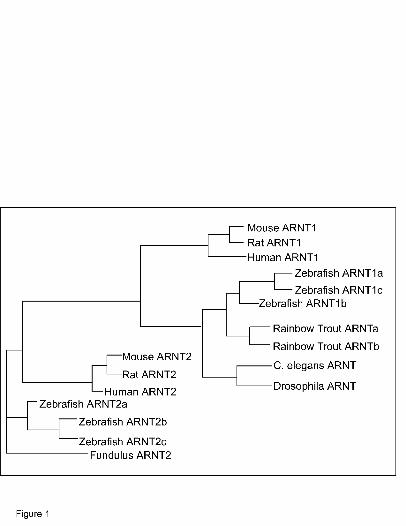
Phylogenetic analysis was used to determine the relationship of the newly identified
zebrafish protein sequences to ARNT protein sequences of other species (Fig. 1). This result
demonstrates that the identified proteins are in fact members of the vertebrate ARNT clade.
Importantly, the protein sequences are most closely related to ARNT1 proteins of other species
and are distinct from the mammalian and fish ARNT2 proteins providing evidence that a
zebrafish homolog of ARNT1 has been identified.
Although it is possible that these three zfARNT1 cDNAs arise from separate genes, it
seems more likely that they are the result of alternate splicing of mRNA from the same gene.
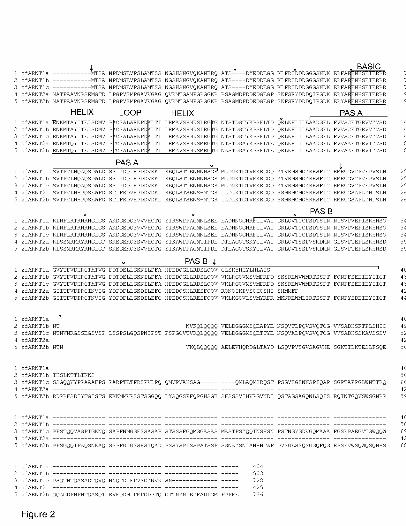
Sequence comparisons demonstrate that the zfARNT1a, zfARNT1b, and zfARNT1c proteins are
identical over the first 390 amino acids and share significant homology with the previously
cloned zfARNT2 proteins in this region (Fig. 2). All three zfARNT1 proteins contain the highly
conserved bHLH and PAS A domains that are known to be important for DNA binding and
MOL # 16873
16
heterodimerization. The zfARNT1a sequence contains a portion of the PAS B domain but
diverges within this domain and truncates with 14 unique amino acids. This divergence occurs at
a site predicted as an intron/exon boundary. Alternate splicing at the same position occurs to
form the zfARNT2a protein (Fig. 2). The zfARNT1b sequence contains the entire PAS B domain
and continues for an additional 64 amino acids before terminating to create a protein that is
shorter than other ARNTs lacking the C-terminal domain found in other ARNT proteins (Pollenz
et al., 1996; Tanguay et al., 2000; Li et al., 1994). The zfARNT1c protein is similar in length to
other known mammalian and fish ARNT proteins containing complete bHLH, PAS A and PAS
B domains and also containing a full length C-terminal domain with sequence similarity to the
C-terminal domains of previously identified mammalian and fish ARNT proteins (Pollenz et al.,
1996; Tanguay et al., 2000; Li et al., 1994).
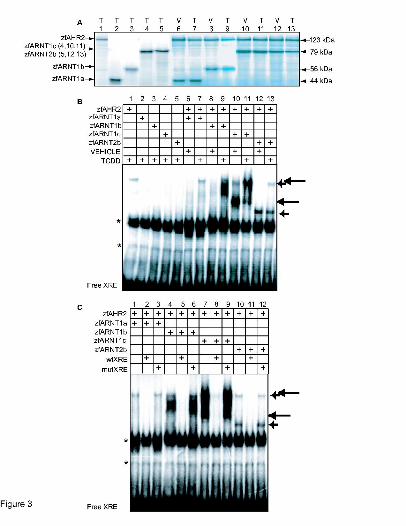
In vitro expression of zfARNT1a and zfARNT1b and in vitro DNA binding. As an initial step
in characterizing the properties of the zfARNT1 proteins, zfARNT1a, zfARNT1b, and
zfARNT1c were produced in vitro from pBKCMV expression constructs using an in vitro
coupled transcription/translation system. As predicted the approximate size of the zfARNT1a
protein was 44 kDa, the zfARNT1b protein was 56kDa, and the zfARNT1c protein was 79 kDa
(Fig. 3A).
To determine if the zfARNT1 proteins are able to form a functional complex with
zfAHR2, DNA binding ability was determined in vitro. ZfARNT2b was included in this
experiment for purposes of comparison. For these experiments the zfARNT proteins were
produced individually in the presence of TCDD (Fig. 3A lanes 1-5) or together with zfAHR2 in
the presence of vehicle or TCDD (Fig. 3A lanes 6-13) and their ability to bind to a radiolabeled
probe containing an XRE sequence from the mouse cyp1a promoter was determined. None of the
MOL # 16873
17
proteins when expressed individually in the presence of TCDD showed significant ability to bind
XRE sequences, although nonspecific lysate derived bands were detected (Fig. 3B lanes 1-5).
The zfARNT1a/zfAHR2 heterodimer did not bind XRE sequences in the absence of TCDD and
showed only weak ability to bind XRE sequences upon TCDD exposure (Fig. 3B lanes 6-7). The
zfARNT1b/zfAHR2 heterodimer also lacked ability to bind XRE in the absence of TCDD,
however, upon TCDD exposure a significant shift in the labeled XRE probe was observed (Fig.
3B lanes 8-9). The zfARNT1c/zfAHR2 heterodimer showed the greatest ability to interact with
the XRE sequence. A complex with the XRE was observed even in the absence of TCDD (Fig
3B, lane 10), and formation of this complex was enhanced upon TCDD exposure (Fig. 3B, lane
11). The zfARNT2b/zfAHR2 heterodimer also showed the ability to interact with zfAHR2 to
bind XRE sequences as has been previously reported (Fig. 3B lane 12-13) (Tanguay et al., 2000),
but this interaction appears less robust than that observed with zfARNT1b and zfARNT1c. The
differential ability to bind DNA cannot be explained by different levels of protein abundance,
because [35S] methionine sublabeling demonstrates that each reaction contains similar levels of
proteins (Fig. 3A), and the proteins have similar numbers of methionine residues. Instead, this
suggests that there are likely structural and functional differences between the proteins that affect
their ability to interact with zfAHR2 and XRE sequences. As has been previously reported
(Andreasen et al., 2002a; Tanguay et al., 2000) this complex migrates as two distinct shifted
bands, although the nature of this doublet is still unclear.
To demonstrate the specificity of the interactions, gel shifts were performed in the
presence of unlabelled wild type XRE (wtXRE) or mutant XRE (mutXRE) oligomer probes (Fig.
3C). The complexes formed by zfAHR2 and each of the zfARNT proteins could be competed by
addition of a 10-fold molar excess of unlabeled wtXRE (Fig. 3C, lanes 2,5,8,11). However, when
MOL # 16873
18
the mutXRE that contained a single base pair change in the core XRE sequence was added, no
competition with the radiolabeled probe was observed (Fig. 3B, lanes 3,6,9,12).
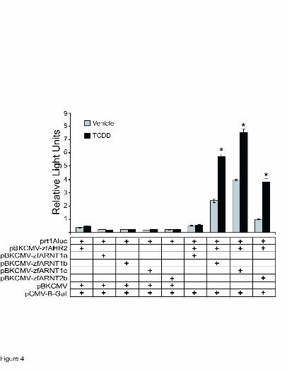
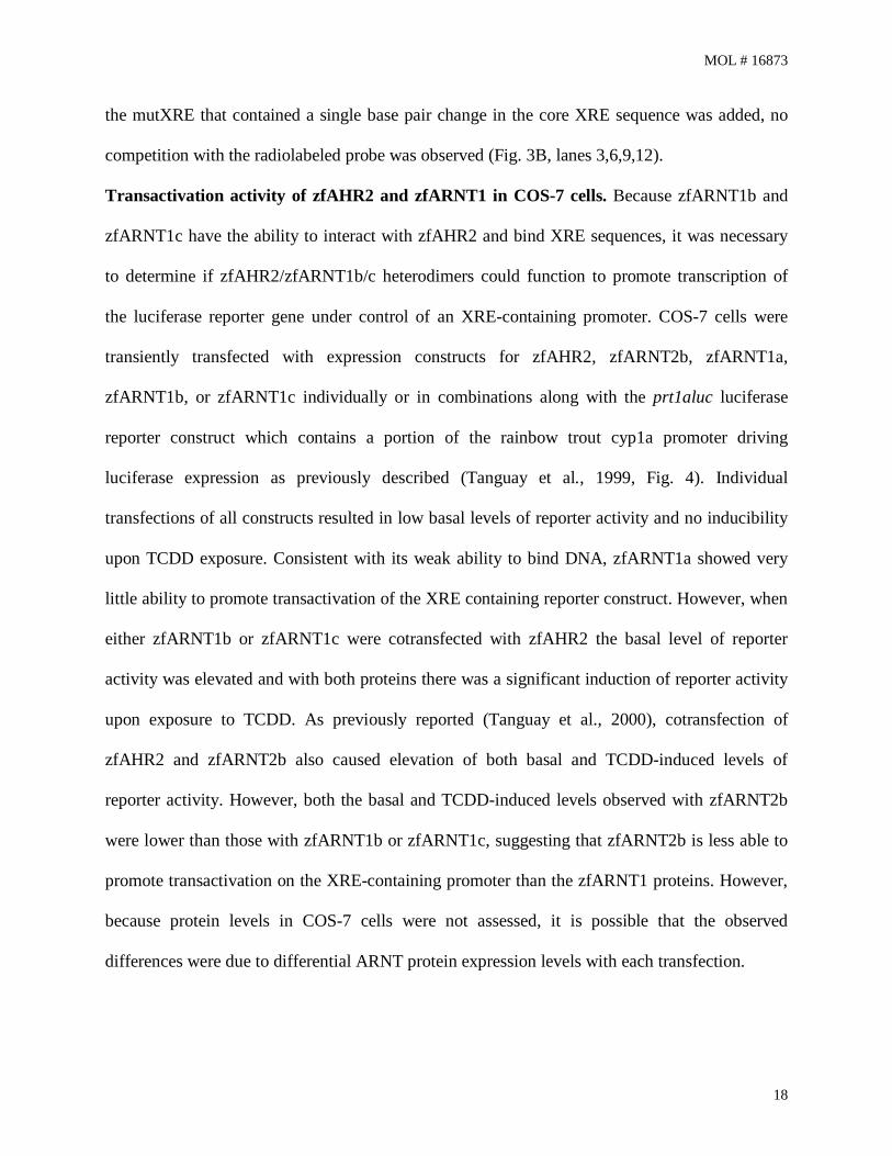
Transactivation activity of zfAHR2 and zfARNT1 in COS-7 cells. Because zfARNT1b and
zfARNT1c have the ability to interact with zfAHR2 and bind XRE sequences, it was necessary
to determine if zfAHR2/zfARNT1b/c heterodimers could function to promote transcription of
the luciferase reporter gene under control of an XRE-containing promoter. COS-7 cells were
transiently transfected with expression constructs for zfAHR2, zfARNT2b, zfARNT1a,
zfARNT1b, or zfARNT1c individually or in combinations along with the prt1aluc luciferase
reporter construct which contains a portion of the rainbow trout cyp1a promoter driving
luciferase expression as previously described (Tanguay et al., 1999, Fig. 4). Individual
transfections of all constructs resulted in low basal levels of reporter activity and no inducibility
upon TCDD exposure. Consistent with its weak ability to bind DNA, zfARNT1a showed very
little ability to promote transactivation of the XRE containing reporter construct. However, when
either zfARNT1b or zfARNT1c were cotransfected with zfAHR2 the basal level of reporter
activity was elevated and with both proteins there was a significant induction of reporter activity
upon exposure to TCDD. As previously reported (Tanguay et al., 2000), cotransfection of
zfAHR2 and zfARNT2b also caused elevation of both basal and TCDD-induced levels of
reporter activity. However, both the basal and TCDD-induced levels observed with zfARNT2b
were lower than those with zfARNT1b or zfARNT1c, suggesting that zfARNT2b is less able to
promote transactivation on the XRE-containing promoter than the zfARNT1 proteins. However,
because protein levels in COS-7 cells were not assessed, it is possible that the observed
differences were due to differential ARNT protein expression levels with each transfection.
MOL # 16873
19
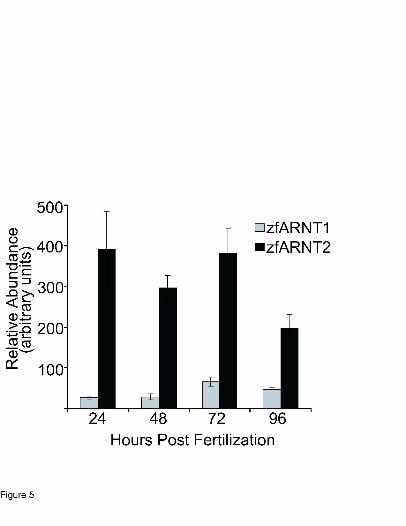
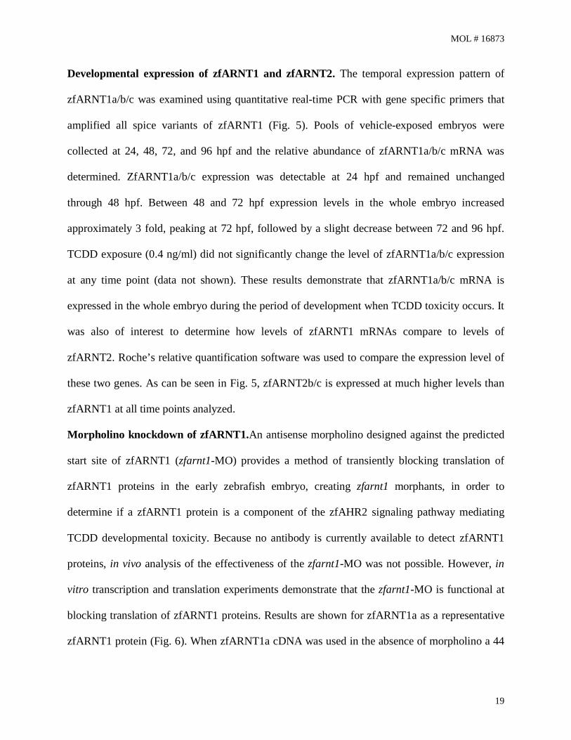
Developmental expression of zfARNT1 and zfARNT2. The temporal expression pattern of
zfARNT1a/b/c was examined using quantitative real-time PCR with gene specific primers that
amplified all spice variants of zfARNT1 (Fig. 5). Pools of vehicle-exposed embryos were
collected at 24, 48, 72, and 96 hpf and the relative abundance of zfARNT1a/b/c mRNA was
determined. ZfARNT1a/b/c expression was detectable at 24 hpf and remained unchanged
through 48 hpf. Between 48 and 72 hpf expression levels in the whole embryo increased
approximately 3 fold, peaking at 72 hpf, followed by a slight decrease between 72 and 96 hpf.
TCDD exposure (0.4 ng/ml) did not significantly change the level of zfARNT1a/b/c expression
at any time point (data not shown). These results demonstrate that zfARNT1a/b/c mRNA is
expressed in the whole embryo during the period of development when TCDD toxicity occurs. It
was also of interest to determine how levels of zfARNT1 mRNAs compare to levels of
zfARNT2. Roche’s relative quantification software was used to compare the expression level of
these two genes. As can be seen in Fig. 5, zfARNT2b/c is expressed at much higher levels than
zfARNT1 at all time points analyzed.
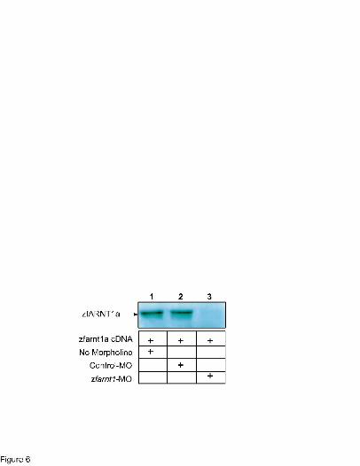
Morpholino knockdown of zfARNT1.An antisense morpholino designed against the predicted
start site of zfARNT1 (zfarnt1-MO) provides a method of transiently blocking translation of
zfARNT1 proteins in the early zebrafish embryo, creating zfarnt1 morphants, in order to
determine if a zfARNT1 protein is a component of the zfAHR2 signaling pathway mediating
TCDD developmental toxicity. Because no antibody is currently available to detect zfARNT1
proteins, in vivo analysis of the effectiveness of the zfarnt1-MO was not possible. However, in
vitro transcription and translation experiments demonstrate that the zfarnt1-MO is functional at
blocking translation of zfARNT1 proteins. Results are shown for zfARNT1a as a representative
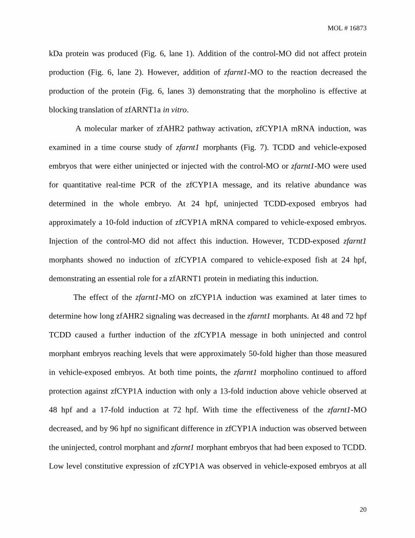
zfARNT1 protein (Fig. 6). When zfARNT1a cDNA was used in the absence of morpholino a 44
MOL # 16873
20
kDa protein was produced (Fig. 6, lane 1). Addition of the control-MO did not affect protein
production (Fig. 6, lane 2). However, addition of zfarnt1-MO to the reaction decreased the
production of the protein (Fig. 6, lanes 3) demonstrating that the morpholino is effective at
blocking translation of zfARNT1a in vitro.
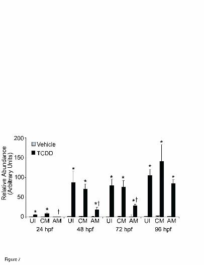
A molecular marker of zfAHR2 pathway activation, zfCYP1A mRNA induction, was
examined in a time course study of zfarnt1 morphants (Fig. 7). TCDD and vehicle-exposed
embryos that were either uninjected or injected with the control-MO or zfarnt1-MO were used
for quantitative real-time PCR of the zfCYP1A message, and its relative abundance was
determined in the whole embryo. At 24 hpf, uninjected TCDD-exposed embryos had
approximately a 10-fold induction of zfCYP1A mRNA compared to vehicle-exposed embryos.
Injection of the control-MO did not affect this induction. However, TCDD-exposed zfarnt1
morphants showed no induction of zfCYP1A compared to vehicle-exposed fish at 24 hpf,
demonstrating an essential role for a zfARNT1 protein in mediating this induction.
The effect of the zfarnt1-MO on zfCYP1A induction was examined at later times to
determine how long zfAHR2 signaling was decreased in the zfarnt1 morphants. At 48 and 72 hpf
TCDD caused a further induction of the zfCYP1A message in both uninjected and control
morphant embryos reaching levels that were approximately 50-fold higher than those measured
in vehicle-exposed embryos. At both time points, the zfarnt1 morpholino continued to afford
protection against zfCYP1A induction with only a 13-fold induction above vehicle observed at
48 hpf and a 17-fold induction at 72 hpf. With time the effectiveness of the zfarnt1-MO
decreased, and by 96 hpf no significant difference in zfCYP1A induction was observed between
the uninjected, control morphant and zfarnt1 morphant embryos that had been exposed to TCDD.
Low level constitutive expression of zfCYP1A was observed in vehicle-exposed embryos at all
MOL # 16873
21
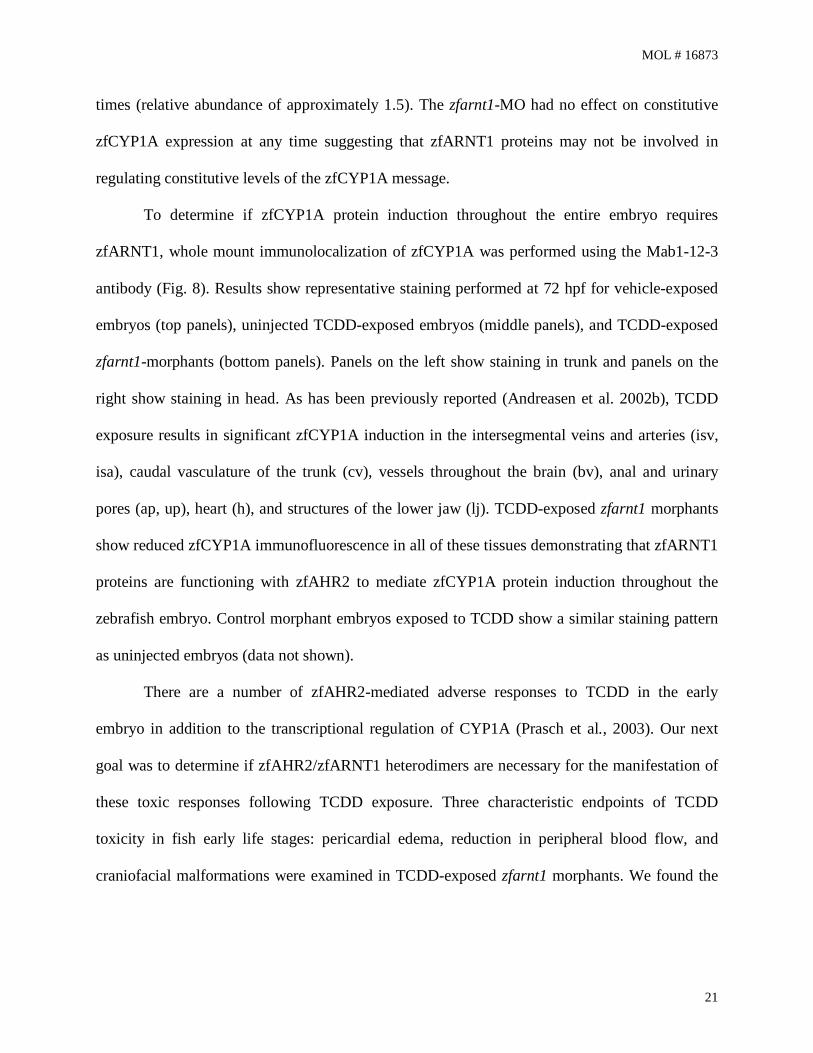
times (relative abundance of approximately 1.5). The zfarnt1-MO had no effect on constitutive
zfCYP1A expression at any time suggesting that zfARNT1 proteins may not be involved in
regulating constitutive levels of the zfCYP1A message.
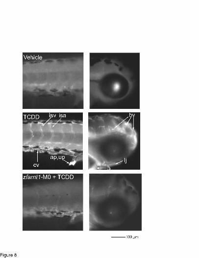
To determine if zfCYP1A protein induction throughout the entire embryo requires
zfARNT1, whole mount immunolocalization of zfCYP1A was performed using the Mab1-12-3
antibody (Fig. 8). Results show representative staining performed at 72 hpf for vehicle-exposed
embryos (top panels), uninjected TCDD-exposed embryos (middle panels), and TCDD-exposed
zfarnt1-morphants (bottom panels). Panels on the left show staining in trunk and panels on the
right show staining in head. As has been previously reported (Andreasen et al. 2002b), TCDD
exposure results in significant zfCYP1A induction in the intersegmental veins and arteries (isv,
isa), caudal vasculature of the trunk (cv), vessels throughout the brain (bv), anal and urinary
pores (ap, up), heart (h), and structures of the lower jaw (lj). TCDD-exposed zfarnt1 morphants
show reduced zfCYP1A immunofluorescence in all of these tissues demonstrating that zfARNT1
proteins are functioning with zfAHR2 to mediate zfCYP1A protein induction throughout the
zebrafish embryo. Control morphant embryos exposed to TCDD show a similar staining pattern
as uninjected embryos (data not shown).
There are a number of zfAHR2-mediated adverse responses to TCDD in the early
embryo in addition to the transcriptional regulation of CYP1A (Prasch et al., 2003). Our next
goal was to determine if zfAHR2/zfARNT1 heterodimers are necessary for the manifestation of
these toxic responses following TCDD exposure. Three characteristic endpoints of TCDD
toxicity in fish early life stages: pericardial edema, reduction in peripheral blood flow, and
craniofacial malformations were examined in TCDD-exposed zfarnt1 morphants. We found the
MOL # 16873
22
zfarnt1-MO provided partial to complete protection against these endpoints demonstrating that
one or more forms of the zfARNT1 protein also play a role in mediating these effects of TCDD.
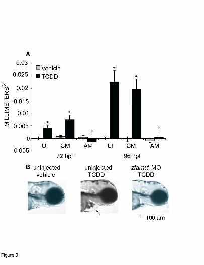
The effect of the zfarnt1-MO on TCDD-induced pericardial edema formation was
examined at 72 and 96 hpf (Fig. 9). Significant formation of pericardial edema can be seen in
TCDD-exposed uninjected and control-MO embryos at 72 hpf indicating that these embryos are
beginning to show the onset of TCDD toxicity (Fig. 9A). The edema fluid continued to
accumulate in the pericardial sac of these embryos causing the area of the pericardium to
increase in size to approximately twice that observed in vehicle-exposed embryos by 96 hpf.
Strikingly, the zfarnt1 morphants showed complete protection against the TCDD-induced
increase in pericardial sac area. The pericardial sac in TCDD-treated zfarnt1 morphants was
similar to that seen in vehicle-exposed embryos at both 72 and 96 hpf. The protection that the
zfarnt1-MO provides against TCDD-induced pericardial edema is depicted in photographs of
representative embryos at 96 hpf (Fig. 9B). The TCDD-exposed zfarnt1 morphants were
observed through 144 hpf and no pericardial or yolk sac edema ever developed (data not shown).
A final point is that injection of the zfarnt1-MO alone did not cause the formation of any types of
edema.
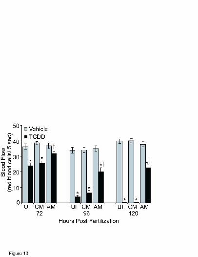
A second hallmark endpoint of TCDD developmental toxicity examined was peripheral
blood flow (Fig. 10). Because peripheral blood flow is reduced following TCDD exposure
earliest in the trunk, the number of red blood cells passing through an intersegmental vein in the
posterior quarter of the trunk in 7.5 s were counted to determine red blood cell perfusion rates as
an index of blood flow. At 72 hpf intersegmental vein blood flow in TCDD exposed, uninjected
and control morphants had decreased to approximately 65% of that observed in vehicle-exposed
embryos. In contrast, no significant reduction was observed in the TCDD-exposed zfarnt1
MOL # 16873
23
morphants. By 96 hpf intersegmental vein blood flow in TCDD-exposed uninjected embryos was
more substantially decreased to 11% of that observed in vehicle-exposed embryos, and injection
of the control-MO did not affect this decrease. A reduction in intersegmental vein blood flow
was observed in the TCDD-exposed zfarnt1 morphants at 96 hpf that was less severe than that
observed in the other treatment groups, being reduced to approximately 56% of that observed in
vehicle-exposed embryos. By 120 hpf there was almost no blood flowing in the TCDD-exposed
uninjected or control-MO injected fish with the average blood flow being only 0.5% of that
measured in vehicle-exposed embryos. Blood flow continued to be less severely reduced in the
TCDD-exposed zfarnt1 morphants and remained at approximately 57% of that observed in
vehicle-exposed embryos.
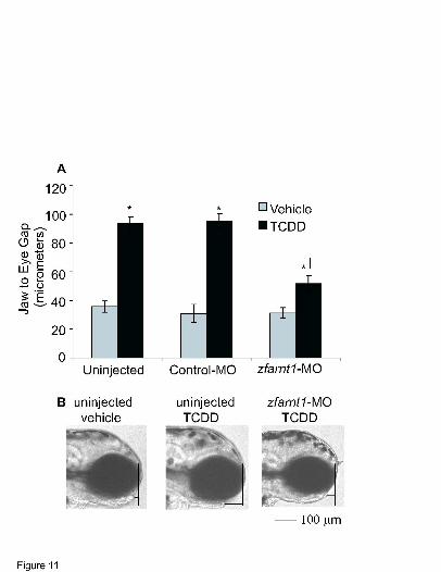
Lower jaw growth in zebrafish is known to be impaired by TCDD exposure (Henry et al.,
1997; Teraoka et al., 2002), and so the role of zfARNT1 proteins in mediating this specific
TCDD response was evaluated (Fig. 11). The distance between the anterior edge of the lower
jaw and the anterior edge of the eye was measured at 96 hpf, and this lower jaw to eye gap was
used as an index of lower jaw growth. In the lateral view of vehicle-exposed embryos, the lower
jaw can be seen extending near to the anterior edge of the eye (Fig. 11B) creating a lower jaw to
eye gap that is quite small, about 10 micrometers (Fig. 11A). However, in TCDD-exposed
embryos the growth of the lower jaw is reduced causing the lower jaw to be shorter and to extend
to varying distances behind the anterior edge of the eye. Because the shorter jaw is farther behind
the eye, a greater jaw to eye gap is obtained upon TCDD treatment.
In uninjected embryos TCDD causes growth of the lower jaw to be reduced so that the
lower jaw to eye gap is almost 3 times greater than that seen in vehicle-exposed embryos (Fig.
11A). A similar effect of TCDD is evident in control morphant embryos. In contrast, the zfarnt1
MOL # 16873
24
morphants exposed to TCDD showed a phenotype that was intermediate between that observed
in vehicle and TCDD-exposed uninjected and control morphant embryos. Lower jaw growth was
decreased in the TCDD-exposed zfarnt1 morphants so that the jaw to eye gap was approximately
1.5-fold greater than that seen in vehicle-exposed embryos. However this was still significantly
less than the 3-fold increase in jaw to eye gap observed in uninjected and control morphant
TCDD-exposed embryos indicating that the reduction in lower jaw growth was less severe in the
zfarnt1 morphants.
DISCUSSION Characteristics of the zfARNT1 proteins. In vitro assays were initially used to assess the
functional characteristics of the zfARNT1 proteins. The DNA binding assay demonstrates that
zfARNT1b and zfARNT1c are both able to function with zfAHR2 to form a strong complex with
XRE-containing DNA, while zfARNT1a forms only a weak interaction. The results of the
transactivation assay are consistent with the DNA binding results and demonstrate that both
zfARNT1b and zfARNT1c are able to promote transactivation on the XRE-containing reporter
construct when expressed with zfAHR2, while zfARNT1a is inactive. Overall, these results
demonstrate that either zfARNT1b or zfARNT1c could be functioning in vivo as a dimerization
partner for zfAHR2, whereas zfARNT1a likely plays no in vivo role in mediating TCDD
toxicity. In addition, the results of the DNA binding and transactivation assays suggest that
zfARNT1b and zfARNT1c may interact more strongly with zfAHR2 on XRE-containing
promoter sequences than zfARNT2b.
In vitro characterization of the zfARNT1 splice variants also provides information about
the functional domains of these proteins. The DNA binding results demonstrate that even though
the N-terminus containing the expected DNA binding domain is identical between the three
zfARNT1 proteins, only zfARNT1b and zfARNT1c are able to interact with zfAHR2 to bind
MOL # 16873
25
XRE-containing DNA. This finding is consistent with what has been observed for alternate
splice variants of zebrafish arnt2 (Tanguay et al., 2000) and rainbow trout arnt (Pollenz et al.,
1996) where ARNT proteins with identical bHLH and PAS domains, but divergent C-terminal
domains, showed differential ability to interact with DNA. Clearly C-terminal sequences are
important for the formation of protein/DNA complexes in vitro, although the identity of these
sequences is unknown in zfARNT1.
Previous deletion studies have demonstrated that a potent transactivation domain
localizes to the C-terminal end of ARNT proteins (Sogawa et al., 1995; Nacela and Pollenz,
1999; Li et al., 1996; Jain et al., 1994). Interestingly, both zfARNT1b and zfARNT1c have
significant ability to promote transactivation, even though zfARNT1b contains a truncated C-
terminal domain compared to other ARNT proteins (Pollenz et al., 1996; Tanguay et al., 2000; Li
et al., 1994). Further deletion studies need to be performed on the zfARNT1 proteins in order to
determine the functional transactivation domains and the way in which these domains interact
with transcription factors and co-regulators to alter gene expression.
In vivo role of zfARNT1 proteins in mediating TCDD developmental toxicity. The
experiments with the zfarnt1-MO clearly demonstrate that some form(s) of the zfARNT1 protein
is required to dimerize with zfAHR2 in vivo to mediate the molecular and physiological
endpoints of TCDD toxicity examined in this study. The protection that the zfarnt1-MO provides
against the induction of both zfCYP1A mRNA and protein demonstrates that a zfARNT1 protein
is essential for this change in gene expression to occur upon TCDD exposure. The zfCYP1A
time course study also provides information about the effectiveness of the morpholino and
demonstrates that the zfarnt1-MO was no longer significantly blocking the zfAHR2 signaling
pathway by 96 hpf. The zfarnt1 morphants also show protection against three endpoints of
MOL # 16873
26
TCDD toxicity: pericardial edema, reduced blood flow and reduced lower jaw growth
demonstrating the essential role of a zfARNT1 protein in mediating toxicity. Importantly, the
results with the zfarnt1-MO demonstrate that one or more splice variant of the zfARNT1 protein
mediates toxicity, but they do not provide information about the role that each splice variant of
zfARNT1 is playing.
Differential requirement of zfARNT1 and zfARNT2 in mediating TCDD toxicity. Although
the zfARNT1b, zfARNT1c and zfARNT2b proteins are all functional with zfAHR2 in vitro, the
in vivo manipulation of these proteins described here and by Prasch et al. (2004a) has revealed
that zfARNT1, not zfARNT2, proteins are an essential component of the zfAHR2 signaling
pathway mediating the endpoints of TCDD toxicity assessed in these studies. The essential role
of zfARNT1 proteins in mediating the TCDD-dependent block of regenerative growth has also
been confirmed (Mathew et al., accepted). In addition, it has been demonstrated that knocking
down expression of ARNT1 in mice provides protection against several endpoints of TCDD
toxicity demonstrating that in a mammalian species ARNT1 is also required for mediating many
endpoints of AHR-dependent toxicity (Walisser et al., 2004).
On possible explanation for the differential requirement of the zfARNT proteins in
mediating zfAHR2 signaling is that there is differential expression patterns of the proteins as
has been observed in mammals (Hirose et al., 1996; Jain et al., 1998; Aitola et al., 2003).
Quantitative real-time PCR of whole embryo tissue demonstrates that both zfARNT1a/b/c and
zfARNT2 are expressed during the period of development when toxicity occurs, and zfARNT2
is a more abundant transcript. However, in order for zfARNT proteins to function with zfAHR2
in vivo to mediate TCDD toxicity, they must be coexpressed with the receptor in target tissues.
Unfortunately, attempts at using in situ hybridization techniques to detect the tissue distribution
MOL # 16873
27
of zfARNT1a/b/c mRNA in the early zebrafish embryo were unsuccessful, likely due to the low
expression levels of these transcripts, and so it was not possible to compare the temporal and
spatial expression pattern of zfARNT1a/b/c to that of zfAHR2 and zfARNT2 (Andreasen et al.,
2002b).
It is also possible that zfARNT1 and zfARNT2 proteins are expressed in similar tissues,
but zfARNT1 proteins are the preferred dimerization partner for zfAHR2 in vivo. The results
from the in vitro assays suggest that zfARNT1b and zfARNT1c may interact more strongly with
zfAHR2 on XRE containing promoters and can promote a more robust level of transactivation
on reporter constructs than zfARNT2b. Therefore, even if zfARNT1 and zfARNT2 proteins are
coexpressed in the same tissues, zfARNT1 proteins may be the preferred dimerization partner
for zfAHR2.
Mechanism of TCDD toxicity. The results with the zfahr2 and zfarnt1 morpholinos clearly
demonstrate that zfAHR2 and one or more forms of zfARNT1 are necessary to induce TCDD-
dependent responses in zebrafish embryos. This provides insight into the mechanism by which
activation of zfAHR2 by TCDD causes developmental toxicity in zebrafish. It has been
hypothesized that the toxic effects of zfAHR2 activation could be independent of XRE-binding
and transcriptional activation and instead due to interactions of zfAHR2 with other proteins
(Prasch et al., 2004b). In mammals the AHR has been shown to interact with a variety of other
signaling pathways and proteins including hormone, NFκB, and hypoxia signaling pathways, and
cell cycle proteins such as Rb (reviewed by Carlson and Perdew, 2002). It as been speculated
that cross-talk with other pathways may actually be the mechanism of TCDD toxicity. However,
results from the current study demonstrate that both zfAHR2 and zfARNT1 are essential for the
generation of developmental toxicity in zebrafish. Although it is possible that the
MOL # 16873
28
zfAHR2/zfARNT1 heterodimer is uniquely capable of tethering other transcription factors
leading to altered gene expression without binding to XRE sequences, it seems more likely that
endpoints of toxicity are in fact dependent on XRE binding and alterations in gene expression
caused by the zfAHR2/ARNT1 heterodimer. It has been shown that blocking induction of the
zfCYP1A protein in zebrafish provides no protection against TCDD developmental toxicity
(Carney et al., 2004) suggesting that altered regulation of other, currently unknown genes, may
be important. However, until these genes and their roles in TCDD developmental toxicity have
been clearly identified, mechanisms of cross-talk with other signaling pathways cannot be ruled
out.
Role of zfARNT1 proteins in normal development. Studies with arnt1-/- knock out mice lines
clearly demonstrate that ARNT1 plays a role in normal mammalian development. Arnt1-/- knock
out mice are not viable past embryonic day 10.5 and show defects in developmental angiogenesis
(Kozak et al., 1997; Maltepe et al., 1997). The current study does not provide any insight into the
role zfARNT1 proteins may play in mediating normal zebrafish development. The zfarnt1
morphants were able to develop completely with no obvious gross abnormalities ever observed.
Given the severity of the arnt1-/- mouse phenotype it is surprising that the zfarnt1 morphants
developed normally. It is possible that our gross observations of the morphant embryos did not
detect abnormalities that are more subtle. Alternate possibilities are that enough zfARNT1
protein remained present in the morpholino studies for normal physiological functions to be
carried out or that there is functional redundancy between zfARNT1 and other zebrafish ARNT
proteins.
The results of this study provide further insight into the initial molecular signaling events
that occur in zebrafish embryos upon exposure to TCDD. Although we now know that both
MOL # 16873
29
zfAHR2 and some form of zfARNT1 are required for toxicity to develop, the way in which
activation of these proteins by TCDD leads to toxicity is still unclear and remains a focus of
future research. In addition, the way in which each of the zfARNT1 splice variants functions to
mediate zfAHR2 signaling in vivo is also unknown. Finally the role(s) that zfARNT1 proteins
may play in dimerizing with other PAS proteins to mediate responses to other changing
environmental or developmental conditions remains to be determined.
ACKNOWLEDGMENTS
We thank Dorothy Nesbit for her excellent technical assistance, Dr. John Stegeman from the
Woods Hole Oceanographic Institute for the generous supply of the Mab 1-12-3 antibody, and
Sara Carney for helpful discussions on the manuscript. Contribution number _ _ _ , Molecular
and Environmental Toxicology Center, University of Wisconsin, Madison, WI 53726-4087.
MOL # 16873
30
REFERENCES Aitola M. H. and Pelto-Huikko M. T. (2003) Expression of Arnt and Arnt2 mRNA in developing
murine tissues. J Histochem Cytochem 51: 41-54. Andreasen, E. A., Hahn, M. E., Heideman, W., Peterson, R. E., and Tanguay, R. L. (2002a). The
zebrafish (Danio rerio) aryl hydrocarbon receptor type 1 is a novel vertebrate receptor. Mol Pharmacol 62, 234-49.
Andreasen, E. A., Spitsbergen, J. M., Tanguay, R. L., Stegeman, J. J., Heideman, W., and
Peterson, R. E. (2002b). Tissue-specific expression of AHR2, ARNT2, and CYP1A in zebrafish embryos and larvae: effects of developmental stage and 2,3,7,8- tetrachlorodibenzo-p-dioxin exposure. Toxicol Sci 68, 403-419.
Belair, C. D., Peterson, R. E., and Heideman, W. (2001). Disruption of erythropoiesis by dioxin
in the zebrafish. Dev Dyn 222, 581-94. Carlson, D. B., and Perdew, G. H. (2002). A dynamic role for the Ah receptor in cell signaling?
Insights from a diverse group of Ah receptor interacting proteins. J Biochem Mol Toxicol 16, 317-25.
Carney, S. A., Peterson, R. E., and Heideman, W. (2004). TCDD activation of the AHR/ARNT
pathway causes developmental toxicity through CYP1A-independent mechanism in zebrafish. Mol Pharmacol 66, 512-521.
Cook, P. M., Robbins, J. A., Endicott, D. D., Lodge, K. B., Guiney, P. D., Walker, M. K., Zabel,
E. W., and Peterson, R. E. (2003). Effects of aryl hydrocarbon receptor-mediated early life stage toxicity on lake trout populations in Lake Ontario during the 20th century. Environ Sci & Technol 37, 3864-77.
Dong, W., Teraoka, H., Tsujimoto, Y., Stegeman, J. J., and Hiraga, T. (2004). Role of aryl
hydrocarbon receptor in mesencephalic circulation failure and apoptosis in zebrafish embryos exposed to 2,3,7,8-tetrachlorodibenzo-p-dioxin. Toxicol Sci 77, 109-16.
Evans, B. R., Karchner, S. I., Franks, D. G., Hahn, M. E. (2005). Duplicate aryl hydrocarbon
receptor repressor genes (ahrr1 and ahrr2) in the zebrafish Danio rerio: Structure, function, evolution, and AHR-dependent regulation in vivo. Arch Biochem Biophys 441, 151-67.
Fernandez-Salguero, P. M., Hilbert, D. M., Rudikoff, S., Ward, J. M., and Gonzalez, F. J. (1996).
Aryl-hydrocarbon receptor-deficient mice are resistant to 2,3,7,8- tetrachlorodibenzo-p-dioxin-induced toxicity. Toxicol Appl Pharmacol 140, 173-9.
Gu, Y. Z., Hogenesch, J. B., and Bradfield, C. A. (2000). The PAS superfamily: sensors of
environmental and developmental signals. Annu Rev Pharmacol Toxicol 40, 519-61.
MOL # 16873
31
Henry, T. R., Spitsbergen, J. M., Hornung, M. W., Abnet, C. C., and Peterson, R. E. (1997). Early life stage toxicity of 2,3,7,8-tetrachlorodibenzo-p-dioxin in zebrafish (Danio rerio). Toxicol Appl Pharmacol 142, 56-68.
Hirose, K., Morita, M., Ema, M., Mimura, J., Hamada, H., Fujii, H., Saijo, Y., Gotoh, O.,
Sogawa, K., and Fujii-Kuriyama, Y. (1996). cDNA cloning and tissue-specific expression of a novel basic helix-loop- helix/PAS factor (Arnt2) with close sequence similarity to the aryl hydrocarbon receptor nuclear translocator (Arnt). Mol Cell Biol 16, 1706-13.
Jain, S., Maltepe, E., Lu, M. M., Simon, C., and Bradfield, C. A. (1998). Expression of ARNT,
ARNT2, HIF1 alpha, HIF2 alpha and Ah receptor mRNAs in the developing mouse. Mech Dev 73, 117-23.
Jain, S., Dolwick, K. M., Schmidt, J. V., and Bradfield, C. A. (1994). Potent transactivation
domains of the Ah receptor and the Ah receptor nuclear translocator map to their carboxyl termini. J. Biol Chem 269, 31518-31524.
Karchner, S. I., Franks, D. G., Hahn, M. E. (2005). AHR1B, a new functional aryl hydrocarbon
receptor in zebrafish: tandem arrangement of ahr1b and ahr2 genes. Biochem J. (In press) Kozak, K. R., Abbott, B., and Hankinson, O. (1997). ARNT-deficient mice and placental
differentiation. Dev Biol 191, 297-305. Li, H., Dong, L., and Whitlock, J. P., Jr. (1994). Transcriptional activation function of the mouse
Ah receptor nuclear translocator. J Biol Chem 269, 28098-105. Maltepe, E., Schmidt, J. V., Baunoch, D., Bradfield, C. A., and Simon, M. C. (1997). Abnormal
angiogenesis and responses to glucose and oxygen deprivation in mice lacking the protein ARNT. Nature 386, 403-7.
Mathew, L., Andreasen, E. A., and Tanguay, R. L. (accepted). Aryl hydrocarbon receptor
activation inhibits regenerative growth. Mol Pharmacol. Mimura, J., Yamashita, K., Nakamura, K., Morita, M., Takagi, T. N., Nakao, K., Ema, M.,
Sogawa, K., Yasuda, M., Katsuki, M., and Fujii-Kuriyama, Y. (1997). Loss of teratogenic response to 2,3,7,8-tetrachlorodibenzo-p-dioxin (TCDD) in mice lacking the Ah (dioxin) receptor. Genes Cells 2, 645-54.
Nasevicius, A. and Ekker, S. C. (2000). Effective targeted gene 'knockdown' in zebrafish. Nat
Genet 26, 216-220. Nacela, B. and Pollenz, R. S. (1999), Functional analysis of activation and repression domains of
the rainbow trout aryl hydrocarbon receptor nuclear translocator (rtARNT) protein isoforms. Biochem Pharmacol 57, 1177-1190.
MOL # 16873
32
Park S. S., Miller, H., Klotz, A. V., Kloepper-Sams, P. J., Stegeman, J. J., and Gelboin, H. V. (1986). Monoclonal antibodies to liver microsomal cytochrome P-450 E of the marine fish Stenotomus chrysops (scup): cross reactivity with 3 methylcholanthrene induced rat cytochrome P-450. Arch Biochem Biophys 249, 339-350.
Peterson, R. E., Theobald, H. M., and Kimmel, G. L. (1993). Developmental and reproductive
toxicity of dioxins and related compounds: cross-species comparisons. Crit Rev Toxicol 23, 283-335.
Pollenz, R. S., Sullivan, H. R., Holmes, J., Necela, B., and Peterson, R. E. (1996). Isolation and
expression of cDNAs from rainbow trout (Oncorhynchus mykiss) that encode two novel basic helix-loop-Helix/PER-ARNT-SIM (bHLH/PAS) proteins with distinct functions in the presence of the aryl hydrocarbon receptor. Evidence for alternative mRNA splicing and dominant negative activity in the bHLH/PAS family. J Biol Chem 271, 30886-96.
Prasch, A. L., Heideman, W., and Peterson, R. E. (2004a). ARNT2 is not required for TCDD
developmental toxicity in zebrafish. Toxicol Sci 82, 250-258. Prasch, A. L., Andreasen, E. A., Peterson, R. E., and Heideman, W. (2004b). Interactions
between 2,3,7,8-tetrachlorodibenzo-p-dioxin (TCDD) and hypoxia signaling pathways in zebrafish: hypoxia decreases responses to TCDD in zebrafish embryos. Toxicol Sci 78, 68-77.
Prasch, A. L., Teraoka, H., Carney, S. A., Dong, W., Hiraga, W., Stegeman, J. J., Heideman, W.,
and Peterson, R. E. (2003). Aryl hydrocarbon receptor 2 mediates 2,3,7,8-tetrachlorodibenzo-p-dioxin developmental toxicity in zebrafish. Toxicol Sci 76, 138-150.
Schmidt, J. V. and Bradfield, C. A. (1996). Ah receptor signaling pathways. Annu Rev Cell Dev
Biol 12, 55-89. Sogawa, K., Iwabuchi, K., Abe, H., and Fujii-Kuriyama, Y. (1995). Transcriptional activation
domains of the Ah receptor and Ah receptor nuclear translocator. J Cancer Res Clin Oncol 121, 612-620.
Tanguay, R. L., Abnet, C. C., Heideman, W., and Peterson, R. E. (1999). Cloning and
characterization of the zebrafish (Danio rerio) aryl hydrocarbon receptor. Biochim Biophys Acta 1444, 35-48.
Tanguay, R. L., Andreasen, E., Heideman, W., and Peterson, R. E. (2000). Identification and
expression of alternatively spliced aryl hydrocarbon nuclear translocator 2 (ARNT2) cDNAs from zebrafish with distinct functions. Biochim Biophys Acta 1494, 117-28.
Tanguay, R. L., Andreasen, E.A, Walker M.K., and Peterson R.E. (2003). Dioxin Toxicity and
Aryl Hydrocarbon Receptor Signaling in Fish. In Dioxins and Health, 2nd edition (A. Schecter and T. A. Gasiewicz, Eds) pp. 603-628. John Wiley & Sons, New York.
MOL # 16873
33
Teraoka, H., Dong, W., Ogawa, S., Tsukiyama, S., Okuhara, Y., Niiyama, M., Ueno, N., Peterson, R. E., and Hiraga, T. (2002). 2,3,7,8-Tetrachlorodibenzo-p-dioxin toxicity in the zebrafish embryo: altered regional blood flow and impaired lower jaw development. Toxicol Sci 65, 192-199.
Teraoka, H., Dong, W., Tsujimoto, Y., Iwasa, H., Endoh, D., Ueno, N., Stegeman, J. J., Peterson,
R. E., and Hiraga, T. (2003). Induction of cytochrome P450 1A is required for circulation failure and edema by 2,3,7,8-tetrachlorodibenzo-p-dioxin in zebrafish. Biochem Biophys Res Commun 304, 223-8.
Walisser, J. A., Bunger, M. K., Glover, E., Harstad, E. B., and Bradfield, C. A. (2004). Patent
Ductus Venosus and Dioxin Resistance in Mice Harboring a Hypomorphic Arnt Allele. J Biol Chem 279, 16326-16331.
Wang W. D., Wu, J., C., Hsu, H. J., Kon, Z. L., Hu C. H. (2000). Overexpression of a zebrafish
ARNT2-like factor represses CYP1A transcription in ZLE cells. Marine Biotech 2, 376-386. Wentworth, J. N., Buzzeo, R., and Pollenz, R. S. (2004). Functional characterization of aryl
hydrocarbon receptor (zfAHR2) localization and degradation in zebrafish (Danio rerio). Biochem Pharmacol 67, 1363-1372.
Westerfield, M. (1995). The Zebrafish Book. University of Oregon Press, Eugene, OR.
MOL # 16873
34
Footnote This work was supported by the University of Wisconsin Sea Grant College Program, National
Oceanic and Atmospheric Administration, U.S. Department of Commerce, Sea Grant Project
Numbers R/BT-16 and R/BT-17 (W.H. and R.E.P.). This research was also supported by
NIH/NIEHS grants T32 ES07015 and RO1 ES0127716 (W.H. and R.E.P), and RO1 ES10820,
ES00210, and ES03850 (R.L.T).
MOL # 16873
35
FIGURE LEGENDS Figure 1. Phylogenetic analysis of aryl hydrocarbon receptor nuclear translocator (ARNT)
proteins. Using Vector NTI software the amino acid sequences of various ARNT proteins were
aligned using Align X alignment program and this alignment was used to construct the
phylogenetic tree using the Neighbor Joining Method. The Genbank Accession numbers for the
ARNT sequences used are: Homo sapiens (human) ARNT1:BC060838, ARNT2:BC036099;
Mus musculus (mouse) ARNT1: BC012870, ARNT2: BC054546; Rattus norvegicus (Rat)
ARNT1: U61184, ARNT2: U61405; Danio rerio (zebrafish) ARNT2a: AF219987, ARNT2b:
AF219988, ARNT2c: AF219989, ARNT1a, AY707650, ARNT1b, AY707649, ARNT1c,
DQ140179; Fundulus heteroclitus (Fundulus) ARNT2: AF079311; Oncorhynchus mykiss
(rainbow trout) ARNTa: U73840, ARNTb: U73841.; Drosophila melanogaster ARNT:
AF154417; Caenorhabditis elegans ARNT: NM060286.
Figure 2. Amino acid sequence alignment of the predicted zfARNT1a, zfARNT1b, and
zfARNT1c sequences with zfARNT2a and zfARNT2b. Sequence alignment was performed
using the ClustalW 1.8 alignment program. Alignment gaps are represented by dashes. The
regions encoding the conserved basic helix loop helix (bHLH), PAS A, and PAS B domains are
indicated. The position of predicted exon-exon boundaries derived from the mouse ARNT1
exon-exon boundaries and also from information available in the Ensembl release of the
zebrafish genome are indicated by arrows.
Figure 3. In vitro translation and gel shift analysis to determine interactions between zfAHR2,
zfARNT1a, zfARNT1b, zfARNT1c and a xenobiotic response element (XRE) in response to
TCDD exposure. (A) Proteins used in the gel-shift analysis were synthesized in vitro either alone
MOL # 16873
36
or in combination with zfAHR2 in the presence of either vehicle (V) or 10 nM TCDD (T). A
subfraction of the protein was expressed with [35S] methionine, resolved on an 8% SDS
polyacrylamide gel, and the dried gel was phosphorimaged. (B) In vitro translated proteins,
either alone or in combination, were incubated in the presence of [32P] labeled DNA probes
containing an XRE from the mouse cyp1a enhancer in the presence or absence of TCDD as
indicated. (C) ZfAHR2 and the indicated zfARNT protein were incubated with the [32P] labeled
DNA probe and a 10-fold molar excess of unlabeled probe containing wild type (wt) XRE or
mutant (mut) XRE in the presence of TCDD as indicated. The two long arrows indicate specific
interactions that occur between zfAHR2 and zfARNT1c. The two shorter arrows indicate
specific interactions that occur between zfAHR2 and zfARNT2b. Asterisks indicate nonspecific
lysate-derived bands
Figure 4. Transactivation ability of zfARNT1 and zfARNT2 proteins with zfAHR2 in COS-7
cells exposed to TCDD. Cells were transiently transfected with pBK-CMV expression constructs
for zfAHR2, zfARNT2b, zfARNT1a, zfARNT1b, zfARNT1c, the prt1Aluc reporter construct
containing the luciferase gene under control of the rainbow trout cyp1a promoter and enhancer,
and a β-galactosidase reporter construct as indicated. Numbers represent the amount of each
construct (ng) transfected into each well of a 24 well plate. Twenty-two hr after transfection,
cells were exposed to vehicle or 10 nM TCDD for 20 hr and then collected for luciferase and β-
galactosidase assays. Bars represent relative light units normalized to β-galactosidase activity
and are mean ± SE of n=6. * indicates a significant difference between vehicle and TCDD (p ≤
0.05).
MOL # 16873
37
Figure 5. Time course of zfARNT1a/b/c and zfARNT2b/c mRNA abundance. Quantitative real-
time PCR was performed on cDNA using gene-specific primer sets that detected zfARNT1a/b/c,
zfARNT2b/c and β-actin. Samples were run concurrently with standard curves derived from
plasmid cDNA dilutions. Relative quantification software (Roche) was used to compare the
expression level of the zfARNT1 and zfARNT2 transcripts. RNA was extracted from pools of 10
vehicle-exposed embryos at each time (n = pool of 10 embryos). Values are mean ± SE of n = 4
pools.
Figure 6. Effect of the zfarnt1-MO on in vitro translation of the zfARNT1a protein. To
determine effectiveness of the zfarnt1-MO at blocking translation of zfARNT1 proteins,
zfARNT1 cDNAs were transcribed and translated in vitro in the presence of [35S]-methionine.
Results are shown for zfARNT1a as a representative zfARNT1 protein. The in vitro translation
reaction was performed either in the absence of morpholino (lane 1) or in the presence of the
control-MO (lane 2) or zfarnt1-MO (lane 3). Both morpholinos were used at a final
concentration of 500 nM. The [35S]-labeled proteins were resolved on an 8% SDS
polyacrylamide gel, and the dried gel was phosphorimaged.
Figure 7. Effect of the zfarnt1-MO on the time course of TCDD-induced zfCYP1A mRNA
abundance in the zebrafish embryo. Quantitative real-time PCR was performed on cDNA using
gene-specific primers for zfCYP1A and β-actin. Samples were run concurrently with standard
curves derived from plasmid cDNA dilutions. Values obtained with the zfCYP1A primers were
normalized to those obtained with β-actin primers to control for differences in loading and obtain
relative values. RNA was extracted from pools of 15 embryos from each treatment group at each
time (n = pool of 15 embryos) and reverse transcribed into cDNA. Values are mean ± SE of n=4
MOL # 16873
38
pools. * indicates a significant difference between TCDD and its respective vehicle control, †
indicates a significant difference between TCDD and TCDD + zfarnt1-MO (p ≤ 0.05).
Figure 8. Whole mount immunolocalization of zfCYP1A in vehicle and TCDD-exposed (0.4
ng/ml) uninjected embryos and TCDD-exposed zfarnt1 morphants at 72 hpf using Mab 1-12-3.
Images on the left are lateral views of embryos taken posterior to the yolk extension, and images
on the right are lateral views of the head taken anterior to the yolk sac. For each group, images
are representative of 12 embryos from 3 distinct vehicle/TCDD exposure experiments.
Abbreviations: ap, anal pore; bv, brain vessels; cv, caudal vasculature; h, heart; isa,
intersegmental artery; isv, intersegmental vein; lj, lower jaw; up, urinary pore. Bar = 100 µm
Figure 9. Effect of the zfarnt1-MO on TCDD-induced increases in pericardial sac edema at 72
and 96 hpf in the zebrafish embryo. (A) Lateral views of embryos were photographed and area of
the pericardial sac was quantitated at 72 and 96 hpf, respectively. The average area obtained for
vehicle-exposed uninjected embryos was subtracted from the area values obtained for TCDD-
exposed embryos in the other treatment groups to get a measure of the increase in pericardial
area caused by edema. (B) Images are representative of the pericardial area observed in embryos
exposed to vehicle, TCDD, or TCDD + zfarnt2-MO at 96 hpf. The arrow indicates the
pericardial edema in uninjected TCDD-treated embryos. Values for edema quantitation are mean
± SE of n=12. ∗ indicates a significant difference between TCDD (0.4 ng/ml) and its respective
vehicle control (p ≤ 0.05). † indicates a significant difference between TCDD and TCDD +
zfarnt1-MO. Abbreviations: UI = uninjected, CM = control-MO, AM = zfarnt1-MO. Bar = 100
µm.
MOL # 16873
39
Figure 10. Effect of the zfarnt1-MO on TCDD-induced reductions in peripheral blood flow at
72, 96, and 120 hpf in the zebrafish embryo. As an index of peripheral blood flow, red blood cell
perfusion rates were determined in an intersegmental vein in the posterior quarter of the trunk.
Values are mean ± SE of n=12. Abbreviations: UI = uninjected; CM = control-MO; AM =
zfarnt1-MO. Other conditions as in Fig. 9 legend.
Figure 11. Effect of the zfarnt1-MO on TCDD-induced increases in the lower jaw to eye gap.
(A) Lateral views of the embryos were photographed and the lower jaw to eye gap was measured
as an index of lower jaw growth at 96 hpf. (B) Representative lateral-view photographs showing
the length of the lower jaw in vehicle and TCDD-exposed (0.4 ng / ml) uninjected embryos and
TCDD-exposed zfarnt1 morphants are included. The way in which the jaw to eye distance was
obtained is also depicted (B). First, a straight vertical line was drawn along the anterior most
edge of the eye, then a second straight horizontal line was drawn from the anterior most edge of
the lower jaw to the intersection of the first vertical line. The length of this second horizontal line
was used as the measure of lower jaw to eye gap. When the lower jaw protruded in front of the
eye the measure was a negative value, and if it remained behind the eye it was given a positive
value. Values are mean ± SE of n=12. Bar = 100 µm. Other conditions as in Fig. 9 legend.