Embed Size (px)
Citation preview

Nutrients 2013, 5, 4184-4205; doi:10.3390/nu5104184
nutrients ISSN 2072-6643
www.mdpi.com/journal/nutrients
Review
IGF-1, the Cross Road of the Nutritional, Inflammatory and Hormonal Pathways to Frailty
Marcello Maggio 1,2,†,*, Francesca De Vita 1,†, Fulvio Lauretani 1, Valeria Buttò 2,
Giuliana Bondi 2, Chiara Cattabiani 3, Antonio Nouvenne 1, Tiziana Meschi 2,
Elisabetta Dall’Aglio 2 and Gian Paolo Ceda 1,2
1 Geriatric Rehabilitation Department, University Hospital of Parma, Via Gramsci, 14,
Parma (PR) 43126, Italy; E-Mails: [email protected] (F.V.); [email protected] (F.L.);
[email protected] (A.N.); [email protected] (G.P.C.) 2 Department of Clinical and Experimental Medicine, Section of Geriatrics, Food Sciences Unit and
Endocrinology of Aging Unit, University of Parma, Via Gramsci, 14, Parma (PR) 43126, Italy;
E-Mails: [email protected] (V.B.); [email protected] (G.B.);
[email protected] (T.M.); [email protected] (E.D.A.) 3 Azienda USL Piacenza, Via Taverna, 49, Piacenza (PC) 23121, Italy;
E-Mail: [email protected]
† These authors contributed equally to this work.
* Author to whom correspondence should be addressed; E-Mail: [email protected];
Tel.: +0039-0521-703-916; Fax: +0039-0521-987-562.
Received: 3 July 2013; in revised form: 1 October 2013 / Accepted: 1 October 2013 /
Published: 21 October 2013
Abstract: The decline in functional capacity is a heterogeneous phenomenon in the elderly.
An accelerated ageing determines a frail status. It results in an increased vulnerability to
stressors for decreased physiological reserves. The early identification of a frail status is
essential for preventing loss of functional capacity, and its clinical consequences. Frailty and
mobility limitation result from an interplay of different pathways including multiple anabolic
deficiency, inflammation, oxidative stress, and a poor nutritional status. However, the
age-related decline in insulin-like growth factor 1 (IGF-1) bioactivity deserves special
attention as it could represent the ideal crossroad of endocrine, inflammatory, and nutritional
pathways to frailty. Several minerals, namely magnesium, selenium, and zinc, appear to be
important determinants of IGF-1 bioactivity. This review aims to provide an overview of the
OPEN ACCESS

Nutrients 2013, 5 4185
potential usefulness of nutrients modulating IGF-1 as potential therapeutic targets in the
prevention of mobility limitation occurring in frail older subjects.
Keywords: ageing; IGF-1; selenium; magnesium; zinc; micronutrients; frailty
1. Introduction
Aging is characterized by multiple changes in physiological pathways that concur to a decline in the
functional capacity of older individuals. This is a heterogeneous phenomenon that predicts a large
number of adverse outcomes including functional limitation, decreased cognitive function, reduced
quality of life, and nursing home placement [1,2]. It is widely recognized that changes in physical
performance and cognitive status are strongly predictors of mortality, independently of other
traditional indicators of disease. Several aspects related to the impairment of functional capacity are
included in the current definition of frailty proposed by Fried and colleagues [3]. Frailty represents a
biological syndrome of increased vulnerability to stressors resulting from decreased physiological
reserves. The main features of frailty are not well defined, but are likely to include an accelerated
decline in physical function where sarcopenia, defined as the presence of both low muscle mass and
low muscle function (strength or performance), is one of its most important clinical correlates [4,5].
Recent studies have demonstrated that frailty is a geriatric syndrome occurring in the early stages of
mobility impairment and is not a synonymous or consequence of co-morbidity or disability.
In the elderly, frailty and mobility limitation, result from an interplay of multiple factors; the
alteration of hormonal levels, pro-inflammatory state, oxidative stress, and nutritional status, seem to
be the main important pathways of this complex phenomenon.
The multiple hormonal anabolic deficiency occurring with age, and characterized by a decline in
dehydroepiandrosterone sulphate (DHEAS), testosterone (T), and insulin-like growth factor 1 (IGF-1),
represents a peculiar mediator of mobility impairment in elderly population [6,7].
However, changes in GH/IGF-1 axis bioactivity deserve special attention since this axis is involved
in the integration of endocrine, immune, and nutritional pathways. IGF-1 is an anabolic hormone
that plays an active role in the maintenance of muscle mass and strength, in preventing apoptosis and
in the protection from oxidative stress [8]. Both the secretion and the biological actions of IGF-1 are
also modulated by the main pro-inflammatory cytokines [9]. Moreover, IGF-1 has been shown as a
sensitive marker of nutritional status [10], especially in the elderly. In this context, IGF-1, due to its
unique characteristics, may be assumed as an ideal cross-road of nutritional, hormonal, and
inflammatory pathways to frailty. Interestingly, minerals, namely selenium, magnesium, and zinc have
been shown to exert beneficial independent actions on muscle function and IGF-1 levels, playing a
potential role in physical performance in the elderly. In our review we will particularly address the
permissive role of each of these minerals on tissue production or liver secretion of IGF-1. Based on the
current preliminary data, we will try to hypothesize a potential therapeutic implication of minerals in
the modulation of IGF-1 bioactivity in frail older subjects at risk of mobility impairment.
The model of critical illness will be presented as an ideal context where the decline in IGF-1 levels
occurring with age is worsened.

Nutrients 2013, 5 4186
2. Insulin-Like Growth Factor
2.1. Insulin-Like Growth Factor Anabolic Hormone
IGF-1 is an anabolic hormone mainly synthesized in the liver and local expressed in peripheral
tissues [11] under the control of pituitary growth hormone (GH). IGF-1 in turn seems to modulate GH
secretion through a negative feedback mechanism [11]. GH/IGF-1 signaling is essential for normal
growth in children and for the maintenance of anabolic processes in adults [11]. IGF-1 bioactivity and
bioavailability are influenced by six binding proteins (IGFBPs), which also have independent
biological actions [12]. The most abundant IGFBP in serum is IGFBP-3, which is a circulating IGF-1
reservoir with potential IGF-1-indipendent cell survival and proliferation effects [13]. During aging
there is the gradual decline and alteration in GH secretion pattern and IGF-1 production. This
phenomenon, called “somatopause” [14], contributes to a characteristic relative anabolic hormone
deficiency in the elderly. The somatopause is associated to changes in body composition and
metabolism similarly to those observed in adults with GH deficiency, namely a reduction of bone and
muscle mass and strength, an increased fat mass, dyslipidemia, arterial hypertension, cardiovascular
diseases, and cognitive decline [15]. The impaired activity of the GH/IGF-1 axis could be the basis of
the multiple metabolic, biochemical, and functional changes that characterize the aging model [14]. As
the relationship between the GH/IGF-1 axis and the musculoskeletal system is influenced by many
variables, the results of clinical trials in humans are conflicting. Moreover, data regarding frail older
subjects are very limited, and the hormonal cut-offs for IGF-1 associated with mobility limitation have
to be established. Observational studies in frail elderly populations have shown a robust relationship
between low circulating IGF-1 levels and sarcopenia, poor muscle strength, and physical performance.
In healthy and frail older women (70–79 years old), low IGF-1 levels have been associated with poor
knee extensor strength and self-reported difficulty on several mobility-related tasks but not with
anthropometry or other strength measures [16]. In their study, Cappola et al. [16] showed a significant
correlation between IGF-1 levels and muscle strength, only below IGF-1 threshold limit of 50 µg/L.
Data from the Health, Aging and Body Composition Study showed a cross-sectional relationship
between low circulating IGF-1 levels and poor thigh muscle area and density [17] in a cohort of elderly
subjects of both sexes.
2.2. Insulin-Like Growth Factor and Inflammation
Interestingly, when both low IGF-1 levels and chronic inflammation are present there is a higher
associated risk of progressive disability. Inflammatory cytokines increase with age, leading to a state
of subclinical inflammation [18]. Upregulation of the inflammatory response plays a major role in the
age-related decline of physical performance. Interleukin-6 (IL-6) is a key proinflammatory cytokine
with negative effects on muscle function [19–21]. In vitro studies demonstrate that an over expression
of IL-6 is one of the mechanisms implicated in the down-regulation of IGF-1 and IGFBP-3, suggesting
that the negative effect of IL-6 on muscle function may be realized through IGF-1 [22]. Barbieri,
Ferrucci et al. [23] evaluated the joint effect of IGF-1 and IL-6 on muscle function in a population-based
sample of 526 participants, with a wide age range (20–102 years) from the InCHIANTI study, a
prospective population-based study aimed at identifying the biological and clinical determinants of

Nutrients 2013, 5 4187
poor mobility and disability. They have documented the importance of inflammatory response in the
decline of physical performance that occurs with aging. IL-6, IGF-1, and their interaction were
significant predictors of handgrip and muscle power independent of potential confounders, such as age,
sex, body mass index, soluble IL-6 receptor, and IL-6 promoter polymorphism. In analyses stratified
by IL-6 tertiles, IGF-1 was an independent predictor of muscle function only in subjects in the lowest
IL-6 tertiles, suggesting that the effect of IGF-1 on muscle function depends on IL-6 levels.
Recent data have confirmed that elevated levels of multiple catabolic biomarkers, including IL-6,
are important predictors of the decline of muscle strength with aging [24]. In a cohort of 718 disabled
older women, enrolled in the Women’s Health and Aging Study I [25], the group with low IGF-1 and
high IL-6 levels had a greater risk for functional impairment compared with that having high IGF-1
and low IL-6 levels. In the Framingham Heart Study, composed of community-dwelling elderly
subjects, an increased mortality rate was also observed in those participants with higher levels of
TNF-α and IL-6, and low levels of IGF-1 [26]. Moreover, several lines of experimental evidence
suggest that optimal circulating levels of IGF-1 are associated with a reduction of oxidative stress.
Thus, IGF-1 may be implicated in various pathological conditions commonly associated with oxidative
tissue damage [27]. It is known that free radicals exert adverse cellular effects interfering with
oxidative phosphorylation [28].
2.3. Insulin-Like Growth Factor: A Sensitive Nutritional Marker in the Elderly
Nutrition seems to be a key regulator of circulating levels of IGF-1 [10]. A suboptimal nutrient
intake negatively affects IGF-1 bioactivity and the anabolic milieu in the elderly. Nutrition regulates
IGF-1 concentrations that may reflect changes in nitrogen balance induced by manipulation of nutrient
intake. In humans, IGF-1 concentrations are reduced to the hypopituitary range after few days of
fasting, and their normalization depends on the qualitative and quantitative composition of the
refeeding diet [29]. In malnourished subjects undergoing repletion food, the increase in IGF-1 levels
has been shown to be much stronger than changes of serum nutritional markers [29]. The decline of
IGF-1 levels during dietary restriction seems to be independent of changes in pituitary GH secretion.
Moreover, the role of the hepatic GH receptor does not seem to be related to the severity of nutritional
insult. The marked reduction of the number of somatogenic receptors observed during fasting supports
the hypothesis of IGF-1 receptor deficiency. In contrast, in selected forms of dietary restriction
(protein restriction), the decline in IGF-1 is a consequence of a post-receptor defect in GH action at the
liver level [29]. When an energy and/or protein deprivation occurs, there is a significant reduction in
plasma levels of IGF-1 [30]. The dietary content of essential amino acids seems to be critical for
optimal IGF-1 recovery after fasting. However, there is evidence of a threshold energy requirement
under which the optimal protein intake fails to increase IGF-1 [31]. Holmes et al. [32], in their
cross-sectional study population of more than one thousand female subjects, have shown significant
lower IGFBP-3 levels in patients with higher fat intake, especially saturated fat. The hypothesis that
IGF-1 is not only implicated in the regulation of cell growth, differentiation, and apoptosis, but it is
also a potential and useful marker of malnutrition status, by defect or by excess, is supported by the
U-shape relationship existing between IGF-1 serum concentrations and body mass index [33]. Lower
IGF-1 levels are found during obesity, metabolic syndrome and diabetes [33]. Similarly, impaired

Nutrients 2013, 5 4188
IGF-1 levels have been documented in patients with HIV [34] and inflammatory bowel diseases [35].
In subjects undergoing protein-caloric restriction, it has been observed a significant reduction in
circulating IGF-1 levels that has been correlated to the typical alteration in urea nitrogen urinary
excretion occurring in malnutrition [36,37]. These data suggest that IGF-1 levels, reflecting acute
directional changes in nitrogen balance and dietary energy, may represent a potential marker of
nitrogen losses, useful for monitoring changes in protein metabolism In a study of malnourished patients
with renal failure, IGF-1 was found to be a more sensitive indicator of malnutrition than classical
nutritional markers such as albumin, pre-albumin, transferrin, and retinol-binding protein [38]. In
children with growth failure due to chronic inflammatory intestinal diseases, IGF-1 has been used as
marker of proper nutrition and reversibility of growth retardation [35]. An increased caloric intake
produced an improvement in the levels of IGF-1 and human growth. The tight relationship between
low levels of IGF-1 and malnutrition has been also described during celiac disease, where a rapid
normalization of serum IGF-1 followed a gluten-free diet [39]. All these data support the potential
usefulness of a positive modulation of IGF-1 levels especially in frail older individuals.
3. Minerals and IGF-1 in the Fire of Frailty
There is evidence suggesting that several minerals, namely magnesium, selenium, and zinc, along
with optimal protein and caloric intake, could profoundly influence IGF-1 levels, IGF-1 bioactivity
and the IGF-1 trophic actions on skeletal muscle. These minerals exert a wide range of pleiotropic
functions on human cells and tissues playing a key role in the structure and function of the body.
The interrelation between nutritional, hormonal and inflammatory pathways is especially evident
in the ageing model as depicted in the Figure 1. Data suggest that a poor mineral status, frequently
observed in older population, may exacerbate the age-related decline in IGF-1, influencing the multiple
anabolic hormonal deficiency, one of main determinants of frailty.
We will try to describe in the following paragraphs that a mineral deficiency may trigger a
low-grade chronic inflammation. This is especially true for magnesium and selenium. It is very well
known that inflammatory cytokines act as negative regulatory signals that impair IGF-1 bioactivity and
temper the action of other anabolic hormones and growth factors [40].
The interaction between immune and endocrine systems is necessary to maintain homeostasis.
On the one hand GH and IGF-I regulate a variety of immune events [41] and, on the other hand,
the inflammatory cytokines released from the innate immune system seem also to affect the
neuroendocrine system [40]. The dysregulation of inflammatory response has been documented as
a potential pathogenic factor in the development of several chronic inflammatory diseases, and of
several phenomena of accelerating ageing, such as sarcopenia and poor mobility.
Moreover, a selenium deficiency could also promote oxidative stress and cellular damage by
decreasing selenoproteins concentration, such as glutathione peroxidase, that in turn have been shown
to affect IGF-1 bioactivity. Similarly the link between zinc and IGF-1 could be at least in part
attributed to the known antioxidant activity of zinc. Selenium and zinc by protecting tissues, including
the endocrine cells, from oxidative stress, could positively modulate IGF-1 cell-releasing by
preventing cells degeneration and limiting the production of oxygen reactive species. Thus, these
actions may exert an important role in the age-related inflammatory conditions.
Nutrients 2013, 5 4189
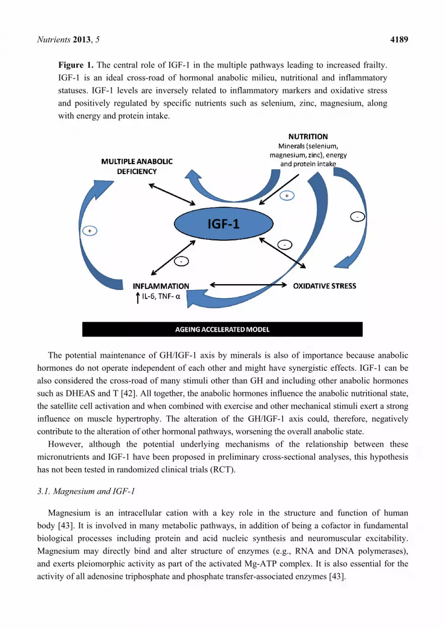
Figure 1. The central role of IGF-1 in the multiple pathways leading to increased frailty.
IGF-1 is an ideal cross-road of hormonal anabolic milieu, nutritional and inflammatory
statuses. IGF-1 levels are inversely related to inflammatory markers and oxidative stress
and positively regulated by specific nutrients such as selenium, zinc, magnesium, along
with energy and protein intake.
The potential maintenance of GH/IGF-1 axis by minerals is also of importance because anabolic
hormones do not operate independent of each other and might have synergistic effects. IGF-1 can be
also considered the cross-road of many stimuli other than GH and including other anabolic hormones
such as DHEAS and T [42]. All together, the anabolic hormones influence the anabolic nutritional state,
the satellite cell activation and when combined with exercise and other mechanical stimuli exert a strong
influence on muscle hypertrophy. The alteration of the GH/IGF-1 axis could, therefore, negatively
contribute to the alteration of other hormonal pathways, worsening the overall anabolic state.
However, although the potential underlying mechanisms of the relationship between these
micronutrients and IGF-1 have been proposed in preliminary cross-sectional analyses, this hypothesis
has not been tested in randomized clinical trials (RCT).
3.1. Magnesium and IGF-1
Magnesium is an intracellular cation with a key role in the structure and function of human
body [43]. It is involved in many metabolic pathways, in addition of being a cofactor in fundamental
biological processes including protein and acid nucleic synthesis and neuromuscular excitability.
Magnesium may directly bind and alter structure of enzymes (e.g., RNA and DNA polymerases),
and exerts pleiomorphic activity as part of the activated Mg-ATP complex. It is also essential for the
activity of all adenosine triphosphate and phosphate transfer-associated enzymes [43].

Nutrients 2013, 5 4190
Epidemiological, experimental, and clinical studies support the presence of low magnesium intake and
reduced serum magnesium levels in numerous clinical and preclinical conditions including defective
membrane function, type-2 diabetes, metabolic syndrome, elevated C-reactive protein (CRP), hypertension,
atherosclerotic vascular disease, increased oxidative stress, and immune dysfunction [44,45].
Interestingly, magnesium has been shown to exert, as IGF-1 does, beneficial independent actions on
muscle function and could play a role on physical performance in the elderly. This hypothesis is
consistent with clinical and epidemiologic data supporting the importance of the magnesium ion as a
determinant of muscle performance in young subjects. Structural damages to muscle cells are associated
to magnesium depletion and an optimal magnesium status seems to be necessary for maintaining muscle
performance and exercise tolerance in young individuals, with evidence of significant increasing in
muscle strength after magnesium supplementation [46,47]. However, the role of magnesium in
maintaining muscle integrity and function has not been fully investigated in older adults. This is
surprising given the higher prevalence of poor magnesium status in this specific population [48].
Dominguez et al. [49] found, in a representative cohort of older subjects, a significant independent
and strong positive relationship between circulating magnesium levels and measures of muscle
performance, including grip strength (β = 2.0 ± 0.5, p = 0.0002), lower-leg muscle power (β = 8.8 ± 2.7,
p = 0.001), knee extension torque (β = 31.2 ± 7.9, p < 0.0001), and ankle extension strength (β = 3.8 ± 0.5,
p < 0.0001).
There is also evidence that some of beneficial effects of magnesium on muscle function, especially
in older population, could be exerted through the stimulation of IGF-1.
Maggio and colleagues [50] have investigated for the first time the relationship between magnesium
and IGF-1 using a cohort of 399 older men ≥65 years of the InCHIANTI study. They found that
magnesium levels were strongly and independently associated with total IGF-1 levels (β ± SE, 15.9 ± 4.8;
p = 0.001).
In fasting normotensive subjects IGF-1 was able to increase intracellular magnesium levels in a
dose- and time-dependent fashion also reversing the blunted response to insulin of hypertensive
cells [51]. IGF-1 potentiates insulin-induced stimulation of magnesium at doses that, themselves, do
not raise magnesium, supporting both the hypothesis of a role for IGF-1 in cellular magnesium
metabolism and the importance of magnesium as a determinant of insulin action. This intriguing
relationship might primarily depend of a poor magnesium status that, by increasing systemic oxidative
stress and inflammation, may contribute to down-regulate IGF-1 secretion. Interestingly, magnesium,
but also IGF-1, are strongly related with antioxidant capacity. IGF-1 levels rapidly fall after exposure
to oxidative stress [28]. Moreover, magnesium deficiency has been associated with increased
oxygen-derived free radicals, oxygen peroxide production, decreased antioxidant enzyme expression,
and activity [52,53] and higher levels of interleukin 1-β, tumor necrosis factors-α, and IL-6 [54],
suggesting again a potential mechanistic link between IGF-1 and inflammation.
Since magnesium status seems to be strictly related to muscle ATP and IGF-1 levels, it becomes
important to check whether the contemporary presence of both magnesium deficiency and low IGF-1
levels can be related to age-related phenomena. It is possible that poor magnesium status contributes to
the onset of late-life sarcopenia. Magnesium deficiency might exacerbate the age-related decline in
IGF-1 levels, playing an additive negative effect in impairing physical performance. This is of
particular importance in the older population, which is more vulnerable to develop frailty.

Nutrients 2013, 5 4191
In the elderly, the reduced nutrient intake along with impaired magnesium absorption and/or
increased urinary loss, as well as polypharmacotherapy, could explain why a poor magnesium status is
frequently observed. Although magnesium requirement does not change with age [55], data from the
National Health and Nutrition Examination Survey (NHANES) III have reported an age-related
decrease in daily magnesium intake, that frequently does not reach dietary reference intake values
(420 and 320 mg/day for men and women respectively) [56]. The ageing model is also characterized
by a major risk of magnesium deficit due to the age-dependent fall of intracellular magnesium levels
and the increasing in magnesium retention [57]. This phenomenon suggests a subclinical magnesium
deficit in the elderly, which is difficult to be detected by total serum magnesium [58]. With age, there
is a decline in magnesium absorption efficiency from the intestine and in tubular resorption at the renal
level. This condition could be influenced by alterations of vitamin D metabolism or latent primary
renal disorder, which is frequently observed in older persons [57]. It is also known that medications
such as loop diuretics [59], digitals [59], and proton pump inhibitors [60,61] could interfere with
magnesium absorption and excretion leading to a secondary magnesium deficiency.
These preliminary data require further investigations in order to confirm if magnesium could be
assumed as predictor of anabolic hormones, such as IGF-1, and represents a potential therapeutic
option in order to prevent mobility limitation in older persons.
3.2. Selenium and IGF-1
Selenium is a micronutrient with a wide range of multiple functions on cells and tissues by
integration into 25 selenoproteins that have selenocysteine residues at their active core [62]. Selenoproteins
are involved in the glutathione peroxidases family, a group of antioxidant enzymes [63]. Selenium is
also an essential cofactor of many metabolic pathways. After being transformed into its bioactive
metabolites, selenium acts on the nuclear transcription factor NF-κB [64], signal transduction, cell
cycle checkpoint and apoptosis [65]. Selenium is implicated in the iodothyronine deiodinases function,
enzymes on which depends the production of active thyroid hormone (triiodothyronine or T3) in the
peripheral tissue [66,67]. Selenium status, measured by plasma or serum selenium, varies by country
and corresponds to dietary intakes. Dietary selenium intakes ranges from 7 µg per day to 49,990 µg
per day, with mean values of 40 µg per day in Europe and 1135 µg per day in USA, where 50% of the
population takes dietary supplements [68]. Recommended selenium intake is 60 µg per day for men
and 53 µg per day for women [68]. Food sources of selenium are meats and fish (typical selenium
content 25–150 mg/kg), cereals and grains (typical selenium content 10–75 mg/kg), milk and dairy
products (typical selenium content 10–25 mg/kg), and vegetables and fruits (typical selenium content
0–20 mg/kg) [69].
Rayman and colleagues in a recent review [70], analyzed a wide number of studies investigating the
relationship between selenium status and human health. Low selenium status has been associated with
increased risk of mortality [71–73] and cognitive impairment [67]. On the other side, optimal selenium
status seems to exert a protective effect on viral infection [74], autoimmune thyroid disease [75] and
cancer [76,77].
There is increasing evidence that selenium could influence skeletal muscle function even if its role
in maintaining functional muscle efficiency is still unclear.

Nutrients 2013, 5 4192
Selenium may contribute to skeletal muscle function through the maintenance of an optimal
concentration of glutathione peroxidase and other selenoproteins, particularly the selenoprotein N,
located in endoplasmic reticulum that seems to regulate calcium mobilization required for early muscle
development [63].
Many studies have detected that selenium deficiency contributes to the pathogenesis of
myopathies [78]. An important and surprising observation came from Moosman and Behl [79], who
noticed that muscular symptoms associated with selenium deficiency are very similar to those often
encountered during statin use. The authors suggested that molecules of the family of statins may
interfere with the isopentenil tRNASec, inhibiting its maturation in functional tRNASec, and leading to
a defect in the selenoproteins synthesis and low muscle function.
Beck et al. [80] have shown a significant positive association between selenium levels and handgrip
values in a population of elderly women with moderate to severe levels of disability.
Lauretani et al. [81], analysing data form 891 participants (77.1% of all population enrolled) of the
InCHIANTI study, found a linear association between plasma selenium levels and muscle strength,
namely hip flexion and hip extension for lower extremity muscle strength, shoulder adduction and
handgrip for upper-extremity muscle strength. After adjustment for age, sex, BMI, total energy intake,
chronic diseases, and IL-6 levels, participants in the lowest quartile of plasma selenium concentration
had a greater risk of poor knee, hip, and grip strength decline than those in the highest quartile. More
than 30% of the study population presented selenium concentration <0.88 mmol/L (<70 µg/L), a level
below which selenoproteins synthesis could be significantly reduced. It is possible that the reduced
selenoproteins activity can increase stress and oxidative damage to DNA, proteins and cellular lipids.
In the Uppsala Longitudinal Study of Adult Men [82], high serum levels of selenium were predictive
of low concentrations of urinary F2 isoprostane, a marker of lipid peroxidation and oxidative stress.
The accumulation of damage at the mitochondrial level and nuclear DNA is one of the main etiological
hypotheses by which loss of myocytes and a consequent impaired skeletal muscle function is realized.
Selenium, being an important cofactor in cellular protection, could be particularly involved in muscle
protection, especially in the elderly where the cellular oxidative damage is more prevalent.
Recent findings support the hypothesis of a possible role of selenium in IGF-1 bioactivity. A recent
cross-sectional study that involved 44 young women showed a significant association between IGF-1
serum levels and selenium from dietary sources (r = 0.41, p < 0.01) [83].
Maggio et al. [84], in a recent study have explored this hypothetical link in 951 participants (90% of
enrolled) of the InCHIANTI Study. Factors statistically correlated with IGF-1 levels were identified
and introduced in the multivariate analysis. This study found a strong correlation between selenium
and IGF-1 levels in older adults that was independent of age, sex, body mass index, total energy and
alcohol intakes, smoking, alanine aminotransferase, thyroid stimulating hormone, free thyroxine, and
free triiodothyronine, CRP, IL-6 and the major chronic diseases (congestive heart failure, chronic
obstructive pulmonary disease, and cancer). This finding raises the hypothesis that selenium is capable
of modulating IGF-1 bioactivity also at skeletal muscle level.
The hypothesis of a link between selenium and IGF-1 is supported by the observation of a fine
balance between selenium and GH in ensuring normal growth. Animal studies suggest that selenium
deficiency is independently associated to growth retardation, compromising triiodothyronine
availability and GH/IGF-1 axis [85].

Nutrients 2013, 5 4193
Long-term sodium selenite treatment in rats reduced serum GH levels, IGF-1 and IGF binding
protein-1, -2, and -3 and determined a growth delay [86]. The underlying mechanism of this
phenomenon has not been fully elucidated in the study, but was probably caused by the accumulation
of selenium in secreting vesicles that inhibited GH secretion. Moreover, a selenium deficiency causes
an increased T4 (67%) and a reduction of T3 (23%). By using second-generation rat pups with
selenium deficiency, maintaining adequate vitamin E and methionine levels, administration of 0.1 or
0.2 mg/g selenium normalized thyroid hormones serum levels, selenium liver content, and glutathione
peroxidase activity [86]. The exclusive T3 administration did not restore the growth, suggesting that
factors other than serum T3 might be involved in growth-related disorders.
These findings are particularly interesting since IGF-1 and selenium play a significant role in the
pathophysiology of some age-related phenomena such as sarcopenia. A common target for selenium
and IGF-1 is the skeletal muscle tissue. As previously described, both selenium and IGF-1 are involved
in muscle protein synthesis and their deficiencies may lead to sarcopenia and disability during aging.
Moreover, both low selenium and IGF-1 levels are associated with anemia, another potential
determinant of sarcopenia [87–89].
The positive association between selenium levels and IGF-1 primarily depends on the positive
action exerted by selenium levels on IGF-1. Selenoenzymes such as glutathione peroxidase and
glutathione reductase protecting tissues from oxidative stress [82] could positively affect IGF-1
cell-releasing. Selenoenzymes seem to prevent cell degeneration and oxygen reactive species
production. An alternative hypothesis to explain the relationship between selenium and IGF-1
considers inflammation as the common negative regulator of these two factors. In fact, selenium
deficiency worsens during increased NF-κB activity [64] and by this mechanism can determine the
up-regulation of IL-6. In turn, IL-6 could negatively influence IGF-1 levels. In Women’s Health and
Aging Study I, subjects with low levels of selenium had higher levels of IL-6 [90]. As previously
described, a similar inverse association was also found between IGF-1 and IL-6 [23]. We cannot also
ignore the role played by dietary intake. Primary dietary sources of selenium are proteins [69], which
are also important positive modulators of serum IGF-1 concentrations [31].
Future longitudinal and intervention studies will clarify the mechanisms of the intriguing
association between selenium and IGF-1 and the effects of dietary and supplementary selenium
sources on IGF-1. These data will create the rationale for a replacement treatment combining selenium
and IGF-1 in the prevention and treatment of sarcopenia and frailty.
3.3. Zinc and IGF-1
Zinc is an essential micronutrient with a structural and functional role in a large number of
macromolecules and enzymatic reactions. More than 200 enzymes implicated in major metabolic
processes require zinc as a functional component. Zinc is involved in growth, protein and
DNA synthesis, neuro-sensory functions, cell-mediated immunity, thyroid function, and bone
metabolism [91]. Suboptimal zinc status is associated with impaired wound healing, dermatitis,
inflammation, and depressed immune response and might be responsible for the high incidence of
age-related infections and degenerative diseases [92]. Interestingly, zinc seems also to be involved in
nutritional regulation of IGF-1 bioactivity. In cultured bone cells, some studies suggest that zinc

Nutrients 2013, 5 4194
potentiates the action of IGF-1 [93] and increases endogenous IGF-1 synthesis [94]. In animal models,
severe zinc deficiency has been shown to decrease hepatic IGF-1 gene expression and to impair
intracellular GH signaling pathway [95–97]. By these mechanisms, zinc may affect circulating
concentrations of insulin, IGF-1 and GH. Roth and Kirchgessner [98] showed that force-feeding a
zinc-depleted diet to rats for 14 days results in a 28% decrease in serum IGF-1 compared with rats fed
a zinc-adequate diet, although there was no difference in caloric food intake. Similarly, Droup et al.
demonstrated that low IGF-1 levels were related to decreased zinc concentration [99]. However,
maintaining serum IGF-1 levels by exogenous administration or by inducing food intake, or both, in
zinc-deficient rats, was not sufficient to correct the growth inhibition induced by zinc deficiency [100].
Thus, zinc could be involved in some aspects of growth regulation at the cellular level independently
of those observed on circulating IGF-1 and GH. Changes in the GH-IGF axis alone might not explain
the growth inhibition observed in zinc deficiency. In humans, poor zinc status has been associated with
low circulating IGF-1 concentrations even in presence of adequate caloric intake [101]. In zinc
deficient short children, zinc supplementation was effective in inducing growth. In these subjects, the
growth velocity was associated with increased plasma IGF-1 concentrations [102–104]. These data
suggest that growth-stimulating effect of zinc might be mediated through changes in circulating IGF-1.
However, when optimal serum zinc levels are present in children with idiopathic short stature, zinc
supplementation increased basal IGF-1, IGFBP-3, alkaline phosphatase, and osteocalcin levels but did
not significantly change height or weight standard deviations during a 6–12 month follow-up
period [105]. Thus, zinc supplementation may be beneficial in selected categories of subjects with low
baseline zinc serum levels. A moderate zinc deficiency is also often observed in elderly subjects [106].
During ageing, nutritional deficits coupled with decreased absorptive efficiency contribute to zinc
depletion. More evidence suggests that an increased rate of bone resorption with age is an important
negative determinant of zinc excretion. In postmenopausal osteoporotic women has been reported a
parallel decline of bone zinc and calcium, with a marked hyperzincuria [107–110]. A zinc deficit could
exacerbate the age-related decline in IGF-1 serum levels. However, despite suboptimal zinc intake is
widely reported in the elderly, the relationship between zinc status and IGF-1 has not been adequately
investigated. Devine et al. [111] examined the relation between caloric intake and IGF-1 and IGFBPs
concentrations in 119 postmenopausal women. IGF-1 concentrations were significantly correlated with
mean protein and zinc intake at baseline (r = 0.313, p = 0.001; r = 0.298, p = 0.001, respectively) and
after two years (r = 0.256, p = 0.008; r = 0.331, p = 0.001, respectively), even after adjustment for age,
weight, and other nutrients. In sixty-one hospitalized frail elderly aged 66.7 to 105.8, with a
mini-nutritional assessment score between 17 and 24, Rodondi and colleagues [112] determined the
effects of dietary zinc intake (30 mg/day) on IGF-1 and bone turnover responses to four-week essential
amino acids-whey protein supplementation After one week, zinc supplementation increased the IGF-1
response to essential amino acids-whey protein (+48.2 ± 14.3 and +22.4 ± 4.7%, p < 0.05), and
significantly decreased markers of bone resorption.
The link between zinc and IGF-1 can be at least in part attributed to antioxidant activity of zinc.
In vitro, animal and human studies show that zinc decreases the oxidative damage of membrane
fractions probably through a protection of sulphhydryl groups against oxidation and the inhibition of
the production of reactive oxygen by transition metals [113–115]. Although zinc may act as a
biological antioxidant, high levels of zinc could also be a pro-oxidant agent by eliciting a decline in

Nutrients 2013, 5 4195
erythrocyte Cu–Zinc superoxide dismutase (SOD) [116]. Moreover, zinc is provided by protein foods,
especially animal products [117], known to increase IGF-1 serum levels. In population of healthy
well-nourished middle-aged and elderly men, Larsson and coworkers [118] tested the association
between total energy, alcohol, vitamins, protein, nutrient-dense core foods (including red meat, fish
and seafood, poultry, and milk), and serum IGF-1 concentrations. Interestingly, these authors observed
a statistically significant positive association between protein intake, zinc, and serum IGF-1 concentrations
independent of age.
However, zinc assessment is difficult because a sensitive, specific biomarker for this mineral has
not been identified yet [119]. Plasma (or serum) zinc concentration, being responsive to both zinc
supplementation and zinc depletion, seems to be the most widely reported biomarker for assessing zinc
status. However, data at this regard are not very strong. Hair and urine zinc concentrations are also
considered useful biomarkers of zinc supplementation [119].
Dietary zinc recommendations vary widely across Europe due to the heterogeneity of approaches
used by expert panels [120]. The determination of zinc requirements is traditional based on a factorial
approach that estimates the zinc intake required to meet physiological requirements for growth,
metabolism and tissue repair while replacing obligatory losses. An alternative approach is based on the
examination of the dose–response relationship between intake and biomarkers of zinc status and also
between intake and health outcomes. The relationship between zinc and IGF-1 in ageing population
warrants further investigation in order to confirm whether zinc could be identified as a modulator of
IGF-1 bioactivity in frail older persons at risk of mobility limitation.
4. Perspectives
Based on the data presented here it can be hypothesized a potential therapeutic implication of
mineral supplementation or replacement in well-defined groups of older persons, and particularly in
those with a decreased physiological reserve and increased vulnerability to stressors. These subjects
are more prone to develop mobility limitation, and often show a suboptimal nutritional status and
lower baseline IGF-1 levels. The perfect context where micronutrients might be useful is the model of
critical illness. Stress, acute illness, surgery or trauma produce major changes in the metabolic milieu of
the body such as altered substrate utilization and synthesis, hypermetabolism, and catabolism [121–123].
The robust response of the anterior pituitary gland, cytokines, and the sympathetic nervous system at
the outset of critical illness dramatically changes over time. Older frail individuals have a reduced
ability and require more energetic costs to recover from acute stressors with prolonged acute critical
illness and chronic critical illness [124]. Severe muscle atrophy, weakness, and disability are direct
consequences of this reduced homeostatic reserve [125]. During an acute critical illness, cortisol, as
well as other counter-regulatory hormones and cytokines, support the inflammatory response and vital
organ function [126] shunting substrate away to promote anabolism. Impaired IGF-1 levels along with
the rise in GH, augments the lipolytic and insulin-antagonizing effects of GH, while postponing the
anabolic effect of IGF-1 [126]. In prolonged acute and chronic critical illnesses the loss of pulse GH
amplitude profoundly affects IGF-1 levels, which tend to drop further, worsening the catabolic milieu
of the critically ill older patient [126]. The resulted higher anabolic threshold to stress is particularly
important in very frail elderly subjects, characterized by lower IGF-1 levels and inadequate nutritional

Nutrients 2013, 5 4196
status even before the acute events. Moreover, acute critical illness is also followed by a prolonged bed
rest that emphasizes the catabolic process and favors the onset of disability [127]. The use of certain
micronutrients as modulators of IGF-1 levels and anabolic threshold might be an intriguing future
therapeutic option. This concept it is graphically depicted in Figure 2.
Figure 2. Potential therapeutic implication of mineral supplementation during critical illness.
In both panel, a and b, the lines represent the decline in IGF-1 levels which is realized during a
critical illness. In panel a, the higher anabolic thresholds resulting from prolonged acute and chronic
critical illness is described. Panel b underlines the potential therapeutic implication of mineral
supplementation in lowering such anabolic threshold.
5. Conclusions
The age-related gradual decline in IGF-1 levels is part of the multiple anabolic hormone deficiency.
This phenomenon could be implicated in the development of sarcopenia, mobility limitation, and
frailty syndrome. IGF-1 is a sensitive nutritional marker rather than just an anabolic hormone and is
negatively influenced by poor mineral and global nutritional statuses and subclinical low-grade
inflammation. These conditions may influence the physical performance in older individuals by
exacerbating the negative anabolic milieu. IGF-1, by representing a crossroad between hormonal,
inflammatory, and nutritional pathways of frailty, might contribute to an earlier interception of those
subjects more sensitive to tailored interventions. The increasing evidence that magnesium, selenium,
and zinc along with optimal protein and energy requirement, are implicated in sustaining IGF-1 levels
is of importance especially in elderly population.
These preliminary findings suggest that both, minerals and IGF-1, could be considered specific
therapeutic targets to improve muscle strength and physical performance in the elderly. Longitudinal
studies and RCT are needed to test the precise contribution of multiple mineral deficiencies to frailty,
and to establish the specific cut-offs of micronutrients and the optimal dosages required to increase the
anabolic status and physical function in older individuals. The findings of these upcoming studies will

Nutrients 2013, 5 4197
create the rationale for an effective combined approach in the prevention and treatment of frailty and
other conditions of accelerated aging.
Conflicts of Interest
The authors declare no conflict of interest.
References
1. Delmonico, M.J.; Harris, T.B.; Lee, J.S.; Visser, M.; Nevitt, M.; Kritchevsky, S.B.;
Tylavsky, F.A.; Newman, A.B. Health, aging and body composition study. Alternative
definitions of sarcopenia, lower extremity performance, and functional impairment with aging in
older men and women. J. Am. Geriatr. Soc. 2007, 55, 769–774.
2. Goodpaster, B.H.; Park, S.W.; Harris, T.B.; Kritchevsky, S.B.; Nevitt, M.; Schwartz, A.V.;
Simonsick, E.M.; Tylavsky, F.A.; Visser, M.; Newman, A.B. The loss of skeletal muscle
strength, mass, and quality in older adults: The health, aging and body composition study.
J. Gerontol. 2006, 61, 1059–1064.
3. Fried, L.P.; Tangen, C.M.; Walston, J.; Newman, A.B.; Hirsch, C.; Gottdiener, J.; Seeman, T.;
Tracy, R.; Kop, W.J.; Burke, G.; et al. Frailty in older adults: Evidence for a phenotype.
J. Gerontol. 2001, 56, M146–M156.
4. Cruz-Jentoft, A.J.; Baeyens, J.P.; Bauuer, J.M.; Boirie, Y.; Cederholm, T.; Landi, F.; Martin, F.C.;
Michel, J.P.; Rolland, Y.; Schneider, S.M.; et al. Sarcopenia: European consensus on definition
and diagnosis: Report of the European Working Group on Sarcopenia in Older People. Age Ageing
2010, 1, 412–423.
5. Rosenberg, I.H. Sarcopenia: Origins and clinical relevance. J. Nutr. 1997, 127, 990S–991S.
6. Maggio, M.; Lauretani, F.; Ceda, G.P.; Bandinelli, S.; Ling, S.M.; Metter, E.J.; Artoni, A.;
Carassale, L.; Cazzato, A.; Ceresini, G.; et al. Relationship between low levels of anabolic
hormones and 6-year mortality in older men: The aging in the Chianti Area (InCHIANTI) study.
Arch. Intern. Med. 2007, 167, 2249–2254.
7. Maggio, M.; Dall’Aglio, E.; Lauretani, F.; Cattabiani, C.; Ceresini, G.; Caffarra, P.; Valenti, G.;
Volpi, R.; Vignali, A; Schiavi, G.; et al. The hormonal pathway to cognitive impairment in older
men. J. Nutr. Health Aging 2012, 16, 40–54.
8. Arvat, E.; Broglio, F.; Ghigo, E. Insulin-like growth factor I: Implications in aging. Drugs Aging
2000, 16, 29–40.
9. Rosen, C.J. Serum insulin-like grow factor and insulin like grow factor-binding proteins: Clinical
implication. Clin. Chem. 1999, 45, 1384–1390.
10. Estívariz, C.F.; Ziegler, T.R. Nutrition and the insulin-like growth factor system. Endocrine
1997, 7, 65–71.
11. Sherlock, M.; Toogood, A.A. Aging and the growth hormone/insulin like growth factor-I axis.
Pituitary 2007, 10, 189–203.
12. Lee, P.D.; Giudice, L.C.; Conover, C.A.; Powell, D.R. Insulin-like growth factor binding
protein 1: Recent findings and new directions. Proc. Soc. Exp. Biol. Med. 1997, 216, 319–357.

Nutrients 2013, 5 4198
13. Franklin, S.L.; Ferry, R.J.; Cohen, P. Rapid insulin-like growth factor (IGF)-independent effects
of IGF binding protein-3 on endothelial cell survival. J. Clin. Endocrinol. Metab. 2003, 88,
900–907.
14. Hoffman, A.R.; Pyka, G.; Liecerman, S.A.; Ceda, G.P.; Marcus, R. The Somatopause. In Growth
Hormone and Somatomedins during Lifespan; Muller, E.E., Cocchi, D., Locatelli, V., Eds.;
Springer: Berlin/Heidelberg, Germany, 1993; pp. 265–274.
15. De Marinis, L.; Mancini, A.; Giampietro, A.; Gentilella, R.; Bianchi, A.; Perrelli, M.; Vezzosi, C.;
Milardi, D.; Fusco, A.; Valle, D.; et al. GH deficiency syndrome in elderly patients. J. Endocrinol.
Investig. 2002, 25, 40–41.
16. Cappola, A.R.; Bandeen-Roche, K.; Wand, G.S.; Volpato, S.; Fried, L.P. Association of IGF-I
levels with muscle strength and mobility in older women. J. Clin. Endocrinol. Metab. 2001, 86,
4139–4146.
17. Colbert, L.H.; Rosen, C.J.; Goodpaster, B.H.; Newman, A.B.; Kritchevsky, S.B.; Satterfield, S.;
Kanaya, A.M.; Taaffe, D.R.; Harris, T.B. Insulin-like growth factor-1. J. Am. Geriatr. Soc. 2004,
52, 1962–1963.
18. Ferrucci, L.; Corsi, A.; Lauretani, F.; Bandinelli, S.; Bartali, B.; Taub, D.D.; Guralnik, J.M.;
Longo, D.L. The origins of age-related proinflammatory state. Blood 2005, 105, 2294–2299.
19. Elosua, R.; Bartali, B.; Ordovas, J.M.; Corsi, A.M.; Lauretani, F.; Ferrucci, L. Association
between physical activity, physical performance, and inflammatory biomarkers in an elderly
population: The InCHIANTI study. J. Gerontol. 2005, 60, 760–767.
20. Cesari, M.; Penninx, B.W.; Pahor, M.; Lauretani, F.; Corsi, A.M.; Rhys Williams, G.; Guralnik, J.M.;
Ferrucci, L. Inflammatory markers and physical performance in older persons: The InCHIANTI
study. J. Gerontol. 2004, 59, 242–248.
21. Ferrucci, L.; Harris, T.; Guralnik, J.; Tracy, R.P.; Corti, M.C.; Cohen, H.J.; Cohen, B.; Penninx, B.;
Pahor, M.; Wallace, R.; et al. Serum IL-6 level and the development of disability in older
persons. J. Am. Geriatr. Soc. 1999, 47, 662–668.
22. Lazarus, D.D.; Moldawer, L.L.; Lowry, S.F. Insulin like growth factor-1 activity is inhibited by
interleukine alpha, tumor necrosis factor-alpha, and interleukin-6. Lymphokine Cytokine Res.
1993, 12, 219–223.
23. Barbieri, M.; Ferrucci, L.; Ragno, E.; Corsi, A.; Bandinelli, S.; Bonafè, M.; Olivieri, F.;
Giovagnetti, S.; Franceschi, C.; Guralnik, J.M.; et al. Chronic inflammation and the effect of
IGF-I on muscle strength and power in older persons. Am. J. Physiol. Endocrinol. Metab. 2003,
284, E481–E487.
24. Stenholm, S.; Maggio, M.; Lauretani, F.; Bandinelli, S.; Ceda, G.P.; Di Iorio, A.; Giallauria, F.;
Guralnik, J.M.; Ferrucci, L. Anabolic and catabolic biomarkers as predictors of muscle strength
decline: The InCHIANTI study. Rejuvenation Res. 2010, 13, 3–11.
25. Cappola, A.R.; Xue, Q.L.; Ferrucci, L.; Guralnik, J.M.; Volpato, S.; Fried, L.P. Insulin-like
growth factor I and interleukin-6 contribute synergistically to disability and mortality in older
women. J. Clin. Endocrinol. Metab. 2003, 88, 2019–2025.

Nutrients 2013, 5 4199
26. Roubenoff, R.; Parise, H.; Payette, H.A.; Abad, L.W.; D’Agostino, R.; Jacques, P.F.; Wilson, P.W.;
Dinarello, C.A.; Harris, T.B. Cytokines, insulin-like growth factor 1, sarcopenia, and mortality in
very old community-dwelling men and women: The Framingham Heart Study. Am. J. Med.
2003, 115, 429–435.
27. Fernandez, S.; Fernandez, A.M.; Lopez-Lopez, C.; Torres-Aleman, I. Emerging roles of
insulin-like growth factor 1 in the adult brain. Growth Horm. IGF Res. 2007, 17, 89–95.
28. Yanase, S.; Ishii, N. Hyperoxia exposure induced hormesis decreases mithocondrial
superoxide radical levels via Ins/IGF-1 signaling pathway in a long-lived age-1 mutant on
Caenorhabditis elegans. J. Radiat. Res. 2008, 49, 211–218.
29. Underwood, L.E.; Clemmons, D.R.; Maes, M.; D’Ercole, A.J.; Ketelslegers, J.M. Regulation of
somatomedin-C/insulin-like growth factor I by nutrients. Horm. Res. 1986, 24, 166–176.
30. Norat, T.; Dossus, L.; Rinaldi, S.; Overvad, K.; Grønbaek, H.; Tjønneland, A.; Olsen, A.;
Clavel-Chapelon, F.; Boutron-Ruault, M.C.; Boeing, H.; et al. Diet, serum insulin-like growth
factor-I and IGF-binding protein-3 in European women. Eur. J. Clin. Nutr. 2007, 61, 91–98.
31. Thissen, J.P.; Ketelslegers, J.M.; Underwood, L.E. Nutritional regulation of the insulin-like
growth factors. Endocr. Rev. 1994, 15, 80–101.
32. Holmes, M.D.; Pollack, M.N.; Willett, W.C.; Hankinson, S.E. Dietary correlates of plasma
insulin-like growth factor 1 and insulin-like growth factor binding protein 3 concentrations.
Cancer Epidemiol. Biomark. Prev. 2002, 11, 852–861.
33. Gran, I.T.; Norat, T.; Rinaldi, S.; Dossus, L.; Ulanova, A.; Téhard, B.; Clavel-Chapelon, F.;
van Gils, C.H.; van Noord, P.A.; Peeters, P.H.; et al. Body mass index, waist circumference and
waist-hip ratio and serum levels of IGF-I and IGFBP-3 in European women. Int. J. Obes. 2006,
30, 1623–1631.
34. Salbe, A.D.; Kotler, D.P.; Wang, J.; Pierson, R.N.; Campbell, R.G. Predictive value of IGF-1
concentrations in HIV-infected patients. Clin. Res. 1991, 39, 385A.
35. Kirschner, B.S.; Sutton, M.M. Somatomedin-C levels in growth-impaired children and
adolescents with chronic inflammatory bowel disease. Gastroenterology 1986, 91, 830–836.
36. Clemmons, D.R.; Klibanski, A.; Underwood, L.E.; McArthur, J.W.; Ridgway, E.C.; Beitins, I.Z.;
van Wyk, J.J. Reduction of plasma immunoreactive somatomedin-C during fasting in humans.
J. Clin. Endorinol. Metab. 1981, 53, 1247–1250.
37. Tirapegui, J.; Fukushima, S.E.; Grimaldi, G. Growth, somatomedin and nutrition. Arch.
Latinoam. Nutr. 1993, 43, 94–104.
38. Vehe, K.L.; Brown, R.O.; Moore, L.W.; Acchiardo, S.R.; Luther, R.W. The efficacy of nutrition
support in infected patients with chronic renal failure. Pharmacotherapy 1991, 11, 303–307.
39. Lecornu, M.; David, L.; Francois, R. Low serum somatomedin activity in celiac disease.
Helv. Paediatr. Acta 1978, 33, 509–516.
40. O’Connor, J.C.; McCusker, R.H.; Strle, K.; Johnson, R.W.; Dantzer, R.; Kelley, K.W.
Regulation of IGF-I function by proinflammatory cytokines: At the interface of immunology and
endocrinology. Cell. Immunol. 2008, 252, 91–110.
41. Ni, F.; Sun, R.; Fu, B.; Wang, F.; Guo, C.; Tian, Z.; Wei, H. IGF-1 promotes the development
and cytotoxic activity of human NK cells. Nat. Commun. 2013, 4, doi:10.1038/ncomms2484.

Nutrients 2013, 5 4200
42. Brill, K.T.; Weltman, A.L.; Gentili, A.; Patrie, J.T.; Fryburg, D.A.; Hanks, J.B.; Urban, R.J.;
Veldhuis, J.D. Single and combined effects of growth hormone and testosterone administration
on measures of body composition, physical performance, mood, sexual function, bone turnover,
and muscle gene expression in healthy older men. Clin. Endocrinol. Metab. 2002, 87, 5649–5657.
43. Wolf, F.I.; Cittadini, A. Chemistry and biochemistry of magnesium. Mol. Asp. Med. 2003, 24,
3–9.
44. Barbagallo, M.; Dominguez, L.J.; Galioto, A.; Ferlisi, A.; Cani, C.; Malfa, L.; Pineo, A.;
Busardo’, A.; Paolisso, G. Role of magnesium in insulin action, diabetes and cardiometabolic
syndrome X. Mol. Asp. Med. 2003, 24, 39–52.
45. Champagne, C.M. Magnesium in hypertension, cardiovascular disease, metabolic syndrome, and
other conditions: A review. Nutr. Clin. Pract. 2008, 23, 142–151.
46. Lukaski, H.C.; Nielsen, F.H. Dietary magnesium depletion affects metabolic responses during
submaximal exercise in postmenopausal women. J. Nutr. 2003, 132, 930–935.
47. Brilla, L.R.; Haley, T.F. Effect of magnesium supplementation on strength training in humans.
J. Am. Coll. Nutr. 1992, 11, 326–329.
48. Vaquero, M.P. Magnesium and trace elements in the elderly: Intake, status and recommendations.
J. Nutr. Health Aging 2002, 6, 147–153.
49. Dominguez, L.J.; Barbagallo, M.; Lauretani, F.; Bandinelli, S.; Bos, A.; Corsi, A.M.;
Simonsick, E.M.; Ferrucci, L. Magnesium and muscle performance in older persons: The
InCHIANTI study. Am. J. Clin. Nutr. 2006, 84, 419–426.
50. Maggio, M.; Ceda, G.P.; Lauretani, F.; Cattabiani, C.; Avantaggiato, E.; Morganti, S.; Ablondi, F.;
Bandinelli, S.; Dominguez, L.J.; Barbagallo, M.; et al. Magnesium and anabolic hormones in
older men. Int. J. Androl. 2011, 34, e594–e600.
51. Dominguez, L.J.; Barbagallo, M.; Sowers, J.R.; Resnick, L.M. Magnesium responsiveness to
insulin and insulin-like growth factor I in erythrocytes from normotensive and hypertensive
subjects. J. Clin. Endocrinol. Metab. 1998, 83, 4402–4407.
52. Rayssiguier, Y.; Durlach, J.; Gueux, E.; Rock, E.; Mazur, A. Magnesium and aging. I.
Experimental data: Importance of oxidative damage. Magnes. Res. 1993, 6, 369–378.
53. Yang, Y.; Wu, Z.; Chen, Y.; Qiao, J.; Gao, M.; Yuan, J.; Nie, W.; Guo, Y. Magnesium
deficiency enhances hydrogen peroxide production and oxidative damage in chick embryo
hepatocyte in vitro. Biometals 2006, 19, 71–81.
54. Barbagallo, M.; Dominguez, L.J. Magnesium and aging. Curr. Pharm. Des. 2010, 16, 832–839.
55. Hunt, C.D.; Johnsom, L.K. Magnesium requirements: New estimations for men and women by
cross-sectional statistical analyses of metabolic magnesium balance data. Am. J. Clin. Nutr. 2006,
84, 843–852.
56. Ford, E.S.; Mokdad, A.H. Dietary magnesium intake in a national sample of US adults.
J. Nutr. 2003, 133, 2879–2882.
57. Barbagallo, M.; Belvedere, M.; Dominguez, L.J. Magnesium homeostasis and aging.
Magnes. Res. 2009, 22, 235–246.
58. Gullestad, L.; Nes, M.; Rønneberg, R.; Midtvedt, K.; Falch, D.; Kjekshus, J. Magnesium status in
healthy free-living elderly Norwegians. J. Am. Coll. Nutr. 1994, 13, 45–50.

Nutrients 2013, 5 4201
59. Lameris, A.L.; Monnens, L.A.; Bindels, R.J.; Hoenderop, J.G. Drug-induced alterations in Mg2+
homoeostasis. Clin. Sci. 2012, 123, 1–14.
60. Cundy, T.; Dissanayake, A. Severe hypomagnesaemia in long-term users of proton-pump inhibitors.
Clin. Endocrinol. 2008, 69, 338–341.
61. Hoorn, E.J.; van der Hoek, J.; de Man, R.A.; Kuipers, E.J.; Bolwerk, C.; Zietse, R. A case series
of proton pump inhibitor-induced hypomagnesemia. Am. J. Kidney Dis. 2010, 56, 112–116.
62. Kryukov, G.V.; Castellano, S.; Novoselov, S.V.; Lobanov, A.V.; Zehtab, O.; Guigó, R.;
Gladyshev, V.N. Characterization of mammalian selenoproteomes. Science 2003, 300, 1439–1443.
63. Reeves, M.A.; Hoffmann, P.R. The human selenoproteome: Recent insights into functions and
regulation. Cell. Mol. Life Sci. 2009, 66, 2457–2478.
64. Markropoulos, V.; Bruning, T.; Schulze-Osthoff, K. Selenium-mediated inhibition of
transcription factor NK-kappa B and HIV-1 LTR promoter activity. Arch. Toxicol. 1996, 70,
277–283.
65. Kaeck, M.; Lu, J.; Strange, R.; Ganther, H.E.; Thompson, H.J. Differential induction of growth
arrest inducible genes by selenium compounds. Biochem. Pharmacol. 1997, 53, 921–926.
66. Schomburg, L.; Köhrle, J. On the importance of selenium and iodine metabolism for thyroid
hormone biosynthesis and human health. Mol. Nutr. Food Res. 2008, 52, 1235–1246.
67. Burk, R.F.; Hill, K.E. Selenoprotein P-expression, functions, and roles in mammals. Biochim.
Biophys. Acta 2009, 1790, 1441–1447.
68. Rayman, M.P. Food-chain selenium and human health: Emphasis on intake. Br. J. Nutr. 2008,
100, 254–268.
69. World Health Organization (WHO). International Programme on Chemical Safety;
WHO: Geneva, Switzerland, 1987.
70. Rayman, M.P. Selenium and human health. Lancet 2012, 379, 1256–1268.
71. Bleys, J.; Navas-Acien, A.; Guallar, E. Serum selenium levels and all-cause, cancer, and
cardiovascular mortality among US adults. Arch. Intern. Med. 2008, 168, 404–410.
72. Akbaraly, N.T.; Arnaud, J.; Hininger-Favier, I.; Gourlet, V.; Roussel, A.M.; Berr, C. Selenium
and mortality in the elderly: Results from the EVA study. Clin. Chem. 2005, 51, 2117–2123.
73. Lauretani, F.; Semba, R.D.; Bandinelli, S.; Ray, A.L.; Ruggiero, C.; Cherubini, A.; Guralnik, J.M.;
Ferrucci, L. Low plasma selenium concentrations and mortality among older community-dwelling
adults: The InCHIANTI study. Aging Clin. Exp. Res. 2008, 20, 153–158.
74. Hurwitz, B.E.; Klaus, J.R.; Llabre, M.M.; Gonzalez, A.; Lawrence, P.J.; Maher, K.J.;
Greeson, J.M.; Baum, M.K.; Shor-Posner, G.; Skyler, J.S.; et al. Suppression of human
immunodeficiency virus type 1 viral load with selenium supplementation: A randomized
controlled trial. Arch. Intern. Med. 2007, 167, 148–154.
75. Toulis, K.A.; Anastasilakis, A.D.; Tzellos, T.G.; Goulis, D.G.; Kouvelas, D. Selenium
supplementation in the treatment of Hashimoto’s thyroiditis: A systematic review and a
meta-analysis. Thyroid 2010, 20, 1163–1173.
76. Amaral, A.F.; Cantor, K.P.; Silverman, D.T.; Malats, N. Selenium and bladder cancer risk: A
meta-analysis. Cancer Epidemiol. Biomark. Prev. 2010, 19, 2407–2415.

Nutrients 2013, 5 4202
77. Etminan, M.; FitzGerald, J.M.; Gleave, M.; Chambers, K. Intake of selenium in the prevention
of prostate cancer: A systematic review and meta-analysis. Cancer Causes Control 2005, 16,
1125–1131.
78. Rederstorff, M.; Krol, A.; Lescure, A. Understanding the importance of selenium and
selenoproteins in muscle function. Cell. Mol. Life Sci. 2006, 63, 52–59.
79. Moosmann, B.; Behl, C. Selenoprotein synthesis and side-effects of statins. Lancet 2004, 363,
892–894.
80. Beck, J.; Ferrucci, L.; Sun, K.; Walston, J.; Fried, L.P.; Varadhan, R.; Guralnik, J.; Semba, R.D.
Low serum selenium concentrations are associated with poor grip strength among older women
living in the community. Biofactors 2007, 29, 37–44.
81. Lauretani, F.; Semba, R.D.; Bandinelli, S.; Ray, A.L.; Guralnik, J.M.; Ferrucci, L. Association of
low plasma selenium concentrations with poor muscle strength in older community-dwelling
adults: The InCHIANTI study. Am. J. Clin. Nutr. 2007, 86, 347–352.
82. Helmersson, J.; Arnlov, J.; Vessby, B.; Larsson, A.; Alfthan, G.; Basu, S. Serum selenium
predicts levels of F2-isoprostanes and prostaglandin F2alpha in a 27 year follow-up study of
Swedish men. Free Radic. Res. 2005, 39, 763–770.
83. Karl, J.P.; Alemany, J.A.; Koenig, C.; Kraemer, W.J.; Frystyk, J.; Flyvbjerg, A.; Young, A.J.;
Nindl, B.C. Diet, body composition, and physical fitness influences on IGF-I bioactivity in
women. Growth Horm. IGF Res. 2009, 19, 491–496.
84. Maggio, M.; Ceda, G.P.; Lauretani, F.; Bandinelli, S.; Dall’Aglio, E.; Guralnik, J.M.; Paolisso, G.;
Semba, R.D.; Nouvenne, A.; Borghi, L.; et al. Association of plasma selenium concentrations
with total IGF-1 among older community-dwelling adults: The InCHIANTI study. Clin. Nutr.
2010, 29, 674–677.
85. Moreno-Reyes, R.; Egrise, D.; Nève, J.; Pasteels, J.L.; Schoutens, A. Selenium deficiency-induced
growth retardation is associated with an impaired bone metabolism and osteopenia. J. Bone
Miner. Res. 2001, 16, 1556–1563.
86. Thompson, K.M.; Haibach, H.; Sunde, R.A. Growth and plasma triiodothyronine concentrations
are modified by selenium deficiency and repletion in second-generation selenium-deficient rats.
J. Nutr. 1995, 125, 864–873.
87. Semba, R.D.; Ricks, M.O.; Ferrucci, L.; Xue, Q.L.; Guralnik, J.M.; Fried, L.P. Low serum
selenium is associated with anemia among older adults in the United State. Eur. J. Clin. Nutr.
2009, 63, 93–99.
88. Vihervuori, E.; Virtanem, M.; Koistinen, H.; Koistinen, R.; Seppala, M.; Siimes, M.A.
Haemoglobin level is linked to growth hormone-dependent proteins in short children. Blood
1996, 87, 2075–2081.
89. Nilsson-Ehle, H.; Bengtsson, B.A.; Lindstedt, G.; Mellstrom, D. Insulin-like growth factor-1 is a
predictor of blood haemoglobin concentration in 70-yr-old subjects. Eur. J. Haematol. 2005, 74,
111–116.
90. Walston, J.; Xue, Q.; Semba, R.D.; Ferrucci, L.; Cappola, A.R.; Ricks, M.; Guralnik, J.;
Fried, L.P. Serum antioxidants, inflammation, and mortality in older women. Am. J. Epidemiol.
2006, 163, 18–26.

Nutrients 2013, 5 4203
91. Meunier, N.; O’Connor, J.M.; Maiani, G.; Cashman, K.D.; Secker, D.L.; Ferry, M.;
Roussel, A.M.; Coudray, C. Importance of zinc in the elderly: The ZENITH study. Eur. J. Clin. Nutr.
2005, 59, S1–S4.
92. Hambidge, M. Human zinc deficiency. J. Nutr. 2000, 130, 1344S–1349S.
93. Matsui, T.; Yamaguchi, M. Zinc modulation of insulin-like growth factor’s effect in osteoblastic
MC3T3–E1 cells. Peptides 1995, 16, 1063–1068.
94. Yamaguchi, M.; Hashizume, M. Effect of beta-alanyl-L-histidinato zinc on protein components
in osteoblastic MC3T3-El cells: Increase in osteocalcin, insulin-like growth factor-I and
transforming growth factor-beta. Mol. Cell. Biochem. 1994, 136, 163–169.
95. McNall, A.D.; Etherton, T.D.; Fosmire, G.J. The impaired growth induced by zinc deficiency in
rats is associated with decreased expression of the hepatic insulin-like growth factor I and growth
hormone receptor genes. J. Nutr. 1995, 125, 874–879.
96. Ninh, N.X.; Thissen, J.P.; Maiter, D.; Adam, E.; Mulumba, N.; Ketelslegers, J.M. Reduced liver
insulin-like growth factor-I gene expression in young zinc deprived rats is associated with a
decrease in liver growth hormone (GH) receptors and serum GH-binding protein. J. Endocrinol.
1995, 144, 449–456.
97. Droke, E.A.; Spears, J.W.; Armstrong, J.D.; Kegley, E.B.; Simpson, R.B. Dietary zinc affects
serum concentrations of insulin and insulin-like growth factor I in growing lambs. J. Nutr. 1993,
123, 13–19.
98. Roth, H.P.; Kirchgessner, M. Influence of alimentary zinc deficiency on the concentration of
growth hormone (GH), insulin-like growth factor I (IGF-I) and insulin in the serum of force-fed
rats. Horm. Metab. Res. 1994, 26, 404–408.
99. Dorup, I.; Flyvbjerg, A.; Everts, M.E.; Clausen, T. Role of insulin-like growth factor-1
and growth hormone in growth inhibition induced by magnesium and zinc deficiencies.
Br. J. Nutr. 1991, 66, 505–552.
100. Browning, J.D.; MacDonald, R.S.; Thornton, W.H.; O’Dell, B.L. Reduced food intake in zinc
deficient rats is normalized by megestrol acetate but not by insulin-like growth factor-I.
J. Nutr. 1998, 128, 136–142.
101. Cossack, Z.T. Decline in somatomedin-C, insulin-like growth factor-1, with experimentally
induced zinc deficiency in human subjects. Clin. Nutr. 1991, 10, 284–291.
102. Nakamura, T.; Nishiyama, S.; Futagoishi-Suginohara, Y.; Matsuda, I.; Higashi, A. Mild to
moderate zinc deficiency in short children: Effect of zinc supplementation on linear growth
velocity. J. Pediatr. 1993, 123, 65–69.
103. Ninh, N.X.; Thissen, J.P.; Collette, L.; Gerard, G.; Khoi, H.H.; Ketelslegers, J.M. Zinc
supplementation increases growth and circulating insulin-like growth factor I (IGF-I) in
growth-retarded Vietnamese children. Am. J. Clin. Nutr. 1996, 63, 514–519.
104. Hamza, R.T.; Hamed, A.I.; Sallam, M.T. Effect of zinc supplementation on growth hormone-insulin
growth factor axis in short Egyptian children with zinc deficiency. Ital. J. Pediatr. 2012, 24, 38.
105. Imamoğlu, S.; Bereket, A.; Turan, S.; Taga, Y.; Haklar, G. Effect of zinc supplementation on
growth hormone secretion, IGF-I, IGFBP-3, somatomedin generation, alkaline phosphatase,
osteocalcin and growth in prepubertal children with idiopathic short stature. J. Pediatr.
Endocrinol. Metab. 2005, 18, 69–74.

Nutrients 2013, 5 4204
106. Blumberg, J. Nutritional needs of seniors. J. Am. Coll. Nutr. 1997, 16, 517–523.
107. Herzberg, M.; Foldes, J.; Steinberg, R.; Menczel, J. Zinc excretion in osteoporotic women.
J. Bone Miner. Res. 1990, 3, 251–257.
108. Walhvork, J.C.; Sandstead, H.H. Zinc. In Nutrition and Bone Development; Simmons, D.J., Ed.;
Oxford University Press: New York, NY, USA, 1990; p. 316.
109. Gur, A.; Colpan, L.; Nas, K.; Cevik, R.; Sarac, J.; Erdogan, F.; Duz, M.Z. The role of trace
minerals in the pathogenesis of postmenopausal osteoporosis and a new effect of calcitonin.
J. Bone Miner. Metab. 2002, 20, 39–43.
110. Lowe, N.M.; Fraser, W.D.; Jackson, M.J. Is there a potential therapeutic value of copper and zinc
for osteoporosis? Proc. Nutr. Soc. 2002, 61, 181–185.
111. Devine, A.; Rosen, C.; Mohan, S.; Baylink, D.; Prince, R.L. Effects of zinc and other nutritional
factors on insulin-like growth factor I and insulin-like growth factor binding proteins in
postmenopausal women. Am. J. Clin. Nutr. 1998, 68, 200–206.
112. Rodondi, A.; Ammann, P.; Ghilardi-Beuret, S.; Rizzoli, R. Zinc increases the effects of essential
amino acids-whey protein supplements in frail elderly. J. Nutr. Health Aging 2009, 6, 491–497.
113. Powell, S.R. The antioxidant properties of zinc. J. Nutr. 2000, 130, 1447S–1454S.
114. Klotz, L.O.; Kröncke, K.D.; Buchczyk, D.P.; Sies, H. Role of copper, zinc, selenium and
tellurium in the cellular defense against oxidative and nitrosative stress. J. Nutr. 2003, 133,
1448S–1451S.
115. Bettger, W.J. Zinc and selenium, site specific vs. general antioxidant. Can. J. Physiol. Pharmacol.
1993, 71, 721–724.
116. Abdallah, S.M.; Samman, S. The effect of increasing dietary zinc on the activity of superoxide
dismutase and zinc concentration in erythrocytes of healthy female subjects. Eur. J. Clin. Nutr.
1993, 47, 327–332.
117. Department of Community Services and Health. National Dietary Survey of Adults: 1983. No. 2.
Nutrient intakes, 1st ed.; Australian Government Publishing Service: Canberra, Australia, 1987.
118. Larsson, S.C.; Wolk, K.; Brismar, K.; Wolk, A. Association of diet with serum insulin-like
growth factor I in middle-aged and elderly men. Am. J. Clin. Nutr. 2005, 81, 1163–1167.
119. King, J.C. Zinc: An essential but elusive nutrient. Am. J. Clin. Nutr. 2011, 94, 679S–684S.
120. Lowe, N.M.; Medina, M.W.; Stammers, A.L.; Patel, S.; Souverein, O.W.; Dullemeijer, C.;
Serra-Majem, L.; Nissensohn, M.; Hall Moran, V. The relationship between zinc intake and
serum/plasma zinc concentration in adults: A systematic review and dose-response meta-analysis
by the EURRECA Network. Br. J. Nutr. 2012, 108, 1962–1971.
121. Seeman, T.E.; Robbins, R.J. Aging and hypothalamic-pituitary-adrenal response to challenge in
humans. Endocr. Rev. 1994, 15, 233–260.
122. Maggio, M.; Ceda, G.P.; de Cicco, G.; Cattadori, E.; Visioli, S.; Ablondi, F.; Beghi, C.;
Gherli, T.; Basaria, S.; Ceresini, G.; et al. Acute changes in circulating hormones in older
patients with impaired ventricular function undergoing on-pump coronary artery bypass grafting.
J. Parenter. Enter. Nutr. 2006, 30, 157–163.
123. McEwen, B.S.; Wingfield, J.C. The concept of allostasis in biology and biomedicine. Horm.
Behav. 2003, 43, 2–15.
Nutrients 2013, 5 4205
124. Hollander, J.M.; Mechanick, J.I. Nutrition support and the chronic critical illness syndrome.
Nutr. Clin. Pract. 2006, 21, 587–604.
125. Wolfe, R.R. Optimal nutrition, exercise, and hormonal therapy promote muscle anabolism in the
elderly. J. Am. Coll. Surg. 2006, 202, 176–180.
126. Van den Berhe, G. The neuroendocrine stress response and modern intensive care: The concept
revisited. Burns 1999, 25, 7–16.
127. Ferrando, A.A.; Paddon-Jones, D.; Wolfe, R.R. Bed rest and myopathies. Curr. Opin. Clin. Nutr.
Metab. Care 2006, 9, 410–415.
© 2013 by the authors; licensee MDPI, Basel, Switzerland. This article is an open access article
distributed under the terms and conditions of the Creative Commons Attribution license
(http://creativecommons.org/licenses/by/3.0/).