Embed Size (px)
Citation preview

BioMed CentralJournal of Translational Medicine
ss
Open AcceResearchImmature monocyte derived dendritic cells gene expression profile in response to Virus-Like Particles stimulationEleonora Aricò1,2, Ena Wang1, Maria Lina Tornesello3, Maria Tagliamonte3, George K Lewis4,5, Francesco M Marincola1, Franco M Buonaguro3 and Luigi Buonaguro*3,4Address: 1Immunogenetics Section, Department of Transfusion Medicine, Clinical Center, National Institutes of Health, Bethesda, MD 20892-1502, USA, 2Department of Cell Biolology and Neurosciences, Istituto Superiore di Sanita, Rome, Italy, 3Lab. Viral Oncogenesis and Immunotherapies & AIDS Reference Center, Department of Experimental Oncology, Istituto Nazionale Tumori "Fond. G. Pascale", 80131 Napoli, Italy, 4Institute of Human Virology, University of Maryland Biotechnology Institute and 5Department of Microbiology and Immunology, University of Maryland School of Medicine, University of Maryland Baltimore, Baltimore, MD 21201, USA
Email: Eleonora Aricò - [email protected]; Ena Wang - [email protected]; Maria Lina Tornesello - [email protected]; Maria Tagliamonte - [email protected]; George K Lewis - [email protected]; Francesco M Marincola - [email protected]; Franco M Buonaguro - [email protected]; Luigi Buonaguro* - [email protected]
* Corresponding author
AbstractWe have recently developed a candidate HIV-1 vaccine model based on HIV-1 Pr55gag Virus-LikeParticles (HIV-VLPs), produced in a baculovirus expression system and presenting a gp120molecule from an Ugandan HIV-1 isolate of the clade A (HIV-VLPAs).
The HIV-VLPAs induce in Balb/c mice systemic and mucosal neutralizing Antibodies as well ascytotoxic T lymphocytes, by intra-peritoneal as well as intra-nasal administration. Moreover, wehave recently shown that the baculovirus-expressed HIV-VLPs induce maturation and activation ofmonocyte-derived dendritic cells (MDDCs) which, in turn, produce Th1- and Th2-specificcytokines and stimulate in vitro a primary and secondary response in autologous CD4+ T cells.
In the present manuscript, the effects of the baculovirus-expressed HIV-VLPAs on the genomictranscriptional profile of MDDCs obtained from normal healthy donors have been evaluated. TheHIV-VLPA stimulation, compared to both PBS and LPS treatment, modulate the expression of genesinvolved in the morphological and functional changes characterizing the MDDCs activation andmaturation.
The results of gene profiling analysis here presented are highly informative on the global pattern ofgene expression alteration underlying the activation of MDDCs by HIV-VLPAs at the early stagesof the immune response and may be extremely helpful for the identification of exclusive activationmarkers.
IntroductionVirus-like particles (VLPs) represent a peculiar form of
subunit vaccine based on viral capsid and envelope pro-teins which show the ability to self-assemble into highly
Published: 29 December 2005
Journal of Translational Medicine 2005, 3:45 doi:10.1186/1479-5876-3-45
Received: 30 September 2005Accepted: 29 December 2005
This article is available from: http://www.translational-medicine.com/content/3/1/45
© 2005 Aricò et al; licensee BioMed Central Ltd. This is an Open Access article distributed under the terms of the Creative Commons Attribution License (http://creativecommons.org/licenses/by/2.0), which permits unrestricted use, distribution, and reproduction in any medium, provided the original work is properly cited.
Page 1 of 14(page number not for citation purposes)
Journal of Translational Medicine 2005, 3:45 http://www.translational-medicine.com/content/3/1/45
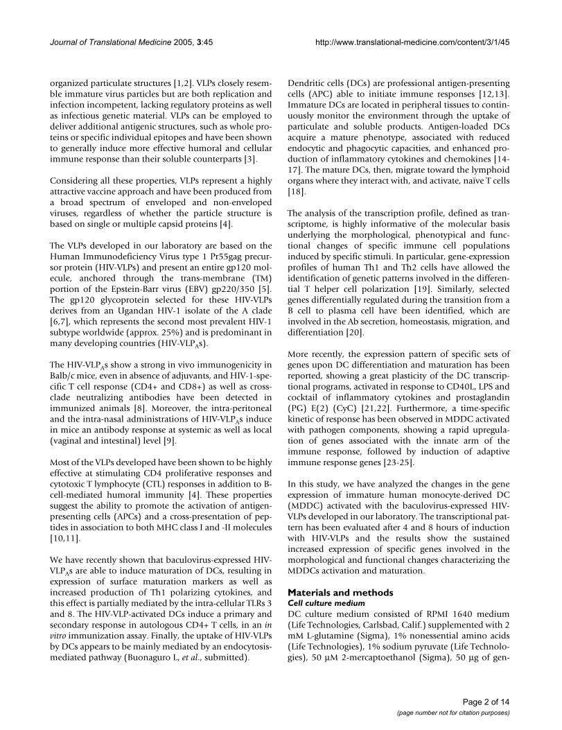
organized particulate structures [1,2]. VLPs closely resem-ble immature virus particles but are both replication andinfection incompetent, lacking regulatory proteins as wellas infectious genetic material. VLPs can be employed todeliver additional antigenic structures, such as whole pro-teins or specific individual epitopes and have been shownto generally induce more effective humoral and cellularimmune response than their soluble counterparts [3].
Considering all these properties, VLPs represent a highlyattractive vaccine approach and have been produced froma broad spectrum of enveloped and non-envelopedviruses, regardless of whether the particle structure isbased on single or multiple capsid proteins [4].
The VLPs developed in our laboratory are based on theHuman Immunodeficiency Virus type 1 Pr55gag precur-sor protein (HIV-VLPs) and present an entire gp120 mol-ecule, anchored through the trans-membrane (TM)portion of the Epstein-Barr virus (EBV) gp220/350 [5].The gp120 glycoprotein selected for these HIV-VLPsderives from an Ugandan HIV-1 isolate of the A clade[6,7], which represents the second most prevalent HIV-1subtype worldwide (approx. 25%) and is predominant inmany developing countries (HIV-VLPAs).
The HIV-VLPAs show a strong in vivo immunogenicity inBalb/c mice, even in absence of adjuvants, and HIV-1-spe-cific T cell response (CD4+ and CD8+) as well as cross-clade neutralizing antibodies have been detected inimmunized animals [8]. Moreover, the intra-peritonealand the intra-nasal administrations of HIV-VLPAs inducein mice an antibody response at systemic as well as local(vaginal and intestinal) level [9].
Most of the VLPs developed have been shown to be highlyeffective at stimulating CD4 proliferative responses andcytotoxic T lymphocyte (CTL) responses in addition to B-cell-mediated humoral immunity [4]. These propertiessuggest the ability to promote the activation of antigen-presenting cells (APCs) and a cross-presentation of pep-tides in association to both MHC class I and -II molecules[10,11].
We have recently shown that baculovirus-expressed HIV-VLPAs are able to induce maturation of DCs, resulting inexpression of surface maturation markers as well asincreased production of Th1 polarizing cytokines, andthis effect is partially mediated by the intra-cellular TLRs 3and 8. The HIV-VLP-activated DCs induce a primary andsecondary response in autologous CD4+ T cells, in an invitro immunization assay. Finally, the uptake of HIV-VLPsby DCs appears to be mainly mediated by an endocytosis-mediated pathway (Buonaguro L, et al., submitted).
Dendritic cells (DCs) are professional antigen-presentingcells (APC) able to initiate immune responses [12,13].Immature DCs are located in peripheral tissues to contin-uously monitor the environment through the uptake ofparticulate and soluble products. Antigen-loaded DCsacquire a mature phenotype, associated with reducedendocytic and phagocytic capacities, and enhanced pro-duction of inflammatory cytokines and chemokines [14-17]. The mature DCs, then, migrate toward the lymphoidorgans where they interact with, and activate, naïve T cells[18].
The analysis of the transcription profile, defined as tran-scriptome, is highly informative of the molecular basisunderlying the morphological, phenotypical and func-tional changes of specific immune cell populationsinduced by specific stimuli. In particular, gene-expressionprofiles of human Th1 and Th2 cells have allowed theidentification of genetic patterns involved in the differen-tial T helper cell polarization [19]. Similarly, selectedgenes differentially regulated during the transition from aB cell to plasma cell have been identified, which areinvolved in the Ab secretion, homeostasis, migration, anddifferentiation [20].
More recently, the expression pattern of specific sets ofgenes upon DC differentiation and maturation has beenreported, showing a great plasticity of the DC transcrip-tional programs, activated in response to CD40L, LPS andcocktail of inflammatory cytokines and prostaglandin(PG) E(2) (CyC) [21,22]. Furthermore, a time-specifickinetic of response has been observed in MDDC activatedwith pathogen components, showing a rapid upregula-tion of genes associated with the innate arm of theimmune response, followed by induction of adaptiveimmune response genes [23-25].
In this study, we have analyzed the changes in the geneexpression of immature human monocyte-derived DC(MDDC) activated with the baculovirus-expressed HIV-VLPs developed in our laboratory. The transcriptional pat-tern has been evaluated after 4 and 8 hours of inductionwith HIV-VLPs and the results show the sustainedincreased expression of specific genes involved in themorphological and functional changes characterizing theMDDCs activation and maturation.
Materials and methodsCell culture mediumDC culture medium consisted of RPMI 1640 medium(Life Technologies, Carlsbad, Calif.) supplemented with 2mM L-glutamine (Sigma), 1% nonessential amino acids(Life Technologies), 1% sodium pyruvate (Life Technolo-gies), 50 µM 2-mercaptoethanol (Sigma), 50 µg of gen-
Page 2 of 14(page number not for citation purposes)

Journal of Translational Medicine 2005, 3:45 http://www.translational-medicine.com/content/3/1/45
tamicin (Life Technologies) per ml, and 10% fetal calfserum (Life Technologies).
DC preparation and treatmentAll human specimens were obtained under informed con-sent, as approved by the University of Maryland BaltimoreInstitutional Review Board. Monocyte-derived DCs weregenerated as described previously (17), with minor mod-ifications. Briefly, human peripheral blood mononuclearcells were isolated, from three independent normalhealthy donors, by Ficoll-Hypaque density gradient cen-trifugation and were enriched for CD14+ monocytes bynegative selection with a cocktail of monoclonal antibod-ies from StemCell Technologies (Vancouver, BritishColumbia, Canada), according to the instructions of themanufacturer. Typically, greater than 80% of the cellswere CD14+ after enrichment (data not shown). The iso-lated monocytes were allowed to adhere to plastic by plat-ing 106 cells per/ml in RPMI 1640 medium for 2 h.Adherent monocytes were washed with RPMI 1640medium and were then cultured for 6 days at 106 cells per/ml in DC culture medium supplemented with 50 ng ofrecombinant GM-CSF (rGM-CSF, R&D Systems, Minne-apolis, Minn.) per ml and 1,000 U of recombinant IL-4(rIL-4; R&D Systems, Minneapolis, Minn.) per ml.
After 6 days in culture, MDDCs were pulsed with either 5µg/ml of HIV-VLPs or 1 µg/ml of LPS for 4 and 8 hours,
for gene microarray analysis, and for 16 hours for matura-tion and activation phenotype analysis.
Analysis of DC phenotypeDCs were incubated for 30 min at 4°C with murine mon-oclonal antibodies specific for CD80, CD83, CD86, andHLA-DR (BD Pharmingen, San Diego, CA), washed andthen fixed with 2% paraformaldehyde for analysis with aFACScalibur flow cytometer (BD Pharmingen). Data anal-ysis was carried out with FlowJo software (Tree Star Inc.,San Carlos, CA). The fraction of MDDCs that respondedby upregulation of activation markers on the cell surfacewas calculated by overlaying the histograms of treated anduntreated MDDCs and Overton subtraction of the curves.
RNA preparation and microarray hybridizationDCs were harvested, washed twice in PBS and lysed in 350ul RLT buffer with fresh addition of 2-Mercaptoethanolper each well of the 6-well plate. Total RNA was isolatedusing RNeasy minikits (Qiagen), according to the manu-facturer's protocol, and RNA quality and quantity was esti-mated by Agilent Bioanlayzer (Agilent Technologies, PaloAlto, CA) and NonoDrop. Amplified antisense RNA(aRNA) was obtained from total RNA (0.5–3 µg) via tworound of in vitro transcription, according the protocolpreviously described by us [26]. 6 ug of amplified testsamples aRNA were labeled with Cy5 (Amersham) whilethe same amount of reference sample (pooled normal
Maturation of DCs by baculovirus-expressed HIV-VLPsFigure 1Maturation of DCs by baculovirus-expressed HIV-VLPs. Immature MDDCs were incubated in the presence of the indi-cated stimulus for 16 hours. The expression of CD80, CD83, CD86 and HLA-DR was analyzed on fixed cells by FACScalibur flow cytometer and data analysis was carried out with FlowJo software. The results of a representative experiment are shown; the shadowed curve represents the untreated cells.
PBS
CD 80 CD 83 CD 86 HLA-DR
100 101 102 103 1040
20
40
60
80
100
100 101 102 103 1040
20
40
60
80
100
100 101 102 103 1040
20
40
60
80
100
100 101 102 103 1040
20
40
60
80
100
%o
fM
ax
%o
fM
ax
%o
fM
ax
%o
fM
ax
HIV-VLPs
100 101 102 103 1040
20
40
60
80
100
100 101 102 103 1040
20
40
60
80
100
100 101 102 103 1040
20
40
60
80
100
100 101 102 103 1040
20
40
60
80
100
%o
fM
ax
%o
fM
ax
%o
fM
ax
%o
fM
ax
Page 3 of 14(page number not for citation purposes)

Journal of Translational Medicine 2005, 3:45 http://www.translational-medicine.com/content/3/1/45
donor PBMCs) was labeled with Cy3. Test-reference sam-ple pairs were mixed and co-hybridized to 17K cDNAmicroarrays [27].
Microarrays and statistical analysesHybridized arrays were scanned at 10-µm resolution on aGenePix 4000 scanner (Axon Instruments) at variablePMT voltage to obtain maximal signal intensities with lessthan 1% probe saturation. Resulting jpeg and data fileswere deposited at microarray data base (mAdb) http://nciarray.nci.nih.gov and retrieved after median centered, fil-tering of intensity (>300) and spot elimination (bad andno signal). Data were further analyzed using Cluster andTreeView software [27] and Partek Pro software (Partek).
Subsequent low-stringency filtering (80% gene presenceacross all experiments and at least one experiment withratio fold change >3), 3,119 genes were selected for fur-ther analysis. Hierarchical cluster analysis was conductedon these genes according to Eisen et al. [28]; differentialexpressed genes were visualized by Treeview and dis-played according to the central method [29].
ResultsBaculovirus-HIV-VLP induces a maturation phenotype of DCsImmature MDDCs were obtained from three independentdonors and, after 6 days of culture in IL-4- and GM-CSF-enrichment medium, were incubated with 5 µg/ml of bac-ulovirus-expressed HIV-VLPs or 1µg/ml of LPS. In parallel,control MDDCs were loaded with PBS. After a 16 hr-induction, the expression of surface maturation/activa-tion markers, such as CD80, CD83, CD86 and HLA-DR,was examined. The expression of all the four markers wasupregulated by treatment with HIV-VLPs, compared toPBS (Fig. 1). The level of cytokines involved in the Th1/Th2 polarization was assessed in the supernatant ofMDDCs loaded with HIV-VLPs or LPS. In particular, theTNF-α, IL-12 p70, IL-10 were produced at higher levels inHIV-VLP-loaded MDDCs compared to LPS-loadedMDDCs (data not shown).
These results indicate that baculovirus HIV-VLPs induce aspecific MDDC maturation pathway, distinct from theone induced by the LPS.
Pattern of MDDCs response to HIV-VLPsGene expression profiles were generated from treated andcontrol MDDC harvested 4 and 8 hrs after stimulation.These time points were selected to evaluate a possiblebiphasic response, as described for LPS-induced DCs andmononuclear phagocytes (MPs) [23,25]. Amplified anti-sense RNA (aRNA) was obtained from total RNA extracts[26] and hybridized to a custom-made 17,000 (17K)-clone cDNA microarray chip enriched with genes relevantto immune function. Stringent filtering were furtherapplied to eliminate genes with missing value in >20% ofall the experiments and >3 fold change in at least oneexperiment. The remaining 3,119 genes were thus used forstatistic analysis.
Unsupervised cluster analysis obtained on MDDCs stimu-lated for 4 and 8 hr with HIV-VLPs or LPS shows the seg-regation of the untreated (PBS) and the treated (HIV-VLPsor LPS) MDDCs in two independent clusters. However,within the treated MDDCs cluster, the samples subclus-tered according to the treatment (HIV-VLPs vs LPS), withthe exception of a single HIV-VLP-treated sample whichformed an independent cluster closer to the LPS-treatedsample, indicating the induction of two distinct transcrip-
Unsupervised hierarchical clustering of all filtered dataFigure 2Unsupervised hierarchical clustering of all filtered data. The clusterogram represents 3,119 genes obtained by Eisen hierarchical clustering of the complete 17K dataset fil-tered for genes that are expressed in a minimum of 80% of the samples 4 and 8 h after HIV-VLPs or LPS stimulation. The PBS treatments was evaluated after 8 h of stimulation. The clustering is defined by the dendrogram and each treatment/time point is represented by a single branch.
PBS (CTR)LPSVLP
DC#8023-PBS
DC#8002-PBS
DC#8016-PBS
DC#8002-VLP8h
DC#8002-VLP4h
DC#8016-VLP8h
DC#8016-VLP4h
DC#8023-VLP4h
DC#8002-LPS8h
DC#8016-LPS8h
DC#8016-LPS4h
DC#8002-LPS4h
DC#8023-LPS8h
DC#8023-LPS4h
DC#8034-VLP8h
DC#8034-VLP4h
Page 4 of 14(page number not for citation purposes)

Journal of Translational Medicine 2005, 3:45 http://www.translational-medicine.com/content/3/1/45
tion machinery. Furthermore, while the HIV-VLP-treatedMDDCs clustered according to the donor, the LPS-treatedMDDCs clustered preferentially according to the treat-ment duration (4 or 8 hr) of stimulation (Fig. 2). Thiswould suggest an individual donor susceptibility to HIV-VLP-treatment and a possible delayed transcription or sec-ondary response to LPS treatment.
Gene expression changes induced in MDDCs by HIV-VLP treatmentThe differential gene expression in HIV-VLP-treatedMDDCs, compared to either LPS- or PBS-treated MDDCs,was considered statistically significant only when sup-ported by a p < 0.005.
Treatment-induced changes in gene profiling were ana-lyzed using student t test, based on simple two-treatmentcomparison or one versus a combined two-treatmentcomparison. Considering only genes showing at least a 2-fold modulation (increase or reduction) in the transcrip-
tional levels, it has been possible to identify unique andcommon genes in the profile induced by the differenttreatments. In particular, the HIV-VLP treatment, inducedthe upregulation of 140 and 72 genes as well as the down-regulation of 108 and 46 genes, compared to PBS and LPSrespectively. A sub-set of upregulated and downregulatedgenes are specific to the HIV-VLP treatment (Fig. 3), indi-cating that, besides transcriptional patterns shared withthe LPS treatment, HIV-VLPs induce a specific reprogram-ming of the MDDCs transcriptional profile.
Visualization of the gene expression among differenttreatments revealed a transcriptional profile pattern inHIV-VLP-induced MDDCs distinct from either PBS or LPS(4 hr and 8 hr) induction (Fig. 4A). Furthermore, cluster-ing analysis performed based on 217 genes selectivelyexpressed (p < 0.005) in HIV-VLP induced MDDCs, com-pared to LPS treatment, confirmed a distinct transcrip-tional profiles signature according to the treatment,regardless of the length of stimulation (Fig. 4B).
Pattern of gene expression in human monocyte-derived DCsFigure 3Pattern of gene expression in human monocyte-derived DCs. Regulated genes by HIV-VLPs or LPS treatment in MDDCs, showing at least a 2-fold modulation (up or downregulation), have been evaluated. Each circle represents the whole set of genes identified in the indicated comparisons, based on the 2-fold modulation parameter. Numbers in overlapping regions represent common regulated genes. Numbers in non-overlapping regions represent unique regulated genes. Circles are drawn in arbitrary scale.
UpregulationUpregulation
VLP vs PBSVLP vs PBS
DownregulationDownregulation
VLP vs LPSVLP vs LPS
LPS vs PBSLPS vs PBS
2626
1818
7979
2727
6262
9898
5454
222626
165165
LPS vs PBSLPS vs PBSVLP vs LPSVLP vs LPS
VLP vs PBSVLP vs PBS
1616
22
Page 5 of 14(page number not for citation purposes)

Journal of Translational Medicine 2005, 3:45 http://www.translational-medicine.com/content/3/1/45
Pathways modulation in MDDCs in response to HIV-VLPsGene expression changes in response to stimuli are gener-ally pathways directed. In order to analyse pathways mod-ulated in HIV-VLPs induced MDDCs, we dissected genesaccording to cluster nodes derived on the basis of expres-sion similarity. Supervised cluster analysis based on the3,119 genes were further analyzed according to patternrecognition. Genes uniquely upregulated in MDDCs byHIV-VLPs only or by HIV-VLPs and LPS (either at 4 h or 8h post-induction) are indicated in Fig. 5A and 5B respec-tively. Similarly, nodes were identified including genesdown-regulated by HIV-VLPs only or by HIV-VLPs andLPS at 4 h post-induction (Fig. 5C top and bottom, respec-tively). Most of the genes present in these nodes arestrictly associated to the functional and morphological
changes characterizing the MDDCs maturation and acti-vation process.
In this regard, the MDDCs transcriptional profile was ana-lyzed on the basis of the cellular pathways modulated bythe HIV-VLP-treatment, compared to either PBS or LPStreatment. In respect to the main focus of this study, onlythe pathways involved in the MDDCs-activation by HIV-VLPs have been evaluated in more detail. These can bedivided into main super-families concerning the cell rep-lication/death, the transcriptional control, the phagocyto-sis and pathogen recognition, the immunologicalfunction of dendritic cells, the migration and the controlof the inflammatory responses (Table I).
Supervised hierarchical clustering of genes differentially expressed in HIV-VLP-treated MDDCsFigure 4Supervised hierarchical clustering of genes differentially expressed in HIV-VLP-treated MDDCs. The clustero-grams represent an Eisen hierarchical clustering of the 281 genes differentially expressed (p < 0.005) in HIV-VLPs-treated MDDCs, compared to PBS treatment (A), and of 217 genes differentially expressed (p < 0.005) in HIV-VLPs-treated MDDCs, compared to LPS treatment (B). The clustering is defined by the dendrogram on the top and on the side of the clusterogram.
DC#8016-VLP8h
DC#8034-VLP8h
DC#8034-VLP4h
DC#8002-VLP8h
DC#8002-VLP4h
DC#8023-VLP4h
DC#8016-VLP4h
DC#8023-PBS
DC#8002-PBS
DC#8016-PBS
DC#8023-LPS4h
DC#8016-LPS4h
DC#8002-LPS4h
DC#8016-LPS8h
DC#8002-LPS8h
DC#8023-LPS8h
PBS (CTR)LPSVLP
PBS (CTR)LPS 4h
VLP
LPS 8h
DC#8016-LPS8h
DC#8002-LPS8h
DC#8023-LPS8h
DC#8016-PBS
DC#8023-PBS
DC#8002-PBS
DC#8016-LPS4h
DC#8002-LPS4h
DC#8023-LPS4h
DC#8034-VLP8h
DC#8034-VLP4h
DC#8023-VLP4h
DC#8016-VLP8h
DC#8016-VLP4h
DC#8002-VLP8h
DC#8002-VLP4h
A B
Page 6 of 14(page number not for citation purposes)
Journal of Translational Medicine 2005, 3:45 http://www.translational-medicine.com/content/3/1/45
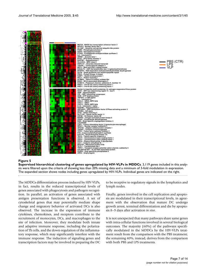
The MDDCs differentiation process induced by HIV-VLPs,in fact, results in the reduced transcriptional levels ofgenes associated with phagocytosis and pathogen recogni-tion. In parallel, an activation of genes associated withantigen presentation functions is observed. A set ofcytoskeletal genes that may potentially mediate shapechange and migratory behavior of activated DCs is alsoobserved. The increase in the expression of immunecytokines, chemokines, and receptors contribute to therecruitment of monocytes, DCs, and macrophages to thesite of infection. Moreover, they modulate both innateand adaptive immune response, including the polariza-tion of Th cells, and the down-regulation of the inflamma-tory response, which may significantly interfere with theimmune response. The induction of signaling genes andtranscription factors may be involved in preparing the DC
to be receptive to regulatory signals in the lymphatics andlymph nodes.
Finally, genes involved in the cell replication and apopto-sis are modulated in their transcriptional levels, in agree-ment with the observation that mature DC undergogrowth arrest, terminal differentiation and die by apopto-sis 8–9 days after activation in vivo.
It is not unexpected that many pathways share same geneswith intra-cellular functions involved in several biologicaloutcomes. The majority (60%) of the pathways specifi-cally modulated in the MDDCs by the HIV-VLPs treat-ment result from the comparison with the PBS treatment;the remaining 40%, instead, derives from the comparisonwith both PBS and LPS treatments.
Supervised hierarchical clustering of genes upregulated by HIV-VLPs in MDDCsFigure 5Supervised hierarchical clustering of genes upregulated by HIV-VLPs in MDDCs. 3,119 genes included in this analy-sis were filtered upon the criteria of showing less than 20% missing data and a minimum of 3-fold modulation in expression. The expanded section shows nodes including genes upregulated by HIV-VLPs. Individual genes are indicated on the right.
PBS (CTR)LPSVLP
MEF2A - MADS box transcription enhancer factor 2NFE2L2 - Nuclear factor-like 2DC-UbP – Dendritic cell-derived ubiquitin-like proteinPGM3 - Phosphoglucomutase 3DGKH - Diacylglycerol kinasePTGS2 - Prostaglandin-endoperoxidase synthase 2IL8 – Interleukin 8ARRB1 – Arrestin beta 1BTG3 – BTG family member 3SOSTM1 – Sequestosome 1HLA-DOA – MHC class IIHSPC121 – Butyrate-induced transcriptTADA3L – Transcriptional adaptor 3MST4 – Mst3 and SOK1-related kinaseB4GALT5 – UDP-Gal:betaGlcNAc bea 1.4-galactosyltransferaseMTHFD2 – NAD-dependent methylenetetrahydrofolate deydrogenaseSGTB – Small glutamine-rich tetratricopeptide repeatPRKX – Protein Kinase, X-linkedN4BP1 – Nedd4 binding protein 1PELI1 – Pellino homolog 1SC4MOL – Sterol-C4-methyl oxidase-likeBAG3 – BCL2-associated athanogene 3KCNK10 – Potassium channel, subfamily K, member 10PBEF1 – Pre-B-cell colony enhancing factor 1PBEF1 – Pre-B-cell colony enhancing factor 1
Similar to tripartite motif-containing 16; estrogen-responsive B box proteinSOX4 – SRY (sex determining region Y) – box 4ADM – AdrenomedullinCIR – CBF1 interacting corepressorGEM – GTP binding proteinTNFSF9 – 4-1BB ligandIL6 – Interleukin 6CXCL3 – GRO3, MIP2 betaCXCL1 – GRO1CXCL3 – GRO3, MIP2 betaIL1A – Interleukin 1 alphaSOSTM1 – Sequestosome 1ARFGAP3 – ADP-ribosylation factor GTPase activating protein 3NFKBIA – IkB alphaDTX2 – Deltex homolog 2SLC11A2 – Solute carrier family 11H3F3B – H3 Histone, family 3BMAPK6 – mitogen-activated protein kinase 6OGDH – Oxoglutarate dehydrogenaseBAI3 – Brain-specific angiogenesis inhibitor 3LOC440309CSF2 – Colony stimulating factor 2 (granulocyte-macrophage)NR3C1 – Nuclear receptor subfamily 3APA1S2 – Adaptor-related protein complex 1YWHAZ – Tyrosine 3-monooxygenaseTFPI2 – Tissue factor pathway inhibitor 2PTS – 6-pyruvoyltetrahydropterin synthaseSTK17A – Serine/threonine kinase 17aTBCA – Tubulin-specific chaperon aC8orf1 – Chromosome 8 open reading frame 1C8orf1 – Chromosome 8 open reading frame 1RHCG – Rhesus blood group, C glycoproteinCA11 – Carbonic anhydrase XIRPS15A – Ribosomal protein S15aTKTL1 – Transketolase-like 1TRPC1 – Transient receptor potential cation channel, subfamily CGALNAC4S-6ST – B cell RAG associated proteinDUSP6 – Dual specificity phosphatase 6AREG - Amphyregulin
Page 7 of 14(page number not for citation purposes)

Page
8 o
f 14
(pag
e nu
mbe
r not
for c
itatio
n pu
rpos
es)
enome Anatomy Project at http://o consideration. Upregulated genes in bold
HIV-VLP vs PBS HIV-VLP vs LPS
/SignalingPathway FOXO3A
NFKB1CASP9
YWHAHCell Surface CD40
FCGR2AICAM1LTBRITGALPTPRC
NFKBIA
g Pathway CD40DUSP1
TNFAIP3TRAF6IKBKB
DUSP1MAP3K1NFKBIATNFAIP3
immune st target cells
ICAM1B2M
LTBRITGAL
hemokine n in HMC-1
FPR1MAP2K2NFKB1CAMK1MAP2K6MAPK1
its Surface ICAM1ICAM2CD44LTBRSELECD83
HLA-DOAITGAL
PECAM1ell Surface CD8A
ICAM1LTBR
CD2 ITGALPTPRC
Surface ICAM1LTBRCD2CD4
ITGALPTPRC
endent se
IFNGNFKB1TNFSF9TRAF2
Jour
nal o
f Tra
nsla
tiona
l Med
icin
e 20
05, 3
:45
http
://w
ww
.tran
slat
iona
l-med
icin
e.co
m/c
onte
nt/3
/1/4
5
Table 1: Pathways involved in the HIV-VLPs-activated MDDCs. The pathways are derived from the BioCarta through the Cancer Gcgap.nci.nih.gov/Pathways/BioCarta_Pathways. Genes with at least a 2-fold modulation (up or downregulation) have been taken intand underline; Downregulated genes in plain text.
Pathways HIV-VLP vs PBS HIV-VLP vs LPS Pathways HIV-VLP vs PBS HIV-VLP vs LPS Pathways
Cell cycle, death Transcription ImmunologyCadmium induces DNA synthesis and proliferation in macrophages
NFKB1MAPK1
TNF Acetylation and Deacetylation of RelA in The Nucleus
NFKB1FADD
NFKBIATNFFADD
AKT Signaling
Caspase Cascade in Apoptosis BIRC3CASP7CASP2CASP9PARP1
Activation of PKC through G protein coupled receptor
GNAQNFKB1
B Lymphocyte Molecules
Free Radical Induced Apoptosis GSRIL8
NFKB1
Chaperones modulate interferon Signaling Pathway
DNAJA3IFNG
NFKB1
IFNGLIN7A
NFKBIARB1TNF
CD40L Signalin
Induction of apoptosis through DR3 and DR4/5 Death Receptors
BIDCASP10CASP7TRAF2CASP9FADD
Double Stranded RNA Induced Gene Expression
NFKB1 CTL mediated response again
Neuropeptides VIP and PACAP inhibit the apoptosis of activated T cells
GNAQNFKB1
PRKAR1BCAMK1
Human Cytomegalovirus and Map Kinase Pathways
MAP2K3NFKB1MAP2K6MAPK1
fMLP induced cgene expressiocells
Regulation of BAD phosphorylation
BCL2L1IGF1R
PRKAR1BMAPK1
RPS6KA1YWHAH
NFkB activation by Nontypeable Hemophilus influenzae
DUSP1EP300
MAP2K3NFKB1NR3C1SMAD4MAP2K6SMAD3TGFBR2
DUSP1IL8
NFKBIASMAD4
TNFIKBKB
MAP2K6SMAD3
Monocyte and Molecules
Role of Mitochondria in Apoptotic Signaling
BCL2L1BID
BIRC3CASP7
ENDOGCASP9PDCD8
NF-kB Signaling Pathway IRAK1NFKB1
TNFAIP3TRAF6FADD
IL1AMAP3K1NFKBIA
TNFTNFAIP3
FADD
T Cytotoxic CMolecules
SODD/TNFR1 Signaling Pathway BIRC3TRAF2FADD
Signal transduction through IL1R
IL1BMAP2K3NFKB1TRAF6IL1RAP
MAP2K6
IL1AMAP3K1NFKBIA
TNFIL1RAP
MAP2K6
T Helper Cell Molecules
Stress Induction of HSP Regulation
CASP9MAPKAPK3
IL1ATNF
CASP9CYC1
TNFR1 Signaling Pathway PAK2TRAF2CASP2FADDPARP1
The 4-1BB-depimmune respon

Page
9 o
f 14
(pag
e nu
mbe
r not
for c
itatio
n pu
rpos
es)
ptor Pathway IRAK1MAP2K3
MAP3K7IP2NFKB1TRAF6MAP2K
enome Anatomy Project at http://o consideration. Upregulated genes in bold
Jour
nal o
f Tra
nsla
tiona
l Med
icin
e 20
05, 3
:45
http
://w
ww
.tran
slat
iona
l-med
icin
e.co
m/c
onte
nt/3
/1/4
5
TNF/Stress Related Signaling MAP2K3NFKB1TANKTRAF2CASP2
MAP2K6
TNFR2 Signaling Pathway DUSP1NFKB1TANK
TNFAIP3TRAF2
DUSP1MAP3K1NFKBIATANK
TNFAIP3TNFRSF1B
Toll-Like Rece
Pathways HIV-VLP vs PBS HIV-VLP vs LPS Pathways HIV-VLP vs PBS HIV-VLP vs LPS
Cytokine Network PhagocytosisCells and Molecules involved in local acute inflammatory response
ICAM1IL8
LTBRCERKLITGAL
Instracellular protein transport
APG9L1RERERNF4
Cytokine Network IFNGIL18IL8
Cytoskeleton
Cytokines and Inflammatory Response
CSF1CSF2IFNGIL7IL8IL15
IL15RAIL23
PTGS2CD4
Cellular motiliyshape ARHGEF2ARPC2
C14orf43FLJ39378MARCKSPDE4BPDE5APLEK
PPP1R15APPP1R15BSLC11A2SLC1A3SLC20A1SLC43A3
IL 17 Signaling Pathway CD8AIL8
CD58CD2CD4
Th1/Th2 Differentiation IL18IL18R
Table 1: Pathways involved in the HIV-VLPs-activated MDDCs. The pathways are derived from the BioCarta through the Cancer Gcgap.nci.nih.gov/Pathways/BioCarta_Pathways. Genes with at least a 2-fold modulation (up or downregulation) have been taken intand underline; Downregulated genes in plain text. (Continued)

Journal of Translational Medicine 2005, 3:45 http://www.translational-medicine.com/content/3/1/45
Functional implementation of activated genes in MDDCs in response to HIV-VLPsThe effects of the HIV-VLP treatment on MDDCs, com-pared to the PBS and LPS treatments, has been analyzedalso considering the individual genes showing increasedtranscriptional levels. The presumed biological implica-tion of selected genes involved in the specific functions ofDendritic Cells as professional APCs, has been evaluatedin more detail according to their function (Table II).
STAT2 is activated upon the binding of type-I IFN, themajor component of the innate immune system, to itsreceptor and participates to the formation of interferon-stimulated gene factor 3 (ISGF3) complex, composed ofSTAT1, STAT2, and IFN regulatory factor 9, which pro-motes serial synthesis of selected proteins that inhibitviral replication [30,31].
Macrophage and Granulocyte-Macrophage Colony Stim-ulating Factor, CSF1 and CSF2, represent the main factorsinvolved in proliferation and differentiation of myeloidlineage progenitor cells [32-35]. They may contribute toan increased production and recruitment of monocytes,DCs, and macrophages to the site of infection.
Formyl Peptide Receptor (FPR) is responsible for the DCchemoattraction to the Gram-negative bacteria Formylpeptide N-formyl-Met-Leu-Phe (fMLP) [35] and to addi-tional pathogen-derived peptides, including HIV [36,37].E-Selectin (SELE) and Chemokine (C-C motif) receptor 7(CCR7) are selectively expressed on mature DCs, follow-ing encounter with pathogens. They favor the interactionwith the endothelial cells as well as the migration fromperipheral tissues to the T cell areas of secondary lym-phoid organs, where they produce regulatory cytokines
Table 2: Functional categories of genes differentially upregulated in HIV-VLPs-induced MDDCs. (+) 1.5-fold upregulation; (++) 2–5-fold upregulation; (+++) >5-fold upregulation.
Pathway HIV-VLP vs PBS HIV-VLP vs LPS Pathway HIV-VLP vs PBS HIV-VLP vs LPS
Cell cycle, death Chemokine/ImmunologyTNF ++ + Innate responseBIRC3 ++ IL8 +++ ++CASP7 + CXCL1 ++ ++BID ++ IL1a ++ ++PAK2 ++ IL1b ++TRAF2 ++ IL6 ++BCL2L1 + CCL4 ++IGF1R ++ IFNG +TNFAIP3 ++ PTGER4TANK ++ Adaptive responsePSEN1 + EBI2 ++TNFRSF1B +++ + ICAM1 ++Transcription B2M +NFKB1 + ReceptorsNFKBIA ++ IL15RA ++IRAK1 + IL18R1 ++MAP3K1 + IL23A ++JUNB + IL2RB ++Signaling FCGR2A ++MAP2K7IP2 + IL7R ++DUSP1 ++ + CD83 ++MAP2K3 + Growth FactorsPAK2 ++ CSF1 ++RB1 + CSF2 +FOXO3A + Migration and
HomeostasisFYN + CCR7 +GNAQ + FPR1 ++MEF2A + SELE ++RAPGEF2 + CD44 ++
LTBR +T-cell activation/polarizationCD40 ++TNFSF9 +IL18 +
Page 10 of 14(page number not for citation purposes)

Journal of Translational Medicine 2005, 3:45 http://www.translational-medicine.com/content/3/1/45
and prime naïve T lymphocytes [38,39]. Lymphotoxin-beta receptor (LTBR) plays a central role in the DC home-ostasis in lymphoid tissues (spleen and LN), promotingand sustaining local DC replication [40]. Furthermore, aset of cytokine receptors that share a common γ chain (IL-2R, IL-7R, IL-15R, and IL-4R) were also induced and theirexpression may allow DCs to respond to lymphocyte-derived interleukins within the lymph node.
Beta-2-microglobulin (B2M) is one of the components ofthe multimolecular peptide-loading MHC class-I complexwithin the endoplasmic reticulum (ER) of DCs [41], rep-
resenting the essential process for antigen presentation tocytotoxic T lymphocytes.
CD40 and TNFSF9 may act as co-stimulatory moleculeswhich, interacting with the corresponding T cell expressedligands, have been shown to play a relevant role in theactivation of T cells. In particular, the CD40:CD40L inter-action activates the CD4+ cells to promote the sensitiza-tion of CD8+ cells as well as to induce memory CD8+ Tcell proliferation [42,43]. ICAM-1 is required on the sur-face of exosomes produced by mature DCs to prime naïveT cells and trigger effector T-cell response [44].
Supervised hierarchical clustering of genes upregulated by HIV-VLPs and LPS in MDDCsFigure 6Supervised hierarchical clustering of genes upregulated by HIV-VLPs and LPS in MDDCs. 3,119 genes included in this analysis were filtered upon the criteria of showing less than 20% missing data and a minimum of 3-fold modulation in expression. The expanded section shows nodes including genes upregulated by HIV-VLPs and LPS-4 h (top) or by HIV-VLPs and LPS (bottom). Individual genes are indicated on the right.
PBS (CTR)LPS 4h
VLPLPS 8h
Tumor Necrosis Facto 2
TNF – Tumor necrosis Factor
CD79B – CD79B
F3 – Coagulation factor III
CCRL2 – Chemokine receptor 2
PALM2-AKAP2 – Paralemmin 2
IL23A – Interleukin 23a
ARPC2 – Actin related protein 2
BCL2A1 – Bcl-2 related protein
BCL2A1 – BCL2-related protein A1
NFKBIZ – Nuclear factor
RASGRP1- RAS guanyl releasing protein 1
WTAP – Wilms tumor 1 associate protein
ARHGAP4 – Rho GTPase activating protein 4
CSF1 – colony stimulating factor 1 (macrophage)
EHD1 – EH-domain containing 1EHD1 - EH-domain contaning 1TNIP2 – TNFAIP3 interacting protein 2NINJ1 – Ninjurin 1CCL5 – Chemokine ligand 5CSF1 – Colony stimulating factor 1 (macrophage)HSGT1 – Suppressor of S. CerevisiaeASCL1 – Acyl-CoA synthetase long-chain family member 1PLEK – PleckstrinKLRC3 – Killer cell lectin-like receptor subfamily C, member 2PDE5A – Phosphodiesterase 5ASDC4 – Syndecan-4ITGAV – integrin, alpha VICAM1 – CD54ICAM1 – CD54DUSP5 – dual specificity phosphatase 5Putative lymphocyte G0/G1 switch geneCCL3 – LD78 betaCCL3 – Chemokine ligand 3CCL2 – MCP-1CCL4 – MIP-1 beta
CCL20 – Chemokine ligand 20TNFAIP3 – Tumor necrosis factor, alpha-induced protein 3SOD2 – Superoxidase dismutase 2SOD2 – Superoxidase dismutase 2CREM – cAMP responsive element modulatorUNG – uracil DNA-glycosylaseCD44 – Pgp-1 extracellular matrix receptorSGPP2 – Sphingosine-1-phosphataseTNFAIP3 – Tumor necrosis factor, alpha-induced protein 3ITGA6 – integrin alpha 6DEAF1 – Deformed epidermal autoregulatory factor 1
BIRC3 – Baculovirus IAP repeat-containing 3SEMA5A – Sema domain, seven thromspondin repeatsPTPLB – Protein tyrosine phosphatase-likeIBRDC2 – IBR domain containing 2KYNU – KynureninaseKYNU - Kynureninase
Page 11 of 14(page number not for citation purposes)
Journal of Translational Medicine 2005, 3:45 http://www.translational-medicine.com/content/3/1/45
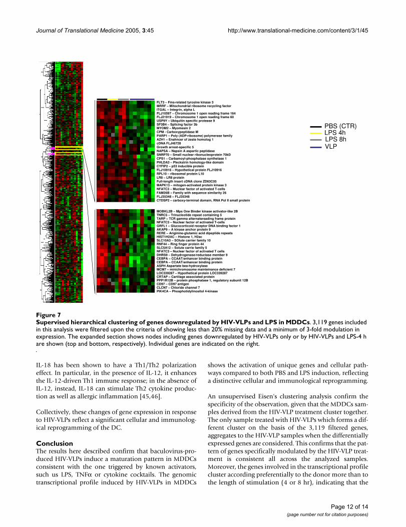
IL-18 has been shown to have a Th1/Th2 polarizationeffect. In particular, in the presence of IL-12, it enhancesthe IL-12-driven Th1 immune response; in the absence ofIL-12, instead, IL-18 can stimulate Th2 cytokine produc-tion as well as allergic inflammation [45,46].
Collectively, these changes of gene expression in responseto HIV-VLPs reflect a significant cellular and immunolog-ical reprogramming of the DC.
ConclusionThe results here described confirm that baculovirus-pro-duced HIV-VLPs induce a maturation pattern in MDDCsconsistent with the one triggered by known activators,such us LPS, TNFα or cytokine cocktails. The genomictranscriptional profile induced by HIV-VLPs in MDDCs
shows the activation of unique genes and cellular path-ways compared to both PBS and LPS induction, reflectinga distinctive cellular and immunological reprogramming.
An unsupervised Eisen's clustering analysis confirm thespecificity of the observation, given that the MDDCs sam-ples derived from the HIV-VLP treatment cluster together.The only sample treated with HIV-VLPs which forms a dif-ferent cluster on the basis of the 3,119 filtered genes,aggregates to the HIV-VLP samples when the differentiallyexpressed genes are considered. This confirms that the pat-tern of genes specifically modulated by the HIV-VLP treat-ment is consistent all across the analyzed samples.Moreover, the genes involved in the transcriptional profilecluster according preferentially to the donor more than tothe length of stimulation (4 or 8 hr), indicating that the
Supervised hierarchical clustering of genes downregulated by HIV-VLPs and LPS in MDDCsFigure 7Supervised hierarchical clustering of genes downregulated by HIV-VLPs and LPS in MDDCs. 3,119 genes included in this analysis were filtered upon the criteria of showing less than 20% missing data and a minimum of 3-fold modulation in expression. The expanded section shows nodes including genes downregulated by HIV-VLPs only or by HIV-VLPs and LPS-4 h are shown (top and bottom, respectively). Individual genes are indicated on the right.
PBS (CTR)LPS 4h
VLPLPS 8h
FLT3 – Fms-related tyrosine kinase 3MRRF – Mitochondrial ribosome recycling factorITGAL – Integrin, alpha LFLJ10597 – Chromosome 1 open reading frame 164FLJ21919 – Chromosome 1 open reading frame 60USP9Y – Ubiquitin specific protease 9SF3B4 – Splicing factor 3bMYOM2 – Myomesin 2CPM - Carboxypeptidase MPARP1 – Poly (ADP-ribosome) polymerase familyAZH1 – Enahncer of zeste homolog 1cDNA FLJ46728Growth arrest-specific 5NAPSA – Napsin A aspartic peptidaseSNRP70 – Small nuclear ribonucleoprotein 70kDCPS1 – Carbamoyl-phosphatase synthetase 1PHLDA3 – Pleckstrin homology-like domainCYFIP2 – p53 inducible proteinFLJ10916 – Hypothetical protein FLJ10916RPL10 – ribosomal protein L10LR8 – LR8 proteinFull-length insert cDNA clone ZD63C05MAPK13 – mitogen-activated protein kinase 3NFATC3 – Muclear factor of activated T-cellsFAM26B – Family with sequence similarity 26FLJ35348 – FLJ35348CTDSP2 – carboxy-terminal domain, RNA Pol II small protein
MOBKL2B – Mps One Binder kinase activator-like 2BTNRC5 – Trinucleotide repeat containing 5TARP – TCR gamma alternatereading frame proteinNFATC3 – Nuclear factor of activated T-cellsGRFL1 – Glucocorticoid receptor DNA binding factor 1AKAP9 – A kinase anchor protein 9RERE – Ariginine-glutamic acid dipeptide repeatsHIST1H2AC – Histone 1, H2acSLC10A3 – SOlute carrier family 10RNF44 – Ring finger protein 44SLC5A12 – Solute carrie family 5NFATC3 – Nuclear factor of activated T cellsDHRS9 – Dehydrogenase/reductase member 9CEBPA – CCAAT/enhancer binding proteinCEBPA – CCAAT/enhancer binding proteinASPH Aspartate bea-hydroxylaseMCM7 – minichromosome mainteinance deficient 7LOC339287 – Hypothetical protein LOC339287CRTAP – Cartilage associated proteinPPP1R12B – protein phosphatase 1, regulatory subunit 12BCD97 – CD97 antigenCLCN7 – Chloride channel 7PIK4CA – Phosphotidylinositol 4-kinase
Page 12 of 14(page number not for citation purposes)
Journal of Translational Medicine 2005, 3:45 http://www.translational-medicine.com/content/3/1/45
induced modulation of transcriptional activity is effectiveafter as early as 4 hours and is persistent for the length ofthe observation. Furthermore, the observed differentdonor susceptibility to HIV-VLP-treatment may suggestthe possibility to select specific gene patterns useful for theidentification of "responsive" vaccinees. This would beextremely helpful in understanding the eventual failure inindividuals enrolled in the clinical trials.
Among the pathways and specific genes activated inMDDCs treated with HIV-VLPs, those directly involved inthe biological functions as antigen presenting cells (APCs)have been analyzed in detail. The full functional matura-tion and activation of MDDCs by HIV-VLPs has been con-firmed and, in particular, the activation of genes involvedin cellular control (proliferation, differentiation, migra-tion and homeostasis) as well as in functional activity(antigen presentation, T cell activation and Th polariza-tion) has been observed.
The ability of the HIV-VLP, as exogenous antigen, toinduce a CD8+ cytotoxic T lymphocyte (CTL) response, aspreviously demonstrated in vivo, is supported by the acti-vation of genes (such as beta 2 microglobulin) involved inthe antigen presentation within the context of major his-tocompatibility complex (MHC) class I molecules as wellas in the Th1 polarization (IFN gamma and IL-18).
The presentation of an exogenous antigen in the contextof major histocompatibility complex (MHC) class I mole-cules, to induce a CD8+ cytotoxic T lymphocyte (CTL)response, is referred to as "cross-presentation", in alterna-tive to the "direct" or "classic" presentation route forendogenously synthesized proteins. The cross-presenta-tion is a strategy employed only by DCs, among all theantigen presenting cells, to ensure an anti-virus CTLresponse also for those viruses which do not infect DCs[47]. The CD8+ DC subset seems to be primarily involvedin this strategy [48,49], but the molecular mechanismsunderlying the cross-presentation are not yet fully eluci-dated [50-52].
Microarray approach allows quantitative and simultane-ous analysis of gene expression of a large amount of genesand the systematic studies of expression patterns areextremely useful for identify molecular events and keypathways involved in cellular functions induced by spe-cific stimuli. In this study, informative data on the globalpattern of gene expression underlying the activation ofMDDCs by HIV-VLPs at the early stages of the immuneresponse have been obtained. They may be extremelyhelpful for the identification of exclusive activation mark-ers to trace the biological effects of modifications/optimi-zations of the HIV-VLP vaccination strategy.
References1. Miyanohara A, Imamura T, Araki M, Sugawara K, Ohtomo N, Matsub-
ara K: Expression of hepatitis B virus core antigen gene inSaccharomyces cerevisiae: synthesis of two polypeptidestranslated from different initiation codons. J Virol 1986,59:176-180.
2. Gheysen D, Jacobs E, de Foresta F, Thiriart C, Francotte M, Thines D,De Wilde M: Assembly and release of HIV-1 precursor Pr55gag
virus-like particles from recombinant baculovirus-infectedinsect cells. Cell 1989, 59:103-112.
3. Buonaguro L, Tornesello ML, Buonaguro FM: Virus-like particles(VLPs) as anti-viral vaccines: an effective approach for a HIV-1 vaccine strategy. ASHI Quarterly 3rd Quarter 2005, 10:.
4. Noad R, Roy P: Virus-like particles as immunogens. TrendsMicrobiol 2003, 11:438-444.
5. Buonaguro L, Buonaguro FM, Tornesello ML, Mantas D, Beth-GiraldoE, Wagner R, Michelson S, Prevost M-C, Wolf H, Giraldo G: Highefficient production of Pr55gag Virus-like Particles express-ing multiple HIV-1 epitopes, including a gp120 proteinderived from an Ugandan HIV-1 isolate of subtype A. AntiviralResearch 2001, 49:35-47.
6. Buonaguro L, Del Gaudio E, Monaco M, Greco D, Corti P, Beth-Giraldo E, Buonaguro FM, Giraldo G: Heteroduplex mobilityassay and phylogenetic analysis of V3 region sequences ofHIV 1 isolates from Gulu – Northern Uganda. J Virol 1995,69:7971-7981.
7. Buonaguro L, Buonaguro FM, Russo F, Tornesello ML, Beth-GiraldoE, Wagner R, Wolf H, Giraldo G: A novel gp120 sequence froman HIV-1 isolate of the A clade identified in North Uganda.AIDS Res Hum Retroviruses 1998, 14:1287-1289.
8. Buonaguro L, Racioppi L, Tornesello ML, Arra C, Visciano ML, Bir-yahwaho B, Sempala SDK, Giraldo G, Buonaguro FM: Induction ofneutralizing antibodies and CTLs in Balb/c mice immunizedwith Virus-like Particles presenting a gp120 molecule from aHIV-1 isolate of clade A (HIV-VLPAs). Antiviral Research 2002,54:189-201.
9. Buonaguro L, Visciano ML, Tornesello ML, Tagliamonte M, Bir-yahwaho B, Buonaguro FM: Induction of systemic and mucosalcross-clade neutralizing antibodies in BALB/c mice immu-nized with human immunodeficiency virus type 1 clade Avirus-like particles administered by different routes of inoc-ulation. J Virol 2005, 79:7059-7067.
10. Heath WR, Carbone FR: Cross-presentation in viral immunityand self-tolerance. Nat Rev Immunol 2001, 1:126-134.
11. Moron VG, Rueda P, Sedlik C, Leclerc C: In vivo, dendritic cellscan cross-present virus-like particles using an endosome-to-cytosol pathway. J Immunol 2003, 171:2242-2250.
12. Banchereau J, Steinman RM: Dendritic cells and the control ofimmunity. Nature 1998, 392:245-252.
13. Rescigno M, Granucci F, Citterio S, Foti M, Ricciardi-Castagnoli P:Coordinated events during bacteria-induced DC matura-tion. Immunology Today 1999, 20:200-203.
14. Sparwasser T, Koch ES, Vabulas RM, Heeg K, Lipford G, Ellwart JW,Wagner H: Bacterial DNA and immunostimulatory CpG oli-gonucleotides trigger maturation and activation of murinedendritic cells. European Journal of Immunology 1998, 28:2045-2054.
15. Cella M, Salio M, Sakakibara Y, Langen H, Julkunen I, Lanzavecchia A:Maturation, activation and protection of dendritic cellsinduced by double-stranded RNA. J Exp Med 1999,189:821-829.
16. Verdijk RM, Mutis T, Esendam B, Kamp J, Melief CJ, Brand A, GoulmyE: Polyribosinic polyribocytidylic acid (poly(I:C)) induces sta-ble maturation of functionally active human dendritic cells. JImmunol 1999, 163:57-61.
17. Cella M, Scheidegger D, Palmer-Lehmann K, Lane P, Lanzavecchia A,Alber G: Ligation of CD40 on dendritic cells triggers produc-tion of high levels of interleukin-12 and enhances T cell stim-ulatory capacity: T-T help via APC activation. J Exp Med 1996,184:747-752.
18. Sallusto F, Lanzavecchia A: Understanding dendritic cells and Tlymphocyte traffic through the analysis of chemokine recep-tor expression. Immunology Review 2000, 177:134-140.
19. Rogge L, Bianchi E, Biffi M, Bono E, Chang SY, Alexander H, Santini C,Ferrari G, Sinigaglia L, Seiler M, et al.: Transcript imaging of thedevelopment of human T helper cells using oligonucleotidearrays. Nat Genetics 2000, 25:96-101.
Page 13 of 14(page number not for citation purposes)

Journal of Translational Medicine 2005, 3:45 http://www.translational-medicine.com/content/3/1/45
Publish with BioMed Central and every scientist can read your work free of charge
"BioMed Central will be the most significant development for disseminating the results of biomedical research in our lifetime."
Sir Paul Nurse, Cancer Research UK
Your research papers will be:
available free of charge to the entire biomedical community
peer reviewed and published immediately upon acceptance
cited in PubMed and archived on PubMed Central
yours — you keep the copyright
Submit your manuscript here:http://www.biomedcentral.com/info/publishing_adv.asp
BioMedcentral
20. Sciammas R, Davis MM: Modular nature of Blimp-1 in the regu-lation of gene expression during B cell maturation. J Immunol2005, 172:5427-5240.
21. Messmer D, Messmer B, Chiorazzi N: The global transcritpionalmaturation program and stimuli-specific gene expressionprofiles of human myeloid dendritic cells. International Immunol-ogy 2003, 15:491-503.
22. Tureci O, Bian H, Nestle FO, Raddrizzani L, Rosinski JA, Tassis A,Hilton H, Walstead M, Sahin U, Hammer J: Cascades of transcrip-tional induction during dendritic cell maturation revealed bygenome-wide expression analysis. FASEB 2003, 17:836-847.
23. Huang Q, Liu D, Majewski P, Schulte LC, Korn JM, Young RA, LanderES, Hacohen N: The plasticity of dendritic cell responses topatogens and their components. Science 2001, 294:870-875.
24. Langenkamp A, Messi M, Lanzavecchia A, Sallusto F: Kinetics of den-dritic cell activation: impact on priming of Th1, Th2 andpolarization T cells. Nature Immunology 2000, 1:311-316.
25. Nagorsen D, Deola S, Smith K, Wang E, Monsurro V, Zanovello P,Marincola FM, Panelli MC: Polarized monocyte response tocytokine stimulation. Genome Biology 2005, 6:R15.
26. Wang E, Miller LD, Ohnmacht GA, Liu ET, Marincola FM: High-fidel-ity mRNA amplification for gene profiling. Nature Biotechnology2000, 18:457-459.
27. Wang E, Miller LD, Ohnmacht GA, Mocellin S, Perez-Diez A,Petersen D, Zhao Y, Simon R, Powell JI, Asaki E, Alexander HR, DurayPH, Herlyn M, Restifo NP, Liu ET, Rosenberg SA, Marincola FM: Pro-spective molecular profiling of melanoma metastases sug-gests classifiers of immune responsiveness. Cancer Res 2005,62:3581-3586.
28. Eisen MB, Spellman PT, Brown PO, Botstein D: Cluster analysisand display of genome-wide expression patterns. Proc NatlAcad Sci USA 1998, 95:14863-14868.
29. Ross DT, Scherf U, Eisen MB, Perou CM, Rees C, Spellman PT, IyerV, Jeffrey SS, Van de Rijn M, Waltham M, et al.: Systematic varia-tion in gene expression patterns in human cancer cell lines.Nat Genetics 2000, 24:227-235.
30. Aaronson DS, Horvath CM: A road map for those who don'tknow JAK-STAT. Science 2002, 296:1653-1655.
31. Darnell JE, Kerr IM, Stark GR: Jak-STAT pathways and transcrip-tional activation in response to IFNs and other extracellularsignaling proteins. Science 1994, 264:1415-1421.
32. Caux C, Dezutter-Dambuyant C, Schmitt D, Banchereau J: GM-CSFand TNF-alpha cooperate in the generation of dendriticLangerhans cells. Nature 1992, 360:258-261.
33. Inaba K, Inaba M, Romani N, Aya H, Deguchi M, Ikehara S, MuramatsuS, Steinman RM: Generation of large numbers of dendritic cellsfrom mouse bone marrow cultures supplemented with gran-ulocyte/macrophage colony-stimulating factor. J Exp Med1992, 176:1693-1702.
34. Li G, Kim YJ, Broxmeyer H: Macrophage colony-stimulating fac-tor drives cord blood monocyte differentiation into IL-10(high)IL-12absent dendritic cells with tolerogenic poten-tial. J Immunol 2005, 174:4706-4717.
35. Boulay F, Tardif M, Brouchon L, Vignais P: The human N-formylpeptide receptor. Characterization of two cDNA iso-lates and evidence for a new subfamily of G-protein-coupledreceptors. Biochemistry 1990, 29:11123-11133.
36. Deng X, Ueda H, Su SB, Gong W, Dunlop NM, Gao JL, Murphy P,Wang JM: A synthetic peptide derived from human immuno-deficiency virus type 1 gp120 downregulates the expressionand function of chemokine receptors CCR5 and CXCR4 inmonocytes by activating the 7-transmembrane G-protein-coupled receptor FPRL1/LXA4R. Blood 1999, 94:1165-1173.
37. Su SB, Gong W, Gao JL, Shen WP, Grimm MC, Deng X, Murphy P,Oppenheim J, Wang JM: T20/DP178, an ectodomain peptide ofhuman immunodeficiency virus type 1 gp1, is an activator ofhuman phagocyte N-formyl peptide receptor. Blood 1999,93:3885-3892.
38. Jarrossay D, Napolitani G, Colonna M, Sallusto F, Lanzavecchia A:Specialization and complementarity in microbial moleculerecognition by human myeloid and plasmacytoid dendriticcells. European Journal of Immunology 2001, 31:3388-3393.
39. Pendl GG, Robert C, Steinert M, Thanos R, Eytner R, Borges E, WildMK, Lowe JB, Fuhlbrigge RC, Kupper TS, et al.: Immaure mousedendritic cells enter inflamed tissue, a process that requires
E- and P-selectin, but not P-selectin glycoprotein ligand 1.Blood 2002, 99:946-956.
40. Kabashima K, Banks TA, Ansel KM, Lu TT, Ware CF, Cyster JG:Intrinsic lymphotoxin-beta receptor requirement for home-ostasis of lymphoid tissue dendritic cells. Immunity 2005,22:439-450.
41. Lankat-Buttgereit B, Tampe R: The transporter associated withantigen processing: function and implications in human dis-eases. Physiological Reviews 2002, 82:187-204.
42. Mackey MF, Gunn JR, Ting PP, Kikutani H, Dranoff G, Noelle RJ, BarthRJ: Protective immunity induced by tumor vaccines requiresinteraction between CD40 and its ligand, CD154. CancerResearch 1997, 57:2569-2574.
43. Li Q, Grover AC, Donald EJ, Carr A, Yu J, Whitfiled J, Nelson M,Takeshita N, Chang AE: Simultaneous Targeting of CD3 on TCells and CD40 on B or Dendritic Cells Augments the Anti-tumor Reactivity of Tumor-Primed Lymph Node Cells. JImmunol 2005, 175:1424-1432.
44. Segura E, Nicco C, Lombard B, Veron P, Raposo G, Batteux F, Amig-orena S, Thery C: ICAM-1 on exosomes from mature dendriticcells is critical for efficient naive T-cell priming. Blood 2005,106:216-223.
45. Nakanishi K, Yoshimoto T, Tsutsui H, Okamura H: Interleukin-18regulates both Th1 and Th2 responses. Annual Review in Immu-nology 2001, 19:423-474.
46. Yoshimoto T, Takeda K, Tanaka T, Ohkusu K, Kashiwamura S, Oka-mura H, Akira S, Nakanishi K: IL-12 Up-Regulates IL-18 Recep-tor Expression on T Cells, Th1 Cells, and B Cells: Synergismwith IL-18 for IFN-gamma Production. J Immunol 1998,161:3400-3407.
47. Heath WR, Belz GT, Behrens GM, Smith CM, Forehan SP, Parish IA,Davey GM, Wilson NS, Carbone FR, Villadangos JA: Cross-presen-tation, dendritic cell subsets, and the generation of immu-nity to cellular antigens. Immunol Rev 2004, 199:9-26.
48. Schulz O, Reis E, Sousa C: Cross-presentation of cell-associatedantigens by CD8alpha+ dendritic cells is attributable to theirability to internalize dead cells. Immunology 2002, 107:183-189.
49. Iyoda T, Shimoyama S, Liu K, Omatsu Y, Akiyama Y, Maeda Y, Taka-hara K, Steinman RM, Inaba K: The CD8+ dendritic cell subsetselectively endocytoses dying cells in culture and in vivo. JExp Med 2002, 195:1289-1302.
50. Guermonprez P, Saveanu L, Kleijmeer M, Davoust J, Van Endert P,Amigorena S: ER-phagosome fusion defines an MHC class Icross-presentation compartment in dendritic cells. Nature2003, 425:397-402.
51. Ackerman AL, Kyritsis C, Tampe R, Cresswell P: Early phagosomesin dendritic cells form a cellular compartment sufficient forcross presentation of exogenous antigens. Proc Natl Acad SciUSA 2003, 100:12889-12894.
52. Houde M, Bertholet S, Gagnon E, Brunet S, Goyette G, Laplante A,Princiotta MF, Thibault P, Sacks D, Desjardins M: Phagosomes arecompetent organelles for antigen cross-presentation. Nature2003, 425:402-406.
Page 14 of 14(page number not for citation purposes)


