Embed Size (px)
Citation preview

Iw
DPa
b
a
ARRA
KBIIN
1
ip
7
0d
Veterinary Parasitology 175 (2011) 245–251
Contents lists available at ScienceDirect
Veterinary Parasitology
journa l homepage: www.e lsev ier .com/ locate /vetpar
mmune response to Neospora caninum native antigens formulatedith immune stimulating complexes in calves
.P. Moorea,∗, I. Echaideb, A.E. Vernab, M.R. Leundab, A. Canob, S. Pereyrab,.I. Zamoranob, A.C. Odeónb, C.M. Camperob
Consejo Nacional de Investigaciones Científicas y Técnicas (CONICET), ArgentinaInstituto Nacional de Tecnología Agropecuaria (INTA), Argentina
r t i c l e i n f o
rticle history:eceived 16 June 2010eceived in revised form 19 August 2010ccepted 11 October 2010
eywords:ovine
mmunologySCOMseospora caninum
a b s t r a c t
The aim of this study was to compare the immune responses to live Neospora caninumtachyzoites and N. caninum native antigens formulated with immune stimulating com-plexes matrix (ISCOM-matrix) in calves. Fifteen calves were used in this study: 3 wereintravenously inoculated with 1 × 10⁸ live tachyzoites (Group A), 3 were inoculated twicewith N. caninum native antigens formulated with ISCOMs (Group B); 3 with N. caninumnative antigens in phosphate-buffered saline (PBS) (Group C); 3 received ISCOM-matrix(ISCOMs without antigen) (Group D) and 3 were negative controls receiving PBS (GroupE). The last four groups were inoculated subcutaneously. The specific total IgG and its sub-types were analyzed by an indirect enzyme-linked immunosorbent assays (ELISAs) and byWestern blot. IFN-� levels in plasma was quantified using a commercial kit. All calves werechallenged intravenously with 1 × 10⁸ live tachyzoites at week 11 after receiving the firstdose. Parasitemia was assessed in plasma samples by semi-nested PCR. Neospora-specificantibodies were detected in animals from Groups A and B in the week 2 after inocula-tion. The ELISA OD values were higher in Group B compared with Group A from weeks 6to 11 (P < 0.05). Analysis of the subisotype specific antibodies in experimentally infectedcalves revealed a predominant IgG2 response; however, a predominant IgG1 response wasobserved in animals inoculated with N. caninum native antigens formulated with ISCOM-
matrix. Control calves remained seronegative until challenge infection. The pattern of bandsby Western blot was similar when testing sera from animals in Groups A and B. The levels ofIFN-� production after respective immunization schedules were similar between Groups Aand B. Neospora-DNA was detected in plasma samples shortly after intravenous challengein calves from all groups including those receiving the experimental vaccine formulation.parasi
The duration of the. Introduction
Neospora caninum causes abortion and economic lossesn cattle worldwide. Although there is no treatment orroven vaccine to prevent infections in cattle (Dubey et al.,
∗ Corresponding author. Veterinary Pathology, INTA Balcarce, CC 276,620 Balcarce, Argentina. Tel.: +54 2266 439100; fax: +54 2266 439101.
E-mail address: [email protected] (D.P. Moore).
304-4017/$ – see front matter © 2010 Elsevier B.V. All rights reserved.oi:10.1016/j.vetpar.2010.10.020
temia was similar in all groups.© 2010 Elsevier B.V. All rights reserved.
2007), it has been shown that cattle experimentally inoc-ulated with live tachyzoites prior to mating developedprotective immunity against vertical transmission (Inneset al., 2001). Moreover, cows with latent Neospora-infectiondevelop protective immunity against foetopathy caused by
experimental inoculation (Williams et al., 2003) or a natu-ral second exposure to the parasite (McAllister et al., 2000).Protective mechanisms are associated with induction oftype 1 immune response (IFN-� production and IgG iso-type 2) (Innes et al., 2002). Indeed, the development of an
y Parasi
246 D.P. Moore et al. / Veterinareffective vaccine based in targeting these mechanisms isneeded for the control of bovine neosporosis.
First approaches performed on cattle with the attemptto develop a vaccine against neosporosis were carriedout to analyze humoral and cellular immune responses(Andrianarivo et al., 1999; Choromanski and Block, 2000;Moore et al., 2005). Based on IFN-� production, an exper-imental vaccine formulation with POLYGENTM-adjuvantwas decided to be the best vaccine candidate. Nevertheless,that vaccine formulation administered twice subcuta-neously during gestation failed to prevent fetal infectionin pregnant cattle challenged with virulent tachyzoites(Andrianarivo et al., 2000). Reactivation of a latent infec-tion and vertical transmission occurred in the naturallyinfected heifers regardless of their immunization status(Andrianarivo et al., 2005). Recently, it was shown that fetaldeath was protected by live vaccination; however, immu-nization using whole-tachyzoite lysate in an oil-in-wateremulsion (VSA3) or a mixture of saponins derived fromthe Quilaja saponaria tree (Quil A) failed (Williams et al.,2007). Nevertheless, spread of infection and reversion topathogenicity are only two of the disadvantages of usinglive vaccines. Nowadays, a commercial N. caninum vaccinebased upon inactivated whole tachyzoites is available butits efficacy is only around 50% (Romero et al., 2004).
The immune stimulating complex formulations(ISCOMs), are 40 nm nanoparticles made of saponin, lipidswith the vaccine antigen integrated or not integrated inthe particle, have a promising application in veterinarymedicine (Morein et al., 2004). Regarding the develop-ment of experimental ISCOM vaccine against N. caninum,interesting advancements have been recorded in mice.For instance, a recombinant major immunodominantsurface antigen from N. caninum (NcSRS2) formulatedwith ISCOMs, not only induced specific antibodies tonative NcSRS2 but also generated sufficient immunityto reduce parasitaemia and proliferation of N. caninumin the brains of immunized mice (Pinitkiatisakul et al.,2005, 2008). Moreover, different techniques have beendeveloped to incorporate Neospora-recombinants proteins(Pinitkiatisakul et al., 2007). Nevertheless, neither suchformulations nor ISCOM-matrix formulated vaccine havebeen tested in cattle.
In the present study, we compared some immuneparameters induced in calves inoculated with live tachy-zoites and calves inoculated with Neospora-native antigensformulated with ISCOM-matrix.
2. Materials and methods
2.1. Animals and study design
Fifteen Aberdeen Angus five-month-old calves, belong-ing to a beef herd located at INTA-Balcarce, Argentina wereutilized in the study. Seroepidemiological data performedon that herd from year 2000 to present date, revealed a low
prevalence of Neospora-infection (<1%). The calves wereallocated in dog-proof pens and provided with water adlibitum, standard hay and commercial cattle concentrate.Three animals were randomly assigned to each of5 experimental groups as follow: (A) inoculated with
tology 175 (2011) 245–251
live tachyzoites, (B) inoculated with Neospora-nativeantigens formulated with ISCOMs, (C) inoculated withNeospora-native antigens in phosphate-buffered saline(PBS), (D) inoculated with ISCOMs plus PBS, and (E)inoculated with PBS. All animal usage was according toprotocols from the Animal Ethics Committee at INTA,Argentina.
Calves were observed daily throughout the experimen-tal period. Rectal temperatures were recorded during 7consecutive days after inoculation or challenge. Animalswith temperatures above 39.5 ◦C were considered to befebrile.
Local inflammatory reactions at injection sites werearbitrarily classified according their diameter as slight(≤1 cm) or moderate (1–2 cm).
2.2. Samples
Blood samples (with and without heparin) were col-lected from the jugular vein at 1-week intervals from firstweek (prior to vaccination) to week 12. Serum sampleswere used for an indirect ELISA for total IgG and its subiso-types, and for Western blot. Whole blood samples werestimulated for assessment of N. caninum-specific IFN-�responses.
2.3. Culture of N. caninum tachyzoites
N. caninum tachyzoites of the NC-1 strain were har-vested from VERO cells monolayer when 80% of them wereinfected. Tachyzoites were released by sequential passageof the cell monolayer through 21, 23, 25 and 27 gauge nee-dles. Parasites were washed with sterile PBS, counted witha haemocytometer and finally used either to formulate thelive inoculum or to obtain the native antigen extract.
2.4. Parasite inoculums
Adjusting 1 × 10⁸ protozoa as total dose per calf, tachy-zoites were diluted in 3 ml PBS and packed in 5 ml sterilesyringes. Parasites inoculums were transported in an insu-lated box at room temperature (RT) to the pens for use andadministered to calves within 45 min after harvest fromthe tissue culture. Calves from Group A were inoculatedintravenously with live tachyzoites at week 0.
2.5. Native antigen extract
Tachyzoites (2 × 10⁹) were partially purified usingsephadex columns (SephadexTM G-25 Medium, GE Health-care, Sweden). Pellets were obtained by centrifugation for10 min at 1500 × g. All parasite pellets were suspendedin 1 ml of 10 mM Tris hydrochloride containing 2 mMof phenylmethylsulfonylfluoride (Sigma Chemical Co., St.Louis, MO, USA), disrupted by ultrasonic treatment (Soni-
fier 450, Branson Ultrasonic Co., USA) in an ice-bath, andcentrifuged at 10,000 × g for 20 min at 4 ◦C.Protein content was determined using the Micro BCAprotein assay method (Pierce, Rockford, USA), and thesupernatant aliquoted and cryopreserved at −80 ◦C. This

y Parasi
ptw
2
SPoesccs
2
2I
a(twaaToo20aw4pCwl
1cai3UidwsrO≥
1(EiI1A
D.P. Moore et al. / Veterinar
reparation was used for the formulation of the experimen-al immunogen, for Western blot and for the stimulation ofhole blood to test IFN-� production.
.6. ISCOM-matrix and vaccine formulation
ISCOM-matrix (Abisco-300, ISCONOVA, Uppsla,weden) was mixed with native antigen extract andBS in order to have a final 2 ml vaccine dose (750 �gf ISCOM matrix and 500 �g of Neospora-native antigenxtract/dose). The ISCOM matrix solution mixed with PBServed as the adjuvant control. The vaccine saline controlonsisted of 2 ml PBS. Except animals from Group A, allalves were inoculated twice subcutaneously on alternateides of the neck 30 days apart.
.7. Serological tests
.7.1. Determination of N. caninum specific total IgG,gG1 and IgG2 in serum by indirect ELISAs
One microgram of solubilized N. caninum tachyzoitentigens diluted in 0.06 M carbonate/bicarbonate bufferpH 9.6) was distributed and adsorbed to each flat bot-om well of 96-well plates (Polysorp, Nunc). The platesere sealed and incubated overnight at 28 ◦C and stored
t −20 ◦C until use (Echaide et al., 2002). After one freezingnd thawing the plates were incubated at 37 ◦C for 45 min.he buffer was eliminated and replaced with 200 �l/wellf blocking buffer (0.06 M carbonate/bicarbonate with 4%f skimmed milk) (Nestle®, Argentina) and incubated at8 ◦C for 45 min. The wells were washed four times with.01 M PBS-0.05% Tween-20 (PBS-T) plus 4% milk. Negativend strong positive controls (C++) sera, and serum samplesere diluted 1/100 in PBS/0.75 M EDTA/EGTA (pH 6.3) plus
% skimmed milk. One hundred microliters of each sam-le were distributed and incubated on a shaker as above.onjugate controls were included in duplicate. After fiveashings with PBS-T, two alternative procedures were fol-
owed.To evaluate total bovine IgG, wells were filled with
00 �l of 1/1000 dilution of the anti-bovine IgG poly-lonal antibody conjugated to peroxidase (Sigma, USA)nd incubated on a shaker for 60 min. After four wash-ngs, 100 �l of 3% H2O2/0.04 M ABTS (2,2′-azino-bis-ethylbenzothiazoline-6-sulphonic acid) (Sigma, St. Louis,SA) were added as substrate/chromogen. A kinetic read-
ng (Multiskan RC, Labsystems, Helsinki, Finland) wasetermined at an optical density of 405 nm (OD405)hen N. caninum C++ reached 1.0 ± 25%. The OD405 of
era were expressed as percentage of positivity (PP)elated to C++ according to the formula: PP = (mean serumD405 × 100)/mean C++ OD405. The cut off point used was25 PP.
To assess the IgG1/IgG2 rate, wells were filled with00 �l of 1/100 dilution of anti-bovine IgG1 or IgG2 mAbsSerotecTM, Oxford, UK) in PBS-T and incubated for 30 min.
ach serum was simultaneously evaluated with both mAbsn the same plate. After four washings 100 �l of anti-mousegG mAb conjugated to peroxidase (Jackson®), diluted/1000 was added and incubated on a shaker for 30 min.fter four washings, 100 �l of 3% H2O2/0.04 M ABTS weretology 175 (2011) 245–251 247
added. For IgG1 and IgG2, a kinetic reading was determinedat an OD405 when N. caninum C++ with anti-IgG1 reached1.0 ± 25%. Data were expressed as a ratio of OD values forIgG1/OD value for IgG2.
2.7.2. Western blotN. caninum proteins (2 mg/ml) were mixed with a
reducing sample buffer (Invitrogen, USA) and then boiledfor 5 min. Electrophoresis was performed using 12.5%polyacrylamide gels (Invitrogen, USA). Broad pre-stainedmolecular weight standards (Bio-Rad Laboratories, Cali-fornia, USA) were used. Proteins were electrophoreticallytransferred to a nitrocellulose membrane (Mini Trans-Blot Cell, Invitrogen, USA). Membranes were washed inTris–phosphate-buffered with 0.05% Tween-20 (TBS-T),and then incubated overnight in blocking buffer (TBS-T,containing 5% (w/v) skimmed milk). After being washedin TBS-T, the membranes were incubated with each calfserum diluted to 1:100 in blocking buffer for 90 minat RT. Sera from calves inoculated with live tachy-zoites (Group A) and PBS (Group E) were consideredpositive and negative controls, respectively. After threewashes with TBS-T for 10 min each, the membranes wereexposed to a rabbit polyclonal anti-bovine IgG conju-gated with peroxidase (1:2000) (Sigma, St. Louis, USA)and incubated for 90 min at RT, rewashed as earliermentioned and then developed using 4-chloro-1-naphtol(Bio-Rad Laboratories) as substrate (Alvarez-García et al.,2002).
2.8. Assessment of N. caninum-specific IFN-� responses
Immune stimulation was performed as mentionedSerrano-Martínez et al. (2007). Briefly, 0.9 ml of hep-arinised whole blood was dispensed into each of two wellsof 24-well tissue culture plates (Cellstar Greiner, USA) andcultured with 0.1 ml of PBS (unstimulated control), con-canavalin A (Con-A, Sigma, St. Louis, USA) at 10 �g/ml toensure cellular ability to respond to stimulation and secreteIFN-�, and with native antigen from the N. caninum NC-1strain (1 �g/ml). Heparinised whole blood samples wereincubated in a 5% CO2 atmosphere for 16 h at 37 ◦C. Plasmawas harvested from each well and frozen at −20 ◦C untiltesting. To assess IFN-� production, plasma samples weretested using a commercial ELISA kit (Bovigam IFN-� kit, CSL,Australia), according to the manufacturer’s recommenda-tions. Briefly, 100 ml of each sample (1:2 dilution) wasdispensed in anti-bovine IFN-� antibody-coated plates andincubated for 60 min at RT. Plates were washed six times,incubated for 60 min at RT with 100 ml of horseradishperoxidase-anti-bovine IFN-� antibody conjugate, washedagain and incubated for 30 min at RT with 100 ml of TMBsubstrate. 50 ml/well of stopping solution (0.5 M sulphuricacid) was added, and plates were read at A450 nm inan ELISA plate reader (Labsystems MULTISKAN®, Plus;Finland).
2.9. Challenge
All calves were challenged with 1 × 10⁸ tachyzoites ofNC-1 strain by intravenous inoculation at week 11. Para-

y Parasitology 175 (2011) 245–251
0.00
0.20
0.40
0.60
0.80
1.00
1.20
1.40
121086420
Indi
rect
EL
ISA
OD
Val
ues
Weeks
Group A
Group B
Group C
Group D
Group E
Fig. 1. Neospora specific serum antibodies in calves from Group A (inocu-lated with live tachyzoites once day 0), Group B (inoculated twice 4 weeksapart with Neospora-native antigens formulated with ISCOMs), Group C(inoculated twice 4 weeks apart with Neospora-native antigens in PBS),Group D (inoculated twice 4 weeks apart with ISCOM-matrix (ISCOMs
A showed a ratio < 1; in contrast, a ratio > 1 was observed incalves from Group B. All ratios were > 1 after the challengeexcept for animals from Group A.
Table 1The ratio of Neospora-specific IgG1/IgG2 subclass antibodies in responseto immunization of animals in Group A (inoculated with live tachyzoites)and in Group B (inoculated Neospora-native antigens formulated withISCOMs). Treatments were performed at weeks 0 and 4 and challengewas done at week 11. No more groups shown.
Animal Weeks
0 2 4 6 8 10 12
Group A1 1.42 0.93 0.61 0.42 0.41 0.46 0.71
248 D.P. Moore et al. / Veterinar
sites were grown and maintained as described before (seeSection 2.4).
2.10. Semi-nested PCR (snPCR)
Blood samples were obtained from each calf before thechallenge, and 5 min, 24 h and 48 h after the challenge.DNA was isolated from plasma samples using Qiagen DNAeasy kit according to the manufacturer’s recommendations(Qiagen, USA).
N. caninum DNA was amplified from plasma samplesafter two PCR rounds, in a final volume of 50 �l using aPerkinElmer thermal cycler. Two pair of primers Np21 F(5′GTG CGT CCA ATC CTG TAA C3′), Np6 R (5′CAG TCA ACCTAC GTC TTC T3′) and Np6/Np7 F (5′GGG TGA ACC GAGGGA GTT G3′) were used to amplify 328 pb and 227 pbsequences in the first and the second round, respectively(Yamage et al., 1996). To avoid contamination, each sam-ple was covered by 20 �l of vaseline before the first PCRround. A touchdown protocol was used for both ampli-fication stages. Concluded the first round, samples werecentrifuged at 20,000 × g for 5 min. One microliter of thefirst round product and 25 cycles were used in the secondPCR round. The snPCR amplicons were visualized in a 1.5%agarose gel stained with 0.02% ethidium bromide after beenelectrophoresed in 0.4 M Tris–acetate-0.2 M EDTA.
2.11. Statistical analysis
OD values from serum antibody responses and IFN-� production were compared between groups by usingPROC-MIXED SAS for one-way repeated measures analy-sis of variance (ANOVA) with treatment as the groupingfactor and time as the repeated measures factor (Littellet al., 1998). Data were log10-transformed before statisticalanalyses. Post hoc Tukey’s pair-wise comparisons were per-formed when significant differences between treatmentgroups were detected. All statistical analyses were consid-ered significant at the P < 0.05 level.
3. Results
3.1. Reactions to the various inoculums
No severe reactions were recorded in any of the calves.Most reactions were slight according to our arbitrary scale;however, moderate subcutaneous nodular reactions (>1 cmin diameter) were observed in 1 calf from Group B and2 calves from Group D during 7 days after the first inoc-ulation. These firm nodules were circumscribed to theinoculation site and disappeared a week later. All calvesinoculated or challenged with live parasites developedfever from 24 h to 48 h (data no shown).
3.2. Serum antibody responses
3.2.1. Anti-N. caninum specific total IgGNeospora-specific antibodies were detected in calves
from Group A and Group B as early as week 2 afterimmunization (P = 0.02) and the OD values were of similarmagnitude in both groups until week 5 (Fig. 1). Thereafter
without antigen) and Group E (negative controls receiving PBS). Treat-ments were performed at week 0 and 4 except for calves in Group A.Challenge was done at week 11.
until week 11 (time of challenge), and with the exceptionof week 8, animals in Group B showed significantly higherOD values than those from Group A (P < 0.05). Calves fromGroup C did not respond to the first immunization, but oneweek after the second immunization the OD values becamehigher than those of the control Groups D and E (P < 0.05).Control animals did not respond with increased OD valuesbefore challenge.
After challenge, the OD values increased for animals inall groups (P < 0.05) except for animals in Group B, whichremained at the same levels that those in week 11. At week12 the OD values of calves from Groups D and E were sta-tistically lower than those from other groups (P < 0.05).
3.2.2. Anti-N. caninum specific IgG subisotypesThe ratios of N. caninum specific IgG1 and IgG2 anti-
body responses are shown in Table 1. Calves from Group
2 1.71 0.96 0.78 0.47 0.34 0.34 0.833 1.01 0.60 0.40 0.34 0.36 0.26 0.49
Group B4 1.44 2.52 1.74 5.50 4.45 3.05 1.705 1.75 4.15 2.51 4.53 2.74 2.78 1.276 2.02 2.95 1.35 2.37 1.58 1.39 1.79
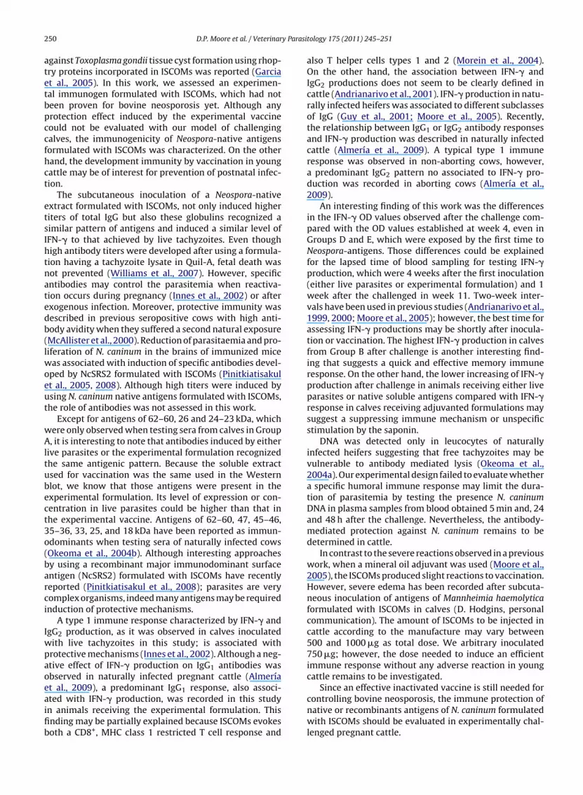
D.P. Moore et al. / Veterinary Parasitology 175 (2011) 245–251 249
Table 2Western blot results when testing sera from calves in each experimental Groups A, B, C, D and E showing weak (+), moderate (++) and strong (+++) signalintensity.
Antigen molecular weight (kDa) Group A Group B Group C Group D Group E
82 + ++78–77 + ++ + +75–74 +++ +++ +62–60 +50 ++ ++43–41 + ++ ++34 ++ ++
++
++
3
tbb
bata35
Gas
3
cBoie(
otCwftG
mgiptglw1�o
2004). Protection against two important viral pathogens(bovine herpes virus and virus of bovine virus diarrheavirus) has been well documented (Trudel et al., 1988;Carlsson et al., 1991). Also in pigs, partial protection
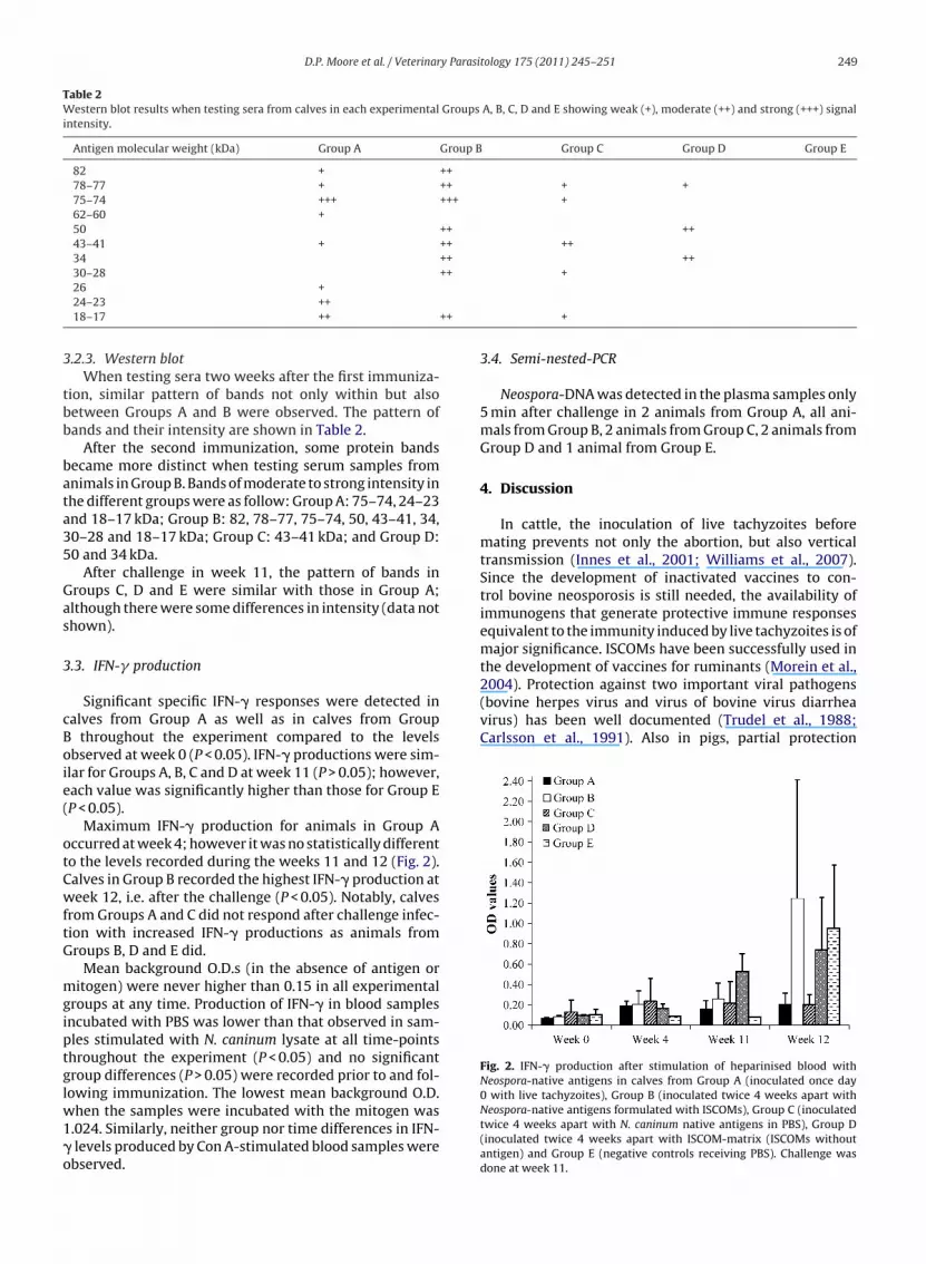
Fig. 2. IFN-� production after stimulation of heparinised blood withNeospora-native antigens in calves from Group A (inoculated once day
30–2826 +24–23 ++18–17 ++
.2.3. Western blotWhen testing sera two weeks after the first immuniza-
ion, similar pattern of bands not only within but alsoetween Groups A and B were observed. The pattern ofands and their intensity are shown in Table 2.
After the second immunization, some protein bandsecame more distinct when testing serum samples fromnimals in Group B. Bands of moderate to strong intensity inhe different groups were as follow: Group A: 75–74, 24–23nd 18–17 kDa; Group B: 82, 78–77, 75–74, 50, 43–41, 34,0–28 and 18–17 kDa; Group C: 43–41 kDa; and Group D:0 and 34 kDa.
After challenge in week 11, the pattern of bands inroups C, D and E were similar with those in Group A;lthough there were some differences in intensity (data nothown).
.3. IFN-� production
Significant specific IFN-� responses were detected inalves from Group A as well as in calves from Group
throughout the experiment compared to the levelsbserved at week 0 (P < 0.05). IFN-� productions were sim-lar for Groups A, B, C and D at week 11 (P > 0.05); however,ach value was significantly higher than those for Group EP < 0.05).
Maximum IFN-� production for animals in Group Accurred at week 4; however it was no statistically differento the levels recorded during the weeks 11 and 12 (Fig. 2).alves in Group B recorded the highest IFN-� production ateek 12, i.e. after the challenge (P < 0.05). Notably, calves
rom Groups A and C did not respond after challenge infec-ion with increased IFN-� productions as animals fromroups B, D and E did.
Mean background O.D.s (in the absence of antigen oritogen) were never higher than 0.15 in all experimental
roups at any time. Production of IFN-� in blood samplesncubated with PBS was lower than that observed in sam-les stimulated with N. caninum lysate at all time-pointshroughout the experiment (P < 0.05) and no significantroup differences (P > 0.05) were recorded prior to and fol-
owing immunization. The lowest mean background O.D.hen the samples were incubated with the mitogen was.024. Similarly, neither group nor time differences in IFN-levels produced by Con A-stimulated blood samples werebserved.
+
+
3.4. Semi-nested-PCR
Neospora-DNA was detected in the plasma samples only5 min after challenge in 2 animals from Group A, all ani-mals from Group B, 2 animals from Group C, 2 animals fromGroup D and 1 animal from Group E.
4. Discussion
In cattle, the inoculation of live tachyzoites beforemating prevents not only the abortion, but also verticaltransmission (Innes et al., 2001; Williams et al., 2007).Since the development of inactivated vaccines to con-trol bovine neosporosis is still needed, the availability ofimmunogens that generate protective immune responsesequivalent to the immunity induced by live tachyzoites is ofmajor significance. ISCOMs have been successfully used inthe development of vaccines for ruminants (Morein et al.,
0 with live tachyzoites), Group B (inoculated twice 4 weeks apart withNeospora-native antigens formulated with ISCOMs), Group C (inoculatedtwice 4 weeks apart with N. caninum native antigens in PBS), Group D(inoculated twice 4 weeks apart with ISCOM-matrix (ISCOMs withoutantigen) and Group E (negative controls receiving PBS). Challenge wasdone at week 11.
y Parasi
250 D.P. Moore et al. / Veterinaragainst Toxoplasma gondii tissue cyst formation using rhop-try proteins incorporated in ISCOMs was reported (Garciaet al., 2005). In this work, we assessed an experimen-tal immunogen formulated with ISCOMs, which had notbeen proven for bovine neosporosis yet. Although anyprotection effect induced by the experimental vaccinecould not be evaluated with our model of challengingcalves, the immunogenicity of Neospora-native antigensformulated with ISCOMs was characterized. On the otherhand, the development immunity by vaccination in youngcattle may be of interest for prevention of postnatal infec-tion.
The subcutaneous inoculation of a Neospora-nativeextract formulated with ISCOMs, not only induced highertiters of total IgG but also these globulins recognized asimilar pattern of antigens and induced a similar level ofIFN-� to that achieved by live tachyzoites. Even thoughhigh antibody titers were developed after using a formula-tion having a tachyzoite lysate in Quil-A, fetal death wasnot prevented (Williams et al., 2007). However, specificantibodies may control the parasitemia when reactiva-tion occurs during pregnancy (Innes et al., 2002) or afterexogenous infection. Moreover, protective immunity wasdescribed in previous seropositive cows with high anti-body avidity when they suffered a second natural exposure(McAllister et al., 2000). Reduction of parasitaemia and pro-liferation of N. caninum in the brains of immunized micewas associated with induction of specific antibodies devel-oped by NcSRS2 formulated with ISCOMs (Pinitkiatisakulet al., 2005, 2008). Although high titers were induced byusing N. caninum native antigens formulated with ISCOMs,the role of antibodies was not assessed in this work.
Except for antigens of 62–60, 26 and 24–23 kDa, whichwere only observed when testing sera from calves in GroupA, it is interesting to note that antibodies induced by eitherlive parasites or the experimental formulation recognizedthe same antigenic pattern. Because the soluble extractused for vaccination was the same used in the Westernblot, we know that those antigens were present in theexperimental formulation. Its level of expression or con-centration in live parasites could be higher than that inthe experimental vaccine. Antigens of 62–60, 47, 45–46,35–36, 33, 25, and 18 kDa have been reported as immun-odominants when testing sera of naturally infected cows(Okeoma et al., 2004b). Although interesting approachesby using a recombinant major immunodominant surfaceantigen (NcSRS2) formulated with ISCOMs have recentlyreported (Pinitkiatisakul et al., 2008); parasites are verycomplex organisms, indeed many antigens may be requiredinduction of protective mechanisms.
A type 1 immune response characterized by IFN-� andIgG2 production, as it was observed in calves inoculatedwith live tachyzoites in this study; is associated withprotective mechanisms (Innes et al., 2002). Although a neg-ative effect of IFN-� production on IgG1 antibodies wasobserved in naturally infected pregnant cattle (Almería
et al., 2009), a predominant IgG1 response, also associ-ated with IFN-� production, was recorded in this studyin animals receiving the experimental formulation. Thisfinding may be partially explained because ISCOMs evokesboth a CD8+, MHC class 1 restricted T cell response andtology 175 (2011) 245–251
also T helper cells types 1 and 2 (Morein et al., 2004).On the other hand, the association between IFN-� andIgG2 productions does not seem to be clearly defined incattle (Andrianarivo et al., 2001). IFN-� production in natu-rally infected heifers was associated to different subclassesof IgG (Guy et al., 2001; Moore et al., 2005). Recently,the relationship between IgG1 or IgG2 antibody responsesand IFN-� production was described in naturally infectedcattle (Almería et al., 2009). A typical type 1 immuneresponse was observed in non-aborting cows, however,a predominant IgG2 pattern no associated to IFN-� pro-duction was recorded in aborting cows (Almería et al.,2009).
An interesting finding of this work was the differencesin the IFN-� OD values observed after the challenge com-pared with the OD values established at week 4, even inGroups D and E, which were exposed by the first time toNeospora-antigens. Those differences could be explainedfor the lapsed time of blood sampling for testing IFN-�production, which were 4 weeks after the first inoculation(either live parasites or experimental formulation) and 1week after the challenged in week 11. Two-week inter-vals have been used in previous studies (Andrianarivo et al.,1999, 2000; Moore et al., 2005); however, the best time forassessing IFN-� productions may be shortly after inocula-tion or vaccination. The highest IFN-� production in calvesfrom Group B after challenge is another interesting find-ing that suggests a quick and effective memory immuneresponse. On the other hand, the lower increasing of IFN-�production after challenge in animals receiving either liveparasites or native soluble antigens compared with IFN-�response in calves receiving adjuvanted formulations maysuggest a suppressing immune mechanism or unspecificstimulation by the saponin.
DNA was detected only in leucocytes of naturallyinfected heifers suggesting that free tachyzoites may bevulnerable to antibody mediated lysis (Okeoma et al.,2004a). Our experimental design failed to evaluate whethera specific humoral immune response may limit the dura-tion of parasitemia by testing the presence N. caninumDNA in plasma samples from blood obtained 5 min and, 24and 48 h after the challenge. Nevertheless, the antibody-mediated protection against N. caninum remains to bedetermined in cattle.
In contrast to the severe reactions observed in a previouswork, when a mineral oil adjuvant was used (Moore et al.,2005), the ISCOMs produced slight reactions to vaccination.However, severe edema has been recorded after subcuta-neous inoculation of antigens of Mannheimia haemolyticaformulated with ISCOMs in calves (D. Hodgins, personalcommunication). The amount of ISCOMs to be injected incattle according to the manufacture may vary between500 and 1000 �g as total dose. We arbitrary inoculated750 �g; however, the dose needed to induce an efficientimmune response without any adverse reaction in youngcattle remains to be investigated.
Since an effective inactivated vaccine is still needed forcontrolling bovine neosporosis, the immune protection ofnative or recombinants antigens of N. caninum formulatedwith ISCOMs should be evaluated in experimentally chal-lenged pregnant cattle.

y Parasi
C
wa
A
cpc3tr
R
A
A
A
A
A
A
C
C
D
E
G
D.P. Moore et al. / Veterinar
onflict of interest statement
There is not any financial and personal relationshipsith other people or organisations that could inappropri-
tely influence this work.
cknowledgments
To the memory of José Luis “Titi” Pereyra, a field techni-ian, who died due acute leukemia during this study. Thisroject was funded by two Research Grants from the Agen-ia Nacional de Promoción Científica y Tecnológica, PICT1681, and from INTA, AESA 3597, Argentina. D.P. Moorehanks Dr. Bror Morein from ISCONOVA, Sweden for criticaleview and helpful comments.
eferences
lmería, S., Nogareda, C., Santolaria, P., Garcia-Ispierto, I., Yániz, J.L.,López-Gatius, F., 2009. Specific anti-Neospora caninum IgG1 and IgG2
antibody responses during gestation in naturally infected cattle andtheir relationship with gamma interferon production. Vet. Immunol.Immunopathol. 130, 35–42.
lvarez-García, G., Pereira-Bueno, J., Gomez-Bautista, M., Ortega-Mora,L.M., 2002. Pattern of recognition of Neospora caninum tachyzoite anti-gens by naturally infected pregnant cattle and aborted foetuses. Vet.Parasitol. 107, 15–27.
ndrianarivo, A.G., Choromanski, L., McDonough, S.P., Packham, A.E., Con-rad, P.A., 1999. Immunogenicity of a killed whole Neospora caninumtachyzoite preparation formulated with different adjuvants. Int. J. Par-asitol. 29, 1613–1625.
ndrianarivo, A.G., Rowe, J.D., Barr, B.C., Anderson, M.L., Packham, A.E.,Sverlow, K.W., Choromanski, L., Loui, C., Grace, A., Conrad, P.A.,2000. A POLYGENTM-adjuvanted killed Neospora caninum tachyzoitepreparation failed to prevent foetal infection in pregnant cattle fol-lowing i.v./i.m. experimental tachyzoite challenge. Int. J. Parasitol. 30,985–990.
ndrianarivo, A.G., Barr, B.C., Anderson, M.L., Rowe, J.D., Packham, A.E.,Sverlow, K.W., Conrad, P.A., 2001. Immune responses in pregnant cat-tle and bovine fetuses following experimental infection with Neosporacaninum. Parasitol. Res. 87, 817–825.
ndrianarivo, A.G., Anderson, M.L., Rowe, J.D., Gardner, I.A., Reynolds, J.P.,Choromanski, L., Conrad, P.A., 2005. Immune responses during preg-nancy in heifers naturally infected with Neospora caninum with andwithout immunization. Parasitol. Res. 96, 24–31.
arlsson, U., Alenius, S., Sundquist, B., 1991. Protective effect of an ISCOMbovine virus diarrhoea virus (BVDV) vaccine against an experimen-tal BVDV infection in vaccinated and non-vaccinated pregnant ewes.Vaccine 9, 577–580.
horomanski, L., Block, W., 2000. Humoral immune responses and safetyof experimental formulations of inactivated Neospora-vaccines. Para-sitol. Res. 86, 851–853.
ubey, J.P., Schares, G., Ortega-Mora, L.M., 2007. Epidemiology and con-trol of neosporosis and Neospora caninum. Clin. Microbiol. Rev. 20,323–367.
chaide, I., Valentini, B., Torioni de Echaide, S., 2002. Neosporosis bov-ina: análisis seroepidemiológico de un hato lechero mediante IFA y
ELISA. In: Mem. XIV Reunión Científica Técnica Asociación Argentinade Veterinarios de Laboratorio de Diagnóstico, 13–15 Noviembre, VillaGeneral Belgrano, Córdoba, Argentina, Secc. Par-01.arcia, J.L., Gennari, S.M., Navarro, I.T., Machado, R.Z., Sinhorini, I.L.,Freire, R.L., Marana, E.R., Tsutsui, V., Contente, A.P., Begale, L.P., 2005.Partial protection against tissue cysts formation in pigs vaccinated
tology 175 (2011) 245–251 251
with crude rhoptry proteins of Toxoplasma gondii. Vet. Parasitol. 129,209–217.
Guy, C.S., Williams, D.J.L., Kelly, D.F., McGarry, J.W., Guy, F., Björkman,C., Smith, R.F., Trees, A.J., 2001. Neospora caninum in persistentlyinfected pregnant cows: spontaneous transplacental infection is asso-ciated with an acute increase in maternal antibody. Vet. Rec. 149,443–449.
Innes, E.A., Wright, S.E., Maley, S., Rae, A., Schock, A., Kirvar, E., Bartley, P.,Hamilton, C., Carey, I.M., Buxton, D., 2001. Protection against verticaltransmission in bovine neosporosis. Int. J. Parasitol. 31, 1523–1534.
Innes, E.A., Andrianarivo, A.G., Björkman, C., Williams, D.J.L., Conrad, P.A.,2002. Immune responses to Neospora caninum and prospects for vac-cination. Trends Parasitol. 18, 497–504.
Littell, R.C., Henry, P.R., Ammerman, C.B., 1998. Statistical analysis ofrepeated measures data using SAS procedures. J. Anim. Sci. 76,1216–1231.
McAllister, M.M., Björkman, C., Anderson-Sprecher, R., Rogers, D.G., 2000.Evidence of point-source exposure to Neospora caninum and protec-tive immunity in a herd of beef cows. J. Am. Vet. Med. Assoc. 217,881–887.
Moore, D.P., Leunda, M.R., Zamorano, P.I., Odeón, A.C., Romera, S.A.,Cano, A., de Yaniz, G., Venturini, M.C., Campero, C.M., 2005. Immuneresponse to Neospora caninum in naturally infected heifers and heifersvaccinated with inactivated antigen during the second trimester ofgestation. Vet. Parasitol. 130, 29–39.
Morein, B., Hu, K., Abusugra, I., 2004. Current status and potential appli-cation of ISCOMs in veterinary medicine. Adv. Drug Deliv. Rev. 56,1367–1382.
Okeoma, C.M., Williamson, N.B., Pomroy, W.E., Stowell, K.M., Gillespie, L.,2004a. The use of PCR to detect Neospora caninum DNA in the bloodof naturally infected cows. Vet. Parasitol. 122, 307–315.
Okeoma, C.M., Williamson, N.B., Pomroy, W.E., Stowell, K.M., 2004b.Recognition patterns of Neospora caninum tachyzoite antigens bybovine IgG at different IFAT titres. Parasite Immunol. 26, 177–185.
Pinitkiatisakul, S., Mattsson, J.G., Wikman, M., Friedman, M., Bengts-son, K.L., Ståhl, S., Lundén, A., 2005. Immunisation of mice againstneosporosis with recombinant NcSRS2 iscoms. Vet. Parasitol. 129,25–34.
Pinitkiatisakul, S., Friedman, M., Wikman, M., Mattsson, J.G., Lövgren-Bengtsson, K., Ståhl, S., Lundén, A., 2007. Immunogenicity andprotective effect against murine cerebral neosporosis of recombinantNcSRS2 in different iscom formulations. Vaccine 25, 3658–3668.
Pinitkiatisakul, S., Mattsson, J.G., Lundén, A., 2008. Quantitative analysis ofparasite DNA in the blood of immunized and naïve mice after infectionwith Neospora caninum. Parasitology 135, 175–182.
Romero, J.J., Pérez, E., Frankena, K., 2004. Effect of a killed whole Neosporacaninum tachyzoite vaccine on the crude abortion rate of Costa Ricandairy cows under field conditions. Vet. Parasitol. 123, 149–159.
Serrano-Martínez, E., Ferre, I., Martínez, A., Osoro, K., Mateos-Sanz, A., del-Pozo, I., Aduriz, G., Tamargo, C., Hidalgo, C.O., Ortega-Mora, L.M., 2007.Experimental neosporosis in bulls: parasite detection in semen andblood and specific antibody and interferon-gamma responses. Theri-ogenology 67, 1175–1184.
Trudel, M., Boulay, G., Seguin, C., Nadon, F., Lussier, G., 1988. Controlof infectious bovine rhinotracheitis in calves with a BHV-1 subunit-ISCOM vaccine. Vaccine 6, 525–529.
Williams, D.J.L., Guy, C.S., Smith, R.F., Guy, F., McGarry, J.W., Macay, J.S.,Trees, A.J., 2003. First demonstration of protective immunity againstfoetopathy in cattle with latent Neospora caninum infection. Int. J.Parasitol. 33, 1059–1065.
Williams, D.J.L., Guy, C.S., Smith, R.F., Ellis, J., Björkman, C., Reichel,M.P., Trees, A.J., 2007. Immunization of cattle with live tachyzoites
of Neospora caninum confers protection against fetal death. Infect.Immun. 75, 1343–1348.Yamage, M., Flechtner, O., Gottstein, B., 1996. Neospora caninum: specificoligonucleotide primers for the detection of brain “cyst” DNA of exper-imentally infected mice by the polymerase chain reaction (PCR). J.Parasitol. 82, 272–279.


