Embed Size (px)
Citation preview
Immunisation with Recombinant PfEMP1 Domains ElicitsFunctional Rosette-Inhibiting and Phagocytosis-InducingAntibodies to Plasmodium falciparumAshfaq Ghumra1, Pongsak Khunrae2¤, Ricardo Ataide3,4, Ahmed Raza1, Stephen J. Rogerson3,
Matthew K. Higgins2, J. Alexandra Rowe1*
1 Centre for Immunity, Infection and Evolution, Institute of Immunology and Infection Research, School of Biological Sciences, University of Edinburgh, Edinburgh, United
Kingdom, 2 Department of Biochemistry, University of Cambridge, Cambridge, United Kingdom, 3 Department of Medicine, University of Melbourne, Post Office Royal
Melbourne Hospital, Melbourne, Australia, 4 Graduate Program in Areas of Basic and Applied Biology, Universidade do Porto, Porto, Portugal
Abstract
Background: Rosetting is a Plasmodium falciparum virulence factor implicated in the pathogenesis of life-threateningmalaria. Rosetting occurs when parasite–derived P. falciparum Erythrocyte Membrane Protein One (PfEMP1) on the surfaceof infected erythrocytes binds to human receptors on uninfected erythrocytes. PfEMP1 is a possible target for a vaccine toinduce antibodies to inhibit rosetting and prevent severe malaria.
Methodology/Findings: We examined the vaccine potential of the six extracellular domains of a rosette-mediating PfEMP1variant (ITvar9/R29var1 from the R29 parasite strain) by immunizing rabbits with recombinant proteins expressed in E. coli.Antibodies raised to each domain were tested for surface fluorescence with live infected erythrocytes, rosette inhibition andphagocytosis-induction. Antibodies to all PfEMP1 domains recognized the surface of live infected erythrocytes down to lowconcentrations (0.02–1.56 mg/ml of total IgG). Antibodies to all PfEMP1 domains except for the second Duffy-Binding-Likeregion inhibited rosetting (50% inhibitory concentration 0.04–4 mg/ml) and were able to opsonize and induce phagocytosisof infected erythrocytes at low concentrations (1.56–6.25 mg/ml). Antibodies to the N-terminal region (NTS-DBL1a) were themost effective in all assays. All antibodies were specific for the R29 parasite strain, and showed no functional activity againstfive other rosetting strains.
Conclusions/Significance: These results are encouraging for vaccine development as they show that potent antibodies canbe generated to recombinant PfEMP1 domains that will inhibit rosetting and induce phagocytosis of infected erythrocytes.However, further work is needed on rosetting mechanisms and cross-reactivity in field isolates to define a set of PfEMP1variants that could induce functional antibodies against a broad range of P. falciparum rosetting parasites.
Citation: Ghumra A, Khunrae P, Ataide R, Raza A, Rogerson SJ, et al. (2011) Immunisation with Recombinant PfEMP1 Domains Elicits Functional Rosette-Inhibitingand Phagocytosis-Inducing Antibodies to Plasmodium falciparum. PLoS ONE 6(1): e16414. doi:10.1371/journal.pone.0016414
Editor: Georges Snounou, Universite Pierre et Marie Curie, France
Received October 18, 2010; Accepted December 14, 2010; Published January 31, 2011
Copyright: � 2011 Ghumra et al. This is an open-access article distributed under the terms of the Creative Commons Attribution License, which permitsunrestricted use, distribution, and reproduction in any medium, provided the original author and source are credited.
Funding: JAR, AG and AR are supported by the Wellcome Trust (http://www.wellcome.ac.uk/) via a Senior Research Fellowship in Basic Biomedical Science toJAR, grant no 084226. MKH is a Royal Society University Research Fellow (http://royalsociety.org/) and this work was supported by grants from the Royal Societyand the Wellcome Trust. PK was funded by a scholarship from the Royal Thai Government. SJR is supported by the National Health and Medical Research Councilof Australia (http://www.nhmrc.gov.au/) and RA is supported by Fundacao para a Ciencia e Tecnologia, Portugal (http://alfa.fct.mctes.pt/), reference SFRH/BD/32985/2006. The funders had no role in study design, data collection and analysis, decision to publish, or preparation of the manuscript.
Competing Interests: The authors have declared that no competing interests exist.
* E-mail: [email protected]
¤ Current address: Microbiology Department, King Mongkut’s University of Technology Thonburi, Bangkok, Thailand
Introduction
The global mortality from malaria continues to be a huge
problem, with Plasmodium falciparum being the major cause of
severe, life-threatening malaria in African children [1]. Rosetting,
the binding of infected erythrocytes to two or more uninfected
erythrocytes, has been shown to be one of the main parasite
virulence phenotypes associated with severe malaria. Initial studies
showed high levels of rosetting in parasite isolates from patients
with cerebral malaria [2,3], with subsequent work showing
rosetting to be linked to all forms of severe malaria [4,5,6,7,8].
Results from human genetic studies have shown that erythrocyte
polymorphisms that reduce rosetting (complement receptor 1
deficiency [9] and blood group O [5]), confer protection against
severe malaria, reducing the odds ratio for severe disease by about
two thirds [10,11]. This protective effect may occur because these
polymorphisms reduce the vaso-occlusive effects of rosetting [12],
thought to be a key pathological process in severe malaria [13].
Together, the association of rosetting with severe malaria, and the
protective effect of human rosette-reducing polymorphisms,
supports a direct role for rosetting in the pathogenesis of severe
malaria. Therapeutic interventions that target rosetting may
therefore have potential to decrease the global burden of severe
malaria [14,15]. This is further supported by the observation that
rosette-inhibiting antibody responses are associated with protec-
tion from severe malaria [2].
PLoS ONE | www.plosone.org 1 January 2011 | Volume 6 | Issue 1 | e16414
Rosetting is mediated by P. falciparum Erythrocyte Membrane
Protein-1 (PfEMP1) expressed on the surface of mature infected
erythrocytes [9]. PfEMP1 variants are 200–400 kDa proteins
encoded by a repertoire of ,60 var genes per haploid parasite
genome, and consisting of tandemly arranged Duffy Binding Like
(DBL) and Cysteine-rich InterDomain Region (CIDR) domains
[16]. Var genes can be classified into groups A, B and C according
to their 59 non-coding sequences, chromosomal location and gene
orientation [16]. Existing data on var gene groups and rosetting are
not entirely consistent. Two well-characterized rosette-mediating
variants are encoded by Group A var genes (ITvar9, also known as
R29var1 [9], and varO [17]), while a third putative rosette-
mediating variant (encoded by FCR3S1.2var1) is group B or C [18].
In P. falciparum field isolates, there is a strong positive correlation
between group A var gene transcription and parasite rosette
frequency [19,20,21,22], suggesting that group A PfEMP1 variants
are common rosetting ligands in natural populations.
Currently, there are few data on the vaccine potential of rosette-
mediating PfEMP1 variants. Previous work has shown that the N-
terminal DBL1a domain is the functional erythrocyte binding
region of rosette-mediating PfEMP1 variants [9,17,23], making
this domain the most promising candidate for an anti-rosetting
vaccine. Antibodies to DBL1a of the VarO variant from the Palo
Alto parasite strain are effective at disrupting rosettes [50%
Inhibitory Concentration (IC50) against Palo Alto, approximately
1/200 dilution of serum [17]], while antibodies to the DBL1adomain of the FCR3S1.2var1 variant have only a modest effect
(IC50 against FCR3S1.2 parasites at 1/2 dilution of serum) [24].
As stated above, FCR3S1.2var1 is a group B or C var gene, and the
majority of the other data suggest that rosetting and severe malaria
are associated with group A var genes [19,20,21,22]. Therefore the
relevance of FCR3S1.2var1 is unclear, and rosette-mediating group
A variants may be better suited for preliminary studies on the
potential for anti-rosetting vaccines.
It remains unclear whether only DBL1a can induce rosette-
disrupting antibodies, or whether the other DBL and CIDR
domains from rosette-mediating PfEMP1 variants can also
generate effective anti-rosetting activity. In addition, it is unknown
whether distinct DBL and CIDR domains differ in their ability to
induce cross-reactive antibodies that are effective against multiple
parasite strains. Finally, the ability of antibodies to recombinant
PfEMP1 domains to promote clearance of infected erythrocytes
via opsonization and phagocytosis, which would also be desirable
in a vaccine, has not previously been studied. We therefore
expressed all of the extracellular domains from a rosette-mediating
group A PfEMP1 variant (ITvar9/R29var1) as recombinant
proteins in E. coli, in order to investigate which domains of
PfEMP1 elicit rosette-inhibiting and phagocytosis-inducing anti-
bodies, and to determine the cross-reactivity of the antibodies for
other rosetting parasite strains.
Results
Expression of DBL and CIDR domains from ITvar9 asrecombinant proteins in E. coli
Domains from the group A rosette-mediating PfEMP1 variant
ITvar9 (also known as R29var1 [9]) were expressed as His-tagged
proteins in E.coli (Figure 1). Previous difficulties in expressing
PfEMP1 domains in E.coli [17,24,25,26,27,28] were overcome
using the methods developed by Higgins [29]. DBL and CIDR
structural information [30,31,32] was used to guide the design of
domain boundaries, and proteins were expressed using a modified
pET15b vector in Origami B cells, supplemented with the pRIG
vector [33] to enrich for rare tRNAs [29,34]. All ITvar9 PfEMP1
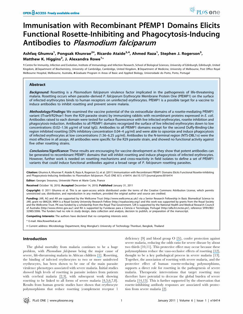
extracellular domains were expressed individually, except for the
first CIDR, which could only be expressed successfully as part of
the NTS-DBL1a-CIDR1c di-domain (Figure 1). The recombinant
proteins were all soluble except for DBL1a, which occurred in
insoluble inclusion bodies and was refolded as described in the
methods. Addition of the short N-terminal sequence (NTS) to
DBL1a resulted in production of soluble protein, suggesting that
the NTS may be an integral part of the DBL1a domain. After
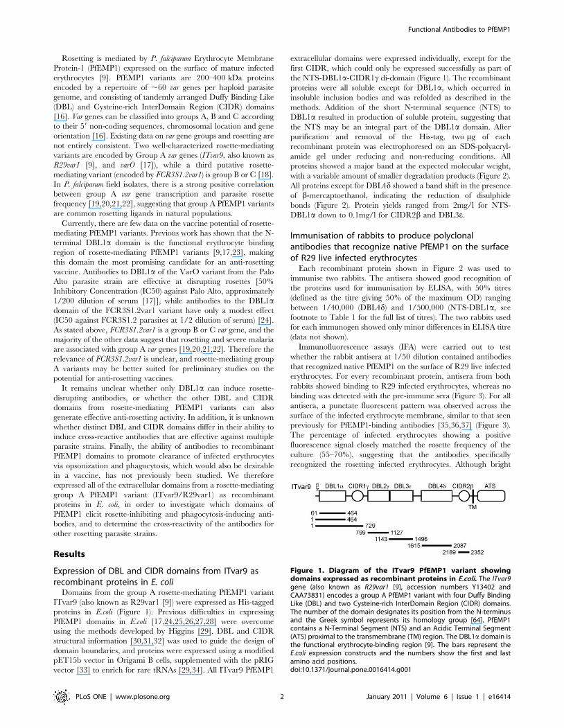
purification and removal of the His-tag, two mg of each
recombinant protein was electrophoresed on an SDS-polyacryl-
amide gel under reducing and non-reducing conditions. All
proteins showed a major band at the expected molecular weight,
with a variable amount of smaller degradation products (Figure 2).
All proteins except for DBL4d showed a band shift in the presence
of b-mercaptoethanol, indicating the reduction of disulphide
bonds (Figure 2). Protein yields ranged from 2mg/l for NTS-
DBL1a down to 0.1mg/l for CIDR2b and DBL3e.
Immunisation of rabbits to produce polyclonalantibodies that recognize native PfEMP1 on the surfaceof R29 live infected erythrocytes
Each recombinant protein shown in Figure 2 was used to
immunise two rabbits. The antisera showed good recognition of
the proteins used for immunisation by ELISA, with 50% titres
(defined as the titre giving 50% of the maximum OD) ranging
between 1/40,000 (DBL4d) and 1/500,000 (NTS-DBL1a, see
footnote to Table 1 for the full list of titres). The two rabbits used
for each immunogen showed only minor differences in ELISA titre
(data not shown).
Immunofluorescence assays (IFA) were carried out to test
whether the rabbit antisera at 1/50 dilution contained antibodies
that recognized native PfEMP1 on the surface of R29 live infected
erythrocytes. For every recombinant protein, antisera from both
rabbits showed binding to R29 infected erythrocytes, whereas no
binding was detected with the pre-immune sera (Figure 3). For all
antisera, a punctate fluorescent pattern was observed across the
surface of the infected erythrocyte membrane, similar to that seen
previously for PfEMP1-binding antibodies [35,36,37] (Figure 3).
The percentage of infected erythrocytes showing a positive
fluorescence signal closely matched the rosette frequency of the
culture (55–70%), suggesting that the antibodies specifically
recognized the rosetting infected erythrocytes. Although bright
Figure 1. Diagram of the ITvar9 PfEMP1 variant showingdomains expressed as recombinant proteins in E.coli. The ITvar9gene (also known as R29var1 [9], accession numbers Y13402 andCAA73831) encodes a group A PfEMP1 variant with four Duffy BindingLike (DBL) and two Cysteine-rich InterDomain Region (CIDR) domains.The number of the domain designates its position from the N-terminusand the Greek symbol represents its homology group [64]. PfEMP1contains a N-Terminal Segment (NTS) and an Acidic Terminal Segment(ATS) proximal to the transmembrane (TM) region. The DBL1a domain isthe functional erythrocyte-binding region [9]. The bars represent theE.coli expression constructs and the numbers show the first and lastamino acid positions.doi:10.1371/journal.pone.0016414.g001
Functional Antibodies to PfEMP1
PLoS ONE | www.plosone.org 2 January 2011 | Volume 6 | Issue 1 | e16414
punctate fluorescence on infected erythrocytes was seen with all
antisera, the intensity of the fluorescence staining was often
stronger for one rabbit out of the pair immunised with each
protein.
As a further negative control, an IFA was carried out with
antibodies to DBL1a domains from several other PfEMP1 variants
(ITvar60, HB3var3, HB3var6, TM180var1, TM284var1 and Muz12-
var1, Ghumra and Rowe, in preparation). These control antibodies
were negative with R29 parasites, however they do recognize live
infected erythrocytes of the parasite strain in which they are
predominantly expressed (Ghumra and Rowe, in preparation).
This indicates that DBL antibodies in general do not recognize
R29, only specific antibodies to ITvar9, the predominantly
expressed variant in R29 parasites [9].
For each ITvar9 recombinant protein, the antiserum giving the
brightest signal by IFA was chosen for purification of total IgG, for
use in rosetting and phagocytosis assays below. In order to
compare the relative abilities of the purified IgG preparations to
recognize live infected erythrocytes, we titred out the IFA signal.
This was done by incubating parasite culture suspension with IgG
at four-fold dilutions, starting at 25 mg/ml then 6.25, 1.56, 0.39,
0.01, 0.024, 0.006 and 0.0015 mg/ml. The end titre was defined
here as the lowest concentration at which .50% of the infected
erythrocytes in the culture showed punctate fluorescence.
Although IFA results can be considered subjective, the end titres
in these experiments were clear, as demonstrated by two
independent observers identifying the same end titre in all cases.
The end titres for positive punctate fluorescence on live R29
infected erythrocytes ranged from 0.024 mg/ml for anti-NTS-
DBL1a to 1.56 mg/ml for anti-CIDR2b (Table 1). Antibodies to
all domains except CIDR2b showed agglutination of infected
erythrocytes at 25 mg/ml (mostly small 5–10 cell agglutinates
under these conditions). Agglutinates did not occur at lower
concentrations.
Figure 2. SDS-PAGE showing recombinant DBL and CIDR domains from ITvar9 expressed in E. coli. The purity and quality of therecombinant DBL and CIDR domains were assessed by electrophoresis of reduced and non-reduced pairs of proteins on 10% SDS-polyacrylamidegels. Two mg of protein was used per well and lanes were as follows: 1) NTS-DBL1a, 2) DBL1a, 3) DBL2c 4) DBL3e, 5) DBL4d, 6) CIDR2b and 7) NTS-DBL1a-CIDR1c. M, molecular weight marker; NR, non-reduced; R, reduced.doi:10.1371/journal.pone.0016414.g002
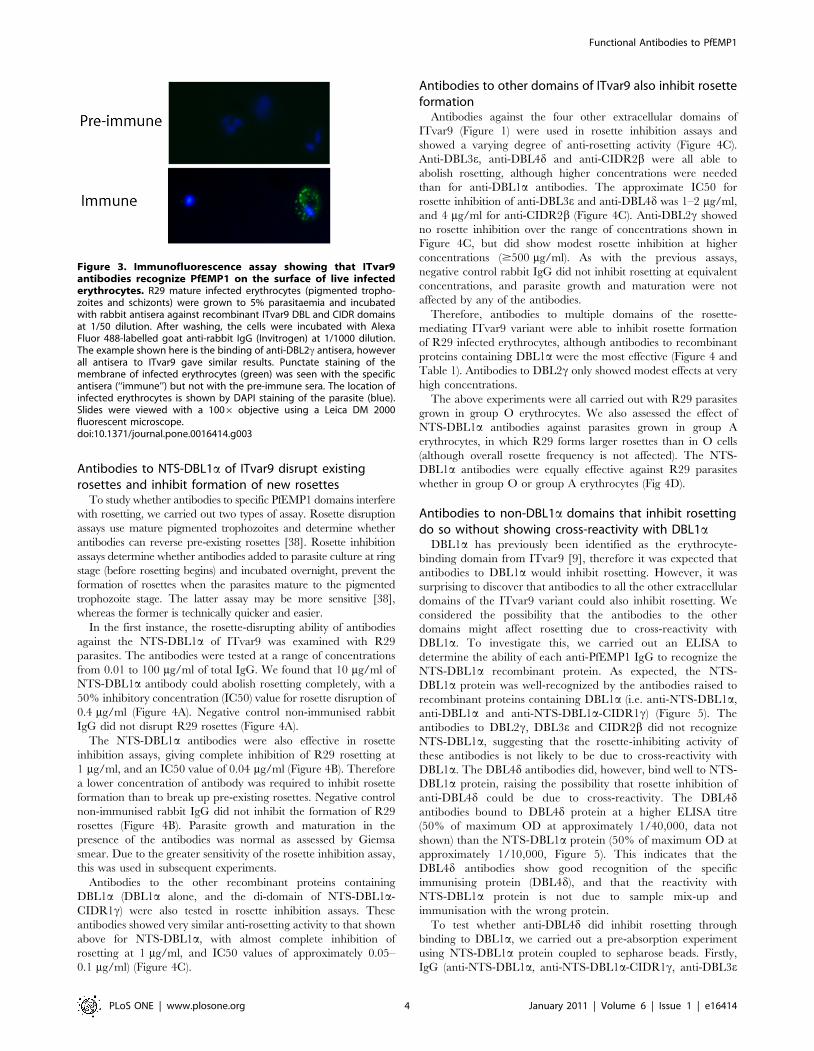
Table 1. Effectiveness of ITvar9 antibodies in various assays#.
Antibodies IFA end titre$ (mg/ml) Rosette Inhibition IC50* (mg/ml)Phagocytosis ,50% of positivecontrol (mg/ml)
Negative control rabbit IgG Negative at 100 Negative at 500 Not done
Anti-NTS-DBL1a 0.024{ 0.04 ,1.56
Anti-DBL1a 0.39 0.1 6.25
Anti-NTS-DBL1a-CIDR1c 0.10 0.05 1.56
Anti-DBL2c 0.39 .500 100
Anti-DBL3e 0.10 2 1.56
Anti-DBL4d 0.10 1 6.25
Anti-CIDR2b 1.56 4 6.25
#50% ELISA titres for the antibodies (defined as the titre giving 50% of the maximum OD) were as follows: NTS-DBL1a 1/500,000; DBL1a 1/250,000; NTS-DBL1a-CIDR1c1/300,000; DBL2c 1/50,000; DBL3e 1/200,000; DBL4d 1/40,000 and CIDR2b 1/200,000.
$The lowest concentration at which .50% of the infected erythrocytes in the culture showed punctate fluorescence by IFA. Values shown are mg/ml of purified totalIgG.
*50% inhibitory concentration (IC50) for rosette inhibition. Values shown are mg/ml of purified total IgG.{The most effective antibodies in each assay are shown in bold. Values shown are mg/ml of purified total IgG.doi:10.1371/journal.pone.0016414.t001
Functional Antibodies to PfEMP1
PLoS ONE | www.plosone.org 3 January 2011 | Volume 6 | Issue 1 | e16414
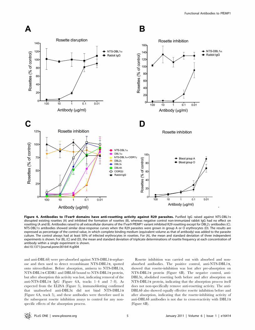
Antibodies to NTS-DBL1a of ITvar9 disrupt existingrosettes and inhibit formation of new rosettes
To study whether antibodies to specific PfEMP1 domains interfere
with rosetting, we carried out two types of assay. Rosette disruption
assays use mature pigmented trophozoites and determine whether
antibodies can reverse pre-existing rosettes [38]. Rosette inhibition
assays determine whether antibodies added to parasite culture at ring
stage (before rosetting begins) and incubated overnight, prevent the
formation of rosettes when the parasites mature to the pigmented
trophozoite stage. The latter assay may be more sensitive [38],
whereas the former is technically quicker and easier.
In the first instance, the rosette-disrupting ability of antibodies
against the NTS-DBL1a of ITvar9 was examined with R29
parasites. The antibodies were tested at a range of concentrations
from 0.01 to 100 mg/ml of total IgG. We found that 10 mg/ml of
NTS-DBL1a antibody could abolish rosetting completely, with a
50% inhibitory concentration (IC50) value for rosette disruption of
0.4 mg/ml (Figure 4A). Negative control non-immunised rabbit
IgG did not disrupt R29 rosettes (Figure 4A).
The NTS-DBL1a antibodies were also effective in rosette
inhibition assays, giving complete inhibition of R29 rosetting at
1 mg/ml, and an IC50 value of 0.04 mg/ml (Figure 4B). Therefore
a lower concentration of antibody was required to inhibit rosette
formation than to break up pre-existing rosettes. Negative control
non-immunised rabbit IgG did not inhibit the formation of R29
rosettes (Figure 4B). Parasite growth and maturation in the
presence of the antibodies was normal as assessed by Giemsa
smear. Due to the greater sensitivity of the rosette inhibition assay,
this was used in subsequent experiments.
Antibodies to the other recombinant proteins containing
DBL1a (DBL1a alone, and the di-domain of NTS-DBL1a-
CIDR1c) were also tested in rosette inhibition assays. These
antibodies showed very similar anti-rosetting activity to that shown
above for NTS-DBL1a, with almost complete inhibition of
rosetting at 1 mg/ml, and IC50 values of approximately 0.05–
0.1 mg/ml) (Figure 4C).
Antibodies to other domains of ITvar9 also inhibit rosetteformation
Antibodies against the four other extracellular domains of
ITvar9 (Figure 1) were used in rosette inhibition assays and
showed a varying degree of anti-rosetting activity (Figure 4C).
Anti-DBL3e, anti-DBL4d and anti-CIDR2b were all able to
abolish rosetting, although higher concentrations were needed
than for anti-DBL1a antibodies. The approximate IC50 for
rosette inhibition of anti-DBL3e and anti-DBL4d was 1–2 mg/ml,
and 4 mg/ml for anti-CIDR2b (Figure 4C). Anti-DBL2c showed
no rosette inhibition over the range of concentrations shown in
Figure 4C, but did show modest rosette inhibition at higher
concentrations ($500 mg/ml). As with the previous assays,
negative control rabbit IgG did not inhibit rosetting at equivalent
concentrations, and parasite growth and maturation were not
affected by any of the antibodies.
Therefore, antibodies to multiple domains of the rosette-
mediating ITvar9 variant were able to inhibit rosette formation
of R29 infected erythrocytes, although antibodies to recombinant
proteins containing DBL1a were the most effective (Figure 4 and
Table 1). Antibodies to DBL2c only showed modest effects at very
high concentrations.
The above experiments were all carried out with R29 parasites
grown in group O erythrocytes. We also assessed the effect of
NTS-DBL1a antibodies against parasites grown in group A
erythrocytes, in which R29 forms larger rosettes than in O cells
(although overall rosette frequency is not affected). The NTS-
DBL1a antibodies were equally effective against R29 parasites
whether in group O or group A erythrocytes (Fig 4D).
Antibodies to non-DBL1a domains that inhibit rosettingdo so without showing cross-reactivity with DBL1a
DBL1a has previously been identified as the erythrocyte-
binding domain from ITvar9 [9], therefore it was expected that
antibodies to DBL1a would inhibit rosetting. However, it was
surprising to discover that antibodies to all the other extracellular
domains of the ITvar9 variant could also inhibit rosetting. We
considered the possibility that the antibodies to the other
domains might affect rosetting due to cross-reactivity with
DBL1a. To investigate this, we carried out an ELISA to
determine the ability of each anti-PfEMP1 IgG to recognize the
NTS-DBL1a recombinant protein. As expected, the NTS-
DBL1a protein was well-recognized by the antibodies raised to
recombinant proteins containing DBL1a (i.e. anti-NTS-DBL1a,
anti-DBL1a and anti-NTS-DBL1a-CIDR1c) (Figure 5). The
antibodies to DBL2c, DBL3e and CIDR2b did not recognize
NTS-DBL1a, suggesting that the rosette-inhibiting activity of
these antibodies is not likely to be due to cross-reactivity with
DBL1a. The DBL4d antibodies did, however, bind well to NTS-
DBL1a protein, raising the possibility that rosette inhibition of
anti-DBL4d could be due to cross-reactivity. The DBL4dantibodies bound to DBL4d protein at a higher ELISA titre
(50% of maximum OD at approximately 1/40,000, data not
shown) than the NTS-DBL1a protein (50% of maximum OD at
approximately 1/10,000, Figure 5). This indicates that the
DBL4d antibodies show good recognition of the specific
immunising protein (DBL4d), and that the reactivity with
NTS-DBL1a protein is not due to sample mix-up and
immunisation with the wrong protein.
To test whether anti-DBL4d did inhibit rosetting through
binding to DBL1a, we carried out a pre-absorption experiment
using NTS-DBL1a protein coupled to sepharose beads. Firstly,
IgG (anti-NTS-DBL1a, anti-NTS-DBL1a-CIDR1c, anti-DBL3e
Figure 3. Immunofluorescence assay showing that ITvar9antibodies recognize PfEMP1 on the surface of live infectederythrocytes. R29 mature infected erythrocytes (pigmented tropho-zoites and schizonts) were grown to 5% parasitaemia and incubatedwith rabbit antisera against recombinant ITvar9 DBL and CIDR domainsat 1/50 dilution. After washing, the cells were incubated with AlexaFluor 488-labelled goat anti-rabbit IgG (Invitrogen) at 1/1000 dilution.The example shown here is the binding of anti-DBL2c antisera, howeverall antisera to ITvar9 gave similar results. Punctate staining of themembrane of infected erythrocytes (green) was seen with the specificantisera (‘‘immune’’) but not with the pre-immune sera. The location ofinfected erythrocytes is shown by DAPI staining of the parasite (blue).Slides were viewed with a 1006 objective using a Leica DM 2000fluorescent microscope.doi:10.1371/journal.pone.0016414.g003
Functional Antibodies to PfEMP1
PLoS ONE | www.plosone.org 4 January 2011 | Volume 6 | Issue 1 | e16414
and anti-DBL4d) were pre-absorbed against NTS-DBL1a-sephar-
ose and then used to detect recombinant NTS-DBL1a, spotted
onto nitrocellulose. Before absorption, antisera to NTS-DBL1a,
NTS-DBL1a-CIDR1 and DBL4d bound to NTS-DBL1a protein,
but after absorption this activity was lost, indicating removal of the
anti-NTS-DBL1a IgG (Figure 6A, tracks 1–4 and 7–8). As
expected from the ELISA (Figure 5), immunoblotting confirmed
that unabsorbed anti-DBL3e did not bind NTS-DBL1a(Figure 6A, track 5), and these antibodies were therefore used in
the subsequent rosette inhibition assays to control for any non-
specific effects of the absorption process.
Rosette inhibition was carried out with absorbed and non-
absorbed antibodies. The positive control, anti-NTS-DBL1a,
showed that rosette-inhibition was lost after pre-absorption on
NTS-DBL1a protein (Figure 6B). The negative control, anti-
DBL3e, abolished rosetting both before and after absorption on
NTS-DBL1a protein, indicating that the absorption process itself
does not non-specifically remove anti-rosetting activity. The anti-
DBL4d also showed equally effective rosette inhibition before and
after absorption, indicating that the rosette-inhibiting activity of
anti-DBL4d antibodies is not due to cross-reactivity with DBL1a(Figure 6B).
Figure 4. Antibodies to ITvar9 domains have anti-rosetting activity against R29 parasites. Purified IgG raised against NTS-DBL1adisrupted existing rosettes (A) and inhibited the formation of rosettes (B), whereas negative control non-immunized rabbit IgG had no effect onrosetting (A and B). Antibodies raised to all extracellular domains of the ITvar9 PfEMP1 variant inhibited R29 rosetting except for DBL2c antibodies (C).NTS-DBL1a antibodies showed similar dose-response curves when the R29 parasites were grown in group A or O erythrocytes (D). The results areexpressed as percentage of the control value, in which complete binding medium (equivalent volume as that of antibody) was added to the parasiteculture. The control always had at least 50% of infected erythrocytes in rosettes. For (A), the mean and standard deviation of three independentexperiments is shown. For (B), (C) and (D), the mean and standard deviation of triplicate determinations of rosette frequency at each concentration ofantibody within a single experiment is shown.doi:10.1371/journal.pone.0016414.g004
Functional Antibodies to PfEMP1
PLoS ONE | www.plosone.org 5 January 2011 | Volume 6 | Issue 1 | e16414
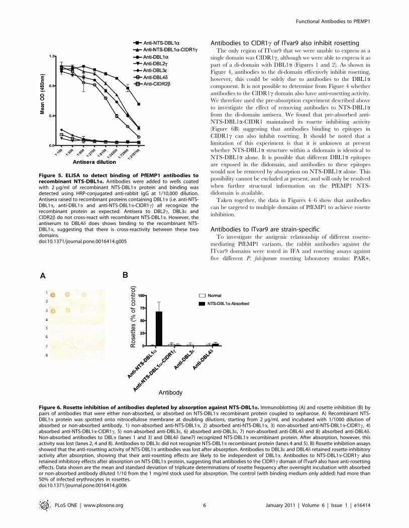
Antibodies to CIDR1c of ITvar9 also inhibit rosettingThe only region of ITvar9 that we were unable to express as a
single domain was CIDR1c, although we were able to express it as
part of a di-domain with DBL1a (Figures 1 and 2). As shown in
Figure 4, antibodies to the di-domain effectively inhibit rosetting,
however, this could be solely due to antibodies to the DBL1acomponent. It is not possible to determine from Figure 4 whether
antibodies to the CIDR1c domain also have anti-rosetting activity.
We therefore used the pre-absorption experiment described above
to investigate the effect of removing antibodies to NTS-DBL1afrom the di-domain antisera. We found that pre-absorbed anti-
NTS-DBL1a-CIDR1 maintained its rosette inhibiting activity
(Figure 6B) suggesting that antibodies binding to epitopes in
CIDR1c can also inhibit rosetting. It should be noted that a
limitation of this experiment is that it is unknown at present
whether NTS-DBL1a structure within a didomain is identical to
NTS-DBL1a alone. It is possible that different DBL1a epitopes
are exposed in the didomain, and antibodies to these epitopes
would not be removed by absorption on NTS-DBL1a alone. This
possibility cannot be excluded at present, and will only be resolved
when further structural information on the PfEMP1 NTS-
didomain is available.
Taken together, the data in Figures 4–6 show that antibodies
can be targeted to multiple domains of PfEMP1 to achieve rosette
inhibition.
Antibodies to ITvar9 are strain-specificTo investigate the antigenic relationship of different rosette-
mediating PfEMP1 variants, the rabbit antibodies against the
ITvar9 domains were tested in IFA and rosetting assays against
five different P. falciparum rosetting laboratory strains: PAR+,
Figure 5. ELISA to detect binding of PfEMP1 antibodies torecombinant NTS-DBL1a. Antibodies were added to wells coatedwith 2 mg/ml of recombinant NTS-DBL1a protein and binding wasdetected using HRP-conjugated anti-rabbit IgG at 1/10,000 dilution.Antisera raised to recombinant proteins containing DBL1a (i.e. anti-NTS-DBL1a, anti-DBL1a and anti-NTS-DBL1a-CIDR1c) all recognize therecombinant protein as expected. Antisera to DBL2c, DBL3e andCIDR2b do not cross-react with recombinant NTS-DBL1a. However, theantiserum to DBL4d does shows binding to the recombinant NTS-DBL1a, suggesting that there is cross-reactivity between these twodomains.doi:10.1371/journal.pone.0016414.g005
Figure 6. Rosette inhibition of antibodies depleted by absorption against NTS-DBL1a. Immunoblotting (A) and rosette inhibition (B) bypairs of antibodies that were either non-absorbed, or absorbed on NTS-DBL1a recombinant protein coupled to sepharose. A) Recombinant NTS-DBL1a protein was spotted onto nitrocellulose membrane at doubling dilutions, starting from 2 mg/ml, and incubated with 1/1000 dilution ofabsorbed or non-absorbed antibody. 1) non-absorbed anti-NTS-DBL1a, 2) absorbed anti-NTS-DBL1a, 3) non-absorbed anti-NTS-DBL1a-CIDR1c, 4)absorbed anti-NTS-DBL1a-CIDR1c, 5) non-absorbed anti-DBL3e, 6) absorbed anti-DBL3e, 7) non-absorbed anti-DBL4d and 8) absorbed anti-DBL4d.Non-absorbed antibodies to DBLa (lanes 1 and 3) and DBL4d (lane7) recognized NTS-DBL1a recombinant protein. After absorption, however, thisactivity was lost (lanes 2, 4 and 8). Antibodies to DBL3e did not recognize NTS-DBL1a recombinant protein (lanes 4 and 5). B) Rosette inhibition assaysshowed that the anti-rosetting activity of NTS-DBL1a antibodies was lost after absorption. Antibodies to DBL3e and DBL4d retained rosette-inhibitoryactivity after absorption, showing that their anti-rosetting effects are likely to be independent of DBL1a. Antibodies to NTS-DBL1a-CIDR1c alsoretained inhibitory effects after absorption on NTS-DBL1a protein, suggesting that antibodies to the CIDR1c domain of ITvar9 also have anti-rosettingeffects. Data shown are the mean and standard deviation of triplicate determinations of rosette frequency after overnight incubation with absorbedor non-absorbed antibody diluted 1/10 from the 1 mg/ml stock used for absorption. The control (with binding medium only added) had more than50% of infected erythrocytes in rosettes.doi:10.1371/journal.pone.0016414.g006
Functional Antibodies to PfEMP1
PLoS ONE | www.plosone.org 6 January 2011 | Volume 6 | Issue 1 | e16414
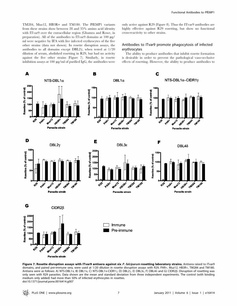
TM284, Muz12, HB3R+ and TM180. The PfEMP1 variants
from these strains show between 28 and 35% amino acid identity
with ITvar9 over the extracellular region (Ghumra and Rowe, in
preparation). All of the antibodies to ITvar9 domains at 100 mg/
ml were negative by IFA with live infected erythrocytes of the five
other strains (data not shown). In rosette disruption assays, the
antibodies to all domains except DBL2c, when tested at 1/20
dilution of serum, abolished rosetting in R29, but had no activity
against the five other strains (Figure 7). Similarly, in rosette
inhibition assays at 100 mg/ml of purified IgG, the antibodies were
only active against R29 (Figure 8). Thus the ITvar9 antibodies are
highly effective against R29 rosetting, but show no functional
cross-reactivity to other strains.
Antibodies to ITvar9 promote phagocytosis of infectederythrocytes
The ability to produce antibodies that inhibit rosette formation
is desirable in order to prevent the pathological vaso-occlusive
effects of rosetting. However, the ability to produce antibodies to
Figure 7. Rosette disruption assays with ITvar9 antisera against six P. falciparum rosetting laboratory strains. Antisera raised to ITvar9domains, and paired pre-immune sera, were used at 1/20 dilution in rosette disruption assays with R29, PAR+, Muz12, HB3R+, TM284 and TM180.Antisera were as follows: A) NTS-DBL1a, B) DBL1a, C) NTS-DBL1a-CIDR1c, D) DBL2c, E) DBL3e, F) DBL4d and G) CIDR2b. Disruption of rosetting wasonly seen with R29 parasites. Data shown are the mean and standard deviation from three independent experiments. The control (with bindingmedium only added) had more than 50% of infected erythrocytes in rosettes.doi:10.1371/journal.pone.0016414.g007
Functional Antibodies to PfEMP1
PLoS ONE | www.plosone.org 7 January 2011 | Volume 6 | Issue 1 | e16414

opsonize infected erythrocytes and target them for clearance by
phagocytosis would also be useful in a vaccine. We therefore
investigated whether the antibodies raised to domains of ITvar9
were effective as opsonins. Fluorescently labelled R29 infected
erythrocytes were preincubated with PfEMP1 antibodies over a
range of concentration from 1.56–100 mg/ml and then mixed
with the phagocytic cell line Thp-1 [39]. The percentage of cells
that had phagocytosed at least one infected erythrocyte after
40 mins co-incubation was determined by flow cytometry. At
high concentration (100 mg/ml) antibodies to all ITvar9 domains
promoted phagocytosis of R29 infected erythrocytes (Figure 9A).
This was a specific effect of the PfEMP1 antibodies, because in
the absence of opsonizing antibodies, no phagocytosis occurred
(Figure 9A, no serum control). Antibodies to NTS-DBL1adomains from other PfEMP1 variants, which do not recognize
R29 live infected erythrocytes by IFA, also did not induce
phagocytosis of R29 infected erythrocytes, even at high
concentrations (Figure 9, control PAR+, TM180, TM284,
Figure 8. Rosette inhibition assays with ITvar9 antisera against six P. falciparum rosetting laboratory strains. Total IgG was used at aconcentration of 100 mg/ml in rosette inhibition assays with R29, PAR+, Muz12, HB3R+, TM284 and TM180. Antisera were as follows: A) NTS-DBL1a, B)DBL1a, C) NTS-DBL1a-CIDR1c, D) DBL2c, E) DBL3e, F) DBL4d and G) CIDR2b. Inhibition of rosetting was only seen with R29 parasites. Data shown arethe mean and standard deviation of triplicate determinations of rosette frequency within a single experiment. The control (with binding medium onlyadded) had more than 50% of infected erythrocytes in rosettes.doi:10.1371/journal.pone.0016414.g008
Functional Antibodies to PfEMP1
PLoS ONE | www.plosone.org 8 January 2011 | Volume 6 | Issue 1 | e16414
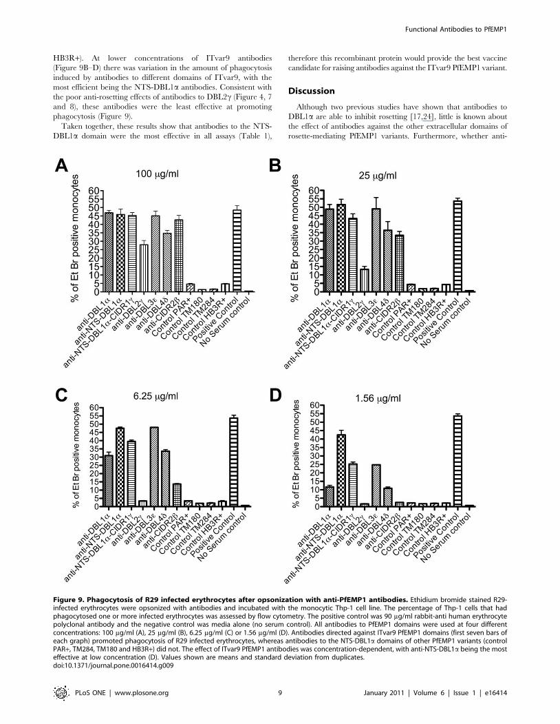
HB3R+). At lower concentrations of ITvar9 antibodies
(Figure 9B–D) there was variation in the amount of phagocytosis
induced by antibodies to different domains of ITvar9, with the
most efficient being the NTS-DBL1a antibodies. Consistent with
the poor anti-rosetting effects of antibodies to DBL2c (Figure 4, 7
and 8), these antibodies were the least effective at promoting
phagocytosis (Figure 9).
Taken together, these results show that antibodies to the NTS-
DBL1a domain were the most effective in all assays (Table 1),
therefore this recombinant protein would provide the best vaccine
candidate for raising antibodies against the ITvar9 PfEMP1 variant.
Discussion
Although two previous studies have shown that antibodies to
DBL1a are able to inhibit rosetting [17,24], little is known about
the effect of antibodies against the other extracellular domains of
rosette-mediating PfEMP1 variants. Furthermore, whether anti-
Figure 9. Phagocytosis of R29 infected erythrocytes after opsonization with anti-PfEMP1 antibodies. Ethidium bromide stained R29-infected erythrocytes were opsonized with antibodies and incubated with the monocytic Thp-1 cell line. The percentage of Thp-1 cells that hadphagocytosed one or more infected erythrocytes was assessed by flow cytometry. The positive control was 90 mg/ml rabbit-anti human erythrocytepolyclonal antibody and the negative control was media alone (no serum control). All antibodies to PfEMP1 domains were used at four differentconcentrations: 100 mg/ml (A), 25 mg/ml (B), 6.25 mg/ml (C) or 1.56 mg/ml (D). Antibodies directed against ITvar9 PfEMP1 domains (first seven bars ofeach graph) promoted phagocytosis of R29 infected erythrocytes, whereas antibodies to the NTS-DBL1a domains of other PfEMP1 variants (controlPAR+, TM284, TM180 and HB3R+) did not. The effect of ITvar9 PfEMP1 antibodies was concentration-dependent, with anti-NTS-DBL1a being the mosteffective at low concentration (D). Values shown are means and standard deviation from duplicates.doi:10.1371/journal.pone.0016414.g009
Functional Antibodies to PfEMP1
PLoS ONE | www.plosone.org 9 January 2011 | Volume 6 | Issue 1 | e16414
PfEMP1 antibodies have opsonizing effects has not been examined
previously. In this study, recombinant domains of the ITvar9
variant were produced in E. coli and used to generate rabbit
polyclonal antibodies for IFA, rosette disruption, rosette inhibition
and phagocytosis assays. All rabbits responded well and gave
antibodies that recognized the surface of R29 live infected
erythrocytes, but not the surface of erythrocytes infected by other
P. falciparum rosetting strains. As expected, antibodies to recom-
binant proteins containing DBL1a had anti-rosetting effects, with
complete inhibition of rosetting at 1mg/ml of total IgG and an
IC50 value of around 0.05 mg/ml. Unexpectedly, antibodies
raised to other extracellular domains of the ITvar9 PfEMP1
variant also inhibited rosetting, with the most effective having
IC50 values of around 1–5 mg/ml. Antibodies to all extracellular
domains of ITvar9 were also able to opsonize infected erythrocytes
for phagocytosis at high concentration. With titration, only
antibodies to the different DBL1a constructs or to DBL3e retained
significant phagocytosis-inducing activity, and this was most
pronounced for the NTS-DBL1a antibodies.
Much of the current work on potential PfEMP1 vaccines focuses
on the generation of adhesion-blocking antibodies [28,40,41,42].
Such antibodies would aim to prevent sequestration and therefore
lead to clearance of mature infected erythrocytes from peripheral
blood due to the filtering action of the spleen [15]. While adhesion-
blocking antibodies are clearly important in preventing microvas-
cular obstruction, it is possible that in vivo, antibodies that opsonize
infected erythrocytes for phagocytosis could have equal or greater
importance as a mechanism for clearance of infected erythrocytes.
Previous work suggests that opsonic phagocytosis of infected
erythrocytes by monocytes and neutrophils is an important
mechanism of immune clearance in malaria [43,44,45,46], and
antibodies that recognize the surface of live infected erythrocytes
have been linked to protective immunity to malaria [47,48]. It
might be expected that PfEMP1 antibodies that recognize the
surface of infected cells and block adhesion would also be able to
promote phagocytosis, but surprisingly, this has not previously been
tested to our knowledge. Our results show that immunisation with
PfEMP1 domains generates antibodies that not only block adhesion
(rosetting) but also effectively opsonize and induce phagocytosis of
infected erythrocytes. This dual activity would be highly advanta-
geous in a vaccine, and it is notable that the most effective
phagoctyosis inducing antibodies (to NTS-DBL1a of ITvar9) were
also the most effective rosette-inhibiting antibodies (Table 1).
One of the most striking features of this study was the very high
activity of the antibodies raised, especially those to NTS-DBL1a,
which gave 50% inhibition of rosetting at 0.04 mg/ml of purified
IgG (Figure 4B). The antibodies were equally effective with R29
parasites grown in group O erythrocytes or group A erythrocytes
(which give larger, stronger rosettes, although do not alter the
rosette frequency) (Figure 4D). This shows that the high activity of
these antibodies is not merely a reflection of weak receptor-ligand
interactions in the R29 rosetting strain in group O erythrocytes.
The results shown here are not unprecedented, because rosette
inhibition at low antibody concentration has been described
previously for a monoclonal antibody to complement receptor 1,
which inhibits rosetting at 1 and 0.1 mg/ml ([38] and JA Rowe,
unpublished data). The high potency of adhesion-blocking
antibodies shown here is, however, in contrast to previous studies
on antibodies to DBL1a from rosetting PfEMP1 variants. One
study found 50% rosette inhibition at 1/200 dilution of serum
(approximately 50 mg/ml of purified IgG, assuming 10 mg/ml of
IgG in normal serum) for the VarO variant in Palo Alto [17],
while a second study required 1/2 dilution of serum (approxi-
mately 5 mg/ml) for the FCR3S1.2var1 variant in FCR3S1.2
parasites [24]. Several factors may contribute to the differences
between studies. Firstly, the rosette inhibition assay used here is ten
times more sensitive than the rosette disruption assay used in
previous studies (compare Figures 4A and 4B). Secondly, it is
possible that R29 rosettes are easier to inhibit than Palo Alto VarO
and FCR3S1.2 rosettes, although these three strains have never
been compared directly in a single laboratory. Thirdly, it is
possible that the recombinant proteins used here contain more
correct conformational epitopes than in previous studies, and are
therefore able to induce more potent anti-rosetting antibodies.
Finally, in the case of the FCR3S1.2var1 variant, in our hands this
variant is expressed by non-rosetting parasites (data not shown),
therefore we think it unlikely that this variant would be capable of
inducing effective rosette-inhibiting antibodies.
The effectiveness of the rosette-inhibiting antibodies shown here
also contrasts with current research on the pregnancy malaria
vaccine candidate var2CSA. Approximately 0.5–1 mg/ml of
antibodies against var2CSA single domains are needed to block
infected erythrocyte adhesion to CSA [28,42], although antibodies
to the full-length protein are more effective [41]. Although
var2CSA is the most promising current PfEMP1 vaccine
candidate because it is relatively well-conserved across strains,
the difficulty in inducing effective adhesion-blocking antibodies
remains an obstacle. Our data suggest that generating adhesion-
blocking antibodies against rosetting can be achieved easily,
however, in this case it is the strain-specific nature of the
antibodies and the between-strain variability in rosetting variants
that remains the major problem.
Previous work has shown that DBL1a is the erythrocyte binding
domain of rosette-mediating PfEMP1 variants [9,17,23]. Why then
do antibodies to other extracellular domains of the ITvar9 variant
inhibit rosetting? It may be that multiple domains of PfEMP1
contribute to a single binding pocket, that multiple domains interact
with different receptors, or that antibodies to other domains interfere
with DBL1a-receptor interactions by steric hindrance or by
disrupting interactions which stabilize higher-order organization of
the protein. There are some examples of PfEMP1 variants where
binding to host receptors is localized to a single domain (for example,
CIDRa binding CD36 [49] or DBLbC2 binding ICAM-1 [50].
However, in the case of CSA-binding, although single domains of the
var2CSA PfEMP1 variant do bind CSA, the full-length molecule
binds with 10,000 fold higher affinity [41,51]. The binding affinity of
full-length rosetting PfEMP1 variants compared to single domains
has not yet been examined. Further work is also needed to determine
if the results shown here, that multiple PfEMP1 domains can induce
anti-rosetting antibodies, are replicated with other rosetting variants.
The only domain of ITvar9 that was notable for its lack of
effectiveness in eliciting rosette-inhibiting and phagocytosis-induc-
ing antibodies was DBL2c. The reason for this is unclear, because
antibodies that recognized the surface of live infected erythrocytes
were generated, which were positive by IFA down to 0.39 mg/ml
(Table 1). One previous study raised antibodies to the second DBL
domain of a rosetting PfEMP1 variant (DBL2bC2 of VarO), and
also found that the antibodies recognized the surface of live infected
cells, but did not disrupt rosettes [52]. Whether this result can be
generalized to all rosetting variants will require further investigation,
but these initial findings do suggest that for these particular variants,
DBL2 domains are not suitable vaccine candidates.
The results shown here indicate that it is straightforward to
generate recombinant PfEMP1 domains in E. coli that induce anti-
rosetting and opsonizing antibodies after immunization. This is an
advance on previous work, in which generation of antibodies
recognizing native PfEMP1 from E. coli-produced protein was
problematic [17,24,25,26,27,28], and researchers were forced to use
Functional Antibodies to PfEMP1
PLoS ONE | www.plosone.org 10 January 2011 | Volume 6 | Issue 1 | e16414
technically more complicated and expensive expression systems
such as Semliki forest virus[24] or Baculovirus [17]. More recently,
Juillerat et al reported successful expression of a rosette-mediating
PfEMP1 domain in E. coli as a fusion protein with maltose-binding
protein, and showed that addition of the NTS to DBL1a resulted in
soluble protein [52]. In our study, methods which combine
structure-lead design of domain boundaries with the use of a
modified pET expression vector in Origami B cells [29], reliably
produced soluble recombinant PfEMP1 protein. Yields of protein
were low (0.1–2 mg/l), however, it is likely that this could be
improved upon by codon optimization of the expression constructs
for E. coli if higher yields are required [53,54].
Developing a strategy to block rosetting of P. falciparum could
lead to a blood-stage vaccine with the potential to combat severe
malaria. However, the sequence polymorphism of PfEMP1
variants, and the ability of the parasite to undergo antigenic
variation and switch to different PfEMP1 variants means that
targeting a single rosette-mediating variant is unlikely to be
successful. An effective vaccine would either induce antibodies that
cross-react with all rosetting variants, or would be composed of
multiple components, each targeting a major rosetting variant
type. Currently there are few data on the cross-reactivity of
antibodies raised to rosette-mediating PfEMP1 variants or on the
number of distinct rosetting mechanisms that occur in natural
parasite populations. Here, although it was easy to generate anti-
rosetting antibodies by immunizing with ITvar9 recombinant
proteins, the antibodies were strain-specific and did not show
functional effects on rosetting in five other laboratory strains
(TM180, Muz12, TM284, HB3R+ or PAR+). Wider testing
against a panel of P. falciparum field isolates from different malaria
endemic countries will be required to determine whether ITvar9
domains induce antibodies that recognize natural parasites, and
could therefore be a useful component of a multi-domain anti-
rosetting vaccine. Previous work has shown that individuals from
malaria endemic countries develop antibodies that recognize
DBL1a of ITvar9 by ELISA [55], suggesting that epitopes within
this variant are similar to those occurring commonly in natural
populations. However, sera from only 3/150 Kenyan children
were able to disrupt R29 rosettes, suggesting that ITvar9-like
variants are rare in the Kenyan P. falciparum population [5].
Although the ITvar9 antibodies generated here appear to be
strain-specific, this cannot be generalized across all rosetting
variants without further study, and our preliminary data suggest
that other rosetting PfEMP1 variants may induce cross-reactive
antibodies (Ghumra and Rowe, unpublished data).
In summary, this study shows that recombinant PfEMP1
(ITvar9) domains produced in E. coli elicit anti-rosetting and
opsonizing antibodies. NTS-DBL1a is the most promising vaccine
candidate because it induces highly effective rosette-inhibiting and
phagocytosis-inducing antibodies. However, several other PfEMP1
domains also generate antibodies that inhibit rosetting at slightly
higher concentrations and are also effective in promoting
phagocytosis. Therefore multiple extracellular domains of ro-
sette-mediating PfEMP1 variants could be considered as vaccine
candidates. Any effort to develop an effective vaccine against P.
falciparum rosetting parasites will require recognition of multiple
strains and further work is needed to fully understand the diversity
of rosette-mediating variants in the field.
Materials and Methods
Ethics statementAnimal immunisations were carried out commercially by BioGenes
GmbH (Berlin, Germany) according to European Union guidelines
86/609/EWG of 24.11.1986 and the European Agreement of
18.3.1996 for protection of animals used for scientific purposes.
ParasitesThe P. falciparum laboratory strains used in this study were R29,
PAR+, TM284, TM180, Muz12 and HB3R+. R29 and PAR+ are
rosetting clones derived from the IT/FCR3 strain, which express
different PfEMP1 variants [[9] and unpublished data]. Muz12 was
collected in Papua New Guinea [56], TM284 was isolated from a
Thai patient with cerebral malaria and TM180 from a Thai
patient with acute malaria [57]. All of the above clones/strains
show high rosette frequency in culture (.50% of infected
erythrocytes in rosettes), but require selection for rosetting 1–2
times per week to maintain the rosetting phenotype. Selection is
carried out by centrifugation through 60% Percoll, or gelatin
flotation [58]. In both cases, the rosetting parasites sediment with
uninfected erythrocytes at the bottom of the tube, whereas non-
rosetting infected erythrocytes are found in the top layer. HB3R+was derived from the HB3 laboratory strain (originally from
Honduras), which naturally shows a low level of rosette formation
(,5%). Repeated rosette selections as described above, over the
course of 3–4 weeks, resulted in the HB3R+ strain with .50% of
infected erythrocytes in rosettes.
Parasite cultureParasites were cultured with group O erythrocytes (Scottish
National Blood Transfusion Service, Edinburgh, UK) at 2%
haematocrit using supplemented RPMI 1640 media as described
[59]. Medium was changed daily, and fresh erythrocytes added
every other day. Parasitaemia was maintained at 5–10% and
cultures were incubated at 37uC in the presence of 3% CO2, 1%
O2 and 96% N2. Cultures were screened regularly to exclude
mycoplasma contamination [60].
Assessment of rosette frequency (RF)The rosette frequency was determined by counting a wet
preparation of stained culture suspension (25mg/ml ethidium
bromide for 5 mins) viewed with simultaneous white light and
fluorescence as described [61]. An infected erythrocyte that bound
to two or more uninfected erythrocytes was counted as a rosette.
The rosette frequency is the percentage of infected erythrocytes
forming rosettes out of 300 infected erythrocytes counted. Samples
were blinded in all experiments to prevent observer bias.
Expression of ITvar9 PfEMP1 domains in E.coliExpression constructs were prepared for each of the six
extracellular domains encoded by the ITvar9 gene (accession
codes Y13402 and CAA73831). Domain boundaries were
predicted by comparing the ITvar9 sequence with known
structures of DBL [30] and CIDR [32] domains. The domain
boundaries were Glu61-Pro464 for DBL1a, Met1-Pro464 for
NTS-DBL1a, Met1-Val729 for NTS-DBL1a-CIDR1c, Gln799-
Thr1127 for DBL2c, Asn1143-Gln1496 for DBL3e, Ala1615-
Pro2087 for DBL4d and Gly2189-Leu2352 for CIDR2b. Primers
were designed to amplify different fragments of ITvar9 from IT
strain genomic DNA by PCR, resulting in products with a BamHI
cleavage site at the 59 end and an NheI site at the 39 end. Primers
were as follows: NTSf: GGGGATCCATGACGCCAAAGCG-
TACAAGTC; DBL1f: CCGGATCCTCGTGTAGTCTTGAT-
CACAAATTC; DBL1r: CCGCTAGCTTAAGGACATGCTT-
CACAATATC; CIDR1f: GGGGATCC
GATAATAAAATTGCATTTAATGTATTG; CIDR1r: GG-
GCTAGCTTATACAC
Functional Antibodies to PfEMP1
PLoS ONE | www.plosone.org 11 January 2011 | Volume 6 | Issue 1 | e16414
TTTTAGGTAATATATCAC; DBL2f: CTGGATCCGCGT-
GTGCTATTGTTAA
GGGTGTT; DBL2r: TTGCTAGCTTATGTGCACCCGC-
AATATTGGTC; DBL3f: CTGGATCCAATTGTTGTGGTC-
TAAACAGTG; DBL3r: AAGCTAGCTCATTGG
CACTCACATCTGTCTTT; DBL4f: AAGGATCCGCATG-
TAACCAAAAA
TATGGTTACCC; DBL4r: AAGCTAGCTCAAGGACACG-
GCGTACAATTTG; CIDR2f: TTGGATCCAATTGTAAAAA-
TGGTAATTGTGGTG; CIDR2r: TTGCTAGCTCAAACA-
CATTTTTCTTCTAGGACTGT. The PCR-amplified products
were digested with restriction enzymes and inserted into a
modified pEt15b vector [29], generating constructs with an N-
terminal His tag and a Tobacco Etch Virus (TEV) protease
cleavage site before the start of the domain [33]. Constructs were
sequenced to ensure that no PCR-generated errors were
incorporated.
Constructs were transformed into Origami B (Novagen,
Nottingham, UK) E. coli containing the pRIG plasmid [33]. Cells
were grown to an optical density of 1.0 at 600nm and then
induced with 1mM IPTG. Expression took place at 25uCovernight. Western blots, in which the proteins were visualised
using an HRP conjugated penta-His antibody (Qiagen, Crawley,
UK), showed that all constructs except for CIDR1c and DBL1agenerated soluble protein. CIDR1c produced heavily degraded
protein that could not be purified while DBL1a was expressed in
inclusion bodies.
Purification of NTS-DBL1a, NTS-DBL1a-CIDR1c, DBL3e,DBL4d and CIDR2b recombinant proteins
Cells were pelleted, resuspended in solubilisation buffer (20mM
Tris pH 8.0, 0.3M NaCl, 10mM imidazole, 0.5% Triton X-100)
and lysed by sonication. The cell lysate was centrifuged for 30 minss
at 45,000g and purified by affinity chromatography using nickel-
NitriloTriacetic Acid (Ni-NTA) sepharose (Qiagen). The protein
was loaded onto the Ni-NTA resin, washed with solubilisation
buffer, and eluted with 20mM Tris pH 8.0, 0.1M NaCl, 0.2M
imidazole. Protein was buffer exchanged in 20mM phosphate
pH 7.0, 150mM NaCl, 3mM reduced glutathione, 0.3 mM
oxidized glutathione and cleaved overnight at room temperature
by the addition of one milligram of His-tagged TEV protease [62]
per ten milligrams of protein. The mixture was passed through a Ni-
NTA affinity column to remove the TEV protease and any
uncleaved protein that retained its His-tag. The cleaved protein in
the flow-through was concentrated using an Amicon Ultra
centifugal filter device (10,000 MWCO) and further purified using
a Superdex 200 16/60 column (GE Healthcare, Little Chalfont,
Buckinghamshire, UK) run using 20mM Tris pH 8.0, 50mM NaCl.
Purification of DBL1a recombinant proteinThe DBL1a domain was expressed as inclusion bodies and was
refolded while bound to a Ni-NTA affinity column. Cells were
resuspended, lysed and centrifuged as above. The pellet was
resuspended by sonication in 20mM Tris pH 8.0, 0.3M NaCl,
10mM imidazole, 6M guanidine hydrochloride and 10mM b-
mercaptoethanol. After clarification by centrifugation for 30 mins
at 45,000g, the supernatant was loaded onto a Ni-NTA column.
The column was then washed with resuspension buffer lacking b-
mercaptoethanol and containing 3mM reduced glutathione and
0.3 mM oxidized glutathione. The guanidine concentration was
slowly reduced from 6M to zero using a continuous gradient over
40 column volumes, while other buffer components (as described
above) were maintained. The column was washed with 0.3M
NaCl, 20mM Tris pH 8.0 and the protein was eluted with 20mM
Tris pH 8.0, 0.1M NaCl, 0.2M imidazole. The protein was then
buffer exchanged, TEV cleaved and gel filtered as above.
SDS-PAGE of recombinant proteinsThe purity of the recombinant ITvar9 DBL/CIDR proteins
was assessed by SDS-PAGE on 10% bis-Tris polyacrylamide gels,
stained with SimplyBlue (Invitrogen). Two micrograms of non-
reduced and reduced (heated to 80uC for 10 mins in the presence
of 5% b-mercaptoethanol) proteins were loaded in each well and
separated by electrophoresis according to the manufacturer’s
protocol (Invitrogen).
Generation of polyclonal antibodies to ITvar9 PfEMP1domains
All immunizations were carried out by BioGenes GmbH (Berlin,
Germany). Because some rabbit sera contain heterophile antibodies
that react with human erythrocytes or P. falciparum infected
erythrocytes [6], rabbits’ sera were screened before immunisation
using immunofluorescence assays (IFA) to select animals with
minimal reactivity to parasite cultures. For each recombinant protein,
pre-immune sera from five rabbits were tested for binding to R29
infected erythrocytes as described below. Two animals whose pre-
immune sera gave no fluorescent signal on infected and uninfected
erythrocytes were chosen for immunizations. Briefly, two rabbits were
immunized with 125 mg of protein on day 0, 7, 14 and 28. Antisera
from both rabbits were collected on day 28 and showed a titre of at
least 1: 10,000 by ELISA (Biogenes GmbH). IFA on live R29 infected
erythrocytes were carried out with pre-immune and post-immuniza-
tion sera for both animals, and the antiserum giving the brightest
positive fluorescence was chosen for Protein-A purification of total
IgG (Biogenes GmbH). IgG from a non-immunised rabbit was also
purified by the same method to use as a negative control, because
there was insufficient pre-immune serum from the immunised rabbits
to provide purified IgG for all experiments. However pre-immune
sera were used (and found to be negative) in the initial
immunofluorescence assays at 1/50 dilution described below, and
in the rosette disruption experiments shown in Figure 7.
Live parasite immunofluorescence assays (IFA)IFA were carried out to i) determine if the antisera recognized
native PfEMP1 on the surface of unfixed cells, ii) determine which
antisera would be chosen for total IgG purification and iii) assess
whether antisera cross-reacted with multiple parasite strains.
Parasite cultures with a parasitaemia of at least 5% mature
pigmented trophozoites and a rosette frequency of at least 50%
were used. IFA were as described previously [6]. Briefly, primary
incubation was for 1 hour on ice with a 1/50 dilution of pre-
immune serum or anti-serum, and after three washes, secondary
incubation was for 45 mins on ice with a 1:1000 dilution of highly
cross-absorbed Alexa Fluor 488-labelled goat anti-rabbit IgG
(catalogue number A11034, Invitrogen Ltd, Paisley, UK) in PBS/
1%BSA containing 1 mg/ml 4, 6-diamidino-2-phenylindole
(DAPI). Positive IFA signals were titred out by starting with a
concentration of 25 mg/ml of purified IgG, and carrying out seven
4-fold dilutions down to 0.0015 mg/ml. The end titre was the
lowest antibody concentration that gave punctate fluorescence
over more than 50% of infected erythrocytes. End titres were
assessed by two independent observers who identified the same
end point in all cases.
Rosette disruption assaysRosette disruption assays were carried out with parasites of
greater than 50% rosette frequency. Parasite cultures were pre-
Functional Antibodies to PfEMP1
PLoS ONE | www.plosone.org 12 January 2011 | Volume 6 | Issue 1 | e16414
stained with 25 mg/ml of ethidium bromide (5 mins at 37uC) then
washed twice with incomplete RPMI 1640 (with supplements for
parasite culture but without serum). The cells were resuspended at
2% haematocrit in complete binding medium (RPMI 1640 with
supplements for parasite culture but lacking sodium bicarbonate),
containing 10% pooled human serum that had been heat-
inactivated at 56uC for 30 mins. The bicarbonate-free binding
medium maintains a more stable pH in non-gassed cultures than
RPMI containing bicarbonate. Rabbit pre-immune and antisera
were added to 50 ml aliquots of synchronous parasite cultures
(mature pigmented trophozoites, 2% haematocrit, between 5–10%
parasitaemia) and incubated at 37uC for 30 mins. Negative
controls were equivalent volumes of complete binding medium
and serum from a non-immunised rabbit. The samples were
blinded and 1.2 ml from each tube was placed on a spot of a
multispot slide (Hendley Essex Ltd, Loughton, UK), and covered
with a 22622 mm coverslip. The rosette frequency was counted as
described above. The data are shown as percentage of the negative
control with complete binding medium added.
Rosette inhibition assaysThese assays were set up using synchronous ring-stage parasites
that had shown a rosette frequency of at least 50% in the previous
cycle. Parasite cultures (parasitaemia 5–10%) were spun down and
resuspended in complete binding media at 2% haematocrit, and
aliquoted into wells of a 96-well tissue culture plate. Total IgG,
purified from antisera raised against ITvar9 recombinant proteins,
was added to triplicate wells to give a final concentration of 100,
10, 0.1 and 0.01 mg/ml and a final assay volume of 100 ml. An
equivalent volume of complete binding media or purified total IgG
from a non-immunised rabbit was added to triplicate wells as
negative controls. Tubes containing working antibody stocks
diluted in complete binding media or negative controls were
blinded prior to addition into the 96 well plate. The plate was put
in a sealed humid chamber, supplied with a gas mixture as
described above and incubated at 37uC overnight. The following
day, wells were incubated with 25 mg/ml ethidium bromide for
5 mins and rosette frequency was assessed as described above. The
data are shown as percentage of the negative control with
complete binding medium.
ELISA for recognition of NTS-DBL1a recombinant proteinWells of an ELISA plate were coated with 2 mg/ml of NTS-
DBL1a recombinant protein in carbonate bicarbonate buffer
(Sigma, Poole, UK) and incubated overnight at 4uC. After
blocking for 1 hr in PBS containing 0.05% Tween 20 (PBST)
and 5% milk, wells were incubated with anti-PfEMP1 rabbit
antisera diluted in PBST containing 1% milk (PBSTM). After 1 hr
incubation at room temperature, wells were washed with PBST
and incubated with 1:10 000 of HRP-conjugated goat anti-rabbit
IgG (Sigma) in PBSTM for another hour. Finally, wells were
washed with PBST and reactions were developed by incubating
the wells with substrate 3,39,5,59-tetramethylbenzidinedihy-
drochloride (Sigma) according to the manufacturer’s instructions
and absorbance was measured at a wavelength of 450 nm.
Absorption of PfEMP1 antibodies on NTS-DBL1arecombinant protein coupled to sepharose
NTS-DBL1a protein was coupled to sepharose using cyanogen
bromide according to the manufacturer’s instructions (Sigma).
Total IgG against NTS-DBL1a, NTS-DBL1a-CIDR1, DBL3eand DBL4d were diluted to a concentration of 1mg/ml and 100 ml
of each was incubated with 100 ml of NTS-DBL1a-sepharose that
had been coated with 0.3 mg of protein. Antibodies were allowed
to bind for 30 mins on a rotator at room temperature. The NTS-
DBL1a-sepharose was pelleted by spinning in a microfuge for
30 sec and the supernatant was collected. Immunoblotting was
used to confirm that anti-NTS-DBL1a binding activity had been
depleted from the absorbed antibody stocks. Doubling dilutions of
recombinant NTS-DBL1a protein were made, starting at 2mg/ml.
Three ml spots of each dilution were placed onto nitrocellulose
(Schleicher and Schuell, Dassel, Germany). After drying, the
nitrocellulose was blocked in PBST containing 5% milk for 1 hr,
followed by 1 hr incubation with absorbed and unabsorbed
PfEMP1 antibodies at 1/1000 dilution. The membrane was then
washed in PBST and incubation with HRP-labelled goat anti-
rabbit IgG (Sigma) diluted in PBSTM 1:10,000 for another hour.
Following a further wash in PBST, blots were developed using
liquid diaminobenzidine (DAB) (DAKO, Ely, UK) according to
the manufacturer’s instructions. Rosette inhibition assays were
carried out with 1:10 dilution of normal (1mg/ml) or absorbed
IgG as described above.
Phagocytosis assays with anti-PfEMP1 antibodiesThe phagocytosis assay was performed as described previously
[39] with modifications. The human pro-monocytic cell line Thp-
1 was maintained in RPMI 1640 (GIBCO, Mulgrave, Australia)
supplemented with 10% heat-inactivated Foetal Bovine Serum
(FBS), 1% penicillin-streptomycin, 2 mM L-glutamine, 25mM
HEPES and 55 mM 2-Mercaptho-Ethanol (Sigma) at a density
below 56105 cells/ml. During the phagocytosis assay, this medium
was supplemented with 100mg/ml of Heparin (T-Hep) in order to
prevent rosetting. Preliminary experiments showed that the
presence of heparin did not affect phagocytosis of a positive
control (erythrocytes opsonized with rabbit anti-erythrocyte
surface antibodies). Synchronised R29 parasites (.70% rosetting)
were washed in RPMI-HEPES medium (RPMI 1640 supple-
mented with 25mM HEPES, 2 mM L-glutamine, 40mg/ml
gentamicin and 50mg/ml hypoxanthine) and resuspended in
RPMI-HEPES containing 100mg/ml of Heparin and 0.5% BSA
(R-Hep) for 15 mins. Wet smears were made to ensure that
rosettes had been disrupted. Mature trophozoites-infected eryth-
rocytes were isolated using a VarioMACS magnet [63], resulting
in .80% purity. Infected erythrocytes were washed in R-Hep and
resuspended at 3.36107 cells/ml in 10mg/ml of ethidium bromide
solution in R-Hep. After 3 further washes, cells were resuspended
at 3.36107 cells/ml in R-Hep. Thirty ml per well of this cell
suspension was distributed to 96-well plates where 3.3 ml of
purified rabbit IgG to ITvar9 domains or control rabbit anti-
human erythrocyte antibodies (ab34858, ABCAM, Cambridge,
UK) had been plated. Opsonization was allowed to occur for
60mins in the dark at RT. During this time, Thp-1 cells were
washed, resuspended at 56105 cells/ml in T-Hep, and 100ml
aliquots were dispensed into wells of a 96-well round-bottom plate
(phagocytosis plate). After opsonization the infected erythrocytes
were washed thrice with R-Hep and resuspended in 100ml of T-
Hep. Two 50ml aliquots were then transferred to the phagocytosis
plate and phagocytosis was allowed to occur for 40 mins at 37uCin a humidified incubator with 5% CO2. Phagocytosis was stopped
by centrifugation at 4uC. After discarding the supernatant, the
cells were resuspended at 37uC in FACS Lysing solution (BD
Biosciences) according to the manufacturer’s instructions. Lysis
was stopped by the addition of 50 ml of cold PBS (2Ca2+, 2Mg2+)
2% FBS and 0.02% NaNO3 (FACS Buffer). After 3 washes with
FACS Buffer the cells were fixed in cold 2% Paraformaldehyde in
PBS and 10,000 Thp-1 cells were acquired using a FACSCalibur
flow cytometer. Only minimal agglutination of infected erythro-
Functional Antibodies to PfEMP1
PLoS ONE | www.plosone.org 13 January 2011 | Volume 6 | Issue 1 | e16414
cytes mixed with antibodies to ITvar9 occurs under the conditions
of this assay (occasional 2–3 cell aggregates at 100 and 25 mg/ml),
which would not be expected to have a major impact on the
results.
Author Contributions
Conceived and designed the experiments: AG PK RA SJR MKK JAR.
Performed the experiments: AG PK RA AR MKK. Analyzed the data: AG
RA JAR. Wrote the paper: AG PK RA SJR MKK JAR.
References
1. Snow RW, Guerra CA, Noor AM, Myint HY, Hay SI (2005) The global
distribution of clinical episodes of Plasmodium falciparum malaria. Nature 434:
214–217.
2. Carlson J, Helmby H, Hill AV, Brewster D, Greenwood BM, et al. (1990)
Human cerebral malaria: association with erythrocyte rosetting and lack of anti-
rosetting antibodies. Lancet 336: 1457–1460.
3. Treutiger CJ, Hedlund I, Helmby H, Carlson J, Jepson A, et al. (1992) Rosette
formation in Plasmodium falciparum isolates and anti-rosette activity of sera from
Gambians with cerebral or uncomplicated malaria. Am J Trop Med Hyg 46:
503–510.
4. Ringwald P, Peyron F, Lepers JP, Rabarison P, Rakotomalala C, et al. (1993)
Parasite virulence factors during falciparum malaria: rosetting, cytoadherence,
and modulation of cytoadherence by cytokines. Infect Immun 61: 5198–5204.
5. Rowe A, Obeiro J, Newbold CI, Marsh K (1995) Plasmodium falciparum rosetting
is associated with malaria severity in Kenya. Infect Immun 63: 2323–2326.
6. Rowe JA, Shafi J, Kai OK, Marsh K, Raza A (2002) Nonimmune IgM, but not
IgG binds to the surface of Plasmodium falciparum-infected erythrocytes and
correlates with rosetting and severe malaria. Am J Trop Med Hyg 66: 692–699.
7. Rowe JA, Obiero J, Marsh K, Raza A (2002) Positive correlation between
rosetting and parasitaemia in Plasmodium falciparum clinical isolates. Am J Trop
Med Hyg 66: 458–460.
8. Doumbo OK, Thera MA, Kone AK, Raza A, Tempest LJ, et al. (2009) High
levels of Plasmodium falciparum rosetting in all clinical forms of severe malaria
in African children. Am J Trop Med Hyg 81: 987–993.
9. Rowe JA, Moulds JM, Newbold CI, Miller LH (1997) P. falciparum rosetting
mediated by a parasite-variant erythrocyte membrane protein and complement-
receptor 1. Nature 388: 292–295.
10. Cockburn IA, Mackinnon MJ, O’Donnell A, Allen SJ, Moulds JM, et al. (2004)
A human complement receptor 1 polymorphism that reduces Plasmodium
falciparum rosetting confers protection against severe malaria. Proc Natl Acad
Sci U S A 101: 272–277.
11. Rowe JA, Handel IG, Thera MA, Deans AM, Lyke KE, et al. (2007) Blood
group O protects against severe Plasmodium falciparum malaria through the
mechanism of reduced rosetting. Proc Natl Acad Sci U S A 104: 17471–17476.
12. Kaul DK, Roth EFJ, Nagel RL, Howard RJ, Handunnetti SM (1991) Rosetting
of Plasmodium falciparum-infected red blood cells with uninfected red blood cells
enhances microvascular obstruction under flow conditions. Blood 78: 812–819.
13. Dondorp AM, Ince C, Charunwatthana P, Hanson J, van Kuijen A, et al. (2008)
Direct in vivo assessment of microcirculatory dysfunction in severe falciparum
malaria. J Infect Dis 197: 79–84.
14. Kyriacou HM, Steen KE, Raza A, Arman M, Warimwe G, et al. (2007) In vitro
inhibition of Plasmodium falciparum rosette formation by Curdlan sulfate.
Antimicrob Agents Chemother 51: 1321–1326.
15. Rowe JA, Claessens A, Corrigan RA, Arman M (2009) Adhesion of Plasmodium
falciparum-infected erythrocytes to human cells: molecular mechanisms and
therapeutic implications. Expert Rev Mol Med 11: e16.
16. Kraemer SM, Smith JD (2006) A family affair: var genes, PfEMP1 binding, and
malaria disease. Curr Opin Microbiol 9: 374–380.
17. Vigan-Womas I, Guillotte M, Le Scanf C, Igonet S, Petres S, et al. (2008) An in
vivo and in vitro model of Plasmodium falciparum rosetting and autoaggluti-
nation mediated by varO, a group A var gene encoding a frequent serotype.
Infect Immun 76: 5565–5580.
18. Chen Q, Barragan A, Fernandez V, Sundstrom A, Schlichtherle M, et al. (1998)
Identification of Plasmodium falciparum erythrocyte membrane protein 1 (PfEMP1)
as the rosetting ligand of the malaria parasite P. falciparum. J Exp Med 187:
15–23.
19. Bull PC, Berriman M, Kyes S, Quail MA, Hall N, et al. (2005) Plasmodium
falciparum Variant Surface Antigen Expression Patterns during Malaria. PLoS
Pathog 1: e26.
20. Kyriacou HM, Stone GN, Challis RJ, Raza A, Lyke KE, et al. (2006)
Differential var gene transcription in Plasmodium falciparum isolates from patients
with cerebral malaria compared to hyperparasitaemia. Mol Biochem Parasitol
150: 211–218.
21. Kaestli M, Cockburn IA, Cortes A, Baea K, Rowe JA, et al. (2006) Virulence of
malaria is associated with differential expression of Plasmodium falciparum var
gene subgroups in a case-control study. J Infect Dis 193: 1567–1574.
22. Warimwe GM, Keane TM, Fegan G, Musyoki JN, Newton CR, et al. (2009)
Plasmodium falciparum var gene expression is modified by host immunity. Proc
Natl Acad Sci U S A 106: 21801–21806.
23. Russell C, Mercereau-Puijalon O, Le Scanf C, Steward M, Arnot DE (2005)
Further definition of PfEMP-1 DBL-1alpha domains mediating rosetting
adhesion of Plasmodium falciparum. Mol Biochem Parasitol 144: 109–113.
24. Chen Q, Pettersson F, Vogt AM, Schmidt B, Ahuja S, et al. (2004)
Immunization with PfEMP1-DBL1alpha generates antibodies that disrupt
rosettes and protect against the sequestration of Plasmodium falciparum-infectederythrocytes. Vaccine 22: 2701–2712.
25. Singh AP, Puri SK, Chitnis CE (2002) Antibodies raised against receptor-binding domain of Plasmodium knowlesi Duffy binding protein inhibit
erythrocyte invasion. Mol Biochem Parasitol 121: 21–31.
26. Oguariri RM, Mattei D, Tena-Tomas C, Uhlemann AC, Kremsner PG, et al.
(2003) Recombinant Duffy binding-like-alpha domains of Plasmodium falci-
parum erythrocyte membrane protein 1 elicit antibodies in rats that recognizeconserved epitopes. Parasitol Res 90: 467–472.
27. Barfod L, Nielsen MA, Turner L, Dahlback M, Jensen AT, et al. (2006)Baculovirus-expressed constructs induce immunoglobulin G that recognizes
VAR2CSA on Plasmodium falciparum-infected erythrocytes. Infect Immun 74:4357–4360.
28. Nielsen MA, Pinto VV, Resende M, Dahlback M, Ditlev SB, et al. (2009)Induction of adhesion-inhibitory antibodies against placental Plasmodium
falciparum parasites by using single domains of VAR2CSA. Infect Immun 77:
2482–2487.
29. Higgins MK (2008) Overproduction, purification and crystallization of a
chondroitin sulfate A-binding DBL domain from a Plasmodium falciparumvar2csa-encoded PfEMP1 protein. Acta Crystallogr Sect F Struct Biol Cryst
Commun 64: 221–223.
30. Tolia NH, Enemark EJ, Sim BK, Joshua-Tor L (2005) Structural basis for the
EBA-175 erythrocyte invasion pathway of the malaria parasite Plasmodium
falciparum. Cell 122: 183–193.
31. Singh SK, Hora R, Belrhali H, Chitnis CE, Sharma A (2006) Structural basis for
Duffy recognition by the malaria parasite Duffy-binding-like domain. Nature439: 741–744.
32. Klein MM, Gittis AG, Su HP, Makobongo MO, Moore JM, et al. (2008) Thecysteine-rich interdomain region from the highly variable plasmodium
falciparum erythrocyte membrane protein-1 exhibits a conserved structure.PLoS Pathog 4: e1000147.
33. Baca AM, Hol WG (2000) Overcominsg codon bias: a method for high-leveloverexpression of Plasmodium and other AT-rich parasite genes in Escherichia
coli. Int J Parasitol 30: 113–118.
34. Khunrae P, Philip JM, Bull DR, Higgins MK (2009) Structural comparison of
two CSPG-binding DBL domains from the VAR2CSA protein important in
malaria during pregnancy. J Mol Biol 393: 202–213.
35. Baruch DI, Pasloske BL, Singh HB, Bi X, Ma XC, et al. (1995) Cloning the P.
falciparum gene encoding PfEMP1, a malarial variant antigen and adherencereceptor on the surface of parasitised human erythrocytes. Cell 82: 77–87.
36. Ghumra A, Semblat JP, McIntosh RS, Raza A, Rasmussen IB, et al. (2008)Identification of residues in the Cmu4 domain of polymeric IgM essential for
interaction with Plasmodium falciparum erythrocyte membrane protein 1(PfEMP1). J Immunol 181: 1988–2000.
37. Joergensen LM, Salanti A, Dobrilovic T, Barfod L, Hassenkam T, et al. (2010)The kinetics of antibody binding to Plasmodium falciparum VAR2CSA
PfEMP1 antigen and modelling of PfEMP1 antigen packing on the membrane
knobs. Malar J 9: 100.
38. Rowe JA, Rogerson SJ, Raza A, Moulds JM, Kazatchkine MD, et al. (2000)
Mapping of the region of complement receptor (CR) 1 required for Plasmodium
falciparum rosetting and demonstration of the importance of CR1 in rosetting in
field isolates. J Immunol 165: 6341–6346.
39. Ataide R, Hasang W, Wilson DW, Beeson JG, Mwapasa V, et al. (2010) Using
an improved phagocytosis assay to evaluate the effect of HIV on specificantibodies to pregnancy-associated malaria. PLoS One 5: e10807.
40. Avril M, Gamain B, Lepolard C, Viaud N, Scherf A, et al. (2006)
Characterization of anti-var2CSA-PfEMP1 cytoadhesion inhibitory mousemonoclonal antibodies. Microbes Infect 8: 2863–2871.
41. Khunrae P, Dahlback M, Nielsen MA, Andersen G, Ditlev SB, et al. (2010) Full-length recombinant Plasmodium falciparum VAR2CSA binds specifically to
CSPG and induces potent parasite adhesion-blocking antibodies. J Mol Biol 397:826–834.
42. Salanti A, Resende M, Ditlev SB, Pinto VV, Dahlback M, et al. (2010) Severaldomains from VAR2CSA can induce Plasmodium falciparum adhesion-
blocking antibodies. Malar J 9: 11.
43. Celada A, Cruchaud A, Perrin LH (1982) Opsonic activity of human immune
serum on in vitro phagocytosis of Plasmodium falciparum infected red blood cells by
monocytes. Clin Exp Immunol 47: 635–644.
44. Celada A, Cruchaud A, Perrin LH (1983) Phagocytosis of Plasmodium falciparum-
parasitized erythrocytes by human polymorphonuclear leukocytes. J Parasitol69: 49–53.
45. Mota MM, Brown KN, Holder AA, Jarra W (1998) Acute Plasmodiumchabaudi chabaudi malaria infection induces antibodies which bind to the
surfaces of parasitized erythrocytes and promote their phagocytosis bymacrophages in vitro. Infect Immun 66: 4080–4086.
Functional Antibodies to PfEMP1
PLoS ONE | www.plosone.org 14 January 2011 | Volume 6 | Issue 1 | e16414

46. Sponaas AM, Freitas do Rosario AP, Voisine C, Mastelic B, Thompson J, et al.
(2009) Migrating monocytes recruited to the spleen play an important role incontrol of blood stage malaria. Blood 114: 5522–5531.
47. Marsh K, Otoo L, Hayes RJ, Carson DC, Greenwood BM (1989) Antibodies to
blood stage antigens of Plasmodium falciparum in rural Gambians and their relationto protection against infection. Trans Roy Soc Trop Med Hyg 83: 293–303.
48. Bull PC, Lowe BS, Kortok M, Molyneux CS, Newbold CI, et al. (1998) Parasiteantigens on the infected red cell surface are targets for naturally acquired
immunity to malaria. Nature Medicine 4: 358–360.
49. Robinson BA, Welch TL, Smith JD (2003) Widespread functional specializationof Plasmodium falciparum erythrocyte membrane protein 1 family members to bind
CD36 analysed across a parasite genome. Mol Microbiol 47: 1265–1278.50. Howell DP, Levin EA, Springer AL, Kraemer SM, Phippard DJ, et al. (2008)
Mapping a common interaction site used by Plasmodium falciparum Duffybinding-like domains to bind diverse host receptors. Mol Microbiol 67: 78–87.
51. Srivastava A, Gangnard S, Round A, Dechavanne S, Juillerat A, et al. (2010)
Full-length extracellular region of the var2CSA variant of PfEMP1 is requiredfor specific, high-affinity binding to CSA. Proc Natl Acad Sci U S A 107:
4884–4889.52. Juillerat A, Igonet S, Vigan-Womas I, Guillotte M, Gangnard S, et al. (2010)
Biochemical and biophysical characterisation of DBL1alpha1-varO, the
rosetting domain of PfEMP1 from the VarO line of Plasmodium falciparum.Mol Biochem Parasitol 170: 84–92.
53. Yadava A, Ockenhouse CF (2003) Effect of codon optimization on expressionlevels of a functionally folded malaria vaccine candidate in prokaryotic and
eukaryotic expression systems. Infect Immun 71: 4961–4969.54. Zhou Z, Schnake P, Xiao L, Lal AA (2004) Enhanced expression of a
recombinant malaria candidate vaccine in Escherichia coli by codon
optimization. Protein Expr Purif 34: 87–94.55. Mayor A, Rovira-Vallbona E, Srivastava A, Sharma SK, Pati SS, et al. (2009)
Functional and immunological characterization of a Duffy binding-like alpha
domain from Plasmodium falciparum erythrocyte membrane protein 1 that
mediates rosetting. Infect Immun 77: 3857–3863.
56. Cox MJ, Kum DE, Tavul L, Narara A, Raiko A, et al. (1994) Dynamics of
malaria parasitaemia associated with febrile illness in children from a rural area
of Madang, Papua New Guinea. Trans R Soc Trop Med Hyg 88: 191–197.
57. Scholander C, Treutiger CJ, Hultenby K, Wahlgren M (1996) Novel fibrillar
structure confers adhesive property to malaria-infected erythrocytes. Nature
Medicine 2: 204–208.
58. Handunnetti SM, Gilladoga AD, van Schravendijk MR, Nakamura K,
Aikawa M, et al. (1992) Purification and in vitro selection of rosette-positive
(R+) and rosette-negative (R2) phenotypes of knob-positive Plasmodium falciparum
parasites. Am J Trop Med Hyg 46: 371–381.
59. Corrigan RA, Rowe JA (2010) Strain variation in early innate cytokine induction
by Plasmodium falciparum. Parasite Immunol 32: 512–527.
60. Rowe JA, Scragg IG, Kwiatkowski D, Ferguson DJP, Carucci DJ, et al. (1998)
Implications of mycoplasma contaminsation in Plasmodium falciparum cultures
and methods for its detection and eradication. Mol Biochem Parasitol 92:
177–180.
61. Deans AM, Rowe JA (2006) Plasmodium falciparum: Rosettes do not protect
merozoites from invasion-inhibitory antibodies. Exp Parasitol 112: 269–273.
62. Kapust RB, Tozser J, Fox JD, Anderson DE, Cherry S, et al. (2001) Tobacco
etch virus protease: mechanism of autolysis and rational design of stable mutants
with wild-type catalytic proficiency. Protein Eng 14: 993–1000.
63. Trang DT, Huy NT, Kariu T, Tajima K, Kamei K (2004) One-step
concentration of malarial parasite-infected red blood cells and removal of
contaminsating white blood cells. Malar J 3: 7.
64. Smith JD, Subramanian G, Gamain B, Baruch DI, Miller LH (2000)
Classification of adhesive domains in the Plasmodium falciparum erythrocyte
membrane protein 1 family. Mol Biochem Parasitol 110: 293–310.
Functional Antibodies to PfEMP1
PLoS ONE | www.plosone.org 15 January 2011 | Volume 6 | Issue 1 | e16414


