Embed Size (px)
Citation preview

Ž .Brain Research 754 1997 171–180
Research report
Immunocytochemical appearance of cytokines, prostaglandin E and2
lipocortin-1 in the CNS during the incubation period of murine scrapiecorrelates with progressive PrP accumulations
Alun Williams a,), Anne-Marie Van Dam b, Diane Ritchie a, Pieter Eikelenboom c, Hugh Fraser a
a Institute for Animal Health, BBSRC and MRC Neuropathogenesis Unit, Ogston Building, West Mains Road, Edinburgh, EH9 3JF, UKb Research Institute Neurosciences Free UniÕersity, Faculty of Medicine, Department of Pharmacology, Van der Boechorststraat 7, 1081 BT Amsterdam,
Netherlandsc Research Institute Neurosciences Free UniÕersity, Faculty of Medicine, Department of Psychiatry, Valeriuskliniek, Valeriusplein 9, 1075 BG Amsterdam,
Netherlands
Accepted 17 December 1996
Abstract
Ž . Ž . Ž .The appearance of immunoreactive interleukin IL -1b, IL-6 and tumour necrosis factor TNF -a , prostaglandin PG E and2
lipocortin-1 in the central nervous system was investigated during the development of lesions in a 301VrVM murine scrapie model.Focal PrPSc deposition was present after 30 days of the 115–120 day incubation period; this immunoreactivity increased in intensity anddistribution thereafter. Staining for IL-1b and TNFa in perivascular macrophages, and PGE immunoreactivity in astrocytes, was2
detected in those areas showing PrPSc deposition from 60 days. Increased GFAP and F4r80 immunoreactivity, indicating activation ofastrocytes and microglia, was also evident in these areas from 60 days. Glial cytokine and lipocortin immunoreactivity was detected after90 days, in the absence of clinical signs. The disease-induced cytokine, PG and lipocortin immunoreactivity occurred only in those brainareas showing PrPSc deposition, glial activation and, in later stages, vacuolation. These findings support the concept that PrPSc depositioninduces glial cytokine production. These glial cytokines may contribute to the development of the pathological lesions in scrapie. q 1997Elsevier Science B.V. All rights reserved.
Keywords: Scrapie; Prion; Cytokine; Prostaglandin; Lipocortin; Neurodegeneration; Glia; Mouse
1. Introduction
Many accounts of scrapie pathology describe a chronicneurodegeneration with vacuolation of neurons and neu-ropil, accumulations of a protease-resistant isoform of the
Ž . w xPrP prion protein and a reactive astrocytosis 21,33 .More recent studies of murine scrapie and of Creutzfeldt-
Ž .Jakob Disease CJD in man have also demonstrated thew xpresence of a marked microglial response 30,46,53 . In-
duction of the cytokines IL-1a , IL-1b, IL-6 and TNFa
has been reported in the brains of mice showing clinicalsigns of scrapie and immunoreactivity for these cytokines,
Ž .prostaglandins PGs E and F , and lipocortin-1 lo-2 2 a
w xcalised to activated glia 9,37,39,55 . Prostaglandin E is2
known to mediate a variety of effects induced by cytokinessuch as IL-1 whereas lipocortins are endogenous peptides
) Ž .Corresponding author. Fax: q44 131 668-3872.
with anti-inflammatory properties, including an inhibitoryw xaction on the effects of cytokines 10,15,19,44 .
It has previously been proposed that cytokines and otheracute phase proteins may contribute to the development ofpathology of chronic neurodegenerative diseases such as
Ž . w xAlzheimer’s disease AD 3,17,49 . Thus in AD, wherethe observed bA4 deposition may be due to overproduc-
Ž .tion of amyloid precursor protein APP andror abnormalw xpost-translational processing 28 , expression of cytokines,
acute phase proteins, activated complement factors and cellw xstress proteins have all been reported 1,2,13,16,17,26,43 .
It has also been shown that the promoter region of the APPgene contains AP-1 sites, acute phase and heat shock
w x Ž .elements 24,45 and that interleukin-1 IL-1 can up-regu-w xlate the synthesis of APP mRNA in vitro 24 .
A number of similar observations has been made inscrapie and CJD where ‘pathological’ post-translationalmodifications of the PrP protein result in PrPSc depositions
0006-8993r97r$17.00 Copyright q 1997 Elsevier Science B.V. All rights reserved.Ž .PII S0006-8993 97 00067-X

( )A. Williams et al.rBrain Research 754 1997 171–180172
in areas of the brain that subsequently show vacuolationw x6,7,12,33,42 . Marked astrocytosis and microglial activa-tion, induction of b -integrins, increased cell stress protein2
expression and ubiquitin conjugation have all been re-w xported 11,14,31,35,39,41,46,53 and several transcription
sites, including AP-1 sites, have been identified in thew xpromoter region of the mouse PrP gene 8 . Increased
IL-1a , IL-1b and TNFa mRNA levels have been reportedin scrapie- or CJD-infected mice during their clinical phase
w xof disease 9,37,39 . Furthermore, intraocular injection ofrecombinant TNFa in mice has been shown to producemyelin ballooning in the optic nerve similar to that seen inthe white matter lesions of the panencephalopathic type of
w xCJD 40 . In addition, IL-6 has been demonstrated toinduce differentiation of, and increase PrP mRNA levels
w xin, PC-12 cells 38 .Thus cytokines may be significant factors in determin-
ing the pathogenesis of the neurodegeneration in scrapieand CJD. However, there are no reports on the presence ofthese factors at the protein level in these diseases duringthe period before clinical disease becomes evident. Thispaper describes an immunocytochemical study investigat-ing the presence and localisation of cytokines, PGE and2
lipocortin-1 in the brains of scrapie-affected mice through-out the course of infection.
2. Materials and methods
VMrDk mice were inoculated intracerebrally with thew x301V strain of scrapie as previously described 47 . Ani-
mals inoculated with normal brain homogenate acted ascontrols. Mice were killed 30, 60 or 90 days after inocula-tion or when showing clinical signs of scrapie, 115–120days after inoculation. Mice used for routine histologicalexamination were killed by cervical dislocation and their
Žbrains fixed by immersion in 10% formol saline ns6.scrapie-inoculated plus 2 control mice for each time point .
Other mice were perfused either with paraformaldehyde-Žlysine-periodate PLP; 2% paraformaldehyde final concen-
.Ž .tration ns4q2 for each time point , PLP containingŽ .0.05% glutaraldehyde ns4q2 or with Tyrode’s salt
Ž .solution followed by Bouin’s fixative ns3 or 4q1 ,under deep chloral hydrate anaesthesia.
5 mm wax-embedded sections of the brains fixed informol saline or with PLP were examined at the five levels
w xused for scrapie lesion profile scoring 22 . Immunostain-Ž .ing for GFAP astrocytes and PrP was performed on
PLP-fixed, wax-embedded sections using rabbit polyclonalŽ .antisera and a standard PAP method. F4r80 microglia
staining was performed on PLPrwax sections and on 10mm cryostat sections of tissue fixed with PLP-glutaralde-
w xhyde, using an ABC method previously described 53 .Cytokine, prostaglandin and lipocortin immunocytochem-istry was performed on 50 mm coronal slices of Bouin’s-fixed brain as free-floating sections using a PAP method
Table 1Primary antibodies used
Dilutions
IL-1b Sheep anti-mouse 1 : 500–1 : 750Rabbit anti-rat 1 : 750
IL-6 Rabbit anti-human 1 : 1000Rabbit anti-mouse 1 : 250
TNFa Sheep anti-human 1 : 1000Rabbit anti-mouse 1 : 200
PGE Rabbit anti-PGE 1 : 5002 2
Lipocortin-1 Rabbit anti-lipocortin 1 : 300GFAP Rabbit anti-human 1 : 800
Ž .Rabbit anti-cow Dako 1 : 400Ž .F4r80 Rat anti-mouse Serotec 1 : 10–1 : 20
aPrP Rabbit anti-mouse SAF 1 : 800
a SAFsscrapie-associated fibrils.
w x52,55 . A nickel ammonium sulphate enhanced DAB visu-w xalisation method 27,29 was used on some of the Bouin’s
fixed sections. GFAP and F4r80 staining was also per-formed on this material. The primary antibodies used aregiven in Table 1. Sections stained with preadsorbed anti-
Ž .body cytokines and PGE , normal serum or without the2
application of primary antibody acted as controls.
3. Results
The results of the cytokine immunoreactivity were com-pared with the development of vacuolation, PrP depositionand activation of astrocytes and microglia during the incu-bation period. It was noted that although cytokineimmunoreactivity was observed at various brain levels,staining patterns were clearest in the thalamus, hippocam-
w xpus and cerebral cortex, as previously described 55 . Thus,whilst increased PrP, GFAP and F4r80 staining occurredin affected areas at all levels of the scrapie-infected brainsexamined, the detailed descriptions of PrP, GFAP andF4r80 immunoreactivity below will only be given for thelevel of the hippocampus rostral to the geniculate nuclei,taken to include the caudal median eminence or infundibu-lum, arcuate hypothalamic nucleus and CA1-CA4 regions
Ž w xof the hippocampus approximately coronal level 301 50 ,one of the standard levels for lesion profile scoring and
.caudal to the injection site .
3.1. Routine H&E sections: Vacuolation
No vacuolation, characteristic of scrapie infection, waspresent in animals killed 30 and 60 days after inoculation.At 90 days, low levels of vacuolation were evident in themedulla, thalamus, dorsal hippocampus and cortex in 4 outof 6 mice. The severity of these low grade vacuolar lesionsin the thalamus was often greater on the ipsilateral side tothe scrapie inoculation site. Mice with clinical signs ofscrapie showed more severe and more widespread vacuola-

( )A. Williams et al.rBrain Research 754 1997 171–180 173
w xtion. The lesion profile 22 obtained for these mice wassimilar to that for the 301VrVM model used in other
w xstudies M. Bruce, personal communication . Vacuolationwas prominent in the thalamus and hippocampus, particu-
Ž .larly in the CA1 and CA2 regions Fig. 1 . Althoughmorphometric techniques were not used, a marked loss ofCA1 pyramical neurons in the hippocampus, which wasnot evident at 90 days, was present in all 6 mice killedwhen showing clinical signs of scrapie. There was littlevariation in the severity of lesions between infected miceat any one time point, precluding the need to performimmunocytochemistry on large numbers of mice.
3.2. Immunocytochemistry: PrP, astrocytes and microglia
There was a focal increase in PrP immunoreactivity inthe dorsal hippocampus and dorsomedial thalamus in 3 outof 4 scrapie-inoculated animals killed 30 days after inocu-lation. This was present as both granular deposits in the
Ž .neuropil and as pericellular staining Fig. 2 . The PrPimmunoreactivity at 60 days was more intense and showedgreater distribution throughout the hippocampus and thala-mus; increased PrP immunoreactivity was also present inthe dorsal parietal cortex, where it tended to show laminarstaining. Thereafter, the intensity of the PrP immuno-staining increased further and became more widespread
Fig. 1. Distribution of vacuolation in hippocampus and thalamus in aterminally affected mouse. Neuronal loss in the CA1 region of the
Ž . Ž .hippocampus is also evident arrow 5 mm wax section . Bars50 mm.
Ž .Table 2; compare Fig. 2a, Fig. 3a, Fig. 4a, Fig. 5a ,occurring in those brain areas that later showed vacuola-tion.
The first differences in GFAP and F4r80 stainingbetween infected and control animals were observed at 60days. At this time, increased GFAP and F4r80 immuno-reactivity was observed in those areas of the thalamus,cortex and dorsal hippocampus showing PrP depositionand PGE immunoreactivity. After 60 days, GFAP and2
F4r80 immunoreactivity became progressively more in-tense, more widespread and occurred predominantly in thesame brain areas as the increased PrP staining and inducedcytokinerPGrlipocortin immunoreactivity. The neuronalloss seen in H&E sections of clinically affected mice wasalso evident in immunostained material, especially waxand cryostat sections.
3.3. Immunocytochemistry: cytokines, prostaglandins andlipocortin
Constitutive staining of IL-1b and IL-6 occurred inneurons of the supra-optic and periventricular nuclei of all
w xanimals studied, as previously recorded 55 . IL-1b im-munoreactivity was also present in cells in the meningesand on ependymal surfaces in both infected and controlmice. Both the IL-6 antibodies tested also stained scatteredneuronal cell bodies and cell processes in the hippocampusand cerebellum of normal and scrapie-affected mice. Noother cytokine, PG or lipocortin immunoreactivity wasobserved in any control mice. Preadsorption of cytokineand PG antibodies with 10y6 M of the appropriate recom-binant protein markedly reduced or abolished both consti-tutive immunoreactivity in normal and scrapie-infectedmice and disease-induced glial immunoreactivity in in-
Ž w xfected animals Fig. 5d preabsorbed IL-1b antibody illus-trates the reduction in disease-induced glial IL-1b
immunoreactivity compared to staining observed with un-w x.absorbed serum Fig. 5b . No staining was detected in the
absence of primary antibody or when normal serum wasused.
The results obtained in scrapie-inoculated mice aresummarised in Table 2. Disease-induced cytokine and PGimmunoreactivity was first detected at 60 days after infec-
Žtion. At this time, the anti-rat IL-1b antibody but not the.anti-mouse IL-1b antibody stained perivascular cells in
the thalamus of infected mice; this staining was particu-larly prominent when the nickel enhancement technique
Ž .was used Fig. 3b . These cells were detected bilaterallybut were more pronounced on the ipsilateral side to thescrapie inoculation site. A smaller number of TNFa im-munoreactive perivascular cells were also present. Thenumber of anti-rat IL-1b immunoreactive perivascular cellsprogressively decreased after 60 days and TNFa-stainedperivascular cells were observed only at 60 days. Bilateral,mild PGE immunoreactivity, absent at 30 days, was2
present in glia in the dorsal hippocampus, dorsal thalamus

( )A. Williams et al.rBrain Research 754 1997 171–180174
Ž . Ž .Fig. 2. 30 days post inoculation. Focal PrP deposition present as diffuse granular deposits arrow and perineuronal staining arrow head in theŽ . Ž . Ž . Ž . Ž .hippocampus A,B and thalamus C,D A–D: 5 mm wax sections . Bars50 mm in A,C ; bars100 mm in B,D .
and in focal areas of the dorsal cerebral cortex in 2 of the 4Ž .mice killed at 60 days Fig. 3c .
Clear, disease-induced IL-1b, IL-6, TNFa andlipocortin immunoreactivity in glial cells was observed 90days after inoculation. Anti-mouse IL-1b immunoreactiveglia were observed bilaterally in the dorsal and dorsolateralhippocampus, dorsal thalamus and in foci in the cerebral
Ž .cortex Fig. 4b . Less intense staining was observed in the
medulla. IL-6, TNFa , and lipocortin-1 immunoreactivityŽ .showed a similar distribution Fig. 4c–e . Glial PGE2
Ž .immunoreactivity was stronger than at 60 days Fig. 4f,g .Staining was more intense in animals killed when show-
ing clinical signs of scrapie. Vacuoles were discernible inmany of the 50 mm sections from infected animals andglial cytokine, PG and lipocortin immunoreactivity waslargely confined to those brain areas showing vacuolation
Table 2Immunoreactivity and lesion severity in infected animals
Days post PrP GFAP F4r80 IL-1b IL-6 TNFa PGE Lc-1 Vac2
injection T H T H T H T H T H T H T H T H T H
30 " " – – – – – – – – – – – – – – – –60 q q q " q " P – – – P – q " – – – –90 qq qq qq qq qq qq q q q q q q q q q q q q115–120 qqq qqq qqq qqq qqq qqq qq qq qq qq qq qq qq qq qq qq qq qq
Lc-1s lipocortin-1; Vacsvacuolation; Ts thalamus a; Hsdorsal hippocampus a. –sno difference between infected animals and controls. PsonlyŽ .perivascular cells immunoreactive The numbers of these decreased at later time points . "s immunoreactivity: low intensity of staining seen in a
Ž .proportion of mice PrP, GFAP, F4r80 3r4; PGE 2r4 . qs immunoreactivity: positive staining in all mice; vacuolation: low level. qqs immuno-2
reactivity: stronger positive staining observed on greater number of individual cells, with a wider distribution of immunoreactivity; vacuolation: high level.qqqs immunoreactivity: very strong staining in all mice with more numerous and more widespread immunoreactive cells than qq.a Ž .At brain level described in detail see text .

( )A. Williams et al.rBrain Research 754 1997 171–180 175
Ž .and PrP accumulation see below . Thus, for example,IL-1b, IL-6, TNFa , PGE and lipocortin-1 staining was2
Žmore pronounced in the dorsal and lateral hippocampus as
.illustrated for IL-1b in Fig. 5b,c; see also Table 2 thanventrally and tended to show a laminar pattern in thelateral parietal and temporal cortex. This immunoreactivity
Ž . Ž .Fig. 3. 60 days post inoculation. A Focal PrP deposition greater than at 30 days, shown here in the hippocampus. B IL-1b immunoreactivity inŽ . Ž . Ž . Žperivascular cells arrow C glial PGE immunoreactivity in hippocampus A: 5 mm wax section; B–D: 50 mm vibratome sections . Bars50 mm in2
. Ž .A ; bars100 mm in B,C .

( )A. Williams et al.rBrain Research 754 1997 171–180176

( )A. Williams et al.rBrain Research 754 1997 171–180 177
Ž . Ž . Ž .Fig. 5. Terminal mice. A Intense, widespread PrP immunoreactivity in the hippocampus. B Strong IL-1b immunoreactivity. C Morphology of IL-1b
Ž . Ž . Ž .immunoreactive cells compare with Fig. 5E,F . D Cytokine immunoreactivity here IL-1b abolished on preabsorption of primary antibody with theŽ . Ž . Ž .appropriate ligand. E GFAP staining of terminal mice. F F4r80 staining of terminal mice A: 5 mm wax section; B–F: 50 mm vibratome sections .
Ž . Ž .Bars50 mm in A ; bars100 mm in B–F .
Žwas abolished when the primary antibody cytokines and.PGE had been preadsorbed with the appropriate recom-2
Ž .binant ligand see Fig. 5d .
A comparison of the morphology of the glia stainedwith the cytokine, prostaglandin and lipocortin antibodieswith the cells stained using the glial markers GFAP and
Ž . Ž . Ž . Ž .Fig. 4. 90 days post inoculation. A PrP immunoreactivity more widespread shown here for hippocampus . B Glial IL-1b immunoreactivity. C glialŽ . Ž . Ž .IL-6 immunoreactivity. D glial TNFa immunoreactivity. E glial lipocortin-1 immunoreactivity. F Glial PGE immunoreactivity more pronounced2
Ž . Ž . Žthan at 60 days. G PGE immunoreactive cells at higher magnification, showing astrocyte morphology compare with Fig. 5E A: 5 mm wax section;2. Ž . Ž .B–G: 50 mm vibratome sections . Bars50 mm in A ; bars100 mm in B–G .
( )A. Williams et al.rBrain Research 754 1997 171–180178
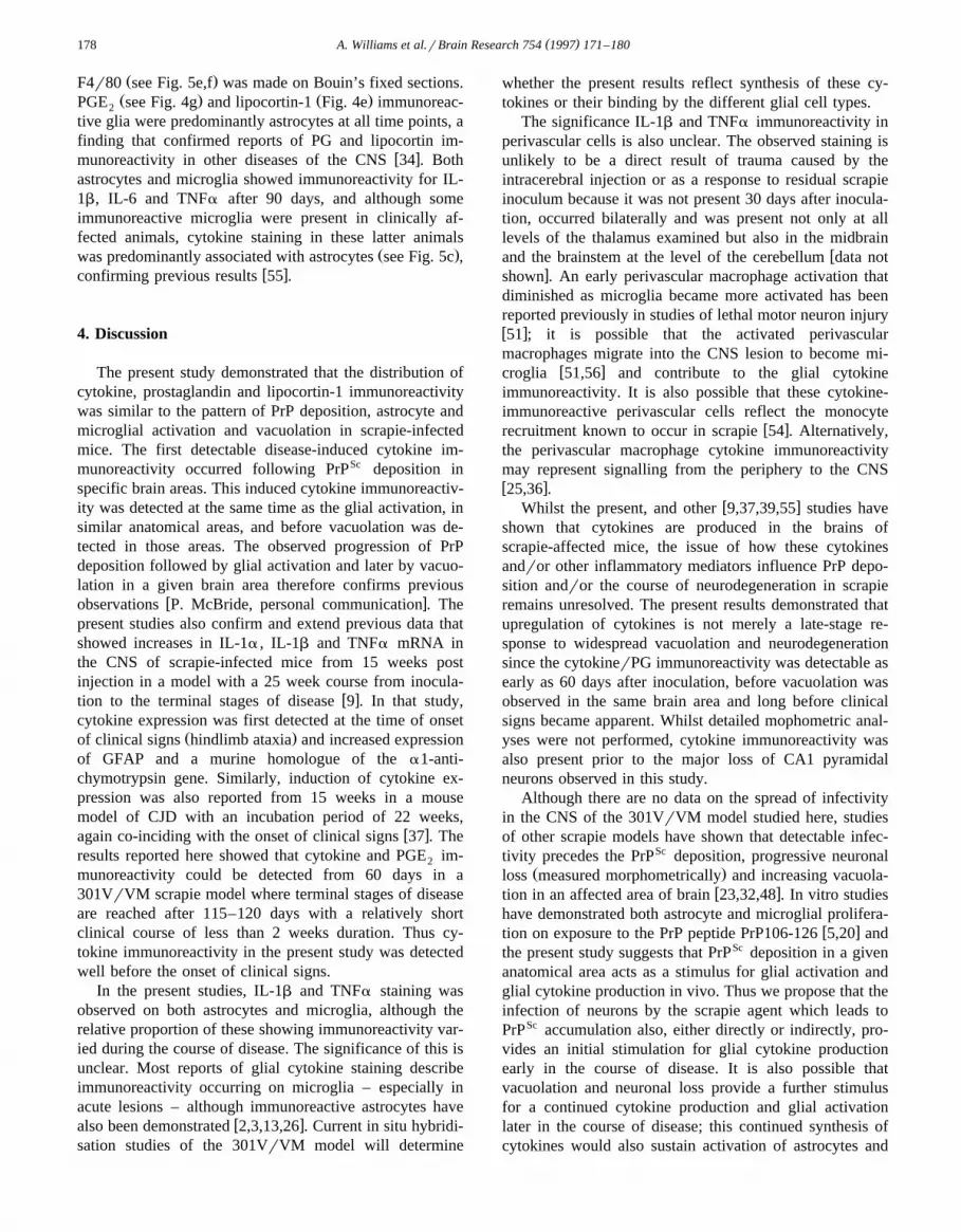
Ž .F4r80 see Fig. 5e,f was made on Bouin’s fixed sections.Ž . Ž .PGE see Fig. 4g and lipocortin-1 Fig. 4e immunoreac-2
tive glia were predominantly astrocytes at all time points, afinding that confirmed reports of PG and lipocortin im-
w xmunoreactivity in other diseases of the CNS 34 . Bothastrocytes and microglia showed immunoreactivity for IL-1b, IL-6 and TNFa after 90 days, and although someimmunoreactive microglia were present in clinically af-fected animals, cytokine staining in these latter animals
Ž .was predominantly associated with astrocytes see Fig. 5c ,w xconfirming previous results 55 .
4. Discussion
The present study demonstrated that the distribution ofcytokine, prostaglandin and lipocortin-1 immunoreactivitywas similar to the pattern of PrP deposition, astrocyte andmicroglial activation and vacuolation in scrapie-infectedmice. The first detectable disease-induced cytokine im-munoreactivity occurred following PrPSc deposition inspecific brain areas. This induced cytokine immunoreactiv-ity was detected at the same time as the glial activation, insimilar anatomical areas, and before vacuolation was de-tected in those areas. The observed progression of PrPdeposition followed by glial activation and later by vacuo-lation in a given brain area therefore confirms previous
w xobservations P. McBride, personal communication . Thepresent studies also confirm and extend previous data thatshowed increases in IL-1a , IL-1b and TNFa mRNA inthe CNS of scrapie-infected mice from 15 weeks postinjection in a model with a 25 week course from inocula-
w xtion to the terminal stages of disease 9 . In that study,cytokine expression was first detected at the time of onset
Ž .of clinical signs hindlimb ataxia and increased expressionof GFAP and a murine homologue of the a1-anti-chymotrypsin gene. Similarly, induction of cytokine ex-pression was also reported from 15 weeks in a mousemodel of CJD with an incubation period of 22 weeks,
w xagain co-inciding with the onset of clinical signs 37 . Theresults reported here showed that cytokine and PGE im-2
munoreactivity could be detected from 60 days in a301VrVM scrapie model where terminal stages of diseaseare reached after 115–120 days with a relatively shortclinical course of less than 2 weeks duration. Thus cy-tokine immunoreactivity in the present study was detectedwell before the onset of clinical signs.
In the present studies, IL-1b and TNFa staining wasobserved on both astrocytes and microglia, although therelative proportion of these showing immunoreactivity var-ied during the course of disease. The significance of this isunclear. Most reports of glial cytokine staining describeimmunoreactivity occurring on microglia – especially inacute lesions – although immunoreactive astrocytes have
w xalso been demonstrated 2,3,13,26 . Current in situ hybridi-sation studies of the 301VrVM model will determine
whether the present results reflect synthesis of these cy-tokines or their binding by the different glial cell types.
The significance IL-1b and TNFa immunoreactivity inperivascular cells is also unclear. The observed staining isunlikely to be a direct result of trauma caused by theintracerebral injection or as a response to residual scrapieinoculum because it was not present 30 days after inocula-tion, occurred bilaterally and was present not only at alllevels of the thalamus examined but also in the midbrain
wand the brainstem at the level of the cerebellum data notxshown . An early perivascular macrophage activation that
diminished as microglia became more activated has beenreported previously in studies of lethal motor neuron injuryw x51 ; it is possible that the activated perivascularmacrophages migrate into the CNS lesion to become mi-
w xcroglia 51,56 and contribute to the glial cytokineimmunoreactivity. It is also possible that these cytokine-immunoreactive perivascular cells reflect the monocyte
w xrecruitment known to occur in scrapie 54 . Alternatively,the perivascular macrophage cytokine immunoreactivitymay represent signalling from the periphery to the CNSw x25,36 .
w xWhilst the present, and other 9,37,39,55 studies haveshown that cytokines are produced in the brains ofscrapie-affected mice, the issue of how these cytokinesandror other inflammatory mediators influence PrP depo-sition andror the course of neurodegeneration in scrapieremains unresolved. The present results demonstrated thatupregulation of cytokines is not merely a late-stage re-sponse to widespread vacuolation and neurodegenerationsince the cytokinerPG immunoreactivity was detectable asearly as 60 days after inoculation, before vacuolation wasobserved in the same brain area and long before clinicalsigns became apparent. Whilst detailed mophometric anal-yses were not performed, cytokine immunoreactivity wasalso present prior to the major loss of CA1 pyramidalneurons observed in this study.
Although there are no data on the spread of infectivityin the CNS of the 301VrVM model studied here, studiesof other scrapie models have shown that detectable infec-tivity precedes the PrPSc deposition, progressive neuronal
Ž .loss measured morphometrically and increasing vacuola-w xtion in an affected area of brain 23,32,48 . In vitro studies
have demonstrated both astrocyte and microglial prolifera-w xtion on exposure to the PrP peptide PrP106-126 5,20 and
the present study suggests that PrPSc deposition in a givenanatomical area acts as a stimulus for glial activation andglial cytokine production in vivo. Thus we propose that theinfection of neurons by the scrapie agent which leads toPrPSc accumulation also, either directly or indirectly, pro-vides an initial stimulation for glial cytokine productionearly in the course of disease. It is also possible thatvacuolation and neuronal loss provide a further stimulusfor a continued cytokine production and glial activationlater in the course of disease; this continued synthesis ofcytokines would also sustain activation of astrocytes and

( )A. Williams et al.rBrain Research 754 1997 171–180 179
microglia that could affect neuronal function by a numberof means. In this context, it has been proposed that mi-croglia activated by PrP may mediate some of the neuronal
w xloss in scrapie 4 . In addition, this synthesis of cytokines,PGs and lipocortin would be sustained by the amplificationof infection in neurons that become irreversibly damaged.Any homeostatic role of lipocortins in limiting the effects
Ž w x.of cytokines see 10,18,19,44 and therefore constrainingthe glial activation andror any cytokine-induced changesin PrP metabolism would be overwhelmed by the contin-ued replication of the scrapie agent and the spread ofinfection through the brain. Clearly, further studies arerequired to determine whether cytokine production by gliacontributes to the neuronal dysfunction and loss, androraffect PrP metabolism and PrPsc accumulation in the brainsof scrapie-infected animals.
Acknowledgements
ŽWe thank C. Farquhar Institute for Animal Health,.BBSRC & MRC Neuropathogenesis Unit, Edinburgh, UK ,
ŽF. Carey and R. Forder Zeneca Pharmaceuticals, Maccles-. Žfield, UK , S. Poole National Institute for Biological
.Standards and Controls, South Mimms, Hertfordshire, UK ,ŽW. Buurman and F. Ramakers State University of Lim-
. Žburg, Maastricht, Netherlands and E. Kawashima Glaxo.IMB, Geneva, Switzerland for their gifts of antibodies.
We also thank L. Coyle, W. Klimek and L. Doughty fortechnical and photographic assistance. This work was sup-ported by the European Community Concerted Actions
ŽGrant BIO2-CT93-0248 co-ordinated by Dr. C. Bostock,.Institute for Animal Health .
References
w x1 Abraham, C.R., Selkoe, D.J. and Potter, H., Immunocytochemicalidentification of the serine protease inhibitor a1-antichymotrypsin in
Ž .brain amyloid deposits of Alzheimer’s disease, Cell, 52 1988484–501.
w x2 Bauer, J., Strauss, S., Schreiter-Gasser, U., Ganter, U., Schegel, P.,Witt, I., Volk, B. and Berger, M., Interleukin-6 and a2-macro-globulin indicate an acute phase state in Alzheimer’s disease cor-
Ž .tices, FEBS Lett., 285 1991 111–114.w x3 Berkenbosch, F., Biewenga, J., Brouns, M., Rozemuller, J.M., Strij-
bos, P. and van Dam, A-.M., Cytokines and inflammatory proteinsŽ .in Alzheimer’s disease, Res. Immunol., 143 1992 657–663.
w x4 Brown, D.R., Schmidt, B. and Kretzschmar, H.A. Role of microgliaand host prion protein in neurotoxicity of a prion protein fragment,
Ž .Nature 380 1996 345–347.w x5 Brown, D.R., Schmidt, B. and Kretzschmar, H.A. A neurotoxic
prion protein fragment enhances proliferation of microglia but notŽ .astrocytes in culture, Glia 18 1996 59–67.
w x6 Bruce, M.E., McBride, P.A. and Farquhar, C.F., Precise targeting ofthe pathology of the sialoglycoprotein, PrP, and vacuolar degenera-
Ž .tion in mouse scrapie, Neurosci. Lett., 102 1989 1–6.w x7 Bruce, M.E., McBride, P.A., Jeffrey, M., Rozemuller, J.M. and
Eikelenboom, P. PrP in scrapie and brA4 in Alzheimer’s disease
show many similar patterns of deposition in the brain. In B. Corain,K. Iqbal, M. Nicolini, B. Winblad, H. Wisniewski and P. ZattaŽ .Eds. , Alzheimer’s Disease: AdÕances in Clinical and Basic Re-search, J. Wiley and Sons Ltd., New York, 1993, pp. 481–487.
w x X8 Cameron, J.D., Characterisation of the 5 flanking region of themurine PrP gene, Ph.D. Dissertation, University of Edinburgh,1995.
w x9 Campbell, I.L., Eddelston, M., Kemper, P., Oldstone, M.B.A. andHobbs, M.V., Activation of cerebral cytokine gene expression andits correlation with onset of reactive astrocyte and acute-phase
Ž .response gene expression in scrapie, J. Virol., 68 1994 2383–2387.w x10 Carey, F., Forder, R., Edge, M.D., Greene, A.R., Horan, M.A.,
Strijbos, P.J.L.M. and Rothwell, N.J., Lipocortin 1 fragment modi-fies pyrogenic actions of cytokines in rats, Am. J. Physiol., 259Ž .1990 R266–R269.
w x Sc11 DeArmond, S.J., Kristensson, K. and Bowler, R.P., PrP causesnerve cell death and stimulates astrocyte proliferation: a paradox,
Ž .Prog. Brain Res., 94 1992 437–446.w x12 DeArmond, S.J., Mobley, W.C., DeMott, D.L., Barry, R.A., Beck-
stead, J.H. and Prusiner, S.B., Changes in the localization of brainŽ .prion proteins during scrapie infection, Neurology, 37 1987 1271–
1280.w x13 Dickson, D.W., Lee, S.C., Mattiace, L.A., Yen, S.C. and Brosnan
C., Microglia and cytokines in neurological disease, with specialŽ .reference to AIDS and Alzheimer’s disease, Glia, 7 1993 75–83.
w x14 Diedrich, J.F., Carp, R.I. and Haase, A.T., Increased expression ofheat shock protein, transferrin and b -microglobulin in astrocytes2
Ž .during scrapie, Microb. Pathogen., 15 1993 1–6.w x15 Dinarello, C.A., Cannon, J.G., Wolff, S.M., Bernheim, H.A., Beut-
let, B., Cerami, A., Figari, I.S., Palladino, M.A. and O’Connor, J.V.,Ž .Tumor necrosis factor cachetin is an endogenous pyrogen and
Ž .induces production of interleukin-1, J. Exp. Med., 163 19861433–1450.
w x16 Eikelenboom, P., Rozemuller, J.M., Fraser, H., Berkenbosch, F.,Ž .Kamphorst, W. and Stam, F.C., 1991 Neuroimmunological mecha-
nisms in cerebral amyloid deposition in Alzheimer’s disease. In T.Ž .Ishii, D. Selkoe and D. Allsop Eds. , Frontiers of Alzheimer’s
Research, Elsevier Science, Amsterdam, 1991, pp. 259–271.w x17 Eikelenboom, P., Zhan, S.S., Van Gool, W.A. and Allsop, D.,
Inflammatory mechanisms in Alzheimer’s disease, Trends Pharma-Ž .col. Sci., 15 1994 447–450.
w x18 Elderfield, A.-J., Newcombe, J., Bolton, C. and Flower, R.J.,Ž .Lipocortins annexins 1, 2, 4 and 5 are increased in the central
Ž .nervous system in multiple sclerosis, J. Neuroimmunol., 39 1992 ,91–100.
w x19 Flower, R.J., Lipocortin and the mechanism of action of the guco-Ž .corticoids, Br. J. Pharmacol., 94 1988 987–1015.
w x20 Forloni, G., Del Bo, R., Angeretti, N., Chiesa, R., Smiroldo, S.,Doni, R., Ghibaudi, E., Salmona, M., Porro, M., Verga, L., Giac-cone, G., Bugiani, O. and Tagliavini, F., A neurotoxic prion proteinfragment induces rat astroglial proliferation and hypertrophy, Eur. J.
Ž .Neurosci., 6 1994 1415–1422.w x21 Fraser, H., The pathology of natural and experimental scrapie. In
Ž .R.H. Kimberlin Ed. , Slow Virus Diseases of Animals and Man,North Holland Publishing Co., AmsterdamrOxford, 1976, pp. 267–305.
w x22 Fraser, H. and Dickinson, A.G., Agent-strain differences in thedistribution and intensity of grey matter vacuolation, J. Comp.
Ž .Pathol., 83 1973 29–40.w x23 Fraser, H. and Dickinson, A.G., Targeting of scrapie lesions and
spread of agent via the retino-tectal projection, Brain Res., 346Ž .1985 32–41.
w x24 Goldgaber, D., Harris, H.W., Hla, T., Maciag, T., Donnelly, R.J.,Jacobsen, J.S., Vitek, M.P. and Gajdusek, D.C., Interleukin 1 regu-lates synthesis of amyloid b-protein precursor mRNA in human
Ž .endothelial cells, Proc. Natl. Acad. Sci. USA, 86 1989 7606–7610.w x25 Graeber, M.B., Streit, W.J. and Kreutzberg, G.W., Identity of ED2-

( )A. Williams et al.rBrain Research 754 1997 171–180180
Ž .positive perivascular cells in rat brain, J. Neurosci. Res., 22 1989103–106.
w x26 Griffin, W.S., Stanley, L.C., Ling, C., White, L., MacLeod, V.,Perrot, L.J., White, C.L. and Araoz, C., Brain interleukin-1 andS-100 immunoreactivity are elevated in Down syndrome and
Ž .Alzheimer disease, Proc. Natl. Acad. Sci. USA, 86 1989 7611–7615.
w x27 Hancock, M.B., DAB-nickel substrate for the differential im-munoperoxidase staining of nerve fibers and fiber terminals, J.
Ž .Histochem. Cytochem., 30 1982 578.w x28 Hardy, J. and Allsop, D., Amyloid deposition as the central event in
the etiology of Alzheimer’s disease, Trends Pharmacol. Sci., 12Ž .1991 383–388.
w x29 Hsu, S.-M. and Soban, E., Color modification of diaminobenzidineŽ .DAB precipitation by metallic ions and its application for double
Ž .immunohistochemistry, J. Histochem. Cytochem., 30 1982 1079–1082.
w x30 Ironside, J.W., Barrie, C., McCardle, L. and Bell, J.E., MicroglialŽ .cell reactions in human spongiform encephalopathies Abstract ,
Ž .Neuropathol. Appl. Neurobiol., 19 1993 , 203.w x31 Ironside, J.W., McCardle, L., Hayward, P.A.R. and Bell, J.E.,
Ubiquitin immunocytochemistry in human spongiform en-Ž .cephalopathies, Neuropathol. Appl. Neurobiol., 19 1993 134–140.
w x32 Jeffrey, M., Fraser, J.R., Halliday, W.G., Fowler, N., Goodsir, C.M.and Brown, D.A., Early unsuspected neuron and axon terminal lossin scrapie–infected mice revealed by morphometry and immuno-
Ž .cytochemistry, Neuropathol. Appl. Neurobiol., 21 1995 41-49.w x33 Jendroska, K., Heinzel, F.P., Torchia, M., Stowring, L., Kret-
zschmar, H.A., Kon, A., Stern, A., Prusiner, S.B. and DeArmond,S.J., Proteinase-resistant prion protein accumulation in Syrian ham-ster brain correlates with regional pathology and scrapie infectivity,
Ž .Neurology, 41 1991 , 1482–1490.w x34 Johnson, M.D., Kamso-Pratt, J.M., Whetsell Jr., W.O. and Pepinksy,
R.B., Lipocortin-1 immunoreactivity in the normal human centralnervous system and lesions with astrocytosis, Am. J. Clin. Pathol.,
Ž .92 1989 424–429.w x35 Kato, S., Hirano, A., Umahara, T., Llena, J.F., Herz, F. and Ohama,
E., Ultrastructual and immunocytochemical studies on balloonedcortical neurons in Creutzfeldt-Jakob disease: expression of ab-crystallin, ubiquitin and stress-response protein 27, Acta Neu-
Ž .ropathol., 84 1992 443–448.w x36 Kida, S., Steart, P.V., Zhang, E.-T. and Weller, R.O., Perivascular
cells act as scavengers in the cerebral perivascular spaces andremain distinct from pericytes, microglia and macrophages, Acta
Ž .Neuropathol., 85 1993 646–652.w x37 Kordek, R., Nerurkar, V.R., Liberski, P.P., Isaacson, S., Yanagihara,
R. and Gajdusek, D.C., Heightened expression of tumor necrosisfactor a , interleukin 1a and glial fibrillary acidic protein in experi-mental Creutzfeldt-Jakob disease in mice, Proc. Natl. Acad. Sci.,
Ž .USA, 93 1996 9754–9758.w x38 Lazarini, F., Castelnau, P., Chermann, J.-F., Deslys, J.-P. and Dor-
mont, D., Modulation of prion protein gene expression by growthfactors in cultured mouse astrocytes and PC-12 cells, Mol. Brain
Ž .Res., 22 1994 268–274.w x39 Liberski, P.P., Nerurkar, V.R., Yanagihara, R. and Gajdusek, D.C.,
Tumor necrosis factor: cytokine-mediated myelin vacuolation inexperimental Creutzfeldt-Jakob Disease, Proc. VIIIth Int. Congr.Virol., Berlin, 1990, Abstract P69-015, p. 421.
w x40 Liberski, P.P., Yanagihara, R., Nerurkar, V.R. and Gajdusek, D.C.,
Tumour necrosis factor-a produces Creutzfeldt-Jakob Disease-likeŽ .lesions in vivo, Neurodegeneration, 2 1993 215–225.
w x41 Lowe, J., Fergusson, J., Kenward, N., Laszlo, L., Landon, M.,Farquhar, C., Brown, J., Hope, J. and Mayer, R.J., Immunoreactivityto ubiquitin-protein conjugates is present early in the disease process
Ž .in the brains of scrapie-infected mice, J. Pathol., 168 1992169–177.
w x42 McBride, P.A., Bruce, M.E. and Fraser, H., Immunostaining ofscrapie cerebral amyloid plaques with antisera raised to scrapie-asso-
Ž . Ž .ciated fibrils SAF , Neuropathol. Appl. Neurobiol., 14 1988 325–336.
w x43 Mann, D.M.A., Younis, N., Jones, D. and Stoddart, R.W., The timecourse of pathological events in Down’s syndrome with particularreference to the involvement of microglial cells and deposits of
Ž .brA4, Neurodegeneration, 1 1992 201–215.w x44 Relton, J.K., Strijbos, P.J.L.M., O’Shaughnessy, C.T., Carey, F.,
Forder, R.A., Tilders, F.J.H. and Rothwell, N.J., Lipocortin-1 is anendogenous inhibitor of ischemic damage in the rat brain, J. Exp.
Ž .Med., 174 1991 305–310.w x45 Salbaum, J.M., Maertens, C.L. and Beyreuther, K., The amyloid
gene of Alzheimer’s disease and neuronal dysfunction. In F. Boller,Ž .R. Katzman, A. Rascol, I.L. Signal and Y. Christensen Eds. ,
Biological Markers of Alzheimer’s Disease, Springer-Verlag, Berlin,1989, pp. 118–122.
w x46 Sasaki, A., Hirato, J. and Nakazato, Y. Immunohistochemical studyof microglia in the Creutzfeldt-Jakob diseased brain, Acta Neu-
Ž .ropathol., 86 1993 337–344.w x47 Scott, J.R. and Fraser, H. Degenerative hippocampal pathology in
( ) Ž .mice infected with scrapie, Acta Neuropathol. Berlin , 65 198462–68.
w x48 Scott, J.R., Jeffrey, M. and Halliday, W.G., Unsuspected earlyneuronal loss in scrapie-infected mice revealed by morphometric
Ž .analysis, Ann. NY Acad. Sci., 724 1994 338–343.w x49 Selkoe, D.J., Normal and abnormal biology of the b-amyloid precur-
Ž .sor protein, Annu. ReÕ. Neurosci., 17 1994 489–517.w x50 Sidman, R.L., Angevine, Jr., J.B. and Pierce, E.T., Atlas of the
Mouse Brain and Spinal Cord, Harvard University Press, Cam-bridge, 1971.
w x51 Streit, W.J., Graeber, M.B. and Kreutzberg, G.W., Expression of Iaantigen on perivascular and microglial cells after sublethal and lethal
Ž .motor neuron injury, Exp. Neurol., 105 1989 115-126.w x52 Van Dam, A.-M., Brouns, M., Louisse, S. and Berkenbosch, F.,
Appearance of interleukin-1 in macrophages and in ramified mi-croglia in the brain of endotoxin-treated rats: a pathway for theinduction of non-specific symptoms of sickness? Brain Res., 588Ž .1992 291–296.
w x53 Williams, A.E., Lawson, L., Perry, V.H. and Fraser, H., Characteri-zation of the microglial response in murine scrapie, Neuropathol.
Ž .Appl. Neurobiol., 20 1994 47–55.w x54 Williams, A.E., Ryder, S. and Blakemore, W.F., Monocyte recruit-
Ž .ment into the scrapie-affected brain, Acta Neuropathol., 90 1995164–169.
w x55 Williams, A.E., Van Dam, A.-M., Man-A-Hing, W.K.H., Berken-bosch, F., Eikelenboom, P. and Fraser, H., Cytokines, prostaglandinsand lipocortin-1 are present in the brains of scrapie-infected mice,
Ž .Brain Res., 654 1994 200–206.w x56 Wisniewski, H.M. and Wegiel, J., Migration of perivascular cells
into the neuropil and their involvement in b-amyloid plaque forma-Ž .tion, Acta Neuropathol., 85 1993 586–595.


