Embed Size (px)
Citation preview
Downloaded from www.microbiologyresearch.org by
IP: 23.22.250.46
On: Mon, 08 Feb 2016 09:08:36
J. gen. Virol. (I98o), 46, 311-324 Printed in Great Britain
3II
Immunoglobul ins in Human Serum Reactive with Murine Friend Leukaemia Virus
By M A U R O B E N D I N E L L I , A N T O N I O TONIOLO, D O N A T E L L A M A T T E U C C I , F R A N C O CELESTE 1 AND
RO BERTO R E V O L T E L L A x*
Institute of Microbiology, University of Pisa, Via S. Zeno, 39 Pisa, and ~Laboratory o f Cell Biology, Tumour lmmunobiology Unit, C.N.R.,
Via Romagnosi I8/A, Rome, Italy
(Accepted 3 September I979)
SUMMARY
Fresh human sera neutralize Friend leukaemia virus (FLV). The activity is abolished by heating (56 °C for 30 min), hydrazine and EDTA treatment, sugges- ting the involvement of complement. Human sera also contain antibody which specifically reacts with FLV. Following incubation with human whole serum, IgG and F(ab')~ fragments, FLV acquire the ability to bind anti-human Ig sera, as shown in neutralization and radioimmunoprecipitation assays. The same is true for FLV-infected cells but not for uninfected control cells, as assessed by immuno- fluorescence. The binding of antibody to FLV-infected cells is prevented by pre-treating the cells with antisera to FLV and FLV-gp7I, and is competed by FLV, FLV-gp7I and, to a lesser extent, by FLV-pI2 and p3o.
I N T R O D U C T I O N
There have been several reports on the presence of antibody to mammalian type-C oncovirus in humans. Positive results have repeatedly been described by using as test antigens primate viruses or various purified structural proteins extracted from them (Lewis et al. I974; Kim, I975; Aoki et al. I976a; Louie et al. I976; Prochownik & Kirsten, 1976; Snyder et al. 1976; Kurth et al. I977; Hirsch et al. I978; Jacquemin et al. 1978; Thiry et al. 1978; Witkin et al. 1978). Less consistent results have been obtained when viruses or virus antigens from lower animals were employed; reactivities attributable to antibody have been detected by some investigations in the serum (Fink et al. I964; Hersh et al. I974; Olsen & Yohn I974; Snyder et al. 1976; Kurth et al. I977; Simkovic et al. I977; Jacque- min et al. 1978; Kurth & Mikschy, 1978) and kidney eluates (Mellors & Mellors, I978) but not by others (Charman et al. 1974; Stephenson & Aaronson, 1976; Krakower & Aaron- son, 1978). Moreover, positive reactivities were generally slight and present in a low pro- portion of subjects. Factors in human sera which inactivate type-C viruses have also been described, but were either not characterized (Bendinelli et al. I974) or recognized as not involving antibody (Fieldsteel, I974; Cooper et al. 1976; Bartholomew et al. I978; Sherwin et al. 1978).
In this paper we show that sera from healthy humans consistently contain antibodies which bind to Friend leukaemia virus (FLV) complex and are mainly directed towards anti- genic determinants on its major envelope glycoprotein, gPTI.
* To whom reprint requests should be addressed.
oo22-13t7/8o/oooo-3734 $o2.oo~) I98O SGM

Downloaded from www.microbiologyresearch.org by
IP: 23.22.250.46
On: Mon, 08 Feb 2016 09:08:36
312 M. BENDINELLI AND OTHERS
METHODS
Friend leukaemia virus (FL V) complex. Two strains of the FLV complex, namely the polycythaemia- (FLV-P) and the anaemia-inducing (FLV-A) strains (for a review see Friend et aL I979) were obtained from Dr C. Friend, New York and Dr M. H. Salaman, London, respectively. FLV-P was propagated in young adult DBA/2 mice (Jackson Labora- tories, Bar Harbor, Maine), purified from plasma filtrates (0"45 ~m membrane filters) by ultracentrifugation (5oooog, SW27 rotor, I'5 h) followed by banding on 2o to 3o% sucrose gradient (7o0oo g, SW27 rotor, 2 h). Virus recovered was analysed for purity by SDS-gel acrylamine electrophoresis. It gave a titre, as assessed by leukaemogenicity in 8-week-old DBA/2 mice, about 5 × Io4 IDr0/o'2 ml, and, as established by 8 to IO day spleen focus assay, about 3 × Io 2 f.f.u./o.2 ml. FLV-A consisted of filtered plasma from 3 week-infected adult BALB/c mice from our own colony and was used without further processing. Its titre, as estimated by 3-week spleen weight assay, was IoS'~IDso/o.2 ml.
FL V-transformed erythroleukaemia cells (FLC). Three clones of mouse FLC were used. Clone 745, a clone producing a poorly leukaemogenic FLV-A, was originally provided by Dr C. Friend. Clone F4-I, a clone releasing in culture a non-leukaemogenic virus defective for the internal p3o protein, was derived from F4-N, a clone of FSD-I from Dr W. Ostertag and was provided by Dr H. Eisen, Paris. Clone 3BM-78-o35, which produces in culture an actively leukaemogenic FLV-P, was derived from the line 3BM-78, originally obtained by in vitro transformation of DBA/2 mouse bone marrow cells by FLV-P (Revol- tella et al. I979). All cells were grown in Dulbecco's modified Eagle's medium supplemented with antibiotics and IO% foetal calf serum (FCS; Microbiological Associates, Bethesda, Maryland) in 7 % COs in air. Primary cultures of bone marrow cells from uninfected DBA/2 mice were prepared according to a modification (Revoltella et al. I975) of the original tech- nique by Golde & Cline 0973).
Preparation of radio-labelled virus. Labelled FLV was prepared by incubating FLC (clones 745 or 3BM-78-35 ) for 24 h in medium containing radioactive precursors. Labelling with SH-leucine (25 #Ci/ml) took place in leucine-free medium, which also contained 2o/zCi /ml of 3H-glucose, 20/~Ci/ml of L-14C-labelled amino acids and only IO % of the standard level of cold amino acids. Isotopes were purchased from New England Nuclear Corp. (Boston, Mass.). Labelled virus was purified from the filtered supernatants as described above.
Sera. Blood was collected aseptically from healthy donors of different ages and from patients with acute lymphatic leukaemia (ALL), lymphoblastoma, neuroblastoma or hypo- gammaglobulinaemia. Human baby cord blood together with the blood of the respective mothers was obtained through the courtesy of Dr S. Fanciulli. The blood was kept at room temperature for 3o rain, at 4 °C for 2 h and then centrifuged at 5oo g for 15 min in the cold. The sera were removed and stored individually at - 7o °C.
Human serum immunoglobulins (Ig) were fractionated first by 50 % (NH~)~SO4 precipi- tation and then by DEAE- and Sephadex G-2oo column chromatography; the eluate fractions were further purified by affinity column chromatography through Sepharose 2B beads (Pharmacia, Uppsala, Sweden) covalently bound with rabbit anti-human IgG or IgM antibodies. The acid-eluted human immunoglobulins were checked for purity by the Ouchterlony technique using reference antisera. Purified IgG and the corresponding F(ab')~ fragments were prepared as described by Nisonoff et aL 0960). The concentrations of IgG and F(ab')2 were adjusted to those present in whole serum.
Rabbit anti-human Ig (Ra. a. HuIg), anti-human IgG (Ra. a. HulgG), anti-human whole serum (Ra. a. HuS), and goat anti-human Ig (G. a. Hulg), anti-human IgM (G. a. HulgM), anti-human IgA (G.a. HulgA) sera were purchased from Hyland Laboratories (Los

Downloaded from www.microbiologyresearch.org by
IP: 23.22.250.46
On: Mon, 08 Feb 2016 09:08:36
Human lg binding to F L V 3 r 3
Angeles, Calif., U.S.A.). Polyspecific rabbit anti-mouse Ig (Ra. a. Molg) was purchased from Meloy Lab (Springfield, Va., U.S.A.). Hyperimmune anti-FLV serum and monospecific antisera against purified FLV-gp7I, -p3o or -pr2 proteins were raised in goats (generous gifts of Dr D. Bolognesi, Durham, North Carolina) and absorbed on DBA/2 mouse spleen, kidney and liver cells at 4 °C for 2 h before use. Mouse anti-FLV serum was raised in BALB/c mice by repeated injections of a formalinized FLV-infected spleen cell extract in Freund's complete adjuvant.
Absorption of human sera. FLC or 5- to 8-day old primary cultures of bone marrow cells from uninfected DBA/2 mice were collected by centrifugation. Spleen, liver and kidney cells were obtained from previously exsanguinated young adult DBA/2 mice. Sheep red blood cells (SRBC), human red blood cells (HRBC), and mouse red blood cells (MRBC) were obtained from Sclavo (Siena, Italy) suspended in Alsever's solution. All cells were washed a minimum of three times in phosphate buffered saline (PBS), pH 7"3, before use. Absorptions were carried out by incubating 0"5 ml of serum diluted I : 1o in PBS with either red cells (8 × roS), nucleated ceils (5 x ios to 5 x Io7), FLV or purified FLV proteins at 37 °C for 30 rain and at 4 °C for 2 h. After centrifugation at 3oo0 g, the supernatant was immedi- ately assayed. Mock absorptions were done identically but without using cells, virus or proteins.
Virus neutralization tests. Three methods were used. Spleen weight assay. FLV diluted in PBS to contain Ioo IDs0 in o.I ml was incubated
with an equal volume of twofold dilutions of the serum under test at 37 °C for 45 min with occasional shaking. Controls with heat-inactivated serum (56 °C for 3 o min) and PBS were always run in parallel. The mixtures were then injected into lots of 3 to 5 BALB/c mice (o.2 ml intravenously) and 3 weeks later the mice were killed and the spleens weighed. The highest dilution of serum which reduced the spleen weight by 5o ~o or more was taken as the neutralizing titre.
S+L - cell assay. FLV diluted to contain IOO (S+L -) f.f.u, in o-2 ml was incubated with serum as in the previous assay. The mixtures (o'4 ml) were then inoculated on to two Falcon plastic dishes (6 cm in diam.) plated the day before with i .6 × IO 5 S+L - D56 cells (Bassin et al. I97I) in 4 m! of McCoy's 5 A medium supplemented with I o % heat-inactivated (at 56 °C for 30 min) FCS and pre-treated with 25/zg/ml DEAE-dextran for 20 rain before inoculation. The culture medium was replaced 2 days later and the cells were stained with Amido black after an additional 2-day incubation. Foci of transformed cells were counted at low magnification. The highest dilution of serum which reduced the number of foci by 5o % or more was taken as the neutralizing titre.
XC cell assay. FLV diluted to contain IOO (XC) p.f.u, in o.2 ml was incubated with serum as above. The mixtures (o'4 ml) were then inoculated on to subconfluent SC-I cells in Falcon dishes (6 cm in diam.) plated the day before and pre-treated with DEAE- dextran as above. Three days later the cells were irradiated with 5ooo rads (cobalt) and super-seeded with I × Io 6 XC cells in 4 ml medium (Rowe et al. I97O). The culture medium was replaced 2 days later and the cells were stained with Amido black after an additional 2-day incubation. The highest dilution of serum which reduced the number of plaques by 5o 0/0 or more was taken as the neutralizing titre.
Radioimrnunoprecipitation (RIP) assay against intact FLV. The method adopted was similar to that described by Ihle et al. (I974). Twofold dilutions of the serum under test (200/~1) were made f rom an initial I : Io dilution in tris-HC1, 0.02 M, pH 7"6, in o.I M-NaCI- o.oot M-EDTA (TNE-A) buffer. SH-labelled intact FLV was then added (20 #1 vol., 5000 to 22 ooo c t /min/2o ng protein) to each dilution. After incubation at 37 °C for I h and at 4 °C for another hour, all the IgG in each dilution was precipitated by adding 20/~1 of an appropriate dilution of G. a. HuIg serum in TNE-A. The mixtures were incubated at 37 °C

Downloaded from www.microbiologyresearch.org by
IP: 23.22.250.46
On: Mon, 08 Feb 2016 09:08:36
314 M. BENDINELLI AND OTHERS
for I h and at 4 °C for 4 h, and then precipitated at 3000 g for 20 min. After two washings in TNE-A, the pellets were dissolved in 1-o M-NaOH and IOO/~l samples were transferred into scintillation vials containing 5 ml Aquasol (New England Nuclear Corp.) and counted. Reactivity was expressed as the percentage of added radioactive virus present in the precipi- tate. In certain cases, the highest dilution of serum which precipitated 5o % or more of the added TCA-precipitable radioactivity was taken as the RIP titre.
R I P assay against purified F L V proteins. Purified virus gp7I and p3o and p I2 from FLV- infected 745 cells were prepared as described by Green et al. (1973) Moennig et al. (1973) and Schafer et al. (I975). Some preparations of gp71 and p3o from FLV-Eveline-STU cells were a generous gift of Dr D. Bolognesi. Proteins were labelled by the chloramine-T procedure and the RIP assays were performed essentially as described previously (Strand et al. I974). The reaction mixture consisted of: (I) 5o/4 of serum diluted in TNE-B buffer (TNE-A buffer supplemented with bovine serum albumin, IO mg/ml) containing o ' 5% NP4o and o-oI M-NaI; (2) Io# l of 125I-labelled virus protein (5 ng, approx. 2 to 5 x io 4 TCA precipitable ct/min) in 4oo #1 plastic Beckman tubes. After incubation for 16 h at 4 °C, 2o/zl of an appropriate dilution of G. a. HuIg serum in TNE-B were added to the mixtures. After additional incubation for I h at 37 °C and I h at 4 °C, the precipitate was pelleted for 3 min in a Beckman microfuge, washed twice in TNE-A supplemented,with o'5 o/o NP4o and tested for the presence of lzSI-labelled antigen in an autogamma LKB spectro- meter.
Competitive R I P assay. Intact FLV or purified gP7I, p3o and pI5 at varying concen- trations, were used as inhibitors of the binding reaction of an optimally labelled FLV- or FLV protein-test serum (or specific antiserum) combination. Test sera were generally used at four times the concentration which precipitated 5o % of the labelled antigen. Reactivity was expressed as the percentage of labelled antigen precipitated by the same serum dilution in the absence of inhibitors.
Immunofluoreseenee (IF). The indirect live cell membrane or, alternatively the acetone- fixed cell IF assay was used as previously described (Cloyd et al. I977). Briefly, thoroughly washed cells were incubated for I h at 37 °C with various dilutions of primary sera, washed three times in PBS and incubated with fluoresceinated secondary antiserum which had been previously absorbed for 2 h at 4 °C on 745 cells and on BALB/c or DBA/2 mouse kidney, spleen and liver cells to prevent non-specific staining. The anti-FLV specificity of the human serum binding to cells was studied by blocking experiments: these were performed by pre- treating the target cells for I h with candidate blocking serum. At least 5oo cells were scored for fluorescence under dark field u.v. illumination with a Zeiss Universal microscope.
R E S U L T S
F L V neutralization
As shown by Table I, the FLV neutralizing activity of pooled fresh human serum was detected by both in vivo and in vitro tests, but the latter were more sensitive. The in vitro S + L - and XC tests exhibited superimposable sensitivities but, due to its simplicity, the for- mer was used in subsequent experiments.
All the individual fresh sera tested, from healthy adults, children or newborns, neutralized FLV (Table 2). There was considerable variability in the activity of individual sera, but on average, adult sera were somewhat more active than infant or newborn sera. Representative titration curves by individual sera are presented in Fig. I ; most sera gave linear curves indi- cating that there was no non-neutralizable virus fraction. Other experiments (data not pre- sented) showed that neutralization required direct contact between serum and virus (pre-

Downloaded from www.microbiologyresearch.org by
IP: 23.22.250.46
On: Mon, 08 Feb 2016 09:08:36
Human Ig binding to F L V 3I 5
Table i. FL V neutralizing activity of fresh and heated human sera as detected by different assays
Neutral izing titre with the test c-
Serum* Spleen weight S+L - cells XC cells
Fresh 1:2 5 I : 1 6 0 I "- 1 6 0
Heated (56 °C for 30 min) < I N.D.T 1:5
* Pool of 5 adult sera. t N.D. = not done.
Table 2. FL V neutralizing activity of individual fresh human sera from different groups of donors
Age in years Neutralizing titre Serum donors (mean + s.d.) (mean + s.d.)
Adults (17)* 44 + 21 1:512+ I65 Children (8) 2+0-6 i :21o_+9o Baby cord (5) - - 1:65 + 20 Babies' mothers (5) zz + 4 < x : 16o
* Number of sera tested given in parenthesis
100 ' ' ' '
S 1 8 ~
)
i
I
f.k
I I 320:160 80 40 20
1/dilution of serum
Fig. I. FLV-neutral izat ion curves f rom individual fresh human sera, as assayed in S+L - cells.
incubation of S+L - cells with serum followed by three washings prior to FLV inoculation or addition of serum to the cultures 2 h after infection had no detectable effect on focus for- mation) and that there was no detectable difference in susceptibility to neutralization between FLV-A and FLV-P, whether harvested f rom mice or from tissue cultures.
The neutralizing activity was nearly completely abolished by treatment of sera at 56 °C for 30 min (Table I) or with hydrazine, or by the presence of EDTA in the neutralization mixture but was little affected by prior heating of the serum at 5 °C for 25 min (Table 3).

Downloaded from www.microbiologyresearch.org by
IP: 23.22.250.46
On: Mon, 08 Feb 2016 09:08:36
316 M. B E N D I N E L L I A N D O T H E R S
T a b l e 3- Effect of complement-active manipulations on the neutralizing activity of fresh human serum
% of FLV infectivity neutralized by serum diluted:
A t
Treatment of fresh serum* ~ :40 1:8o
None 92 6i Hydrazinet 24 17 EDTA ~: 31 12 EDTA-Ca§ 79 55 5o °C for z5 min 72 60
* Pool of five adult sera. t One ml of fresh serum incubated with o'25 ml o-I5 M-NH4OH at 37 °C for 1"5 h and then supplemented
with 0-25 ml o 'I5 M-HCI. :1: Fresh serum was supplemented with an equal vol. of o 'I M-Na2 EDTA in o"5 u-tris-HCl, pH 7"3,
and diluted in Ca- and Mg-free PBS. At the end of incubation, the neutralization mixtures were chilled and supplemented with appropriate dilutions of cold o" 15 M-CaCI~ at the moment of inoculation.
§ Fresh serum was supplemented with a half vol. of 0.2 ~-Naa EDTA and half vol. of 0'3 M-CaCI2 and diluted in Ca- and Mg-free PBS.
T a b l e 4- Effect of anti-human antisera on FL V pre-incubated with hydrazine-decomplemented human sera
% of FLV infectivity
Ist incubation* 2nd incubation-[, neutralized
Fresh serum~ PBS 97 Hydrazine-treated serum PBS 12
I-Iydrazine-treated serum Ra. a. HuS serum 88 PBS Ra. a. HuS serum 16
Hydrazine-treated serum Ra. a. HuIgG serum 96 PBS Ra. a. HulgG serum 18
Hydrazine-treated serum G . a . HulgM serum 66 PBS G .a . HuIgM serum 47
Hydrazine-treated serum G . a . HuIgA serum 19 PBS G . a . HuIgA serum 15
Hydrazine-treated serum Normal Ra. serum z4 PBS Normal Ra. serum 15
* FLV was incubated with human serum (diluted I : 5) or PBS at 37 °C for ! h. Hydrazine treatment of serum was as the footnote of Table 3.
t After the 1st incubation the mixtures were supplemented with an equal vol. of the indicated reagent, incubated for additional 20 rain and then centrifuged in the cold at 700 g for zo min. Before use the antisera were treated with hydrazine and diluted I : IO.
:~ Pool of IO adult sera.
T a b l e 4 p r e s e n t s t h e r e su l t s o f e x p e r i m e n t s in w h i c h F L V was f irst i n c u b a t e d w i t h h y d r a -
z i n e - d e c o m p l e m e n t e d h u m a n s e r u m a n d t h e n i n c u b a t e d fo r o n e f u r t h e r h o u r in t he p r e s e n c e
o f a n t i - h u m a n w h o l e s e r u m or o f m o n o s p e c i f i c a n t i - h u m a n Ig a n t i s e r a (a l so d e c o m p -
l e m e n t e d w i t h h y d r a z i n e ) . D e c o m p l e m e n t e d h u m a n s e r u m , as expec t ed , n e u t r a l i z e d l i t t le
F L V in fec t i v i t y b u t r e n d e r e d it n e u t r a l i z a b l e b y ( in o r d e r o f e f fec t iveness) R a . a . H u l g G
a n d R a . a . H u S s e r u m . G . a . H u l g M s e r u m a f f o r d e d s o m e n e u t r a l i z a t i o n o f b o t h h u m a n
s e r u m - s e n s i t i z e d a n d u n s e n s i t i z e d v i rus . G . a . H u l g A s e r u m a n d c o n t r o l r a b b i t s e r u m d i d n o t n e u t r a l i z e sens i t i zed F L V .

Downloaded from www.microbiologyresearch.org by
IP: 23.22.250.46
On: Mon, 08 Feb 2016 09:08:36
Human Ig binding to FLV
100 , ~ ~ t l ) ~ ~ *
: ~- ( b )
_ v
0 20 80 320 1280 5120 0 20 80 320 1280 5120 1/Dilution of serum
Fig. 2. (a) Titration of four representative normal human sera (A, D, O, A) and of goat anti- FLV serum (O), with intact all-labelled FLV (40 ng protein, 47o0 ct/min). (b) Titration of a pool of five normal human sera (Q) ; IgG purified from this serum pool ( I ) ; F(ab')~ purified from the IgG (A); the same pooled serum after heat-inactivation at 56 °C for 3o min ([]) ; after absorption on 5 x [08 SRBC, MRBC or HRBC (×) , on 1o 7 cells from bone marrow primary cultures from normal DBA/z mice (A), or on lo ~ 745 (C)), 3 BM-78-o35 (*) or F4-I ( + ) FLC.
8o
'~, 60
~ 40
20
317
T a b l e 5. Summary of comparisons between RIP and FL V-neutralization assays of human sera
Neutralization Serum RPI titre* titre
Pool from IO adults 80 20 Pool from 8 adults 80 16o Individual adult 80 io Individual adult ~ 60 160 Individual adult 640 80 Individual adult 320 32o Individual adult 3zo 80 Individual adult (post partum) 32o i6o Individual foetus 8o 8o Five individual adults 80-1- IO N.D.'~ Four individual newborns 8o + to N.D. Pool from 5 adults 320 16o Pool from 5 adults (IgG) 320 16o Hypogammaglobulinaemic
subjects Io 80 Pool from 2 hypogammaglobulin-
aemic subjects 5 32o Three children with ALL 40 + 5 160 + 20 Six children with neuroblastoma 180 q- 3 ° N.D.
* Dilution of serum which precipitated 50 % of SH-FLV input (162oo ct/min, 2o ng). t N.D. = not done.
RIP of intact FL V and purified FL V proteins
H u m a n se ra we re a n a l y s e d fo r i n t a c t F L V - b i n d i n g Ig b y m e a n s o f a sens i t ive a n d specif ic
R I P assay . Al l t he se ra t e s t e d e x h i b i t e d a s ign i f i can t t h o u g h v a r i a b l e b i n d i n g ( r e p r e s e n t a t i v e
r e su l t s s h o w n in Fig. za). T h e a v e r a g e R I P t i t r e o f se ra f r o m h e a l t h y a d u l t d o n o r s was
a b o u t I:ZOO a n d t h a t o f c h i l d r e n , w h e t h e r h e a l t h y o r c a r r i e r s o f n e u r o b l a s t o m a s , a n d o f
n e w b o r n s was n o t m u c h lower . O n l y t h e few t e s t e d se ra f r o m p a t i e n t s w i t h h y p o g a m m a -
g l o b u l i n a e m i a o r A L L h a d l o w e r b i n d i n g ac t iv i ty . T h e r e was n o a p p a r e n t c o r r e l a t i o n
b e t w e e n n e u t r a l i z a t i o n a n d R I P t i t res ( T a b l e 5)-
i2 vr~ 46

Downloaded from www.microbiologyresearch.org by
IP: 23.22.250.46
On: Mon, 08 Feb 2016 09:08:36
318 M. BENDINELLI AND OTHERS
100
~ 80
-~ 60
~ 40 *G
2O
I I I
I i i 103 104 lO s 106 107
No. of cells used for absorption
Fig. 3. Competition RIP assay of a normal human serum with FLC cultures. The assay was performed as described in Methods, using ~H-labelled FLV (20 #g protein, 3000 ct/min), a I : 500 dilution of the serum, which was pre-treated with varying concentrations of absorbing cell cultures: 0 , 745 FLC; Z~, 3BM-78-o35 FLC; O, F4-I FLC; A, bone marrow cultures (6d) from normal DBA/2 mice; I , the same bone marrow cultures but in medium supplemented with IO% FCS.
I00
80
60
e~
"~ 40
20
I 1 I I I
(a)
20 80 320 1280 5120 1/dilution of serum
I I I I I I |
(b)
0"93 1-87 3.75 7.5 15 30 60 Inhibitor (ng)
Fig. 4. (a) Titration of a normal human serum with 8H-labelled FLV or 1251-1abelled purified FLV-antigens: a , 125I-FLV-gp71 (5 ng, 18oooo ct/min); A, SH-FLV (5o ng, 5800 ct/min); I , I~5I- FLV-p3o (5 ng, 9600 ct/min). (b) Competition RIP assay of the same serum tested in (a) with purified FLV fractions. The assay was performed as in Methods, with the serum diluted ~ :4o0, 3H- FLV (20 ng, 22o0 ct/min) and varying concentrations of FLV-pl 2 (O), FLV-p3o (A), FLV-gp71 ( i ) and whole FLV (©).
The Ig nature of the h u m a n serum factor(s) b inding intact FLV was confirmed by controls showing tha t : (I) the RIP titre was no t lowered by heating the sera at 56 °C for I h (Fig. 2b); (2) the b ind ing was almost completely abolished by removal of IgG from the sera by affinity co lumn chromatography (data not shown); (3) solutions of purified IgG and of the derived F(ab')2 at the concentra t ion found in the whole serum presented RIP titres and t i t rat ion curves similar to those of the original serum (Fig. 2b).
Whether the h u m a n Ig b inding was specifically directed to FLV antigens was checked by absorpt ion experiments. The b inding power of pooled sera and of randomly chosen sera was not affected by absorpt ion with MRBC, SRBC and HRBC nor was it significantly reduced by absorpt ion with 6-day cultured bone marrow cells from uninfected D B A / 2
mice (Fig. 2b). It was, instead, abolished by absorpt ion with FLV, both of A or P strain
Downloaded from www.microbiologyresearch.org by
IP: 23.22.250.46
On: Mon, 08 Feb 2016 09:08:36
Human Ig binding to F L V
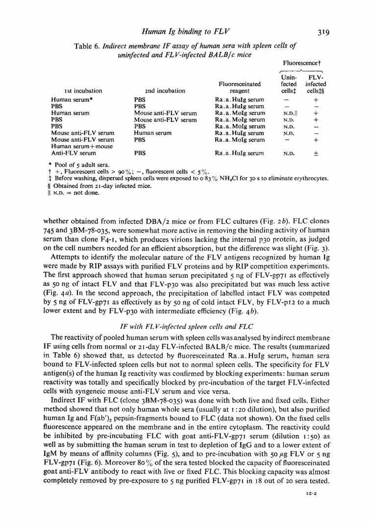
Table 6. Indirect membrane IF assay of human sera with spleen cells of uninfected and FL V-infected BALB/c mice
319
Fluorescence']" _ _ A
Unin- FLV-' Fluoresceinated fected infected
I st incubation znd incubation reagent cells:~ cells~:§ Human serum* PBS Ra. a. HuIg serum -- + PBS PBS Ra. a. Hulg serum - - Human serum Mouse anti-FLV serum Ra. a. MoIg serum N.D.II + PBS Mouse anti-FLV serum Ra. a. MoIg serum N.D. + PBS PBS Ra. a. MoIg serum N.D. -- Mouse anti-FLV serum Human serum Ra. a. HuIg serum N.D. -- Mouse anti-FLV serum PBS Ra.a. MoIg serum -- + Human serum + mouse Anti-FLV serum PBS Ra. a. HuIg serum N.D. +
* Pool of 5 adult sera. t + , Fluorescent cells > 9o % ; - , fluorescent cells < 5 %. :l: Before washing, dispersed spleen cells were exposed to 0"83 % NH4C1 for 30 s to eliminate erythrocytes. § Obtained from 2t-day infected mice. II N.D. = not done.
whether ob ta ined f rom infected D B A / z mice or f rom F L C cultures (Fig. 2 b). F L C clones 745 and 3BM-78-o35, were somewhat more active in removing the b inding act ivi ty of h u m a n serum than clone F4 - l , which produces vir ions lacking the internal p3o protein, as judged on the cell numbers needed for an efficient absorp t ion , bu t the difference was slight (Fig. 3).
A t t empt s to identify the molecular nature o f the FLV antigens recognized by h u m a n Ig were made by RIP assays with purified F L V prote ins and by RIP compet i t ion experiments . The first a p p r o a c h showed tha t human serum precip i ta ted 5 ng of FLV-gp7r as effectively as 5o ng o f intact F L V and tha t FLV-p3o was also prec ip i ta ted but was much less active (Fig. 4a). In the second approach , the prec ip i ta t ion o f label led intact F L V was compe ted by 5 ng o f FLV-gp7~ as effectively as by 50 ng o f cold intact FLV, by F L V - p r 2 to a much lower extent and by FLV-p3o with in termediate efficiency (Fig. 4b).
IF with FL V-infected spleen cells and FLC
The react ivi ty o f pooled human serum with spleen cells was analysed by indirect membrane I F using cells f rom normal or 2 I -day FLV-infected B A L B / c mice. The results ( summar ized in Table 6) showed that , as detected by f luoresceinated R a . a . HuIg serum, human sera bound to FLV-infected spleen cells but not to normal spleen cells. The specificity for F L V antigen(s) o f the h u m a n Ig react ivi ty was confirmed by b locking exper iments : h u m a n serum react ivi ty was total ly and specifically b locked by pre- incubat ion of the target FLV-infec ted cells with syngeneic mouse an t i -FLV serum and vice versa.
Indirect I F with F L C (clone 3BM-78-o35) was done with bo th live and fixed cells. Ei ther me thod showed tha t not only h u m a n whole sera (usually at ~ : zo dilut ion), but also purif ied h u m a n Ig and F(ab')2 pepsin-f ragments bound to F L C (da ta not shown). On the fixed cells fluorescence appea red on the membrane and in the entire cytoplasm. The react ivi ty could be inhibi ted by pre- incubat ing F L C with goat an t i -FLV-gp7I serum (di lut ion I :5o) as well as by submit t ing the h u m a n serum in test to deple t ion o f I g G and to a lower extent o f I g M by means o f affinity columns (Fig. 5), and to pre - incuba t ion with 5o #g F L V or 5 ng F L V - g p 7 I (Fig. 6). Moreove r 8o % of the sera tested b locked the capaci ty o f flu oresceinated goa t a n t i - F L V an t ibody to react with live or fixed FLC. This b locking capac i ty was a lmos t comple te ly removed by pre-exposure to 5 ng purif ied F L V - g p 7 l in J8 out of 2o sera tested.
I2-2

Downloaded from www.microbiologyresearch.org by
IP: 23.22.250.46
On: Mon, 08 Feb 2016 09:08:36
320 M. B E N D I N E L L I A N D O T H E R S
a m
Fig. 5. Membrane immunofluorescence of line 3BM-78-o35 FLC with goat anti-FLV-gp71 antiserum, after staining with fluorescein-labelled rabbit anti-goat IgG antibody: the cells had been previously incubated with a pool from five adult normal humans (a) after and (b) before filtration through Sepharose beads coated with rabbit anti-human IgG antibody. The affinity column completely removed the blocking activity of the serum.
As a control for the FLV-specificity of the human serum binding, human sera were also reacted with 6-day primary cultures of bone marrow cells from DBA/2 mice: only 3 out of 35 sera tested (diluted t :5) stained these cells and their reactivity was totally blocked by pre-incubation with 5 ng of purified FLV-gp7I.
DISCUSSION
It was previously shown that fresh human sera can neutralize FLV infectivity (Bendinelli et al. I974). In the present studies this property of human serum, by analogy with the similar property of guinea pig serum (Bendinelli et al. 1974) and with the retrovirus lyric action of human serum (Cooper et al. I976; Bartholomew et aL 1978; Sherwin et al. 1978), proved strongly dependent on a normal functioning of the classical complement pathway, as indi- cated by its almost complete removal following decomplementation by heat or hydrazine, by its marked reduction when divalent cations in the reaction mixture were chelated with EDTA and by its nearly total resistance to a heat treatment (5o °C for 25 min) known to destroy properdin factor B.
However, the finding that a portion of the neutralizing power consistently resisted serum decomplementation suggested that additional factor(s) might be involved. Natural antibody was an obvious candidate. This possibility was substantiated by the observation that FLV, infected spleen cells or cell lines and purified FLV proteins, following incubation with human sera, acquire the ability to bind anti-human Ig sera in a fashion detectable in neutrali- zation, RIP and IF assays. The same investigations demonstrated that the human Ig which react with FLV mainly belong to the IgG class since: (i) human serum-sensitized FLV was

Downloaded from www.microbiologyresearch.org by
IP: 23.22.250.46
On: Mon, 08 Feb 2016 09:08:36
Human Ig binding to F L V 32I
m W
o
1
t ~
I
Fig. 6. Membrane and cytoplasmic immunofluorescence reaction of acetone-fixed 3BM-78-o35 with a representative normal adult human serum, after staining with fluorescein-labelled rabbit anti- human IgG antibody. The human serum was pre-incubated with (a) FLV (25o ng/o.i ml serum) or (c) FLV-gp7I (20 ng/o.I ml serum) or (b, d) used alone. Fluorescence was completely blocked with FLV or FLV-gp7I.
not neutralized by anti-human IgG serum; (ii) in the RIP test against intact FLV, as well as in the IF assays, purified IgG and the corresponding F(ab')~ fragments at the concen- tration present in whole serum were as active as the unfractionated original serum; (iii) specific removal of IgG from human sera also removed their ability to react with FLV and FLC.
Our present results also provide strong evidence that the antigen(s) recognized by human IgG which react to FLV are viral in nature and the facts which led to this conclusion were: (i) human sera reacted equally well with the anaemia- and the polycythaemia-inducing strains of FLV, whether grown in vivo or in cultured cells; (ii) in IF, human sera reacted strongly with FLV-infected cells, whether producers of complete or of p3o-defective virus, but did not react or reacted very weakly, with uninfected control cells and the reaction could be inhibited by prior incubation of the test cells with homologous or heterologous antibody to FLV. Holder et aL (r976) have described that uninfected mouse cell cultures express low levels of retrovirus glycoprotein antigens on the cell membrane. The occasional reac- tivity of human sera with uninfected mouse bone marrow cells could be explained in accord- ance with these findings: (iii) in neither the RIP nor in the IF assay was the level of reac- tivity between human sera and FLV influenced by performing the tests in the presence of FCS, which can be found linked to in vitro propagated retroviruses (Snyder & Fox, I978); (iv) the reactivity of human sera, as studied in RIP and IF, was entirely absorbed by purified FLV and by FLC, but not by appropriate control mouse cells and could be

Downloaded from www.microbiologyresearch.org by
IP: 23.22.250.46
On: Mon, 08 Feb 2016 09:08:36
3 2 2 M. B E N D I N E L L I A N D O T H E R S
blocked with purified FLV-gp7I ; (v) in RIP assays against purified FLV proteins, human sera reacted strongly with gp71, less with P3O and very poorly with pI2.
It is reasonable to suppose that the natural antibody, unequivocally demonstrated by the present results, is responsible for the low FLV-neutralizing power ofdecomplemented human sera. However, this point is not clarified by the present studies which also do not cast light on its role in the neutralizing activity of fresh serum. In the series of individual sera tested comparatively there was no strict correlation between FLV-neutralizing and RIP titres. For example, sera from hypogammaglobulinaemic patients effectively inhibited FLV but presented low RIP titres. It appears possible that natural antibody contributes only a fraction of the total neutralizing activity of fresh serum, the bulk of which might be due to complement that, according to recent reports (Cooper et al. I976; Bartholomew et aL 1978) is capable of lysing retroviruses independently of the collaboration with antibody.
An implication of our findings is that FLV cross-reacts with antigens which evoke humoral responses in humans. To speculate on the nature of these antigens, until more information is acquired on the precise determinants recognized by natural antibody, is certainly prema- ture but perhaps it is not out of place to mention that natural antibodies to retroviruses have been detected in many animal species and are thought to be elicited by endogenous viruses (Aaronson & Stephenson, 1974; Ihle et aL I974; Lee & Ihle, I975; Aoki et al. I976b). Evidence for the existence of retroviruses in humans is scanty (Hehlmann, i976) but the present results might add to this evidence, considering that antigenic cross-reactions between retroviruses of widely separated mammal species are not rare (Bauer, I974).
Supported in part by a grant from the Italian Research Council (C.N.R.), Progetto Finalizzato Virus.
R E F E R E N C E S
AARONSON, S. A. & STEPHENSON, I. R. (1974). Widespread natural occurrence of high titered neutralizing antibodies to a specif ic c lass o f endogenous murine type-C virus. Proceedings of the National Academy of Sciences of the United States of America 7 x, I 9 5 7 - 1 9 6 I .
AOKI, T., LIU, M., WALLING, M. J., BUSHAR, G. S., BRANDCHAFT, e. B. & KAWAKAMI, T. G. (1976a) . Speci f ic i ty o f naturally occurring antibody in normal gibbon se rum. Science x 9 i , I 18o- I 183.
AOKI, T., WALLING, M. J., BUSHAR, G. S., LIU, M. & HSU, K. C. ( I976b) . Natural antibodies in sera from healthy humans to antigens on surfaces of type-C RNA viruses and cells from primates. Proceedings of the National Academy of Sciences of the United States of America 73, 249 t -2495 .
BARTHOLOMEW, R. M., ESSER, A. F. & MULLER-EBERHARD, H. J. (1978). Lys i s o f oncornaviruses by human se rum. Isolation of the viral complement (CI) receptor and identification as PI5E. Journal of Experimental Medicine x47 , 844-853.
BASSIN, R. N., TLrrTLE, N. & FISCHINGER, P. J. ( I 9 7 I ) . Rapid cell culture assay technique for murine leukemia viruses. Nature, London 229, 564-566.
BAUER, H. (1974). Virion and tumor cell antigens of C - t y p e R N A t u m o u r v i ruses . Advances in Cancer Research 2o, 275 -34 I •
BENDINELLI, M., NARDINI, L. & CAMPA, M. (I974) . N e u t r a l i z a t i o n o f F r i e n d l e u k e m i a virus by sera of u n i m m u - n i zed animals. Journal of General Virology 22, 2o7-214.
CHARMAN, H. P., KIM, N., WFtlTE, M. & GILDEN, R. V. (1974). Failure to detect in human sera, antibodies cross- reactive with group specific antigens of murine leukemia virus. Journal of the National Cancer Institute 52, I 4 0 9 - I 4 1 3 .
CLOYD, M. W., BOLOGNESI, D. P. & BIGNER, D. O. ( t977) . Immunofluoreseent analysis of expression of the R N A t u m o u r virus major glycoprotein, gp7I, on the su r face o f normal murine cells. Cancer Research 37, 931-938 .
COOPER, N. R., JENSEN, V. C., WELSH, R. M. JUN. & OLDSTOm~, M. n. A. ( t976) . Lys i s o f R N A tumor viruses by h u m a n serum: direct antibody-independent triggering of the classical complement pathway. Journal of Experimental Medicine x44 , 97o-984.
FIELDSTEEL, A. H. (I974). N o n s p e c i f i c antiviral substances in human milk active against arbovirus and murine leukemia virus. Cancer Research 34, 712-715.
FINK, M. A., MALGREN, R. A., RAUSCHER, F. J., ORR, H. C. & KARON, M. (1964). Application ofimmunofluorescence to the study of human leukemia. Journal of the National Cancer Institute 33, 581-588.
Downloaded from www.microbiologyresearch.org by
IP: 23.22.250.46
On: Mon, 08 Feb 2016 09:08:36
Human Ig binding to FLV 323
FRIEND, C., SCHER, W., TSUEI, D., HADDAD, J., HOLLAND, J. G., SZRAJER, N. & I-[AUBENSTOCK, H. (1979). Perspec- tives on Friend leukemia virus: pathogenesis in viva and studies on the control of erythrodifferentiation in vitro. In Oji International Seminar on Oncogenic Viruses and Host Cell Genes, 1977 Yamanaka Lake, pp. 279-3o~. Edited by Y. Ikawa and T. Odaka. New York: Academic Press.
GOLDE, D. W. & CLINE, M. S. 0973). Growth of human bone marrow in liquid culture. Blood 4I, 45-52. GREEN, R. W., BOLOGNESI, D. P., SCHAFER, W., PISTER, L., HUNSMANN, G. & DE NORONHA, F. (1973). Polypeptides
of mammalian oncornaviruses. I. Isolation and serological analysis of polypeptides from murine and feline C-type viruses. Virology 45, 565-579.
HEHLMANN, R. (I976). RNA tumour viruses and human cancer. Current Topics in Microbiology and Immu- nology 73, 141--215-
HERSH, E. M., HANNA, M. G., GUTTERMAN, J. U., MAVLIGIT, O., YURCONIC, M. & 6SCHWIND, C. a. 0974). Human immune response to active immunization with Rauscher leukemia virus. II. Humoral immunity. Journal of the National Cancer Institute 53, 327-333.
I~RSCH, M. S., KELLY, A. P., CHAPIN, D. S., FULLER, T. C., BLACK, P. H. & KURTH, R. (I978). Immunity to antigens associated with primate C-type oncornaviruses in pregnant women. Science x99, 1337-I 34o.
HOLDER, W. O., GEER, G. W., BOLOGNESI, D. a. & WELLS, S. A. JU'N. (I976). Detection of the major glycoproteins of Friend leukemia virus (gp71) and the murine mammary tumor virus (gp 52) on the surface of mouse cells. Cancer Research 36, 3217-3224.
JACQUEMIN, P. C., SAXINGER, C. & GALLO, R. C. 0978). Surface antibodies of human myelogenous leukaemia leukocytes reactive with specific type-C viral reverse transcriptases. Nature, London 276, 230-236.
RIM, a. S. 0975)- Presence of antibody to a primate RNA virus in human plasma. Nature, London 257, 614-616.
KRAKOWER, S. M. & AARONSON, S. A. (1978). Seroepidemiological assessment of feline leukaemia virus infection risk for man. Nature, London 273, 463-464.
KURrn, a. & MIKSCHY, O. 0978). Human antibodies reactive with purified envelope antigens of primate type C tumor viruses. Proceedings of the National Academy of Sciences of the United States of America 75, 5692-5696.
KURTH, R., T~ICH, N. M., WEISS, R. & OLlVER, R. T. D. 0977). Natural human antibodies reactive with primate type-C viral antigens. Proceedings of the National Academy of Sciences of the United States of America 74, i237-I241.
IHLE, J. N., HANNA, M. O. JUN., ROBERTSON, L. E. & KENNEY, F. T. 0974). Autogenous immunity to endogenous RNA tumor virus. Identification of antibody reactivity to select viral antigens. Journal of Experimental Medicine x39 , 1568-158I.
LEE, J. C. & IHLE, J. N. (I975). Autogenous immunity to endogenous RNA tumor virus: reactivity of natural immune sera to antigenic determinants of several biologically distinct murine leukemia viruses. Journal of the National Cancer Institute 55, 831-838.
LEWIS, R. M., TANNENBERG, W., SMITYI, C. & SCHWARTZ, R. S. (1974). C-type viruses in systemic lupus erythe- matosus. Nature, London 252, 78-79.
LOUIE, S., CURTIS, J. E. & McCULLOCH, E. A. (1976). Antibodies in human sera to oncornavirus-like proteins from normal or leukemic marrow cell cultures. Journal of Experimental Medicine I44, I243-I253.
MELLORS, R. C. & MELLORS, J. W. (I978). Type-C RNA virus specific antibody in human lupus erythematosus demonstrated by enzymoimmunoassay. Proceedings of the National Academy of Sciences of the United States of America 75, 2463-2467.
MOEN'NIG, V., HUNSMANN, G. & SCr~AFER, W. (1973). Partielle reinigung und biologisch-serologische charak- terisierung Kohnenhydrat kalkiger komponenten aus pr~parater yon Friend-leuk~lmie virus. Zeitschrift far Naturforschung 284, 785-788.
N1SONOFF, A., WISSLER, F. C., IIPMAN, L. N. & WOERNLEY, D. L. (I96o). Separation of univalent fragments from the bivalent rabbit antibody molecule by reduction of disulfide bonds. Archives of Biochemistry 89, 230-244.
OLSEN, R. G. & YOHN, D. S. (1974). Antibodies in haman sera to the mammalian oncornaviruses interspecies antigen. Proceedings of the American Association of Cancer Research x4, 61-67.
PROCHOW~K, E. V. & KmSTEN, W. H. (I976). Inhibition of reverse transcriptases of primate type-C viruses by 7S immunoglobulin from patients with leukaemia. Nature, London 26o, 64-67.
REVOLTELLA, R., PEDICON/, M., BERTOLINI, L. & BOSMAN, C. (I975). In vitro immune response by routine bone marrow cells stimulated against soluble immune complexes. Cellular Immunology zo, I I7-I32.
REVOLTELLA, R., BERTOLINI, L. & FRIEND, C. 0979). In vitro erythroleukemic transformation of mouse bone marrow cells by Friend leukemia virus. Proceedings of the National Academy of Sciences of the United States of America 76, I464-1468.
ROW'E, W. V., PUGH, W. E. & HARTLEY, J. W. (I970). Plaque assay techniques for murine leukemia viruses. Virology 42, tI36-I139 .
SCH.AFER, W., LANGE, J., FISCHINGER, P. J., FRANK, H., BOLOGNESI, D. P., GREEN, R. W. & HUPER, G. (1975). Polypeptides of mammalian oncornaviruses. IL Characterization of a mouse leukemia virus polypeptide (PI5) bearing interspecies reactivity. Virology 63, 48-59.
SHERWIN, S. A., BENVENISTE, R. E. & TODARO, O. J. (1978). Complement-mediated lysis of type-C virus: effect of primate and human sera on various retroviruses. International Journal of Cancer 2x, 6-I I.

Downloaded from www.microbiologyresearch.org by
IP: 23.22.250.46
On: Mon, 08 Feb 2016 09:08:36
324 M. B E N D I N E L L I A N D O T H E R S
SlMKOVIC, D., CrtORVATH, B. & HLUmNOVA, g. (I977). Presence of complement-fixing antibodies against anti- gens of Gross virus-induced rat lymphoma and normal rat thymus in sera of patients with some forms of malignancies. Neoplasma 24, 357-363.
SNYDER, H. W. & FOX, M. (I978). Characterization of foetal calf serum-derived molecule reactive with human natural antibodies: its occurrence in tissue culture-grown type-C RNA viruses. Journal of Immunology xzo, 646--651.
SNVOER, H. W., PINCUS, T. & FLEISSNER, E. (I976). Specificities of human immunoglobulins reactive with anti- gens in preparations of several mammalian type-C viruses. Virology 75, 60-73.
STEPI-IENSON, J. R. & AARONSON, S. A. (I976). Search for antigens and antibodies cross-reactive with type-C viruses of the woolly monkey and gibbon ape in animal models and in humans. Proceedings of the National Academy of Sciences of the United States of America 73, t725-1729.
STRAND, M., LILLY, F. & AUGUST, J. T. (1974) . Host control of endogenous murine leukemia virus gene expres- sion: concentrations of viral proteins in high and low leukemia mouse strains. Proceedings of the National Academy of Sciences of the United States of America 70, 3682-3686.
THIRY, L., SPRECHER-GOLDBERGER, S., BOSSENS, M., COGNIAUX-LE CLERC~ J. & VEREESTRATEN. P. (1978) . N e u t r a l - i z a t i o n of Mason-Pfizer virus by sera from patients treated for renal disease. Journal of General Virology 4 I, 587-597.
WITKIN, S. S., H-IGGINS, P. J. & BENDICH, A. (I978). Inhibition of viral reverse transcriptase and human sperm DNA polymerase by anti-sperm antibodies. Clinical and Experimental Immunology 33, 244-25I.
(Received I7 April I979)


