Embed Size (px)
Citation preview

Summary. Tumorigenesis in human glioblastomamultiforme (GBM) is driven by several geneticabnormalities with disruption of important molecularpathways, such as p53/MDM2/p14ARF andEGFR/PTEN/Akt/mTOR. The malignant progression ofhuman GBM is also primarily associated with a peculiarmultistep pathophysiological process characterized byintratumoral ischemic necrosis (i.e. pseudopalisadingnecrosis) and activation of the hypoxia-inducible factor(HIF)-1α pathway with consequent peritumoralmicrovascular proliferation and infiltrative behaviour.Predictable preclinical animal models of GBM shouldrecapitulate the main pathobiological hallmarks of thehuman disease. In this study we describe two murineorthotopic xenograft models using U87MG and U251human cell lines. Ten Balb/c nude male mice wereorthotopically implanted with either U87MG (5 mice) orU251 (5 mice) cell lines. Intracranial tumor growth wasmonitored through Magnetic Resonance Imaging (MRI).Immunohistopathological examination of the wholecranium was performed 30 days after implantation.U251 orthotopic xenografts recapitulated the salientpathobiological features described for human GBM,including invasive behaviour, wide areas ofpseudopalisading necrosis, florid peripheralangiogenesis, GFAP and vimentin expression,nonfunctional p53 expression, striking active-caspase-3and HIF-1α expression along pseudopalisades. U87MGorthotopic xenografts proved to be very dissimilar fromhuman GBM, showing expansile growth, occasional
necrotic foci without pseudopalisades, intratumorallacunar pattern of angiogenesis, lack of GFAPexpression, fuctional p53 expression and inconsistentHIF-1α expression. Expression of pAkt was upregulatedin both models. The results obtained suggest that theU251 orthotopic model may be proposed as a predictiveand reliable tool in preclinical studies since itrecapitulates the most salient pathobiological featuresreported for human GBM.
Key words: Glioblastoma multiforme, Hypoxia-inducible factor-1, Mouse model, Pseudopalisadingnecrosis
Introduction
Glioblastoma multiforme (GBM) is the mostcommon and highest grade primary brain tumor thatgenerally arises in the cerebral hemispheres (Kleihues etal., 2000). GBM is divided into those that develop fromastrocytic tumors of lower malignancy grade (secondaryGBM), such as diffuse astrocytomas [World HealthOrganization (WHO) grade II] or anaplasticastrocytomas (WHO grade III), and those that developde novo, without evidence of a less malignant precursorlesion (primary GBM) (Ohgaki and Kleihues, 2007).Whether arising de novo or from a lower-gradeprecursor GBM has a truly dismal prognosis with rapidclinical progression leading ultimately to death (Mischelet al., 2003; Brat and Van Meir, 2004). The mainbiologic properties that make GBM fatal are poorresponse to a series of adjuvant therapies and infiltrativebehaviour that makes complete surgical resection nearly
Immunohistopathological and neuroimagingcharacterization of murine orthotopic xenograft models of glioblastoma multiforme recapitulating the most salient features of human diseaseE. Radaelli1, R. Ceruti2, V. Patton2, M. Russo2, A. Degrassi2, V. Croci2, F. Caprera2, G. Stortini2, E. Scanziani1, E. Pesenti2 and R. Alzani21Department of Veterinary Pathology, Hygiene and Public Health, Section of Veterinary and Avian Pathology, Faculty of Veterinary
Medicine, Milano, Italy and 2Department of Pharmacology, Nerviano Medical Sciences S.r.l., Nerviano, Italy
Histol Histopathol (2009) 24: 879-891
Offprint requests to: Alzani Rachele, BU Oncology, Pharmacology Dept,Nerviano Medical Sciences, Viale Pasteur, 10 - 20014, Nerviano(Milan), Italy. e-mail: [email protected]
http://www.hh.um.es
Histology andHistopathology
Cellular and Molecular Biology

impossible (Brat et al., 2004).Specific genetic abnormalities such as LOH 10q,
TP53 mutation, PTEN mutation, p16INK4a deletion andEGFR amplification-overexpression are common inGBM. These genetic aberrations account for a series ofdisrupted molecular pathways (i.e. p53/MDM2/p14ARF
pathway; p16INK4a/RB1 pathway; EGFR/PTEN/Akt/mTOR pathway) that regulate vital cellularprocesses, including apoptosis, cell proliferation and cellmigration (Ohgaki and Kleihues, 2007). Thedevelopment of GBM is also primarly associated with apeculiar multistep pathophysiological process that linksintratumoral ischemic necrosis (i.e. pseudopalisadingnecrosis) and hypoxia to angiogenesis and infiltrativebehaviour (Brat et al., 2004; Rong et al., 2006).Pseudopalisading necrosis is basically characterized byan intratumoral serpentine area of coagulative necrosislined by palisade-oriented GBM cells (pseudopalisades)(Kleihues et al., 2000). Endothelial cell apoptosis inintratumoral vessels and secondary vascular thrombosiscontribute to the ischemic necrosis and hypoxic state(Kaur et al, 2004; Preusser et al 2006). In turn, hypoxiastimulates pseudopalisading cells to activate hypoxia-inducible factor (HIF-1α) leading to overexpression ofangiogenetic factors and peritumoral microvascularproliferations (Brat et al., 2002, 2004). Hypoxia is alsoresponsible for the highly invasive behaviour of GBM,as it elicits migration of GBM cells away from hypoxicareas (Pennacchietti et al., 2003; Brat et al., 2004).Pseudopalisading necrosis and microvascularhyperplasia currently represent two of the most powerfulpredictors of poor prognosis in human GBM (Rong etal., 2006).
Tremendous technical advances have allowed theuse of Magnetic Resonance Imaging (MRI) as a non-invasive method for detection and diagnosis of primarybrain tumors. In astrocytic tumors MRI also gives usefulprognostic indications, detecting the early formation ofpseudopalisading necrosis and microvascularproliferations during the progression of secondary GBMfrom lower grade tumor (Goldbrunner et al., 2000). MRIalso represents a fundamental tool for monitoring blood-brain barrier permeability and the therapeuticresponsiveness of GBM in terms of tumor regression andinhibition of angiogenesis (Zhu et al., 2000; Henson etal., 2005).
Intriguing insights into the pathogenesis of GBMand more accurate prognostic and therapeutic directionsare emerging from the contextual examination ofclinical, immunohistopathological and genomic features.The study of the biological mechanisms underlyingGBM development and the evaluation of new therapiesrequire accurate and reproducible brain tumor animalmodels. Predictable animal models of GBM shouldrecapitulate the main pathobiological hallmarks of thehuman disease concerning progression, kinetics andtumor-host interaction. Although rodent orthotopicxenografts of human GBM are widely employed inpreclinical research, their use remains controversial andthese models have been criticized for not recapitulating
the main morphological and biological aspects of humanGBM (Fomchenko and Holland, 2006). In fact, recentinvestigations confirmed that fundamental immuno-histopathological, neuroimaging and biologicalsimilarities exist between murine orthotopic xenograftmodels and naturally occurring human GBM (Candolfiet al., 2007; Martínez-Murillo and Martínez, 2007). Adetailed characterization and standardization aretherefore mandatory to rule out the predictivity range ofspecific preclinical models.
In this study, we report the histopathological,immunohistochemical and imaging (MRI) characte-rization of mouse orthotopic xenograft models using twodifferent cell lines of human GBM, U251 and U87MG.
Material and methods
Cell lines
The human brain tumor cell lines U251 and U87MG(glioblastomas) were purchased from National CancerInstitute, Bethesda, MD and from American Type CellCulture, Manassas, VA respectively and grown inRPMI1640 medium supplemented with 10% Fetal CalfSerum (FCS), 2 mM L-Glutamine (U251) and Eagle'sMinimal Essential Medium (E-MEM) supplementedwith 10% FCS, 2 mM L-Glutamine and 1% non-essential amino acids (U87MG).
Animals and study design
All procedures adopted for housing and handling theanimals were in strict compliance with EEC and ItalianGuidelines for Laboratory Animal Welfare.
Ten Balb/c nude male mice, aged 6-8 weeks, wereanesthetized and stereotactically implanted in theputamen region with human GBM cell lines, 5 mice withU87MG and 5 mice with U251 (105 cells in 2 µ l ofPBS).
By using the stereotactic X and Y axis controller,bregma (interception between median sagittal andanterior coronal sutures) and lambda (interceptionbetween median sagittal and posterior coronal sutures)were identified as point 0. The following coordinates [+1 mm posterior and + 3 mm lateral (right)] were set inthe apparatus and the point of injection identified. With amicrodrill a small hole was made in the desired point.Cells were aspirated with a Hamilton syringe just priorto injection (a brief mix of cells before aspiration isperformed to avoid precipitation of cells). By pointing tothe hole, Z axis was fixed to 0 and the syringe gentlyinserted into the brain until reaching the correctcoordinate (-3.5 mm depth). Cells were injected at thespeed of 1 µl every minute. The syringe was left for anadditional 5 minutes in the hole before removing it toavoid cell aspiration. The hole was closed using bonewax and the wound was closed with sterile autoclips.Following surgery, mice were monitored for recoveryuntil complete wakening.
880
Orthotopic mouse models of glioblastoma

MR imaging
MRI was performed to visualize and delineatetumors in the nuclei area. In order to monitor tumorprogression in each animal, a serial study was perfomedby weekly imaging sessions starting at day 8 afterintracranial injection. For imaging studies, mice wereanesthetized with gas isofluoran, positioned prone in theanimal bed and inserted in the radiofrequency coil (38mm i.d.) inside the magnet. Images were acquired on aBruker Pharmascan instrument (Bruker, Germany)equipped with a 7.0 T horizontal magnet. Transversaland sagittal spin echo multislice images were acquired aspilot scans for correct localization of brain areas. Then,T2-weighted (T2-W) and T1-weighted (T1-W) scanswere run in both transversal and coronal orientations inorder to better highlight the characteristics and locationof tumors. T1-W images were acquired before and afteriv injection of the contrast agent gadolinium-diethylenetriamine penta-acetic acid (Gd-DTPA,Guerbet). Imaging parameters were as follows:
T2-W: RARE (TR=5000 ms; TEeff=57 ms; rarefactor=8; 4 averages; FOV=2.5*2.5 cm2; Slicethickness=1 mm; matrix=256*128; Total acquisitiontime=5 min 20sec.
T1-W: RARE (TR=800 ms; TEeff=25.5 ms; rarefactor=4; 4 averages; FOV=2.5*2.5 cm2; Slicethickness=1 mm; matrix=256*128; Total acquisitiontime=1 min 42sec.
The whole imaging session lasted around 25 minutesfor each animal.
Histopathology & Immunohistochemistry
For histopathology and immunohistochemistrystudies, mice were sacrificed at a late stage of thedisease corresponding more or less to day 30 afterintracranial injection. The whole cranium from eachanimal was collected and fixed in buffered formalin for 4to 5 hours then decalcified in 5% formic acid diluted inbuffered formalin for 24h. The region between externalauditory meatus and orbit was sliced and a coronalsection of the whole cranium was obtained and
embedded in paraffin. Serial 4 µm-thick sections weresubsequently placed on Superfrost plus slides andstained with haematoxylin and eosin (HE) forhistological examination. For immunohistochemistry,sections were deparaffinized in xylene, rehydrated ingraded ethanol, and transferred to PBS. Heat treatmentantigen retrieval was performed in citrate buffer pH 6(Vector). The slides were rinsed with PBS andendogenous peroxidase was blocked using 3% hydrogenperoxide (Sigma) in PBS for 10 minutes. The tissueswere incubated for 20 minutes at room temperature witha protein-blocking solution consisting of PBS containing0.05% Tween 20 (DAKO) and 10% normal goat serum(Vector). Slices were then incubated with the primaryantibodies as reported in Table 1. The samples were thenrinsed in PBS containing 0,05% Tween 20 and incubatedfor 30 minutes at room temperature with the appropriatesecondary antibody as reported in Table 1. Glialfibrillary acidic protein (GFAP), S100 and Factor VIII(FVIII)-stained slides were additionally incubated for 30minutes at room temperature with streptavidin-peroxidase (Dako). Finally, the reaction was developedwith diaminobenzidine (Dako).
Immunostained and HE-stained sections from eachcase were qualitatively and quantitatively/semi-quantitatively scored as follow (see also Table 2).
Necrosis (HE stained sections):(-) = no intratumoral necrotic foci(+) = <20% of the mass effaced by intratumoral
necrotic foci(++) = 20-50% of the mass effaced by intratumoral
necrotic foci(+++) = >50% of the mass effaced by intratumoral
necrotic fociHIF-1α nuclear expression by neoplastic cells:(-) = No immunohistochemical expression of HIF-
1α by neoplastic cells(+) = Immunohistochemical expression of HIF-1α
by neoplastic cells in perinecrotic areas(++) = Immunohistochemical expression of HIF-1α
by neoplastic cells in perinecrotic areas and in the viableportion of the tumor as well
Tumor-associated thrombi:
881
Orthotopic mouse models of glioblastoma
Table 1. Antibodies used in immunohistochemical assay: source, clone/company code, dilution, incubation time and secondary antibody.
ANTIBODY COMPANY CLONE / WORKING INCUBATION SECONDARYCOMPANY CODE DILUTION TIME ANTIBODY
Vimentin Epitomics SP20 1:50 40 min at 37°C EnVision Rabbit-HRPGFAP Dako Z334 1:10000 40 min at 37°C Biotinylate Anti-RabbitS100 Dako Z311 1:15000 40 min at 37°C Biotinylated Anti-RabbitSynaptophysin Epitomics EP1098Y 1:100 1 hour at room temperature EnVision Rabbit-HRPHLA-abc MBL EMR8-5 1:100 1 hour at room temperature EnVision Mouse-HRPFVIII Dako N1505 1:80 40 min at 37°C Biotinylated Anti-RabbitActive-caspase-3 Cell Signaling Asp175 1:100 1 hour at room temperature EnVision Rabbit-HRPp53 Dako DO-7 1:50 1 hour at room temperature EnVision Mouse-HRPHIF 1α BD Transduction Laboratories 54 1:50 1 hour at room temperature EnVision Mouse-HRPKi-67 Dako MIB-1 1:150 1 hour at room temperature EnVision Mouse-HRPAKT (pS473) Epitomics EP 2109y 1:200 1 hour at room temperature EnVision Rabbit-HRP

(-) = no FVIII positive intratumoral thrombi(+) = <20 FVIII positive intratumoral thrombi(++) = 20-50 FVIII positive intratumoral thrombi(+++) = >50 FVIII positive intratumoral thrombiTumor angiogenesis (microvessel density):The number of FVIII positive vascular channels
were assessed in four 400x microscopic fields both inthe centre and at periphery of the tumor. Mean valuesfrom each case are reported (Table 2).
Ki-67 (MIB-1) and p53 expression by neoplasticcells: Both for Ki-67 and p53, the number ofimmunopositive nuclei were counted on a total amountof about 3000 neoplastic cells at 200x microscopicfields. Mean percentages from each case are reported(Table 2).
Immunohistopathological evaluations wereperformed using Image Pro-Plus 6.0 softwaretechnology. Statistical analysis was performed usingMann-Whitney test (Prism 3.0 software).
Results
Imaging
The development and growth of neoplastic lesionsderived from U251 or U87MG implantation werefollowed in each animal at different time points. Bothcell lines grew very quickly, even if a more aggressivegrowth pattern was noticed in the U251 model.
In terms of MR signal intensity, some differences
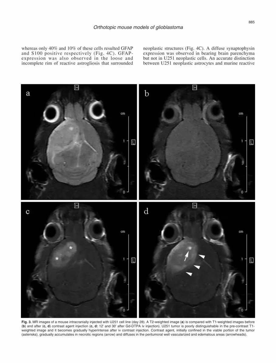
were observed for the two cell lines. The U251 tumorwas delineated from normal brain tissue as an isointenseor lightly hyperintense, poorly demarcated, irregularlyrounded area surrounded by a hyperintense rim(peritumoral edema). The development of intratumoralnecrotic areas was common in this model (Fig. 1a'-d').The U87MG tumor was delineated from normal braintissue as a hyperintense, well demarcated, nodular area,with a regularly expansile growth (tennis ballappearance) (Fig. 2a-d). In both models the compressioncaused by tumor growth leads to internal hydrocephalus,due to blockage of cerebrospinal fluid outflow in theventricles or in the subarachnoid space (Fig. 1d' and 2d).Gd-DTPA uptake profile in U251 xenograft showed thatthe tumor was poorly distinguishable in the pre-contrastT1-weighted image (Fig. 3b) and it became graduallyhyperintense after iv contrast injection in the tail vein.Irregular signal dishomogeneities were present inside thetumor, due to the presence of necrotic areas. Contrastagent, initially confined in the viable portion of thetumor, gradually accumulated in necrotic regions anddiffused in the peritumoral well vascularized andedematous areas (Fig. 3c,d).
Histopathology and immunohistochemistry
U251 orthotopic xenografts were morphologicallycharacterized by a single, densely cellular, irregular,poorly demarcated, unencapsulated, intrahemisphericmass with peripheral medusoid projections invading the
882
Orthotopic mouse models of glioblastoma
Table 2. The table summarizes the scores for the different histopathological and immunohistochemical aspects investigated in U87MG and U251murine orthotopic xenograft models of human glioblastoma multiforme.
GBM line Record Ki-67 p53* Angiogenesis* Necrosis HIF-1α Thrombi
Central Peripheral
U87MG #67-1 45.10% 11.61% 9 3.25 - - ++#663-1 41.69% 24.11% 10.5 1.25 ++ + +++#851-1 56.78% 11.94% 12 2.25 - - +#861-1 41.71% 6.94% 9.25 3 + + ++#899-1 32.85% 9.8% 14.5 2.25 - - +++
Mean 43.63% 12.88% 11.05 2.4
U251 #389-1 36.71% 90.12% 0 6.25 ++ ++ -#952-2 47.74% 99.8% 0 7.5 + ++ -#966-3 51.48% 99.72% 0.25 10.5 ++ ++ -#9614-3 63.66% 95.43% 0 7.5 + ++ -#9615-3 60.35% 97.51% 2 11 + ++ -
Mean 52% 96.51% 0.45 8.55
* Statistically significant differences between U251 and U87MG group. Necrosis (HE stained sections): (-) = no intratumoral necrotic foci, (+) = <20% ofthe mass effaced by intratumoral necrotic foci, (++) = 20-50% of the mass effaced by intratumoral necrotic foci, (+++) = >50% of the mass effaced byintratumoral necrotic foci; HIF-1α nuclear expression by neoplastic cells: (-) = No immunohistochemical expression of HIF-1α by neoplastic cells, (+) =Immunohistochemical expression of HIF-1α by neoplastic cells in perinecrotic areas, (++) = Immunohistochemical expression of HIF-1α by neoplasticcells in perinecrotic areas and in the viable portion of the tumor as well; Tumor-associated thrombi: (-) = no FVIII positive intratumoral thrombi, (+) = <20FVIII positive intratumoral thrombi, (++) = 20-50 FVIII positive intratumoral thrombi, (+++) = >50 FVIII positive intratumoral thrombi; Tumor angiogenesis(microvessel density): The number of FVIII positive vascular channels were assessed in four 400x microscopic fields both in the centre and at peripheryof the tumor. Mean values from each case are reported; Ki-67 and p53 expression by neoplastic cells: Both for Ki-67 and p53. the number ofimmunopositive nuclei were counted on a total amount of about 3000 neoplastic cells at 200x microscopic fields. Mean percentages from each case arereported.

surrounding cerebral parenchyma and often spreadinginto the controlateral hemisphere. This mass consisted oftightly packed, interlacing bundles of pleomorphic,spindle to stellate cells with occasional giant andmultinucleated elements. Findings of atypia, includingmarked anisocytosis, anysokariosis, bizarre mitosis and
multiple irregular nucleoli, were common amongneoplastic cells. In all examined cases, wide intratumoralfoci of vascular-oriented (serpentine) pseudopalisadingnecrosis were observed (Table 2; Fig. 4A). Infiltratingneoplastic projections at the periphery of the mass wereintimately associated with tortuous microvascular
883
Orthotopic mouse models of glioblastoma
Fig. 1. T2-weighted images: coronal sections at different time points showing the progression and growth of the intrahemispheric neoplastic lesions inU251 (a': day 8; b': day 15; c': day 21; d': day 28). U251 xenograft appears as an isointense or lightly hyperintense, poorly demarcated, irregularlyrounded area surrounded by a hyperintense rim (peritumoral edema, asterisks). Note also the presence of intratumoral necrosis (arrow).

proliferations often characterized by hypertrophic liningendothelium. Prominent rarefaction-status spongiosusand edema of the surrounding neuroparenchyma andoccasional intratumoral and peritumoral haemorrhagicfoci were also observed. Infiltrating neoplastic
extensions along the ependimal surface into the lateralventricles (Fig. 4B) as well as along the pial surface intothe subarachnoid space represented a common finding.
In U251 orthotopic xenografts, almost all theneoplastic cells displayed intense vimentin expression,
884
Orthotopic mouse models of glioblastoma
Fig. 2. T2-weighted images: coronal sections at different time points showing the progression and growth of the intrahemispheric neoplastic lesions inU87MG xenografts (a: day 8, b: day 14, c: day 22, d: day 29). U87MG xenograft appears as a hyperintense, well demarcated, nodular area, with aregularly expansile growth and a tennis ball appearance (arrowhead).
whereas only 40% and 10% of these cells resulted GFAPand S100 positive respectively (Fig. 4C). GFAP-expression was also observed in the loose andincomplete rim of reactive astrogliosis that surrounded
neoplastic structures (Fig. 4C). A diffuse synaptophysinexpression was observed in bearing brain parenchymabut not in U251 neoplastic cells. An accurate distinctionbetween U251 neoplastic astrocytes and murine reactive
885
Orthotopic mouse models of glioblastoma
Fig. 3. MR images of a mouse intracranially injected with U251 cell line (day 28). A T2-weighted image (a) is compared with T1-weighted images before(b) and after (c, d) contrast agent injection (c, d: 12' and 30' after Gd-DTPA iv injection). U251 tumor is poorly distinguishable in the pre-contrast T1-weighted image and it becomes gradually hyperintense after iv contrast injection. Contrast agent, initially confined in the viable portion of the tumor(asterisks), gradually accumulates in necrotic regions (arrow) and diffuses in the peritumoral well vascularized and edematous areas (arrowheads).

886
Orthotopic mouse models of glioblastoma
Fig. 4. Histopathology and immunohistochemistry of murine intracranial orthotopic xenograft of glioblastoma multiforme using U251 human cell line. A.Linear intratumoral area of pseudopalisading necrosis. HE staining. B. The human-specific marker HLA-abc defines the infiltrative projections ofintracranial U251 xenograft with invasion of lateral ventricles and spread toward the controlateral hemisphere. HLA-abc immunostaining. C. Approximately 40% of the neoplastic cells within the U251 xenograft display GFAP expression. Marked GFAP expression is also evident in theincomplete and loose peripheral rim of astrogliosis. GFAP immunostaining. D. Disseminated along the infiltrative medusoid neoplastic projections arenumerous FVIII-positive vascular channels. FVIII immunostaining. E. Disseminated along the serpiginous intratumoral pseudopalisading necrosis arenumerous apoptotic cells. Active caspase-3 immunostaining. F. Virtually all neoplastic cells within the U251 xenograft display prominent intranulcearaccumulation of p53 protein. p53 immunostaining. G. Numerous neoplastic cells with prominent HIF-1α-nuclear expression are concentrated along anintratumoral area of pseudopalisading necrosis. HIF-1α immunostaining. Scale bar: A, C-G, 100 µm; B, 250 µm.

astrocytes and gemistocytes was performed using thehuman-specific marker HLA (human leukocyte antigen)-abc. In addition, HLA-abc staining clearly underlinedthe infiltrative intrahemispheric growth of U251xenografts (Fig. 4B). FVIII-staining confirmed the floridangiogenetic processes (high microvessel density)associated with the peripherally infiltrating neoplasticprojections (Table 2; Fig. 4D). Only occasional FVIII-positive intratumoral vascular channels were observed inU251 orthotopic xenografts (Table 2). Multifocal groupsof active caspase-3-positive apoptotic cells, mostlyconcentrated in the necrotic and perinecrotic areas, wereobserved (Fig. 4E ). Scattered apoptotic cells were also
found in the viable neoplastic portions. Almost the entireU251 neoplastic cell population (96,51%) displayed aprominent nuclear accumulation of p53 protein (Table 2;Fig. 4F). Fifty-two % of U251 neoplastic cells alsoshowed nuclear Ki-67 expression (Table 2). MultifocalHIF-1α nuclear expression by U251 neoplastic cells wasrecorded within pseudopalisades along the perinecroticareas, as well as in the surrounding viable portion of thetumor (Table 2; Fig. 4G). For the vast majority of U251neoplastic cells, pAkt-immunohistochemical analysisrevealed a diffuse, finely granular positivity both in thecytoplasm and along the cell membrane (Fig. 6A).
887
Orthotopic mouse models of glioblastoma
Fig. 5. Histopathology and immunohistochemistry of murine intracranial orthotopic xenograft of glioblastoma multiforme using U87MG human cell line.A. U87MG xenograft appears as well-demarcated intrahemispheric expansile mass consisting of tightly packed sheets of round to polygonal,occasionally plump spindle cells with abundant intensely eosinophilic cytoplasm. HE staining. B. U87MG xenograft result GFAP-negative and it isrimmed by a prominent GFAP-positive capsular reaction of astrogliosis. GFAP immunostaining. C. The human-specific marker HLA-abc highlights theintrahemispheric expansile growth of U87MG xenograft. HLA-abc immunostaining. D. The widespread intratumoral network of anastomosing vascularlacunae show an incomplete FVIII-positive endothelial lining. Furthermore, FVIII-staining highligts a large number of non-occlusive vascularmicrothrombi. FVIII immunostaining. Scale bar: 100 µm.

U87MG orthotopic xenografts were morphologicallycharacterized by a single, densely cellular, welldemarcated, non-infiltrating, expansile, intrahemisphericnodular mass surrounded by a compact reaction ofastrogliosis. The mass consisted of tightly packed sheetsof plump, round to polygonal, occasionally spindle cellswith abundant intensely eosinophilic cytoplasm andfrequent giant and multinucleated elements (Fig. 5A).Moderate findings of atypia were observed amongneoplastic cell. These included anisocytosis,anysokariosis, bizarre mitosis and multiple irregularnucleoli. Variable-sized intratumoral foci of coagulativenecrosis were observed in only two cases (Table 2).These were associated with prominent vascularthrombosis and concurrent marked neutrophilicinfiltration. Neoplastic sheets were often separated bydelicate anastomosing vascular lacunae and variabledegrees of edema. Occasional intratumoral andperitumoral haemorrhagic foci were also observed.
In U87MG orthotopic xenografts, almost all theneoplastic cells displayed intense vimentin expression.On the contrary, no concurrent GFAP or S100 expressionwas recorded. Striking GFAP-expression was observedin the compact rim of reactive astrogliosis thatsurrounded neoplastic structures (Fig. 5B). A diffusesynaptophysin expression was observed in bearing brainparenchyma but not in U87MG neoplastic cells. Also, inthese cases, the use of the human-specific marker HLA-abc allowed an accurate distinction between U87MGneoplastic astrocytes and murine reactiveastrocytes/gemistocytes. In addition, human-specific
HLA-abc antibody staining clearly underlined theexpansile intrahemispheric growth of U87MGxenografts (Fig. 5C). FVIII-staining confirmed thepresence of a high intratumoral microvessel density,consisting of a widespread network of anastomosingvascular lacunae. Furthermore, FVIII-staining delineateda large number of non-occlusive vascular microthrombi(Table 2; Fig. 5D). Only occasional FVIII-positiveperitumoral vascular channels were observed in U87MGorthotopic xenografts (Table 2). Numerous groups ofactive caspase-3-positive apoptotic cells, mostlyconcentrated in the occasional necrotic and perinecroticareas, were observed. Scattered apoptotic cells were alsofound in the viable neoplastic portions. Only 12,88% ofthe U87MG neoplastic cell population displayed variabledegrees of p53-nuclear expression (Table 2). About 44%of U87MG cells showed nuclear Ki-67 expression(Table 2). HIF-1α nuclear expression by U87MGneoplastic cells was mostly distributed along theoccasional perinecrotic areas (Table 2). For the vastmajority of U87MG cells, pAkt immunohistochemicalanalysis revealed a diffuse, finely granular positivity,both in the cytoplasm and along the cell membrane (Fig.6B).
Statistical analyses revealed significant differencesbetween U251 and U87MG groups with respect to theperipheral (p=0,0079) and the central (p=0,0079)microvessel density (FVIII-immunostaining; Table 2). Asignificant difference between the two groups was alsoobserved for p53- (p=0,0001) but not for Ki-67-immunopositive cells (p=0,2) (Table 2).
888
Orthotopic mouse models of glioblastoma
Fig. 6. Murine intracranial xenograft models of glioblastoma multiforme using U251 and U87MG human cell lines. Both U251 (A) and U87MG (B) tumorcells show diffuse, finely granular positivity for pAkt within the cytoplasm and along the cell membrane. pAkt immunostaining. Scale bar: 100 µm.

Discussion
In this study, the histopathological, immuno-histochemical and MRI characterization highlightedprofound differences between the two investigatedorthotopic xenograft models of GBM. Indeed, where theU251 orthotopic xenograft model recapitulates most ofthe salient pathobiological features described in humanspontaneous GBM, the U87MG orthotopic xenograftmodel proved to be very different.
MR imaging monitoring of U251 orthotopicxenografts was able to detect the progressive invasivegrowth into the surrounding brain parenchyma, thedevelopment of intratumoral necrotic areas, as well asthe peripheral florid vascular activity with edema.Similar imaging findings are also reported for humanGBM, and are considered important diagnostic andprognostic features (Rong et al., 2006). In U251xenografts, the nature of signal alterations observed onMRI was then confirmed through the histopathologicaland immunohistochemical examination.
Neoplastic cells in U251 orthotopic xenograftsshowed a typical GBM phenotype, staining concurrentlypositive for vimentin, GFAP, S100 and negative forsynaptophysin (Kleihues et al., 2000; Porter et al., 2004).In agreement with the striking interhemispheric invasivespread reported for human spontaneous GBM, in thisstudy, HLA-abc staining precisely defined the prominentinfiltrative behaviour of the growing U251 xenograft andits tendency to invade the ventricular system (Kleihueset al., 2000).
In U251 orthotopic xenografts, intratumoral areas ofcoagulative necrosis, similar to that described for humanspontaneous GBM, displayed a serpiginous morphologyand were peculiarly lined by pseudopalisading neoplasticcells. The coagulative nature and the serpiginousmorphology of the necrotic areas suggest an underlyingvascular damage with ischemic process (Zagzag et al.,2000a; Brat et al., 2004). In human GBM, the activationof a prothrombotic state along neoplastic vasculature isconsidered one of the most likely causes of ischemicnecrosis. However, the molecular mechanismspromoting this peculiar event are largely unclear (Bratand Van Meir, 2004). In U251 xenografts neitherhistopathological examination or FVIII-staining wereable to detect necrosis-associated occlusive thromboticlesions. This finding indicates that vascular thrombosisdoes not represent the mechanisms leading topseudopalisading necrosis in this model. However, othermechanisms such as vascular collapse and markedCD95-CD95 ligand-mediated apoptotic activity shouldbe considered in the pathogenesis of GBMpseudopalisading necrosis (Tachibana et al., 1996;Kleihues et al., 2000; Brat and Van Meir, 2004). In U251orthotopic xenografts, a prominent active caspase-3signal was indeed observed along pseudopalisades andwithin the cellular debris in necrotic areas. A similarimmunohistochemical distribution of active caspase-3
has been described in human spontaneous GBM as wellas in murine subcutaneous xenografts using U251 cellline (Ray et al., 2002; Gdynia et al., 2007). Thesefindings may indicate that not only in human GBM butalso in U251 xenograft models, the apoptotic pathwayplays a remarkable role in the formation ofpseudopalisading necrosis. In addition, recent in vitroand in vivo observations indicated that in glioblastomacells, constitutive caspase-3 activity promotes migrationand invasiveness in a CD95/CD95 ligand independentmanner (Gdynia et al., 2007). U251 intracranial modelcould be therefore proposed as a reasonable preclinicaltool to test the efficacy of caspase-3 inhibition as a novelstrategy for the treatment of glioblastomas.
In U251 orthotopic xenografts, the pattern ofangiogenesis was characterized by florid microvascularproliferations along the neoplastic infiltrativeprojections. This latter finding represents a commonfeature in naturally occurring human GBM (Homma etal., 2006). In these xenografts, pseudopalisadingnecrosis, microvascular proliferations and neoplasticinvasion of the adjacent brain parenchyma representstrictly interconnected features (Brat and Van Meir,2004). Furthermore, as reported for most of the humanspontaneous GBM, these features are associated withwidespread HIF-1α expression along pseudopalisades,as well as in the viable portion of the tumor (Zagzag etal., 2000b, 2006). Taken together, these aspects suggestthat human spontaneous GBM and this model share thesame unique pathophysiological mechanisms that linknecrosis and hypoxia to microvascular proliferations andinvasive behaviour (Rong et al, 2006). For this reason,the U251 orthotopic mouse model could be efficaciouslyemployed in the study of compounds that target HIF-1αdownstream signalling pathway.
TP53 is known to be mutated in most of thesecondary human GBM and also in lower gradeprecursor astrocytic tumors. Furthermore, immuno-histochemical examination demonstrated that mutationof TP53 in secondary GBM generally leads to anonfunctional p53 protein accumulation within nuclei ofneoplastic cells (Kleihues et al., 2000; Ganigi et al.,2005; Brat et al., 2007). The same pattern of expressionwas observed for the U251 orthotopic xenograft model,where p53 immunostaining revealed a marked nuclearstaining in almost the entire neoplastic cell population.The U251 cell line is known to harbour mutated TP53(Lang et al., 1998). Several studies have demonstratedthat GBM harbouring TP53 mutation and non fuctionalp53 may exhibit a specific spectrum of susceptibility toadjuvant therapy and different postsurgical outcomes(Lang et al., 1998; Matsumoto, 1998; Shih et al., 2005).For the reasons mentioned above, the U251 orthotopicmouse model could be proposed as a reasonablepreclinical tool to test the efficacy of novel adjuvanttherapy protocols designed for TP53-mutated GBM.
For the vast majority of the examined features, theU87MG orthotopic xenograft model proved to be very
889
Orthotopic mouse models of glioblastoma

dissimilar from both the U251 orthotopic xenograftmodel and human spontaneous GBM. MR imagingmonitoring of U87MG xenografts detected aprogressively expansile, non-infiltrating regularlygrowing mass (tennis ball appearance) withoutconsistent evidence of intratumoral necrosis,microvascular proliferation and invasiveness. All theseaspects were then confirmed through the histo-pathological and immunohistochemical examination ofserial coronal sections of the whole cranium. U87MGorthotopic xenograft cells showed a vimentin-positive,GFAP and S100-negative phenotype that barely fits withspontaneous GBM phenotype (Kleihues et al., 2000;Porter et al., 2004). Another unusual finding inspontaneous human GBM, but always detected inU87MG xenografts, is represented by a complete andwell-organized GFAP positive astroglial capsularreaction, limiting tumor growth and extension into thesurrounding brain parenchyma.
In U87MG orthotopic xenografts the pattern ofangiogenesis, characterized by a delicate intratumoralnetwork of anastomosing vascular lacunae, is completelydifferent when compared to the florid microvascularproliferations described in the U251 orthotopic modeland in spontaneous human GBM. Although numerousfibrin thrombi were observed lodging in the vascularlacunae of U87MG orthotopic xenografts, occlusiveevents with consequent ischemic phenomena wereuncommon. Unlike U251 xenografts, necrotic areas inU87MG model did not display the peculiarpseudopalisading morphology, and they were notassociated with any other findings typical of high gradeastrocytic tumor, such as pheripheral microvascularproliferation and invasive behaviour (Rong et al., 2006).Necrosis was also accompanied by a concurrent markedneutrophilic infiltration that represents an uncommonfeature in spontaneous human GBM (Rong et al., 2006).For the reasons mentioned above, necrosis in U87MGxenografts should be considered an incidental finding,lacking any pathobiological analogies with necroticlesions in spontaneous human GBM.
In U87MG xenografts, HIF-1α expression wasobserved in perinecrotic neoplastic cells. This pattern ofdistribution is quite similar to that reported for humanGBM (Rong et al., 2006). However, the non-palisadingmorphology, as well as the inconsistency of theintratumoral necrosis in U87MG xenografts, make thepathobiological value of this finding not comparablewith the human counterpart.
U87MG cells are known to be TP53 wild type (Langet al., 1998; Clark et al., 2007; Trog et al., 2007). Thisaspect was also confirmed in this study, where U87MGorthotopic xenografts exhibited a typical p53 wild typeimmunhistochemical pattern with only 13% ofneoplastic cells showing different intensity ofexpression.
Although it is still a matter for debate, severalstudies have demonstrated that in spontaneous GBM, Ki-67 expression is directly correlated with aggressivebehaviour and poor prognosis (Torp, 2002;
Kleinschmidt-DeMasters et al., 2006). In this study, boththe orthotopic models examined exhibited elevatedlevels of Ki-67 expression and no significant differenceswere observed.
As expected for both cell lines studied, orthotopicxenografts displayed a diffuse pAkt-expression (Zinda etal., 2001; de la Peña et al., 2006; Zhang et al., 2007). InGBM, EGFR overexpression-amplification and PTENmutation represent the two major genetic abnormalitiesaccounting for the constitutive activation of PI3K/Aktsignalling pathway. These events result in cellproliferation, increased cell survival and resistance toradiation (Pelloski et al., 2006). For this reason, for boththe GBM xenograft models investigated, immuno-histochemical monitoring of pAkt-expression mayrepresent a useful tool in preclinical studies ofcompounds specifically targeting EGFR overexpression-amplification and PTEN mutation.
In conclusion, this study demonstrates that the U251orthotopic xenograft mouse model may be proposed as apredictive and reliable tool to target human GBM inpreclinical studies, since it recapitulates most of thesalient pathobiological features reported for humanspontaneous GBM.
Acknowledgements. The authors thank Dr Vanessa Marchesi for theEnglish revision of the manuscript
References
Brat D.J. and Van Meir E.G. (2004). Vaso-occlusive and prothromboticmechanisms associated with tumor hypoxia, necrosis, andaccelerated growth in glioblastoma. Lab. Invest. 84, 397-405.
Brat D.J., Castellano-Sanchez A., Kaur B. and Van Meir E.G. (2002).Genetic and biologic progression in astrocytomas and their relationto angiogenic dysregulation. Adv. Anat. Pathol. 9, 24-36.
Brat D.J., Castellano-Sanchez A.A., Hunter S.B., Pecot M., Cohen C.,Hammond E.H., Devi S.N., Kaur B. and Van Meir E.G. (2004).Pseudopalisades in glioblastoma are hypoxic, express extracellularmatrix proteases, and are formed by an actively migrating cellpopulation. Cancer Res. 64, 920-927.
Brat D.J., Shehata B.M., Castellano-Sanchez A.A., Hawkins C., YostR.B., Greco C., Mazewski C., Janss A., Ohgaki H. and Perry A.(2007). Congenital glioblastoma: a clinicopathologic and geneticanalysis. Brain Pathol. 17, 276-281.
Candolfi M., Curtin J.F., Nichols W.S., Muhammad A.G., King G.D.,Pluhar G.E., McNiel E.A., Ohlfest J.R., Freese A.B., Moore P.F.,Lerner J., Lowenstein P.R. and Castro M.G. (2007). Intracranialglioblastoma models in preclinical neuro-oncology: neuro-pathological characterization and tumor progression. J. Neurooncol.85, 133-148.
Clark A.J., Chan D.C., Chen M.Y., Fillmore H., Dos Santos W.G., VanMeter T.E., Graf M.R. and Broaddus W.C. (2007). Down-regulationof Wilms' tumor 1 expression in glioblastoma cells increasesradiosensitivity independently of p53. J. Neurooncol. 83, 163-172.
de la Peña L., Burgan W.E., Carter D.J., Hollingshead M.G., SatyamitraM., Camphausen K. and Tofilon P.J. (2006). Inhibition of Akt by thealkylphospholipid perifosine does not enhance the radiosensitivity ofhuman glioma cells. Mol. Cancer Ther. 5, 1504-1510.
890
Orthotopic mouse models of glioblastoma

Fomcheenko E.I. and Holland E.C. (2006). Mouse models of braintumors and their applications in preclinical trials. Clin. Cancer Res.12, 5288-5297.
Ganigi P.M., Santosh V., Anandh B., Chandramouli B.A. and SastryKolluri V.R. (2005). Expression of p53, EGFR, pRb and bcl-2proteins in pediatric glioblastoma multiforme: a study of 54 patients.Pediatr. Neurosurg. 41, 292-299.
Gdynia G., Grund K., Eckert A., Böck B.C., Funke B., Macher-Goeppinger S., Sieber S., Herold-Mende C., Wiestler B., WiestlerO.D. and Roth W. (2007). Basal caspase activity promotes migrationand invasiveness in glioblastoma cells. Mol. Cancer. Res. 5, 1232-1240.
Goldbrunner R.H., Bendszus M., Sasaki M., Kraemer T., Plate K.H.,Roosen K. and Tonn J.C. (2000). Vascular endothelial growth factor-driven glioma growth and vascularization in an orthotopic rat modelmonitored by magnetic resonance imaging. Neurosurgery 47, 921-929.
Henson J.W., Gaviani P. and Gonzalez R.G. (2005) MRI in treatment ofadult gliomas. Lancet Oncol. 6, 167-175.
Homma T., Fukushima T., Vaccarella S., Yonekawa Y., Di Patre P.L.,Franceschi S. and Ohgaki H. (2006). Correlation among pathology,genotype, and patient outcomes in glioblastoma. J. Neuropathol.Exp. Neurol. 65, 846-854.
Kaur B., Tan C., Brat D.J., Post D.E. and Van Meir E.G. (2004) Geneticand hypoxic regulation of angiogenesis in gliomas. J. Neurooncol.70, 229-243.
Kleihues P., Burger P.C., Collins V.P., Newcomb E.W., Ohgaki H. andCavenee W.K. (2000). Glioblastoma. In: World Health OrganizationClassification of Tumours. Pathology and genetics of tumours of thenervous system. Kleihues P. and Cavenee W.K. (eds). IARC Press,Lyon. pp 29-47.
Kleinschmidt-DeMasters B.K., Meltesen L., McGavran L. and LilleheiK.O. (2006). Characterization of glioblastomas in young adults.Brain Pathol. 16, 273-286.
Lang F.F., Yung W.K., Raju U., Libunao F, Terry N.H. and Tofilon P.J.(1998). Enhancement of radiosensitivity of wild-type p53 humanglioma cells by adenovirus-mediated delivery of the p53 gene. J.Neurosurg. 89, 125-132.
Martínez-Murillo R. and Martínez A. (2007). Standardization of anorthotopic mouse brain tumor model following transplantation of CT-2A astrocytoma cells. Histol. Histopathol. 22, 1309-1326.
Matsumoto R. (1998). Clinical and pathological significance of p53 genemutation in human cerebral glioblastoma multiforme. Hokkaido IgakuZasshi. 73, 407-417.
Mischel P.S., Nelson S.F. and Cloughesy T.F. (2003). Molecularanalysis of glioblastoma: pathway profiling and its implications forpatient therapy. Cancer Biol. Ther. 2, 242-247.
Ohgaki H. and Kleihues P. (2007). Genetic pathways to primary andsecondary glioblastoma. Am. J. Pathol. 170, 1445-1453.
Pelloski C.E., Lin E., Zhang L., Yung W.K., Colman H., Liu J.L., WooS.Y., Heimberger A.B., Suki D., Prados M., Chang S., Barker F.G.3rd, Fuller G.N. and Aldape K.D. (2006). Prognostic associations ofactivated mitogen-activated protein kinase and Akt pathways inglioblastoma. Clin. Cancer Res. 12, 3935-3941.
Pennacchietti S., Michieli P., Galluzzo M., Mazzone M., Giordano S. andComoglio P.M. (2003). Hypoxia promotes invasive growth bytranscriptional activation of the met protooncogene. Cancer Cell 3,347-361.
Porter B.F., Summers B.A., Leland M.M .and Hubbard G.B. (2004).Glioblastoma multiforme in three baboons (Papio spp). Vet. Pathol.41, 424-428
Preusser M., Haberler C. and Hainfellner J.A. (2006). Malignant glioma:neuropathology and neurobiology. Wien Med. Wochenschr. 156,332-337.
Ray S.K., Patel S.J., Welsh C.T., Wilford G.G., Hogan E.L. and BanikN.L. (2002) Molecular evidence of apoptotic death in malignant braintumors including glioblastoma multiforme: upregulation of calpainand caspase-3. J. Neurosci. Res. 15, 197-206.
Rong Y., Durden D.L., Van Meir E.G. and Brat D.J. (2006).'Pseudopalisading' necrosis in glioblastoma: a familiar morphologicfeature that links vascular pathology, hypoxia, and angiogenesis. J.Neuropathol. Exp. Neurol. 65, 529-539.
Shih H.A., Betensky R.A., Dorfman M.V., Louis D.N., Loeffler J.S. andBatchelor T.T. (2005). Genetic analyses for predictors of radiationresponse in glioblastoma. Int. J. Radiat. Oncol. Biol. Phys. 63, 704-710.
Tachibana O., Lampe J., Kleihues P. and Ohgaki H. (1996). Preferentialexpression of Fas/APO1 (CD95) and apoptotic cell death inperinecrotic cells of glioblastoma multiforme. Acta Neuropathol. 92,431-434.
Torp S.H. (2002). Diagnostic and prognostic role of Ki-67immunostaining in human astrocytomas using four differentantibodies. Clin. Neuropathol. 21, 252-257.
Trog D., Moenkemann H., Breipohl W., Schueller H., Schild H. andGolubnitschaja O. (2007). Non-sufficient cell cycle control aspossible clue for the resistance of human malignant glioma cells toclinically relevant treatment conditions. Amino Acids 32, 373-379.
Zagzag D., Amirnovin R., Greco M.A., Yee H., Holash J., Wiegand S.J.,Zabski S., Yancopoulos G.D. and Grumet M. (2000a). Vascularapoptosis and involution in gliomas precede neovascularization: anovel concept for glioma growth and angiogenesis. Lab. Invest. 80,837-849.
Zagzag D., Zhong H., Scalzitti J.M., Laughner E., Simons J.W. andSemenza G.L. (2000b). Expression of hypoxia-inducible factor 1alpha in brain tumors: association with angiogenesis, invasion, andprogression. Cancer 88, 2606-2618.
Zagzag D., Lukyanov Y., Lan L., Ali M.A., Esencay M., Mendez O., YeeH., Voura E.B. and Newcomb E.W. (2006). Hypoxia-inducible factor1 and VEGF upregulate CXCR4 in glioblastoma: implications forangiogenesis and glioma cell invasion. Lab. Invest. 86, 1221-1232.
Zhang R., Banik N.L. and Ray S.K. (2007). Combination of all-transretinoic acid and interferon-gamma suppressed PI3K/Akt survivalpathway in glioblastoma T98G cells whereas NF-kappaB survivalsignaling in glioblastoma U87MG cells for induction of apoptosis.Neurochem. Res. 32, 2194-2202.
Zhu X.P., Li K.L., Kamaly-Asl I.D., Checkley D.R., Tessier J.J., WatertonJ.C. and Jackson A. (2000) Quantif ication of endothelialpermeability, leakage space, and blood volume in brain tumorsusing combined T1 and T2* contrast-enhanced dynamic MRimaging. J. Magn. Reson. Imaging 11, 575-585.
Zinda M.J., Vlahos C.J. and Lai M.T. (2001). Ceramide induces thedephosphorylation and inhibition of constitutively activated Akt inPTEN negative U87mg cells. Biochem. Biophys. Res. Commun.280, 1107-1115.
Accepted February 9, 2009
891
Orthotopic mouse models of glioblastoma

![[Orthotopic heart transplantation. Experience at the Hospital Reina Sofía, Córdoba]](https://img.pdfslide.net/doc/110x75/635618f55108319c870323dc/orthotopic-heart-transplantation-experience-at-the-hospital-reina-sofia-cordoba.jpg)


