Embed Size (px)
Citation preview
Biochimica et Biophysica Acta 1850 (2015) 1354–1361
Contents lists available at ScienceDirect
Biochimica et Biophysica Acta
j ourna l homepage: www.e lsev ie r .com/ locate /bbagen
IMP–GMP specific cytosolic 5′-nucleotidase regulates nucleotide pooland prodrug metabolism
Cividini Federico a,1, Filoni Daniela Nicole a,b,1, Pesi Rossana a, Allegrini Simone b,⁎,Camici Marcella a, Tozzi Maria Grazia a
a Dipartimento di Biologia, Unità di Biochimica, Università di Pisa, Via San Zeno 51, 56127, Pisa, Italyb Dipartimento di Chimica e Farmacia, Università di Sassari, Via Muroni 23A, 07100, Sassari, Italy
Abbreviations: cN-II, type II cytosolic 5′-nucleotidase;phosphoribosyltransferase;ADA, adenosinedeaminase; PNylase;dCK,deoxycytidinekinase; CDA,deoxycytidinedeam3-phosphate dehydrogenase; PRPP, 5-phosphoribosyl-1-acetic acid; MTT, 3-(4,5-dimethyl-2-thiazolyl)-2,5-diphDMEM, Dulbecco's modified Eagle's medium; BPG, 2,3-bβ-γ-methyleneadenosine-5′-triphosphate; dNTP, deoxyNLR family CARDdomain-containing protein 4.⁎ Corresponding author. Tel.: +39 079228716.
E-mail address: [email protected] (A. Simone).1 These authors contributed equally to the work presen
http://dx.doi.org/10.1016/j.bbagen.2015.03.0170304-4165/© 2015 Elsevier B.V. All rights reserved.
a b s t r a c t
a r t i c l e i n f oArticle history:
Received 3 November 2014Received in revised form 26 March 2015Accepted 31 March 2015Available online 7 April 2015Keywords:ChemotherapyResistancecN-IIFludarabineGemcitabineCytarabine
Background: Type II cytosolic 5′-nucleotidase (cN-II) catalyzes the hydrolysis of purine and, to some extent, ofpyrimidine monophosphates. Recently, a number of papers demonstrated the involvement of cN-II in the mech-anisms of resistance to antitumor drugs such as cytarabine, gemcitabine and fludarabine. Furthermore, cN-II isinvolved in drug resistance in patients affected by hematological malignancies influencing the clinical outcome.Although the implication of cN-II expression and/or activity appears to be correlated with drug resistance andpoor prognosis, the molecular mechanism by which cN-II mediates drug resistance is still unknown.Methods: HEK 293 cells carrying an expression vector coding for cN-II linked to green fluorescent protein (GFP)and a control vector without cN-II were utilized. A highly sensitive capillary electrophoresis methodwas appliedfor nucleotide pool determination and cytotoxicity exerted by drugs was determined with 3-(4,5-dimethyl-2-thiazolyl)-2,5-diphenyl-2H-tetrazolium bromide (MTT) assay.Results: Over-expression of cN-II causes a drop of nucleoside triphosphate concentration and a general distur-bance of nucleotide pool. Over-expressing cells were resistant to fludarabine, gemcitabine and cytarabine inde-
pendently of cN-II ability to hydrolyze their monophosphates.Conclusions: An increase of cN-II expression is sufficient to cause both a general disturbance of nucleotide pooland an increase of half maximal inhibitory concentration (IC50) of the drugs. Since the monophosphates ofcytarabine and gemcitabine are not substrates of cN-II, the protection observed cannot be directly ascribed todrug inactivation.General significance: Our results indicate that cN-II exerts a relevant role in nucleotide and drug metabolismthrough not only enzyme activity but also a mechanism involving a protein–protein interaction, thus playing ageneral regulatory role in cell survival.Sentence: Resistance to fludarabine, gemcitabine and cytarabine can be determined by an increase of cN-II boththrough dephosphorylation of active drugs and perturbation of nucleotide pool.© 2015 Elsevier B.V. All rights reserved.
1. Introduction
The intracellular 5′-nucleotidases catalyze the dephosphorylation ofribo- and deoxyribo-nucleoside monophosphates to the correspondingnucleosides providing the catabolic arm of substrate cycles regulating
HGPRT, hypoxanthine-guanineP, purinenucleosidephosphor-inase;GAPDH, glyceraldehyde-pyrophosphate; TCA, trichloro-enyl-2H-tetrazolium bromide;isphosphoglycerate; AMP-PCP,nucleoside triphosphates; Ipaf,
ted in this article.
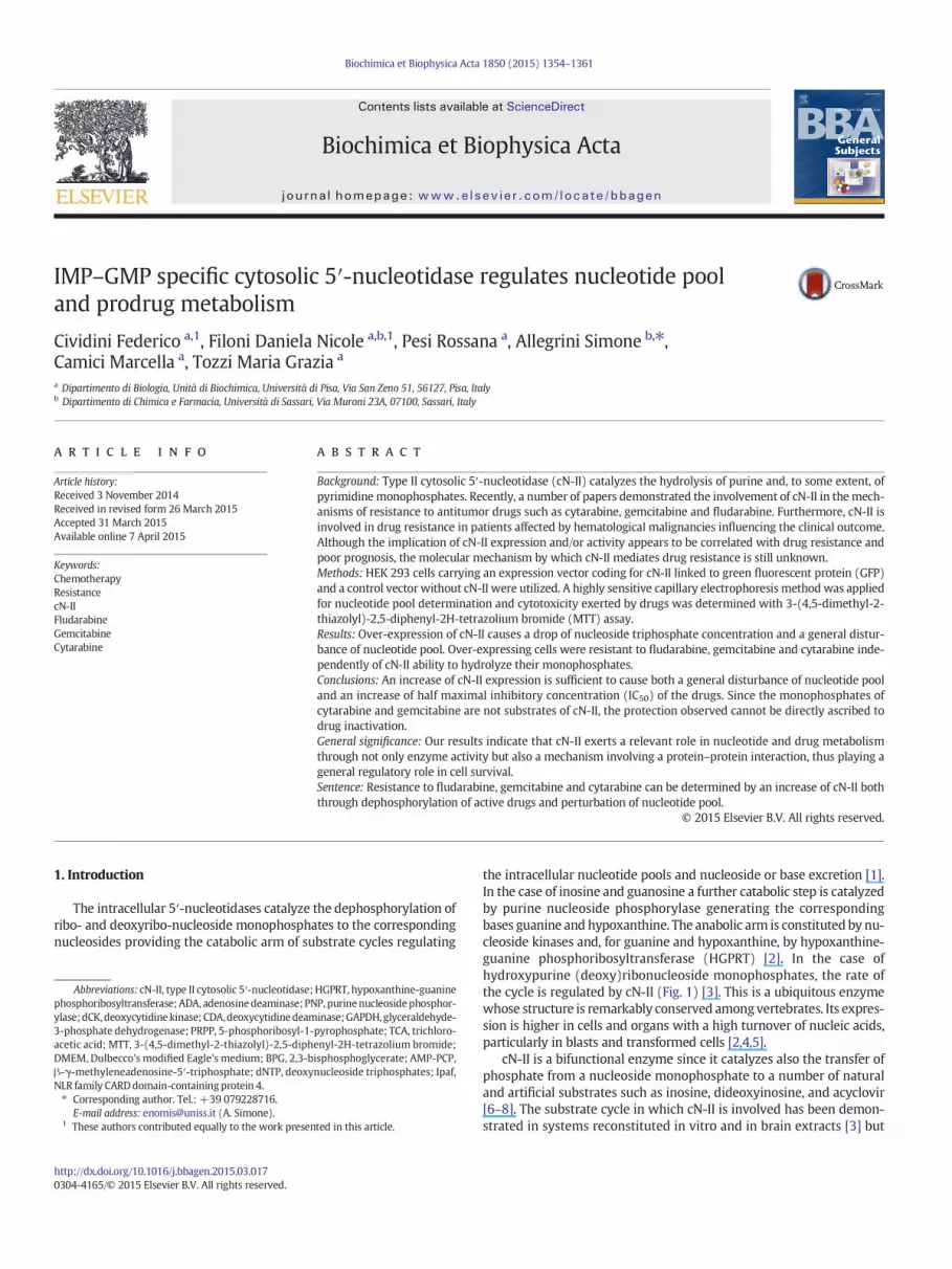
the intracellular nucleotide pools and nucleoside or base excretion [1].In the case of inosine and guanosine a further catabolic step is catalyzedby purine nucleoside phosphorylase generating the correspondingbases guanine and hypoxanthine. The anabolic arm is constituted by nu-cleoside kinases and, for guanine and hypoxanthine, by hypoxanthine-guanine phosphoribosyltransferase (HGPRT) [2]. In the case ofhydroxypurine (deoxy)ribonucleoside monophosphates, the rate ofthe cycle is regulated by cN-II (Fig. 1) [3]. This is a ubiquitous enzymewhose structure is remarkably conserved amongvertebrates. Its expres-sion is higher in cells and organs with a high turnover of nucleic acids,particularly in blasts and transformed cells [2,4,5].
cN-II is a bifunctional enzyme since it catalyzes also the transfer ofphosphate from a nucleoside monophosphate to a number of naturaland artificial substrates such as inosine, dideoxyinosine, and acyclovir[6–8]. The substrate cycle in which cN-II is involved has been demon-strated in systems reconstituted in vitro and in brain extracts [3] but
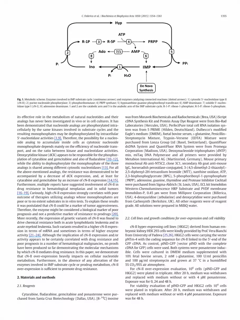
Fig. 1.Metabolic scheme. Enzymes involved in IMP substrate cycle (continuous arrows) and enzymes catalyzing connected reactions (dotted arrows): 1) cytosolic 5′-nucleotidase type II(cN-II); 2) purine nucleoside phosphorylase; 3) phosphoribomutase; 4) PRPP synthase; 5) hypoxanthine-guanine phosphoribosyl transferase; 6) AMP deaminase; 7) soluble 5′-nucleo-tidase type I (cN-I); 8) adenosine deaminase. 1 and 2 are the catabolic arm and 5 is the anabolic arm of the IMP substrate cycle. R-1-P: ribose-1-phosphate; R-5-P: ribose-5-phosphate.
1355C. Federico et al. / Biochimica et Biophysica Acta 1850 (2015) 1354–1361
its effective role in the metabolism of natural nucleotides and theiranalogs has never been investigated in vivo or in cell cultures. It hasbeen demonstrated that nucleoside analogs are phosphorylated intra-cellularly by the same kinases involved in substrate cycles and theresulting monophosphates may be dephosphorylated by intracellular5′-nucleotidase activities [1,9]. Therefore, the possibility for a nucleo-side analog to accumulate inside cells as cytotoxic nucleosidemonophosphate depends mainly on the efficiency of nucleoside trans-port, and on the ratio between kinase and nucleotidase activities.Deoxycytidine kinase (dCK) appears to be responsible for thephosphor-ylation of cytarabine and gemcitabine and also of fludarabine [10–12],while the ability to dephosphorylate the monophosphate of the threeanalogs is shared among different cytosolic nucleotidases [13]. For allthe above-mentioned analogs, the resistance was demonstrated to beaccompanied by a decrease of dCK expression, and, at least forcytarabine and gemcitabine by an increase of cN-II expression [14,15].Furthermore, multiple reports have suggested involvement of cN-II indrug resistance in hematological neoplasias and in solid tumors[16–19]. Curiously, high cN-II expression strongly correlates with pooroutcome of therapies utilizing analogs whose monophosphates arepoor or to no extent substrates in in vitro tests. To explain these resultsit was postulated that cN-II could be a marker of tumor aggressiveness.Therefore, the enzymemight be considered a biological marker of poorprognosis and not a predictive marker of resistance to prodrugs [20].More recently, the expression of genetic variants of cN-II was found todrive chemical resistance both in acute lymphoblastic leukemia and inacutemyeloid leukemia. Such variants resulted in a higher cN-II expres-sion in terms of mRNA and sometimes in terms of higher enzymeactivity [21–24]. Although the implication of cN-II expression and/oractivity appears to be certainly correlated with drug resistance andpoor prognosis in a number of hematological malignancies, no proofshave been produced so far demonstrating the molecular mechanismsby which cN-II mediates drug resistance. In this paper, we demonstratethat cN-II over-expression heavily impacts on cellular nucleotidemetabolism. Furthermore, in the absence of any alteration of theenzymes involved in IMP substrate cycle or prodrug metabolism, cN-IIover-expression is sufficient to promote drug resistance.
2. Materials and methods
2.1. Reagents
Cytarabine, fludarabine, gemcitabine and ponasterone were pur-chased from Santa Cruz Biotechnology (Dallas, USA). [8-14C] inosine
was fromMoravek Biochemicals and Radiochemicals (Brea, USA) iScriptcDNA Synthesis Kit and Protein Assay Dye Reagent were from Bio-RadLaboratories (Hercules, USA). PerfectPure total cell RNA isolation sys-tem was from 5 PRIME (Hilden, Deutschland). Dulbecco's modifiedEagle's medium (DMEM), foetal bovine serum, L-glutamine, Penicillin–Streptomycin Mixture, Trypsin–Versene (EDTA) Mixture werepurchased from Lonza Group Ltd (Basel, Switzerland). QuantiFluordsDNA System and QuantiFluor RNA System were from PromegaCorporation (Madison, USA). Deoxynucleoside triphosphates (dNTP)mix, miTaq DNA Polymerase and all primers were provided byMetabion International AG (Martinsried, Germany). Mouse primarymonoclonal Ab anti-NT5C2, clone 3C1, secondary Ab goat anti-mouseIgG, horseradish peroxidase-conjugated, 3-(4,5-dimethyl-2-thiazolyl)-2,5-diphenyl-2H-tetrazolium bromide (MTT), xanthine oxidase, ATP,2,3-bisphosphoglycerate (BPG), 5-phosphoribosyl-1-pyrophosphate(PRPP), adenosine, guanine, thymidine and Protease Inhibitor Cocktailwere purchased from Sigma-Aldrich (St. Louis, USA). ECL kit ImmobilonWestern-Chemoluminescence HRP Substrate and PVDF membraneImmobilon-P, 0.45 μm were from Millipore Corporation (Billerica,USA). 4-deoxyuridine (zebularine) and deoxycytidine were purchasedfrom Carbosynth (Berkshire, UK). All other reagents were of reagentgrade. All solutions were prepared in MilliQ water.
2.2. Cell lines and growth conditions for over-expression and cell viability
cN-II hyper-expressing cell lines (HKiG2) derived from human em-bryonic kidneyHEK 293 cellswere kindly provided by Prof. Vera Bianchifrom University of Padova [25,26]. HKiG2 cells were carrying the vectorpNTm-4 with the coding sequence for cN-II linked to the 5′-end of theGFP cDNA. As control, pIND-GFP (vector pIND with the completecDNA for GFP) cells were used. Both systems were ponasterone induc-ible. Cells were cultured in DMEM medium supplemented with10% fetal bovine serum, 2 mM L-glutamine, 100 U/ml penicillinand 100 μg/ml streptomycin and grown at 37 °C in a humidified5% CO2/95% air atmosphere.
For cN-II over-expression evaluation, 106 cells (pIND-GFP andHKiG2) were plated in triplicate. After 20 h, medium was withdrawnand replaced with medium without or with 4 μM ponasterone.Exposure was for 0, 24 and 48 h.
For viability evaluation of pIND-GFP and HKiG2 cells 104 cellswere plated in triplicate. After 20 h, medium was withdrawn andreplaced with medium without or with 4 μM ponasterone. Exposurewas for 48 h.

1356 C. Federico et al. / Biochimica et Biophysica Acta 1850 (2015) 1354–1361
2.3. mRNA extraction and evaluation
Total RNA was isolated from cells using PerfectPure total cell RNAisolation system following the manufacturer's instructions and quanti-fied using Quantifluor-ST Fluorometer according to the protocolsdescribed in the respective detection kits. 1 μg of total RNA wasretrotranscribed into cDNA using iScript cDNA Synthesis Kit in a totalvolume of 20 μl. 1 μl of each cDNA preparation was assayed for cN-IIexpression in a total of 20 μl in the presence of deoxynucleoside triphos-phates (50 μM each final concentration), 1 μM of forward and reverseprimer specific for cN-II, 1 × PCR reaction buffer containing 1.5 mMMgCl2, and 1 U mi-Taq DNA Polymerase. For normalization of RNAloading, the housekeeping glyceraldehyde-3-phosphate dehydrogenase(GAPDH) gene was also amplified for each cDNA sample. cN-II andGAPDH primers and PCR amplification procedures are those describedby Careddu et al. [27].
2.4. Protein concentration determination
Protein Assay Dye Reagent was used, according to the providerinstructions, for the determination of protein concentration if not differ-ently stated.
2.5. Immunoblot analysis
SDS-PAGE was performed following the method described byLaemmli [28]. Sample volumes corresponding to 10 μg of proteinsfrom each sample were applied to a 10% SDS polyacrylamide gel. Afterelectrophoresis, Western blotting was performed using PVDFmembrane and processed for immune detection as described byAllegrini et al. [29].
2.6. Crude extract preparation and enzyme activities
For the evaluation of enzyme activities in pIND-GFP and HKiG2 cells,106 cells were plated in triplicate. After 20 h, medium was withdrawnand replaced with medium with 4 μM ponasterone. After 48 h incuba-tion pIND-GFP and HKiG2 were harvested and the pellet was resus-pended in Tris–HCl 100 mM pH 7.4 in the presence of proteaseinhibitor cocktail. For dCK assay, 10mMsodiumfluoride as phosphataseinhibitorwas also added to the resuspensionbuffer. Crude extractswereobtained by 3 freeze/thaw cycles followed by centrifugation at10,000 ×g at 4 °C for 40 min to remove the cell debris. Supernatantwas used for enzyme activities.
Phosphotransferase activity of cN-II in crude extracts was measuredas the rate of [8-14C]IMP formation from 1.4 mM [8-14C] inosine, in thepresence of 2 mM IMP (or GMP), 20 mMMgCl2, 4.5 mMATP and 5 mMdithiothreitol and crude extract (100 μg of protein) as previouslydescribed by Pesi et al. [30].
Phosphatase activity of cN-II was assayed measuring the releaseof phosphate according to Chifflet [31]. The reaction mixture contained2mM IMP, 20mMMgCl2, 5mMBPG, 500 μMβ-γ-methyleneadenosine-5′-triphosphate (AMP-PCP) as ecto-nucleotidase inhibitor, Tris–HCl100 mM pH 7.4 and crude extract (100 μg of protein).
HGPRT assay was performed spectrophotometrically, as previouslydescribed [32]. The reaction mixture contained 90 μM guanine,0.2mMPRPP, 10mMMgCl2, Tris–HCl 100mMpH 7.4 and crude extract(10 μg of protein).
Adenosine deaminase (ADA) was assayed spectrophotometricallyaccording to Kalckar [33]. The reaction mixture contained 100 μMadenosine, Tris–HCl 100 mM pH 7.4 and crude extract (40 μg of protein).
Purine nucleoside phosphorylase (PNP) was assayed spectrophoto-metrically according to Kalckar [34]. The reaction mixture contained300 μM inosine, 5 mM Pi, 100 mU of xanthine oxidase as ancillaryenzyme, Tris–HCl 100 mM pH 7.4 and crude extract (40 μg of protein).
dCK was determined using a radioactive assay: 20 μM[2-14C]deoxycytidine (53 mCi/mmol), 1 mM ATP, 10 mM MgCl2,30 μM thymidine as competitive inhibitor of thymidine kinase 2,20 μM 4-deoxyuridine (Zebularine) as deoxycytidine deaminase(CDA) inhibitor, 100 mM Tris–HCl pH 7.4 and crude extract (40 μg ofprotein). At different times (0′, 20′, 40′ and 60′) 8 μl of assay mixturewas withdrawn and spotted on DE-81 paper disks. After extensivewashing in 1 mM ammonium formate and water, the disks were driedand the radioactivity was determined.
CDA was measured by determining the amount of deoxyuridineformed in the presence of labeled deoxycytidine. The reaction mixturecontained 0.4 mM [2-14C]deoxycytidine (53 mCi/mmol), Tris–HCl100 mM pH 7.4 and crude extract (10 μg of protein). At differenttime intervals (0′, 20′, 40′ and 60′), 10 μl of the assay mixture wasspotted on PEI-cellulose precoated thin-layer plastic sheets and thechromatogram was developed in n-propanol/NH3/trichloroaceticacid (100%)/H2O (75:0.7:5:20, v/v) to separate deoxycytidine anddeoxyuridine [35]. Deoxyuridine standard was used and detected asUV absorbing areas, which were excised and counted for radioactivity.
All the assays were performed at 37 °C.For all the enzymes, 1 mU represents the amount of enzyme re-
quired to convert 1 nmol of substrate to product per min under assayconditions. All the assays were performed in triplicate.
2.7. Intracellular metabolite extraction
106 cells were plated in triplicate. After 20 h, medium was with-drawn and replaced with medium containing 4 μM ponasterone. After48 h of exposure to 4 μM of ponasterone each replicate of platedpIND-GFP andHKiG2 cellswas trypsinized. Pelletswere rapidly separat-ed frommedium by centrifugation (1500 ×g for 5 min) and resuspend-ed in 150 μl 1 M trichloroacetic acid (TCA). It is known that nucleotidesare unstable in highly acidic solutions. To avoid degradation of nucleo-tides we adapted to our needs a protocol described by Friedecky et al.[36], in which the authors show that degradation of ribonucleotides in-cubated for 30 min in 1 M TCA is negligible. After three rounds of rapidfreezing–thawing (3min in dry ice-ethanol−75 °C bath and 3min in awarm 37 °C bath), pellets were separated from supernatants by centri-fugation (14,000 ×g for 1 min at room temperature). 120 μl of eachsupernatant was collected in a 1.5 ml tube and immediately back-extracted three times with 1.2 ml ether vortexing for 20 s at maximumspeed. This procedure produced samples at pH N 5 in which no moredegradation of nucleotides is observed. Samples were kept at roomtemperature under a chemical hood for 5 min and later stored at−20 °C until needed. The whole process of deproteination took nolonger than 25min. The protein content of the pellets previously precip-itated with TCA was determined using a modified Lowry methoddescribed by Peterson [37].
2.8. Capillary electrophoresis analysis
All the experiments were performed using a Beckman P/ACE MDQCapillary Electrophoresis System equipped with an UV detector. Theintracellular nucleotide concentration was determined as previouslydescribed [29].
2.9. Cytotoxicity assay
In cytotoxicity experiments, 104 cells grown as described inSection 2.2, were threefold plated. After 20 h, medium was withdrawnand replaced with mediumwith 4 μMponasterone. After 48 h, mediumwas withdrawn and replaced with medium with 4 μM ponasterone,and different concentrations of nucleoside analogs were added(gemcitabine, cytarabine andfludarabine from0.32 μMto5mM). Expo-sure to the drug was for 48 h. After treatment threefold replicates foreach cell line were incubated at 37 °C, 5% CO2/95% air atmosphere for

Fig. 3. Viability of pIND-GFP and HKiG2 after 48 h of growth in the presence (white) or inthe absence (gray) of 4 μM ponasterone. Data shown represent the mean ± SD and arerepresentative of three independent experiments. ****p b 0.0001.
1357C. Federico et al. / Biochimica et Biophysica Acta 1850 (2015) 1354–1361
2 h with 0.5 mg/ml of MTT previously dissolved in PBS and filteredaccording to Mosmann [38]. Formazan salts produced in the reactionwere dissolved in 0.04 N HCl in isopropanol and quantified spectropho-tometrically by reading the absorbance at 570 nm using the Sunrise™Absorbance Reader (Tecan, Switzerland).
2.10. Statistical analysis
Variance was analyzed by ONE-WAY ANOVA and means werecompared by Dunnet's test (p b 0.05). All statistical analyses wereperformed using the software InStat (ver. 3.05, GraphPad Software,Inc., La Jolla, CA 92037 USA).
3. Results
3.1. Induction of cN-II hyper-expression inHEK 293 cell cultures and impacton viability and nucleotide content
The HEK 293 cell line is widely utilized as cell model for proteinover-expression. An inducible over-expression of cN-II in HEK 293cells was previously obtained by Gazziola et al. [25]. We took advantageof the availability of this model to better understand the role of cN-II innucleotide and prodrug metabolism. HEK 293 cells were stablytransfected with a plasmid containing the ecdysone receptor and thecoding sequence for the human cN-II linked to the 5′-end of the cDNAfor the GFP (HKiG2 cells). Addition of ponasterone led to over-expression of cN-II assayed as phosphotransferase in an exposuretime-dependent manner (Fig. 2). Evaluation of cN-II mRNA and proteinis reported in Supplementary Fig. S1. Our results are similar to those re-ported in the papers describing the preparation of the cellular models[25,26]. Quantitation of mRNA and cN-II protein is in agreement withthe measured cN-II activity indicating that fusion with GFP does notaffect the catalytic properties of the enzyme (see SupplementaryFig. S1 and [25]). To ascertain if cN-II over-expression affects cell vitalityin our experimental conditions, we measured the viability (MTT assay)of both pIND-GFP and HKiG2 cells grown 48 h in the absence and pres-ence of ponasterone and found a significant decrease of cell viabilityupon treatment with ponasterone in both cell lines, but no differencein cell viability was found between control and over-expressing cellsin these growth conditions (Fig. 3).
Fig. 2. cN-II phosphotransferase activity was measured in HEK 293 cell crude extracts ob-tained from cells grown both in the absence of ponasterone (white bars), and in the pres-ence of 4 μMponasterone for 24 h (gray bars) and 48 h (black bars). Data shown representthe mean ± SD and are representative of three independent experiments. Significance isrelated to pIND-GFP for samples not treated with ponasterone (white bars) while forthose treated with ponasterone the significance is related to the respective sampleswithout ponasterone. *p b 0.05; **p b 0.01; ***p b 0.001.
Nucleotide content appears to be significantly affected by the cN-IIover-expression and, as stated above, this result is not ascribable to adifferent sensitivity of the two cell lines to ponasterone treatment.Fig. 4 shows that purine and pyrimidine triphosphates are significantlydecreased in HKiG2 cells expressing the fusion protein. Also GDPappeared significantly decreased while, among monophosphates, onlyIMP, the best substrate of cN-II, was significantly decreased. On thecontrary, AMP levels were increased in cN-II over-expressing cells.ATP depletion was accompanied by a decrease of energy charge[(ATP + 1/2 ADP) / (AMP + ADP + ATP)] and by a loss of adenylatecompounds which is not counterbalanced by the modest increase ofAMP levels in HKiG2 cell. This suggests that the salvage operated bythe anabolic arm of the IMP substrate cycle shown in Fig. 1 was slowerthan the catabolic arm when cN-II was over-expressed.
3.2. Enzyme activities in cells exposed to ponasterone
In order to ascertain if, following cN-II over-expression, compensa-tory mechanisms were activated, we measured the activity of severalenzymes involved in themetabolic pathways shown in Fig. 1. The activ-ity of cN-II, measured both as phosphatase and phosphotransferase,wassignificantly higher in HKiG2 cells. On the other hand, the specific activ-ity of HGPRT, PNP andADA, enzymes involved in themetabolic pathwaydescribed in Fig. 1, was not changed in our experimental conditions(Table 1). With the intent to assess the effect of cN-II over-expressionon cytotoxicity exerted by fludarabine, cytarabine and gemcitabine,we also measured the specific activity of CDA and dCK in both pIND-GFP and HKiG2. CDA can deaminate, and therefore inactivate, bothgemcitabine and cytarabine [39],while dCK is responsible for the activa-tion of the three analogs [40]. CDA exhibited the same specific activity inboth cell lines (Table 1). dCK requires ATP both as a substrate and asactivator, being subjected to phosphorylation at Ser-74 [41,42]. Toascertain if the low ATP level found in HKGi2 cells could affect thedegree of the enzyme phosphorylation, we measured dCK activity inextracts preparedwith andwithout sodium fluoride, an inhibitor of pro-tein phosphatases (dCK can be dephosphorylated by protein phospha-tase 2A [43]). The results indicate in both cases no differences in dCKspecific activities in control an over-expressing cells (Table 1). There-fore, among the enzymes assayed, only cN-II activity was altered inHKGi2 cells upon 48 h incubation with ponasterone.

Fig. 4. Capillary electrophoresis analysis. A) Intracellular nucleotide levels (pIND-GFPwhite bars andHKiG2 black bars). CDP and CMPwere undetectable. Energy charge (B) and adenylateintracellular content (C) after 48 h of exposure to 4 μM ponasterone. C) Data shown represent the mean ± SD and are representative of three independent experiments. Significance isrelated to pINDGFP. *p b 0.05; **p b 0.01; ***p b 0.001.
1358 C. Federico et al. / Biochimica et Biophysica Acta 1850 (2015) 1354–1361
3.3. Cytotoxicity tests
The viability of the two cell lines described above was measured inthe presence of increasing concentrations of fludarabine, gemcitabineand cytarabine and the effect of the ponasterone mediated over-expression of cN-II on cell viability is shown in Fig. 5 and in Table 2.pIND-GFP was susceptible to micromolar concentration of bothfludarabine and gemcitabine while cytarabine exerted a modest effectat higher concentrations. The IC50 measured for fludarabine was of thesame order of magnitude found by other authors [44,45], whereas theIC50 values found for gemcitabine and cytarabine are significantlyhigher than the values found by other authors in cultured tumor cells[45–48]. This observation might be due to an elevated CDA activity inHEK 293 cells (see Table 1) as compared to other cells and organs[49], causing a natural resistance to the cytidine analogs [50,51].The over-expression of cN-II clearly protects (Table 2) from cytotoxicityexerted by fludarabine (IC50 ratio HKiG2/pIND-GFP = 82) andgemcitabine (IC50 ratio HKiG2/pIND-GFP = 63) and to a lower butsignificant extent, by cytarabine (IC50 ratio HKiG2/pIND-GFP = 5.5).
Table 1Enzyme activities in pIND-GFP and HKiG2 cell extracts. Numbers indicate mU/mg ofproteins ± SD.
Enzyme pIND-GFP HKiG2
cN-II phosphotransferase 1.03 ± 0.24 20.53 ± 0.65cN-II phosphatase 6.79 ± 0.34 34.04 ± 0.30HGPRT 34.17 ± 0.49 35.73 ± 4.15PNP 52.87 ± 1.63 56.97 ± 0.95ADA 9.47 ± 0.35 10.00 ± 0.10dCK (with NaF) 0.21 ± 0.03 0.23 ± 0.02dCK 0.05 ± 0.01 0.08 ± 0.03CDA 1.5 ± 0.51 1.35 ± 0.37
4. Discussion
An increase of cN-II activity, as well as a decrease, has been reportedto be toxic at least for dividing cells [25,27]. This observation indicates apivotal role played by the enzyme in cell proliferation and surviving.Furthermore, several reports demonstrated that cN-II over-expressionor hyper-activity correlate with drug resistance in both hematologicalmalignancies and solid tumors [15,17]. The mechanism of drug resis-tance remains to be unraveled since the protection of cN-II over-expression is exerted also upon treatment with nucleoside analogswhose monophosphates are not substrates of the enzyme. We took ad-vantage of the availability of an over-expressingmodel to investigate onthe role of cN-II in both the catabolism of purine compounds and resis-tance to purine and pyrimidine nucleoside analogs used in cancer ther-apy. HEK 293 cells carrying an expression vector coding for cN-II linkedto GFP (HKiG2) and a control vector without cN-II (pIND-GFP) wereused. Both systems are ponasterone inducible as described by Gazziolaet al. [25]. After 48 h of incubation with the inducer, HKiG2 cellsexpressed approximately 20 times more cN-II, measured as phospho-transferase, and 5 times more measured as phosphatase, than thepIND-GFP cells. It is conceivable that this discrepancy depends on anover-estimation of the phosphatase activity in control cells. In fact,while the phosphotransferase assay is specific for cN-II [30], the phos-phatase activity, measured as the rate of IMP hydrolysis, even in thepresence of an ectosolic-5′-nucleotidase inhibitor, depends not onlyon cN-II, but also on other cytosolic 5′-nucleotidases or aspecific phos-phatases [52,53]. To assess the effect of cN-II over-expression on theviability of HEK 293 cells, we measured the viability of cells after 48 hof ponasterone exposure finding no significant differences betweenover-expressing and control cells. This finding is in line with thatobtained by Gazziola et al. [25], who reported no difference after 48 htreatment, while a significant proliferation decrease of cN-II over-expressing cells with respect to control cells was found after several
Fig. 5. HEK 293 cells exposure to nucleoside analogs: cell viability was assayed with MTT.pIND-GFP (empty circles) and HKiG2 (filled circles) were treated with 4 μM ponasteronefor 48 h. Later themediumwas replacedwith freshmedium containing 4 μMponasteroneand the cells were exposed to μM concentrations (from 0.32 to 5000) of A) fludarabine,B) gemcitabine and C) cytarabine for 48 h. Data are reported in graphs as percentageof cell viability respect to control (no drug exposure) and are representative of threeindependent replicates.
1359C. Federico et al. / Biochimica et Biophysica Acta 1850 (2015) 1354–1361
days of exposure to the inducer. Our previous results demonstrated thatan increase of cN-II activitywas followed by PRPP consumption in tissueextracts [3] and ATP consumption in over-expressing yeast cells [29].Accordingly, we found that cN-II over-expression in HEK 293 cellscaused a decrease of all nucleoside triphosphates with a consequentdrop in the adenylate content and energy charge. This result is in linewith that obtained by Gazziola et al. [25] who found a 20% decrease intriphosphate content. The extraction procedure and the analyticalmethod used in the present work allowed us to determine, besidestriphosphates, also diphosphate and monophosphate nucleosides.Nucleoside diphosphates and NAD+ were decreased in cN-II over-expressing cells, and among monophosphates, only IMP was substan-tially decreased while AMP increased significantly. Indeed, AMP
Table 2IC50 values+ SD of the three analogs in pIND-GFP (control) and HKiG2 (over-expressing)cells.
Drug pIND-GFP(μM)
HKiG2a
(μM)HKiG2/pIND-GFP
Fludarabine 44 ± 4 3615 ± 617⁎⁎⁎ 82Gemcitabine 30 ± 19 1905 ± 398⁎⁎ 63Cytarabine 714 ± 272 3929 ± 1428⁎ 5.5
a Significance: ⁎p b 0.05; ⁎⁎p b 0.01; ⁎⁎⁎p b 0.001.
accumulates as the result of ATP dephosphorylation, required for PRPPsynthesis, necessary for the salvage of hypoxanthine, whose formationincreases as a consequence of cN-II over-expression (see Fig. 1).Furthermore, both the low AMP deaminase activity at low ATP concen-trations [54] and the very high Km for AMP of cN-II [55] contribute toAMP accumulation. These results demonstrate that cells try to keep aconstant pool of PRPP at the expense of ATP when the catabolic arm ofthe cycle of Fig. 1, as a consequence of cN-II over-expression, is runningfaster than in normal conditions. To ascertain if any compensatory alter-ation of enzyme expression occurred in cN-II over-expressing cells, wemeasured the activity of several enzymes involved in the metabolicpathways shown in Fig. 1, i.e., cN-II, HGPRT and PNP (IMP substratecycle) and ADA, and found that only phosphatase and phosphotransfer-ase activities specific of cN-II were altered upon 48 h incubation withponasterone.
Over-expressing and control cells were incubated in the presence ofthe analogsfludarabine, gemcitabine and cytarabine. IC50 of control cellsfor fludarabine was in line with the values found by other authors withdifferent cell lines [44,45], while pIND-GFP sensitivity for gemcitabineand cytarabine was significantly lower [45–48]. In fact CDA, that can in-activate both drugs, in HEK 293 cells exhibits a specific activity higherthan that reported for many other cells and organs, (in many tumoraltissues/cells, CDA activity is lower than 0.1 mU/mg) [49]. This findingmay account for the high IC50 measured for cytotoxicity exerted bygemcitabine and cytarabine on these cells.
Cells over-expressing cN-II resulted to be protected from cytotoxiceffect exerted by fludarabine and gemcitabine and, to a lower extent,by cytarabine. The extent of this protective effect does not correlatewith the enzyme ability to dephosphorylate the active phosphorylatedform of the drug analogs. Indeed fludarabine monophosphate hasbeen demonstrated to be substrate of cN-II [56], while gemcitabineand cytarabine monophosphates are not substrates of the enzyme[13]. Wemight therefore infer that the extent of protection can dependon the perturbance of purine and pyrimidine nucleotide pool deter-mined by cN-II over-expression, which might interfere at differentlevels with the transport and intracellular metabolism of the drugs. Ithas been demonstrated that the rate limiting step in the metabolismof the analogs used in the present work is their phosphorylation medi-ated by dCK [40], which can accept as triphosphate substrate either ATPor UTP [57]. Furthermore, dCK is regulated by phosphorylation,resulting in enzyme activation [41]. However, dCK activity was thesame both in pIND-GFP and in HKiG2 cells even when the activatingphosphorylation of the enzymewas preserved using a protein phospha-tase inhibitor for the preparation of the extracts, indicating that the de-crease of ATP in over-expressing cells does not interfere with dCKactivation. Furthermore, being the Km for the best phosphate donor(UTP) very low (1 μM), the observed decrease in the level of this nucle-otide in over-expressing cells is unlikely to affect dCK activity. On theother hand, dCK is a very complex allosteric enzyme [58] and therefore,the possibility that a profound alteration of nucleotide pool might neg-atively reflect on dCK activation and activity, cannot be ruled out. Theobservation that a modest cN-II silencing causes activation of the apo-ptotic program, without any measurable alteration of nucleotide pool[27], suggests that cN-II plays some regulatory role on important cellu-lar mechanisms. Indeed, we recently demonstrated that cN-II interactswith the cytosolic protein NLR family CARD domain-containing protein4 (Ipaf) involved in innate immunity and inflammation [59]. Ipaf, uponactivation exerted by extracellular or intracellular signals, promotesinterleukin production and apoptosis activation [60]. It is therefore con-ceivable that the interaction of Ipaf with cN-II may play a modulatoryrole, possibly reflecting on prodrug metabolism and cytotoxicity.
In conclusion, our results demonstrate that cN-II, besides being di-rectly involved in the inactivation of drugs, is also determinant for theintracellular nucleotide concentration, by regulating the rate of themetabolic pathways shown in Fig. 1. This might reflect on the rate ofdrug intracellular metabolism. Finally, our findings demonstrate that

1360 C. Federico et al. / Biochimica et Biophysica Acta 1850 (2015) 1354–1361
over-expression of cN-II is sufficient to cause cell resistance and mightexplain why a high level of cN-II expression is protective against drugswhose monophosphates are poor or to no extent substrates of theenzyme.
Supplementary data to this article can be found online at http://dx.doi.org/10.1016/j.bbagen.2015.03.017.
Transparency document
The Transparency document associated with this article can befound in the online version.
Acknowledgements
The Authors wish to thank Prof. Vera Bianchi, University of PadovaItaly, for providing pIND-GFP and HKiG2 cells.
This work was financially supported by a grant from the REGIONEAUTONOMA DELLA SARDEGNA, L.R. 07/08/2007, grant numberCRP3360 (TITLE: Modulation of expression by inducible silencing; het-erologous expression in S. cerevisiae; two-hybrid system. Three modelsto make clear the physiological role of cytosolic 5′nucleotidase II), andby local grants from the University of Pisa. The funders had no role instudy design, data collection and analysis, decision to publish, or prepa-ration of the manuscript.
References
[1] V. Bianchi, J. Spychala, Mammalian 5′-nucleotidases, J. Biol. Chem. 278 (2003)46195–46198.
[2] M.G. Tozzi, R. Pesi, S. Allegrini, On the physiological role of cytosolic 5′-nucleotidaseII (cN-II): pathological and therapeutical implications, Curr. Med. Chem. 20 (2013)4285–4291.
[3] C. Barsotti, R. Pesi, F. Felice, P.L. Ipata, The purine nucleoside cycle in cell-freeextracts of rat brain: evidence for the occurrence of an inosine and a guanosinecycle with distinct metabolic roles, Cell. Mol. Life Sci. 60 (2003) 786–793.
[4] R. Itoh, IMP–GMP 5′-nucleotidase, Comp. Biochem. Physiol. B 105 (1993) 13–19.[5] M.G. Tozzi, M. Camici, S. Allegrini, R. Pesi, M. Turriani, A. Del Corso, P.L. Ipata,
Cytosolic 5′-nucleotidase/phosphotransferase of human colon carcinoma, Adv.Exp. Med. Biol. 309B (1991) 173–176.
[6] S. Banditelli, C. Baiocchi, R. Pesi, S. Allegrini, M. Turriani, P.L. Ipata, M. Camici, M.G.Tozzi, The phosphotransferase activity of cytosolic 5′-nucleotidase; a purine analogphosphorylating enzyme, Int. J. Biochem. Cell Biol. 28 (1996) 711–720.
[7] M.A. Johnson, A. Fridland, Phosphorylation of 2′,3′-dideoxyinosine by cytosolic5′-nucleotidase of human lymphoid cells, Mol. Pharmacol. 36 (1989) 291–295.
[8] M. Turriani, R. Pesi, A. Nardone, G. Turchi, F. Sgarrella, P.L. Ipata, M.G. Tozzi, Cytosolic5′-nucleotidase/nucleoside phosphotransferase: a nucleoside analog activatingenzyme? J. Biochem. Toxicol. 9 (1994) 51–57.
[9] H. Kawasaki, C.J. Carrera, L.D. Piro, A. Saven, T.J. Kipps, D.A. Carson, Relationshipof deoxycytidine kinase and cytoplasmic 5′-nucleotidase to the chemotherapeuticefficacy of 2-chlorodeoxyadenosine, Blood 81 (1993) 597–601.
[10] R. Garcia-Carbonero, D.P. Ryan, B.A. Chabner, Cytidine analogs, in: C. B.A., L. D.L.(Eds.), Cancer Chemotherapy and Biotherapy, Lippincott-Raven, Philadelphia1996, pp. 265–294.
[11] W. Plunkett, P.P. Saunders, Metabolism and action of purine nucleoside analogs,Pharmacol. Ther. 49 (1991) 239–268.
[12] C.M. Galmarini, M.L. Clarke, L. Jordheim, C.L. Santos, E. Cros, J.R. Mackey, C.Dumontet, Resistance to gemcitabine in a human follicular lymphoma cell line isdue to partial deletion of the deoxycytidine kinase gene, BMC Pharmacol. 4(2004) 8.
[13] C. Mazzon, C. Rampazzo, M.C. Scaini, L. Gallinaro, A. Karlsson, C. Meier, J. Balzarini, P.Reichard, V. Bianchi, Cytosolic and mitochondrial deoxyribonucleotidases: activitywith substrate analogs, inhibitors and implications for therapy, Biochem. Pharmacol.66 (2003) 471–479.
[14] T. Yamauchi, E. Negoro, S. Kishi, K. Takagi, A. Yoshida, Y. Urasaki, H. Iwasaki, T. Ueda,Intracellular cytarabine triphosphate production correlates to deoxycytidine kinase/cytosolic 5 ′-nucleotidase II expression ratio in primary acute myeloid leukemiacells, Biochem. Pharmacol. 77 (2009) 1780–1786.
[15] P. Seve, J.R. Mackey, S. Isaac, O. Tredan, P.J. Souquet, M. Perol, C. Cass, C. Dumontet,cN-II expression predicts survival in patients receiving gemcitabine for advancednon-small cell lung cancer, Lung Cancer 49 (2005) 363–370.
[16] E. Giovannetti, V. Mey, L. Loni, S. Nannizzi, G. Barsanti, G. Savarino, S. Ricciardi, M.Del Tacca, R. Danesi, Cytotoxic activity of gemcitabine and correlation with expres-sion profile of drug-related genes in human lymphoid cells, Pharmacol. Res. 55(2007) 343–349.
[17] C.M. Galmarini, E. Cros, X. Thomas, L. Jordheim, C. Dumontet, The prognostic value ofcN-II and cN-III enzymes in adult acute myeloid leukemia, Haematologica 90 (2005)1699–1701.
[18] C.M. Galmarini, X. Thomas, K. Graham, A. El Jafaari, E. Cros, L. Jordheim, J.R. Mackey,C. Dumontet, Deoxycytidine kinase and cN-II nucleotidase expression in blast cellspredict survival in acute myeloid leukaemia patients treated with cytarabine, Br. J.Haematol. 122 (2003) 53–60.
[19] A.K. Mitra, M.N. Kirstein, A. Khatri, K.M. Skubitz, A.Z. Dudek, E.W. Greeno, R.A.Kratzke, J.K. Lamba, Pathway-based pharmacogenomics of gemcitabine pharmaco-kinetics in patients with solid tumors, Pharmacogenomics 13 (2012) 1009–1021.
[20] C.M. Galmarini,What does over-expression of cN-II enzyme signify in haematologicalmalignancies? Leuk. Res. 31 (2007) 1325–1326.
[21] A.K. Mitra, K.R. Crews, S. Pounds, X. Cao, T. Feldberg, Y. Ghodke, V. Gandhi, W.Plunkett, M.E. Dolan, C. Hartford, S. Raimondi, D. Campana, J. Downing, J.E.Rubnitz, R.C. Ribeiro, J.K. Lamba, Genetic variants in cytosolic 5′-nucleotidase II areassociated with its expression and cytarabine sensitivity in HapMap cell lines andin patients with acute myeloid leukemia, J. Pharmacol. Exp. Ther. 339 (2011) 9–23.
[22] J.A. Meyer, W.L. Carroll, T. Bhatla, Screening for genemutations: will identification ofNT5C2 mutations help predict the chance of relapse in acute lymphoblasticleukemia? Expert. Rev. Hematol. 6 (2013) 223–224.
[23] J.A. Meyer, J. Wang, L.E. Hogan, J.J. Yang, S. Dandekar, J.P. Patel, Z. Tang, P. Zumbo, S.Li, J. Zavadil, R.L. Levine, T. Cardozo, S.P. Hunger, E.A. Raetz, W.E. Evans, D.J. Morrison,C.E. Mason, W.L. Carroll, Relapse-specific mutations in NT5C2 in childhood acutelymphoblastic leukemia, Nat. Genet. 45 (2013) 290–294.
[24] G. Tzoneva, A. Perez-Garcia, Z. Carpenter, H. Khiabanian, V. Tosello, M. Allegretta, E.Paietta, J. Racevskis, J.M. Rowe, M.S. Tallman, M. Paganin, G. Basso, J. Hof, R.Kirschner-Schwabe, T. Palomero, R. Rabadan, A. Ferrando, Activating mutations inthe NT5C2 nucleotidase gene drive chemotherapy resistance in relapsed ALL, Nat.Med. 19 (2013) 368–371.
[25] C. Gazziola, M. Moras, P. Ferraro, L. Gallinaro, R. Verin, C. Rampazzo, P. Reichard, V.Bianchi, Induction of human high KM 5′-nucleotidase in cultured 293 cells, Exp.Cell Res. 253 (1999) 474–482.
[26] C. Rampazzo, C. Gazziola, P. Ferraro, L. Gallinaro, M. Johansson, P. Reichard, V.Bianchi, Human high-Km 5′-nucleotidase effects of overexpression of the clonedcDNA in cultured human cells, Eur. J. Biochem. 261 (1999) 689–697.
[27] M.G. Careddu, S. Allegrini, R. Pesi, M. Camici, M. Garcia-Gil, M.G. Tozzi, Knockdownof cytosolic 5′-nucleotidase II (cN-II) reveals that its activity is essential for survivalin astrocytoma cells, Biochim. Biophys. Acta 1783 (2008) 1529–1535.
[28] U.K. Laemmli, Cleavage of structural proteins during the assembly of the head ofbacteriophage T4, Nature 227 (1970) 680–685.
[29] S. Allegrini, D.N. Filoni, A. Galli, A. Collavoli, R. Pesi, M. Camici, M.G. Tozzi, Expressionof bovine cytosolic 5′-nucleotidase (cN-II) in yeast: nucleotide pools disturbanceand its consequences on growth and homologous recombination, Plos One 8(2013) e63914.
[30] M.G. Tozzi, M. Camici, R. Pesi, S. Allegrini, F. Sgarrella, P.L. Ipata, Nucleoside phospho-transferase activity of human colon carcinoma cytosolic 5′-nucleotidase, Arch.Biochem. Biophys. 291 (1991) 212–217.
[31] S. Chifflet, A. Torriglia, R. Chiesa, S. Tolosa, A method for the determination ofinorganic phosphate in the presence of labile organic phosphate and high concen-trations of protein: application to lens ATPases, Anal. Biochem. 168 (1988) 1–4.
[32] M.P. Giovannitti, R. Pesi, G. Cercignani, Spectrophotometric assay for hypoxanthine-guanine phosphoribosyltransferase in human-erythrocytes, Ital. J. Biochem. 37(1988) A44–A46.
[33] H.M. Kalckar, Differential spectrophotometry of purine compounds by means ofspecific enzymes; determination of adenine compounds, J. Biol. Chem. 167 (1947)445–459.
[34] H.M. Kalckar, Differential spectrophotometry of purine compounds by means ofspecific enzymes; determination of hydroxypurine compounds, J. Biol. Chem. 167(1947) 429–443.
[35] C. Barsotti, R. Pesi, M. Giannecchini, P.L. Ipata, Evidence for the involvement ofcytosolic 5′-nucleotidase (cN-II) in the synthesis of guanine nucleotides fromxanthosine, J. Biol. Chem. 280 (2005) 13465–13469.
[36] D. Friedecky, J. Tomkova, V. Maier, A. Janost'akova, M. Prochazka, T. Adam, Capillaryelectrophoretic method for nucleotide analysis in cells: application on inheritedmetabolic disorders, Electrophoresis 28 (2007) 373–380.
[37] G.L. Peterson, Determination of total protein, in: C.H.W. Hirs (Ed.), MethodsEnzymol — Enzyme Structure. Part I, vol. 91, Academic Press 1983, pp. 95–104.
[38] T. Mosmann, Rapid colorimetric assay for cellular growth and survival: applicationto proliferation and cytotoxicity assays, J. Immunol. Methods 65 (1983) 55–63.
[39] C.M. Galmarini, J.R. Mackey, C. Dumontet, Nucleoside analogues: mechanisms ofdrug resistance and reversal strategies, Leukemia 15 (2001) 875–890.
[40] B. Munch-Petersen, J. Piskur, Deoxynucleoside kinases and their potential role indeoxynucleoside cytotoxicity, in: G.J. Peters (Ed.), Deoxynucleoside Analogs inCancer Therapy, Humana Press Inc., Totowa, New Jersey, United States 2006,pp. 53–79.
[41] C. Smal, D. Vertommen, L. Bertrand, S. Ntamashimikiro, M.H. Rider, E. Van DenNeste, F. Bontemps, Identification of in vivo phosphorylation sites on humandeoxycytidine kinase — role of Ser-74 in the control of enzyme activity, J. Biol.Chem. 281 (2006) 4887–4893.
[42] C. Smal, E. Van Den Neste, M. Maerevoet, X. Poire, I. Theate, F. Bontemps, Positiveregulation of deoxycytidine kinase activity by phosphorylation of Ser-74 in B-cellchronic lymphocytic leukaemia lymphocytes, Cancer Lett. 253 (2007) 68–73.
[43] R. Amsailale, M. Beyaert, C. Smal, V. Janssens, E. Van Den Neste, F. Bontemps, Proteinphosphatase 2A regulates deoxycytidine kinase activity via Ser-74 dephosphorylation,FEBS Lett. 588 (2014) 727–732.
[44] R. Silber, B. Degar, D. Costin, E.W. Newcomb, M. Mani, C.R. Rosenberg, L. Morse, J.C.Drygas, Z.N. Canellakis, M. Potmesil, Chemosensitivity of lymphocytes from patientswith B-cell chronic lymphocytic leukemia to chlorambucil, fludarabine, andcamptothecin analogs, Blood 84 (1994) 3440–3446.

1361C. Federico et al. / Biochimica et Biophysica Acta 1850 (2015) 1354–1361
[45] L.P. Jordheim, E. Cros, M.H. Gouy, C.M. Galmarini, S. Peyrottes, J. Mackey, C. Perigaud,C. Dumontet, Characterization of a gemcitabine-resistant murine leukemic cell line:reversion of in vitro resistance by a mononucleotide prodrug, Clin. Cancer Res. 10(2004) 5614–5621.
[46] Y. Nakano, S. Tanno, K. Koizumi, T. Nishikawa, K. Nakamura, M. Minoguchi, T. Izawa,Y. Mizukami, T. Okumura, Y. Kohgo, Gemcitabine chemoresistance and molecularmarkers associatedwith gemcitabine transport andmetabolism in humanpancreaticcancer cells, Br. J. Cancer 96 (2007) 457–463.
[47] H. Achiwa, T. Oguri, S. Sato, H. Maeda, T. Niimi, R. Ueda, Determinants of sensitivityand resistance to gemcitabine: the roles of human equilibrative nucleosidetransporter 1 and deoxycytidine kinase in non-small cell lung cancer, Cancer Sci.95 (2004) 753–757.
[48] M. Hewish, S.A. Martin, R. Elliott, D. Cunningham, C.J. Lord, A. Ashworth, Cytosine-based nucleoside analogs are selectively lethal to DNA mismatch repair-deficienttumour cells by enhancing levels of intracellular oxidative stress, Br. J. Cancer 108(2013) 983–992.
[49] J. Sigmond, R.J. Honeywell, T.J. Postma, C.M. Dirven, S.M. de Lange, K. van der Born,A.C. Laan, J.C. Baayen, C.J. Van Groeningen, A.M. Bergman, G. Giaccone, G.J. Peters,Gemcitabine uptake in glioblastoma multiforme: potential as a radiosensitizer,Ann. Oncol. 20 (2009) 182–187.
[50] C.M. Galmarini, J.R. Mackey, C. Dumontet, Nucleoside analogues and nucleobases incancer treatment, Lancet Oncol. 3 (2002) 415–424.
[51] M.H. Tattersall, K. Ganeshaguru, A.V. Hoffbrand, Mechanisms of resistance of humanacute leukaemia cells to cytosine arabinoside, Br. J. Haematol. 27 (1974) 39–46.
[52] R. Pesi, M. Camici, V. Micheli, L. Notarantonio, G. Jacomelli, M.G. Tozzi, Identificationof the nucleotidase responsible for the AMP hydrolysing hyperactivity associat-ed with neurological and developmental disorders, Neurochem. Res. 33 (2008)59–65.
[53] R. Pesi, V. Micheli, G. Jacomelli, L. Peruzzi, M. Camici, M. Garcia-Gil, S. Allegrini, M.G.Tozzi, Cytosolic 5′-nucleotidase hyperactivity in erythrocytes of Lesch–Nyhansyndrome patients, Neuroreport 11 (2000) 1827–1831.
[54] C. Barsotti, P.L. Ipata, Metabolic regulation of ATP breakdown and of adenosineproduction in rat brain extracts, Int. J. Biochem. Cell Biol. 36 (2004)2214–2225.
[55] R. Itoh, Enzymatic properties and physiological roles of cytosolic 5′-nucleotidase II,Curr. Med. Chem. 20 (2013) 4260–4284.
[56] C.M. Galmarini, L. Jordheim, C. Dumontet, Role of IMP-selective 5′-nucleotidase(cN-II) in hematological malignancies, Leuk. Lymphoma 44 (2003) 1105–1111.
[57] E.S. Arner, S. Eriksson, Mammalian deoxyribonucleoside kinases, Pharmacol. Ther.67 (1995) 155–186.
[58] T.L. Hughes, T.M. Hahn, K.K. Reynolds, D.S. Shewach, Kinetic analysis of humandeoxycytidine kinase with the true phosphate donor uridine triphosphate,Biochemistry 36 (1997) 7540–7547.
[59] F. Cividini, M.G. Tozzi, A. Galli, R. Pesi, M. Camici, C. Dumontet, L. Jordheim, S.Allegrini, Cytosolic 5′-nucleotidase II interacts with the leucin rich repeat of NLRfamily member Ipaf, PLoS One 10 (2015) e0121525.
[60] F.S. Sutterwala, R.A. Flavell, NLRC4/IPAF: a CARD carryingmember of the NLR family,Clin. Immunol. 130 (2009) 2–6.


